chest assessment and injuries - prehospital...
TRANSCRIPT

Firefighter Pre-Hospital Care ProgramModule 18
Chest AssessmentAnd Injuries
Firefighter Pre-Hospital Care ProgramModule 18
At the end of the lesson and upon completion of the post test quiz, the participant will demonstrate an understanding of :
• how to accurately assess a patient experiencing chest trauma• how to determine priorities related to chest trauma• how to provide patient care in a safe manner consistent with local standards and Base Hospital direction
• how to evaluated the effectiveness of treatment measures• how to perform ongoing assessments and interventions inresponse to the patient’s presentation, changing treatment requirements and environmental variables.

Organs of the Chest

Skeletal Structures of the Chest
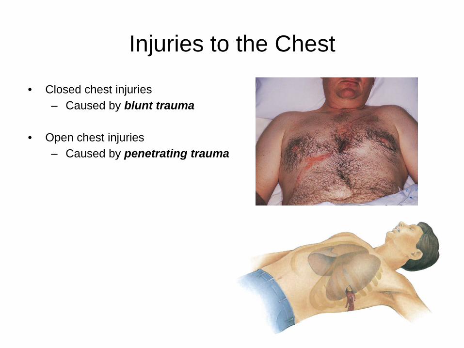
Injuries to the Chest
• Closed chest injuries– Caused by blunt trauma
• Open chest injuries– Caused by penetrating trauma
Signs and Symptoms
• Pain at site of injury
• Pain aggravated by breathing
• Bruising to chest wall
• Crepitus with palpation of chest (due to air bubbles under skin or due to broken ribs)
• Penetrating injury to chest
• Dyspnea
• Failure of chest to expand normally
• Rapid, weak pulse and low blood pressure
• Cyanosis around lips or fingernails
• Unequal breath sounds (compare sounds with stethoscope on left side and right side)
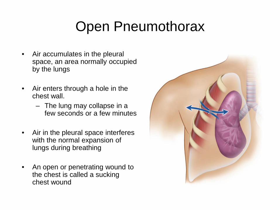
Open Pneumothorax
• Air accumulates in the pleural space, an area normally occupied by the lungs
• Air enters through a hole in the chest wall.– The lung may collapse in a
few seconds or a few minutes
• Air in the pleural space interferes with the normal expansion of lungs during breathing
• An open or penetrating wound to the chest is called a sucking chest wound

Spontaneous Pneumothorax
• Some people with COPD or asthma develop weak areas on the surface of the lungs. Others can be born with lung defects.
• Occasionally, the area will rupture spontaneously, allowing air into the pleural space.
• Patient experiences sudden chest pain and trouble breathing.
• Consider a spontaneous pneumothorax for a patient with chest pain and dyspnea without cause.
Air leak
Listen for decreased or absent breath sounds on the affected side!!!

Tension Pneumothorax
• A complete collapse of the lung that occurs when air enters, but does not leave, the space around the lung (pleural space).
• It is a life-threatening emergency that requires immediate treatment
Tension Pneumothorax
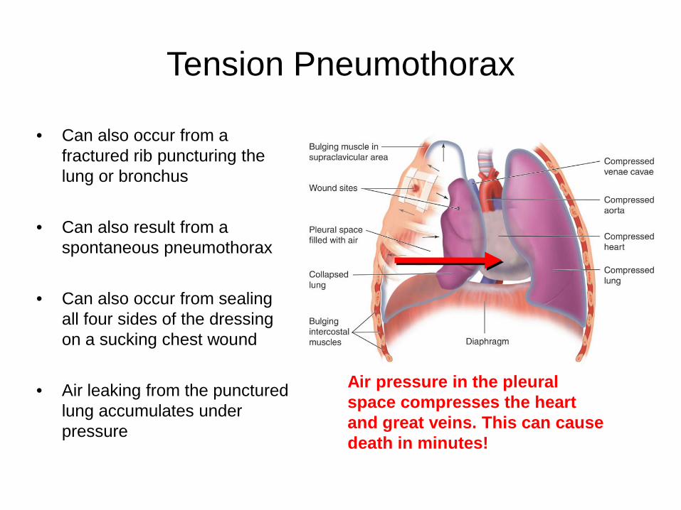
• Can also occur from a fractured rib puncturing the lung or bronchus
• Can also result from a spontaneous pneumothorax
• Can also occur from sealing all four sides of the dressing on a sucking chest wound
• Air leaking from the punctured lung accumulates under pressure
Air pressure in the pleural space compresses the heart and great veins. This can cause death in minutes!

Signs and Symptoms ofTension Pneumothorax
• Respiratory distress• Distended neck veins
• Tracheal deviation
• Tachycardia
• Low blood pressure
• Cyanosis
• Decreased/absent lung sounds

Hemothorax
• Collection of blood in the pleural space
• Can occur from penetrating trauma or blunt trauma from a broken rib
• Suspect if the following are seen:– Signs and symptoms of shock– Decreased breath sounds on
affected side
• If both air and blood are present in the pleural space, it is a hemopneumothorax.

Rib Fractures
• More common in older people, or in young people with major trauma.
• A fractured rib may lacerate the surface of the lung.
• Patients will avoid taking deep breaths and breathing will be rapid and shallow.
• The patient often holds the affected side to minimize discomfort.
Flail Chest
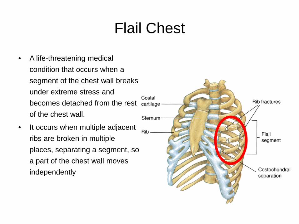
• A life-threatening medical condition that occurs when a segment of the chest wall breaks under extreme stress and becomes detached from the rest of the chest wall.
• It occurs when multiple adjacent ribs are broken in multiple places, separating a segment, so a part of the chest wall moves independently

Flail Chest
• Segment of chest wall detached from rest of thoracic cage
• Occurs when:– Three or more ribs are
fractured in two or more places.
– Sternum is fractured along with several ribs.
• Creates paradoxical motion (broken segments of ribs cannot rise and fall with breathing)
Traumatic Asphyxia
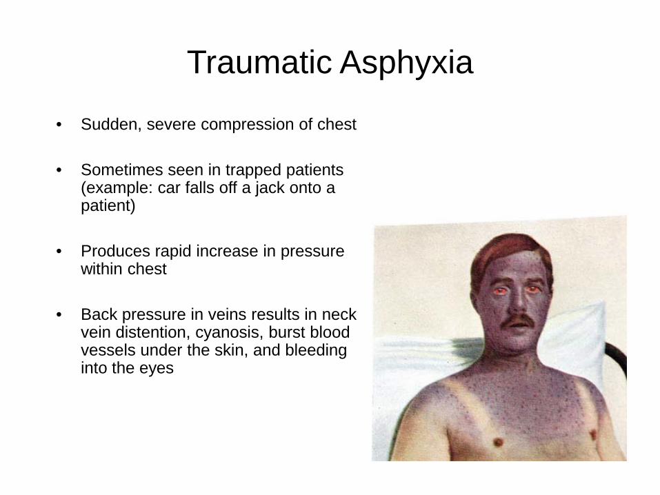
• Sudden, severe compression of chest
• Sometimes seen in trapped patients (example: car falls off a jack onto a patient)
• Produces rapid increase in pressure within chest
• Back pressure in veins results in neck vein distention, cyanosis, burst blood vessels under the skin, and bleeding into the eyes
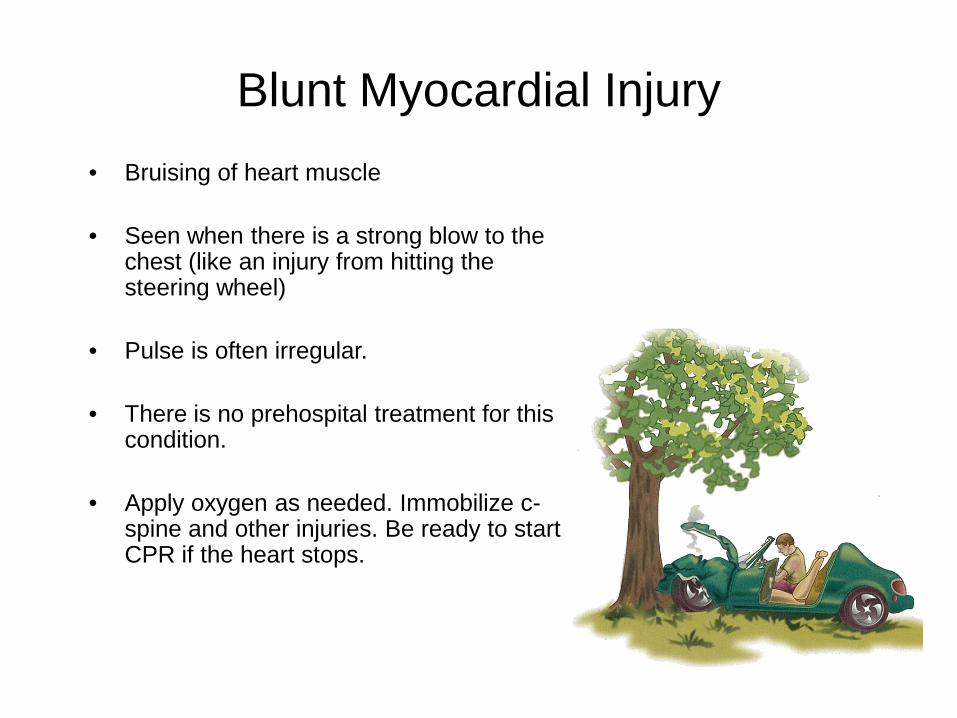
Blunt Myocardial Injury• Bruising of heart muscle
• Seen when there is a strong blow to the chest (like an injury from hitting the steering wheel)
• Pulse is often irregular.
• There is no prehospital treatment for this condition.
• Apply oxygen as needed. Immobilize c-spine and other injuries. Be ready to start CPR if the heart stops.

Pericardial Tamponade• Blood or other fluids collect in the pericardium (sack
surrounding the heart).• Prevents heart from beating effectively.• May become life threatening in minutes.
• Signs and symptoms:
– Very soft and faint heart tones (heard with stethoscope)
– Weak pulse– Low blood pressure– Decreased difference between systolic and
diastolic blood pressure– Jugular vein distention (JVD)
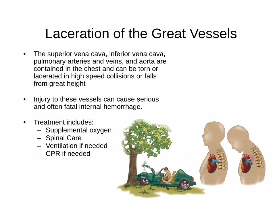
Laceration of the Great Vessels• The superior vena cava, inferior vena cava,
pulmonary arteries and veins, and aorta are contained in the chest and can be torn or lacerated in high speed collisions or falls from great height
• Injury to these vessels can cause serious and often fatal internal hemorrhage.
• Treatment includes:– Supplemental oxygen– Spinal Care– Ventilation if needed– CPR if needed
Firefighter Pre-Hospital Care ProgramModule 18
Treatmentfor
Chest Emergencies
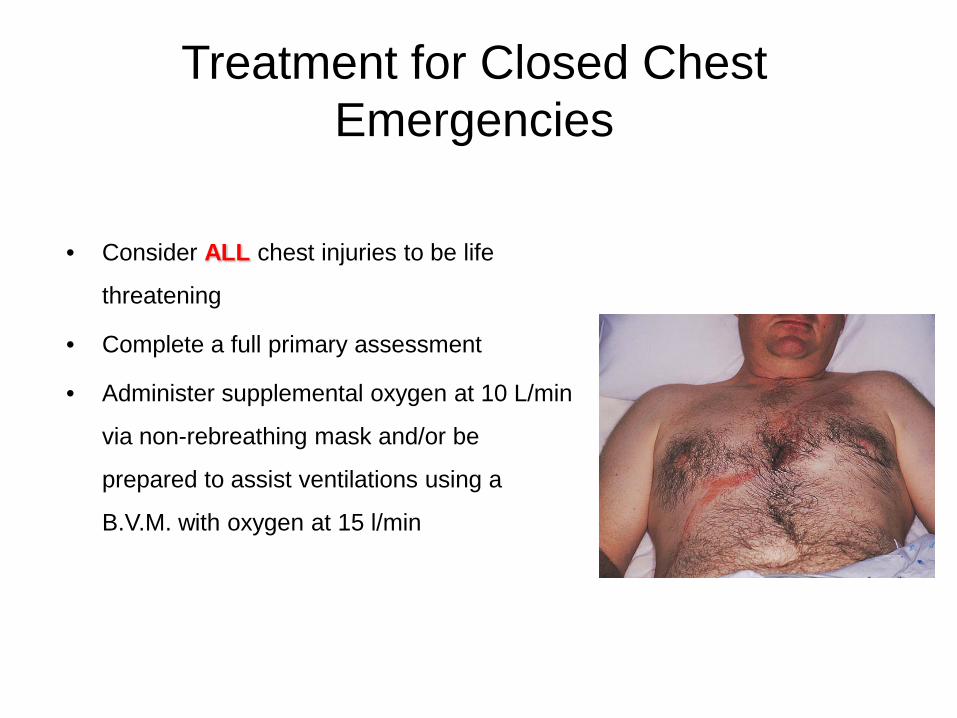
Treatment for Closed Chest Emergencies
• Consider ALL chest injuries to be life
threatening
• Complete a full primary assessment
• Administer supplemental oxygen at 10 L/min
via non-rebreathing mask and/or be
prepared to assist ventilations using a
B.V.M. with oxygen at 15 l/min

Treatment for Closed Chest Emergencies
• Stabilize any paradoxical movement (if one part of the
chest moves in an opposite direction from the rest of the
chest)
• Provide spinal immobilization
• Obtain a detailed history including
SAMPLE and OPQRST assessments
• Be prepared for the patient to go into
cardiac arrest

The Question of a SAMPLE History
• Signs and Symptoms– What things you see and what the patient complains of at onset,
during assessment, and following treatment?
• Allergies– Is the patient allergic to medications, foods, or other?
• Medications– What medications is the patient taking?– Gather them and the patient’s health card

• Pertinent past history– Does the patient have any medical history?
• Last oral intake– When did the patient last eat or drink?
• Events leading to injury or illness– What events led to this incident?
The Question of a SAMPLE History
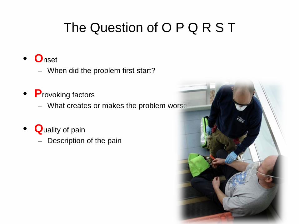
The Question of O P Q R S T
• Onset– When did the problem first start?
• Provoking factors – What creates or makes the problem worse?
• Quality of pain– Description of the pain

• Radiation of pain or discomfort– Do you feel any discomfort anywhere else?
• Severity – Intensity of pain on scale of 1-to-10– This is a relative thing
• Time– How long has the patient had this problem?
The Question of O P Q R S T
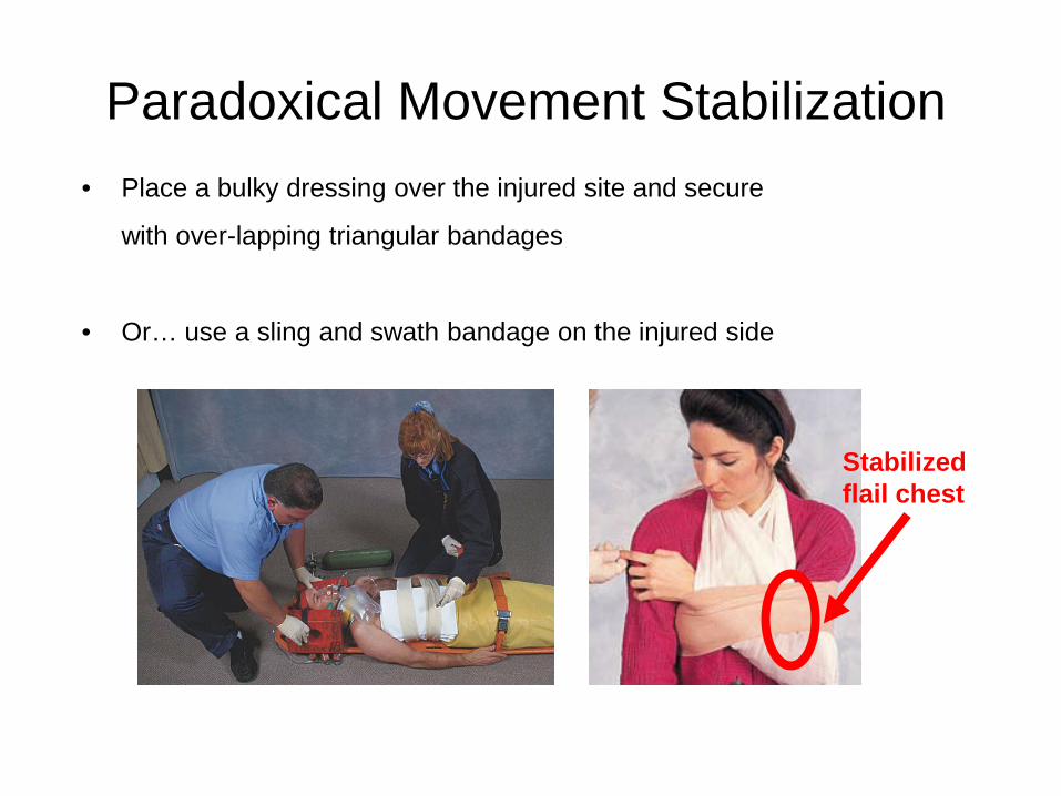
Paradoxical Movement Stabilization • Place a bulky dressing over the injured site and secure
with over-lapping triangular bandages
• Or… use a sling and swath bandage on the injured side
Stabilizedflail chest

Treatment for Open Chest Emergencies
• Consider ALL chest injuries to be life threatening
• Complete a full primary assessment
• Administer supplemental oxygen at 10 L/min via non-
rebreathing mask and/or be prepared to assist
ventilations using a B.V.M. with oxygen
at 15 l/min or with a B.V.M. if required
• Provide spinal immobilization
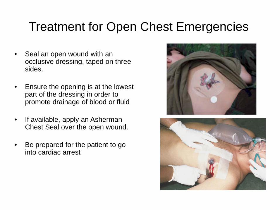
• Seal an open wound with an occlusive dressing, taped on three sides.
• Ensure the opening is at the lowest part of the dressing in order to promote drainage of blood or fluid
• If available, apply an Asherman Chest Seal over the open wound.
• Be prepared for the patient to go into cardiac arrest
Treatment for Open Chest Emergencies

For All Questions Pertaining to this Module,
Contact Your E.M.S. Command Coordinator.
North / West – ( 416 ) 338-9429
South / East – ( 416 ) 338-8796