chemokine receptor ccr5 and cxcr4 expression in hiv- associated
TRANSCRIPT

Chemokine Receptor CCR5 and CXCR4 Expression in HIV-Associated Kidney Disease
FRANK EITNER,* YAN CUI,* KELLY L. HUDKINS,* MICHAEL B. STOKES,*STEPHAN SEGERER,† MATTHIAS MACK, † PAUL L. LEWIS,‡
A. ANDREW ABRAHAM,§ DETLEF SCHLONDORFF,† GLORIA GALLO,i
PAUL L. KIMMEL, ¶ and CHARLES E. ALPERS**Department of Pathology, University of Washington, Seattle, Washington;†Medizinische Poliklinik, KlinikumInnenstadt der LMU, Munich, Germany;‡Division of Pediatric Infectious Disease, Oregon Health SciencesUniversity, Portland, Oregon;§Department of Medicine and Pathology, George Washington UniversityMedical Center, Washington, DC; andiDepartment of Pathology, New York University, New York, New York;and ¶National Institutes of Health, Bethesda, Maryland.
Abstract.The chemokine receptors CCR5 and CXCR4 havebeen identified as essential coreceptors for entry of HIV-1strains into susceptible cells. Direct infection of renal paren-chymal cells has been implicated in the pathogenesis of HIV-associated renal disease, although data are conflicting. Thelocalization of CCR5 and CXCR4 in kidneys with HIV-asso-ciated renal disease is unknown. Formalin-fixed, paraffin-em-bedded renal biopsies from patients with HIV-associated ne-phropathy (HIVAN) (n 5 13), HIV-associated immunecomplex glomerulonephritis (n 5 3), HIV-associated throm-botic microangiopathy (n 5 1), and HIV-negative patients withcollapsing glomerulopathy (n 5 8) were analyzed in this study.Cellular sites of expression of CCR5 and CXCR4 were iden-tified by immunohistochemistry and byin situ hybridization.The presence of HIV-1 was detected by immunohistochemistry
and by in situ hybridization. Expression of both chemokinereceptors CCR5 and CXCR4 was undetectable in intrinsicglomerular, tubular, and renovascular cells in all analyzedcases. In the presence of tubulointerstitial inflammation, CCR5and CXCR4 expression was localized to infiltrating mononu-clear leukocytes. HIV-1 protein was undetectable by immuno-histochemistry in all cases of HIV-associated renal disease.HIV-1 RNA was identified in one case of HIVAN but wasrestricted to infiltrating leukocytes. HIV-1 RNA was not de-tected in intrinsic renal cells in all analyzed cases. Identifyingthe cellular expression of HIV-coreceptors CCR5 and CXCR4may help to clarify which tissues are permissive for direct HIVinfection. These data do not support a role of productive HIV-1infection of renal parenchymal cells in the pathogenesis ofHIV-associated renal disease.
Renal disease may complicate approximately 10% of patientsinfected with HIV, with a striking predominance in African-Americans (1–3). Several clinically and morphologically di-verse renal syndromes have been described in HIV-infectedpatients (4,5). The syndrome most frequently reported in renalbiopsy series from HIV-infected individuals has been termedHIV-associated nephropathy (HIVAN) (5,6). Characteristicpathologic features of HIVAN include a collapsing form offocal and segmental glomerulosclerosis with hypertrophy andhyperplasia of glomerular visceral epithelial cells, extensivetubulointerstitial injury, including microcystic tubular dilation,and prominent endothelial cell tubuloreticular inclusions (5,6).Clinically, such patients frequently show nephrotic-range pro-teinuria with renal insufficiency and rapid progression to end-stage renal disease (2,4). Other lesions reported in renal biop-
sies from HIV-infected patients are immune complex-mediatedrenal diseases, including membranoproliferative glomerulone-phritis, membranous glomerulonephritis, and IgA nephropathy(5,7–9). Renal thrombotic microangiopathy has been increas-ingly reported in HIV-infected humans (reviewed in references(10) and (11). Tubulointerstitial inflammation is present tovariable degrees in all varieties of HIV-associated renal disease(12–14).
The pathogenesis of HIV-associated renal disease is poorlyunderstood (2,3). Proposed mechanisms include: (1) directHIV infection of renal parenchymal cells (e.g., visceral epithe-lial cells, tubular epithelial cells, and renovascular endothelialcells); (2) indirect injury to the kidney by renal cellular uptakeof circulating virally encoded molecules; or (3) indirect injuryto the kidney through release of cytokines by infected mono-nuclear cells in the circulation or infiltrating the kidney (2,3).Furthermore, different mechanisms might be involved in thepathogenesis of the different variants of HIV-associated renaldisease. Circulating immune complex deposition andin situmechanisms of immune-mediated renal disease may underliethe pathogenesis of glomerulonephritis in HIV-infected pa-tients (7). In addition, there may be a role for hepatitis Ccoinfection in some cases of HIV-associated immune complex
Received July 26, 1999. Accepted September 18, 1999.Correspondence to Dr. Charles E. Alpers, University of Washington, Depart-ment of Pathology, Box 356100, 1959 NE Pacific Street, Seattle, WA 98195.Phone: 206-548-6409; Fax: 206-548-4928; E-mail: [email protected]
1046-6673/1105-0856Journal of the American Society of NephrologyCopyright © 2000 by the American Society of Nephrology
J Am Soc Nephrol 11: 856–867, 2000

glomerulonephritis (8). Some renal disease in HIV-infectedindividuals, however, may represent chance occurrence unre-lated to HIV infection itself.
New directions in AIDS research of viral entry, tropism, andpathogenesis were initiated by the recent discovery that mem-bers of the chemokine receptor family act as necessary core-ceptors together with CD4 for entry of HIV-1 into susceptiblecells (15). HIV-1 strains previously characterized as T lym-phocyte-tropic have been shown to bind to the chemokinereceptor CXCR4 as a condition of entry into mammalian cells,while macrophage-tropic strains of HIV-1 require the chemo-kine receptor CCR5. Expression of these molecules appears toplay a key role in determining which tissues are permissive fordirect HIV infection.
Expression of the HIV coreceptors CCR5 and CXCR4 inhuman kidneys with features of HIV-associated renal disease iscompletely unknown. In previous studies from our laborato-ries, expression of both chemokine receptors CCR5 andCXCR4 was not identified by either immunohistochemical(16) or in situ hybridization (17,18) techniques in intrinsicrenal parenchymal cells in normal human kidneys, in renalallograft nephrectomies, and in human renal biopsies with avariety of different glomerular and interstitial diseases. How-ever, in vitro studies show that the expression of certain che-mokine receptors in mononuclear cells is regulated by a num-ber of stimuli (reviewed in references (19) and (20). Anupregulation of CCR5 or CXCR4 expression in intrinsic renalcells might therefore require specific cytokine stimulationpresent in the course of HIV infection.
In this study, we tested the hypothesis that the relevantchemokine coreceptors for HIV infection not constitutivelyexpressed in normal kidney may be upregulated in the settingof HIV infection and thereby permit infection of renal tissue.We assessed the cellular sites of expression of chemokinereceptor CCR5 by immunohistochemistry andin situ hybrid-ization, and CXCR4 byin situ hybridization, in renal biopsiesobtained from HIV-infected patients with features of HIVAN,HIV-associated immune complex glomerulonephritis, andHIV-associated thrombotic microangiopathy. Additional tis-sues examined within this study consisted of human renalbiopsy material with features of a collapsing glomerulopathyfrom patients without clinical evidence of HIV infection. Fur-thermore, we provide corresponding data for the presence ofHIV-1 protein by immunohistochemistry and for the presenceof HIV-1 RNA by in situ hybridization in each of these cases.
Materials and MethodsSource of Tissue
Formalin-fixed, paraffin-embedded renal biopsy tissue specimensobtained between 1984 and 1998 at the George Washington Univer-sity Medical Center (Washington, DC), the New York University(New York, NY), and the University of Washington (Seattle, WA)were included in this study. Several cases had been included inprevious studies investigating HIV-associated renal disease(7,8,14,21–23). All renal biopsy cases with sufficient tissue for com-plete immunohistochemical andin situ hybridization evaluation aftercompletion of diagnostic workup and previous investigations were
included. Renal biopsies from 13 patients with HIVAN were studied.All cases demonstrated variable degrees of a collapsing form of focaland segmental glomerulosclerosis with microcystic tubular dilation.Additionally, renal biopsies from three patients with HIV-associatedimmune complex glomerulonephritis were analyzed, including twocases of diffuse proliferative glomerulonephritis and one case ofmembranous nephropathy. One renal biopsy with HIV-associatedrenal thrombotic microangiopathy was also examined. For HIV-neg-ative disease controls, we analyzed kidney samples from eight patientswith collapsing glomerulopathy and negative HIV serology. Tissuesections contained 1 to 20 glomeruli (mean, 4 glomeruli) from patientswith HIVAN and 2, 4, and 10 glomeruli, respectively, from patientswith HIV-associated glomerulonephritis, and 13 glomeruli from thepatient with HIV-associated thrombotic microangiopathy. Kidneysamples from HIV-negative patients with collapsing glomerulopathyincluded 2 to 12 glomeruli (mean, 5 glomeruli). The demography ofthe patient populations in New York and Washington, DC has beenpublished previously (21,24).
Formalin-fixed, paraffin-embedded HIV-1-infected human periph-eral blood mononuclear cell (PBMC) pellets were generated as con-trols for immunohistochemical detection of HIV-1 p24 antigen and forin situ hybridization of HIV-1 RNA. Human PBMC were obtainedfrom HIV antibody-negative volunteer donors as described previously(25). Briefly, the PBMC layer was removed after centrifugation ofwhole blood, washed, and resuspended in RPMI 1640 containing 16%fetal calf serum, penicillin (100 U/ml), streptomycin (100mg/ml), andglutamine (0.3 mg/ml). The mononuclear cells were stimulated withphytohemagglutinin (5mg/ml) and 5% nonrecombinant human inter-leukin-2 (Pharmacia, Piscataway, NJ) as described (25). Forty-eighthours after stimulation, the human PBMC were infected with a cell-free solution of HIV-1 strain LAI. A full-length clone of HIV-1LAI
[pBRU3] was obtained from Dr. Michael Emerman, Fred HutchinsonCancer Research Center, Seattle, WA (26). Cell-free stock solutionsof HIV-1LAI were generated by transfection of this plasmid into 293Tcells.) Cells were harvested 48 h after infection and counted in ahemocytometer. Several different preparations were generated bymixing defined concentrations of HIV-1-infected PBMC with definedconcentrations of uninfected PBMC. The different HIV-1-infected/uninfected PBMC preparations contained HIV-1-infected PBMC inconcentrations ranging from 5 to 50%. These mixed PBMC sampleswere subsequently centrifuged, and the remaining cell pellet was fixedin 10% phosphate-buffered formalin and embedded in paraffin usingstandard protocols for tissue preparation. Four-micrometer-thick sec-tions were generated as controls for immunohistochemical andin situhybridization procedures.
AntibodiesCCR5. A murine monoclonal antibody MC5 directed against
human chemokine receptor CCR5 has previously been characterizedfor specificity by Western blotting and fluorescence-activated cellsorter analysis (16), and was found suitable for the specific detectionof CCR5 in formalin-fixed, paraffin-embedded tissue sections afterheat-mediated antigen retrieval procedures (16).
HIV-1. A murine monoclonal antibody p24, clone Kal-1, di-rected against an epitope of the core protein p24 of HIV-1 waspurchased from DAKO (Carpinteria, CA). Specificity of this antibodyfor the detection of HIV-1 p24 has been demonstrated previously byimmunoprecipitation, Western blotting, and immunohistochemistry(27), and it has been demonstrated previously to recognize HIV-1 p24in formalin-fixed, paraffin-embedded tissue sections (27,28).
J Am Soc Nephrol 11: 856–867, 2000 Chemokine Receptors in HIV-Associated Renal Disease 857

ImmunohistochemistryImmunohistochemistry was performed on formalin-fixed, paraffin-
embedded tissue sections according to protocols that we have usedpreviously (29). Four-micrometer sections of tissue samples weredeparaffinized in xylene and rehydrated in graded ethanols. Endoge-nous peroxidase was blocked by incubation in 3% hydrogen peroxide.Sections that were subsequently incubated with the anti-CCR5 anti-body were pretreated by steam heating for 20 min in Antigen Un-masking Solution (Vector Laboratories, Burlingame, CA), accordingto the instructions of the manufacturer. Nonspecific binding wasblocked by incubation in 10% normal horse serum (Vector). Thesections were then incubated for 1 h at room temperature with theprimary antibody diluted in phosphate-buffered saline plus 1% bovineserum albumin (Sigma, St. Louis, MO). After washes in phosphate-buffered saline, the sections were incubated with biotinylated horseanti-mouse antibody (Vector). A Tyramide Signal Amplification(TSA™-Indirect, NEN™ Life Science Products, Boston, MA) wasperformed according to the manufacturer’s instructions. Finally, 3,39diaminobenzidine (with nickel chloride enhancement) was used as achromogen. Sections were counterstained with methyl green, dehy-drated, and coverslipped. The complete biopsies were examined, andthe number of CCR5 protein-expressing cells per glomerular cross-section was calculated in each case. Negative controls for the immu-nohistochemical procedures consisted of substitution of the primaryantibody with isotype-matched, irrelevant murine monoclonal anti-bodies (DAKO).
Molecular ProbesCCR5. A 1.1-kb sequence of DNA coding for human CCR5 was
subcloned into pcDNAI/amp (Invitrogen, San Diego, CA) (obtainedthrough the AIDS Research and Reference Reagent Program, Divisionof AIDS, NIAID, and NIH, originally provided by Dr. NathanielLandau) and then linearized withHindIII and transcribed with Sp6 forthe antisense probe or linearized withSphI and transcribed with T7 forthe sense probe. Sensitivity and specificity of the CCR5 antisenseriboprobe has been demonstrated previously by Northern analysis andby in situ hybridization (17).
CXCR4. A 1.1-kb sequence of DNA coding for human CXCR4was subcloned into pcDNAI/amp (Invitrogen) (obtained through theAIDS Research and Reference Reagent Program, Division of AIDS,NIAID, and NIH, originally provided by Dr. Nathaniel Landau) andthen linearized withHindIII and transcribed with Sp6 for the antisenseprobe or linearized withXbaI and transcribed with T7 for the senseprobe. Sensitivity and specificity of the CXCR4 antisense riboprobehave been demonstrated previously by Northern analysis and byinsitu hybridization (18).
HIV-1. Four DNA templates that collectively represent 90% ofthe HIV-1 genome were purchased from Lofstrand Labs (Gaithers-burg, MD): gag, SacI-BglII, 1.4 kb; gag/pol, BglII-EcoRI, 2.6 kb;pol/vif/vpr/rev/tat/vpu, KpnI-KpnI, 2.2 kb;env/vpr/rev/tat/vpu, EcoRI-BamHI, 2.7 kb. Antisense and sense riboprobes were generated fromall templates using Sp6 or T7 according to the manufacturer’s instruc-tions. Radioactivity was introduced by synthesis of RNA using35S-UTP. The generated probes were combined to yield one sense orantisense probe cocktail. Specific detection of HIV-1 RNA in forma-lin-fixed, paraffin-embedded tissue sections byin situ hybridizationwith an estimated sensitivity of 30 to 300 copies of target RNA hasbeen described previously by numerous groups using identical RNAprobes (30–35).
In Situ HybridizationHIV-1 RNA, CCR5 mRNA, and CXCR4 mRNA were detected in
tissue sections usingin situ hybridization techniques according toprotocols used previously (29). Riboprobes forin situ hybridizationwere generated from cDNA using35S-UTP. Four-micrometer sectionsof formalin-fixed, paraffin-embedded tissue samples were deparaf-finized and rehydrated through xylene and graded ethanols, washedwith 0.53 SSC (Life Technologies, Grand Island, NY), and digestedwith proteinase K (5mg/ml; Sigma). Sections that were subsequentlyhybridized with HIV-1-specific riboprobes were analyzed in dupli-cate.
In one section, thein situhybridization was performed according tothe following protocol. The second section was additionally pretreatedby steam heating for 20 min in Antigen Unmasking Solution (Vector)before the hybridization, according to the manufacturer’s instructions.Prehybridization was performed for 2 h by adding 100ml of prehy-bridization buffer (0.3 M NaCl, 20 mM Tris, pH 8.0, 5 mM ethyl-enediaminetetra-acetic acid, 13 Denhardt’s solution, 10% dextransulfate, and 10 mM dithiothreitol). The hybridizations were started byadding 500,000 cpm of35S-labeled riboprobe in 50ml of prehybrid-ization buffer and allowed to proceed overnight at 50°C. After hy-bridization, sections were treated with RNase A (20mg/ml; Sigma),followed by three high-stringency washes in 0.13 SSC/0.5% Tween20 (Sigma) for 40 min each at 50°C, and several 23 SSC washes.After the tissue was dehydrated and air-dried, it was dipped in NTB2nuclear emulsion (Kodak, Rochester, NY) and exposed in the dark at4°C for 2 wk (HIV-1) or 6 wk (CCR5, CXCR4), respectively. Afterdeveloping, the sections were counterstained with hematoxylin andeosin, dehydrated, and coverslipped. Positive cellular labeling wasdefined as five or more silver grains concentrated over a single cell onthe slides hybridized with the antisense probe, and little or no signalpresent on the sense control slides. The complete biopsies wereexamined, and the numbers of CCR5 mRNA and CXCR4 mRNAexpressing cells per glomerular cross-section were calculated in eachcase. Positive controls for the detection of CCR5 and CXCR4 mRNAconsisted of several allograft nephrectomy specimens with features ofsevere rejection, as published previously (17,18).
ResultsCCR5 Expression in HIV-Associated Renal Disease
By both immunohistochemistry andin situ hybridization,CCR5 expression was absent in intrinsic glomerular cells in allcases of HIVAN, HIV-associated glomerulonephritis, HIV-associated thrombotic microangiopathy, and HIV-negative col-lapsing glomerulopathy. Parietal epithelial cells, visceral epi-thelial cells, mesangial cells, and glomerular endothelial cellsin glomeruli, with and without features of collapse, did notexpress detectable CCR5 (Figure 1, A through D). A fewCCR5-positive cells were detected in the capillary lumina ofsome glomeruli in cases of all different groups (Figure 1, A andB, Table 1). These cells likely represented circulating mono-nuclear leukocytes.
All cases of the different disease groups analyzed in thisstudy showed variable degrees of tubulointerstitial inflamma-tion ranging from very mild, focal, perivascular infiltrates ofleukocytes to severe, diffuse interstitial mononuclear cell in-filtrates. CCR5-expressing cells were demonstrable by bothimmunohistochemistry (Figure 1, E and F) andin situ hybrid-ization (Figure 1, G and H) in these infiltrates in the tubulo-
858 Journal of the American Society of Nephrology J Am Soc Nephrol 11: 856–867, 2000
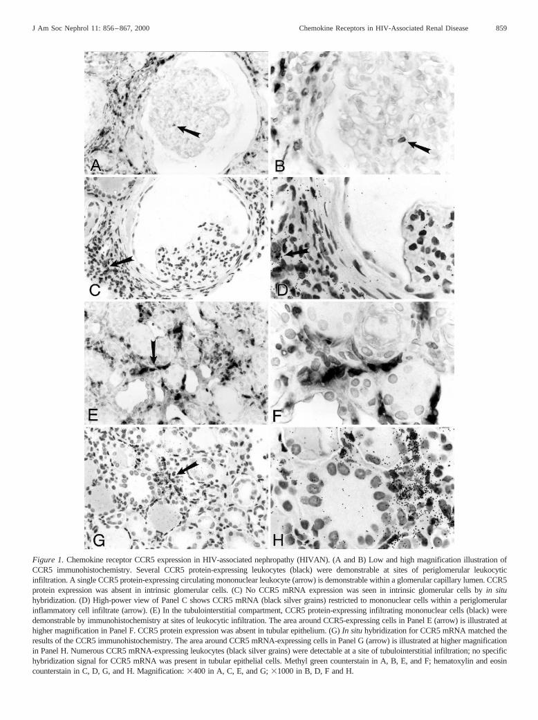
Figure 1.Chemokine receptor CCR5 expression in HIV-associated nephropathy (HIVAN). (A and B) Low and high magnification illustration ofCCR5 immunohistochemistry. Several CCR5 protein-expressing leukocytes (black) were demonstrable at sites of periglomerular leukocyticinfiltration. A single CCR5 protein-expressing circulating mononuclear leukocyte (arrow) is demonstrable within a glomerular capillary lumen. CCR5protein expression was absent in intrinsic glomerular cells. (C) No CCR5 mRNA expression was seen in intrinsic glomerular cells byin situhybridization. (D) High-power view of Panel C shows CCR5 mRNA (black silver grains) restricted to mononuclear cells within a periglomerularinflammatory cell infiltrate (arrow). (E) In the tubulointerstitial compartment, CCR5 protein-expressing infiltrating mononuclear cells (black) weredemonstrable by immunohistochemistry at sites of leukocytic infiltration. The area around CCR5-expressing cells in Panel E (arrow) is illustratedathigher magnification in Panel F. CCR5 protein expression was absent in tubular epithelium. (G)In situ hybridization for CCR5 mRNA matched theresults of the CCR5 immunohistochemistry. The area around CCR5 mRNA-expressing cells in Panel G (arrow) is illustrated at higher magnificationin Panel H. Numerous CCR5 mRNA-expressing leukocytes (black silver grains) were detectable at a site of tubulointerstitial infiltration; no specifichybridization signal for CCR5 mRNA was present in tubular epithelial cells. Methyl green counterstain in A, B, E, and F; hematoxylin and eosincounterstain in C, D, G, and H. Magnification:3400 in A, C, E, and G;31000 in B, D, F and H.
J Am Soc Nephrol 11: 856–867, 2000 Chemokine Receptors in HIV-Associated Renal Disease 859

interstitial compartment of all analyzed cases. The expressionof CCR5 was restricted to infiltrating mononuclear cells (Fig-ure 1, E through H). The number of CCR5-expressing cellscorrelated with the degree of tubulointerstitial mononuclearcell infiltration. No difference in the CCR5 expression patternon infiltrating leukocytes was seen between patients with HIV-associated renal disease or HIV-negative disease controls (datanot shown). Tubular epithelial cells, whether located in areaswith absence of interstitial inflammation, at sites of severetubulointerstitial mononuclear cell infiltration, or at sites ofmicrocystic tubular dilation, showed no detectable CCR5 ex-pression (Figure 1, E through H). CCR5 expression was com-pletely absent in endothelial cells and smooth muscle cells ofthe vascular compartment.
The number of leukocytes exhibiting expression of theCCR5 protein as detected by immunohistochemistry correlatedwith the number of leukocytes synthesizing detectable CCR5mRNA in all individual cases (Figure 1, compare Panels F andH). However, in the glomerular compartment slightly moreCCR5-positive leukocytes were identified by immunohisto-chemistry compared within situ hybridization (Table 1).
CXCR4 Expression in HIV-Associated Renal DiseaseBy in situ hybridization, CXCR4 mRNA expression re-
mained undetectable in cells clearly identifiable as renal pa-renchymal cells of the glomerular, tubular, and vascular com-partments in all analyzed cases. At sites of collapsingglomeruli, CXCR4 mRNA expression was typically absent(Figure 2, A and B). However, within all analyzed cases smallnumbers of individual CXCR4 mRNA-expressing cells wereidentified within glomeruli (Figure 2, C and D) (Table 1). Wewere unable to clearly determine whether these CXCR4mRNA-expressing cells were intrinsic glomerular cells or cir-culating leukocytes, although we favor the latter interpretation.All of the biopsy tissue was consumed during the course of thepresent studies, and so material was unavailable for double-labeling studies that might confirm this interpretation.
Within the tubulointerstitial compartment, CXCR4 mRNAexpression was restricted to infiltrating mononuclear leuko-cytes at sites of tubulointerstitial inflammation (Figure 2, E andF). There was no difference in the number of detectable
CXCR4 mRNA-expressing leukocytes between HIV-1-posi-tive or -negative renal tissues. CXCR4 mRNA expression wasundetectable in tubular epithelial cells, including those at sitesof tubulointerstitial inflammation and at sites of microcystictubular dilation (Figure 2, E and F). Renovascular endothelialcells did not show detectable CXCR4 mRNA expression.
Comparison of CCR5 and CXCR4 expression in serial sec-tions of the analyzed specimens revealed that both chemokinereceptors were expressed in a large percentage of interstitialinfiltrating mononuclear leukocytes. The number of CCR5- orCXCR4-expressing cells, respectively, correlated positivelywith the degree of tubulointerstitial inflammation. No obviousdifferences in the distribution pattern of CCR5- or CXCR4-expressing infiltrating leukocytes were evident. Because of thetechnical limitation of serial sections, we could not properlyaddress the question of whether CCR5 and CXCR4 werecoexpressed in the same subset of infiltrating mononuclearleukocytes, or whether CCR5 and CXCR4 were expressed indifferent leukocyte subsets. As indicated above, exhaustion ofthe biopsy samples precluded double-labeling studies thatwould address this issue.
Specificity of the HIV-1 p24 Immunohistochemistry andof the HIV-1 RNA in Situ Hybridization
Immunohistochemical detection of HIV-1 proteins in forma-lin-fixed, paraffin-embedded renal tissue sections has beenvery unreliable and frequently resulted in nonspecific immu-nostaining in tissue obtained from HIV-negative controls(36,37). We therefore performed a series of experiments tocontrol the specificity of our procedures. As positive controls,formalin-fixed, paraffin-embedded PBMC pellets, containingHIV-1-infected PBMC, reproducibly demonstrated positiveimmunostaining (Figure 3, A through C). Furthermore, thenumber of cells with positive immunohistochemical signalwithin the pellet closely reflected the number of HIV-1-in-fected PBMC mixed in the pellet preparation (Figure 3, Athrough D). No immunostaining was detectable in HIV-1-infected PBMC when the primary antibody was substituted byan irrelevant, isotype-matched mouse IgG preparation (data notshown). PBMC pellets that contained no HIV-1-infected cells
Table 1. Chemokine receptor-expressing cells within glomerulia
Category CXCR4 mRNA-PositiveCells/Glomeruli
CCR5 Protein-PositiveCells/Glomeruli
CCR5 mRNA-PositiveCells/Glomeruli
HIV-associated nephropathy (n 5 13) 0.46 0.6 0.36 0.3 0.16 0.1HIV-associated immune complex
glomerulonephritis (n 5 3)1.46 1.7 0.36 0.3 0.26 0.4
HIV-associated thromboticmicroangiopathy (n 5 1)
4.0 1.1 0.1
Collapsing glomerulopathy, HIV-negative (n 5 8)
0.66 0.4 0.36 0.3 0.16 0.3
a Data are given as mean values6 SD of chemokine receptor-expressing cells per glomerular cross-sections.
860 Journal of the American Society of Nephrology J Am Soc Nephrol 11: 856–867, 2000

showed no detectable immunostaining with the HIV-1 p24antibody (Figure 3D). Renal biopsy sections from patients withcollapsing glomerulopathy and negative HIV serology (n 5 8)consistently showed absent immunohistochemical stainingwhen incubated with the HIV-1 p24 antibody (data not shown).
PBMC pellets containing HIV-1-infected PBMC consis-tently showed detectablein situ hybridization signal for HIV-1RNA (Figure 3E). The number of PBMC with positive hybrid-ization signal closely reflected the number of HIV-1-infected
PBMC mixed in the pellet preparation. No difference in thenumber of HIV-1 RNA-positive PBMC was seen in thein situhybridization procedures with and without heat-mediated pre-treatment of the PBMC pellet specimens before hybridization(data not shown). No hybridization was seen when identicalprocedures were performed with substitution of a control senseprobe for hybridization (Figure 3F). All renal biopsies obtainedfrom HIV-negative control patients showed absent hybridiza-tion signal for HIV-1 RNA (data not shown).
Figure 2. Chemokine receptor CXCR4 expression in HIVAN. (A and B) Low and high magnification illustration of CXCR4in situhybridization. CXCR4 mRNA expression was absent in a glomerulus with typical features of a collapsing glomerulopathy. Severalmononuclear leukocytes within a periglomerular interstitial infiltrate (arrow) showed detectable CXCR4 mRNA expression (black silvergrains). (C and D) Low and high magnification illustration of CXCR4in situ hybridization. CXCR4 mRNA expression (black silver grains)was localized to a cluster of three individual cells (arrows) whose precise identity cannot be ascertained by their location or appearance in thesesections, within the illustrated glomerular cross-section. (E and F) Low and high magnification illustration ofin situ hybridization for CXCR4mRNA. Within the tubulointerstitial compartment, CXCR4 mRNA expression (black silver grains) was clearly restricted to inflammatoryinterstitial mononuclear leukocytes (arrow), whereas tubular epithelial cells did not demonstrate detectable CXCR4 mRNA expression. CXCR4mRNA expression was absent at sites of microcystic tubular dilation (c). Hematoxylin and eosin counterstain in A through F. Magnification:3400 in A, C, and E;31000 in B, D, and F.
J Am Soc Nephrol 11: 856–867, 2000 Chemokine Receptors in HIV-Associated Renal Disease 861

Detection of HIV-1 in Biopsies from Patients with HIV-Associated Renal Disease
By immunohistochemistry, HIV-1 p24 protein was not de-tected in glomerular (Figure 4B), tubulointerstitial (Figure 4E),or vascular compartments of all cases of HIVAN (n 5 13),HIV-associated glomerulonephritis (n 5 3), and HIV-associ-ated thrombotic microangiopathy (n 5 1).
In situ hybridization detected no HIV-1 RNA in glomerular(Figure 4, C and D), tubular (Figure 4F), or vascular cells in allanalyzed cases. However, in one case of HIVAN, HIV-1 RNAwas detectable within one small lymphoid follicle located inthe tubulointerstitium (Figure 4, G and H). HIV-1 RNA wasclearly restricted to infiltrating mononuclear leukocytes.
DiscussionRenal disease is frequently reported in a large percentage of
HIV-infected humans, although the mechanisms leading toHIV-associated renal injury are largely unknown. Direct infec-tion of renal parenchymal cells by HIV in susceptible patientshas been proposed (2,3). The results of the present study do notsupport a role for productive infection of renal cells by HIV inthe pathogenesis of HIV-associated renal disease. The twoprincipal HIV coreceptors, the chemokine receptors CCR5 andCXCR4 that mediate entry of HIV-1 strains into susceptiblecells, were not expressed by intrinsic renal cells in HIV-associated renal disease, but were demonstrable in circulatingand infiltrating leukocytes at sites of tubulointerstitial inflam-mation. Furthermore, using sensitive immunohistochemicaland in situ hybridization techniques, HIV-1 protein and RNAwere undetectable in renal parenchymal cells in biopsies withfeatures of HIV-associated renal disease. Small numbers ofHIV-1 RNA-expressing mononuclear leukocytes were demon-strable in the tubulointerstitium in one case of HIVAN.
Major insights into the mechanisms of HIV infection havebeen obtained from the discovery that members of the chemo-kine receptor family act as necessary coreceptors, together withCD4, for entry of AIDS viruses into mammalian cells (re-viewed in reference (15). Chemokine receptors represent afamily of structurally and functionally related seven transmem-brane-spanning, G protein-coupled receptors (19,20,38). Re-cent observations indicate that certain chemokine receptorsmay also facilitate infection by immunodeficiency viruses in aCD4-independent manner (39,40). By facilitating entry intocells, these receptors determine viral tropism. CXCR4 is acoreceptor for strains of HIV-1 that infect T lymphocyte celllines (T-tropic strains), and CCR5 serves as a coreceptor forHIV-1 isolates that infect macrophages and activated T lym-phocytes (M-tropic strains) (15). Additionalin vivo observa-tions support the central role of CCR5 and CXCR4 in thepathogenesis of HIV infection in humans. Individuals homozy-gous for a mutant allele of the CCR5 gene bearing a 32-nucleotide deletion (CCR5 delta 32), which leads to an inactivevariant of CCR5, have been linked to protection from HIV-1infection (41,42). The consequence of the heterozygous state isnot clear, but it may delay the progression to AIDS in infectedindividuals (41–43). Additionally, individuals homozygous fora mutation of the only identified CXCR4 ligand, stromal cell-derived factor-1, appear to have some degree of protectionagainst disease progression after HIV infection (44).
The expression of chemokine receptors is not limited toleukocytes and lymphoid tissues. Tissue-specific expression ofthe HIV coreceptors CCR5 and CXCR4 most likely play a rolein determining whether tissues are permissive for direct HIVinfection in vivo. CCR5 mRNA was detectable by Northernblotting in RNA isolated from small intestine, ovary, and lung(45), and CCR5 protein was detectable by immunohistochem-istry in the central nervous system (on neurons, astrocytes, andmicroglia) and on endothelium, vascular smooth muscle cells,and fibroblasts in several analyzed parenchymal tissues (46).Two recent studies by members of our group investigated the
Figure 3. Specificity of the HIV-1 p24 immunohistochemistry (Athrough D) and HIV-1 RNAin situ hybridization (E through F).Formalin-fixed, paraffin-embedded peripheral blood mononuclear cell(PBMC) pellet preparations containing defined concentrations ofHIV-1-infected PBMC were used. (A) Positive immunostaining forHIV-1 p24 (black) was detectable in approximately 50% of the cellsin a section of a formalin-fixed, paraffin-embedded PBMC pelletcontaining 50% HIV-1-infected PBMC. (B) Immunohistochemistryfor HIV-1 p24 labels approximately 33% of the PBMC in a cell pelletcontaining 33% HIV-1-infected PBMC. (C) Approximately 5% of thePBMC are immunohistochemically labeled in a cell pellet containing5% HIV-1-infected PBMC. (D) PBMC pellets that contained noHIV-1-infected cells showed no detectable immunostaining with theHIV-1 p24 antibody. (E)In situ hybridization detects HIV-1 RNA(black silver grains) in numerous HIV-1-infected cells in a section ofa formalin-fixed, paraffin-embedded PBMC pellet containing 33%HIV-1-infected PBMC. (F) No hybridization signal was seen in thesame PBMC pellet as illustrated in Panel E when identical procedureswere performed with substitution of a control sense probe for hybrid-ization. Methyl green counterstain in A through D; hematoxylin andeosin counterstain in E and F. Magnification:31000.
862 Journal of the American Society of Nephrology J Am Soc Nephrol 11: 856–867, 2000
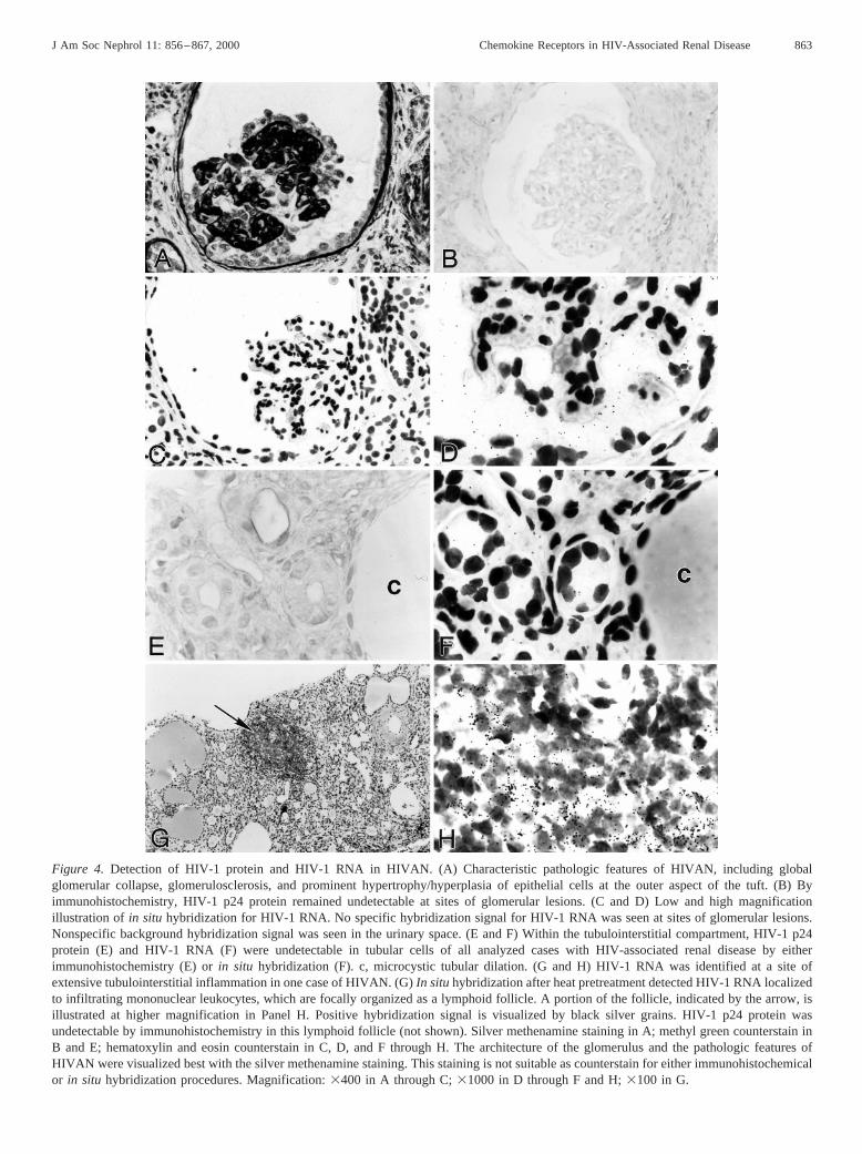
Figure 4. Detection of HIV-1 protein and HIV-1 RNA in HIVAN. (A) Characteristic pathologic features of HIVAN, including globalglomerular collapse, glomerulosclerosis, and prominent hypertrophy/hyperplasia of epithelial cells at the outer aspect of the tuft. (B) Byimmunohistochemistry, HIV-1 p24 protein remained undetectable at sites of glomerular lesions. (C and D) Low and high magnificationillustration of in situ hybridization for HIV-1 RNA. No specific hybridization signal for HIV-1 RNA was seen at sites of glomerular lesions.Nonspecific background hybridization signal was seen in the urinary space. (E and F) Within the tubulointerstitial compartment, HIV-1 p24protein (E) and HIV-1 RNA (F) were undetectable in tubular cells of all analyzed cases with HIV-associated renal disease by eitherimmunohistochemistry (E) orin situ hybridization (F). c, microcystic tubular dilation. (G and H) HIV-1 RNA was identified at a site ofextensive tubulointerstitial inflammation in one case of HIVAN. (G)In situhybridization after heat pretreatment detected HIV-1 RNA localizedto infiltrating mononuclear leukocytes, which are focally organized as a lymphoid follicle. A portion of the follicle, indicated by the arrow, isillustrated at higher magnification in Panel H. Positive hybridization signal is visualized by black silver grains. HIV-1 p24 protein wasundetectable by immunohistochemistry in this lymphoid follicle (not shown). Silver methenamine staining in A; methyl green counterstain inB and E; hematoxylin and eosin counterstain in C, D, and F through H. The architecture of the glomerulus and the pathologic features ofHIVAN were visualized best with the silver methenamine staining. This staining is not suitable as counterstain for either immunohistochemicalor in situ hybridization procedures. Magnification:3400 in A through C;31000 in D through F and H;3100 in G.
J Am Soc Nephrol 11: 856–867, 2000 Chemokine Receptors in HIV-Associated Renal Disease 863

expression of CCR5 in normal kidneys, kidneys with variousglomerular diseases, kidneys with interstitial diseases, andrejected allograft nephrectomies (16,17). By eitherin situ hy-bridization (17) or immunohistochemistry (16), CCR5 wasundetectable in intrinsic renal cell types of the glomerular,tubular, or vascular compartments. The expression of CCR5was restricted to infiltrating mononuclear leukocytes at sites oftubulointerstitial and vascular injury. The total number ofCCR5-expressing cells correlated with the number of CD3-positive T lymphocytes at sites of tubulointerstitial inflamma-tion (16).
T lymphocytes, monocytes, and neutrophils express the che-mokine receptor CXCR4 (47), however, CXCR4 is by far themost widely expressed of the functional chemokine receptorsin nonhematopoietic cells (38). High transcript levels havebeen demonstrated in several tissues, including heart, brain,liver, and colon (48). We have recently reported that CXCR4mRNA expression is absent in intrinsic glomerular, tubular,and renovascular cells in native normal kidneys and in rejectedallograft nephrectomies (18). In the presence of renal intersti-tial inflammation, CXCR4 expression was localized to a largefraction of infiltrating leukocytes (18).
The present study does not provide evidence for an upregu-lated renal parenchymal expression of the HIV coreceptorsCCR5 or CXCR4 in kidneys with features of HIV-associatedrenal disease. Expression of the two analyzed chemokine re-ceptors was clearly limited to infiltrating leukocytes at sites oftubulointerstitial inflammation. Individual CCR5- andCXCR4-expressing cells were identified within glomeruli.Those cells likely represented circulating leukocytes. Becauseof the complete exhaustion of the biopsy material, a combina-tion of in situ hybridization for chemokine receptor mRNAwith immunohistochemical labeling of cellular phenotypes asdescribed previously (18) was impossible. However, a previousdetailed immunohistochemical analysis of the immune cellpopulations present in HIV-associated kidney disease (14)quantified the numbers of macrophages and T lymphocytes inglomeruli obtained from patients with HIVAN and with HIV-associated immune complex glomerulonephritis. In that study(14), the total number of leukocytes per glomerular cross-section was 2.76 1.1 (mean6 SEM) in HIVAN and 5.56 2.8in HIV-associated immune complex glomerulonephritis. Thenumbers of chemokine receptor CCR5- and CXCR4-express-ing cells in glomeruli, identified in the present study (as de-tailed in Table 1), correlate well with the previously publishednumber of circulating leukocytes in HIV-associated renal dis-ease.
Chemoattraction of CCR5- and CXCR4-expressing mono-nuclear leukocytes to sites of tubulointerstitial inflammation isnot specific for HIV-associated renal disease. Similar numbersof CCR5- or CXCR4-expressing leukocytes were detected inHIV-negative collapsing glomerulopathy (present study) and inHIV-negative patients with a variety of different glomerularand interstitial diseases (16–18).
We have previously investigated whether the major HIVreceptor CD4 is expressed by intrinsic renal parenchymal cells.Unpublished studies by our group of immunohistochemically
detectable expression in human renal tissues and publishedstudies of cultured human mesangial cells (49) have failed todemonstrate detectable expression of CD4 on any cells exceptinfiltrating leukocytes in kidney tissues. A single report, pub-lished as a letter but without further corroboration in theliterature, has asserted that CD4 can be expressed by humanmesangial cells (50).
Previous investigations that were aimed to detect HIV-1proteins or genomic material in human renal biopsies fromHIV-infected individuals revealed conflicting data. Cohenetal. demonstrated the presence of HIV-1 protein in the cyto-plasm of tubular epithelium and in a few glomerular visceralepithelial cells in human biopsies with HIVAN by immuno-histochemistry and of HIV-1 genomic material within a smallnumber of tubular epithelial cells and within both glomerularparietal epithelial and visceral epithelial cells byin situhybrid-ization (51). Kimmelet al. detected HIV-1 protein by immu-nofluorescence in acid-treated kidney sections obtained frompatients with HIV-associated immune complex glomerulone-phritis (7) and HIV-associated IgA nephropathy (9). Others,however, have not been able to replicate these results (52,53).Furthermore, Nadasdyet al. and a recent study by Yamamotoet al. testing several different HIV-1 antibody preparationsdetected positive immunostaining not only in renal biopsieswith features of HIVAN but also in uninfected control tissues,raising serious concerns regarding the specificity of immuno-histochemical staining with some HIV-1 antibodies (36,37). Ina previous study, two of the authors of the present study wereable to demonstrate HIV-1 DNA in microdissected glomeruli,tubules, interstitial cells, and inflammatory infiltrating cellsfrom renal biopsies of HIV-infected patients using PCR am-plification techniques (22). Identification of this genomic ma-terial did not correspond to manifestations of HIV-associatedrenal disease, as HIV genome was equally detectable in renaltissues of HIV-infected patients with and without clinical orpathologic evidence of renal disease (22). Although PCR am-plification is likely to be the most sensitive technique for thedetection of HIV genomic material, the difficult microdissec-tion procedures entail a risk of contamination by circulating orinfiltrating leukocytes, and therefore there remains some un-certainty about whether infection of renal parenchymal cellshas been conclusively demonstrated in this study. Our study,using less sensitive techniques, cannot exclude the possibilityof latent infection of tissues or the possibility of low-levelreplication with virus copy numbers below the threshold fordetection by either immunohistochemistry orin situ hybridiza-tion.
A second part of this present study therefore readdressed thestill conflicting question of whether productive HIV-1 infec-tion (detection of HIV-1 protein or RNA) occurs in renalparenchymal cells in HIV-associated renal diseasein vivo. Weused highly sensitive immunohistochemistry andin situ hy-bridization techniques that identified productive HIV-1 infec-tion in lymphoid and neuronal tissues (30–35,54) and that havenot reportedly been used for studies in kidney tissues. Immu-nohistochemical detection of HIV-1 was performed with awell-characterized, commercially available murine monoclonal
864 Journal of the American Society of Nephrology J Am Soc Nephrol 11: 856–867, 2000

antibody (DAKO, anti-p24, clone Kal-1) (27,28) according tothe recently published protocol of Strappeet al. (54). In situhybridization utilized radiolabeled RNA probes generated fromcommercially available cDNA templates of the HIV-1 genome(30) according to the initial protocol of Foxet al. with somemodifications to achieve a higher sensitivity. These modifica-tions included pretreatment of the formalin-fixed, paraffin-embedded tissue sections with proteinase K and heat-mediatedprocedures as described (34).
Both immunohistochemistry andin situ hybridization dem-onstrated excellent specificity in PBMC control pellets anduninfected control tissues and were sensitive enough to easilydetect productively infected leukocytes in the PBMC pelletpreparations. However, both HIV-1 p24 antigen and HIV-1RNA remained completely undetectable in renal parenchymalcells in all analyzed cases of HIV-associated renal disease. Inone single renal biopsy with HIVAN, HIV-1 RNA was local-ized to mononuclear cells at a site of extensive tubulointersti-tial inflammation.
Our data do not provide evidence for productive HIV-1infection of renal parenchymal cells in the pathogenesis ofHIV-associated renal disease. We recognize that our methodsof HIV-1 detection, although successfully established in lym-phoid organs, have some limits to their sensitivity. Both im-munohistochemistry andin situ hybridization are likely suit-able to identify productive infection,i.e., infection associatedwith viral replication and antigen expression. These techniquesdo not detect a true latent infection of cells, which might bedetectable only by such sensitive techniques as PCR. Theinability to detect HIV-1 RNA in more than one case withHIV-associated renal disease might further be explained by thelimited tissue material that was available in some cases.
Renal parenchymal expression of the HIV cofactors CCR5and CXCR4 is not upregulated in HIV-associated renal dis-ease, whereas large numbers of infiltrating mononuclear leu-kocytes at sites of tubulointerstitial inflammation expressCCR5 or CXCR4. Although productive HIV-1 infection ofkidney parenchyma is undetectable, small numbers of HIV-1-infected mononuclear cells can be detected at sites of tubulo-interstitial inflammation in kidneys with features of HIV-asso-ciated renal disease. Whether this finding is related to thepathogenesis of the renal injury and whether the presence ofHIV in the kidney is capable of inducing cytopathic effects insusceptible individuals independent of direct renal infection, orwhether it just represents nonspecific trapping of some HIV-infected leukocytes as “innocent bystanders” has not yet beenascertained (14). We believe that infected leukocytes circulat-ing within the kidney, the altered cytokine milieu within thehost occurring as a consequence of HIV infection and thedevelopment of AIDS, and certain host susceptibility factorssuch as racial background all contribute to the development ofHIVAN. It has been difficult to directly test the importance ofeach of these variables in isolation, and hence the pathogenesisof HIV-associated renal disease remains far from being under-stood.
AcknowledgmentsThis work was supported in part by Grants DK 49514, DK 47659,
HL63652, and RR 00166 from the National Institutes of Health(NIH). Provision of reagents by the AIDS Research and ReferenceReagents Program of the National Institute of Allergy and InfectiousDiseases, NIH, and by Dr. Michael Emerman (Fred Hutchinson Can-cer Research Center, Seattle, WA) is gratefully acknowledged.
References1. Winston JA, Klotman PE: Are we missing an epidemic of HIV-
associated nephropathy?J Am Soc Nephrol7: 1–7, 19962. D’Agati V, Appel GB: HIV infection and the kidney.J Am Soc
Nephrol8: 138–152, 19973. Schwartz EJ, Klotman PE: Pathogenesis of human immunodefi-
ciency virus (HIV)-associated nephropathy.Semin Nephrol18:436–445, 1998
4. Rao TK: Renal complications in HIV disease.Med Clin NorthAm 80: 1437–1451, 1996
5. D’Agati V, Appel GB: Renal pathology of human immunodefi-ciency virus infection.Semin Nephrol18: 406–421, 1998
6. Humphreys MH: Human immunodeficiency virus-associatedglomerulosclerosis.Kidney Int48: 311–320, 1995
7. Kimmel PL, Phillips TM, Ferreira-Centeno A, Farkas-Szallasi T,Abraham AA, Garrett CT: HIV-associated immune-mediatedrenal disease.Kidney Int44: 1327–1340, 1993
8. Stokes MB, Chawla H, Brody RI, Kumar A, Gertner R, GoldfarbDS, Gallo G: Immune complex glomerulonephritis in patientscoinfected with human immunodeficiency virus and hepatitis Cvirus. Am J Kidney Dis29: 514–525, 1997
9. Kimmel PL, Phillips TM, Ferreira-Centeno A, Farkas-Szallasi T,Abraham AA, Garrett CT: Brief report: Idiotypic IgA nephrop-athy in patients with human immunodeficiency virus infection.N Engl J Med327: 702–706, 1992
10. Hymes KB, Karpatkin S: Human immunodeficiency virus infec-tion and thrombotic microangiopathy.Semin Hematol34: 117–125, 1997
11. Ruggenenti P, Lutz J, Remuzzi G: Pathogenesis and treatment ofthrombotic microangiopathy.Kidney Int Suppl58: S97–S101,1997
12. Cohen AH, Nast CC: HIV-associated nephropathy: A uniquecombined glomerular, tubular, and interstitial lesion.Mod Pathol1: 87–97, 1988
13. D’Agati V, Suh JI, Carbone L, Cheng JT, Appel G: Pathology ofHIV-associated nephropathy: A detailed morphologic and com-parative study.Kidney Int35: 1358–1370, 1989
14. Bodi I, Abraham AA, Kimmel PL: Macrophages in humanimmunodeficiency virus-associated kidney diseases.Am J Kid-ney Dis24: 762–767, 1994
15. Doms RW, Peiper SC: Unwelcomed guests with master keys:How HIV uses chemokine receptors for cellular entry.Virology235: 179–190, 1997
16. Segerer S, Mack M, Regele H, Kerjaschki D, Schlondorff D:Expression of the C-C chemokine receptor 5 in human kidneydiseases.Kidney Int56: 52–64, 1999
17. Eitner F, Cui Y, Hudkins KL, Anderson DM, Schmidt A, MortonWR, Alpers CE: Chemokine receptor (CCR5) expression inhuman kidneys and in the HIV infected macaque.Kidney Int54:1945–1954, 1998
18. Eitner F, Cui Y, Hudkins KL, Alpers CE: Chemokine receptor(CXCR4) mRNA expressing leukocytes are increased in humanrenal allograft rejection.Transplantation66: 1551–1557, 1998
J Am Soc Nephrol 11: 856–867, 2000 Chemokine Receptors in HIV-Associated Renal Disease 865

19. Rollins BJ: Chemokines.Blood 90: 909–928, 199720. Luster AD: Chemokines: Chemotactic cytokines that mediate
inflammation.N Engl J Med338: 436–445, 199821. Langs C, Gallo GR, Schacht RG, Sidhu G, Baldwin DS: Rapid
renal failure in AIDS-associated focal glomerulosclerosis.ArchIntern Med150: 287–292, 1990
22. Kimmel PL, Ferreira-Centeno A, Farkas-Szallasi T, AbrahamAA, Garrett CT: Viral DNA in microdissected renal biopsy tissuefrom HIV infected patients with nephrotic syndrome.Kidney Int43: 1347–1352, 1993
23. Bodi I, Abraham AA, Kimmel PL: Apoptosis in human immu-nodeficiency virus-associated nephropathy.Am J Kidney Dis26:286–291, 1995
24. Cantor ES, Kimmel PL, Bosch JP: Effect of race on expressionof acquired immunodeficiency syndrome-associated nephropa-thy. Arch Intern Med151: 125–128, 1991
25. Coombs RW, Collier AC, Allain JP, Nikora B, Leuther M,Gjerset GF, Corey L: Plasma viremia in human immunodefi-ciency virus infection.N Engl J Med321: 1626–1631, 1989
26. Peden K, Emerman M, Montagnier L: Changes in growth prop-erties on passage in tissue culture of viruses derived from infec-tious molecular clones of HIV-1LAI, HIV-1MAL, and HIV-1ELI. Virology 185: 661–672, 1991
27. Kaluza G, Willems WR, Lohmeyer J, Altmannsberger M, BohleRM, Lubke S: A monoclonal antibody that recognizes a forma-lin-resistant epitope on the p 24 core protein of HIV-1.PatholRes Pract188: 91–96, 1992
28. Daugharty H, Long EG, Swisher BL, Warfield DT, Feorino PM:Comparative study with in situ hybridization and immunocyto-chemistry in detection of HIV-1 in formalin-fixed paraffin-em-bedded cell cultures.J Clin Lab Anal4: 283–288, 1990
29. Alpers CE, Davis CL, Barr D, Marsh CL, Hudkins KL: Identi-fication of platelet-derived growth factor A and B chains inhuman renal vascular rejection.Am J Pathol148: 439–451, 1996
30. Fox CH, Tenner-Racz K, Racz P, Firpo A, Pizzo PA, Fauci AS:Lymphoid germinal centers are reservoirs of human immunode-ficiency virus type 1 RNA.J Infect Dis164: 1051–1057, 1991
31. Burke AP, Benson W, Ribas JL, Anderson D, Chu WS, SmialekJ, Virmani R: Postmortem localization of HIV-1 RNA by in situhybridization in lymphoid tissues of intravenous drug addictswho died unexpectedly.Am J Pathol142: 1701–1713, 1993
32. Pantaleo G, Graziosi C, Demarest JF, Butini L, Montroni M, FoxCH, Orenstein JM, Kotler DP, Fauci AS: HIV infection is activeand progressive in lymphoid tissue during the clinically latentstage of disease.Nature362: 355–358, 1993
33. Embretson J, Zupancic M, Ribas JL, Burke A, Racz P, Tenner-Racz K, Haase AT: Massive covert infection of helper T lym-phocytes and macrophages by HIV during the incubation periodof AIDS. Nature362: 359–362, 1993
34. Tenner-Racz K, Stellbrink HJ, van Lunzen J, Schneider C, Ja-cobs JP, Raschdorff B, Grosschupff G, Steinman RM, Racz P:The unenlarged lymph nodes of HIV-1-infected, asymptomaticpatients with high CD4 T cell counts are sites for virus replica-tion and CD4 T cell proliferation: The impact of highly activeantiretroviral therapy.J Exp Med187: 949–959, 1998
35. Pantaleo G, Cohen OJ, Schacker T, Vaccarezza M, Graziosi C,Rizzardi GP, Kahn J, Fox CH, Schnittman SM, Schwartz DH,Corey L, Fauci AS: Evolutionary pattern of human immunode-ficiency virus (HIV) replication and distribution in lymph nodesfollowing primary infection: Implications for antiviral therapy.Nat Med4: 341–345, 1998
36. Nadasdy T, Hanson-Painton O, Davis LD, Miller KW, DeBault
LE, Burns DK, Silva FG: Conditions affecting the immunohis-tochemical detection of HIV in fixed and embedded renal andnonrenal tissues.Mod Pathol5: 283–291, 1992
37. Yamamoto T, Noble NA, Miller DE, Gold LI, Hishida A, NagaseM, Cohen AH, Border WA: Increased levels of transforminggrowth factor-beta in HIV-associated nephropathy.Kidney Int55: 579–592, 1999
38. Baggiolini M, Dewald B, Moser B: Human chemokines: Anupdate.Annu Rev Immunol15: 675–705, 1997
39. Endres MJ, Clapham PR, Marsh M, Ahuja M, Turner JD, McK-night A, Thomas JF, Stoebenau-Haggarty B, Choe S, Vance PJ,Wells TN, Power CA, Sutterwala SS, Doms RW, Landau NR,Hoxie JA: CD4-independent infection by HIV-2 is mediated byfusin/CXCR4.Cell 87: 745–756, 1996
40. Edinger AL, Mankowski JL, Doranz BJ, Margulies BJ, Lee B,Rucker J, Sharron M, Hoffman TL, Berson JF, Zink MC, HirschVM, Clements JE, Doms RW: CD4-independent, CCR5-depen-dent infection of brain capillary endothelial cells by a neuroviru-lent simian immunodeficiency virus strain.Proc Natl Acad SciUSA94: 14742–14747, 1997
41. Huang Y, Paxton WA, Wolinsky SM, Neumann AU, Zhang L,He T, Kang S, Ceradini D, Jin Z, Yazdanbakhsh K, Kunstman K,Erickson D, Dragon E, Landau NR, Phair J, Ho DD, Koup RA:The role of a mutant CCR5 allele in HIV-1 transmission anddisease progression.Nat Med2: 1240–1243, 1996
42. Dean M, Carrington M, Winkler C, Huttley GA, Smith MW,Allikmets R, Goedert JJ, Buchbinder SP, Vittinghoff E, Gomp-erts E, Donfield S, Vlahov D, Kaslow R, Saah A, Rinaldo C,Detels R, O’Brien SJ: Genetic restriction of HIV-1 infection andprogression to AIDS by a deletion allele of the CKR5 structuralgene.Science273: 1856–1862, 1996
43. Walli R, Reinhart B, Luckow B, Lederer E, Loch O, Malo A,Wank R, Schlondorff D, Goebel FD: HIV-1-infected long-termslow progressors heterozygous for delta32-CCR5 show signifi-cantly lower plasma viral load than wild-type slow progressors.J Acquir Immune Defic Syndr Hum Retrovirol18: 229–233,1998
44. Winkler C, Modi W, Smith MW, Nelson GW, Wu X, CarringtonM, Dean M, Honjo T, Tashiro K, Yabe D, Buchbinder S, Vit-tinghoff E, Goedert JJ, O’Brien TR, Jacobson LP, Detels R,Donfield S, Willoughby A, Gomperts E, Vlahov D, Phair J,O’Brien SJ: Genetic restriction of AIDS pathogenesis by anSDF-1 chemokine gene variant. ALIVE Study, HemophiliaGrowth and Development Study (HGDS), Multicenter AIDSCohort Study (MACS), Multicenter Hemophilia Cohort Study(MHCS), San Francisco City Cohort (SFCC).Science279: 389–393, 1998
45. Raport CJ, Gosling J, Schweickart VL, Gray PW, Charo IF:Molecular cloning and functional characterization of a novelhuman CC chemokine receptor (CCR5) for RANTES, MIP-1beta, and MIP-1alpha.J Biol Chem 271: 17161–17166,1996
46. Rottman JB, Ganley KP, Williams K, Wu L, Mackay CR, Ring-ler DJ: Cellular localization of the chemokine receptor CCR5:Correlation to cellular targets of HIV-1 infection.Am J Pathol151: 1341–1351, 1997
47. Loetscher M, Geiser T, O’Reilly T, Zwahlen R, Baggiolini M,Moser B: Cloning of a human seven-transmembrane domainreceptor, LESTR, that is highly expressed in leukocytes.J BiolChem269: 232–237, 1994
866 Journal of the American Society of Nephrology J Am Soc Nephrol 11: 856–867, 2000

48. Federsppiel B, Melhado IG, Duncan AM, Delaney A, SchappertK, Clark-Lewis I, Jirik FR: Molecular cloning of the cDNA andchromosomal localization of the gene for a putative seven-transmembrane segment (7-TMS) receptor isolated from humanspleen.Genomics16: 707–712, 1993
49. Alpers CE, McClure J, Bursten SL: Human mesangial cells areresistant to productive infection by multiple strains of humanimmunodeficiency virus types 1 and 2.Am J Kidney Dis19:126–130, 1992
50. Karlsson-Parra A, Dimeny E, Fellstrom B, Klareskog L: HIVreceptors (CD4 antigen) in normal human glomerular cells [Let-ter]. N Engl J Med320: 741, 1989
51. Cohen AH, Sun NC, Shapshak P, Imagawa DT: Demonstration
of human immunodeficiency virus in renal epithelium in HIV-associated nephropathy.Mod Pathol2: 125–128, 1989
52. Barbiano di Belgiojoso G, Genderini A, Vago L, Parravicini C,Bertoli S, Landriani N: Absence of HIV antigens in renal tissuefrom patients with HIV-associated nephropathy.Nephrol DialTransplant5: 489–492, 1990
53. Pardo V, Shapshak P, Yoshioka M, Strauss J: HIV associatednephropathy (HIVAN): Direct renal invasion or indirect glomer-ular involvement? [Abstract]FASEB J5: A907, 1991
54. Strappe PM, Wang TH, McKenzie CA, Lowrie S, Simmonds P,Bell JE: Enhancement of immunohistochemical detection ofHIV-1 p24 antigen in brain by tyramide signal amplification.J Virol Methods67: 103–112, 1997
Access to UpToDate on-line is available for additional clinical informationat http://www.lww.com/JASN.
J Am Soc Nephrol 11: 856–867, 2000 Chemokine Receptors in HIV-Associated Renal Disease 867