chemokine rantes/ccl5 as an unknown link between · pdf filedental decay, but it is just the...
TRANSCRIPT

Lechner and von Baehr The EPMA Journal (2015) 6:10 DOI 10.1186/s13167-015-0032-4
RESEARCH Open Access
Chemokine RANTES/CCL5 as an unknown linkbetween wound healing in the jawbone andsystemic disease: is prediction and tailoredtreatments in the horizon?Johann Lechner1* and Volker von Baehr2
Abstract
Background: This research elucidates the question of whether common and widespread dental procedures (DP)like root filling (RF) and the removal of wisdom teeth (WT) contribute to chronic inflammation in the jawbone.Dentists, in carrying out these DP, can set off defective wound healing in the jawbone in ignorance of its connectionto inflammatory mediators and the possibility of it being a hidden cause of chronic systemic diseases (SYD).
Materials and methods: We examined samples of the jawbone for seven cytokines by multiplex analysis in threegroups of jawbone areas. In order to clarify systemic interrelations, specimens from 16 patients were analyzed inareas of former surgery in the retromolar wisdom tooth area; specimens from 16 patients were analyzed in thejawbone, apically of teeth with RF; and specimens from 19 patients were of the healthy jawbone. Each of theretromolar and the apical jawbone samples showed clinically fatty degenerated and osteonecrotic medullary changes.
Results: All fatty necrotic and osteolytic jawbone (FDOJ) samples showed regulated on activation, normal T-cellexpressed and secreted (RANTES) and fibroblast growth factor (FGF)-2 as the only extremely overexpressed cytokines.FDOJ cohorts showed a 30-fold mean overexpression of RANTES and a 20-fold overexpressed level of FGF-2 whencompared to healthy controls.
Conclusions: As RANTES is discussed in the literature as a possible contributor to inflammatory diseases, and thoughit might have oncogenic effects, we hypothesize that FDOJ in areas of improper and incomplete wound healing inthe jawbone might act as hyperactivated signaling pathways, while serving as an unknown source of “silentinflammation”. Because of the wide range of RANTES in immune diseases, treating FDOJ can cover manypotential prediction or prognosis of individual outcomes.
Keywords: Chronic inflammation in the jawbone, Fatty necrotic osteolytic jawbone, Hyperactivated signalingpathways, RANTES/CCL5, Predictive preventive personalized medicine
OverviewAcute disease is unavoidable given our interaction withdental decay, but it is just the tip of a disease iceberg.Below the surface lie hidden chronic diseases (cancer,autoimmune diseases, etc.), the products of an immunesystem that is being constantly triggered by overex-pressed cytokines. These triggers lead to the stimulation
* Correspondence: [email protected] for Integrative Dentistry, Gruenwalder Str. 10A, 81547 Munich,GermanyFull list of author information is available at the end of the article
© 2015 Lechner and von Baehr; licensee BioMCreative Commons Attribution License (http:/distribution, and reproduction in any mediumDomain Dedication waiver (http://creativecomarticle, unless otherwise stated.
of different signaling pathways, which are instrumentalin the development of chronic disease. In general, thecell communication systems are organized as cascades insequential stages [1]. The signal messengers, like thecytokines, carry instructions that are received by cellswith specific receptors, which are able to detect them.Most dental procedures consist in eliminating acute in-flammation in situations that do not feature typical signsof inflammation like pain and tissue swelling. This is thecase with root fillings (RF) and surgical procedures likewisdom tooth surgery (WTS). The use of antibiotics
ed Central. This is an Open Access article distributed under the terms of the/creativecommons.org/licenses/by/4.0), which permits unrestricted use,, provided the original work is properly credited. The Creative Commons Publicmons.org/publicdomain/zero/1.0/) applies to the data made available in this

Lechner and von Baehr The EPMA Journal (2015) 6:10 Page 2 of 9
helps the dentist and the patient overcome inflammationafter dental procedures and during acute infections (AIs)in daily practice. This research tries to elucidate the con-version of AI into chronic inflammation (CI) in the jaw-bone during common dental procedures like RF andWTS. In daily dental practice, the effects of CI on over-all health are normally not of interest because localproblems seem to be resolved after the symptoms of AIare gone. Here, we try to define the characteristics of CIby reference to the possible cytokine content of fatty de-generative osteonecrotic jawbone (FDOJ) found in oldWTS extraction or RF operation sites with insufficientwound healing (IWH). Our hypothesis is that the reduc-tion of AI might serve as the beginning of a possible de-velopment of CI in the jawbone. Persons with certainrisk factors might be prone to developing subsequentsystemic diseases (SYD). Treatments tailored to the per-son and individually targeted prevention is a crucial partof this therapeutic concept.
MethodsGroups of patients examinedIn this study, we focused on IWH in former wisdomtooth areas that were mostly in danger of developing AIfollowing dental surgery in a group of patients withrheumatoid arthritis (RA) (number [n] = 16). The inclu-sion criteria for the studied population with RA were(1) patients with clinical symptoms of joint pain, (2) adiagnosis of “rheumatoid arthritis” as determined by in-ternal doctors, and (3) a local diagnosis of FDOJ in thejawbone in retromolar areas with former WTS. Demo-graphic data of the RA/WTS cohort were as follows: anaverage age of 56 years (standard deviation [SD] = 11.4years) and a gender ratio of 8:8 (females/males). We alsofocused on areas of the jawbone underneath RF in agroup of patients with the following SYD (n = 16):RA = 6; neurodegenerative diseases like chronic fa-tigue syndrome and amyotrophic lateral sclerosis = 4;allergies = 2; breast cancer (BC) = 2; and Hashimoto’sthyroiditis = 2. The inclusion criteria of the studiedcohort with root-filled tooth (RFT) included patientswith systemic immunological or neurodegenerative dis-eases and a local diagnosis of FDOJ in the jawbone, api-cally, of one RF. Demographic data of the RF/SYD cohortincluded an average age of 60 years (SD = 13.2 years) anda gender ratio of 14:1 (females/males).We collected tissue samples from these 32 patients
(RA/WTS group = 16; RF/SYD group = 16) from FDOJregions. Mandatory inclusion criteria for both groupswere the availability of two-dimensional orthopantomo-grams (2D-OPG), cone beam 3D-DVT, and measure-ment of the bone density of the jawbone withtransalveolar ultrasound (TAU) technology. TAU is auseful tool when establishing FDOJ [2].
The third cohort was a group of patients with samplesof the healthy jawbone (HJB), which were taken in theform of drill cores during normal dental implantationsurgery. Inclusion criteria for this group were no radio-logically distinctive feature in 2D-OPG and incon-spicuous TAU measurements of bone density in theimplantation range. The age range of the control groupconsisting of 19 patients without FDOJ was 38–71 years,with an average age of 54 years (SD = 12.4 years), and therewas a gender ratio (female/male) of 11/8.The taking by patients of any medications due to sys-
temic complaints did not serve as an exclusion criterion.Use of bisphosphonate medication was the central exclu-sion criterion for all three groups. The research wasbased on data retrieved from patients during normaldental surgery. All patients gave their written informedconsent. This study was performed as a randomizedcontrolled trial. Statistical analyses were performedusing IBM SPSS, version 19 (IBM Corporation, Armonk,NY, USA).
Clinical features of fatty degenerative jawbone definitionand diagnostic criteriaThe softening in FDOJ bone marrow is so distinct thatthe marrow space can actually be sucked and spoonedout. Hollow cavitations with fatty degenerated adipocyteshave undergone dystrophic changes, accompanied by de-myelination of the bony sheath of the infra-alveolarnerve. All 32 FDOJ samples in the wisdom tooth (WT)and RF groups presented themselves clinically andmacroscopically as fatty lumps. Figure 1 shows a type ofspecimen with a predominantly fatty transformation ofthe jawbone in the left part. The often impressive extentof FDOJ lesions is documented in the right part by X-raywith a contrast medium.To obtain a better understanding of this marrow
disorder, Figure 2 shows a characteristic photomicro-graph of FDOJ lesions. Following previous research[3], it can be seen that FDOJ is a similar lesion tothose found in the long bones, which are primarilydefined by bone marrow edema and chronic nonsup-purative osteomyelitis.
Sampling of FDOJ tissueCurrent treatment of the FDOJ lesion consists of curet-tage of the bony cavity [4,5]. To elucidate the cytokinepatterns in the jawbone in the corresponding author’sClinic for Integrative Dentistry, 32 patients diagnosedwith FDOJ in sites of former WTS or in the apical areaof RFT had surgery on the affected area of the jaw. Fol-lowing local anesthesia and the folding of a mucoperios-tal flap, the cortical layer was removed. All patientsshowed FDOJ inside the bone marrow, which was quitesimilar to the samples described in literature [6,7] and as

Fatty-degenerative medullary spongial bone - necrotic adipocytes form yellow osteolytic and softened tissue.
Contrast medium
Figure 1 FDOJ sample of fatty and osteolytic degenerated bone marrow (left part) and a contrast medium in the FDOJ cavity after curettage(right part).
Lechner and von Baehr The EPMA Journal (2015) 6:10 Page 3 of 9
illustrated in Figure 1. In all cases, surgery was per-formed on edentulous jaw areas in the regions of formerWT, adjacent retromolar areas, or the area underneathteeth with root fillings. The FDOJ samples with a volumeup to 0.5 cm3 were stored in a dry, sterile, 2-mL collectingvial (Sarstedt AG & Co., Nümbrecht, Germany), whichwas made airtight, and frozen at −20°C.
Processing of necrotic tissue samples and measurementof cytokinesAt the examining Institute for Medical Diagnostics,Nikolaistr. 22, D-12247 Berlin (inspected by DAKKS[Deutsche Akkreditierungsstelle GmbH; accredited to
Figure 2 Photomicrograph of an FDOJ lesion with typical signs ofosteonecrosis and fatty degenerated and necrobiotic adipocytes,centered in a hollow cavitation of the jawbone. The red circle showsfatty degenerated and necrobiotic adipocytes centered in a hollowcavitation of the jawbone with clear signs of osteonecrosis (1:200).
DIN EN ISO/IEC 17025:2005 and DIN EN ISO15189:2007]), the samples were homogenized by mech-anical force in 200 μL of cold protease inhibitor buffer(Complete Mini Protease Inhibitor Cocktail; RocheDiagnostics GmbH, Penzberg, Germany). The homogen-ate was centrifuged for 15 min at 13,400 rpm. After-wards, the supernatant was collected and centrifuged fora further 25 min at 13,400 rpm. In the 15 supernatantsof tissue homogenate, we measured regulated on activa-tion, normal T-cell expressed and secreted (RANTES),fibroblast growth factor (FGF)-2, interleukin (IL)-1 re-ceptor antagonist (ra), IL-6, IL-8, monocyte chemotacticprotein-1 (MCP1), and tumor necrosis factor-alpha(TNF-α). Measurement was performed using the HumanCytokine/Chemokine Panel I (MPXHCYTO-60K; MerckKGaA, Darmstadt, Germany) according to the manufac-turer’s instructions, and these findings were analyzedusing the Luminex® 200™ with xPonent® Software (LuminexCo, Austin, TX, USA).
Results of seven cytokine panel evaluations in theosteonecrotic and healthy jawboneThe mean values of 19 samples of HJB were as follows(pg/mL): FGF-2, 27.6; IL-1ra, 196.5; IL-6, 101.0; IL-8,7.5; MCP-1, 20.3; TNF-α, 11.0; RANTES, 149.9 (seeFigure 3). Values for healthy patients and HJB werenot available for comparison from the literature.
Expression of seven cytokines in the wisdom tooth areaof the jawbone in 16 patients with rheumatoid arthritisThe results of the multiplex analysis of the seven cytokinesin the WTS/RA cohort (n = 16) are shown in Figure 3,and they were compared to the mean values of HJB.WTS/RA patients show elevated inflammatory signals in

.7,6.96,5 101.0 7.5 20.3 11
149,9
690.9 631.2
3.2 35.8 106.33.2
4.297.4
0.0
500.0
1000.0
1500.0
2000.0
2500.0
3000.0
3500.0
4000.0
4500.0
5000.0
Norm (n=19)
FDOJ/WT(n=16)
pg/mL*
Figure 3 Distribution of seven cytokines in HJB (n = 19) (pg/mL) and in FDOJ (n = 16).
Lechner and von Baehr The EPMA Journal (2015) 6:10 Page 4 of 9
the FDOJ samples deriving from the WTS areas with obvi-ous IWH in the jawbone with an average RANTES/CCL5value of 4.297,4 pg/mL (SD = ±2,145.7) when compared tothat of the randomized controlled sample of 149.9 pg/mLin HJB. All other cytokines—except FGF-2 and IL-1ra—were not derailed. The most striking result of thisanalysis is the high concentration of RANTES. No otherproinflammatory messengers, such as IL-6, IL-8, MCP-1,or TNF-α, were detected at such elevated levels fromthe jawbones of a group of patients with formerWTS and RA.
Fatty degenerated bon
Single bony tprotrude frommarrow tissue
Figure 4 FDOJ sample from the former area of WTS with focal loss of medosteoporotic marrow defect. Single bony trabeculae protrude from the sof
Case report: RANTES overexpression in the jawbone inthe former wisdom tooth areaThe FDOJ sample in Figure 4 was removed from a36-year-old patient; her right shoulder had beentreated with cortisone injections because of RA. Thepicture shows the FDOJ sample after surgery in theright lower wisdom tooth area, with focal loss of themedullary bone structure, with ischemic and fattychanges in the remaining osteoporotic marrow defect.Single bony trabeculae protrude from the softenedand yellowish altered surrounding marrow tissue. In
2.204
Normal(n=19)
Area #48/49
e marrow
rabeculae softened
RANTES RANTESpg/mL pg/mL
ullary bone structure, as well as fatty changes in the remainingtened and yellowish altered surrounding marrow tissue.

Lechner and von Baehr The EPMA Journal (2015) 6:10 Page 5 of 9
the columns in the right image, multiplex analysis ofthis FDOJ sample indicated a 15-fold overexpressionof RANTES in the retromolar area with former WTSwhen compared to HJB. Inflammatory pain in the pa-tient’s right shoulder disappeared shortly after curet-ting this FDOJ.
Expression of seven cytokines in the apical area ofroot-filled teeth in the jawboneThe results of the multiplex analysis of the seven cytokinesin the RFT/SYD cohort (n = 16) are shown in Figure 5 andcompared to mean values of HJB. RFT/SYD patients showelevated inflammatory signals in the FDOJ samples that arederived from the RFT areas, and obvious IWH is evident inthe jawbone with an average RANTES/CCL5 value of4,953.1 pg/mL (SD = ±2,645.2) when compared to that ofthe randomized controlled sample of 149.9 pg/mL in HJB.All other cytokines—except FGF-2 and IL-1ra—were notderailed. The most striking result of this analysis is the highconcentration of RANTES. No other proinflammatorymessengers, such as IL-6, IL-8, MCP-1, or TNF-α, weredetected at such elevated levels.
Case report: RANTES overexpression in the jawboneunderneath a root-filled toothIn order to document the noncoincidental representationof FDOJ in normal dental 2D-OPG and to determine theintramedullary extent of FDOJ, we injected a contrastagent into the FDOJ area after curetting the softenedcancellous bone underneath the previously extracted RFT(see Figure 6). The upper X-ray shows the same area with
27.6196.5 101.0 7.5 20.3
771.4 410.4 3.4 25.8 92.5
0.0
1000.0
2000.0
3000.0
4000.0
5000.0
6000.0 pg/mL
Figure 5 Distribution of seven cytokines in HJB (n = 19) (pg/mL) and in FDand RFT.
inconspicuous bony structures; optical analysis of the X-raydiagnosed a “normal/healthy” jawbone. The lower X-ray inFigure 6 shows the dimension of the contrast agent fillingup the FDOJ after curetting of FDOJ, which was not ad-equately recorded by the X-ray before surgery. Theright columns compare RANTES expression for thisFDOJ area (RFT #47) with RANTES in HJB (n = 19).
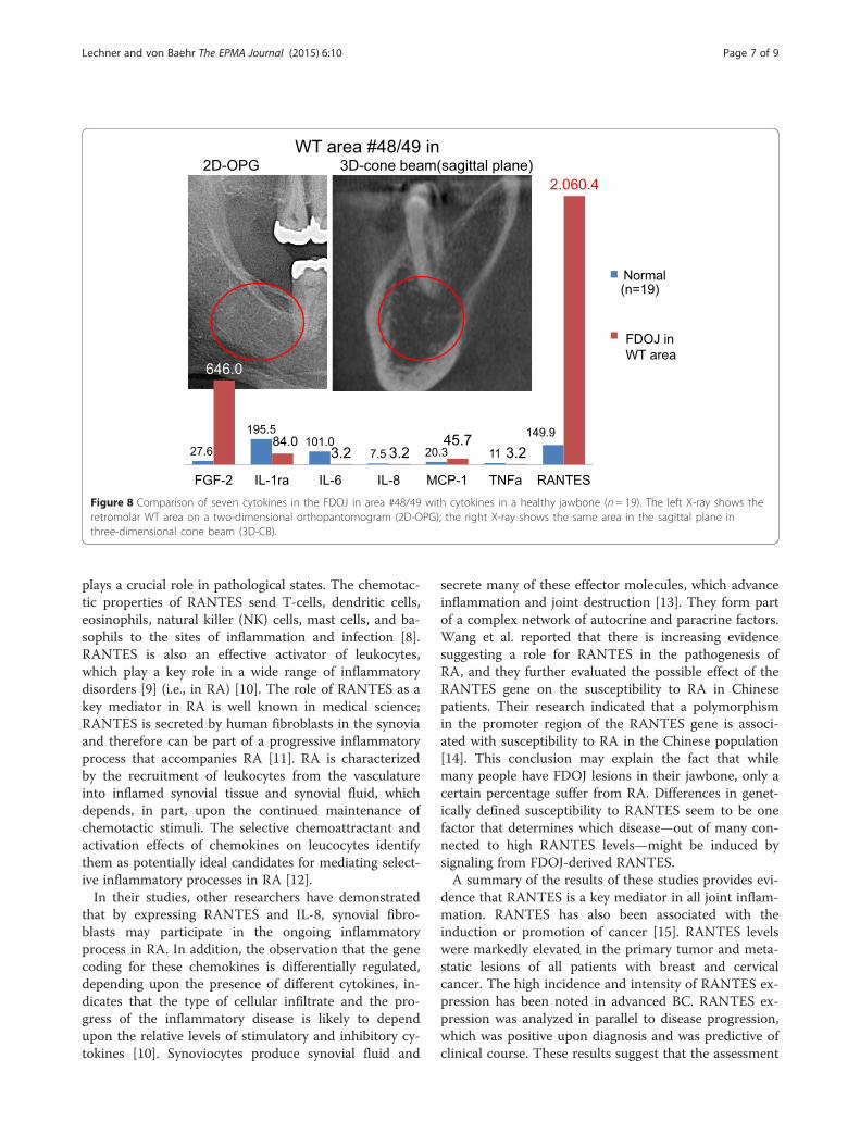
Case report: recurring breast cancer and comparison ofRANTES overexpression in the wisdom tooth area and inthe jawbone underneath a root-filled toothThe patient, 69 years old, had recurring BC on the rightside. This case is of interest because of the difference inRANTES expression in the jawbone underneath end-odontically treated tooth #46 (US #30) and in retromolararea #48/49 (US #32). In Figure 7, X-rays show theinconspicuous apical area of RFT #46 with only verydiscrete changes in the trabecular structures. The columnsin the same figure compare the distribution of seven cyto-kines in this FDOJ sample (in red) and 19 healthy controls(in blue). The X-rays in Figure 8 show the inconspicuousretromolar wisdom tooth area #48/49. As in Figure 7, thecolumns in Figure 8 compare the distribution of seven cy-tokines in the FDOJ sample of wisdom tooth area #48/49(in red) and in 19 healthy controls (in blue). Amazingly,RANTES expression in area #46 underneath the RFT wasthreefold higher (6,178 pg/mL) than RANTES expressionin area #48/49 (only 2,060 pg/mL), with obvious IWH inthe former area of WTS. Please note again the comprehen-sive failing of any X-ray diagnosis in both of the presentedjawbone areas.
11149.9
3.2
4.953.1
Norm (n=19)
FDOJ/RFT(n=16)
*
OJ (n = 16) from the jawbones of a group of patients with SYD

27.6 195.5 101.0 7.5 20.3 11 149.9
723.2 233.9 3.2 16.6 62.9 3.2
9.902.7
Normal(n=19)
FDOJarea(RFT#47)
Figure 6 Comparison of seven cytokines in FDOJ underneath RFT #47 with the cytokines in the healthy jawbone (n= 19). Intraoperative documentationof extension of FDOJ in the right lower jawbone, area #47 apically of RFT #47, by contrast agent after the surgical removal of RFT #47.
Lechner and von Baehr The EPMA Journal (2015) 6:10 Page 6 of 9
ResultsRole of RANTES in inflammatory diseaseRANTES belongs to the family of chemotactic cytokinesknown as CC pattern chemokines. It is expressed by anearly response gene. RANTES is chemotactic for T-cells,
27.6195.5 101.0 7.5 2
1.160.0
391.53.2 8.8
FGF-2 IL-1ra IL-6 IL-8 M
Apical area of RFT2D-OPG 3D-cone bea
Figure 7 Comparison of the seven cytokines in the FDOJ underneath RFTshows the apical area of RFT #46 in a red circle on a two-dimensional orthsagittal plane in three-dimensional cone beam (3D-CB).
eosinophils, and basophils, and it plays a key role inrecruiting leukocytes to inflammatory sites. The signifi-cance of RANTES for the development of diseasesseems to be enormous; RANTES interferes with im-mune responses on a number of levels and therefore
0.3 11149.9131.6 3.2
6.176.0
CP-1 RANTES
(n=19)
#46 inm(sagittal plane)
TNF-a
Normal
ApicalFDOJRFT #46
#46 with the cytokines in the healthy jawbone (n = 19). The left X-rayopantomogram (2D-OPG); the right X-ray shows the same area on the
27.6
195.5101.0
7.5 20.3 11
149.9
646.0
84.03.2 3.2
45.73.2
2.060.4
FGF-2 IL-1ra IL-6 IL-8 MCP-1 TNFa RANTES
(n=19)
FDOJ inWT area
Normal
WT area #48/49 in2D-OPG 3D-cone beam(sagittal plane)
Figure 8 Comparison of seven cytokines in the FDOJ in area #48/49 with cytokines in a healthy jawbone (n = 19). The left X-ray shows theretromolar WT area on a two-dimensional orthopantomogram (2D-OPG); the right X-ray shows the same area in the sagittal plane inthree-dimensional cone beam (3D-CB).
Lechner and von Baehr The EPMA Journal (2015) 6:10 Page 7 of 9
plays a crucial role in pathological states. The chemotac-tic properties of RANTES send T-cells, dendritic cells,eosinophils, natural killer (NK) cells, mast cells, and ba-sophils to the sites of inflammation and infection [8].RANTES is also an effective activator of leukocytes,which play a key role in a wide range of inflammatorydisorders [9] (i.e., in RA) [10]. The role of RANTES as akey mediator in RA is well known in medical science;RANTES is secreted by human fibroblasts in the synoviaand therefore can be part of a progressive inflammatoryprocess that accompanies RA [11]. RA is characterizedby the recruitment of leukocytes from the vasculatureinto inflamed synovial tissue and synovial fluid, whichdepends, in part, upon the continued maintenance ofchemotactic stimuli. The selective chemoattractant andactivation effects of chemokines on leucocytes identifythem as potentially ideal candidates for mediating select-ive inflammatory processes in RA [12].In their studies, other researchers have demonstrated
that by expressing RANTES and IL-8, synovial fibro-blasts may participate in the ongoing inflammatoryprocess in RA. In addition, the observation that the genecoding for these chemokines is differentially regulated,depending upon the presence of different cytokines, in-dicates that the type of cellular infiltrate and the pro-gress of the inflammatory disease is likely to dependupon the relative levels of stimulatory and inhibitory cy-tokines [10]. Synoviocytes produce synovial fluid and
secrete many of these effector molecules, which advanceinflammation and joint destruction [13]. They form partof a complex network of autocrine and paracrine factors.Wang et al. reported that there is increasing evidencesuggesting a role for RANTES in the pathogenesis ofRA, and they further evaluated the possible effect of theRANTES gene on the susceptibility to RA in Chinesepatients. Their research indicated that a polymorphismin the promoter region of the RANTES gene is associ-ated with susceptibility to RA in the Chinese population[14]. This conclusion may explain the fact that whilemany people have FDOJ lesions in their jawbone, only acertain percentage suffer from RA. Differences in genet-ically defined susceptibility to RANTES seem to be onefactor that determines which disease—out of many con-nected to high RANTES levels—might be induced bysignaling from FDOJ-derived RANTES.A summary of the results of these studies provides evi-
dence that RANTES is a key mediator in all joint inflam-mation. RANTES has also been associated with theinduction or promotion of cancer [15]. RANTES levelswere markedly elevated in the primary tumor and meta-static lesions of all patients with breast and cervicalcancer. The high incidence and intensity of RANTES ex-pression has been noted in advanced BC. RANTES ex-pression was analyzed in parallel to disease progression,which was positive upon diagnosis and was predictive ofclinical course. These results suggest that the assessment
Lechner and von Baehr The EPMA Journal (2015) 6:10 Page 8 of 9
of RANTES expression may be a useful prognostic indi-cator for the identification of patients with an apparentlypoor prognosis [16]. The development of BC may, inpart, be due to the ability of RANTES to act directly onthe tumor cells and to promote tumor progression [17].
Origin of RANTES in FDOJ and adipocytesThat the inflammatory response of adipose tissue is as-sociated with a systemic inflammatory response is wellknown and widely discussed. In obesity, this systemic in-flammatory response originates in the adipose tissueitself. Secretion of inflammatory cytokines mediates thesystemic effects of adipose tissue inflammation. Huberet al. found an increased expression of RANTES in fattytissue in obese patients [18]. Our findings regardingRANTES/CCL5 secretion by FDOJ deserve further dis-cussion: reduced blood flow and capillary densityfollowed by ischemia in the jawbone may lead to a hyp-oxic situation [19]. Adipocytes and the necrotic parts offat cells are considered by many studies to be immuno-logically effective ingredients. The role of these immuneeffects on understanding FDOJ, RANTES/CCL5, andSYD is an evident issue and needs further illuminationin the discussion. While proinflammatory cytokines suchas TNF-α, IL-6, and prostaglandins are already distrib-uted early in the acute stage of an injury or tissue infec-tion, there are many indications that chemokines likeRANTES are activated at a later time and that they canact in the conversion of acute pain into a more chronicphenomenon. In fact, recent data suggest that, in con-junction with tissue damage or infection, ischemia-induced chemokine expression causes an increase ininflammatory cytokines [20].
FDOJ as a systemic threat from RANTES overexpressionFDOJ is similar to silent inflammation or subclinical in-flammation without typical signs of acute inflammation.In CI, the local production of proinflammatory cytokinesoverstrains regulatory and compensating mechanisms bybuilding FDOJ in the bone marrow. This phenomenonseems to be more widespread than dentists and doctorspresumed in the past. It is generally accepted that an im-balance between cytokines and their specific inhibitors ischaracteristic of chronic inflammatory conditions [21].Cytokines merge to release the immune response and toinduce acute inflammatory events in the transition orpersistence of the CI. This means that when maintaininghealthy conditions, the cytokine-producing mechanismsmust be controlled [22]. FDOJ represents a new cellularresponse phenomenon in inflammation, in that the cyto-kines are not released bacterially or virally, but by per-sisting metabolic derailments in the medullary space ofthe jawbone. If the body does not succeed in revising themetabolic disturbances in the IWH area of the jawbone
within a certain period, increasing numbers of immunecells are recruited in the fatty tissue. The chronicallysilent inflammation of the medullary fatty tissue leadsto the local development of proinflammatory signal-ing mediators—in particular, RANTES/CCL5. Thesesystemically affect the organism and can result inchronic inflammatory processes or provoke furtherpathophysiological mechanisms.
The problem of diagnosing FDOJ lesions by X-rayIn earlier research, we already demonstrated the nonvisi-bility and lack of radiographic appearance of FDOJ,which makes it difficult to obtain an accurate diagnosisby common dental radiographic means [23]. Thus, theexistence of FDOJ is largely neglected today in main-stream dentistry, as is its systemic relevance. The reasonfor this is that conventional X-ray techniques are limitedin their ability to reveal the actual extent and location ofFDOJ. To aid the practitioner in diagnosing the debilitat-ing effects of bone marrow softening inside FDOJlesions, a computer-assisted through transalveolar ultra-sound (TAU) device is available [24]. TAU precisely im-ages and identifies cavitational porosity in the jawbone.TAU imaging proved significantly superior to radiologyfor the detection of microscopically confirmed FDOJ.The efficiency and reliability of TAU in the diagnosisand imaging of FDOJ has been presented in numerouspublications [25]. Because of these diagnostic difficulties,jawbone disease is underdiagnosed by dentists in generaland, consequently, by doctors in SYD cases. The missingcoincidence between inconspicuous X-rays and the over-expression of proinflammatory signaling pathways incorresponding FDOJ areas—as shown in Figures 6, 7,and 8—lends this phenomenon importance in the dis-cussion about “silent inflammation”.
ConclusionsIn our eyes, this is one of the first investigations to showa potential correlation between RA (of any type) and is-chemic or inflammatory jawbone lesions—an associationthat has been speculated on for decades. Additionally,chemokine overexpression in the jawbone connected toRFT seems to be a possible danger for immune preserva-tion, which is needed to maintain a balanced system andto prevent different types of SYD. IWH in old extractionsites and underneath RFT might also provoke immunemodulation, which hinders the restoration of an alreadydisease-modified immune system. The presence of cyto-kine imbalances in the jawbone leads to internal signal-ing through the accessory pathways via overexpressedRANTES/CCL5, which can lead to chronic pathologiessuch as cancer, diabetes, and cardiovascular diseases inthe long term, as well as neurodegenerative or inflam-matory processes. Once a chronic disease has been
Lechner and von Baehr The EPMA Journal (2015) 6:10 Page 9 of 9
established, the deterioration produced by the undiscov-ered “silent inflammation” in the jawbone progressivelycreates a set of pathological conditions that worsens theoverall condition and leads to further deficiencies. It is avicious circle. This is the reason why restoring a modi-fied system might require synergistic actions at the den-tal level, including surgical curettage of FDOJ or theelimination of RF with additional cleaning of the sur-rounding jawbone. Thus, dentists can help to not onlyalleviate the symptoms of acute inflammation but alsoput their patients on track to avoid the devastating ef-fects of CI, which exists below the threshold of perceivedpain and can smolder silently for decades. Novel thera-peutic strategies that specifically target the inflammatoryreaction in FDOJ could considerably contribute to thereduction of morbidity in patients. A more critical atti-tude inside the dental community to wound healing afterdental surgery is a potential clinical implementation ofthe achieved results. The actual paper follows recom-mendations of the “EPMA White Paper” [26].
AbbreviationsAI: acute infection; BC: breast cancer; CI: chronic inflammation; FDOJ: fattydegenerative osteonecrotic jawbone; FGF-2: fibroblast growth factor-2;HJB: healthy jawbone; IWH: insufficient wound healing; RA: rheumatoidarthritis; RF: root filling; RFT: root-filled tooth; SYD: systemic diseases;WT: wisdom tooth; WTS: wisdom tooth surgery.
Competing interestsThe authors declare that they have no competing interests.
Authors’ contributionsJL conceived of the design of the study and carried out the dentalinvestigations and the surgery in the jawbone. He performed the statisticalanalysis and drafted the manuscript. VB carried out the Luminex® analysis ofthe jawbone samples. He participated in the design of the study andcoordination and helped to draft the manuscript. All authors read andapproved the final manuscript.
Author details1Clinic for Integrative Dentistry, Gruenwalder Str. 10A, 81547 Munich,Germany. 2Compartment of Immunology and Allergology, Institute forMedical Diagnostics, MVZ GbR, Nicolaistrasse 22, 12247 Berlin, Germany.
Received: 6 February 2015 Accepted: 25 March 2015
References1. Townsend MJ, McKenzie AN. Unravelling the net? Cytokines and diseases.
J Cell Sci. 2000;113(Pt 20):3549–50.2. Imbeau J. Introduction to through-transmission alveolar ultrasonography
(TAU) in dental medicine. Cranio. 2005;23(2):100–12.3. Ratner EJ, Langer B, Evins ML. Alveolar cavitational osteopathosis:
manifestations of an infectious process and its implication in thecausation of chronic pain. J Periodontol. 1986;57:593.
4. Mankin HJ. Nontraumatic necrosis of bone (osteonecrosis). N Engl J Med.1992;326:1473.
5. Ono K. Symposium: recent advances in avascular osteonecrosis. Clin Orthop.1992;277:2.
6. Graff-Radford SB, Simmons M, Fox L, et al. Are bony cavities exclusivelyassociated with atypical facial pain and trigeminal neuralgia? Proceedings ofAnnual Meeting. Santa Fe, New Mexico: Western USA Pain Society; 1988.
7. Bouquot JE, Roberts AM, Person P, Christian J. NICO (neuralgia-inducingcavitational osteonecrosis): osteomyelitis in 224 jawbone samples frompatients with facial neuralgias. Oral Surg Oral Med Oral Pathol. 1992;73:307.
8. Levy JA. The unexpected pleiotropic activities of RANTES. J Immunol.2009;182(7):3945–6.
9. von Luettichau I, Nelson PJ, Pattison JM, van de Rijn M, Huie P, Warnke R,et al. RANTES chemokine expression in diseased and normal human tissues.Cytokine. 1996;8:89–98.
10. Rathanaswami P, Hachicha M, Sadick M, Schall TJ, McColl SR. Expression ofthe cytokine RANTES in human rheumatoid synovial fibroblasts. Differentialregulation of RANTES genes by inflammatory cytokines. J Biol Chem.1993;268(8):5834–9.
11. Hirano F, Kobayashi A, Hirano Y, Nomura Y, Fukawa E, Makino I. Thrombin-induced expression of RANTES mRNA through protease activated receptor-1in human synovial fibroblasts. Ann Rheum Dis. 2002;61(9):834–7.
12. Robinson E, Keystone EC, Schall TJ, Gillett N, Fish EN. Chemokine expressionin rheumatoid arthritis (RA): evidence of RANTES and macrophageinflammatory protein (MIP)-1β production by synovial T cells. Clin ExpImmunol. 1995;101(3):398–407.
13. Chicheportiche Y, Chicheportiche R, Sizing I, Thompson J, Benjamin CB,Ambrose C, et al. Proinflammatory activity of TWEAK on human dermalfibroblasts and synoviocytes: blocking and enhancing effects of anti-TWEAKmonoclonal antibodies. Arthritis Res. 2002;4(2):126–33.
14. Wang CR, Guo HR, Liu MF. RANTES promoter polymorphism as a geneticrisk factor for rheumatoid arthritis in the Chinese. Clin Exp Rheumatol.2005;23(3):379–84.
15. Soria G, Ben-Baruch A. The inflammatory chemokines CCL2 and CCL5 inbreast cancer. Cancer Lett. 2008;267:271–85.
16. Wigler N. Breast carcinoma: a report on the potential usage of the CCchemokine RANTES as a marker for a progressive disease. Isr Med Assoc J.2002;4(11 Suppl):940–3.
17. Niwa Y, Akamatsu H, Niwa H, Sumi H, Ozaki Y, Abe A. Correlation of tissueand plasma RANTES levels with disease course in patients with breast orcervical cancer. Clin Cancer Res. 2001;7(2):285–9.
18. Huber J, Kiefer FW, Zeyda M, Ludvik B, Silberhumer GR, Prager G, et al. CCchemokine and CC chemokine receptor profiles in visceral andsubcutaneous adipose tissue are altered in human obesity. J ClinEndocrinol Metab. 2008;93:3215–21.
19. Ye J. Emerging role of adipose tissue hypoxia in obesity and insulinresistance. Int J Obes (Lond). 2009;33:54–66.
20. Kiguchi N, Kobayashi Y, Kishioka S. Chemokines and cytokines inneuroinflammation leading to neuropathic pain. Curr Opin Pharmacol.2012;12(1):55–61.
21. Tilg H, Moschen RA. Adipocytokines: mediators linking adipose tissue,inflammation and immunity. Nat Rev Immunol. 2006;6(10):772–83.
22. Ramesh G, MacLean A, Philipp M. Cytokines and chemokines at thecrossroads of neuroinflammation, neurodegeneration, and neuropathic pain.Mediators Inflamm. 2013;2013:480739.
23. Lechner J. Validation of dental X-ray by cytokine RANTES—comparison ofX-ray findings with cytokine overexpression in jawbone. Clin CosmetInvestig Dent. 2014;6:71–9.
24. Bouquot JE, Margolis M, Shankland W, Imbeau J. Through-transmissionalveolar ultrasonography (TAU): new technology for evaluation of medullarydiseases. Correlation with histopathology of 285 scanned jaw sites. Oral SurgOral Med Oral Pathol Oral Radiol Endod. 2002;94:210.
25. Bouquot J, Martin W, Wrobleski G. Computer-based thru-transmissionsonography (CTS) imaging of ischemic osteonecrosis of the jaws—apreliminary investigation of 6 cadaver jaws and 15 pain patients. OralSurg Oral Med Oral Pathol Oral Radiol Endod. 2001;92:550.
26. Golubnitschaja O, Costigliola V, EPMA. General Report & Recommendationsin Predictive, Preventive and Personalised Medicine 2012: White Paper ofthe European Association for Predictive, Preventive and PersonalisedMedicine. EPMA J 2012, 3:14. doi:10.1186/1878-5085-3-14