chemical priming by isothiocyanates protects against
TRANSCRIPT

fpls-11-00887 June 26, 2020 Time: 15:36 # 1
ORIGINAL RESEARCHpublished: 26 June 2020
doi: 10.3389/fpls.2020.00887
Edited by:Stephan Pollmann,
Center for Plant Biotechnologyand Genomics, National
Institute for Agricultural and FoodResearch and Technology, Spain
Reviewed by:Ute Wittstock,
Technische Universitat Braunschweig,Germany
Jonathan Gershenzon,Max Planck Institute for Chemical
Ecology, Germany
*Correspondence:Martin J. Mueller
Specialty section:This article was submitted to
Plant Physiology,a section of the journal
Frontiers in Plant Science
Received: 13 March 2020Accepted: 29 May 2020
Published: 26 June 2020
Citation:Ferber E, Gerhards J, Sauer M,
Krischke M, Dittrich MT, Müller T,Berger S, Fekete A and Mueller MJ
(2020) Chemical Priming byIsothiocyanates Protects Against
Intoxication by Products of theMustard Oil Bomb.
Front. Plant Sci. 11:887.doi: 10.3389/fpls.2020.00887
Chemical Priming by IsothiocyanatesProtects Against Intoxication byProducts of the Mustard Oil BombElena Ferber1, Julian Gerhards1, Miriam Sauer1, Markus Krischke1, Marcus T. Dittrich2,3,Tobias Müller2, Susanne Berger1, Agnes Fekete1 and Martin J. Mueller1*
1 Julius-von-Sachs-Institute of Biosciences, Biocenter, Pharmaceutical Biology, University of Würzburg, Würzburg, Germany,2 Department of Boinformatics, Biocenter, University of Würzburg, Würzburg, Germany, 3 Institute of Clinical Biochemistry,University of Würzburg, Würzburg, Germany
In Brassicaceae, tissue damage triggers the mustard oil bomb i.e., activates thedegradation of glucosinolates by myrosinases leading to a rapid accumulation ofisothiocyanates at the site of damage. Isothiocyanates are reactive electrophilic species(RES) known to covalently bind to thiols in proteins and glutathione, a process thatis not only toxic to herbivores and microbes but can also cause cell death of healthyplant tissues. Previously, it has been shown that subtoxic isothiocyanate concentrationscan induce transcriptional reprogramming in intact plant cells. Glutathione depletionby RES leading to breakdown of the redox potential has been proposed as a centraland common RES signal transduction mechanism. Using transcriptome analyses,we show that after exposure of Arabidopsis seedlings (grown in liquid culture) tosubtoxic concentrations of sulforaphane hundreds of genes were regulated withoutdepletion of the cellular glutathione pool. Heat shock genes were among the mosthighly up-regulated genes and this response was found to be dependent on thecanonical heat shock factors A1 (HSFA1). HSFA1-deficient plants were more sensitive toisothiocyanates than wild type plants. Moreover, pretreatment of Arabidopsis seedlingswith subtoxic concentrations of isothiocyanates increased resistance against exposureto toxic levels of isothiocyanates and, hence, may reduce the autotoxicity of the mustardoil bomb by inducing cell protection mechanisms.
Keywords: autotoxicity, heat shock response, isothiocyanates, mustard oil bomb, reactive electrophilic species,redox homeostasis, sulforaphane
INTRODUCTION
Chemical defense strategies in plants often involve both inducible secondary metabolites, classifiedas phytoalexins, as well as the constitutive accumulation of secondary defense metabolites, termedphytoanticipins. Since the biological activity of the vast majority of plant secondary metabolites isunspecific and not directed against specific foreign target proteins, plants cells need to protect their
Abbreviations: AITC, allyl isothiocyanate; BITC, benzyl isothiocyanate; BSO, buthionine sulfoximine; GSH, glutathione;GST, glutathione S-transferase; GSSG, glutathione disulfide; HSF, heat shock factor; HSP, heat shock protein; ITC,isothiocyanate; PGA1, prostaglandin A1; RES, reactive electrophilic species; SF, sulforaphane; TGG, ß-thioglucosideglucohydrolases.
Frontiers in Plant Science | www.frontiersin.org 1 June 2020 | Volume 11 | Article 887
fpls-11-00887 June 26, 2020 Time: 15:36 # 2
Ferber et al. Chemical Priming by Isothiocyanates
own tissues from self-intoxication by their defense chemicals.Besides storage of these compounds in safe compartments, acommon strategy is to store biologically inactive secondarymetabolite precursors that release biologically activephytochemicals after activation of enzymes triggered by tissuedamage, infection or pest attack. An example of this plant defensestrategy is the glucosinolate-myrosinase system in Arabidopsisalso known as the mustard oil bomb (Shirakawa and Hara-Nishimura, 2018). After tissue damage, however, biologicallyactive phytochemicals can cause cell death and tissue necrosis inplant tissues close to the damage site (Andersson et al., 2015).
Up to 40 biologically inactive glucosinolates, localizedin specialized lacticifer-like S-cells in close vicinity to thevasculature and along the leaf margins, have been identifiedas most abundant phytoanticipins in different Arabidopsisaccessions, reviewed in Burow and Halkier (2017). Thesespecialized cells have been shown to contain extremely highlevels (>130 mM) of glucosinolates (Koroleva et al., 2010).Arabidopsis thaliana Columbia (Col-0) synthesizes aliphatic andindolic glucosinolates in a ratio of about 8:2 in leaves (Brownet al., 2003) and the predominant glucosinolate is the aliphaticglucosinolate glucoraphanin that releases the isothiocyanate(ITC) sulforaphane (SF). In Arabidopsis Col-0, nitriles appearto be major breakdown products besides isothiocyanates, areaction that is promoted by nitrile-specifier proteins afterglucosinolate hydrolysis (Wittstock et al., 2016). However,biological activities are likely associated with the correspondingisothiocyanates.
Glucosinolates can be degraded by the enzymatic activityof myrosinases (ß-thioglucoside glucohydrolases, TGGs) thatare localized in guard cells and myrosin phloem cell idioblasts(Thangstad et al., 2004). In Arabidopsis leaves, two redundantmyrosinases (TGG1 and TGG2) have been shown to be essentialfor the breakdown of the predominant aliphatic glucosinolates.In the absence of TGG1 and TGG2, degradation of indolicglucosinolates was also greatly affected (Barth and Jander, 2006).
Tissue damage results in high local production of ITCs. Forinstance after complete mechanical tissue disruption, about 12µmol aliphatic and 3 µmol indolic glucosinolates per gramfresh weight (FW) (Barth and Jander, 2006) were degradedwithin 1 min leading to a corresponding local concentrationof glucosinulolate degradation products in damaged tissues ofabout 15 mM. The toxicity of extracellular or exogenously appliedITCs depends on the structure of the side chain and the way ofapplication (vapor, infiltration, spraying). For instance, exposureto the volatile allyl isothiocyanate (AITC) >0.5 M (Overbyet al., 2015b) or infiltration of SF >0.5 mM caused large visiblelesions (Andersson et al., 2015) and spraying of different ITCsin the concentration range of 10–100 mM resulted in growthinhibition and leaf bleaching (Hara et al., 2010). However, dueto the application mode, the absolute dose of exogenous ITCstaken up by plants is difficult to estimate and to correlate withdamage-induced ITC accumulation.
A common feature of ITCs is their lipophilicity andelectrophilicity. Due to their high chemical reactivity, exposedthiol groups in glutathione and proteins can be covalentlymodified. Because of their reactivity, ITCs can be classified as
reactive electrophilic species (RES) that display concentration-dependent, broad spectrum toxicity in microorganisms, animalsand plants. Toxicity of ITCs also depends on the capacityof cells to detoxify these RES mainly via conjugation toglutathione (GSH). Coupling to GSH can take place as anon-enzymatic reaction or can be catalyzed by an array ofglutathione S-transferases (GSTs) (Wagner et al., 2002; Haraet al., 2010) which can be transcriptionally upregulated by ITCs(Overby et al., 2015b).
It has been proposed that the major mode of action ofSF in mammalian (Valgimigli and Iori, 2009) as well as inplant cells (Andersson et al., 2015) is to decrease the cellularglutathione pool leading to an increase of the redox potential andeventually to cell death. Since all cell permeable and structurallydiverse RES have the concentration-dependent ability to depletethe cellular GSH pool, the resulting increased redox potentialcould potentially act as a common signal triggering RES-responsive genes.
Indeed, several ITCs (Hara et al., 2010) and structurallydifferent RES oxylipins (Mueller et al., 2008; Muench et al.,2016) have been shown to induce expression of heat shockand detoxification genes at low concentrations while sprayinghigher concentrations were highly toxic. The magnitude of geneinduction and cell death varied when different ITCs were tested.Exposure to AITC vapor has recently been shown to upregulatemore than 3900 genes in Arabidopsis seedlings including heatstress, oxidative and general stress as well as detoxificationgenes (Kissen et al., 2016). However, AITC does not occur inArabidopsis Col-0 and transcriptome data on Arabidopsis ITCsincluding SF is not available.
It has been shown that SF both possesses direct antimicrobialproperties and, at lower concentrations, acts directly on plantcells to trigger defense responses (Andersson et al., 2015;Schillheim et al., 2018). In addition, we hypothesized that SFand other ITCs produced after herbivore or pathogen attack mayalso serve as local signals or as metabolic regulators that reducecollateral damage of the mustard oil bomb. We show that ITC –at subtoxic concentrations – trigger cell rescue mechanisms thatconfer protection against ITC intoxication.
RESULTS
Uptake and Metabolism of ExogenousSulforaphane by Arabidopsis SeedlingsIn order to study the effect of extracellular SF on plant tissueswe employed a liquid culture system for Arabidopsis seedlingsto which exogenous ITCs and RES could be easily applied fortreatment and, after defined exposure times, could be removedfor recovery. Previously, ITCs were applied by spraying, leafinoculation, growth on ITC-containing agar or exposure toisothiocyanate vapors. All these application systems have thedisadvantage that the absolute treatment dosage per gram of planttissue is difficult to estimate. Using the liquid culture system, wefirst determined uptake of SF by 10 seedlings (123 ± 33 mg,mean ± SD) exposed to 500 µl treatment solution containing100 µM SF (50 nmol) which corresponds to an absolute dosage
Frontiers in Plant Science | www.frontiersin.org 2 June 2020 | Volume 11 | Article 887
fpls-11-00887 June 26, 2020 Time: 15:36 # 3
Ferber et al. Chemical Priming by Isothiocyanates
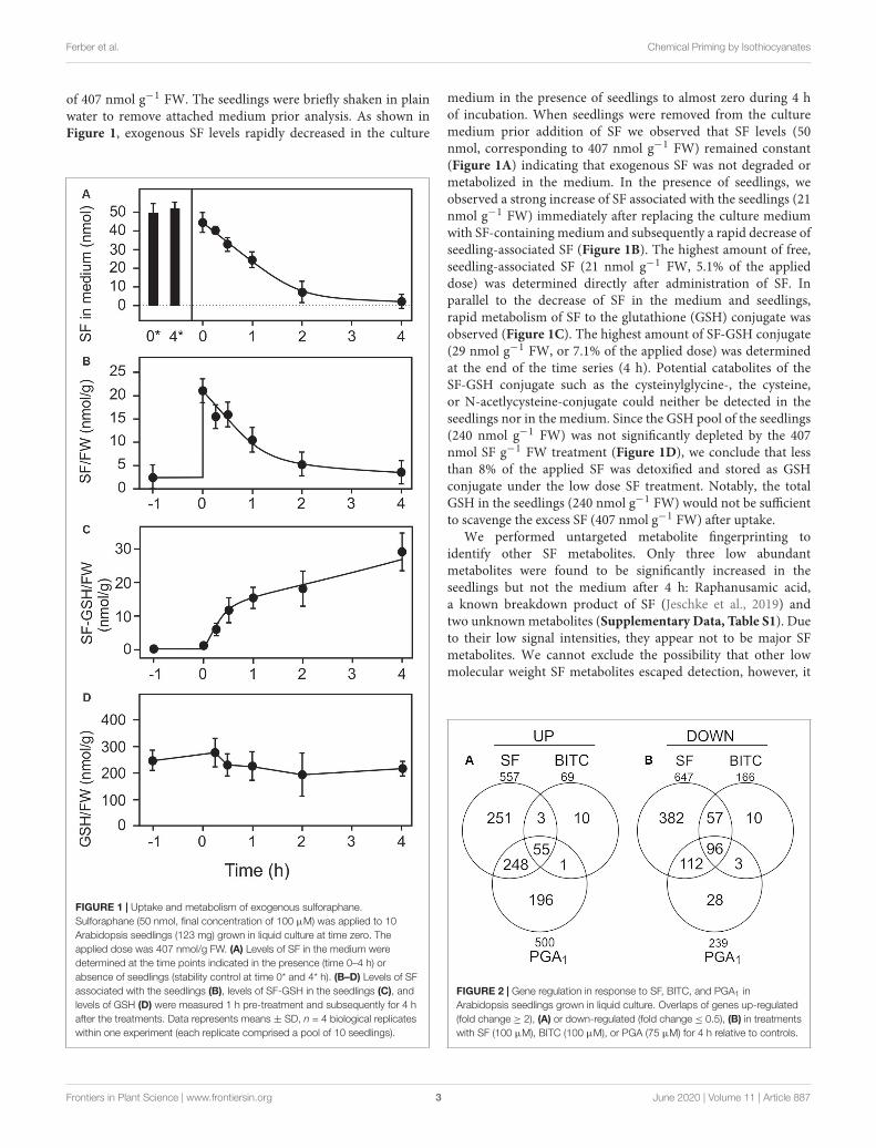
of 407 nmol g−1 FW. The seedlings were briefly shaken in plainwater to remove attached medium prior analysis. As shown inFigure 1, exogenous SF levels rapidly decreased in the culture
FIGURE 1 | Uptake and metabolism of exogenous sulforaphane.Sulforaphane (50 nmol, final concentration of 100 µM) was applied to 10Arabidopsis seedlings (123 mg) grown in liquid culture at time zero. Theapplied dose was 407 nmol/g FW. (A) Levels of SF in the medium weredetermined at the time points indicated in the presence (time 0–4 h) orabsence of seedlings (stability control at time 0* and 4* h). (B–D) Levels of SFassociated with the seedlings (B), levels of SF-GSH in the seedlings (C), andlevels of GSH (D) were measured 1 h pre-treatment and subsequently for 4 hafter the treatments. Data represents means ± SD, n = 4 biological replicateswithin one experiment (each replicate comprised a pool of 10 seedlings).
medium in the presence of seedlings to almost zero during 4 hof incubation. When seedlings were removed from the culturemedium prior addition of SF we observed that SF levels (50nmol, corresponding to 407 nmol g−1 FW) remained constant(Figure 1A) indicating that exogenous SF was not degraded ormetabolized in the medium. In the presence of seedlings, weobserved a strong increase of SF associated with the seedlings (21nmol g−1 FW) immediately after replacing the culture mediumwith SF-containing medium and subsequently a rapid decrease ofseedling-associated SF (Figure 1B). The highest amount of free,seedling-associated SF (21 nmol g−1 FW, 5.1% of the applieddose) was determined directly after administration of SF. Inparallel to the decrease of SF in the medium and seedlings,rapid metabolism of SF to the glutathione (GSH) conjugate wasobserved (Figure 1C). The highest amount of SF-GSH conjugate(29 nmol g−1 FW, or 7.1% of the applied dose) was determinedat the end of the time series (4 h). Potential catabolites of theSF-GSH conjugate such as the cysteinylglycine-, the cysteine,or N-acetlycysteine-conjugate could neither be detected in theseedlings nor in the medium. Since the GSH pool of the seedlings(240 nmol g−1 FW) was not significantly depleted by the 407nmol SF g−1 FW treatment (Figure 1D), we conclude that lessthan 8% of the applied SF was detoxified and stored as GSHconjugate under the low dose SF treatment. Notably, the totalGSH in the seedlings (240 nmol g−1 FW) would not be sufficientto scavenge the excess SF (407 nmol g−1 FW) after uptake.
We performed untargeted metabolite fingerprinting toidentify other SF metabolites. Only three low abundantmetabolites were found to be significantly increased in theseedlings but not the medium after 4 h: Raphanusamic acid,a known breakdown product of SF (Jeschke et al., 2019) andtwo unknown metabolites (Supplementary Data, Table S1). Dueto their low signal intensities, they appear not to be major SFmetabolites. We cannot exclude the possibility that other lowmolecular weight SF metabolites escaped detection, however, it
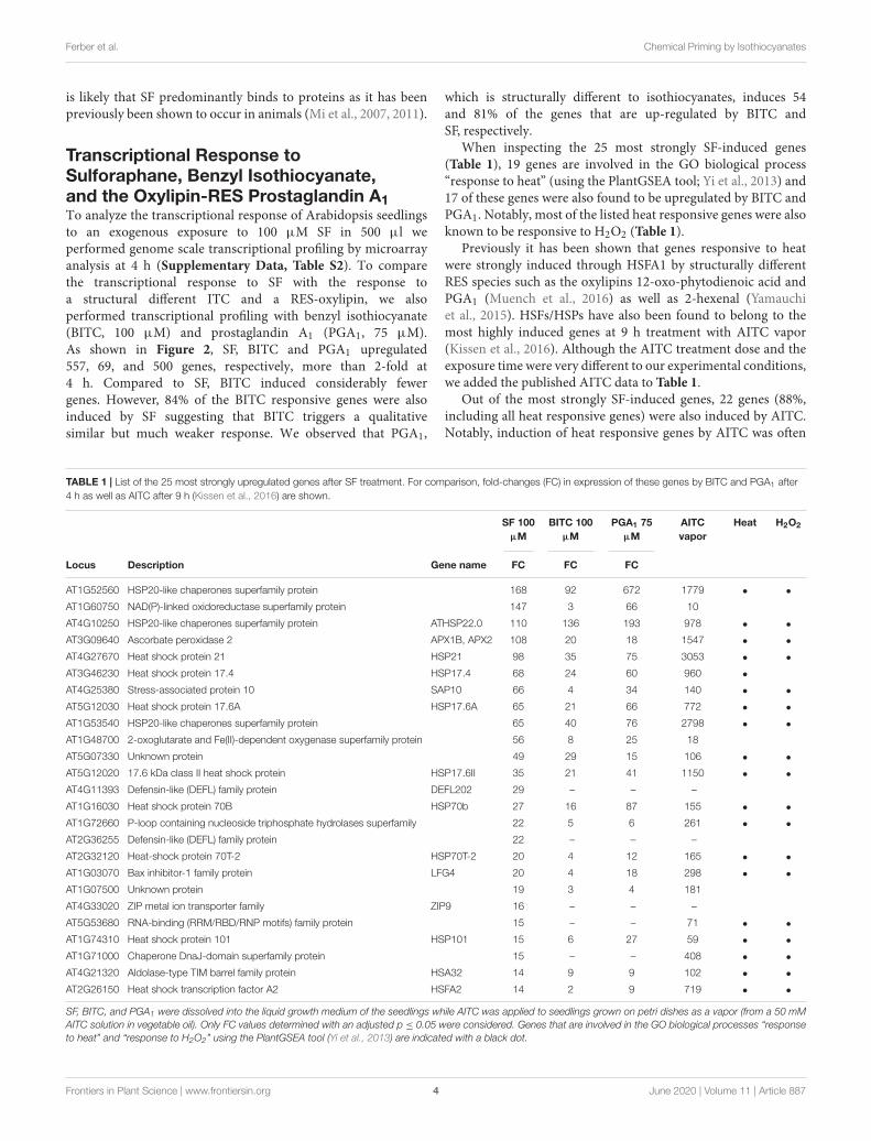
FIGURE 2 | Gene regulation in response to SF, BITC, and PGA1 inArabidopsis seedlings grown in liquid culture. Overlaps of genes up-regulated(fold change ≥ 2), (A) or down-regulated (fold change ≤ 0.5), (B) in treatmentswith SF (100 µM), BITC (100 µM), or PGA (75 µM) for 4 h relative to controls.
Frontiers in Plant Science | www.frontiersin.org 3 June 2020 | Volume 11 | Article 887
fpls-11-00887 June 26, 2020 Time: 15:36 # 4
Ferber et al. Chemical Priming by Isothiocyanates
is likely that SF predominantly binds to proteins as it has beenpreviously been shown to occur in animals (Mi et al., 2007, 2011).
Transcriptional Response toSulforaphane, Benzyl Isothiocyanate,and the Oxylipin-RES Prostaglandin A1To analyze the transcriptional response of Arabidopsis seedlingsto an exogenous exposure to 100 µM SF in 500 µl weperformed genome scale transcriptional profiling by microarrayanalysis at 4 h (Supplementary Data, Table S2). To comparethe transcriptional response to SF with the response toa structural different ITC and a RES-oxylipin, we alsoperformed transcriptional profiling with benzyl isothiocyanate(BITC, 100 µM) and prostaglandin A1 (PGA1, 75 µM).As shown in Figure 2, SF, BITC and PGA1 upregulated557, 69, and 500 genes, respectively, more than 2-fold at4 h. Compared to SF, BITC induced considerably fewergenes. However, 84% of the BITC responsive genes were alsoinduced by SF suggesting that BITC triggers a qualitativesimilar but much weaker response. We observed that PGA1,
which is structurally different to isothiocyanates, induces 54and 81% of the genes that are up-regulated by BITC andSF, respectively.
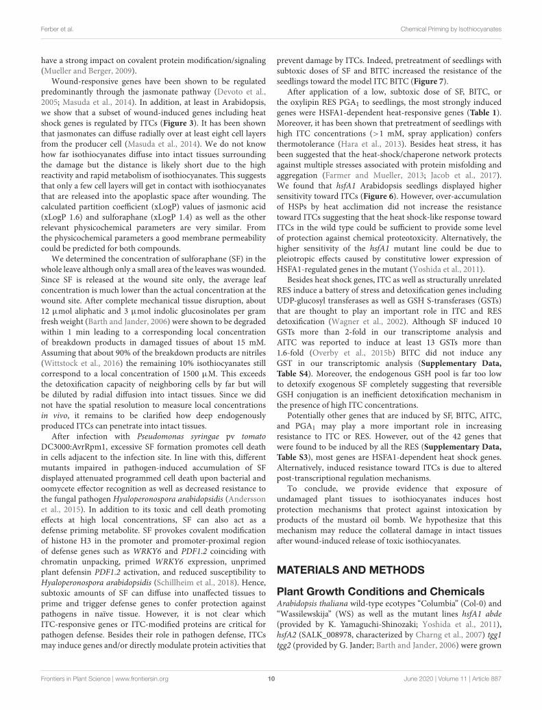
When inspecting the 25 most strongly SF-induced genes(Table 1), 19 genes are involved in the GO biological process“response to heat” (using the PlantGSEA tool; Yi et al., 2013) and17 of these genes were also found to be upregulated by BITC andPGA1. Notably, most of the listed heat responsive genes were alsoknown to be responsive to H2O2 (Table 1).
Previously it has been shown that genes responsive to heatwere strongly induced through HSFA1 by structurally differentRES species such as the oxylipins 12-oxo-phytodienoic acid andPGA1 (Muench et al., 2016) as well as 2-hexenal (Yamauchiet al., 2015). HSFs/HSPs have also been found to belong to themost highly induced genes at 9 h treatment with AITC vapor(Kissen et al., 2016). Although the AITC treatment dose and theexposure time were very different to our experimental conditions,we added the published AITC data to Table 1.
Out of the most strongly SF-induced genes, 22 genes (88%,including all heat responsive genes) were also induced by AITC.Notably, induction of heat responsive genes by AITC was often
TABLE 1 | List of the 25 most strongly upregulated genes after SF treatment. For comparison, fold-changes (FC) in expression of these genes by BITC and PGA1 after4 h as well as AITC after 9 h (Kissen et al., 2016) are shown.
SF 100µM
BITC 100µM
PGA1 75µM
AITCvapor
Heat H2O2
Locus Description Gene name FC FC FC
AT1G52560 HSP20-like chaperones superfamily protein 168 92 672 1779 • •
AT1G60750 NAD(P)-linked oxidoreductase superfamily protein 147 3 66 10
AT4G10250 HSP20-like chaperones superfamily protein ATHSP22.0 110 136 193 978 • •
AT3G09640 Ascorbate peroxidase 2 APX1B, APX2 108 20 18 1547 • •
AT4G27670 Heat shock protein 21 HSP21 98 35 75 3053 • •
AT3G46230 Heat shock protein 17.4 HSP17.4 68 24 60 960 •
AT4G25380 Stress-associated protein 10 SAP10 66 4 34 140 • •
AT5G12030 Heat shock protein 17.6A HSP17.6A 65 21 66 772 • •
AT1G53540 HSP20-like chaperones superfamily protein 65 40 76 2798 • •
AT1G48700 2-oxoglutarate and Fe(II)-dependent oxygenase superfamily protein 56 8 25 18
AT5G07330 Unknown protein 49 29 15 106 • •
AT5G12020 17.6 kDa class II heat shock protein HSP17.6II 35 21 41 1150 • •
AT4G11393 Defensin-like (DEFL) family protein DEFL202 29 – – –
AT1G16030 Heat shock protein 70B HSP70b 27 16 87 155 • •
AT1G72660 P-loop containing nucleoside triphosphate hydrolases superfamily 22 5 6 261 • •
AT2G36255 Defensin-like (DEFL) family protein 22 – – –
AT2G32120 Heat-shock protein 70T-2 HSP70T-2 20 4 12 165 • •
AT1G03070 Bax inhibitor-1 family protein LFG4 20 4 18 298 • •
AT1G07500 Unknown protein 19 3 4 181
AT4G33020 ZIP metal ion transporter family ZIP9 16 – – –
AT5G53680 RNA-binding (RRM/RBD/RNP motifs) family protein 15 – – 71 • •
AT1G74310 Heat shock protein 101 HSP101 15 6 27 59 • •
AT1G71000 Chaperone DnaJ-domain superfamily protein 15 – – 408 • •
AT4G21320 Aldolase-type TIM barrel family protein HSA32 14 9 9 102 • •
AT2G26150 Heat shock transcription factor A2 HSFA2 14 2 9 719 • •
SF, BITC, and PGA1 were dissolved into the liquid growth medium of the seedlings while AITC was applied to seedlings grown on petri dishes as a vapor (from a 50 mMAITC solution in vegetable oil). Only FC values determined with an adjusted p ≤ 0.05 were considered. Genes that are involved in the GO biological processes “responseto heat” and “response to H2O2” using the PlantGSEA tool (Yi et al., 2013) are indicated with a black dot.
Frontiers in Plant Science | www.frontiersin.org 4 June 2020 | Volume 11 | Article 887

fpls-11-00887 June 26, 2020 Time: 15:36 # 5
Ferber et al. Chemical Priming by Isothiocyanates
more than 10-fold stronger than after treatment with SF, BITC,or PGA1 (Table 1) which might be due to structural differences,higher dosage or longer exposure time. Stronger response toAITC is also reflected by the absolute number of genes inducedafter 9 h AITC vapor treatment (2352 genes) compared to 407nmol g−1 FW SF treatment for 4 h (557 genes) (SupplementaryData, Figure S1). The core set of genes induced by SF, BITC,PGA1, and AITC included 42 genes of which 28 (67%) wereheat responsive genes (Supplementary Data, Table S3). Hence,induction of heat shock responsive genes appears to be commonand most strongly affected after treatment with structurallydifferent RES. However, a considerable number of genes was notinduced by any of the other RES-treatments: SF (125 genes, 22%),BITC (10 genes, 14%), PGA1 (143 genes, 28%) and AITC (1970genes, 85%). This suggests that structurally different RES inducea set of common genes but also genes that are RES-specific anddependent on the structure (Supplementary Data, Figure S1).
Wound-Induced GlucosinolateBreakdown Triggers Gene InductionA major glucosinolate breakdown product in Arabidopsis Col-0 leaves besides nitriles is SF (Wittstock et al., 2016). Sinceexogenously administered SF can act as a signal at lowconcentrations and is toxic at high concentrations (Anderssonet al., 2015) we first determined endogenous SF levels inArabidopsis seedlings and plants. As shown in Figure 3A, wecould detect SF in apparently undamaged seedlings and plantsgrown under optimal conditions. Endogenous levels of SF werearound 0.5 nmol g−1 FW in seedlings cultured in liquid mediumand up to approximately 3 nmol g−1 FW in leaves of soil-grownplants (Figure 3A). To determine endogenous SF levels afterpartial leaf damage by wounding, leaves from 6-week old plantswere wounded three times with forceps over the leaf lamina. Thistreatment led to an accumulation of SF of up to 50 nmol g−1
FW corresponding to an average concentration in the woundedleaves of about 50 µM (Figure 3B) in wild type plants. Onlya small part of the leaf was directly damaged while SF levelswere measured in the whole leave. As expected, the myrosinasedeficient double mutant tgg1 tgg2 was not able to accumulate SF.Notably, SF is expected to accumulate at the wound site close tothe midrib and, hence, the SF concentration near the midrib likelyreaches much higher concentrations than the levels determinedin extracts from whole leaves.
To test whether wound-induced glucosinolate breakdownproducts induce transcription in wounded leaves, we determinedthe expression of genes known to be induced by SF and AITCas well as many RES species (Mueller et al., 2008; Kissen et al.,2016) in wild type and tgg1 tgg2 leaves. Relative expressionof HSFA2, HSP17.6II, and HSP26.5 was determined 0.5 hafter wounding since these genes display a rapid and transientexpression (Muench et al., 2016). Relative expression of mostother genes including Cyp81D11 is often highest between 2and 4 h after treatment. We therefore measured expression ofCyp81D11 4 h after wounding (Figure 3C). Wound-induction ofthe heat shock marker genes was abolished or strongly reduced inthe ITC-deficient tgg1 tgg2 line, indicating that wound-induced
FIGURE 3 | Endogenous SF levels and ITC-induced gene expression. (A) SFlevels were determined in seedlings (each sample comprised a pool of 10seedlings) grown in liquid MS medium (2 and 3 weeks) or leaves of plantsgrown on soil (4–8 weeks), data represent means ± SD, n = 3 biologicalreplicates within one experiment. (B) Increase of SF levels in leaves of 7-weekold wild-type and tgg1/tgg2 plants grown on soil after wounding, datarepresent means ± SD, n = 4 plants. Time 0 is a no treatment control (leaveswere collected and immediately shock frozen). Wounded leaves werecollected from the plants after wounding at the times indicated. (C) Inductionof RES-inducible marker genes in leaves of 6-week old wild-type andtgg1/tgg2 plants after wounding. Gene expression was determined byRT-qPCR after 0.5 h (HSFA2, HSP17.6II, and HSP26.5) or 4 h (Cyp81D11).Values were normalized to AtActin2/8, data represent means ± SD, n = 3–6plants. Asterisks indicate p ≤ 0.05 (*), and ≤ 0.005 (***).
Frontiers in Plant Science | www.frontiersin.org 5 June 2020 | Volume 11 | Article 887

fpls-11-00887 June 26, 2020 Time: 15:36 # 6
Ferber et al. Chemical Priming by Isothiocyanates
endogenous glucosinolate breakdown products, most likely SFand other ITCs, act as signals that modulate gene expressionin wounded leaves. Interestingly, expression of the RES- andjasmonate-inducible Cyp81D11 gene was also markedly reducedin the tgg1 tgg2 line although the jasmonate pathway is intact inthis line suggesting that more than one signal can regulate thatgene after wounding.
Sulforaphane Strongly Induces HeatShock Response Marker Genes ThroughHSFA1Induction of most heat-responsive genes including the inducibletranscription factors HSFA2 and dehydration-responsive element-binding protein 2A (DREB2A) as well as HSP101 and HSP26.5 isdependent on the four constitutive HSFA1 transcription factors(a, b, d, and e). To compare the activation of these marker genesby moderate heat and SF, we treated seedlings with moderate heat(37◦C) or SF (100 µM, corresponding to a treatment dose of407 nmol g−1 FW) for 4 h. The control sample did not receive
FIGURE 4 | Induction of heat-responsive genes by SF and moderate heat inwild-type (Col-0, WS), hsfA1 abde, and hsfA2 plants. Seedlings were treatedwith SF (100 µM, corresponding to a treatment dose of 407 nmol/g FW) ormoderate heat (37◦C) for 4 h and gene expression was analyzed by RT-qPCR.Values were normalized to AtSAND. Data represent means ± SD, n = 3biological replicates within one experiment (each replicate comprised a pool of10 seedlings). Asterisks indicate p ≤ 0.05 (*), ≤ 0.01 (**), and ≤ 0.005 (***).
methanol. We previously tested the effect of different solvents onheat shock gene expression when studying reactive electrophilesdissolved in methanol or DMSO (Muench et al., 2016). Methanol(1%) had no significant effect on gene expression of the selectedgenes. In addition, we tested expression of these genes in thehsfA1 abde quadruple and hsfA2 mutant plants. Since the hsfA1quadruple mutant was generated by crossing single mutants fromthe ecotype backgrounds Col-0 and Wassilewskija (WS), we usedboth wild types as controls (Figure 4).
In the wild types Col-0 and WS, SF-induced expression ofHSFA2, HSP101, and HSP26.5 was about 2–30% of the expressionof these genes after 37◦C for 4 h. Notably, HSP101 which hasbeen shown to be essential and sufficient to establish acquiredthermotolerance was strongly induced at 37◦C (68-fold) andSF (15-fold) in A. thaliana Col-0 seedlings. The expression ofthe DREB2A gene is induced by heat shock and dehydrationvia HSFA1-dependent and -independent signaling pathways,respectively (Yoshida et al., 2011). In contrast to the other markergenes, induction of DREB2A by SF was more similar to the 37◦Ctreatment in the wild types.
SF-induced expression of HSFA2, HSP101, HSP26.5, andDREB2A was strictly dependent on HSFA1 master regulatorgenes of the heat shock response. In addition, the inducibletranscriptions factor HSFA2 was not found to be requiredfor up-regulation of HSP101, HSP26.5 and DREB2A by SF.However, HSFA2 was required for the full up-regulation of thesmall HSP26.5 gene by heat. Hence, these results indicate thatHSFA1s but not HSFA2 are essential for the induction of heat-responsive HSPs by SF.
Glutathione Depletion and GlutathioneRedox Potential Alterations Are NotInvolved in the Regulation of theTranscriptional Response to SubtoxicConcentrations of Sulforaphane, BenzylIsothiocyanate, and Prostaglandin A1Due to their thiol reactivity, RES can deplete the cellular GSHpool in a concentration dependent manner. Thereby, the redoxpotential could be increased and sensed as a common signalto triggering RES-responsive genes. To test this hypothesis, wetreated 10 day-old Arabidopsis seedlings with SF, BITC andPGA1 under treatment conditions similar to the microarrayexperiments and determined the GSH and the oxidized GSH(GSSG) concentration of the seedlings 4 h after the treatment.As shown in Figure 5, we observed a concentration-dependentdecline of the endogenous GSH concentration. In addition, wecalculated the GSH/GSSG redox potential (Queval and Noctor,2007) from the measured endogenous GSH and GSSG levels 4 hafter the treatment. However, at concentrations (75–100 µM)that triggered gene regulation, we did not observe a significantdecrease of GSH or a change of the calculated redox potential.
The transcriptional response to low GSH content associatedwith increased redox potential has previously been determinedboth in GSH-deficient root meristemless 1-1 (rml1-1) seedlings(Schnaubelt et al., 2015) as well as in seedlings that have been
Frontiers in Plant Science | www.frontiersin.org 6 June 2020 | Volume 11 | Article 887

fpls-11-00887 June 26, 2020 Time: 15:36 # 7
Ferber et al. Chemical Priming by Isothiocyanates
FIGURE 5 | SF, BITC, and PGA1 treatments cause oxidation of the cellularglutathione pool and change of the cellular redox potential. Wild-typeArabidopsis seedlings grown in liquid culture in MS medium were treated withSF (A), BITC (B), and PGA1 (C) at the indicated concentrations. Reduced(GSH) and oxidized glutathione (GSSG) contents per gram fresh weight (FW)were measured 4 h after treatment, and the redox potential was calculated.Data represent means ± SD, n = 3 biological replicates within one experiment(each replicate comprised a pool of 10 seedlings) (D) Expression of low GSHmarker genes was determined by RT-qPCR 4 h after treatments with solvent(Con), SF or BITC. Values were normalized to the expression of AtActin2/8.Data represent means ± SD, n = 4–6 biological replicates within oneexperiment (each replicate comprised a pool of 10 seedlings). Asterisksindicate p ≤ 0.05 (*).
treated with the GSH-synthesis inhibitor buthionine sulfoximine(BSO) (Koprivova et al., 2010). When comparing our list ofSF-induced genes with the published microarray data of thesestudies, only 13% of the SF-induced genes were found tobe up-regulated in rml1-1 or BSO-treated seedlings. We alsoanalyzed the expression of two marker genes, a cytosolic h-typethioredoxin (TRX, TH8) and a glucosyltransferase (UGT74E2),that were found to be strongly up-regulated in seedlings withlow GSH content. However, both genes were not found tobe up-regulated at the concentration used in our microarrayexperiment. Moreover, heat shock genes are the most strongly up-regulated genes after ITC treatment while these genes appearednot to be regulated after BSO treatment (Koprivova et al., 2010)or were even down-regulated in rml1-1 seedlings (Schnaubeltet al., 2015). Hence, we conclude that signaling by low glutathionecontent is not involved in the gene regulation in response to lowdose ITC treatment.
Arabidopsis HSFA1 Transcription FactorsAre Important for Resistance AgainstIsothiocyanate IntoxicationITCs strongly up-regulate heat shock proteins, a process thatcould be important to maintain protein integrity under chemicalstress. To test this hypothesis, we compared the resilience ofwild type (Col-0 and WS) and hsfA1 abde seedlings towardBITC intoxication. We observed, that in the liquid culturesystem, 100 µM BITC was well tolerated by wild type seedlingswhile photosynthetic efficiency (Fv/Fm) and the survival ratedecreased after treatment with BITC concentrations around 200µM or higher. Moreover, leaf bleaching started to increase atconcentrations higher than 200 µM. In contrast to the wildtype lines, we observed that in hsfA1 abde seedlings Fv/Fm aswells as the survival rate already decreased after exposure to 100µM BITC. Photosynthetic efficiency collapsed almost completelyand no plants survived the 200 µM treatment (as indicated bycomplete bleaching of the cotelydons and the first true leaves)indicating a much higher sensitivity of the HSFA1-deficientseedling toward BITC intoxication (Figure 6).
Chemical Priming by Sulforaphane andBenzyl Isothiocyanate but Not HeatAcclimation Confers Protection AgainstIsothiocyanate IntoxicationTo further test if induction of heat shock genes in wild typeseedlings is sufficient to protect against BITC intoxication weheat-acclimated wild type seedlings at 37◦C for 2 h. After2 h of recovery at 22◦C, seedlings were exposed to differentconcentrations of BITC for 2 h. As shown in Figure 4, a 37◦C pre-treatment induced a much stronger expression of HSPs than a SFpretreatment. However, we found that strong induction of heatshock genes in seedlings after heat acclimation did not increasethe BITC tolerance (Supplementary Data, Figure S2). Hence,rapid HSFA1-mediated up-regulation of HSP by ITC in the wildtype may be required and already sufficient to confer someresistance against BITC intoxication. Alternatively, the higherBITC-sensitivity of the hsfA1 abde seedlings could be due to a
Frontiers in Plant Science | www.frontiersin.org 7 June 2020 | Volume 11 | Article 887

fpls-11-00887 June 26, 2020 Time: 15:36 # 8
Ferber et al. Chemical Priming by Isothiocyanates
FIGURE 6 | Toxicity of BITC treatments in wild-type and hsfA1 abdeArabidopsis seedlings. Wild-type (Col-0, white bars; WS, gray bars) and hsfA1abde (black bars) seedlings were grown in liquid culture in MS medium andtreated with BITC at the concentrations indicated for 2 h. Thereafter, the BITCtreatment solution was replaced by MS medium for recovery. The effect ofBITC treatment on photosynthesis (Fv/Fm) was determined after 4 h ofrecovery. Data represent means ± SD, n = 8 biological replicates within oneexperiment and repeated two times with similar results. (A) Survival rates (B)were determined 7 days after the treatments; statistical analysis wasperformed on seedlings using the χ2 test with n ≥ 55 seedlings. Asterisksindicate p ≤ 0.05 (*), and ≤ 0.005 (***). The experiment was repeated threetimes with similar results.
constitutive lower expression of HSFA1-regulated genes. It hasbeen shown that expression of 310 genes was decreased (>2-fold)in the hsfA1 abde mutant compared to the wild type in non-stress conditions, and many of them were heat-inducible genes(Yoshida et al., 2011).
Besides heat responsive genes, ITCs induce a great variety ofother genes including GSTs that potentially could protect againstautotoxicity. To test this hypothesis, we pretreated wild typeseedlings with a non-toxic concentration (100 µM) of SF or BITCfor 24 h prior to application of toxic concentrations of BITC(200 and 300 µM). As shown in Figures 7A–D, photosyntheticefficiency as well as the survival rates of the SF- or BITC-primedseedlings after intoxication with the model ITC BITC was muchhigher compared with naïve plants. We also tested radicicol (50µM) as a chemical priming agents and found that it protectedagainst BITC intoxication (Figures 7E,F). Radicicol has been wellstudied as a HSP90 inhibitor that strongly induces heat shock
FIGURE 7 | Chemical priming induces resistance toward BITC. Arabidopsisseedlings grown in liquid culture in MS medium were pretreated with solventcontrol (white bars) or different RES: 100 µM BITC (A,B), 100 µM SF (C,D),or 50 µM radicicol (E,F) for 24 h (black bars). Thereafter, seedlings weretreated with BITC at the concentration indicated for 2 h and, subsequently,allowed to recover in MS medium. The effect of the treatment combination onphotosynthesis (Fv/Fm) was determined after 4 h of recovery. Data representmeans ± SE, n = 12 biological replicates within one experiment (eachcomprising 10 seedlings) (A,C,E). Survival rates (B,D,F) were determined 7days after the treatments; statistical analysis was performed on seedlingsusing the χ2 test with n ≥53 seedlings. Asterisks indicate p ≤ 0.05 (*)and ≤ 0.005 (***). Experiments (A–F) were repeated two times with similarresults.
genes and increases thermotolerance in Arabidopsis (Yamadaet al., 2007). However, radicicol can also be classified as anRES. In fact, microarray analyses revealed that the majority ofradicicol-induced genes (73%) was not induced by heat (Yamadaet al., 2007). Hence, priming by ITC and potentially otherthiol reactive RES can confer protection against ITC to reducetheir autotoxicity.
Frontiers in Plant Science | www.frontiersin.org 8 June 2020 | Volume 11 | Article 887

fpls-11-00887 June 26, 2020 Time: 15:36 # 9
Ferber et al. Chemical Priming by Isothiocyanates
DISCUSSION
The myrosinase-glucosinolate system is a powerful defensesystem of Brassicales plants. S-cells in the vasculature ofleaves and along leaf margins can accumulate up to 130 mMglucosinolates (Nintemann et al., 2018) which in contact withmyrosinases can potentially produce local concentrations of ITCsthat are at least 2 orders of magnitude higher than the toxicconcentrations for microorganisms (Dufour et al., 2015) andplant cells (Andersson et al., 2015). To ensure safe storage of theITC precursors, myrosinases are separated from glucosinolatesin different specialized cells. We cannot completely exclude thepossibility that SF levels (Figure 3) determined in intact tissuesreflect an artifact such as an extremely rapid wound responseduring sampling or release of SF from glutathione- or protein-conjugates during extraction. Alternatively, the low levels offree SF (2–5 nmol g−1 FW in mature leaves of undamagedArabidopsis plants) could be a result of a basal glucoraphaninturnover which potentially could also explain the formation ofraphanusamic acid (Jeschke et al., 2019). Since SF is rapidlymetabolized (Figure 1), this scenario would imply a slim butcontinuous degradation of glucoraphanine in intact tissues.
Herbivore feeding, wounding or pathogenic microorganismsthat induce a hypersensitive response break S-cells andmyrosinase cells open resulting in release of ITCs into theextracellular space around lesions. For instance, wounding(Figure 3) or Pseudomonas syringae pv tomato DC3000 infection(Andersson et al., 2015) triggered a total SF release of 50–170 nmol g−1 FW. SF levels remained elevated for severalhours both after wounding (Figure 3) and pathogen infection(Andersson et al., 2015) indicating that the Arabidopsis SFdetoxification capacity is not sufficient to remove the highdamage-induced local amounts of SF immediately. High local,cytotoxic SF concentrations likely become diluted to subtoxic SFconcentrations by radial diffusion into unaffected tissues.
Undamaged tissues of seedlings (10 seedlings, 123 mg FW)grown in liquid culture almost completely absorbed exogenousSF (50 nmol) from the exogenous medium after 2–4 h (Figure 1).The total GSH content in the seedlings (30 nmol) was notsignificantly decreased after treatment of the seedlings with alow non-toxic SF dose (100 µM solution corresponding to anabsolute dose of 50 nmol) but was strongly decreased aftertreatment with cytotoxic concentrations higher than 200 µM(Figures 1, 5). The predominant SF metabolite that could bedetected in the seedlings but not in the medium was the SF-GSH conjugate. However, the major SF metabolite in planta isunlikely the GSH conjugate because the endogenous formation ofGSH GSH pool is not significantly affected by low concentrationsof SF and is by far not sufficient to absorb the excess SFupon treatment after exposure to higher concentrations. Tocheck the possibility of de novo GSH formation to compensatefor GSH consumption, we checked expression of the twokey GSH forming enzymes (glutamate-cysteine ligase (GSH1,AT4G23100), glutathione synthetase (GSH2, AT5G27380) andall five cysteine forming enzymes of the serine acetyltransferase(SAT) gene family: SERAT1;1 (At5g56760, SAT-c), SERAT2;1(At1g55920, SAT-p), SERAT2;2 (At3g13110, SAT-m), SERAT3;1
(At2g17640, SAT-106) and SERAT3;2 (At4g35640). Except forSERAT2;1 which was found to be induced by 1.6-fold by SF 4 hafter treatment, none of these genes appeared to be regulated bySF in our microarray experiments. We therefore assume that theGSH pool did not respond to the treatment.
Depletion of the endogenous GSH pool associated with anincrease of the redox potential has been suggested as a potentialtrigger of redox-regulated genes. However, exogenous applicationof subtoxic concentrations of SF, BITC and PGA1 that weresufficient to induce transcriptional reprogramming (Figure 2 andSupplementary Data, Table S2) did not induce a marked changein GSH and the calculated GSH redox potential. Hence, GSHdepletion is not a relevant signaling mechanism after exposureto low, subtoxic ITC doses while treatment with cytotoxic ITCconcentrations potentially triggers low GSH responsive genes(Koprivova et al., 2010) and inhibits growth (Urbancsok et al.,2018). Plants with low GSH levels have been shown to be moresensitive to SF or AITC treatment suggesting that severe GSHdepletion leads to cell death (Urbancsok et al., 2018).
Application of 14C-labeled SF to animal cell lines revealed thatextracellular SF was rapidly depleted in the medium and thatintracellular enrichment of SF was driven by conjugation of SFto GSH. Initially, the reversible conjugation of SF to GSH wasfound to be faster than SF binding to proteins. However, after4 h of incubation, proteins became the major targets of SF whileintracellular levels of the SF-GSH conjugate declined (Mi et al.,2007, 2011; Nakamura et al., 2018). This is because the resultingSF-GSH conjugates are still biologically active metabolites thatmodify protein thiols through transthiocarbamoylation (Shibataet al., 2011). We hypothesize that proteins are also the majortargets of SF in plants. Untargeted proteomics has been usedto identify – thus far – more than 30 SF-modified proteins inanimals (reviewed in Mi et al., 2011). For instance, tubulin wasidentified as an abundant SF-modified protein in animal cells andITCs have been shown to disrupt the tubulin network leadingto cell growth inhibition and cell death both in animal (Xiaoet al., 2012) and plant cells (Overby et al., 2015a). Althoughmany proteins are affected by SF in animals, only the Kelch-likeECH-associated protein 1 (KEAP1) – Nuclear factor erythroid2-related factor 2 (NRF2) signaling system that triggers cellsurvival responses to endogenous and exogenous stressors can beconsidered a validated target at this time (Dinkova-Kostova et al.,2017). It is still unclear how SF or the SF-GSH conjugates inducegene activation in Arabidopsis. We propose that the mechanismlikely involves covalent modification of yet unkown proteins.
To this end, we note that structurally different electrophilesinduce a similar subset of genes likely through common signalingmechanisms. SF, BITC, AITC, and PGA1 share 42 induced genes,of which 71% are classified as heat responsive (SupplementaryData, Table S3). However, a rather high number of genesinduced by each electrophile species was not induced by anyof the three other electrophiles: AITC (1970, 85%), SF (125,22%), BITC (10, 14%), and PGA1 (143, 29%) suggesting thatstructural features other than the thiol reactivity have a strongimpact on gene regulation. RES species-specific responses can becaused by different absorption and elimination kinetics, structurespecific affinities to target proteins and steric properties that can
Frontiers in Plant Science | www.frontiersin.org 9 June 2020 | Volume 11 | Article 887
fpls-11-00887 June 26, 2020 Time: 15:36 # 10
Ferber et al. Chemical Priming by Isothiocyanates
have a strong impact on covalent protein modification/signaling(Mueller and Berger, 2009).
Wound-responsive genes have been shown to be regulatedpredominantly through the jasmonate pathway (Devoto et al.,2005; Masuda et al., 2014). In addition, at least in Arabidopsis,we show that a subset of wound-induced genes including heatshock genes is regulated by ITCs (Figure 3). It has been shownthat jasmonates can diffuse radially over at least eight cell layersfrom the producer cell (Masuda et al., 2014). We do not knowhow far isothiocyanates diffuse into intact tissues surroundingthe damage but the distance is likely short due to the highreactivity and rapid metabolism of isothiocyanates. This suggeststhat only a few cell layers will get in contact with isothiocyanatesthat are released into the apoplastic space after wounding. Thecalculated partition coefficient (xLogP) values of jasmonic acid(xLogP 1.6) and sulforaphane (xLogP 1.4) as well as the otherrelevant physicochemical parameters are very similar. Fromthe physicochemical parameters a good membrane permeabilitycould be predicted for both compounds.
We determined the concentration of sulforaphane (SF) in thewhole leave although only a small area of the leaves was wounded.Since SF is released at the wound site only, the average leafconcentration is much lower than the actual concentration at thewound site. After complete mechanical tissue disruption, about12 µmol aliphatic and 3 µmol indolic glucosinolates per gramfresh weight (Barth and Jander, 2006) were shown to be degradedwithin 1 min leading to a corresponding local concentrationof breakdown products in damaged tissues of about 15 mM.Assuming that about 90% of the breakdown products are nitriles(Wittstock et al., 2016) the remaining 10% isothiocyanates stillcorrespond to a local concentration of 1500 µM. This exceedsthe detoxification capacity of neighboring cells by far but willbe diluted by radial diffusion into intact tissues. Since we didnot have the spatial resolution to measure local concentrationsin vivo, it remains to be clarified how deep endogenouslyproduced ITCs can penetrate into intact tissues.
After infection with Pseudomonas syringae pv tomatoDC3000:AvrRpm1, excessive SF formation promotes cell deathin cells adjacent to the infection site. In line with this, differentmutants impaired in pathogen-induced accumulation of SFdisplayed attenuated programmed cell death upon bacterial andoomycete effector recognition as well as decreased resistance tothe fungal pathogen Hyaloperonospora arabidopsidis (Anderssonet al., 2015). In addition to its toxic and cell death promotingeffects at high local concentrations, SF can also act as adefense priming metabolite. SF provokes covalent modificationof histone H3 in the promoter and promoter-proximal regionof defense genes such as WRKY6 and PDF1.2 coinciding withchromatin unpacking, primed WRKY6 expression, unprimedplant defensin PDF1.2 activation, and reduced susceptibility toHyaloperonospora arabidopsidis (Schillheim et al., 2018). Hence,subtoxic amounts of SF can diffuse into unaffected tissues toprime and trigger defense genes to confer protection againstpathogens in naïve tissue. However, it is not clear whichITC-responsive genes or ITC-modified proteins are critical forpathogen defense. Besides their role in pathogen defense, ITCsmay induce genes and/or directly modulate protein activities that
prevent damage by ITCs. Indeed, pretreatment of seedlings withsubtoxic doses of SF and BITC increased the resistance of theseedlings toward the model ITC BITC (Figure 7).
After application of a low, subtoxic dose of SF, BITC, orthe oxylipin RES PGA1 to seedlings, the most strongly inducedgenes were HSFA1-dependent heat-responsive genes (Table 1).Moreover, it has been shown that pretreatment of seedlings withhigh ITC concentrations (>1 mM, spray application) confersthermotolerance (Hara et al., 2013). Besides heat stress, it hasbeen suggested that the heat-shock/chaperone network protectsagainst multiple stresses associated with protein misfolding andaggregation (Farmer and Mueller, 2013; Jacob et al., 2017).We found that hsfA1 Arabidopsis seedlings displayed highersensitivity toward ITCs (Figure 6). However, over-accumulationof HSPs by heat acclimation did not increase the resistancetoward ITCs suggesting that the heat shock-like response towardITCs in the wild type could be sufficient to provide some levelof protection against chemical proteotoxicity. Alternatively, thehigher sensitivity of the hsfA1 mutant line could be due topleiotropic effects caused by constitutive lower expression ofHSFA1-regulated genes in the mutant (Yoshida et al., 2011).
Besides heat shock genes, ITC as well as structurally unrelatedRES induce a battery of stress and detoxification genes includingUDP-glucosyl transferases as well as GSH S-transferases (GSTs)that are thought to play an important role in ITC and RESdetoxification (Wagner et al., 2002). Although SF induced 10GSTs more than 2-fold in our transcriptome analysis andAITC was reported to induce at least 13 GSTs more than1.6-fold (Overby et al., 2015b) BITC did not induce anyGST in our transcriptomic analysis (Supplementary Data,Table S4). Moreover, the endogenous GSH pool is far too lowto detoxify exogenous SF completely suggesting that reversibleGSH conjugation is an inefficient detoxification mechanism inthe presence of high ITC concentrations.
Potentially other genes that are induced by SF, BITC, AITC,and PGA1 may play a more important role in increasingresistance to ITC or RES. However, out of the 42 genes thatwere found to be induced by all the RES (Supplementary Data,Table S3), most genes are HSFA1-dependent heat shock genes.Alternatively, induced resistance toward ITCs is due to alteredpost-transcriptional regulation mechanisms.
To conclude, we provide evidence that exposure ofundamaged plant tissues to isothiocyanates induces hostprotection mechanisms that protect against intoxication byproducts of the mustard oil bomb. We hypothesize that thismechanism may reduce the collateral damage in intact tissuesafter wound-induced release of toxic isothiocyanates.
MATERIALS AND METHODS
Plant Growth Conditions and ChemicalsArabidopsis thaliana wild-type ecotypes “Columbia” (Col-0) and“Wassilewskija” (WS) as well as the mutant lines hsfA1 abde(provided by K. Yamaguchi-Shinozaki; Yoshida et al., 2011),hsfA2 (SALK_008978, characterized by Charng et al., 2007) tgg1tgg2 (provided by G. Jander; Barth and Jander, 2006) were grown
Frontiers in Plant Science | www.frontiersin.org 10 June 2020 | Volume 11 | Article 887

fpls-11-00887 June 26, 2020 Time: 15:36 # 11
Ferber et al. Chemical Priming by Isothiocyanates
in a growth chamber under an 8 h/16 h short-day cycle at 22◦C(80 µE)/20◦C and either grown on soil or in liquid medium in24-well plates. For liquid culture, seedlings (10 seeds per well)were grown in 500 µl of sterile MES buffered Murashige andSkoog (MS) medium, pH 5,7 (Duchefa Biochemie BV, Haarlem,Netherlands) supplemented with 3% sucrose for 7 days on arotary shaker (100 rounds per minute). Thereafter, the mediumwas replaced by fresh medium with 3% sucrose and experimentswere performed on day 10. The average weight of 10 seedlingswas 123 ± 33 mg FW, experiments were performed biologicallyindependent at least in triplicate (each replicate comprised 10seedlings, see figure legends). For the isothiocyanate toxicityand chemical priming experiments the medium was replaced byliquid MS medium without sucrose on day 7 and at least threebiologically independent experiments were performed on day 10.Wounding experiments were performed with plants (6 weeks old)grown on soil by wounding leaves three times with a forceps(4 mm broad) across the leaf lamina (90% angle to the midrib).Sulforaphane, radicicol and PGA1 were purchased from CaymanChemical (Ann Arbor, United States). Benzyl isothiocyanate wasfrom Merck KGaA (Darmstadt, Germany). DL-[D8]sulforaphanewas obtained from Lipidox (Stockholm, Sweden). All solventswere at least HPLC grade and were purchased from Biosolve(Valkenswaard, Netherlands).
Chemical TreatmentsThe chemicals were freshly dissolved in methanol andthe stock solution was diluted into liquid MS mediumwithout sucrose to yield the final treatment solution.The final methanol concentration was 1% (v/v) in allexperiments except the chemical priming experiments. Inthese experiments (Figure 7), the methanol concentrationduring the pretreatment, the high dose treatment andrecovery was 1, 2, and 0%, respectively. For controls,treatment solutions without chemicals but the same methanolconcentrations were prepared. Ten seedlings (10-day-old)per well were exposed to the chemicals by replacing themedium with the treatment or control solution (500 µl). Forrecovery, treatment solutions were replaced by MS mediumwithout sucrose at the times indicated and cultivated foradditional 7 days. Seedlings were considered to be dead whenthe cotelydons and true leaves were completely bleachedby the treatment.
Gene Expression AnalysisExtraction of total RNA from plant material (ten 10-day-old-seedlings/sample; three biological replicates in oneexperiment) was performed by using peqGOLD TriFastTM
reagent (PEQLAB). RNA concentration was determinedspectrophotometrically. Remaining DNA was removed usingRNase-free DNase I (Fermentas, Waltham, United States).RNA was reverse transcribed to cDNA using RNA M-MLVreverse transcriptase (Promega, Madison, United States).Real-time PCR was performed using ABsolute SYBR CapillaryMix (Thermo Fisher Scientific, Waltham, United States) anda CFX 96 Real-Time System C1000 Thermal Cycler (Bio-Rad, Hercules, United States). Sequences of primers (TIB
MOLBIOL, Berlin, Germany) are given in SupplementaryData, Table S5. The efficiency of the reaction for eachprimer pair was monitored by testing sequential dilutionsof a preparation of the product with a defined concentration.The amplified fragment of each primer pair was sequenced.The annealing temperature for all primers was 59◦C. Geneexpression relative to AtSAND or ACTIN 2/8 was measuredby using the delta cycle threshold method (Pfaffl, 2001).ACTIN is not regulated by heat (Li et al., 2019). Ourmicroarray studies revealed that both ACTIN and SANDare not regulated by isothiocyanates and appear to be suitablereference genes.
Microarray Hybridization and AnalysisFor transcriptome profiling, three biological replicates withinone experiment samples were hybridized on an Agilent Platformusing the Agilent Arabidopsis V4 (design number 021169)microarrays1. RNA quantity was measured with a ND-100Spectrophotometer v3.3.0 (NanoDrop Technologies). RNAintegrity was confirmed using an Agilent RNA 6000 NanoChip on an Agilent 2100 BioAnalyzer (vB.02.03 BSI307).Total RNA (200 ng) was used for each sample labeling.Labeling and preparation of samples for hybridization wasperformed as described in the one-color microarray-basedgene expression analysis protocol provided by Agilentincluding the one-color RNA spike-in kit (v5.0.1, 2006;Agilent Technologies, Santa Clara, united States). Slides werescanned on the Agilent Microarray Scanner with extendeddynamic range (XDR) at high resolution (5 µm). Datasets were extracted by feature extraction software package(v11.5.1.1/ Agilent Technologies) using a standard protocol(GE1_1105_Oct12).
Data preprocessing was performed using the Bioconductorsoftware (Huber et al., 2015) with the statistical programmingenvironment R (R Development Core). Normalization hasbeen performed using negative control probes and quantilenormalization using negative and positive control probes asimplemented in the neqc function (Shi et al., 2010) of theLimma package (Ritchie et al., 2015). Differential gene expressionfor all stimuli was calculated using the moderated t-statisticapproach as implemented in the R-package Limma, which hasbeen specifically developed for the analysis of small samplesize experiments. The P-values of all results were correctedfor multiple testing by using the false discovery rate (FDR)(Benjamini and Hochberg, 1995). The data discussed in thispublication has been deposited in NCBI’s Gene ExpressionOmnibus (Edgar et al., 2002) and are accessible through GEOSeries accession number GSE1178692.
Analysis of SFSeedlings (100 mg, 10 seedlings/sample) or leaves (100 mg) wereshock-frozen in liquid nitrogen and incubated with 500 µl ofmethanol/water/formic acid (9:1:0.1, v/v) at 80◦C for 1 min. DL-[D8]SF (50 pmol) was added as internal standard. Biological
1http://www.agilent.com2http://www.ncbi.nlm.nih.gov/geo/query/acc.cgi?acc=GSE117869
Frontiers in Plant Science | www.frontiersin.org 11 June 2020 | Volume 11 | Article 887
fpls-11-00887 June 26, 2020 Time: 15:36 # 12
Ferber et al. Chemical Priming by Isothiocyanates
replicates within one experiment were performed in triplicate.SF was extracted using a ball mill (Retsch Inc., Germany)operated at 23 Hz for 2 min. After centrifugation (10 min at10,000 g), the supernatant was stored at −20◦C until analysis.SF was analyzed with an Acquity Ultra Performance LiquidChromatography system (UPLC) coupled to a triple quadrupolemass spectrometer (Quatro Premier, Waters, Milford, MA,United States). Chromatographic separation was carried out ona BEH C18 column (2.1 × 50 mm, 1.7 µm, Waters) with a linearbinary solvent gradient of 5–60% eluent B over 5 min at a flowrate of 0.2 mL min−1. Eluent A consisted of 1 mM ammoniumacetate in water and eluent B was methanol.
SF was detected by Multiple Reaction Monitoring (MRM) inthe positive electrospray mode with a capillary voltage of 2.75 kV.Argon was used for collision-induced dissociation (CID) (flowrate of 0.3 mL min−1, 3 × 10⁻³ mBar). The cone voltage andcollision energy were 32 V and 16 V, respectively. The followingions were recorded (m/z precursor ion, m/z product ion): SF (178,114), [D8]SF (186, 122).
Quantification of GSH, GlutathioneDisulfide (GSSG), and SF Glutathione(SF-GSH) ConjugateGSH, GSSG, and SF-GSH were extracted from 100 mgof seedlings (10 seedlings/sample) with 500 µl ofmethanol/water/formic acid (9:1:0.1, v/v) containing 10nmol glutathione ethylester (GSH-EE internal standard) and8.5 mM S-methyl methanethiosulfonate (for conversion of thethiol groups of GSH and GSH-EE into dithiomethanes) at 80◦Cfor 1 min. Biological replicates within one experiment wereperformed in triplicate. Analytes were extracted using a ballmill (23 Hz, 2 min) and the homogenizate was centrifugated(10 min at 10,000 g). The supernatants were analyzed usingan Acquity UPLC coupled to a quadrupole/time-of-flight massspectrometer (qTOF-MS, Synapt G2 HDMS, Waters, Milford,MA, United States). Chromatographic separation was carriedout on a BEH C18 column (2.1 × 100 mm, 1.7 µm, Waters)with a linear binary solvent gradient of 0–60% eluent B over5 min at a flow rate of 0.3 mL min−1. Eluent A consisted of0.1% formic acid in water and eluent B was methanol. Themass spectrometer was operated in the positive electrosprayionization mode. Peak areas were integrated in the extracted ionchromatograms of GSH dithiomethane (m/z of 354.074 ± 0.03,retention time of 2.9 min), GSSG (m/z of 613.160 ± 0.03,retention time of 2.0 min), SF-GSH (m/z of 485.112 ± 0.03,retention time of 3.1 min) and GSH-EE dithiomethane(m/z of 382.110 ± 0.030, retention time of 4.1 min). Foranalyte quantification, response factors were determined fromcalibration curves using authentic reference compounds (GSH,GSSG, and GSH-EE). SF-GSH was synthetized by incubationof SF with GSH (molar ratio of 1:10) in a 1:1 (v/v) mixtureof methanol and phosphate buffer (50 mM, pH 8.0) at roomtemperature for 10 min (remaining SF < 2%). The reactionwas terminated by acidifying the sample with formic acidto pH 4. SF-GSH was stable for at least 24 h at 10◦C and5 days at −20◦C.
Measurement of ChlorophyllFluorescencePulse amplitude modulation fluorometry (PAM) was used tomeasure chlorophyll fluorescence in seedlings. Chlorophyllfluorescence was measured with a Maxi Imaging PAMChlorophyll Fluorometer (Walz GmbH, Germany) usingthe saturation pulse method as described (Schreiber, 2004;Bonfig et al., 2006). Seedlings were dark adapted for 10 minprior to the measurements. The optimal quantum yield of PSII(Fv/Fm) was determined using the software ImagingWin version2.41a (Walz GmbH) as described (Van Kooten and Snel, 1990).
DATA AVAILABILITY STATEMENT
The datasets generated for this study can be found in theNCBI’s Gene Expression Omnibus under accession numberGSE117869 (https://www.ncbi.nlm.nih.gov/geo/query/acc.cgi?acc=GSE117869).
AUTHOR CONTRIBUTIONS
SB, AF, and MM: conceptualization. MK: developed themethodology. EF, JG, MS, MK, and AF: performed theexperiments. EF, MS, MK, MD, TM, and SB: data analysisand formal analysis. MM: wrote the original draft, fundingacquisition, and supervision. SB and MM: reviewing andediting. All authors contributed to the article and approved thesubmitted version.
FUNDING
This work was financially supported by the DeutscheForschungsgemeinschaft (DFG), Research Training Group (GRK1342 to MS and MM). This publication was supported by theOpen Access Publication Fund of the University of Wuerzburg.
ACKNOWLEDGMENTS
We thank Dr. Kazuko Yamaguchi-Shinozaki (University ofTokyo) for the mutant line hsfA1 abde and Dr. Georg Jander(Cornell University) for the tgg1tgg2 line. Lipid analyses wereperformed in the Metabolomics Core Unit of the UniversityWürzburg. We thank Sophia Sonnewald for performing themicroarray analysis on the Agilent DNA microarray platform atthe Department of Biology, University Erlangen-Nürnberg. Wealso like to thank Maria Lesch for technical support.
SUPPLEMENTARY MATERIAL
The Supplementary Material for this article can be found onlineat: https://www.frontiersin.org/articles/10.3389/fpls.2020.00887/full#supplementary-material
Frontiers in Plant Science | www.frontiersin.org 12 June 2020 | Volume 11 | Article 887

fpls-11-00887 June 26, 2020 Time: 15:36 # 13
Ferber et al. Chemical Priming by Isothiocyanates
REFERENCESAndersson, M. X., Nilsson, A. K., Johansson, O. N., Boztas, G., Adolfsson,
L. E., Pinosa, F., et al. (2015). Involvement of the electrophilic isothiocyanatesulforaphane in Arabidopsis local defense responses. Plant Physiol. 167, 251–261. doi: 10.1104/pp.114.251892
Barth, C., and Jander, G. (2006). Arabidopsis myrosinases TGG1 and TGG2 haveredundant function in glucosinolate breakdown and insect defense. Plant J. 46,549–562. doi: 10.1111/j.1365-313x.2006.02716.x
Benjamini, Y., and Hochberg, Y. (1995). Controlling the false discovery rate: apractical and powerful approach to multiple testing. J. R. Stat. Soc. Ser. B 57,289–300. doi: 10.1111/j.2517-6161.1995.tb02031.x
Bonfig, K. B., Schreiber, U., Gabler, A., Roitsch, T., and Berger, S. (2006).Infection with virulent and avirulent P. syringae strains differentially affectsphotosynthesis and sink metabolism in Arabidopsis leaves. Planta 225, 1–12.doi: 10.1007/s00425-006-0303-3
Brown, P. D., Tokuhisa, J. G., Reichelt, M., and Gershenzon, J. (2003). Variation ofglucosinolate accumulation among different organs and developmental stagesof Arabidopsis thaliana. Phytochemistry 62, 471–481. doi: 10.1016/s0031-9422(02)00549-6
Burow, M., and Halkier, B. A. (2017). How does a plant orchestrate defense in timeand space? Using glucosinolates in Arabidopsis as case study. Curr. Opin. PlantBiol. 38, 142–147. doi: 10.1016/j.pbi.2017.04.009
Charng, Y. Y., Liu, H. C., Liu, N. Y., Chi, W. T., Wang, C. N., Chang, S. H., et al.(2007). A heat-inducible transcription factor, HsfA2, is required for extensionof acquired thermotolerance in Arabidopsis. Plant Physiol. 143, 251–262. doi:10.1104/pp.106.091322
Devoto, A., Ellis, C., Magusin, A., Chang, H. S., Chilcott, C., Zhu, T., et al. (2005).Expression profiling reveals COI1 to be a key regulator of genes involved inwound- and methyl jasmonate-induced secondary metabolism, defence, andhormone interactions. Plant Mol. Biol. 58, 497–513. doi: 10.1007/s11103-005-7306-5
Dinkova-Kostova, A. T., Fahey, J. W., Kostov, R. V., and Kensler, T. W. (2017).KEAP1 and Done? targeting the NRF2 Pathway with sulforaphane. Trends FoodSci. Technol. 69, 257–269. doi: 10.1016/j.tifs.2017.02.002
Dufour, V., Stahl, M., and Baysse, C. (2015). The antibacterial properties ofisothiocyanates. Microbiology 161, 229–243. doi: 10.1099/mic.0.082362-0
Edgar, R., Domrachev, M., and Lash, A. E. (2002). Gene expression omnibus: NCBIgene expression and hybridization array data repository. Nucleic Acids Res. 30,207–210. doi: 10.1093/nar/30.1.207
Farmer, E. E., and Mueller, M. J. (2013). ROS-mediated lipid peroxidation and RES-activated signaling. Annu. Rev. Plant Biol. 64, 429–450. doi: 10.1146/annurev-arplant-050312-120132
Hara, M., Harazaki, A., and Tabata, K. (2013). Administration of isothiocyanatesenhances heat tolerance in Arabidopsis thaliana. Plant Growth Regul. 69, 71–77.doi: 10.1007/s10725-012-9748-5
Hara, M., Yatsuzuka, Y., Tabata, K., and Kuboi, T. (2010). Exogenously appliedisothiocyanates enhance glutathione S-transferase expression in Arabidopsisbut act as herbicides at higher concentrations. J. Plant Physiol. 167, 643–649.doi: 10.1016/j.jplph.2009.11.006
Huber, W., Carey, V. J., Gentleman, R., Anders, S., Carlson, M., Carvalho,B. S., et al. (2015). Orchestrating high-throughput genomic analysis withBioconductor. Nat. Methods 12, 115–121. doi: 10.1038/nmeth.3252
Jacob, P., Hirt, H., and Bendahmane, A. (2017). The heat-shock protein/chaperonenetwork and multiple stress resistance. Plant Biotechnol. J. 15, 405–414. doi:10.1111/pbi.12659
Jeschke, V., Weber, K., Moore, S. S., and Burow, M. (2019). Coordination ofglucosinolate biosynthesis and turnover under different nutrient conditions.Front. Plant Sci. 10:1560. doi: 10.3389/fpls.2019.01560
Kissen, R., Overby, A., Winge, P., and Bones, A. M. (2016). Allyl-isothiocyanatetreatment induces a complex transcriptional reprogramming including heatstress, oxidative stress and plant defence responses in Arabidopsis thaliana.BMC Genomics 17:740. doi: 10.1186/s12864-016-3039-x
Koprivova, A., Mugford, S. T., and Kopriva, S. (2010). Arabidopsis root growthdependence on glutathione is linked to auxin transport. Plant Cell Rep. 29,1157–1167. doi: 10.1007/s00299-010-0902-0
Koroleva, O. A., Gibson, T. M., Cramer, R., and Stain, C. (2010). Glucosinolate-accumulating S-cells in Arabidopsis leaves and flower stalks undergo
programmed cell death at early stages of differentiation. Plant J. 64, 456–469.doi: 10.1111/j.1365-313x.2010.04339.x
Li, B., Gao, Z., Liu, X., Sun, D., and Tang, W. (2019). transcriptional profilingreveals a time-of-day-specific role of REVEILLE 4/8 in regulating the first waveof heat shock-induced gene expression in Arabidopsis. Plant Cell 31, 2353–2369.doi: 10.1105/tpc.19.00519
Masuda, S., Tokaji, Y., Kobayashi, Y., and Ohta, H. (2014). Mechanismsof induction of the stress-responsive transcription factors HsfA2and DREB2A by 12-oxo-phytodienoic acid in Arabidopsis thaliana.Biosci. Biotechnol. Biochem. 78, 647–650. doi: 10.1080/09168451.2014.891929
Mi, L., Wang, X., Govind, S., Hood, B. L., Veenstra, T. D., Conrads, T. P., et al.(2007). The role of protein binding in induction of apoptosis by phenethylisothiocyanate and sulforaphane in human non-small lung cancer cells. CancerRes. 67, 6409–6416. doi: 10.1158/0008-5472.can-07-0340
Mi, L., Xiao, Z., Veenstra, T. D., and Chung, F. L. (2011). Proteomic identificationof binding targets of isothiocyanates: a perspective on techniques. J. Proteom.74, 1036–1044. doi: 10.1016/j.jprot.2011.04.015
Mueller, M. J., and Berger, S. (2009). Reactive electrophilic oxylipins: patternrecognition and signalling. Phytochemistry 70, 1511–1521. doi: 10.1016/j.phytochem.2009.05.018
Mueller, S., Hilbert, B., Dueckershoff, K., Roitsch, T., Krischke, M., Mueller, M. J.,et al. (2008). General detoxification and stress responses are mediated byoxidized lipids through TGA transcription factors in Arabidopsis. Plant Cell 20,768–785. doi: 10.1105/tpc.107.054809
Muench, M., Hsin, C. H., Ferber, E., Berger, S., and Mueller, M. J. (2016). Reactiveelectrophilic oxylipins trigger a heat stress-like response through HSFA1transcription factors. J. Exp. Bot. 67, 6139–6148. doi: 10.1093/jxb/erw376
Nakamura, T., Abe-Kanoh, N., and Nakamura, Y. (2018). Physiological relevanceof covalent protein modification by dietary isothiocyanates. J. Clin. Biochem.Nutr. 62, 11–19. doi: 10.3164/jcbn.17-91
Nintemann, S. J., Hunziker, P., Andersen, T. G., Schulz, A., Burow, M., andHalkier, B. A. (2018). Localization of the glucosinolate biosynthetic enzymesreveals distinct spatial patterns for the biosynthesis of indole and aliphaticglucosinolates. Physiol. Plant. 163, 138–154. doi: 10.1111/ppl.12672
Overby, A., Baevre, M. S., Thangstad, O. P., and Bones, A. M. (2015a).Disintegration of microtubules in Arabidopsis thaliana and bladder cancer cellsby isothiocyanates. Front. Plant Sci. 6:6. doi: 10.3389/fpls.2015.00006
Overby, A., Stokland, R. A., Asberg, S. E., Sporsheim, B., and Bones, A. M.(2015b). Allyl isothiocyanate depletes glutathione and upregulates expressionof glutathione S-transferases in Arabidopsis thaliana. Fronti. Plant Sci. 6:277.doi: 10.3389/fpls.2015.00277
Pfaffl, M. W. (2001). A new mathematical model for relative quantification inreal-time RT-PCR. Nucleic Acids Res. 29:e45.
Queval, G., and Noctor, G. (2007). A plate reader method for the measurement ofNAD, NADP, glutathione, and ascorbate in tissue extracts: application to redoxprofiling during Arabidopsis rosette development. Anal. Biochem. 363, 58–69.doi: 10.1016/j.ab.2007.01.005
Ritchie, M. E., Phipson, B., Wu, D., Hu, Y., Law, C. W., Shi, W., et al. (2015). limmapowers differential expression analyses for RNA-sequencing and microarraystudies. Nucleic Acids Res. 43:e47. doi: 10.1093/nar/gkv007
Schillheim, B., Jansen, I., Baum, S., Beesley, A., Bolm, C., and Conrath, U. (2018).Sulforaphane modifies histone H3, unpacks chromatin, and primes defense.Plant Physiol. 176, 2395–2405. doi: 10.1104/pp.17.00124
Schnaubelt, D., Queval, G., Dong, Y., Diaz-Vivancos, P., Makgopa, M. E., Howell,G., et al. (2015). Low glutathione regulates gene expression and the redoxpotentials of the nucleus and cytosol in Arabidopsis thaliana. Plant Cell Environ.38, 266–279. doi: 10.1111/pce.12252
Schreiber, U. (2004). “Pulse-amplitude-modulation (PAM) fluorometry andsaturation pulse method: an overview,” in Chlorophyll a Fluorescence: ASignature of Photosynthesis, eds G. C. Papageorgiou and Govinjee (TheNetherlands: Springer).
Shi, W., Oshlack, A., and Smyth, G. K. (2010). Optimizing the noise versus biastrade-off for Illumina whole genome expression BeadChips. Nucleic Acids Res.38, e204. doi: 10.1093/nar/gkq871
Shibata, T., Kimura, Y., Mukai, A., Mori, H., Ito, S., Asaka, Y., et al. (2011).Transthiocarbamoylation of proteins by thiolated isothiocyanates. J. Biol.Chem. 286, 42150–42161. doi: 10.1074/jbc.m111.308049
Frontiers in Plant Science | www.frontiersin.org 13 June 2020 | Volume 11 | Article 887

fpls-11-00887 June 26, 2020 Time: 15:36 # 14
Ferber et al. Chemical Priming by Isothiocyanates
Shirakawa, M., and Hara-Nishimura, I. (2018). Specialized vacuoles of myrosincells: chemical defense strategy in Brassicales plants. Plant Cell Physiol. 59,1309–1316.
Thangstad, O. P., Gilde, B., Chadchawan, S., Seem, M., Husebye, H., Bradley,D., et al. (2004). Cell specific, cross-species expression of myrosinases inBrassica napus, Arabidopsis thaliana and Nicotiana tabacum. Plant Mol. Biol.54, 597–611. doi: 10.1023/b:plan.0000038272.99590.10
Urbancsok, J., Bones, A. M., and Kissen, R. (2018). Arabidopsis mutants impairedin glutathione biosynthesis exhibit higher sensitivity towards the glucosinolatehydrolysis product allyl-isothiocyanate. Sci. Rep. 8:9809.
Valgimigli, L., and Iori, R. (2009). Antioxidant and pro-oxidant capacities of ITCs.Environ. Mol. Mutagen. 50, 222–237. doi: 10.1002/em.20468
Van Kooten, O., and Snel, J. F. (1990). The use of chlorophyll fluorescencenomenclature in plant stress physiology. Photosynth. Res. 25, 147–150. doi:10.1007/bf00033156
Wagner, U., Edwards, R., Dixon, D. P., and Mauch, F. (2002). Probing the diversityof the Arabidopsis glutathione S-transferase gene family. Plant Mol. Biol. 49,515–532.
Wittstock, U., Meier, K., Dorr, F., and Ravindran, B. M. (2016). NSP-DependentSimple nitrile formation dominates upon breakdown of major aliphaticglucosinolates in roots, seeds, and seedlings ofArabidopsis thaliana Columbia-0.Front. Plant Sci. 7:1821. doi: 10.3389/fpls.2016.01821
Xiao, Z., Mi, L., Chung, F. L., and Veenstra, T. D. (2012). Proteomic analysisof covalent modifications of tubulins by isothiocyanates. J. Nutr. 142, 1377S–1381S. doi: 10.3945/jn.111.152041
Yamada, K., Fukao, Y., Hayashi, M., Fukazawa, M., Suzuki, I., and Nishimura, M.(2007). Cytosolic HSP90 regulates the heat shock response that is responsiblefor heat acclimation in Arabidopsis thaliana. J. Biolig. Chem. 282, 37794–37804.doi: 10.1074/jbc.m707168200
Yamauchi, Y., Kunishima, M., Mizutani, M., and Sugimoto, Y. (2015). Reactiveshort-chain leaf volatiles act as powerful inducers of abiotic stress-related geneexpression. Sci. Rep. 5:8030.
Yi, X., Du, Z., and Su, Z. (2013). PlantGSEA: a gene set enrichment analysis toolkitfor plant community. Nucleic Acids Res. 41, W98–W103.
Yoshida, T., Ohama, N., Nakajima, J., Kidokoro, S., Mizoi, J., Nakashima, K., et al.(2011). Arabidopsis HsfA1 transcription factors function as the main positiveregulators in heat shock-responsive gene expression. Mol. Genet. Genom. 286,321–332. doi: 10.1007/s00438-011-0647-7
Conflict of Interest: The authors declare that the research was conducted in theabsence of any commercial or financial relationships that could be construed as apotential conflict of interest.
Copyright © 2020 Ferber, Gerhards, Sauer, Krischke, Dittrich, Müller, Berger, FeketeandMueller. This is an open-access article distributed under the terms of the CreativeCommons Attribution License (CC BY). The use, distribution or reproduction inother forums is permitted, provided the original author(s) and the copyright owner(s)are credited and that the original publication in this journal is cited, in accordancewith accepted academic practice. No use, distribution or reproduction is permittedwhich does not comply with these terms.
Frontiers in Plant Science | www.frontiersin.org 14 June 2020 | Volume 11 | Article 887