chemical examination of urine books/urinalysis and... · finally urobilin. 43 12467c05.pgs 7/11/02...
TRANSCRIPT

C H A P T E R
Chemical Examinationof Urine
__ ls__ le__ ll
5
L E A R N I N G O B J E C T I V E S
Upon completion of this chapter, the reader will be able to:
1 Describe the proper technique for performing reagent strip testing.2 List four causes of premature deterioration of reagent strips, and tell how to avoid
them.3 List five quality-control procedures routinely performed with reagent strip testing.4 Name two reasons for measuring urinary pH, and discuss their clinical applications.5 Discuss the principle of pH testing by reagent strip.6 Differentiate between prerenal, renal, and postrenal proteinuria, and give clinical
examples of each.7 Explain the “protein error of indicators,” and list any sources of interference that may
occur with this method of protein testing.8 Discuss the sulfosalicylic acid (SSA) test for urine protein, including interpretation
and sources of interference.9 Describe the unique solubility characteristics of Bence Jones protein, and tell how
they can be used to perform a screening test for the presence of this protein.10 Explain why glucose that is normally reabsorbed in the proximal convoluted tubule
may appear in the urine, and state the renal threshold levels for glucose.11 Describe the principle of the glucose oxidase method of reagent strip testing for
glucose, and name possible causes of interference with this method.12 Describe the copper reduction method for detection of urinary reducing substances,
and list possible causes of interference.13 Interpret matching and nonmatching results between the glucose oxidase and the
copper reduction tests for glucose.14 Name the three “ketone bodies” appearing in urine and three causes of ketonuria.15 Discuss the principle of the sodium nitroprusside reaction, including sensitivity and
possible causes of interference.16 Differentiate between hematuria, hemoglobinuria, and myoglobinuria with regard to
the appearance of urine and serum and clinical significance.17 Describe the chemical principle of the reagent strip method for blood testing, and list
possible causes of interference.18 Discuss methods used to differentiate between hemoglobinuria and myoglobinuria.19 Outline the steps in the degradation of hemoglobin to bilirubin, urobilinogen, and
finally urobilin.
43
12467C05.PGS 7/11/02 4:14 PM Page 43

Reagent StripsRoutine chemical examination of the urine has changeddramatically since the early days of urine testing, owing tothe development of the reagent strip method for chemicalanalysis. Reagent strips currently provide a simple, rapidmeans for performing medically significant chemical analy-sis, including pH, protein, glucose, ketones, blood, biliru-bin, urobilinogen, nitrite, leukocytes, and specific gravity.The two major types of reagent strips are manufacturedunder the tradenames Multistix (Bayer Corporation, Elk-hart, IN) and Chemstrip (Roche-Boehringer MannheimDiagnostics, Indianapolis, IN). These products are avail-able with single- or multiple-testing areas, and the brandand number of tests used are a matter of laboratory prefer-ence. Certain variations relating to chemical reactions,sensitivity, specificity, and interfering substances occuramong the products and are discussed in the following sec-tions. Reagent strip brands are also specified by instrumen-tation manufacturers.
Reagent strips consist of chemical-impregnated ab-sorbent pads attached to a plastic strip. A color-producingchemical reaction takes place when the absorbent padcomes in contact with urine. Color reactions are inter-preted by comparing the color produced on the pad with achart supplied by the manufacturer. Several colors or inten-sities of a color for each substance being tested appear onthe chart. By careful comparison of the colors on the chartand the strip, a semiquantitative value of trace, 1+, 2+, 3+,or 4+ can be reported. An estimate of the milligrams perdeciliter present is available for appropriate testing areas.Automated reagent strip readers also provide Système In-ternational units.
REAGENT STRIP TECHNIQUE
Testing methodology includes dipping the reagent stripcompletely, but briefly, into a well-mixed specimen; remov-ing excess urine from the strip when withdrawing it fromthe specimen; waiting the specified length of time for reac-
44 Urinalysis and Body Fluids•
20 Describe the relationship of urinary bilirubin and urobilinogen to the diagnosis of bileduct obstruction, liver disease, and hemolytic disorders.
21 Discuss the principle of the reagent strip test for urinary bilirubin, including possiblesources of error.
22 Discuss the advantages and disadvantages of performing an Ictotest for detection ofurine bilirubin.
23 State two reasons for increased urine urobilinogen and one reason for a decreasedurine urobilinogen.
24 Describe the Watson-Schwartz test used to differentiate among urobilinogen,porphobilinogen, Ehrlich reactive compounds, and the Hoesch screening test forporphobilinogen.
25 Discuss the principle of the nitrite-reagent-strip test for bacteriuria.26 List five possible causes of a false-negative results in the reagent-strip test for nitrite.27 State the principle of the reagent strip test for leukocytes.28 Discuss the advantages and sources of error of the reagent strip test for leukocytes.29 Explain the principle of the chemical test for specific gravity.30 Compare reagent strip testing for urine specific gravity with urinometer and
refractometer testing.31 Correlate physical and chemical urinalysis results.
bacteriuriabilirubinglycosuriahematuriahemoglobinuriaketonurialeukocyturiamicroalbuminuria
myoglobinuriaorthostatic proteinuriapostrenal proteinuriaprerenal proteinuriaprotein error of indicatorsproteinuriarenal proteinuriaurobilinogen
K E Y T E R M S
12467C05.PGS 7/11/02 4:14 PM Page 44

tions to take place; and comparing the colored reactionsagainst the manufacturer’s chart using a good light source.
Improper technique can result in errors. Formed ele-ments such as red and white blood cells sink to the bottomof the specimen and will be undetected in an unmixedspecimen. Allowing the strip to remain in the urine for anextended period may cause leaching of reagents from thepads. Likewise, excess urine remaining on the strip after itsremoval from the specimen can produce a runover betweenchemicals on adjacent pads, producing distortion of thecolors. To ensure against runover, blotting the edge of thestrip on absorbent paper and holding the strip horizontallywhile comparing it with the color chart is recommended.The amount of time needed for reactions to take placevaries between tests and manufacturers and ranges from animmediate reaction for pH to 120 seconds for leukocytes.For the best semiquantitative results, the manufacturer’sstated time should be followed; however, when precise tim-ing cannot be adhered to, manufacturers recommend thatreactions be read between 60 and 120 seconds, with theleukocyte reaction read at 120 seconds. A good light sourceis, of course, essential for accurate interpretation of colorreactions. The strip must be held close to the color chartwithout actually placing it on the chart. Reagent strips andcolor charts from different manufacturers are not inter-changeable. Specimens that have been refrigerated must beallowed to return to room temperature prior to reagentstrip testing, as the enzymatic reactions on the strips aretemperature dependent.
HANDLING AND STORAGE OFREAGENT STRIPS
In addition to the use of correct testing technique, reagentstrips must be protected from deterioration caused by mois-ture, volatile chemicals, heat, and light. Reagent strips arepackaged in opaque containers with a desiccant to protectthem from light and moisture. Strips are removed just priorto testing, and the bottle is tightly resealed immediately.Bottles should not be opened in the presence of volatilefumes. Manufacturers recommend that reagent strips bestored at room temperature below 30°C. All bottles arestamped with an expiration date that represents the func-tional life expectancy of the chemical pads. Reagent strips
must not be used past the expiration date. Care must betaken not to touch the chemical pads when removing thestrips.
QUALITY CONTROL OF REAGENT STRIPS
Reagent strips must be checked with both positive and neg-ative controls a minimum of once every 24 hours. Manylaboratories perform this check at the beginning of eachshift.12 Testing is also performed when a new bottle ofreagent strips is opened, questionable results are obtained,or there is concern about the integrity of the strips. Allquality control results must be recorded following labora-tory protocol. Several companies manufacture both posi-tive and negative controls, and many methods of preparingand preserving in-house controls have been published.8Distilled water is not recommended as a negative controlbecause reagent strip chemical reactions are designed toperform at ionic concentrations similar to urine.18 All read-ings of the negative control must be negative, and positivecontrol readings should agree with the published value by ±one color block.12 Results that do not agree with the pub-lished values must be resolved through the testing of addi-tional strips and controls (see Chap. 7).
Chemical Examination of Urine • 45
P R O C E D U R E
Reagent Strip Technique
• Dip the reagent strip briefly into a well-mixeduncentrifuged urine specimen at roomtemperature.
• Remove excess urine by touching the edge ofthe strip to the container as the strip iswithdrawn. Blot the edge of the strip.
• Wait the specified amount of time for thereaction to occur.
• Compare the color reaction of the strip pads tothe manufacturer’s color chart in good lighting.
Summary of Reagent Strip Testing
Care of Reagent Strips
1. Store with desiccant in an opaque, tightly closed con-tainer.
2. Store below 30°C; do not freeze.3. Do not expose to volatile fumes.4. Do not use past the expiration date.5. Do not use if chemical pads become discolored.6. Remove strips immediately prior to use.
Technique
1. Mix specimen well.2. Dip the strip completely, but briefly, into specimen.3. Remove excess urine by withdrawing the strip against
the rim of the container and by blotting the edge of thestrip.
4. Compare reaction colors with the manufacturer’s chartunder a good light source at the specified time.
5. Perform backup tests when indicated.6. Be alert for the presence of interfering substances.7. Understand the principles and significance of the test.8. Relate chemical findings to each other and to the physi-
cal and microscopic urinalysis results.
Quality Control
1. Test open bottles of reagent strips with known positiveand negative controls every 24 h.
2. Resolve control results that are out of range by furthertesting.
3. Test reagents used in backup tests with positive and neg-ative controls.
4. Perform positive and negative controls on new reagentsand newly opened bottles of reagent strips.
5. Record all control results and reagent lot numbers.
12467C05.PGS 7/11/02 4:14 PM Page 45

Demonstration of chemically acceptable reagent stripsdoes not entirely rule out the possibility of inaccurate re-sults. Interfering substances in the urine, technical care-lessness, and color blindness also will produce errors.Reagent strip manufacturers have published informationconcerning the limitations of their chemical reactions, andlaboratory personnel should be aware of these conditions.As mentioned in Chapter 4, a primary example of reagentstrip interference is the masking of color reactions by theorange pigment present in the urine of persons takingphenazopyridine compounds. If laboratory personnel donot recognize the presence of this pigment or other pig-ments, they will report many erroneous results.
Nonreagent strip testing procedures using tablets andliquid chemicals are available when questionable resultsare obtained or highly pigmented specimens are encoun-tered. In the past, many of these procedures were used rou-tinely to confirm positive results. Increased specificity andsensitivity of reagent strips and the use of automated stripreaders have reduced the need for routine use of these pro-cedures.14 The chemical reliability of these procedures alsomust be checked using positive and negative controls. Spe-cific backup tests are discussed in this chapter under thesections devoted to the chemical parameters for whichthey are used.
pH
Along with the lungs, the kidneys are the major regulatorsof the acid-base content in the body. They do this throughthe secretion of hydrogen in the form of ammonium ions,hydrogen phosphate, and weak organic acids, and by thereabsorption of bicarbonate from the filtrate in the convo-luted tubules (see Chap. 2). A healthy individual will usu-ally produce a first morning specimen with a slightly acidicpH of 5.0 to 6.0; a more alkaline pH is found followingmeals (alkaline tide). The pH of normal random samplescan range from 4.5 to 8.0. Consequently, no normal valuesare assigned to urinary pH, and it must be considered inconjunction with other patient information, such as theacid-base content of the blood, the patient’s renal function,the presence of a urinary tract infection, the patient’s di-etary intake, and the age of the specimen (Table 5–1).
CLINICAL SIGNIFICANCE
The importance of urinary pH is primarily as an aid in de-termining the existence of systemic acid-base disorders ofmetabolic or respiratory origin and in the management ofurinary conditions that require the urine to be maintainedat a specific pH. In respiratory or metabolic acidosis not re-lated to renal function disorders, the urine will be acidic;conversely, if respiratory or metabolic alkalosis is present,the urine will be alkaline. Therefore, a urinary pH thatdoes not conform to this pattern may be used to rule outthe suspected condition, or, as discussed in Chapter 2, itmay indicate a disorder resulting from the kidneys’ inabilityto secrete or to reabsorb acid or base.
The precipitation of inorganic chemicals dissolved inthe urine forms urinary crystals and renal calculi. This pre-cipitation depends on urinary pH and can be controlled by
maintaining the urine at a pH that is incompatible withthe precipitation of the particular chemicals causing thecalculi formation. For example, calcium oxalate, a frequentconstituent of renal calculi, precipitates primarily in acidicand not alkaline urine. Therefore, maintaining urine at analkaline pH will discourage formation of the calculi.Knowledge of urinary pH is important in the identifica-tion of crystals observed during microscopic examinationof the urine sediment. This will be discussed in detail inChapter 6.
The maintenance of an acidic urine can be of value inthe treatment of urinary tract infections caused by urea-splitting organisms because they do not multiply as readilyin an acidic medium. These same organisms are also re-sponsible for the highly alkaline pH found in specimensthat have been allowed to sit unpreserved for extended pe-riods. Urinary pH is controlled primarily by dietary regula-tion, although medications also may be used. Persons onhigh-protein and high-meat diets tend to produce acidicurine, whereas urine from vegetarians is more alkaline,owing to the formation of bicarbonate following digestionof many fruits and vegetables.13 An exception to the rule iscranberry juice, which produces an acidic urine and haslong been used as a home remedy for minor bladder infec-tions. Medications prescribed for urinary tract infections,such as methenamine mandelate (Mandelamine) and fos-
46 Urinalysis and Body Fluids•
T A B L E 5 – 1 Causes of Acid and Alkaline Urine
Acid Urine Alkaline Urine
Emphysema HyperventilationDiabetes mellitus VomitingStarvation Renal tubular acidosisDehydration Presence of urease-producing bacteriaDiarrhea Vegetarian dietPresence of acid- Old specimens
producing bacteria(Escherichia coli)
High-protein dietCranberry juiceMedications
(methenamine mande-late [Mandelamine], fosfomycin tromethamine)
Summary of Clinical Significance of Urine pH
1. Respiratory or metabolic acidosis/ketosis2. Respiratory or metabolic alkalosis3. Defects in renal tubular secretion and reabsorption of
acids and bases—renal tubular acidosis4. Renal calculi formation5. Treatment of urinary tract infections6. Precipitation/identification of crystals7. Determination of unsatisfactory specimens
12467C05.PGS 7/11/02 4:14 PM Page 46

fomycin tromethamine, are metabolized to produce anacidic urine.
The pH of freshly excreted urine does not reach 9 innormal or abnormal conditions. A pH of 9 is associatedwith an improperly preserved specimen and indicates thata fresh specimen should be obtained to ensure the validityof the analysis.
REAGENT STRIP REACTIONS
The Multistix and Chemstrip brands of reagent strips mea-sure urine pH in 0.5- or 1-unit increments between pH 5and 9. To provide differentiation of pH units throughoutthis wide range, both manufacturers use a double-indicatorsystem of methyl red and bromthymol blue. Methyl redproduces a color change from red to yellow in the pH range4 to 6, and bromthymol blue turns from yellow to blue inthe range of 6 to 9. Therefore, in the pH range 5 to 9 mea-sured by the reagent strips, one will see colors progressingfrom orange at pH 5 through yellow and green to a finaldeep blue at pH 9.
Methyl red + H+ → Bromthymol blue �H+
(Red → Yellow) (Yellow → Blue)
No known substances interfere with urinary pH mea-surements performed by reagent strips. Care must be taken,however, to prevent runover between the pH testing areaand the adjacent, highly acidic protein testing area onMultistix, as this may produce a falsely acidic reading in analkaline urine.
Protein
Of the routine chemical tests performed on urine, the mostindicative of renal disease is the protein determination.The presence of proteinuria is often associated with earlyrenal disease, making the urinary protein test an importantpart of any physical examination. Normal urine containsvery little protein: usually, less than 10 mg/dL or 100 mgper 24 hours is excreted. This protein consists primarily oflow-molecular-weight serum proteins that have been fil-tered by the glomerulus and proteins produced in the geni-tourinary tract. Owing to its low molecular weight, albu-
min is the major serum protein found in normal urine.Even though it is present in high concentrations in theplasma, the normal urinary albumin content is low becausethe majority of the albumin presented to the glomerulus isnot filtered, and much of the filtered albumin is reabsorbedby the tubules. Other proteins include small amounts ofserum and tubular microglobulins, Tamm-Horsfall proteinproduced by the tubules, and proteins from prostatic, semi-nal, and vaginal secretions.
CLINICAL SIGNIFICANCE
Demonstration of proteinuria in a routine analysis does notalways signify renal disease; however, its presence does re-quire additional testing to determine whether the proteinrepresents a normal or a pathologic condition. The causesof proteinuria are varied and can be grouped into threemajor categories: prerenal, renal, and postrenal, based onthe origin of the protein.
PRERENAL PROTEINURIA
As the name implies, prerenal proteinuria is caused by con-ditions affecting the plasma prior to its reaching the kidneyand, therefore, is not indicative of actual renal disease.This condition is frequently transient, caused by increasedlevels of low-molecular-weight plasma proteins such as he-moglobin, myoglobin, and the acute phase reactants asso-ciated with infection and inflammation. The increased fil-tration of these proteins exceeds the normal reabsorptivecapacity of the renal tubules resulting in an overflow of theproteins into the urine. Because reagent strips detect pri-marily albumin, prerenal proteinuria is usually not discov-ered in a routine urinalysis.
Bence Jones Protein
A primary example of proteinuria due to increased serumprotein levels is the excretion of Bence Jones proteinby persons with multiple myeloma. In multiple myeloma, aproliferative disorder of the immunoglobulin-producingplasma cells, the serum contains markedly elevated levelsof monoclonal immunoglobulin light chains (Bence Jonesprotein). This low-molecular-weight protein is filtered inquantities exceeding the tubular reabsorption capacity andis excreted in the urine.
When Bence Jones protein is suspected, a screening testthat uses the unique solubility characteristics of the proteincan be performed. Unlike other proteins, which coagulateand remain coagulated when exposed to heat, Bence Jonesprotein coagulates at temperatures between 40°C and 60°Cand dissolves when the temperature reaches 100°C. There-fore, a specimen that appears turbid between 40°C and60°C and clear at 100°C can be suspected of containingBence Jones protein. Interference due to other precipitatedproteins can be removed by filtering the specimen at100°C and observing the specimen for turbidity as it coolsto between 40°C and 60°C. Not all persons with multiplemyeloma excrete detectable levels of Bence Jones protein.Suspected cases of multiple myeloma must be diagnosed byperforming serum electrophoresis.
Chemical Examination of Urine • 47
pH Reagent Strip Summary
Reagents Methyl red, bromthymol blue
Sensitivity 5–9Sources of error/interference No known interfering
substancesRunover from adjacent
padsOld specimens
Correlations with other tests NitriteLeukocytesMicroscopic
12467C05.PGS 7/11/02 4:14 PM Page 47

RENAL PROTEINURIA
Proteinuria associated with true renal disease may be theresult of either glomerular or tubular damage. When theglomerular membrane is damaged, selective filtration is im-paired, and increased amounts of serum albumin and even-tually red and white blood cells pass through the mem-brane and are excreted in the urine. Conditions thatpresent the glomerular membrane with abnormal sub-stances (e.g., amyloid material, toxic substances, and theimmune complexes found in lupus erythematosus andstreptococcal glomerulonephritis) are the major causes ofproteinuria due to glomerular damage.
Increased pressure from the blood entering the glomeru-lus may override the selective filtration of the glomerulus,causing increased albumin to enter the filtrate. This condi-tion may be reversible, such as occurs during strenuous ex-ercise and dehydration or associated with hypertension.Proteinuria that occurs during the latter months of preg-nancy may indicate a pre-eclamptic state and should beconsidered in conjunction with other clinical symptoms,such as hypertension, to determine if this condition exists.
Increased albumin is also present in disorders affectingtubular reabsorption because the normally filtered albumincan no longer be reabsorbed. Other low-molecular-weightproteins that are usually reabsorbed also will be present.Causes of tubular dysfunction include exposure to toxicsubstances and heavy metals, severe viral infections, andFanconi’s syndrome. The amount of protein that appearsin the urine following glomerular damage will range fromslightly above normal to 4 g/day, whereas markedly ele-vated protein levels are seldom seen in tubular disorders.
The discovery of protein, particularly in a random sam-ple, is not always of pathologic significance, because sev-eral benign causes of renal proteinuria exist. Benign pro-teinuria is usually transient and can be produced byconditions such as exposure to cold, strenuous exercise,high fever, and dehydration.
Orthostatic (Postural) Proteinuria
A persistent benign proteinuria occurs frequently in youngadults and is termed orthostatic, or postural, proteinuria. Itoccurs following periods spent in a vertical posture and dis-appears when a horizontal position is assumed. Increasedpressure on the renal vein when in the vertical position isbelieved to account for this condition. Patients suspectedof orthostatic proteinuria are requested to empty theirbladder before going to bed, collect a specimen immedi-ately upon arising in the morning, and collect a secondspecimen after remaining in a vertical position for severalhours. Both specimens are tested for protein, and, if ortho-static proteinuria is present, a negative reading will be seenon the first morning specimen and a positive result will befound on the second specimen.
Microalbuminuria
The development of diabetic nephropathy leading to re-duced glomerular filtration and eventual renal failure is acommon occurrence in persons with both type 1 and type 2
diabetes mellitus. Onset of renal complications can first bepredicted by detection of microalbuminuria, and the pro-gression of renal disease can be prevented through betterstabilization of blood glucose levels and controlling of hy-pertension.19 The presence of microalbuminuria is also as-sociated with an increased risk of cardiovascular disease.3
The term microalbuminuria is used to denote protein-uria that cannot be detected by routinely used reagentstrips. Values are reported as the albumin excretion rate(AER) in µg/min, mg/24 h, and the albumin:creatinineratio, depending on the testing methodology in use. Mi-croalbuminuria is considered to be significant when theAER is 20 to 200 µg/min, 30 to 300 mg of albumin are ex-creted in 24 hours, or the albumin:creatinine ratio isgreater than 3.4 mg/mmol.
Determination of AER and 24-hour albumin excretionrequires collection of timed specimens. The albumin:crea-tinine ratio and the semiquantitative Micral-Test (BMC,Indianapolis, IN) can be performed on random specimens.To avoid the presence of orthostatic protein, overnighttimed specimens and first morning specimens are recom-mended for testing. The Clinitek 50 or Clinitek 100 mi-croalbumin reagent strips (Bayer Diagnostics, Elkhart, IN)provide an automated calculation of the albumin:creati-nine ratio using semiquantitative results for albumin andcreatinine obtained by reactions on the chemical pads(Appendix A). The Micral-Test is a reagent strip test em-ploying an antibody-enzyme conjugate to bind human al-bumin. The resulting conjugate reacts with substrate toproduce a colored reaction that can be compared to a colorchart calibrated between 0 to 10 mg/dL. Development ofrapid testing methods is increasing the routine testingof patients with diabetes for the presence of microalbu-minuria.
48 Urinalysis and Body Fluids•
Summary of Clinical Significance of UrineProtein
Prerenal Tubular Disorders
Intravascular hemolysis Fanconi’s syndromeMuscle injury Toxic agents/heavy metalsSevere infection and Severe viral infections
inflammationMultiple myeloma
Renal Postrenal
Glomerular disorders Lower urinary tract infections/inflammation
Immune complex Injury/traumadisorders Menstrual contamination
Amyloidosis Prostatic fluid/spermatozoaToxic agents Vaginal secretionsDiabetic nephropathyStrenuous exerciseDehydrationHypertensionPre-eclampsiaOrthostatic or postural
proteinuria
12467C05.PGS 7/11/02 4:14 PM Page 48

POSTRENAL PROTEINURIA
Protein can be added to a urine specimen as it passesthrough the structures of the lower urinary tract (ureters,bladder, urethra, prostate, and vagina). Bacterial and fun-gal infections and inflammations produce exudates con-taining protein from the interstitial fluid. The presence ofblood as the result of injury or menstrual contaminationcontributes protein, as does the presence of prostatic fluidand large amounts of spermatozoa.
REAGENT STRIP REACTIONS
Reagent strip testing for protein uses the principle of theprotein error of indicators to produce a visible colorimetricreaction. Contrary to the general belief that indicators pro-duce specific colors in response to particular pH levels, cer-tain indicators change color in the presence of proteineven though the pH of the medium remains constant. Thisis because protein (primarily albumin) accepts ions fromthe indicator. Depending on the manufacturer, the proteinarea of the strip contains either tetrabromphenol blue or3�, 3�, 5�, 5�-tetrachlorophenol-3, 4, 5, 6-tetrabromosulfon-phthalein and an acid buffer to maintain the pH at a con-stant level. At a pH level of 3, both indicators will appearyellow in the absence of protein; however, as the proteinconcentration increases, the color will progress throughvarious shades of green and finally to blue. Readings areusually reported in terms of negative, trace, 1+, 2+, 3+, and4+; however, the manufacturers also supply a semiquantita-tive value in milligrams per deciliter corresponding to eachcolor change. Interpretation of trace readings can be diffi-cult. The specific gravity of the specimen should be consid-ered because a trace protein in a dilute specimen is moresignificant than in a concentrated specimen.
pH 3.0Indicator + Protein → Protein + H+
(Yellow) Indicator � H+
(Blue-green)
REACTION INTERFERENCE
The major source of error with reagent strips occurs withhighly buffered alkaline urine that overrides the acid buffersystem, producing a rise in pH and a color change unre-lated to protein concentration. Likewise, a technical errorof allowing the reagent pad to remain in contact with theurine for a prolonged period may remove the buffer. False-positive readings will be obtained when the reaction doesnot take place under acidic conditions. Highly pigmentedurine and contamination of the container with quaternaryammonium compounds, detergents, and antiseptics willalso cause false-positive readings. When using Multistix, afalse-positive trace reading may occur in specimens with ahigh specific gravity. The fact that reagent strips detect pri-marily albumin can result in a false-negative reading in thepresence of proteins other than albumin.
Traditionally most laboratories chose to confirm all pos-itive protein results using the sulfosalicyclic acid (SSA)precipitation test. Currently this practice is being replaced
by more selective criteria to determine the need for addi-tional testing. For example, some laboratories perform SSAtesting only on highly alkaline urines, and others acidifythe specimen and retest using a reagent strip. Also, a labo-ratory with an automated strip reader can opt not to recordtrace readings.
SULFOSALICYLIC ACID PRECIPITATIONTEST
The SSA test is a cold precipitation test that reacts equallywith all forms of protein (Table 5–2). Various concentra-tions and amounts of SSA can be used to precipitateprotein, and methods vary greatly among laboratories. All
Chemical Examination of Urine • 49
Protein Reagent Strip Summary
Reagents Multistix: Tetrabromphenol blueChemstrip: 3�, 3�, 5�,
5� tetrachlorophenol3, 4, 5, 6-tetrabro-mosulfophthalein
Sensitivity Multistix: 15–30 mg/dL albuminChemstrip: 6 mg/dL albumin
Sources of error/ False-positive: Highly buffered interference alkaline urine
Pigmented specimens, phenazopyridine
Quaternary am-monium compounds(detergents)
Antiseptics, chlorhexidine
Loss of buffer from prolonged exposure of the reagent strip to the specimen
High specific gravityFalse-negative: Proteins other than
albuminCorrelations with Blood
other tests NitriteLeukocytesMicroscopic
T A B L E 5 – 2 Reporting SSA Turbidity
Protein Range Grade Turbidity (mg/dL)
Negative No increase in turbidity <6Trace Noticeable turbidity 6–301+ Distinct turbidity with no 30–100
granulation2+ Turbidity with granulation with 100–200
no flocculation3+ Turbidity with granulation and 200–400
flocculation4+ Clumps of protein >400
12467C05.PGS 7/11/02 4:14 PM Page 49

precipitation tests must be performed on centrifuged speci-mens to remove any extraneous contamination.
Any substance precipitated by acid will, of course,produce false turbidity in the SSA test. The most fre-quently encountered substances are radiographic dyes, tol-butamide metabolites, cephalosporins, penicillins, and sul-fonamides.1 The presence of radiographic material can besuspected when a markedly elevated specific gravity is ob-tained. In the presence of radiographic dye, the turbiditywill also increase on standing due to the precipitation ofcrystals rather than protein. The patient’s history will pro-vide the necessary information on tolbutamide and antibi-otic ingestion. In contrast to the reagent strip test, a highlyalkaline urine will produce false-negative readings in pre-cipitation tests as the higher pH interferes with precipita-tion. Use of a more concentrated solution of SSA mayovercome the effect of a highly buffered, alkaline urine.
Glucose
Because of its value in the detection and monitoring of dia-betes mellitus, the glucose test is the most frequent chemi-cal analysis performed on urine. Owing to the nonspecificsymptoms associated with the onset of diabetes, it is esti-mated that more than half of the cases in the world are un-diagnosed. Therefore, blood and urine glucose tests are in-cluded in all physical examinations and are often the focusof mass health screening programs. Early diagnosis of dia-betes mellitus through blood and urine glucose tests pro-vides a greatly improved prognosis. Using currently avail-able reagent strip methods for both blood and urine glucosetesting, patients can monitor themselves at home and candetect regulatory problems prior to the development of se-rious complications.
CLINICAL SIGNIFICANCE
Under normal circumstances, almost all the glucose filteredby the glomerulus is reabsorbed in the proximal convolutedtubule; therefore, urine contains only minute amounts ofglucose. Tubular reabsorption of glucose is by active trans-port in response to the body’s need to maintain an adequateconcentration of glucose. Should the blood level of glucosebecome elevated (hyperglycemia), as occurs in diabetesmellitus, the tubular transport of glucose ceases, and glu-cose appears in the urine. The blood level at which tubularreabsorption stops (renal threshold) for glucose is approxi-
mately 160 to 180 mg/dL. Blood glucose levels will fluctu-ate, and a normal person may have glycosuria following ameal with a high glucose content. Therefore, the most in-formative glucose results are obtained from specimens col-lected under controlled conditions. Fasting prior to the col-lection of samples for screening tests is recommended. Forpurposes of diabetes monitoring, specimens are usuallytested 2 hours after meals. A first morning specimen doesnot always represent a fasting specimen because glucosefrom an evening meal may remain in the bladder over-night, and patients should be advised to empty the bladderand collect the second specimen.5 Urine for glucose testingalso may be collected in conjunction with the blood sam-ples drawn during the course of a glucose tolerance test,which is used to confirm the diagnosis of diabetes mellitusor hypoglycemia.
Hyperglycemia that occurs during pregnancy and disap-pears after delivery is called gestational diabetes. The onsetof the hyperglycemia and glycosuria is normally around thesixth month of pregnancy. Hormones secreted by the pla-centa are believed to block the action of insulin, resultingin hyperglycemia. Detection of gestational diabetes is im-portant to the welfare of the baby, because glucose willcross the placenta whereas insulin does not. Women whohave gestational diabetes are prone to developing type 2 di-abetes mellitus in later years.
Hyperglycemia of nondiabetic origin is seen in a varietyof disorders and also will produce glycosuria. Many of thesedisorders are associated with hormonal function and in-clude pancreatitis, pancreatic cancer, acromegaly, Cush-ing’s syndrome, hyperthyroidism, and pheochromocytoma.The hormones glucagon, epinephrine, cortisol, thyroxine,and growth hormone, which are increased in these disor-ders, work in opposition to insulin, thereby producing hy-perglycemia and glucosuria. Whereas a primary function ofinsulin is to convert glucose to glycogen for storage (glyco-genesis), these opposing hormones cause the breakdown of
50 Urinalysis and Body Fluids•
P R O C E D U R E
Sulfosalicylic Acid (SSA)Precipitation Test
• Add 3 mL of 3% SSA reagent to 3 mL ofcentrifuged urine.
• Mix by inversion and observe for cloudiness.• Grade the degree of turbidity (see Table 5–2).
Summary of Clinical Significance of Urine Glucose
Hyperglycemia Associated
Diabetes mellitusPancreatitisPancreatic cancerAcromegalyCushing’s syndromeHyperthyroidismPheochromocytomaCentral nervous system damageStressGestational diabetes
Renal Associated
Fanconi’s syndromeAdvanced renal diseaseOsteomalaciaPregnancy
12467C05.PGS 7/11/02 4:14 PM Page 50

glycogen to glucose (glycogenolysis), resulting in increasedlevels of circulating glucose. Epinephrine is also a stronginhibitor of insulin secretion and is increased when thebody is subjected to severe stress, which accounts for theglucosuria seen in conjunction with cerebrovasculartrauma and myocardial infarction.
Glycosuria will occur in the absence of hyperglycemiawhen the reabsorption of glucose by the renal tubules iscompromised. This is frequently referred to as “renal glyco-suria” and is seen in end-stage renal disease, osteomalacia,and Fanconi’s syndrome. Glycosuria not associated withgestational diabetes is occasionally seen as a result of a tem-porary lowering of the renal threshold for glucose duringpregnancy.
REAGENT STRIP (GLUCOSE OXIDASE)REACTIONS
Two very different tests have been used by laboratories tomeasure urinary glucose. The glucose oxidase procedureprovides a specific test for glucose, and the copper reduc-tion test is a general test for glucose and other reducingsubstances. Reagent strips employ the glucose oxidase test-ing method by impregnating the testing area with a mix-ture of glucose oxidase, peroxidase, chromogen, and bufferto produce a double sequential enzyme reaction. In the firststep, glucose oxidase catalyzes a reaction between glucoseand room air to produce gluconic acid and peroxide. In thesecond step, peroxidase catalyzes the reaction between per-oxide and chromogen to form an oxidized colored com-pound that represents the presence of glucose.
glucose1. Glucose + O2 (air) → gluconic acid + H2O2oxidase
peroxidase2. H2O2 + chromogen → oxidized colored chromogen
+ H2O
Reagent strip manufacturers use several different chro-mogens, including potassium iodide (green to brown) andtetramethylbenzidine (yellow to green). Urine glucose maybe reported in terms of negative, trace, 1+, 2+, 3+, and 4+;however, the color charts also provide quantitative mea-surements ranging from 100 mg/dL to 2 g/dL, or 0.1 percentto 2 percent. The American Diabetes Association recom-mends quantitative reporting.
REACTION INTERFERENCE
Because the glucose oxidase method is specific for glucose,false-positive reactions will not be obtained from other uri-nary constituents, including other sugars that may be pres-ent. False-positive reactions may occur, however, if con-tainers become contaminated with peroxide or strongoxidizing detergents.
Substances that interfere with the enzymatic reaction orreducing agents, such as ascorbic acid, that prevent oxida-tion of the chromogen may produce false-negative results.To minimize interference from ascorbic acid, reagent strip
manufacturers are incorporating additional chemicals suchas iodate, which oxidizes ascorbic acid into the test pads.Product literature should be carefully reviewed for currentinformation regarding all interfering substances. High lev-els of ketones also affect glucose oxidase tests at low glu-cose concentrations; however, because ketones are usuallyaccompanied by marked glycosuria, this seldom presents aproblem. High specific gravity and low temperature maydecrease the sensitivity of the test. By far the greatestsource of false-negative glucose results is the technicalerror of allowing specimens to remain unpreserved at roomtemperature for extended periods. False-negative resultswill be obtained with both the glucose oxidase and thecopper reduction methods owing to the rapid glycolysis ofglucose.
COPPER REDUCTION TEST
Measurement of glucose by the copper reduction methodwas one of the earliest chemical tests performed on urine.The test relies on the ability of glucose and other sub-stances to reduce copper sulfate to cuprous oxide in thepresence of alkali and heat. A color change progressingfrom a negative blue (CuSO4) through green, yellow, andorange/red (Cu2O) occurs when the reaction takes place.
heatCuSO4 (cupric ions) + reducing substance →
alkali
Cu2O (cuprous ions) + oxidized substance → color(blue/green → orange/red)
The classic Benedict’s solution was developed in 1908and contained copper sulfate, sodium carbonate, and so-dium citrate buffer.2 Urine was then added to the solution,
Chemical Examination of Urine • 51
Glucose Reagent Strip Summary
Reagents Multistix: Glucose oxidasePeroxidasePotassium iodide
Chemstrip: Glucose oxidasePeroxidaseTetramethylbenzidine
Sensitivity Multistix: 75–125 mg/dLChemstrip: 40 mg/dL
Interference False-positive: Contamination by oxidizing agents and detergents
False-negative: High levels of ascorbic acid
High levels of ketonesHigh specific gravityLow temperaturesImproperly preserved
specimensCorrelations Ketones
with othertests
12467C05.PGS 7/11/02 4:14 PM Page 51

heat was applied, and the resulting precipitate was ob-served for color. A more convenient method that employsBenedict’s principle is the Clinitest tablet (Bayer Diagnos-tics, Elkhart, IN). The tablets contain copper sulfate, so-dium carbonate, sodium citrate, and sodium hydroxide.Upon addition of the tablet to water and urine, heat is pro-duced by the hydrolysis of sodium hydroxide and its reac-tion with sodium citrate, and carbon dioxide is releasedfrom the sodium carbonate to prevent room air from inter-fering with the reduction reaction. Tubes should be placedin a rack and not held in the hand because the reactionheat could cause a burn. At the conclusion of the efferves-cent reaction, the tube is gently shaken, and the colorranging from blue to orange/red can be compared with themanufacturer’s color chart to determine the approximateamount of reducing substance.
Care must be taken to observe the reaction closely as itis taking place, because at high glucose levels, a phenome-non known as “pass through” may occur. When this hap-pens, the color produced passes through the orange/redstage and returns to a blue or blue-green color, and if notobserved, a high glucose level may be reported as negative.The manufacturers of Clinitest have suggested a methodusing two drops instead of five drops of urine to minimizethe occurrence of “pass through.” A separate color chartmust be used to interpret the reaction.
The sensitivity of Clinitest to glucose is reduced to aminimum of 200 mg/dL. As a nonspecific test for reducingsubstances, Clinitest is subject to interference from otherreducing sugars, including galactose, lactose, fructose, mal-tose, and pentoses, ascorbic acid, certain drug metabolites,and antibiotics such as the cephalosporins. Therefore,Clinitest does not provide a confirmatory test for glucose.
Clinitest tablets are very hygroscopic and should bestored in their tightly closed packages. A strong blue colorin the unused tablets suggests deterioration due to moistureaccumulation, as does vigorous tablet fizzing.
COMPARISON OF GLUCOSE OXIDASEAND CLINITEST
Several reasons exist to explain the finding of conflictingresults between the two glucose tests. As stated previously,the Clinitest is not as sensitive as the glucose oxidase test,so the finding of a 1+ reagent strip reading and a negativeClinitest should not be surprising. A strongly positivereagent strip and a negative Clinitest, however, shouldcause concern about the possible contamination by strongoxidizing agents. The most significant discrepancy is thenegative reagent strip with a positive Clinitest. Althoughinterfering substances affecting either test may cause thisproblem, the most frequent cause is the presence of otherreducing sugars in the urine. Commonly found reducingsugars include galactose, fructose, pentose, and lactose, ofwhich galactose is the most clinically significant. Galac-tose in the urine of a newborn represents an “inborn errorof metabolism” in which lack of the enzyme galactose-1-phosphate uridyl transferase prevents breakdown of in-gested galactose and results in failure to thrive and othercomplications, including death. All newborns should bescreened for galactosuria because early detection followedby dietary restriction will control the condition. Depend-ing on the laboratory population, Clinitest is routinely per-formed on pediatric specimens from patients up to at leastthe age of 2 years. The appearance of other reducing sugarsis usually of minimal clinical significance, and lactose isfrequently found in the urine of nursing mothers. Keep inmind that table sugar is sucrose, a nonreducing sugar, andwill not react with Clinitest or glucose oxidase strips.
Ketones
The term ketones represents three intermediate products offat metabolism, namely, acetone, acetoacetic acid, andbeta-hydroxybutyric acid. Normally, measurable amountsof ketones do not appear in the urine, because all the me-tabolized fat is completely broken down into carbon diox-ide and water. However, when the use of available carbohy-drate as the major source of energy becomes compromisedand body stores of fat must be metabolized to supply energy,ketones will be detected in urine.
52 Urinalysis and Body Fluids•
P R O C E D U R E
Clinitest Procedure
• Place five drops of urine into a glass test tube.• Add 10 drops of distilled water to the urine in
the test tube.• Drop one Clinitest tablet into the test tube and
observe the reaction until completion (cessationof boiling).
CAUTION: The reaction mixture gets very hot.Do not touch the bottom area of the test tube.Use glass test tube only.
• Wait 15 seconds after boiling has stopped andgently shake the contents of the tube.
• Compare the color of the mixture to theClinitest color chart and record the result(negative, trace, 1+, 2+, 3+, 4+).
• Observe for the possibility of the “pass-through” phenomenon.
Summary of Glucose Oxidase andClinitest Reactions
Glucose Oxidase Clinitest Interpretation
1+ positive Negative Small amount of glucose present
4+ positive Negative Possible oxidizing agentinterference onreagent strip
Negative Positive Nonglucose reducing substance present
Possible interfering substance for reagentstrip
12467C05.PGS 7/11/02 4:14 PM Page 52

CLINICAL SIGNIFICANCE
Clinical reasons for increased fat metabolism include theinability to metabolize carbohydrate, as occurs in diabetesmellitus; increased loss of carbohydrate from vomiting; andinadequate intake of carbohydrate associated with starva-tion and malabsorption.
Testing for urinary ketones is most valuable in the man-agement and monitoring of insulin-dependent (type 1) dia-betes mellitus. Ketonuria shows a deficiency in insulin, in-dicating the need to regulate dosage. It is often an earlyindicator of insufficient insulin dosage in type 1 diabetesand in patients with diabetes experiencing medical prob-lems in addition to their diabetes. Increased accumulationof ketones in the blood leads to electrolyte imbalance, de-hydration, and, if not corrected, acidosis and eventual dia-betic coma. To aid in the monitoring of diabetes, ketonetests not only are included in all multiple-test strips butalso are combined with glucose on strips used primarily forat-home testing by diagnosed patients with diabetes.
The use of multiple-test strips in hospital laboratorieswill often produce positive ketone tests unrelated to dia-betes because the patient’s illness is either preventing ade-quate intake or absorption of carbohydrates or is producingan accelerated loss, as in the case of vomiting. Obesity clin-ics can use a practical application of ketonuria produced bystarvation to determine whether patients on high-proteinor fasting diets have been cheating. Frequent strenuous ex-ercise can cause overuse of available carbohydrates andproduce ketonuria.
REAGENT STRIP REACTIONS
The three ketone compounds are not present in equalamounts in urine. Both acetone and beta-hydroxybutyricacid are produced from acetoacetic acid, and the propor-tions of 78 percent beta-hydroxybutyric acid, 20 percentacetoacetic acid, and 2 percent acetone are relatively con-stant in all specimens.
Reagent strip tests use the sodium nitroprusside (nitro-ferricyanide) reaction to measure ketones. In this reaction,acetoacetic acid in an alkaline medium will react with so-dium nitroprusside to produce a purple color. The test doesnot measure beta-hydroxybutyric acid and is only slightlysensitive to acetone when glycine is also present; however,inasmuch as these compounds are derived from acetoaceticacid, their presence can be assumed, and it is not necessaryto perform individual tests. Results are reported qualita-tively as negative, small, moderate, or large, or as negative,1+, 2+, or 3+.
In cases of severe ketosis, it may be necessary to performtests on serial dilutions to provide more information as tothe degree of ketosis.
alkalineacetoacetate + sodium nitroprusside + (glycine) →
(and acetone) purple color
Acetest (Bayer Diagnostics, Elkhart, IN) provides so-dium nitroprusside, glycine, disodium phosphate, and lac-tose in tablet form. The addition of lactose gives bettercolor differentiation; however, the primary advantage ofthe Acetest tablets is that they can be used for serum andother body fluid testing. Acetest tablets are hygroscopic,and, if the specimen is not completely absorbed within 30seconds, a new tablet should be used.
REACTION INTERFERENCE
Specimens obtained following diagnostic procedures usingthe dyes phenolsulfonphthalein and bromsulphalein mayproduce an interfering red color in the alkaline testmedium, as will highly pigmented red urine. Large amountsof levodopa and medications containing sulfhydryl groups,including mercaptoethane sulfonate sodium (MESNA) andcaptopril, may produce atypical color reactions. Reactions
Chemical Examination of Urine • 53
→
OH O O +2H �CO2
CH3 –C –CH2–COOH ← CH3–C–CH2–COOH → CH3–C–CH3 �2Η
H
BETA-HYDROXYBUTYRIC ACETOACETIC ACID ACETONE
ACID
Summary of Clinical Significance of Urine Ketones
1. Diabetic acidosis2. Insulin dosage monitoring3. Starvation4. Malabsorption/pancreatic disorders5. Strenuous exercise6. Vomiting7. Inborn errors of amino acid metabolism (see Chap. 9)
P R O C E D U R E
Acetest Procedure
• Remove the Acetest tablet from the bottle andplace on a clean dry piece of white paper.
• Place one drop of urine on top of the tablet.• Wait 30 seconds.• Compare the tablet color with the
manufacturer-supplied color chart.• Report as negative, small, moderate, or large.
12467C05.PGS 7/11/02 4:14 PM Page 53

with interfering substances frequently fade on standing,whereas color development from acetoacetic acid increases,resulting in false-positive results from improperly timedreadings. Falsely decreased values due to the volatilizationof acetone and the breakdown of acetoacetic acid by bacte-ria will be seen in improperly preserved specimens.
Blood
Blood may be present in the urine either in the form of in-tact red blood cells (hematuria) or as the product of redblood cell destruction, hemoglobin (hemoglobinuria). Asdiscussed in Chapter 4, blood present in large quantitiescan be detected visually; hematuria produces a cloudy redurine, and hemoglobinuria appears as a clear red specimen.Because any amount of blood greater than five cells per mi-croliter of urine is considered clinically significant, visualexamination cannot be relied on to detect the presence ofblood. Microscopic examination of the urinary sedimentwill show intact red blood cells, but free hemoglobin pro-duced either by hemolytic disorders or lysis of red bloodcells will not be detected. Therefore, chemical tests for he-moglobin provide the most accurate means for determiningthe presence of blood. Once blood has been detected, themicroscopic examination can be used to differentiate be-tween hematuria and hemoglobinuria.
CLINICAL SIGNIFICANCE
The finding of a positive reagent strip test result for bloodindicates the presence of red blood cells, hemoglobin, ormyoglobin. Each of these has a different clinical signifi-cance.
HEMATURIA
Hematuria is most closely related to disorders of renal orgenitourinary origin in which bleeding is the result oftrauma or damage to the organs of these systems. Majorcauses of hematuria include renal calculi, glomerular dis-
eases, tumors, trauma, pyelonephritis, exposure to toxicchemicals, and anticoagulant therapy. The laboratory isfrequently requested to perform a urinalysis when patientspresenting with severe back and abdominal pain are sus-pected of having renal calculi. In such cases, hematuria isusually of a small to moderate degree, but its presence canbe essential to the diagnosis. Hematuria of nonpathologicsignificance is observed following strenuous exercise andduring menstruation.
HEMOGLOBINURIA
Hemoglobinuria may result from the lysis of red blood cellsproduced in the urinary tract, particularly in dilute, alka-line urine. It also may result from intravascular hemolysisand the subsequent filtering of hemoglobin through theglomerulus. Lysis of red blood cells in the urine will usuallyshow a mixture of hemoglobinuria and hematuria, whereasno red blood cells will be seen in cases of intravascular he-molysis. Under normal conditions, the formation of largehemoglobin-haptoglobin complexes in the circulation pre-vents the glomerular filtration of hemoglobin. When theamount of free hemoglobin present exceeds the haptoglo-bin content—as occurs in hemolytic anemias, transfusionreactions, severe burns, infections, and strenuous exer-cise—hemoglobin is available for glomerular filtration. Re-absorption of filtered hemoglobin also will result in the ap-pearance of large yellow-brown granules of denaturedferritin called hemosiderin in the renal tubular epithelialcells and in the urine sediment.
54 Urinalysis and Body Fluids•
Ketone Reagent Strip Summary
Reagents Sodium nitroprussideGlycine (Chemstrip)
Sensitivity Multistix: 5–10 mg/dL acetoacetic acidChemstrip: 9 mg/dL acetoacetic acid
70 mg/dL acetoneInterference False-positive: Phthalein dyes
Highly pigmented red urineLevodopaMedications containing free
sulfhydryl groupsFalse-negative: Improperly preserved
specimensCorrelations Glucose
with othertests
Summary of Clinical Significance of a Positive Reaction for Blood
Hematuria
1. Renal calculi2. Glomerulonephritis3. Pyelonephritis4. Tumors5. Trauma6. Exposure to toxic chemicals7. Anticoagulants8. Strenuous exercise
Hemoglobinuria
1. Transfusion reactions2. Hemolytic anemias3. Severe burns4. Infections/malaria5. Strenuous exercise/red blood cell trauma
Myoglobinuria
1. Muscular trauma/crush syndromes2. Prolonged coma3. Convulsions4. Muscle-wasting diseases5. Alcoholism/overdose6. Drug abuse7. Extensive exertion
12467C05.PGS 7/11/02 4:14 PM Page 54

MYOGLOBINURIA
Myoglobin, a heme-containing protein found in muscle tis-sue, not only reacts positively with the reagent strip test forblood but also produces a clear red-brown urine. The pres-ence of myoglobin rather than hemoglobin should be sus-pected in patients with conditions associated with muscledestruction (rhabdomyolysis). Examples of these condi-tions include trauma, crush syndromes, prolonged coma,convulsions, muscle-wasting diseases, alcoholism, heroinabuse, and extensive exertion. The heme portion of myo-globin is toxic to the renal tubules, and high concentra-tions can cause acute renal failure. The massive hemoglo-binuria seen in hemolytic transfusion reactions also isassociated with acute renal failure.
HEMOGLOBINURIA VERSUSMYOGLOBINURIA
Occasionally the laboratory may be requested to differenti-ate between the presence of hemoglobin and myoglobin ina urine specimen. The diagnosis of myoglobinuria usually isbased on the patient’s history and elevated serum levels ofthe enzymes creatinine kinase and lactic dehydrogenase.The appearance of the patient’s plasma also can aid in thedifferentiation. The kidneys rapidly clear myoglobin fromthe plasma, leaving a normal appearing plasma, whereashemoglobin bound to haptoglobin remains in the plasmaand imparts a red color.
The concentration of myoglobin in the urine must be atleast 25 mg/dL before the red pigmentation can be visual-ized. At this concentration, a precipitation test can be usedto screen for the presence of myoglobin; 2.8 g of ammo-nium sulfate are added to 5 mL of centrifuged urine. Aftermixing and allowing the specimen to sit for 5 minutes, theurine is filtered or centrifuged, and the supernatant istested for a reaction for blood with a reagent strip. Theprinciple of this screening test is based on the fact that thelarger hemoglobin molecules will be precipitated by theammonium sulfate, and myoglobin will remain in the su-pernatant. Therefore, when myoglobin is present, the su-pernatant will retain the red color and give a positivereagent strip test for blood. Conversely, hemoglobin willproduce a red precipitate and a supernatant that tests nega-tive for blood. Myoglobin is not stable in acid urine and, ifdenatured, may precipitate with the ammonium sulfate.Specimens that cannot be tested immediately should beneutralized and frozen.
REAGENT STRIP REACTIONS
Chemical tests for blood use the pseudoperoxidase activityof hemoglobin to catalyze a reaction between hydrogenperoxide and the chromogen tetramethylbenzidine to pro-duce an oxidized chromogen, which has a green-blue color.
hemoglobinH2O2 + chromogen → oxidized chromogen + H2Operoxidase
Reagent strip manufacturers incorporate peroxide, tetra-methylbenzidine, and buffer into the blood testing area.
Two color charts are provided that correspond to the reac-tions that occur with hemoglobinuria and myoglobinuriaand hematuria. In the presence of free hemoglobin/myoglo-bin, uniform color ranging from a negative yellow throughgreen to a strongly positive green-blue will appear on thepad. In contrast, intact red blood cells are lysed when theycome in contact with the pad, and the liberated hemoglo-bin produces an isolated reaction that results in a speckledpattern on the pad. The degree of hematuria can then beestimated by the intensity of the speckled pattern. Reagentstrip tests can detect concentrations as low as five redblood cells per microliter; however, care must be takenwhen comparing these figures with the actual microscopicvalues, because the absorbent nature of the pad will attractmore than 1 �L of urine. The value of the test lies primar-ily in its ability to differentiate between hemoglobinuria/myoglobinuria and hematuria, not in the quantitation. Theterms trace, small, moderate, and large or trace, 1+, 2+ and3+ are used for reporting.
REACTION INTERFERENCE
False-positive reactions owing to menstrual contaminationmay be seen. They also will occur if strong oxidizing deter-gents are present in the specimen container. Vegetable per-oxidase and bacterial enzymes, including an Escherichia coliperoxidase, also may cause false-positive reactions. There-fore, sediments containing bacteria should be checkedclosely for the presence of red blood cells.21
Traditionally ascorbic acid has been associated withfalse-negative reagent strip reactions for blood. Both Mul-
Chemical Examination of Urine • 55
Blood Reagent Strip Summary
Reagents Multistix: Diisopropylbenzene dehydroperoxide tetramethylbenzidine
Chemstrip: 2,5-dimethyl-2,5-dihydroperoxide tetramethylbenzidine
Sensitivity Multistix: 5–20 RBCs/�L, 0.015–0.062 mg/dL hemoglobin
Chemstrip: 5 RBCs/�L, hemoglobin corresponding to 10 RBCs/�L
Interference False-positive: Strong oxidizing agentsBacterial peroxidasesMenstrual contamination
False-negative: High specific gravity/crenated cells
FormalinCaptoprilHigh concentrations of
nitriteAscorbic acid >25 mg/dLUnmixed specimens
Correlations Proteinwith other Microscopictests
12467C05.PGS 7/11/02 4:14 PM Page 55

tistix and Chemstrip have modified their reagent strips toreduce this interference to very high levels (25 mg/dL) ofascorbic acid. Multistix uses a peroxide that is less subjectto reduction by ascorbic acid, and Chemstrip overlays thereagent pad with an iodate-impregnated mesh that oxidizesthe ascorbic acid prior to its reaching the reaction pad.False-negative reactions can result when urine with a highspecific gravity contains crenated red blood cells that donot lyse when they come in contact with the reagent pad.Decreased reactivity also may be seen when formalin isused as a preservative or when the hypertension medica-tion, captopril, or high concentrations of nitrite (greaterthan 10 mg/dL) are present. Red blood cells settle to thebottom of the specimen container, and failure to mix thespecimen prior to testing will cause a falsely decreasedreading.
Bilirubin
The appearance of bilirubin in the urine can provide anearly indication of liver disease. It is often detected longbefore the development of jaundice.
PRODUCTION OF BILIRUBIN
Bilirubin, a highly pigmented yellow compound, is a degra-dation product of hemoglobin. Under normal conditions,the life span of red blood cells is approximately 120 days, atwhich time they are destroyed in the spleen and liver bythe phagocytic cells of the reticuloendothelial system. Theliberated hemoglobin is broken down into its componentparts: iron, protein, and protoporphyrin. The body reusesthe iron and protein, and the cells of the reticuloendothe-
lial system convert the remaining protoporphyrin to biliru-bin. The bilirubin is then released into the circulation,where it binds with albumin and is transported to the liver.At this point, the kidneys cannot excrete the circulatingbilirubin because not only is it bound to albumin but also itis water insoluble. In the liver, bilirubin is conjugated withglucuronic acid by the action of glucuronyl transferase toform water-soluble bilirubin diglucuronide (conjugatedbilirubin). Usually, this conjugated bilirubin will not ap-pear in the urine because it is passed directly from the liverinto the bile duct and on to the intestine. In the intestine,intestinal bacteria reduce bilirubin to urobilinogen, whichis then oxidized and excreted in the feces in the form ofurobilin. In Figure 5–1, bilirubin metabolism is illustratedfor reference with this section and the subsequent discus-sion of urobilinogen.
CLINICAL SIGNIFICANCE
Conjugated bilirubin will appear in the urine when thenormal degradation cycle is disrupted by obstruction of thebile duct (e.g., gallstones or cancer) or when the integrityof the liver is damaged, allowing leakage of conjugatedbilirubin into the circulation. Hepatitis and cirrhosis arecommon examples of conditions that produce liver damageresulting in bilirubinuria. Not only does the detection ofurinary bilirubin provide an early indication of liver dis-ease, but also its presence or absence can be used in deter-mining the cause of clinical jaundice. As shown in Table5–3, this determination can be even more significant whenbilirubin results are combined with urinary urobilinogen.Jaundice due to increased destruction of red blood cellsdoes not produce bilirubinuria. This is because the serumbilirubin is present in the unconjugated form and the kid-neys cannot excrete it.
56 Urinalysis and Body Fluids•
F I G U R E 5 – 1 Hemoglobin degradation.
12467C05.PGS 7/11/02 4:14 PM Page 56

REAGENT STRIP (DIAZO) REACTIONS
Routine testing for urinary bilirubin by reagent stripuses the diazo reaction. Bilirubin combines with 2,4-dichloroaniline diazonium salt or 2,6-dichlorobenzene-diazonium-tetrafluoroborate in an acid medium to producean azodye, with colors ranging from increasing degrees oftan or pink to violet, respectively. Qualitative results arereported as negative, small, moderate, or large, or as nega-tive, 1+, 2+, or 3+. Reagent strip color reactions for biliru-bin are more difficult to interpret than other reagent stripreactions and are easily influenced by other pigments pres-ent in the urine. Atypical color reactions are frequentlynoted on visual examination and are measured by auto-mated readers. Further testing should be performed on anyquestionable results.15
acidbilirubin glucuronide + diazonium salt → azodye
Questionable results can be repeated using the Ictotest(Bayer Diagnostics, Elkhart, IN). The Ictotest is less sub-ject to interference and is sensitive to 0.05 to 0.10 mg/dLof bilirubin, whereas the reagent strips have a lower sensi-tivity level of 0.40 mg/dL. Ictotest kits consist of testingmats and tablets containing p-nitrobenzene-diazonium-p-toluenesulfonate, SSA, sodium carbonate, and boric acid.Ten drops of urine are added to the mat, which has specialabsorbent properties that cause bilirubin to remain on thesurface as the urine absorbs into the mat. Followingthe chemical reaction, a blue-to-purple color will appearon the mat when bilirubin is present. Colors other thanblue or purple appearing on the mat are considered to be anegative result. If interference in the Ictotest is suspected,it can usually be removed by adding water directly tothe mat after the urine has been added. Interfering sub-stances will be washed into the mat, and only bilirubinwill remain on the surface. An Ictotest may be requested
when early cases of liver disorders, such as hepatitis, aresuspected.
REACTION INTERFERENCE
As discussed previously, false-positive reactions are primar-ily due to urine pigments. Of particular concern are the yel-low-orange urines from persons taking phenazopyridinecompounds, because the thick pigment produced may bemistaken for bilirubin on initial examination. The pres-ence of indican and metabolites of the medication Lodinemay cause false-positive readings.
The false-negative results caused by the testing of speci-mens that are not fresh are the most frequent errors associ-ated with bilirubin testing. Bilirubin is an unstable com-
Chemical Examination of Urine • 57
T A B L E 5 – 3 Urine Bilirubin and Urobilinogen in Jaundice
Urine Bilirubin Urine Urobilinogen
Bile duct obstruction + + + NormalLiver damage + or � + +Hemolytic disease Negative + + +
Summary of Clinical Significance ofUrine Bilirubin
1. Hepatitis2. Cirrhosis3. Other liver disorders4. Biliary obstruction (gallstones, carcinoma)
P R O C E D U R E
Ictotest Procedure
• Place 10 drops of urine onto one square of theabsorbent test mat.
• Using forceps, remove one Ictotest reagenttablet, recap the bottle promptly, and place thetablet in the center of the moistened area.
• Place one drop of water onto the tablet andwait 5 seconds.
• Place a second drop of water onto the tablet sothat the water runs off the tablet onto the mat.
• Observe the color of the mat around the tablet atthe end of 60 seconds. The presence of a blue-to-purple color on the mat indicates that bilirubin ispresent. A slight pink or red color should beignored. Report as positive or negative.
Bilirubin Reagent Strip Summary
Reagents Multistix: 2,4-dichloroaniline diazonium salt
Chemstrip: 2,6-dichlorobenzene-diazonium-tetrafluoroborate
Sensitivity Multistix: 0.4–0.8 mg/dL bilirubinChemstrip: 0.5 mg/dL bilirubin
Interference False-positive: Highly pigmented urines, phenazopyridine
Indican, (intestinal disorders)
Metabolites of LodineFalse-negative: Specimen exposure
to lightAscorbic acid >25 mg/dLHigh concentrations of
nitriteCorrelations Urobilinogen
with othertests
12467C05.PGS 7/11/02 4:14 PM Page 57

pound that is rapidly photo-oxidized to biliverdin when ex-posed to light. Biliverdin does not react with diazo tests.False-negative results also will occur when hydrolysis ofbilirubin diglucuronide produces free bilirubin, becausethis is less reactive in the reagent strip tests.4 High concen-trations of ascorbic acid (greater than 25 mg/dL) and ni-trite may lower the sensitivity of the test, because theycombine with the diazonium salt and prevent its reactionwith bilirubin.
Urobilinogen
Like bilirubin, urobilinogen is a bile pigment that resultsfrom the degradation of hemoglobin. As shown in Figure5–1, it is produced in the intestine from the reduction ofbilirubin by the intestinal bacteria. Approximately half ofthe urobilinogen is reabsorbed from the intestine into theblood, recirculates to the liver, and is excreted back into theintestine through the bile duct. The urobilinogen remainingin the intestine is excreted in the feces, where it is oxidizedto urobilin, the pigment responsible for the characteristicbrown color of the feces. Urobilinogen appears in the urinebecause, as it circulates in the blood en route to the liver, itpasses through the kidney and is filtered by the glomerulus.Therefore, a small amount of urobilinogen—less than 1mg/dL or Ehrlich unit—is normally found in the urine.
CLINICAL SIGNIFICANCE
Increased urine urobilinogen (greater than 1 mg/dL) is seenin liver disease and hemolytic disorders. Measurement ofurine urobilinogen can be valuable in the detection of earlyliver disease; however, studies have shown that when uro-bilinogen tests are routinely performed, 1 percent of thenonhospitalized population and 9 percent of a hospitalizedpopulation exhibit elevated results.6 This is frequentlycaused by constipation.
Impairment of liver function decreases the ability of theliver to process the urobilinogen recirculated from the in-testine. The excess urobilinogen remaining in the blood isfiltered by the kidneys and appears in the urine.
The clinical jaundice associated with hemolytic disor-ders results from the increased amount of circulating un-conjugated bilirubin. This unconjugated bilirubin is pre-sented to the liver for conjugation, resulting in a markedlyincreased amount of conjugated bilirubin entering the in-testines. As a result, increased urobilinogen is produced,and increased amounts of urobilinogen are reabsorbed intothe blood and circulated through the kidneys where filtra-tion takes place. In addition, the overworked liver does notprocess the reabsorbed urobilinogen as efficiently, and ad-ditional urobilinogen is presented for urinary excretion.
Although it cannot be determined by reagent strip, theabsence of urobilinogen in the urine and feces also is diag-nostically significant and represents an obstruction of thebile duct that prevents the normal passage of bilirubin intothe intestine. See Table 5–3 for an outline of the relation-ship of urine urobilinogen and bilirubin to the pathologicconditions associated with them.
REAGENT STRIP REACTIONS ANDINTERFERENCE
The reagent strip reactions for urobilinogen differ betweenMultistix and Chemstrip much more significantly thanwith other reagent strip parameters. Multistix uses Ehr-lich’s aldehyde reaction, in which urobilinogen reacts withp-diethylaminobenzaldehyde (Ehrlich’s reagent) to pro-duce colors ranging from light to dark pink. Results are re-ported as Ehrlich units (EU), which are equal to mg/dL,ranging from normal readings of 0.2 and 1 through ab-normal readings of 2, 4, and 8. Chemstrip incorporates anazo-coupling (diazo) reaction using 4-methoxybenzene-diazonium-tetrafluoroborate to react with urobilinogen,producing colors ranging from white to pink. The reactionis much more specific for urobilinogen than the Ehrlich re-action. Results are reported in mg/dL. Both tests will detecturobilinogen present in normal quantities, and color com-parisons are provided for the upper limits of normal as wellas abnormal concentrations. Reagent strip tests cannot de-termine the absence of urobilinogen, which is significant inbiliary obstruction.
MULTISTIX:acid
urobilinogen + p-diethylaminobenzaldehyde →red color
(Ehrlich’s (Ehrlich’s reagent)reactive
substances)
CHEMSTRIP:acid
urobilinogen + diazonium salt → red azodye
REACTION INTERFERENCE
The Ehrlich reaction on Multistix is subject to a variety ofinterferences, referred to as Ehrlich-reactive compounds,that will produce false-positive reactions. These includeporphobilinogen, indican, p-aminosalicylic acid, sulfon-amides, methyldopa, procaine, and chlorpromazine com-pounds. The presence of porphobilinogen is clinically sig-nificant; however, the reagent strip test is not considered areliable method to screen for its presence. Porphobilinogenis discussed later in this section and in Chapter 9.
The sensitivity of the Ehrlich reaction increases withtemperature, and testing should be performed at room tem-perature. Highly pigmented urines will cause atypical read-ings with both brands of reagent strips.
58 Urinalysis and Body Fluids•
Summary of Clinical Significance ofUrine Urobilinogen
1. Early detection of liver disease2. Liver disorders, hepatitis, cirrhosis, carcinoma3. Hemolytic disorders
12467C05.PGS 7/11/02 4:14 PM Page 58

False-negative results occur most frequently when speci-mens are improperly preserved, allowing urobilinogen to bephoto-oxidized to urobilin. High concentrations of nitritewill interfere with the azo-coupling reaction on Chemstrip.False-negative readings also are obtained with both stripswhen formalin is used as a preservative.
EHRLICH’S TUBE TEST
Until development of reagent strip methods, tests for uro-bilinogen were not performed routinely because the avail-able procedures were time consuming and nonspecific. Thetube test described here was used when clinically necessaryand serves as the basis for the Watson-Schwartz andHoesch screening tests for porphobilinogen. The reagentused in both tests is p-diethylaminobenzaldehyde (Ehr-lich’s reagent). Addition of Ehrlich’s reagent to urine con-taining urobilinogen produces a cherry-red color, as do theEhrlich-reactive compounds. To produce a semiquantita-tive measurement, the original method of adding one partEhrlich’s reagent to 10 parts urine and observing against awhite background for the presence of a red color was modi-fied to test serial dilutions of urine. Positive results in dilu-tions greater than 1 to 20 were considered significant. Toavoid missing the presence of a faint pink color in higherdilutions, tubes should be examined by looking downthrough the top while holding the bottom against a whitebackground. Sodium acetate can be added to enhance thereaction. Results are reported in Ehrlich units, which areessentially equivalent to 1 mg/mL of urobilinogen.Whereas reagent strip testing for urobilinogen is usuallyperformed on random or first morning specimens, the rec-ommended specimen for quantitative testing is one col-
Chemical Examination of Urine • 59
Urobilinogen Reagent Strip Summary
Reagents Multistix: p-diethylaminobenzaldehydeChemstrip: 4-methoxybenzene-diazonium-
tetrafluroborateSensitivity Multistix: 0.2 mg/dL urobilinogen
Chemstrip: 0.4 mg/dL urobilinogenInterference Multistix: False-positive: Porphobilinogen
Indicanp-aminosalicylic
acidSulfonamidesMethyldopaProcaineChlorpromazineHighly
pigmentedurine
False-negative: Old specimensPreservation in
formalinChemstrip: False-positive: Highly
pigmentedurine
False-negative: Old specimensPreservation
in formalinHigh concentra-
tions of nitrate
Correlations Bilirubinwith other tests
F I G U R E 5 – 2 Typical Ehr-lich reactions. (Adapted fromHenry, Lauzon, and Schumann,7p. 434.)
12467C05.PGS 7/11/02 4:14 PM Page 59

lected after the noon meal, between 2 PM and 4 PM. This isthe time of greatest urobilinogen excretion. As mentionedpreviously, the Ehrlich reaction is not specific for uro-bilinogen, and a red color also will be seen in the presenceof porphobilinogen and other Ehrlich-reactive compounds.A positive reaction may require additional testing to deter-mine which compound is present.
WATSON-SCHWARTZ DIFFERENTIATIONTEST
The classic test for differentiating between urobilinogenand porphobilinogen is the Watson-Schwartz test.20 Afterproduction of the cherry-red color using sodium acetateand Ehrlich’s reagent, the specimen is divided into twotubes. The addition of chloroform to one tube will result inthe extraction of urobilinogen into the chloroform (bot-tom) layer, producing a colorless urine (top) layer, and ared chloroform layer on the bottom. Neither porphobilino-gen nor other Ehrlich-reactive compounds are soluble inchloroform. Porphobilinogen is also not soluble in butanol;however, urobilinogen and other Ehrlich reactive com-pounds will be extracted into butanol. Therefore, the addi-tion of butanol to the second tube will produce a red(upper) butanol layer if urobilinogen or Ehrlich reactivecompounds are present and a colorless butanol layer ifporphobilinogen is present. As shown in Figure 5–2 andTable 5–4, urobilinogen is soluble in both chloroform andbutanol, and porphobilinogen is soluble in neither. Ifboth urobilinogen and porphobilinogen are present, bothlayers will appear red. Before reporting the test as positivefor both substances, an additional chloroform extractionshould be performed on the red urine layer to ensure thatthe red color is not due to excess urobilinogen.
HOESCH SCREENING TEST FORPORPHOBILINOGEN
The Hoesch test is used for rapid screening or monitoringof urinary porphobilinogen. Two drops of urine are addedto approximately 2 mL of Hoesch reagent (Ehrlich’s
reagent dissolved in 6 M HCl), and the top of the solutionis immediately observed for the appearance of a red color,which indicates the presence of porphobilinogen. Whenthe tube is shaken, the red color is seen throughout the so-lution. The test will detect approximately 2 mg/dL of por-phobilinogen, and urobilinogen is inhibited by the highlyacidic pH. High concentrations of methyldopa and indicanand highly pigmented urines may produce false-positiveresults.
Nitrite
CLINICAL SIGNIFICANCE
The reagent strip test for nitrite provides a rapid screeningtest for the presence of urinary tract infection (UTI). Thetest is designed to detect those cases in which the need fora culture may not be apparent and is not intended to re-place the urine culture as the primary test for diagnosingand monitoring bacterial infection. Most UTIs are believedto start in the bladder as a result of external contaminationand, if untreated, to progress upward through the ureters tothe tubules, renal pelvis, and kidney. The nitrite test isvaluable for detecting initial bladder infection (cystitis),because patients are often asymptomatic or have vaguesymptoms that would not lead the physician to order aurine culture. Pyelonephritis, an inflammatory process ofthe kidney and adjacent renal pelvis, is a frequent compli-cation of untreated cystitis and can lead to renal tissuedamage, impairment of renal function, hypertension, andeven septicemia. Therefore, detection of bacteriuriathrough the use of the nitrite screening test and subsequentantibiotic therapy can prevent these serious complications.The nitrite test also can be used to evaluate the success ofantibiotic therapy and to periodically screen persons withrecurrent infections, patients with diabetes, and pregnantwomen, all of whom are considered to be at high risk forUTI.10 As discussed in the following section, many labora-tories use the nitrite test in combination with the leuko-cyte esterase test to determine the necessity of performingurine cultures.
60 Urinalysis and Body Fluids•
T A B L E 5 – 4 Watson-Schwartz Test Interpretation
Other Ehrlich-Urobilinogen Reactive Substances Porphobilinogen
Chloroform ExtractionUrine Colorless Red Red
(Top Layer)Chloroform Red Colorless Colorless
(Bottom Layer)
Butanol ExtractionButanol Red Red Colorless
(Top Layer)Urine Colorless Colorless Red
(Bottom Layer)
12467C05.PGS 7/11/02 4:14 PM Page 60

REAGENT STRIP REACTIONS
The chemical basis of the nitrite test is the ability of cer-tain bacteria to reduce nitrate, a normal constituent ofurine, to nitrite, which does nor normally appear in theurine. Nitrite is detected by the Greiss reaction, in whichnitrite at an acidic pH reacts with an aromatic amine(para-arsanilic acid or sulfanilamide) to form a diazoniumcompound that then reacts with tetrahydrobenzoquinolincompounds to produce a pink-colored azodye. To preventfalse-positive reactions in externally contaminated speci-mens, the sensitivity of the test is standardized to corre-spond with a quantitative bacterial culture criterion of100,000 organisms per milliliter. Although different shadesof pink may be produced, the test does not measure the de-gree of bacteriuria, and any shade of pink is considered torepresent a clinically significant amount of bacteria. Re-sults are reported only as negative or positive.
acidpara-arsanilic acid or sulfanilamide + NO2 → diazonium salt
(nitrite)acid
diazonium salt + tetrahydrobenzoquinolin → pink azodye
REACTION INTERFERENCE
Several major factors can influence the reliability of the ni-trite test, and tests with negative results in the presence ofeven vaguely suspicious clinical symptoms should always berepeated or followed by a urine culture.
1. Bacteria that lack the enzyme reductase do not pos-sess the ability to reduce nitrate to nitrite. Reductaseis found in the gram-negative bacteria (Enterobacteri-aceae) that most frequently cause UTIs. Non-nitrate-reducing gram-positive bacteria and yeasts, however,cause a significant number of infections, and the ni-trite test will not detect the presence of these organ-isms.
2. Bacteria capable of reducing nitrate must remain incontact with the urinary nitrate long enough to pro-duce nitrite. Therefore, all nitrite tests should be per-formed on first morning specimens because urine willhave been held in the bladder for several hours. Thecorrelation between positive cultures and positivenitrite test results is significantly lower when testingis performed on random samples.11
3. The reliability of the test depends on the presence ofadequate amounts of nitrate in the urine. This is sel-dom a problem in patients on a normal diet that con-tains green vegetables; however, because diet usuallyis not controlled prior to testing, the possibility of afalse-negative result owing to lack of dietary nitratedoes exist.
4. Further reduction of nitrite to nitrogen may occurwhen large numbers of bacteria are present and causea false-negative reaction.
5. Other causes of false-negative results include inhibi-tion of bacterial metabolism by the presence of an-tibiotics, large quantities of ascorbic acid interferingwith the diazo reaction, and decreased sensitivity inspecimens with a high specific gravity.
False-positive results will be obtained if nitrite testing isnot performed on fresh samples, because multiplication ofcontaminant bacteria will soon produce measurableamounts of nitrite. When fresh urine is used, false-positiveresults will not be obtained, even if a nonsterile containeris used. Pink discoloration or spotting on the edges of thereagent pad should not be considered a positive reaction.Highly pigmented urines will produce atypical color reac-tions. Visual examination of the strip will determine thatthe characteristic pink color is not present. Automatedstrip readers will report any color change as positive, andstrips should be visually examined when discrepancies areobserved.
Chemical Examination of Urine • 61
Summary of Clinical Significance of Urine Nitrite
1. Cystitis2. Pyelonephritis3. Evaluation of antibiotic therapy4. Monitoring of patients at high risk for urinary tract
infection5. Screening of urine culture specimens
Nitrite Reagent Strip Summary
Reagents Multistix: p-arsanilic acidTetrahydrobenzo(h)-quinolin-3-ol
Chemstrip: Sulfanilamide 3-hydroxy-1,2,3,4-
tetrahydro-7,8benzoquinoline
Sensitivity Multistix: 0.06–0.1 mg/dL nitrite ionChemstrip: 0.05 mg/dL nitrite ion
Interference False-negative: Nonreductase-containing bacteria
Insufficient contact time between bacteria and urinary nitrate
Lack of urinary nitrateLarge quantities of
bacteria converting nitrite to nitrogen
Presence of antibioticsHigh concentrations of
ascorbic acidHigh specific gravity
False-positive: Improperly preserved specimens
Highly pigmented urineCorrelations Protein
with other Leukocytestests Microscopic
12467C05.PGS 7/11/02 4:14 PM Page 61
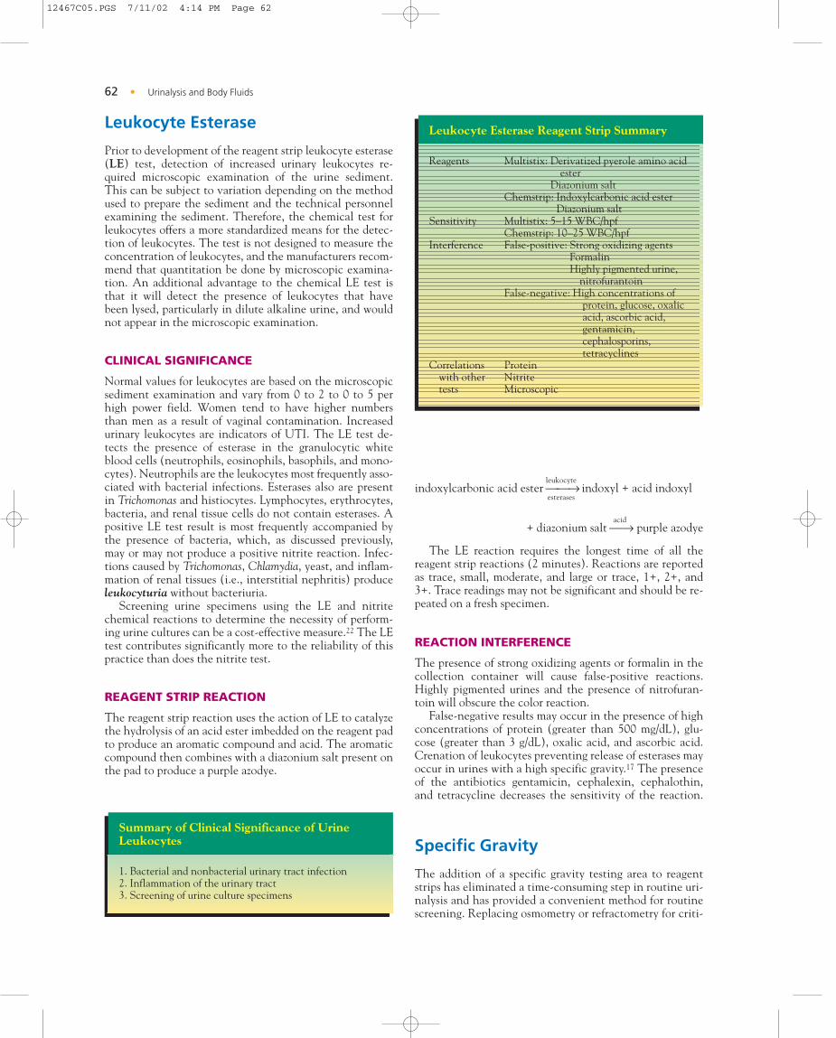
Leukocyte Esterase
Prior to development of the reagent strip leukocyte esterase(LE) test, detection of increased urinary leukocytes re-quired microscopic examination of the urine sediment.This can be subject to variation depending on the methodused to prepare the sediment and the technical personnelexamining the sediment. Therefore, the chemical test forleukocytes offers a more standardized means for the detec-tion of leukocytes. The test is not designed to measure theconcentration of leukocytes, and the manufacturers recom-mend that quantitation be done by microscopic examina-tion. An additional advantage to the chemical LE test isthat it will detect the presence of leukocytes that havebeen lysed, particularly in dilute alkaline urine, and wouldnot appear in the microscopic examination.
CLINICAL SIGNIFICANCE
Normal values for leukocytes are based on the microscopicsediment examination and vary from 0 to 2 to 0 to 5 perhigh power field. Women tend to have higher numbersthan men as a result of vaginal contamination. Increasedurinary leukocytes are indicators of UTI. The LE test de-tects the presence of esterase in the granulocytic whiteblood cells (neutrophils, eosinophils, basophils, and mono-cytes). Neutrophils are the leukocytes most frequently asso-ciated with bacterial infections. Esterases also are presentin Trichomonas and histiocytes. Lymphocytes, erythrocytes,bacteria, and renal tissue cells do not contain esterases. Apositive LE test result is most frequently accompanied bythe presence of bacteria, which, as discussed previously,may or may not produce a positive nitrite reaction. Infec-tions caused by Trichomonas, Chlamydia, yeast, and inflam-mation of renal tissues (i.e., interstitial nephritis) produceleukocyturia without bacteriuria.
Screening urine specimens using the LE and nitritechemical reactions to determine the necessity of perform-ing urine cultures can be a cost-effective measure.22 The LEtest contributes significantly more to the reliability of thispractice than does the nitrite test.
REAGENT STRIP REACTION
The reagent strip reaction uses the action of LE to catalyzethe hydrolysis of an acid ester imbedded on the reagent padto produce an aromatic compound and acid. The aromaticcompound then combines with a diazonium salt present onthe pad to produce a purple azodye.
leukocyteindoxylcarbonic acid ester → indoxyl + acid indoxyl
esterases
acid+ diazonium salt → purple azodye
The LE reaction requires the longest time of all thereagent strip reactions (2 minutes). Reactions are reportedas trace, small, moderate, and large or trace, 1+, 2+, and3+. Trace readings may not be significant and should be re-peated on a fresh specimen.
REACTION INTERFERENCE
The presence of strong oxidizing agents or formalin in thecollection container will cause false-positive reactions.Highly pigmented urines and the presence of nitrofuran-toin will obscure the color reaction.
False-negative results may occur in the presence of highconcentrations of protein (greater than 500 mg/dL), glu-cose (greater than 3 g/dL), oxalic acid, and ascorbic acid.Crenation of leukocytes preventing release of esterases mayoccur in urines with a high specific gravity.17 The presenceof the antibiotics gentamicin, cephalexin, cephalothin,and tetracycline decreases the sensitivity of the reaction.
Specific Gravity
The addition of a specific gravity testing area to reagentstrips has eliminated a time-consuming step in routine uri-nalysis and has provided a convenient method for routinescreening. Replacing osmometry or refractometry for criti-
62 Urinalysis and Body Fluids•
Summary of Clinical Significance of UrineLeukocytes
1. Bacterial and nonbacterial urinary tract infection2. Inflammation of the urinary tract3. Screening of urine culture specimens
Leukocyte Esterase Reagent Strip Summary
Reagents Multistix: Derivatized pyerole amino acid ester
Diazonium saltChemstrip: Indoxylcarbonic acid ester
Diazonium saltSensitivity Multistix: 5–15 WBC/hpf
Chemstrip: 10–25 WBC/hpfInterference False-positive: Strong oxidizing agents
FormalinHighly pigmented urine,
nitrofurantoinFalse-negative: High concentrations of
protein, glucose, oxalicacid, ascorbic acid,gentamicin,cephalosporins,tetracyclines
Correlations Proteinwith other Nitritetests Microscopic
12467C05.PGS 7/11/02 4:14 PM Page 62

cal fluid monitoring is not recommended.16 The clinicalsignificance of the specific gravity test is discussed inChapter 4.
REAGENT STRIP REACTION
The reagent strip reaction is based on the change in pKa(dissociation constant) of a polyelectrolyte in an alkalinemedium. The polyelectrolyte ionizes releasing hydrogenions in proportion to the number of ions in the solution.The higher the concentration of urine, the more hydrogenions are released, thereby lowering the pH. Incorporationof the indicator bromthymol blue on the reagent pad mea-sures the change in pH. As the specific gravity increases,the indicator changes from blue (1.000 [alkaline]), throughshades of green, to yellow (1.030 [acid]). Readings can bemade in 0.005 intervals by careful comparison with thecolor chart. The specific gravity reaction is diagramed inFigure 5–3.
REACTION INTERFERENCE
The reagent strip specific gravity measures only ionicsolutes, thereby eliminating the interference by the largeorganic molecules, such as urea and glucose, and by radio-graphic contrast media and plasma expanders that are in-cluded in physical measurements of specific gravity.9 Thisdifference must be considered when comparing specificgravity results obtained by a different method. Elevatedconcentrations of protein will slightly increase the readingsas a result of protein anions.
Specimens with a pH of 6.5 or higher will have de-creased readings caused by interference with the bromthy-mol blue indicator (the blue-green readings associated withan alkaline pH correspond to a low specific gravity read-ing). Therefore, manufacturers recommend adding 0.005 tospecific gravity readings when the pH is 6.5 or higher.
REFERENCES
1. Abuela, G: Proteinuria: Diagnostic principles and procedures. AnnIntern Med 98:1986–1991, 1983.
2. Benedict, SR: A reagent for the detection of reducing sugars. J BiolChem 5:485–487, 1909.
3. Bianchi, S, et al: Microalbuminurea in essential hypertension. JNephrol 10(4):216–219, 1997.
4. Free, AH, and Free, HM: Urodynamics: Concepts Relating to Rou-tine Urine Chemistry. Ames Division, Miles Laboratories, Elkhart,IN, 1978.
5. Guthrie, D, Hinnen, D, and Guthrie, R: Single-voided vs. double-voided urine testing. Diabetes Care 2(3):269–271, 1979.
6. Hager, CB, and Free, AH: Urine urobilinogen as a component of rou-tine urinalysis. Am J Med Technol 36(5):227–233, 1970.
7. Henry, JB, Lauzon, RB, and Schumann, BG: Basic examination ofurine. In Henry, JB (ed): Clinical Diagnosis and Management byLaboratory Methods. WB Saunders, Philadelphia, 1996.
8. Hoeltge, GA, and Ersts, A: A quality control system for the generalurinalysis laboratory. Am J Clin Pathol 73(3):404–408, 1980.
9. Kavelman, DA: A representative of Ames responds. Clin Chem29(1):210–211, 1983.
10. Kunin, CM, and DeGroot, JE: Self-screening for significant bacteri-uria. JAMA 231(13):1349–1353, 1975.
11. Monte-Verde, D, and Nosanchuk, JS: The sensitivity and specificityof nitrite testing for bacteriuria. Lab Med 12(2):755–757, 1981.
12. National Committee for Clinical Laboratory Standards ApprovedGuideline GP16-A: Urinalysis and Collection, Transportation, andPreservation of Urine Specimens. NCCLS, Villanova, PA, 1995.
13. Rauber, AP, and Maroncelli, RD: Effects of dietary changes on alka-linization of urine pH. Vet Hum Toxicol (Suppl) 23:46–47, 1981.
Chemical Examination of Urine • 63
Summary of Clinical Significance of UrineSpecific Gravity
1. Monitoring patient hydration and dehydration2. Loss of renal tubular concentrating ability3. Diabetes insipidus4. Determination of unsatisfactory specimens due to low
concentration
F I G U R E 5 – 3 Diagram of reagent strip specific gravityreaction.
Urine Specific Gravity Reagent Strip Summary
Reagents Multistix: Poly (methyl vinyl ether/maleic anhydride) bromthymol blue
Chemstrip: Ethyleneglycol-Bis (aminoethylether) bromthymol blue
Sensitivity 1.000–1.030Interference False-positive: High concentrations of
proteinFalse-negative: Highly alkaline urines
(>6.5)
12467C05.PGS 7/11/02 4:14 PM Page 63

14. Renfrew, G: Confirmatory testing in urinalysis. Urinalysis News10(3), 1994.
15. Riddhimat, R, Hiranras, S, and Petchclair, B: Retesting atypical reac-tions in urine bilirubin tests. Clin Lab Sci 5(5):310–312, 1992.
16. Romolo, D, et al: Refractometry, test strip and osmometery comparedas measures of relative density of urine. Clin Chem 33(1):190, 1987.
17. Scheer, WD: The detection of leukocyte esterase activity in urinewith a new reagent strip. Am J Clin Pathol 87(1):86–93, 1987.
18. TechniTips, Miles Diagnostics, Elkhart, IN. October, 1992.19. Ward, KM: Microalbuminuria: Clinical aspects. Clin Lab Sci 2(4):
212–213, 1989.20. Watson, CJ, and Schwartz, S: A simple test for urinary porphobilino-
gen. Proc Soc Exp Biol Med 47:393–394, 1941.21. Weaver, MR, and Gibb, I: Urinalysis for blood: Questionable inter-
pretation of reagent strip results. Clin Chem 29(2):401–402, 1983.22. Wise, KA, Sagert, LA, and Grammens, GL: Urine leukocyte esterase
and nitrite tests as an aid to predict urine culture results. Lab Med15(3):186–187, 1984.
T U D Y Q U E S T I O N S
1. Describe how each of the following errors in reagentstrip technique will affect test results:a. Leaving the reagent strip in the urine specimen
while recording results of the previous specimenb. Failing to remove excess urine while withdrawing
the strip from the specimenc. Recording all results immediately after withdraw-
ing the strip from the specimend. Using a Chemstrip color chart with a Multistix
reagent strip
2. How will failure to allow a refrigerated specimen towarm to room temperature before testing affectreagent strip testing? Why?
3. How are reagent strips protected from deteriorationcaused by:a. moistureb. volatile chemicalsc. lightd. heat
4. What is the significance of the expiration datestamped on reagent strip containers?
5. List four times when positive and negative controlsmust be run on reagent strips.
6. Explain the relationship of urine pH to the formationof crystals and renal calculi.
7. What is the significance of a urine pH of 9?
8. Why do reagent strip pH reactions use both methylred and bromthymol blue indicators?
9. Indicate the source of the following proteinurias byplacing a 1 for prerenal, 2 for renal, or 3 for postrenalin front of the condition.a. _____ Microalbuminuriab. _____ Acute phase reactantsc. _____ Pre-eclampsiad. _____ Vaginal inflammatione. _____ Multiple myelomaf. _____ Orthostatic proteinuriag. _____ Prostatitis
10. Describe the unique solubility characteristics ofBence Jones protein.
11. Differentiate between glomerular and tubular protein-uria.
12. How does detection of microalbuminuria affect pa-tient treatment?
13. Briefly explain the principle of the protein error of in-dicators. How does a highly alkaline urine affect this?
14. State a pathologic reason that would cause a negativereagent strip protein reaction and a positive SSA testresult.
15. Explain why glycosuria occurs in the presence of hy-perglycemia.
16. Why do persons with hyperthyroidism exhibit hyper-glycemia and glucosuria? Why do persons under ex-treme stress exhibit hyperglycemia and glucosuria?
17. How can glycosuria occur in the absence of hyper-glycemia?
18. Explain the purpose of glucose oxidase and peroxidasein the reagent strip test for glucose.
19. What is the primary cause of a false-negative test re-sult for glucosuria?
20. Does the Clinitest detect oxidizing or reducing sub-stances? How does it do this?
21. How will failure to detect “pass through” affect re-sults?
22. What is the primary reason that laboratories performthe Clinitest?
23. State two reasons for a positive reagent strip test re-sult for glucose and a negative Clinitest result.
24. State the three basic reasons for the presence of ke-tonuria.
25. What is the primary substance detected by sodium ni-troprusside? Why is glycine added to the reaction?
26. How do the reactions between ketones and interfer-ing substances differ on reagent strips?
27. Name three pathologic substances detected by thereagent strip blood reaction. State a reason why eachof these appears in the urine.
Substance Reasona. __________ ____________________b. __________ ____________________c. __________ ____________________
28. Why is hemoglobinuria associated with pink plasmaand myoglobinuria with normal-colored plasma?
29. Why may yellow-brown granules appear in the urinesediment?
30. Following precipitation of a clear red urine with am-monium sulfate, what is the significance of a red su-pernatant?
64 Urinalysis and Body Fluids•
S
12467C05.PGS 7/11/02 4:14 PM Page 64

31. What is the purpose of peroxide on the reagent strippad for blood?
32. What is the significance of a speckled reaction on theblood pad?
33. Why are high levels of ascorbic acid of concern withboth the glucose and blood reactions?
34. List in order the three products formed in the degra-dation of hemoglobin to urobilin.
35. Name two causes of jaundice that produce biliru-binuria and one cause that does not produce bili-rubinuria.
36. State two advantages of the Ictotest over the reagentstrip bilirubin test.
37. What is the major cause of false-negative test resultsfor bilirubin?
38. Why is it normal to have 0.1 to 1 mg/dL of urobilino-gen in the urine?
39. How does hemolytic anemia affect urine urobilino-gen? Why? How does biliary obstruction affect urineurobilinogen? Why?
40. How do the reagent strip reactions for urobilinogendiffer between Multistix and Chemstrip? What otherpathologic substance does Multistix detect?
41. Describe the reactions obtained with urobilinogen,porphobilinogen, and Ehrlich-reactive compounds inthe Watson-Schwartz test.
42. How would you perform a rapid screening test for thepresence of porphobilinogen?
43. Describe the diagnostic value of the nitrite test.
44. Why might an automated nitrite reading of positivebe changed to negative when the strip is visually ex-amined?
45. State four reasons why a specimen with a largeamount of bacteria could have a negative nitrite reac-tion.
46. Can a nitrite test be performed on a fresh specimencollected in an unsterile container? Why or why not?
47. Why is it possible to have a positive LE reaction andnot see any leukocytes in the urine microscopic ex-amination?
48. When is it possible to have a positive LE test in theabsence of bacteria?
49. Why do large quantities of ascorbic acid cause false-negative reactions for bilirubin, urobilinogen byChemstrip, nitrite, and LE?
50. Why does a urine specimen with a low specific gravityproduce an alkaline reaction with bromthymol bluein the specific gravity test?
51. How do specific gravity readings differ betweenreagent strips and refractometers?
52. Explain the need to add 0.005 to the specific gravityreadings in urines with a pH of 6.5 or higher.
C A S E S T U D I E S A N DC L I N I C A L S I T U A T I O N S
1. Preadmission laboratory work on an obese patientscheduled for surgery shows a fasting blood glucoselevel of 230 mg/dL. Results of the routine urinalysis areas follows:COLOR: Pale yellow KETONES: NegativeCLARITY: Clear BLOOD: NegativeSP. GRAVITY: 1.030 BILIRUBIN: NegativePH: 5.0 UROBILINOGEN: NegativePROTEIN: 1+ NITRITE: NegativeGLUCOSE: 100 mg/dL LEUKOCYTES: Negativea. Explain the correlation between the patient’s blood
and urine glucose results.b. What is the most probable metabolic disorder asso-
ciated with this patient?c. Considering the patient’s condition, what is the sig-
nificance of the patient’s protein result?d. What could have been done to delay the onset of
proteinuria in this patient?e. If the patient in this study had a normal blood glu-
cose level, to what would the urinary glucose be at-tributed?
2. Results of a urinalysis performed on a patient scheduledfor gallbladder surgery are as follows:COLOR: Amber KETONES: NegativeCLARITY: Hazy BLOOD: NegativeSP. GRAVITY: 1.022 BILIRUBIN: ModeratePH: 6.0 UROBILINOGEN: NormalPROTEIN: Negative NITRITE: NegativeGLUCOSE: Negative LEUKOCYTES: Negativea. What would be observed if this specimen were
shaken?b. What confirmatory test could be performed on this
specimen?c. Explain the correlation between the patient’s sched-
uled surgery and the normal urobilinogen.d. If blood were drawn from this patient, how might
the appearance of the serum be described?e. What special handling is needed for serum and
urine specimens from this patient?
3. Results of a urinalysis on a very anemic and jaundicedpatient are as follows:COLOR: Red KETONES: NegativeCLARITY: Clear BLOOD: LargeSP. GRAVITY: 1.020 BILIRUBIN: NegativePH: 6.0 UROBILINOGEN: 12 EUPROTEIN: Negative NITRITE: NegativeGLUCOSE: Negative LEUKOCYTES: Negativea. Would these results be indicative of hematuria or
hemoglobinuria?b. Correlate the patient’s condition with the uro-
bilinogen result.
Chemical Examination of Urine • 65
12467C05.PGS 7/11/02 4:14 PM Page 65

c. Why is the urine bilirubin result negative in thisjaundiced patient?
d. If interference by porphyrins was suspected in thisspecimen, how could this be resolved? State twomethods.
4. A female patient arrives at the outpatient clinic withsymptoms of lower back pain and urinary frequencywith a burning sensation. She is a firm believer in thecurative powers of vitamins. She has tripled her usualdosage of vitamins in an effort to alleviate her symp-toms; however, the symptoms have persisted. She isgiven a sterile container and asked to collect a mid-stream clean-catch urine specimen. Results of this rou-tine urinalysis are as follows:COLOR: Dark yellow KETONES: NegativeCLARITY: Hazy BLOOD: TraceSP. GRAVITY: 1.012 BILIRUBIN: NegativePH: 7.0 UROBILINOGEN: NormalPROTEIN: Trace NITRITE: NegativeGLUCOSE: Negative LEUKOCYTES: 2+Microscopic8 TO 12 RBC/HPF Heavy bacteria40 TO 50 WBC/HPF Moderate squamous epithelial cellsa. What discrepancies between the chemical and mi-
croscopic test results are present?State a possible reason for each discrepancy.
b. What additional chemical tests could be affected bythe patient’s vitamin dosage?
c. Discuss the urine color and specific gravity resultswith regard to correlation, and give a possible causefor any discrepancy.
d. State three additional reasons not previously givenfor a negative nitrite test in the presence of in-creased bacteria.
5. Results of a urinalysis collected following practice froma 20-year-old college athlete are as follows:COLOR: Dark yellow KETONES: NegativeCLARITY: Hazy BLOOD: 1+SP. GRAVITY: 1.029 BILIRUBIN: NegativePH: 6.5 UROBILINOGEN: 1 EUPROTEIN: 2+ NITRITE: NegativeGLUCOSE: Negative LEUKOCYTES: Negative
The physician requests the athlete to collect anotherspecimen in the morning prior to classes and practice.a. What is the purpose of the second sample?b. What changes would you expect in the second sam-
ple?c. Is the proteinuria present in the first sample of pre-
renal, renal, or postrenal origin?
6. A construction worker is pinned under collapsed scaf-folding for several hours prior to being taken to theemergency room. His abdomen and upper legs are se-verely bruised, but no fractures are detected. A speci-men for urinalysis obtained by catheterization has thefollowing results:COLOR: Red-brown KETONES: NegativeCLARITY: Clear BLOOD: 4+SP. GRAVITY: 1.017 BILIRUBIN: NegativePH: 6.5 UROBILINOGEN: 0.4 EUPROTEIN: Trace NITRITE: NegativeGLUCOSE: Negative LEUKOCYTES: Negativea. Would hematuria be suspected in this specimen?
Why or why not?b. What is the most probable cause of the positive
blood reaction?c. What is the source of the substance causing the pos-
itive blood reaction and the name of the condition?d. Would this patient be monitored for changes in
renal function? Why or why not?
7. Considering the correct procedures for care, technique,and quality control for reagent strips, state a possiblecause for each of the following scenarios.a. The urinalysis supervisor notices that an unusually
large number of reagent strips are becoming discol-ored before the expiration date has been reached.
b. A physician’s office is consistently reporting posi-tive nitrite test results with negative LE test results.
c. A student’s results for reagent strip blood and LE areconsistently lower than those of the laboratory staff.
d. A physician questions the significant number of re-ports indicating elevated automated reagent stripbilirubin results accompanied by negative Ictotestresults.
66 Urinalysis and Body Fluids•
12467C05.PGS 7/11/02 4:14 PM Page 66