chemical ablation in adenoid surgery
TRANSCRIPT

http://www.revistadechimie.ro REV.CHIM.(Bucharest)♦ 69♦ No.10♦ 20182722
*email: [email protected]; Phone: 0040721241122 All authors have contributed equally to this paper
Chemical Ablation in Adenoid Surgery
TEODORA IOANA GHINDEA1, ALEXANDRU DUMITRAS MEIUS3, DRAGOS CRISTIAN STEFANESCU4, CATALINA PIETROSANU2,IRINA IONITA2*, ANDRA IULIA SUCEVEANU5, ANDREEA RUSESCU2, VIOREL ZAINEA2,3, RAZVAN HAINAROSIE2,3
1CMDTAMP, 8th-10th Washington Str., 011794, Bucharest, Romania2Carol Davila University of Medicine and Pharmacy, 8th Eroii Sanitari Blvd., 050474, Bucharest, Romania3Prof. Dr. D. Hociota Institute of Phonoaudiology and Functional ENT Surgery, 21st Mihail Cioranu Str., 050751,Bucharest, Romania4Gen. Dr. Aviator Victor Anastasiu National Institute of Aeronautical and Space Medicine, 88th Mircea Vulcanescu Str., 010825,Bucharest, Romania5Ovidius University, Faculty of Medicine, Universitatii Str., 900470, Constanta Romania
The pharyngeal tonsil is a lymphatic tissue mass located in the roof of the nasopharynx. The function of thepharyngeal tonsil is to prevent infections with the help of antibodies. Chronic inflammation and allergies leadto hyperplasia of the adenoids that is found almost exclusively in children. The treatment of choice is surgicaltreatment; conservative treatment is only indicated preoperatively or if surgery is contraindicated. In thispaper, we will present a new surgical method for adenoidectomy performed trans-orally with coblation,under endoscopic control.
Keywords: nasopharynx, adenoids, coblation
The pharyngeal tonsil, or the adenoid, along with thetubal tonsils, the palatine tonsils and the lingual tonsil formWaldeyer’s ring[1].
The pharyngeal tonsil is a lymphatic tissue mass locatedin the roof of the nasopharynx, above and behind the uvula.Histologically it consists of a layer of ciliated epithelial cellscovered by mucus. The cilia move constantly and propelthe mucus to the pharynx and from there the mucus isswallowed and sent in the stomach. The role of the mucusis to carry the infectious agents or dust particles inhaledthrough the nose in the pharynx where there is a moreresistant epithelium. To replenish the mucus blanket, thenasopharyngeal tonsil also contains glands that secretemucus [1,2].
The development of the adenoid starts in the embryoniclife, after the 16th week. After birth the enlargement processbegins until the age of 5 to 7 years. After the age of 7 thepharyngeal tonsil starts to decrease and by puberty they itis almost completely gone [3-5].
The function of the pharyngeal tonsil is to preventinfections with the help of antibodies [4].
Chronic inflammation and allergies lead to hyperplasiaof the adenoids that is found almost exclusively in children.The main causative organisms are Streptococcuspneumoniae, Haemophilus influenzae, Streptococcus typeA or viruses [6,7].
During childhood the size of the oro-nasopharyngealspace is not fully developed and enlarged adenoids canlead to problems that affect the quality of life [7].
The main symptoms of chronic adenoiditis are: nasalobstruction, mouth breathing, sleep disorders, snoring,obstructive sleep apnea, mucopurulent rhinorrhea, reducedsmell ability, lack of appetite, delayed development [8].
Hyperplasia of the adenoids can also cause a series ofcomplications like sinusitis, purulent rhino-pharyngitis, otitismedia caused by blockage of the Eustachian tube, gothicpalate with malocclusion and dryness of the oral andpharyngeal mucosa due to moth breathing [8].
The diagnostic procedures include inspection, physicalexamination of the ear, nose and throat and hearing tests.
Children with adenoid faces usually have a pale skin, withopen mouth and sunken eyes. The examination of the earreveals a retracted tympanic membrane that can beaccompanied by middle ear effusion. Mucopurulentsecretions can be found in the nasal cavity. Examination ofthe oral cavity can show gothic palate, tonsillar hyperplasia,secretions on the posterior wall of the pharynx [8].
The examination of the nasopharynx can be performedthrough posterior rhinoscopy or nasal endoscopy. Theseprocedures reveal the shape, dimensions and aspect ofthe adenoids [8].
The treatment of choice for chronic adenoiditis issurgical treatment, conservative treatment is only indicatedpreoperatively or if surgery is contraindicated. Theconservative treatment uses antibiotics, decongestantnose drops and immunostimulants. The surgical treatmentwas usually the curettage of the adenoids using the adenoidcurette. Nowadays with the improvement of medicaldevices new methods of adenoidectomy are available [9].
In this paper we will present a new surgical method foradenoidectomy performed trans-orally with coblation,under endoscopic control.
Experimental partAlthough adenoidectomy is a common surgical
procedure in ENT pediatric surgery, it is associated withmany complications [10].
In the last years different techniques were developed toreduce risks and morbidity [10,11].
Coblation is a new technology that can be used in ENTpediatric surgery for adenoidectomy and tonsillectomy. Itis minimally invasive and it reduces intraoperative andpostoperative complications like blood loss, nasalregurgitation, permanent change of voice, leasions tonearby structures like torus tubaris, incomplete excisionof the adenoid tissue [11].
Coblation is a non-heat, minimally invasive technologyused for dissection, ablation of tissue and coagulation byusing radiofrequency energy combined with saline solution[12].
REV.CHIM.(Bucharest)♦ 69♦ No.10♦ 2018 http://www.revistadechimie.ro 2723
Fig. 1. 70° endoscopic tubeview of adenoid tissue
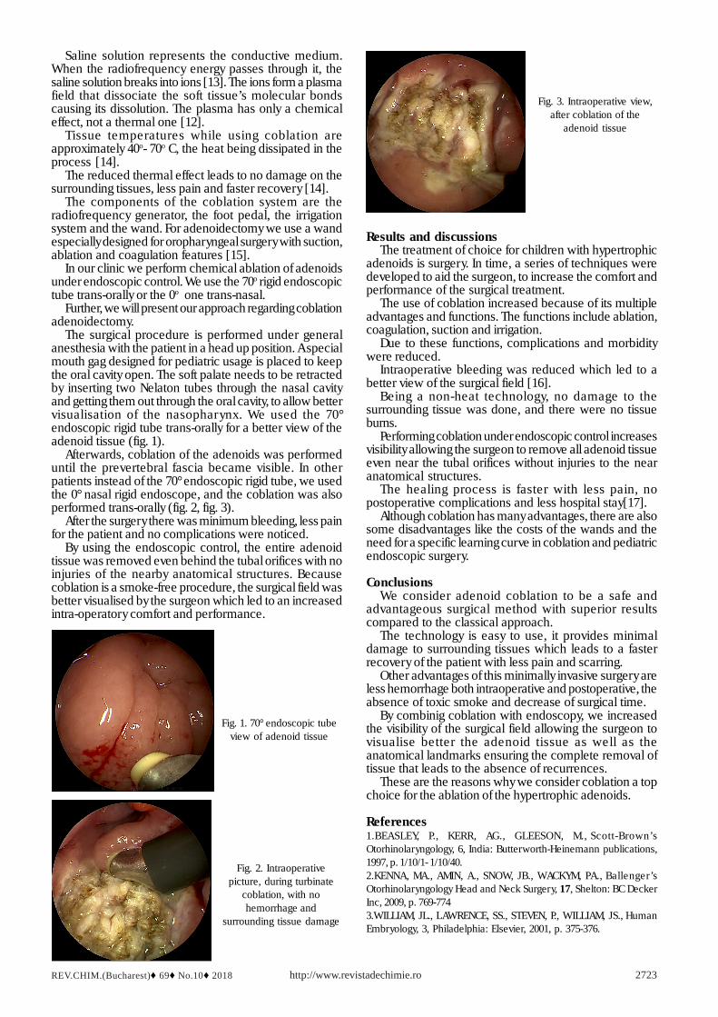
Fig. 3. Intraoperative view,after coblation of the
adenoid tissue
Fig. 2. Intraoperativepicture, during turbinate
coblation, with nohemorrhage and
surrounding tissue damage
Saline solution represents the conductive medium.When the radiofrequency energy passes through it, thesaline solution breaks into ions [13]. The ions form a plasmafield that dissociate the soft tissue’s molecular bondscausing its dissolution. The plasma has only a chemicaleffect, not a thermal one [12].
Tissue temperatures while using coblation areapproximately 40o- 70o C, the heat being dissipated in theprocess [14].
The reduced thermal effect leads to no damage on thesurrounding tissues, less pain and faster recovery [14].
The components of the coblation system are theradiofrequency generator, the foot pedal, the irrigationsystem and the wand. For adenoidectomy we use a wandespecially designed for oropharyngeal surgery with suction,ablation and coagulation features [15].
In our clinic we perform chemical ablation of adenoidsunder endoscopic control. We use the 70o rigid endoscopictube trans-orally or the 0o one trans-nasal.
Further, we will present our approach regarding coblationadenoidectomy.
The surgical procedure is performed under generalanesthesia with the patient in a head up position. A specialmouth gag designed for pediatric usage is placed to keepthe oral cavity open. The soft palate needs to be retractedby inserting two Nelaton tubes through the nasal cavityand getting them out through the oral cavity, to allow bettervisualisation of the nasopharynx. We used the 70°endoscopic rigid tube trans-orally for a better view of theadenoid tissue (fig. 1).
Afterwards, coblation of the adenoids was performeduntil the prevertebral fascia became visible. In otherpatients instead of the 70° endoscopic rigid tube, we usedthe 0° nasal rigid endoscope, and the coblation was alsoperformed trans-orally (fig. 2, fig. 3).
After the surgery there was minimum bleeding, less painfor the patient and no complications were noticed.
By using the endoscopic control, the entire adenoidtissue was removed even behind the tubal orifices with noinjuries of the nearby anatomical structures. Becausecoblation is a smoke-free procedure, the surgical field wasbetter visualised by the surgeon which led to an increasedintra-operatory comfort and performance.
Results and discussionsThe treatment of choice for children with hypertrophic
adenoids is surgery. In time, a series of techniques weredeveloped to aid the surgeon, to increase the comfort andperformance of the surgical treatment.
The use of coblation increased because of its multipleadvantages and functions. The functions include ablation,coagulation, suction and irrigation.
Due to these functions, complications and morbiditywere reduced.
Intraoperative bleeding was reduced which led to abetter view of the surgical field [16].
Being a non-heat technology, no damage to thesurrounding tissue was done, and there were no tissueburns.
Performing coblation under endoscopic control increasesvisibility allowing the surgeon to remove all adenoid tissueeven near the tubal orifices without injuries to the nearanatomical structures.
The healing process is faster with less pain, nopostoperative complications and less hospital stay[17].
Although coblation has many advantages, there are alsosome disadvantages like the costs of the wands and theneed for a specific learning curve in coblation and pediatricendoscopic surgery.
ConclusionsWe consider adenoid coblation to be a safe and
advantageous surgical method with superior resultscompared to the classical approach.
The technology is easy to use, it provides minimaldamage to surrounding tissues which leads to a fasterrecovery of the patient with less pain and scarring.
Other advantages of this minimally invasive surgery areless hemorrhage both intraoperative and postoperative, theabsence of toxic smoke and decrease of surgical time.
By combinig coblation with endoscopy, we increasedthe visibility of the surgical field allowing the surgeon tovisualise better the adenoid tissue as well as theanatomical landmarks ensuring the complete removal oftissue that leads to the absence of recurrences.
These are the reasons why we consider coblation a topchoice for the ablation of the hypertrophic adenoids.
References1.BEASLEY, P., KERR, AG., GLEESON, M., Scott-Brown’sOtorhinolaryngology, 6, India: Butterworth-Heinemann publications,1997, p. 1/10/1- 1/10/40.2.KENNA, MA., AMIN, A., SNOW, JB., WACKYM, PA., Ballenger ’sOtorhinolaryngology Head and Neck Surgery, 17, Shelton: BC DeckerInc, 2009, p. 769-7743.WILLIAM, JL., LAWRENCE, SS., STEVEN, P., WILLIAM, JS., HumanEmbryology, 3, Philadelphia: Elsevier, 2001, p. 375-376.

http://www.revistadechimie.ro REV.CHIM.(Bucharest)♦ 69♦ No.10♦ 20182724
4.SUSAN, S., HAROLD, E., JERMIAH, CH., DAVID, J., ANDREW, W.,Gray’s Anatomy: The Anatomical Basis of Clinical Practice, 39, 35,Philadelphia: Elsevier, 2005, p. 619-6315.GINGHINA,O., NEGREI,C., HUDITA,A .,et al., Farmacia, 2017,65(6),p.947-9536.KHALID, A., AL-MAZROU, ABDULAZIZ, S., AL-KHATTAF, ArchOtolaryngol Head Neck Surg., 2008, 134, p. 20–30.7.BROOK, I., Laryngoscope, 1981, 91, p. 377–82.8.WIATRAK, BJ., WOOLLEY, AL., Cummings, CW., Fredrickson, JM. ,Harker, LA ., Crause, CJ., Schuller, DE., Richardson,MA., Otolaryngology Head and Neck Surgery, 3, London: Mosby,1998,p. 188-215.9.BROOK, I., J Antimicrob Chemother, 2003, 51, p. 1331–7.10.GALLAGHER, TQ., WILCOX, L., MCGUIRE, E., et al., OtolaryngolHead Neck Surg., 2010, 142, p. 886–892.
11.GLADE, RS., PEARSON, SE., ZALZAL, GH., et al., Otolaryngol HeadNeck Surg., 2006, 134, p. 852–855.12.WOLOSZKO, J., GILBRIDE, C., Rep.Arthro Care Corp., 2001, p.102- 114.13.NITIPIR,C., NICULAE,D., ORLOV,C., etal., Oncology Letters, 2017,vol.14(6),p.7011-701514.BELOV, S. V., Biomedical Engineering, 38, nr. 2, 2004, p. 80-85.15.SERGEEV, V.N., BELOV, S.V, Biomedical Engineering, 37, Nr. 1, 2003,p. 22-25.16.DIACONU,C., DRAGOI,C.M., BRATU,O.,et al.,Farmacia,2018, 66(3),p.408-41517.ZUGRAVU,C.A., BACIU,A., PATRASCU,D.,et al., European Journal ofPublic Health, 2012,vol.22(2),p.272
Manuscript received: 21.03.2018