chem 100 unit 5 biochemistry - cerritos college
TRANSCRIPT

4/17/08 1
Chem 100 Unit 5 Biochemistry
Lipids Lipids are large molecules that are not soluble in water. They are soluble in nonpolar solvents. The most common lipid is fat. But steroids and fat soluble vitamins are also classed with lipids. Function of lipids Important part of almost all cells Found in cell membranes and brain and nervous tissue Long-term energy storage in the body Serve as insulation of body’s organs against temperature change and shock
Fats and oils generally provide 9 Cal/g of energy in our diet. These can be converted to glucose.
Classes of Lipids Triglycerides Phosphoglycerides Sphingolipids Glycolipids Steroids Fat Soluble Vitamins The first four classes of lipids have at least one fatty acid Fatty Acids
HOC
CH2
H2C
CH2
H2C
CH2
H2C
CH2
H2C
CH2
H2C
CH2
H2C
CH2
H2C
CH2
H2C
O
CH3
Will be simplified to:
HO
O

4/17/08 2
Fatty Acid Melting point
Source
Saturated Fatty Acid Example: Stearic acid No double bonds
HO
O
69oC solid @RT
pig fat
Monounsaturated fatty acid Example: oleic acid 1 double bond cis form puts a bend in the molecule
C
HO
O
14 oC Liquid @ RT
from olive oil
Monounsaturated fatty acid1 double bond trans form no bend
C
O
OH
43 oC
Polyunsaturated fatty acid 2 double bonds Example linoleic acid
C
O
OH
-5 oC liquid @ RT
Polyunsaturated fatty acid 3 double bonds Example linolenic acid
C
O
OH
-11 oC liquid @ RT

4/17/08 3
HO
O
HO
O
HO
O
Saturated fatty acids stack together very easily so it is easy to form a solid so they are solid at room temperature. Saturated fatty acids raise the cholesterol in your blood.
C
HO
O
C
HO
O
C
HO
O
Cis Unsaturated fatty acids do not stack together well at all so they tend to be liquids at room temperature. Vegatable oils contain cis fatty acids. The double bond tends to oxidize and the oil becomes rancid. The oil can be “hydrogenated” and then become more saturated and resist oxidation.
C
O
OH
C
O
OH
C
O
OH
Trans fatty acids stack together well like unsaturated fatty acids. When cis fatty acids are hydrogenated some of the cis double bonds become trans. Trans fatty acids raise the levels of low density lipoproteins (LDL) in the blood LDL contain cholesterol which accumulates in the arteries leading to heart disease. These fatty acids are found in milk, fried foods, butter, cookies, crackers and vegetable shortening. Many restaurants are using less trans fatty acids. You should limit these fatty acids in your diet
Hydrogenation
C
HO
O
HH
+ H2 Catalyst
C
O
OH
H
H
+
HO
O
Both the saturated fatty acid and the trans isomer are produced
Glycerol H2C
HC
H2C
OH
OH
OH
4/17/08 4
H2C
HC
H2C
O
O
O
C
O
C
O
C
O
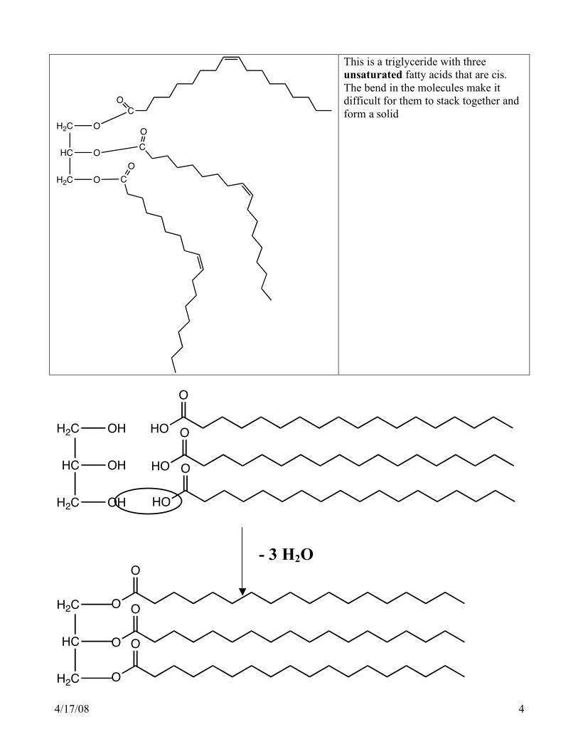
This is a triglyceride with three unsaturated fatty acids that are cis. The bend in the molecules make it difficult for them to stack together and form a solid
H2C
HC
H2C
OH
OH
OH
HO
O
HO
O
HO
O
- 3 H2O
O
O
H2C
HC
H2C
O
O
O
O

4/17/08 5
Saponification
The hydrolysis of a triglyceride with a strong base produces a molecule of glycerol and 3 salts of a fatty acid
O
O
H2C
HC
H2C
O
O
O
O
+ 3 NaOH
3
O
O-
Na+
- +
H2C
HC
H2C
OH
OH
OH In this reaction glyceryl tristearate is hydrolyzed by sodium hydroxide to form sodium stearate Soap
O
O-
Na+
- Soap is the salt of a fatty acid. It is unique because it has an ionic end and a long tail that is nonpolar. So it has both a water loving (hydrophilic) part and a water hating (hydrophobic) part.
Na +
Oil droplet
The polar “head” will be attracted to water. The nonpolar “tail” will be attracted to oil. This is how soap is able to wash away oil from skin or dishes.
4/17/08 6
H O
H
H O
H
H O
H
H O
H
H O
H
H O
H
H O
H
H O
H
H O
H
H O
H
H O
HH O
H
H O
H
H O
H
H O
H
H O
H
O H
H
O H
H
O H
H
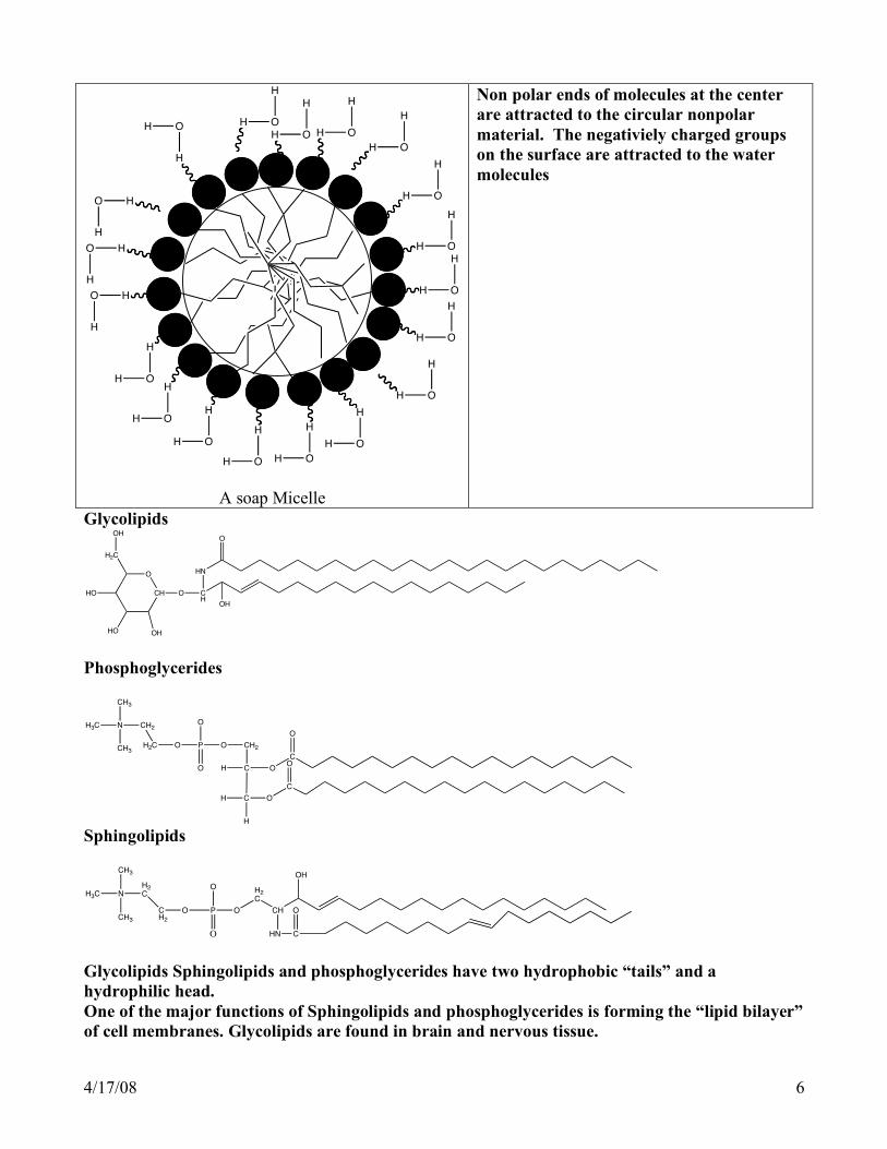
A soap Micelle
Non polar ends of molecules at the center are attracted to the circular nonpolar material. The negativiely charged groups on the surface are attracted to the water molecules
Glycolipids
CH
O
O CH
HN
O
OH
OHHO
HO
H2C
OH
Phosphoglycerides
N
CH3
H3C
CH3
CH2
H2C O P
O
O
O CH2
C
CH
H
H
O
C
O
C
O
O
Sphingolipids
O
H2C
CH
HN
OH
C
OP
O
O
OCH2
H2CN
CH3
H3C
CH3
Glycolipids Sphingolipids and phosphoglycerides have two hydrophobic “tails” and a hydrophilic head. One of the major functions of Sphingolipids and phosphoglycerides is forming the “lipid bilayer” of cell membranes. Glycolipids are found in brain and nervous tissue.
4/17/08 7
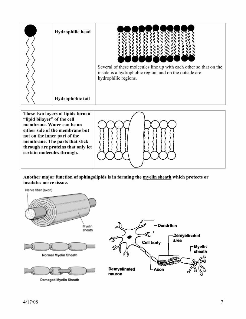
Hydrophilic head Hydrophobic tail
Several of these molecules line up with each other so that on the inside is a hydrophobic region, and on the outside are hydrophilic regions.
These two layers of lipids form a “lipid bilayer” of the cell membrane. Water can be on either side of the membrane but not on the inner part of the membrane. The parts that stick through are proteins that only let certain molecules through.
Another major function of sphingolipids is in forming the myelin sheath which protects or insulates nerve tissue.
4/17/08 8
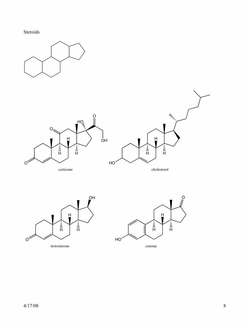
Steroids
O
O
OH
HO
O
H
H
H
cortisone
HO
H
H
H
cholesterol
O
OH
H
H
H
testosterone
HO
O
H
H
H
estrone

4/17/08 9
Carbohydrates Carbohydrates make up ______% of our diet. They represent a major part of all of the matter on earth that is organic. Carbohydrates contain _______ functional groups Carbohydrates are produced in the process called _________:
__________ + ____________ + energy (CH2O)n + ________ n is usually 3, 4, 5, or 6. Function of Carbohydrates In animals and humans 1. 2. 3. Generally carbohydrates provide _____Cal/g of energy In Plants 1. 2. 3. 3 Types of Carbohydrates Monosaccharides Disaccharides Polysaccharides Structures Monosaccharides
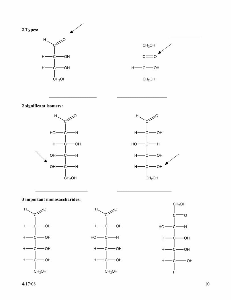
4/17/08 10
2 Types:
C
C
OH
C
H
CH2OH
H O
OHH
C
C
O
CH2OH
CH2OH
OHH
_____________________ ______________________ 2 significant isomers:
C
C
H
C
HO
C
H O
OHH
C
CH2OH
OH
OH H
H
C
C
OH
C
H
C
H O
HHO
C
CH2OH
H
H OH
OH
_______________________ ________________________ 3 important monosaccharides:
C
C
OH
C
H
C
H O
OHH
C
CH2OH
H
H OH
OH
C
C
OH
C
H
C
H O
HHO
C
CH2OH
H
H OH
OH
C
C
O
CH2OH
C
HHO
C
C
H
H OH
OH
H
H OH

4/17/08 11
Glycosidic Linkage Hemiacetal bond
C
C
OH
C
H
C
H O
HHO
C
C
H
H OH
OH
H
OHH
C
C
OH
C
H
C
H O
HHO
C
C
HO
H OH
H
H
OHH
C
C
O
C
C
HHO
C
C
H
H OH
OH
H
H OH
H
OHH
The Hemiacetal bond
R C O
H
+ O R 1H
R C O R 1
OH
H
Ring Structures
C
C C
C
OC
CH2OH
H
OH
H
HH
OH
H
OH
O
H
C
C C
C
OC
CH2OH
H
OH
H
HH
OH
H
OH
OH

4/17/08 12
α and β forms of glucose
C
C C
C
OC
CH2OH
H
OH
H
HH
OH
H
OH
OH
C
C C
C
OC
CH2OH
H
OH
H
HOH
H
H
OH
OH
________________________ _______________________ Glucose is a _________________sugar These differ only in the position of one hydroxyl group. But starch foods like pasta, bread, and rice contain the ______form. We can digest these foods. The _______form is found in wood and cellulose which we cannot digest. We have an enzyme that can digest the ____form but not the ____form. α and β forms of galactose
C
C C
C
OC
CH2OH
H
OH
H
HH
OH
OH
H
OH
C
C C
C
OC
CH2OH
H
OH
H
HOH
H
OH
H
OH
_____________________ _________________________ Galactose is a ____________________sugar.

4/17/08 13
α and β forms of fructose
C
C
C
O
C
CH2OH
H
CH2OH
OH
H
OH
OH
H
C
C
C
O
C
CH2OH
H
OH
CH2OH
H
OH
OH
H ______________________ _____________________ Fructose is a ____________________sugar. Reducing Sugars: These are sugars that contain a free carbonyl group are known as reducing sugars. The oxygen in the carbonyl can react with certain reagents that give a positive test for reducing sugars. Benedict’s solution is one of those reagents. The three monosaccharides are reducing sugars. Lactose and maltose are reducing sugars. Sucrose and the polysaccharides are not. But if those non reducing sugars are hydrolyzed into monoscaccharides, then the product is a reducing surgar. This reaction is also responsible for the browning of certain foods during the cooking process. Function of the monosaccharides glucose, galactose, and fructose.
1. Fructose Found in fruits and honey Sweeter than sucrose or glucose and other carbohydrates Converted to glucose in the liver
2. Galactose
Obtained from the disaccharide lactose found in milk Found on surfaces of cell membranes
3. Glucose Main carbohydrate in our blood Found in honey and fruit It is the major building block of polysaccharides The brain uses only glucose for fuel, but the brain does not store glucose so the blood glucose level must be maintained. Below 25% of normal, coma can occur. This could be caused by an overdose of insulin
Disaccharides

4/17/08 14
The three important disaccharides are maltose, lactose and sucrose. Function Maltose Obtained by hydrolyzing starch Used in cereals, candy, and brewing beverages Lactose Found in milk (human milk 6-8% , cow milk 4-5%) Some people do not have the enzyme needed to hydrolyze lactose and are considered lactose intolerant. Lactose is the least sweet sugar Sucrose
Mostly obtained from sugar cane (20% sucrose) and sugar beets (15% sucrose)
Commonly referred to as “table sugar”. In the year 1700 Americans consumed _____lbs of sugar per person per year. In 1780 it was______lbs. In 1960 it was_______. By 2005 Americans consumed ________lbs per person pear year of sugar and other sweeteners!
Structure
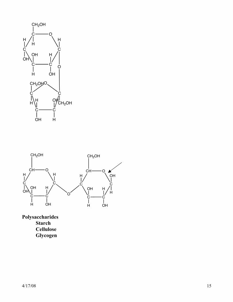
Each of these disaccharides are made of 2 monosaccharides held together by a glycosidic or ether bond.
glucose + glucose maltose glucose + galactose lactose glucose + fructose sucrose
CH O
C C
CC
CH O
C C
CCO
CH2OH
CH2OH
OH
HOH
H
H
OH
H
OH
H
H
OH
OH
H
H
4/17/08 15
C C
C
O
C
C
C C
C
OC
O
H
OH
OH
CH2OH
HH
OH
OH
H
H
CH2OH
H
CH2OH
H OH
H
CH O
C C
CC
CH O
C C
CCO
CH2OHCH2OH
H
OH
OH
H
H
OH
OH
H
H
OH
OH
H
HH
Polysaccharides Starch Cellulose Glycogen
4/17/08 16
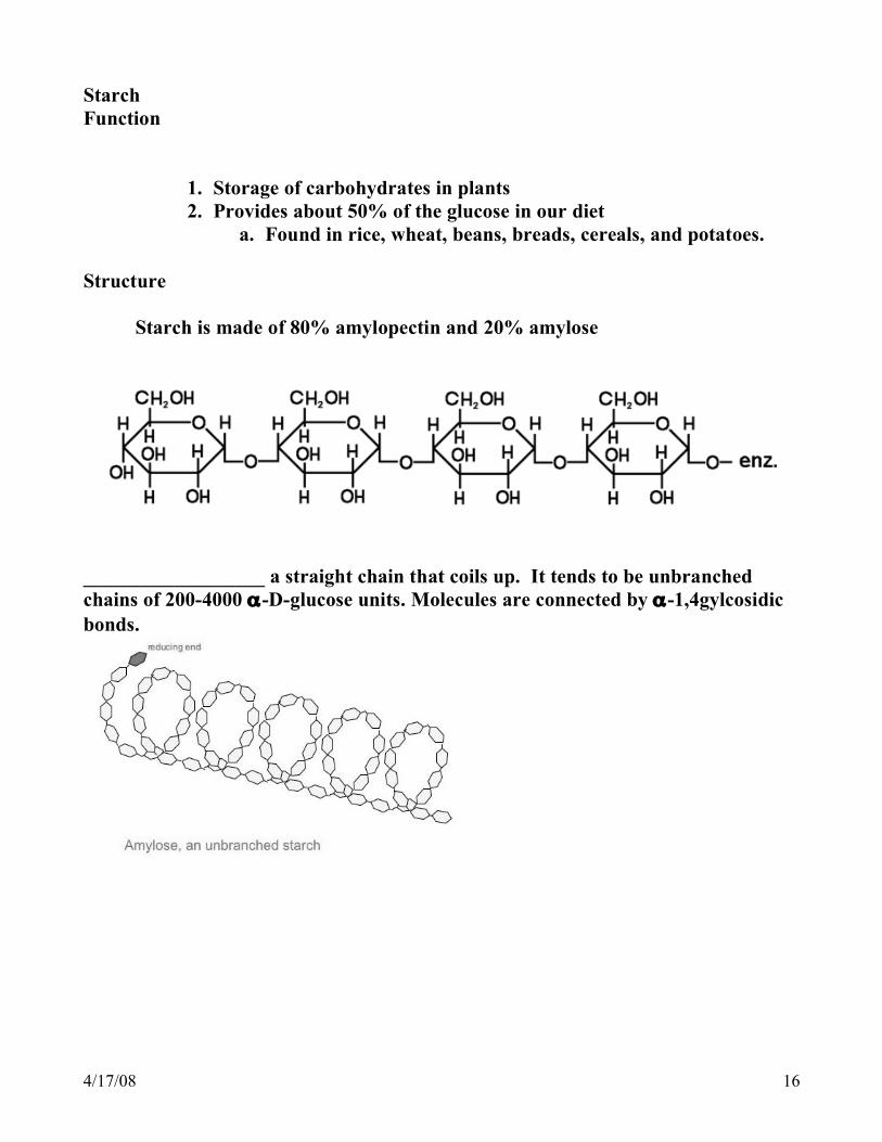
Starch Function
1. Storage of carbohydrates in plants 2. Provides about 50% of the glucose in our diet
a. Found in rice, wheat, beans, breads, cereals, and potatoes.
Structure Starch is made of 80% amylopectin and 20% amylose
__________________ a straight chain that coils up. It tends to be unbranched chains of 200-4000 α-D-glucose units. Molecules are connected by α-1,4gylcosidic bonds.
4/17/08 17
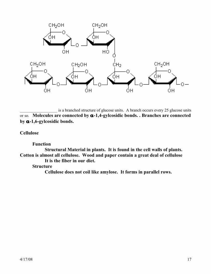
__________________ is a branched structure of glucose units. A branch occurs every 25 glucose units or so. Molecules are connected by α-1,4-gylcosidic bonds. . Branches are connected by α-1,6-gylcosidic bonds. Cellulose Function Structural Material in plants. It is found in the cell walls of plants. Cotton is almost all cellulose. Wood and paper contain a great deal of cellulose It is the fiber in our diet. Structure Cellulose does not coil like amylose. It forms in parallel rows.

4/17/08 18
Cellulose . Molecules are connected by β-1,4gylcosidic bonds. Our bodies have enzymes that can hydrolyze the α-1,4gylcosidic bonds of starch but we do not have enzymes to hydrolyze the β-1,4gylcosidic bonds found in cellulose . It is still and important part of our diet. The rows are held together by hydrogen bonds and then bundles of the rows of chains are twisted into fibers. Cellulose is the fiber in our diet Glycogen Function The way carbohydrates are stored in humans and animals Helps maintain glucose level in blood and muscle tissue
Stored in the liver and in muscles Structure Glucose molecules are connected by α-1,4-gylcosidic bonds. Branching occurs every 10-15 units. So there is much more branching in glycogen than in amlopectin
4/17/08 19
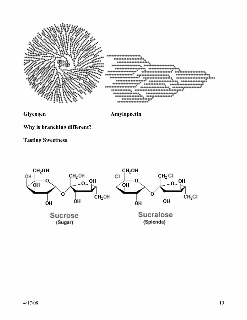
Glycogen Amylopectin Why is branching different? Tasting Sweetness
4/17/08 20
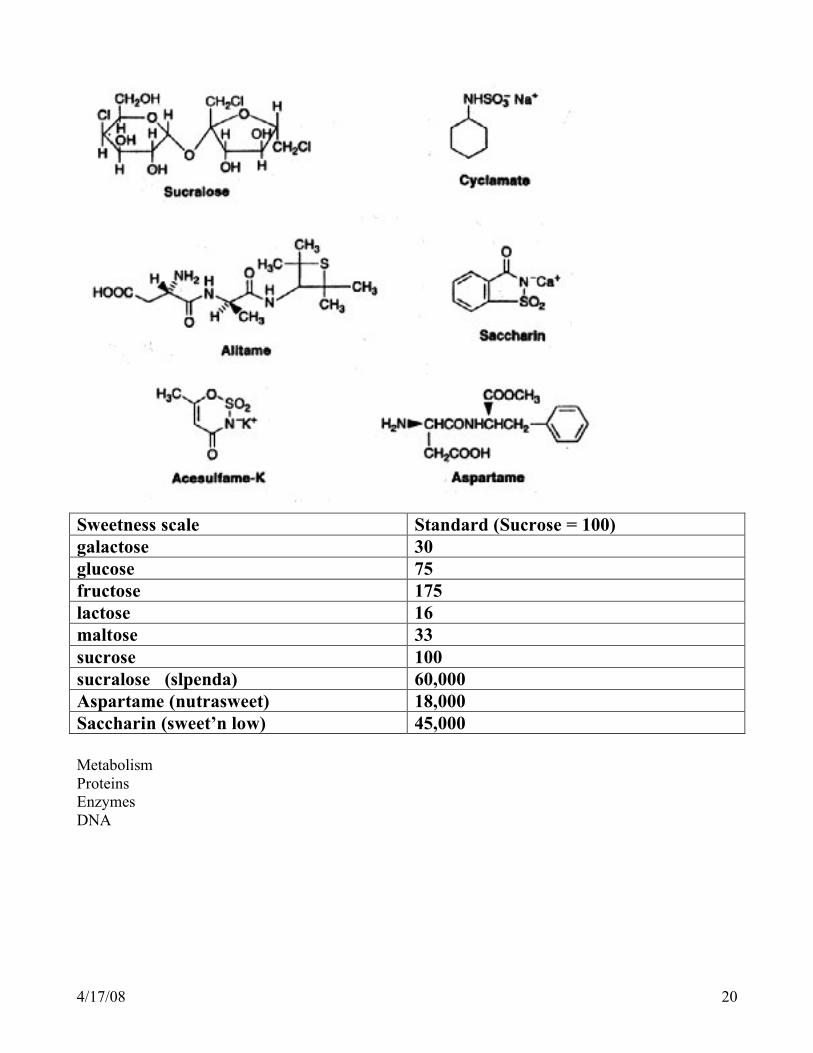
Sweetness scale Standard (Sucrose = 100) galactose 30 glucose 75 fructose 175 lactose 16 maltose 33 sucrose 100 sucralose (slpenda) 60,000 Aspartame (nutrasweet) 18,000 Saccharin (sweet’n low) 45,000 Metabolism Proteins Enzymes DNA
4/17/08 21
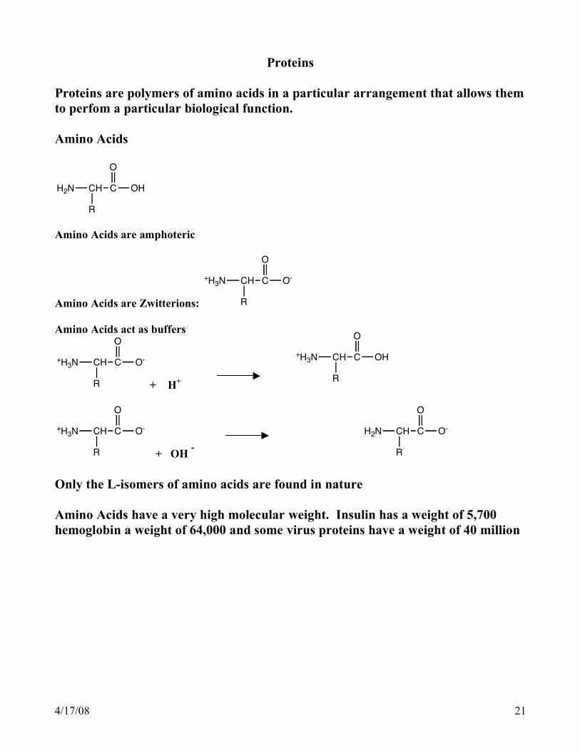
Proteins
Proteins are polymers of amino acids in a particular arrangement that allows them to perfom a particular biological function. Amino Acids
H2N CH C
R
OH
O
Amino Acids are amphoteric
Amino Acids are Zwitterions:
+H3N CH C
R
O-
O
Amino Acids act as buffers
+H3N CH C
R
O-
O
+ H+
+H3N CH C
R
OH
O
+H3N CH C
R
O-
O
+ OH - H2N CH C
R
O-
O
Only the L-isomers of amino acids are found in nature Amino Acids have a very high molecular weight. Insulin has a weight of 5,700 hemoglobin a weight of 64,000 and some virus proteins have a weight of 40 million
4/17/08 22
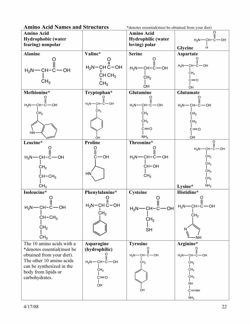
Amino Acid Names and Structures *denotes essential(must be obtained from your diet) Amino Acid Hydrophobic (water fearing) nonpolar
Amino Acid Hydrophilic (water loving) polar
Glycine
H2N CH C
H
OH
O
Alanine
H2N CH C
CH3
OH
O
Valine*
H2N CH C
CH
OH
O
CH3
CH3
Serine
H2N CH C
CH2
OH
O
OH
Aspartate
H2N CH C
CH2
OH
O
C
OH
O
Methionine*
H2N CH C
CH2
OH
O
HN
Tryptophan*
H2N CH C
CH2
OH
O
OH
Glutamine
H2N CH C
CH2
OH
O
CH2
C
NH2
O
Glutamate
H2N CH C
CH2
OH
O
CH2
C
OH
O
Leucine*
H2N CH C
CH2
OH
O
CH CH3
CH3
Proline
HN
C OH
O
Threonine*
H2N CH C
CH
OH
O
OH
CH3
Lysine*
H2N CH C
CH2
OH
O
CH2
CH2
CH2
NH2 Isoleucine*
H2N CH C
CH
OH
O
CH3
CH2
CH3
Phenylalanine*
H2N CH C
CH2
OH
O
Cysteine
H2N CH C
CH2
OH
O
SH
Histidine*
H2N CH C
CH2
OH
O
N
NH The 10 amino acids with a *denotes essential(must be obtained from your diet). The other 10 amino acids can be synthesized in the body from lipids or carbohydrates.
Asparagine (hydrophilic)
H2N CH C
CH2
OH
O
C
OH
O
Tyrosine
H2N CH C
CH2
OH
O
OH
Arginine*
H2N CH C
CH2
OH
O
CH2
CH2
NH
C
NH2
NH

4/17/08 23
Peptide bonds. The bond that holds amino acids together in a chain which becomes a protein is called the peptide bond or amid linkage. This bond is between the amino group of one amino acid and the carboxyl group of another amino acid
H2N CH C
CH3
OH
O
H2N CH C
CH3
OH
O
+H2N CH C
CH3
O
HN CH C
CH3
OH
O
2 amino acids dipeptide with amide linkage
If a polypeptide chain is hydrolyzed the products are amino acids Types of proteins Fibrous protein Long, linear, polypeptide chains that are side by side Insoluble in water Structural proteins Examples: hair, muscle Globular Proteins Polypeptide chains folded up Attracted to water These proteins can be moved from one place to another Examples: enzymes, hemoglobin, insulin, antibodies Structure: There are 4 levels of protein structure Primary structure The sequence/order of amino acids Maintained by peptide bonds Other levels of structure depend on primary structure
H2N CH C
R
O
HN CH C
R'
O
HN CH C
R''
O
HN CH C
R"""
O
HN CH C
R''''
OH
O
4/17/08 24
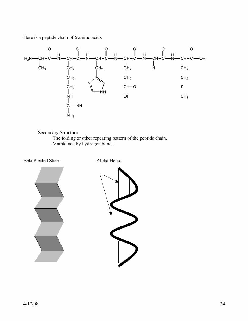
Here is a peptide chain of 6 amino acids
H2N CH C
CH3
O
HN CH C
CH2
O
CH2
CH2
NH
C
NH2
NH
HN CH C
CH2
O
N
NH
HN CH C
CH2
O
CH2
C
OH
O
HN CH C
H
O
HN CH C
CH2
OH
O
CH2
S
CH3
Secondary Structure The folding or other repeating pattern of the peptide chain. Maintained by hydrogen bonds Beta Pleated Sheet Alpha Helix
4/17/08 25
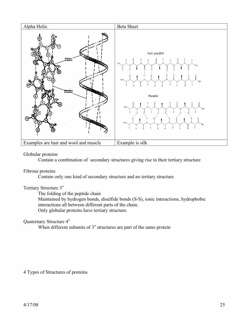
Alpha Helix Beta Sheet
Examples are hair and wool and muscle Example is silk Globular proteins Contain a combination of secondary structures giving rise to their tertiary structure Fibrous proteins
Contain only one kind of secondary structure and no tertiary structure
Tertiary Structure 3o The folding of the peptide chain
Maintained by hydrogen bonds, disulfide bonds (S-S), ionic interactions, hydrophobic interactions all between different parts of the chain.
Only globular proteins have tertiary structure. Quaternary Structure 4o When different subunits of 3o structures are part of the same protein 4 Types of Structures of proteins

4/17/08 26
Denaturing Protein Breaking down the 3o, 3o and 4o structures but not the amino acid sequence.
Losing structure caused by the hydrogen bonds, disulfide bonds, folding, etc. The shape of the protein is lost.
The peptide bonds are not broken so 1o structure stays the same.

4/17/08 27
Effects Protein is no longer biologically active No longer soluble Causes of denaturing Extreme heat as in cooking Extreme pH Presence of certain heavy metal ions Ag+, Pb2+, Hg2+ Examples of Proteins
Function
Enzymes sucrase lipase protease
hydrolyzes sucrose hydrolyzes lipids hydrolyzes peptide bond
Storage Proteins ovalbumin casein ferritin
egg-white protein milk protein iron storage protein
Transport Proteins hemoglobin myoglobin serum albumin
transports oxygen in blood transports oxygen in muscle transports fatty acids in blood
Contractile Proteins myosin actin
thick filaments in muscle thin filaments in muscle
Protective Protein antibodies fibrinogen
form complexes with foreign proteins like viruses protein used for blood clotting
Hormones growth hormone insulin
stimulates growth of bone regulates glucose in blood
Structural Protiens α-keratin collagen
Skin, hair, feathers, horns, nails, wool, hooves Fibrous connective tissue: tendons, bone, cartilage

4/17/08 28
How Proteins are made 1. Nucleic Acids
Nucleic acids carry the information that is the blueprint needed to make the primary structure of proteins
a. Nucleotides Sugar + base + phosphate -> nucleotide Nitrogen containing bases
1. Adenine (A) 2. Thymine (T) 3. Guanine (G) 4. Cytosine (C) 5. Uracil (U)
DNA contains A, G, C, T RNA contains A, G, C, U
b. Structure of Nucleic acids
Polymers of nucleotides Base Base Base | | | Phosphate—Sugar—Phosphate--Sugar—Phosphate—Sugar--Phosphate
c. Double Helix DNA is a spiral molecule in which to strands of the polymer are hooked together by hydrogen bonds. Adenine hydrogen bonds with thymine Guanine hydrogen bonds with cytosine -A—C—G—A—T—C—T- : : : : : : : -T—G—C—T—A—G—A-

4/17/08 29
d. DNA Replication e. Transcription
DNA mRNA
1. Types of RNA
Messenger RNA (mRNA) Ribosomal RNA (rRNA) Transfer RNA (tRNA)
2. The genetic code The genetic information carried by the Nucleic acids to make proteins is coded. There are only 4 bases in mRNA and there are 20 amino acids. The genetic code is a system of 3 bases in a particular order that corresponds to an amino acid.
Code Guanine-guanine-cytosine (GGC) is the code for the amino acid gly. GAG is the code for the amino acid glutamic acid.
There are 64 code words or codons for the 20 amino acids.
f. Translation
Initiation-Elongation-Termination

4/17/08 30
g. Issues involving Nucleic Acids
1. Recombinant DNA technology
2. Muatations X rays, UV sunlight, Mutagens, Viruses
DNA > alteration of DNA>defective protein>genetic disease (germ cells) Cancer (somatic cells) Normal Sequence Mutation DNA ACA—CCC—AGG—TTT ACA—CAC—AGG—TTT ↓ mRNA UGU—GGG—UCC—AAA UGU—GUG—UCC—AAA ↓ Amino Acid Cys—Gly—Ser—Lys Cys—Val—Ser—Lys sequence 3. Viruses