characterization oficam-2 and evidence for a third counter ... · characterization oficam-2 and...
TRANSCRIPT

Characterization of ICAM-2 and Evidence for aThird Counter-Receptor for LFA1By Antonin R. de Fougerolles,* Steven A. Stacker,*Roland Schwarting,$ and Timothy A . Springer*
From the "Committee on Immunology, Department ofPathology, Harvard Medical School, andCenter for Blood Research, Boston, Massachusetts 02115, and the $Department ofPathology,Thomas Jeferson University, Philadelphia, I+nnsylvania 19107
SummaryIn an endeavor to further characterize human intercellular adhesion molecule-2 (ICAM-2), twomurine monoclonal antibodies (mAb) were generated to ICAM-2 transfected COS cells, anddesignated CBR-IC2/1 and CBR-IC2/2 . Immunoprecipitated, reduced ICAM-2 migrated asa broad band of M, 60,000 in sodium dodecyl sulfate-polyacrylamide gel electrophoresis . Treat-ment with N-glycanase revealed a peptide backbone of MI 31,000, consistent with the sizepredicted from the cDNA. ICAM-2 had a broad distribution on hematopoietic cell lines andlittle expression on other cell lines, the sole exception being cultured endothelial cells whichpossess high levels of ICAM-2. Resting lymphocytes and monocytes expressed ICAM-2, whileneutrophils did not . Staining of tissue sections with anti-ICAM-2 mAb confirmed their strongreactivity to vascular endothelium, but demonstrated a lack ofICAM-2 expression on other tissues.Small clusters of ICAM-2 positive cells were, however, seen in germinal centers . In contrast toICAM-1 there was little or no induction of ICAM-2 expression on lymphocytes or culturedendothelium upon stimulation with inflammatory mediators . One ofthe two mAb, CBRIC2/2,was found to totally inhibit binding of ICAM-2 * COS cells to purified lymphocytefunction-associated antigen-1 (LFA-1) . Using this mAb, LFA-1-dependent binding to bothstimulated and unstimulated endothelium was found to be totally accounted for by ICAM-1and ICAM-2. Homotypic aggregation of an Epstein-Barr virus-transformed B cell line, JY, wasfound to be solely ICAM-1 and ICAM-2-dependent, while in the case of the T cell lymphomacell line, SKW3, anti- ICAM-2 mAb in conjunction with anti-ICAM-1 mAb could not inhibitthe LFA-1-dependent aggregation. This suggests an additional LFA-1 ligand exists. Using a cellbinding assay to purified LFA-1 in conjunction with anti-ICAM-1 and anti-ICAM-2 mAb, wehave demonstrated that this putative third ligand for LFA1 exists on SKW3 and other cell lines.
Adhesion molecules play a central role in the functions ofthe immune system . These molecules direct cell-cell in-
teractions critical for antigen presentation, lymphocyte acti-vation, localization, migration, and effector/target functionsat the site of inflammation (reviewed in reference 1) . The inte-grin family of adhesion receptors are involved in the cell-celland cell-matrix interactions of a wide variety of cell types(1-4) . A subgroup of the integrins, consisting of lympho-cyte function-associated antigen-1 (LFA-1), 1 Mac-1, andp150,95 (the "leukocyte integrins") play a major role in leu-kocyte adhesion (5) . They share a common 0 subunit (CD18)noncovalently associated with homologous cr subunits (LFA1
t Abbreviations used in this paper. AP, alkaline phosphatase; FDC, folliculardendritic cells; HUVEC, human umbilical vein endothelial cells ; ICAM,intercellular adhesion molecule; LFA, lymphocyte function-associatedantigen .
ci, CD11a; Mac1 u, CD11b ; p150,95 ci, CD11c) (6) . Theirimportance in leukocyte adhesion is most aptly demonstratedby a clinical condition, known as leukocyte adhesion deficiency(7) . Patients have recurrent life-threatening bacterial infec-tions due to mutations in the common a subunit, resultingin lack of expression of all three leukocyte integrins (8) .Of the three leukocyte integrins, LFA1 has the best charac-
terized role in cell adhesion . It was first defined by the abilityof mAb to block both T cell-mediated killing and prolifera-tion, and is required for numerous other functions includingT helper and B lymphocyte responses, natural killing, Ab-dependent cellular cytotoxicity by monocytes and granulocytes,and adherence of leukocytes to endothelial cells, fibroblasts,and epithelial cells (5, 9) . An inducible molecule, intercel-lular adhesion molecule-1 (ICAM-1) (CD54), was the firstligand discovered for LFA-1 (10, 11) . A member of the im-munoglobulin superfamily (12, 13), ICAM-1 has five Ig-like
253
J. Exp. Med . ® The Rockefeller University Press " 0022-1007/91/07/0253/15 $2.00Volume 174 July 1991 253-267
on Decem
ber 6, 2004 w
ww
.jem.org
Dow
nloaded from

domains with the binding site for LFA-1 localized to specificresidues in the first N-terminal domain (14) . Most LFA1-de-pendent phenomena could be inhibited by blocking mAb toICAM-1 (10, 15, 16) . However, binding of lymphocytes tocultured human umbilical vein endothelial cells (HUVEC)showed that the ICAM-1 blocking mAb, RRl/1, could notinhibit all the LFA1-dependent adhesion (15) . This implieda second ligand for LFA-1 existed on endothelial cells . Simi-larly, LFA-1 mAb but not ICAM-1 mAb blocked PMA-induced aggregation of a T cell lymphoblastoid cell line,SKW3 (10) . Lastly, LFAI-dependent cytolysis ofcertain targetcells by T cells was not inhibitable with mAb to ICAM-1(16), further evidence for additional LFA-1 ligands . A cDNAclone for a second LFA-1 ligand, termed ICAM-2, was ob-tained by transfecting COS cells with an endothelial cDNAlibrary and selecting for binding to LFA-1 coated plates inthe presence ofblocking ICAM-1 mAb (17) . Like ICAM-1,ICAM-2 is a member of the immunoglobulin superfamily.It has only two immunoglobulin-like domains that are mosthomologous to the two N-terminal domains ofICAM-1, with35% amino acid identity (17) .
In this study we report on the biochemical and cellularnature of ICAM-2 using mAb generated to COS cells tran-siently expressing the ICAM-2 cDNA. Two murine mAbto ICAM-2, CBR-IC2/1 and CBRIC2/2, were generated .Using these antibodies, the cellular and tissue distribution,biochemical properties and functional role in cell-cell adhe-sion of ICAM-2 could be studied . ICAM-2 was found todiffer from ICAM-1 in both its distribution and inducibility,implying a functional difference between the two molecules .Several LFA-1-dependent, ICAM-1-independent cell inter-actions could be accounted for by ICAM-2, whereas othersrevealed the presence of a third, as yet undefined, ligand forLFA-1 .
Materials and MethodsMonoclonal Antibodies.
The following previously described mu-rine mAbs to human antigens were used : TS2/9 (anti-LFA-3, IgGl)(18), TS2/16 (anti-CD29, IgGl) (18), TSl/22 (anti-CDlla, IgGl)(18), RR1/1 (anti-ICAM-1, IgGl) (10), W6/32 (anti-HLAA,B,C,IgG2a) (19), and X63 (nonbinding antibody, IgGl) .
Cell Culture.
The murine myeloma P3X63Ag8.653 (20) wasmaintained in DMEM supplemented with 10% Fetal Bovine Serum(FBS), 5 mM glutamine and 50 kg/ml gentamyein (supplementedDMEM) at 37°C in a humidified 10% COZ atmosphere. Hybrid-omas were initially grown in supplemented DMEM under HATselection (100 FM hypoxanthine, 400 nM aminopterin, 16 /AMthymidine), transferred to supplemented DMEM under HT selec-tion (100/AM hypoxanthine and 16 AM thymidine) and later grownsolely in supplemented 10% FBS/DMEM. The human fibrosar-coma cell line, FS 1,2,3 and epitheloid carcinoma cell line, HeLa,were grown in 10% FBS/DMEM plus supplements at 37°C and10% C02 . All other human cell lines were grown in RPMI 1640medium supplemented with 10% FBS, 5 mM glutamine and 50Fcg/ml gentamycin at 37°C and 5% C02 . All cell lines used inthis study are listed in Table 2 .Human umbilical vein endothelial cells (HUVEC, passage
number 2-4) were maintained as a monolayer on fibronectin-coateddishes in M199 medium, 20% PBS (Hyclone Laboratories, Inc .,
Logan, UT; LPS = 0.025 ng/ml), 5 mM glutamine, 50 14g/mlgentamycin, 100 Pg/ml endothelial growth supplement (Biomed-ical Technologies, Inc., Stoughton, MA) and 100 jig/ml heparinat 37°C and 5% CO2 . For stimulation, 5 U/ml of recombinanthuman IIr1s (Boehringer Mannheim, Indianapolis, IN), 10,ag/mlof LPS (Sigma Chemical Co., St. Louis, MO), 20 ng/ml ofrecom-binant human TNF-a (Genzyme, Boston, MA) or 103 U/ml ofrecombinant IFN-y (Genzyme) was added to the medium at ei-ther 4 or 24 h before harvesting . Stimulation of HUVEC was moni-tored by flow cytometry analysis of ICAM-1 on treated and un-treated cells.
Peripheral blood mononuclear cells (PBMC) were obtained bydextran sedimentation and Ficoll-Hypaque (1 .077) centrifugationas described (15) . Granulocytes were recovered from the cell pellet;contaminating erythrocytes were removed by hypotonic lysis . Lym-phocytes were enriched by incubating the PBMC in 10%FBS/RPMI 1640 on tissue culture-treated plastic Petri dishes for2 h and saving the nonadherent cells . To enrich for T lymphocytes,the nonadherent cells were passed through nylon wool (21) . NKcells were further isolated from plastic and nylon wool nonadherentmononuclear cells by Percoll (Pharmacia Fine Chemicals, Piscataway,NJ) gradient centrifugation as described (22) . CD3' cells were re-moved by labelling with CD3 mAb (OKT3) and removing themAb bound cells by rosetting with magnetic beads (Dynal; Bob-bins Scientific, Mountain View, CA) coated with F(ab') 2 goatanti-mouse IgG and IgM (Tago Inc., Burlingame, CA) . Immuno-fluorescence flow cytometry showed the purified NK cells to be>75% CD16' with few (<10%) contaminating CD3' cells. PHAblasts were prepared from isolated PBMC, incubated in supple-mented RPMI, plus 10% FBS and 10 Fig/ml PHA-P (Sigma Chem-ical Co.) and assayed at the indicated times (23) . Plastic adherentmonocytes were either analyzed immediately or cultured in vitrofor 10 d to induce differentiation towards macrophage-like cells(24) . The adherent cells were removed with HBSS/EDTA and sur-face antigen expression examined by immunofluorescence flowcytometry.cDNA and Transfection .
ICAM-1 and ICAM-2 cDNAs in thetransient expression vector CDM8 (13, 17) or vector alone (mock)were transfected into COS cells using DEAE-dextran (25) . 3 d aftertransfection, cells were detached with 10 mM HBSS/EDTA, washedthree times in 10%FBS/RPMI 1640 and then used for immuniza-tion, flow cytometric analysis, "5 I-labeling, or binding to LFA-1-coated plates . Cells were washed twice with PBS, pH 7.3, beforeeither immunization or . .. I-labeling .
Development ofICAM-2 Hybridomas.
Transfected COS cells ex-pressing ICAM-2 were used to immunize 3-12-wk-old BALB/cfemale mice (Charles River Laboratories, Wilmington, MA) . Im-munizations (105-106 cells/i .p. immunization) were performed fourtimes at 3-wk intervals . 3 d before fusion with the murine my-eloma P3X63Ag8.653, the mice were injected both i .p . and i.v.with 5 x 10 5 COS cells transiently expressing ICAM-2 . Thesetransfectants were tested for ICAM-2 expression by specific bindingto LFA-1 . The protocol for fusion and subsequent maintenance ofhybridomas is as previously described (26) . Differential reactivityto ICAM-2 transfected COS cells and untransfected cells was usedinitially to screen for ICAM-2 mAbs. Initial screening was per-formed by ELISA, followed by flow cytometric analysis of putativeICAM-2 reactive hybridoma supernatants. Positive mAbs were thenscreened for reactivity to cell lines known to be either positive(SKW3) or negative (HeLa) for ICAM-2 by Northern blotting anal-ysis (17) . MAbs selected for further analysis were cloned twice bylimiting dilution and isotyped by ELISA using affinity purified an-tibodies to mouse immunoglobulins (Zymed Immunochemicals,San Francisco, CA) .
254
Characterization of ICAM-2 and Evidence for a Third Counter-Receptor
on Decem
ber 6, 2004 w
ww
.jem.org
Dow
nloaded from

Immunohistochemical Staining.
Fresh tissues were received at theDepartment of Surgical Pathology at Thomas Jefferson Universityand snap-frozen in liquid nitrogen . The tissues were stored at-70°C until use. Immunohistological staining was carried out fol-lowing the alkaline phosphatase (AP) anti-alkaline phosphatase pro-cedure (27) . Briefly, 7 jLm frozen tissue sections were air-dried over-night and fixed in acetone for 10 min. ICAM-1 (RR1/1) andICAM-2 (CBRIC2/1) mAb were applied at 1/12,000 and 1/2,000dilutions of ascites fluid, respectively, for 30 min followed by briefwashing in Tris-buffered saline, pH 7.6 . As a negative control, mouseserum at a 1/5,000 dilution was applied . After washing, the sec-tions were incubated with a 1/40 dilution of rabbit anti-mouseIg mAb (Dakopatts, Carpinteria, CA) for 30 min. Finally, afterintermittent washing, the cells were incubated with APAAP-complex (anti-AP mouse mAb preincubated with AP). The lasttwo steps were repeated in order to enhance the sensitivity of thisprocedure. Bound alkaline phosphatase was visualized using newfuchsin (Sigma Chemical Co.) as substrate. Levamisole was appliedin order to block endogenous alkaline phosphatase activity andMeyer's acid hematoxylin (Sigma Chemical Co.) was used as coun-terstain .
Flow Cytometric Analysis .
Adherent cells were removed with10 mM HBSS/EDTA, washed with PBS/2% FBS and 50 Al ofa 0.5-1.0 x 10⁶ cells/ml suspension was added to either 50 ul ofmAb supernatant or 50 P1 of a 1/200 dilution of mAbcontainingascites fluid. After 45 min incubation at 4°C, the cells were washedand incubated with 100 P1 of a 1/20 dilution of FITC-labeled goatanti-mouse Ig (Zymed Immunochemicals) for 45 min at 4°C. Thecells were rewashed and fixed in 1% paraformaldehyde/PBS . Sampleswere analyzed using an Epics V (Coulter Diagnostics, Hialeah, FL)flow cytometer. As both primary and secondary mAb were usedat saturating concentrations, membrane antigen expression couldbe quantitated as a measure of mean fluorescence intensity usingEPICS Immuno-Brite fluorescent beads (Coulter Diagnostics) tocalibrate the cytometer, and X63 control antibody staining usedto subtract nonspecific fluorescence .
Surface Iodination .
Surface labeling of cells with 1a'I was per-formed as described using Iodogen (Pierce Chemical Co., Rock-ford, IL) (28) . Cells were labeled with 1251 (1 .0 mCi Na 1251; Amer-sham Corp ., Arlington Heights, IL) and lysed for 45 min at 4°Cin 1 ml of lysis buffer (10 mM Tris HCl pH 8.0, 150 mM NaCl,1% Triton-X-100, 1% hemoglobin, 1 mM iodoacetamide, 1 mMPMSF, 0.24 TIU/ml aprotonin, 0.025% azide) . Nuclei and insolubledebris were removed by centrifugation at 14,000 g for 20 min andlysates precleared overnight at 4'C with 100 Al of packed bovineIgG coupled-Sepharose. The lysate was then incubated with 30 P1packed mAb bound to Sepharose for 2 h. The beads were washedfour times in 25 mM Tris HClpH 8.0,150 mM NaCl, 0.1% Triton-X-100, twice in 25 mM Tris pH 8 .0, 150 mM NaCl, and oncein 50 mM Tris pH 8.0. The beads were then treated with an equalvolume of 2 x SDS-PAGE sample buffer, boiled for 2 min andanalyzed on 8% vertical slab polyacrylamide gels as previously de-scribed (29) . Proteins were visualized by autoradiography. Treat-ment of samples with N-glycanase (Genzyme Corp.) was as previ-ously described (33), using a concentration (3 U/ml) determinedto give optimal cleavage of all N-linked oligosaccharides from thepeptide backbone .
Purification ofLFA-1 .
LFA1 was purified from JY lysates onTS2/4-Sepharose as described previously (30) . The LFA-1 boundto TS2/4-Sepharose was eluted with 50 mM triethylamine pH 11 .5,150 mM NaCl, 2 mM MgCl2, and 1% octyl (3-D-glucopyranoside(OG). Samples were neutralized and stored frozen at -70°C.
255
de Fougerolles et al .
Adhesion Assay.
Purified LFA1 in 1% OG was diluted 1/10or 1/20 in 25 mM Tris HCl pH 8.0, 150 mM NaCl, 2 mMMgCl2 (TSM) and 50 Al absorbed onto 96-well polystyrene micro-titre plates (LinbroTitertek ; Flow Laboratories, McLean, VA) for2 h at room temperature. Nonspecific binding sites were blockedfor 2 h at room temperature with TSM/1% BSA and then washedtwice with PBS/5% FBS/2 mM MgC12/0.5% HSA (assay media) .Specific inhibition of LFA-1 was achieved by an incubation for 30min at room temperature with a 1/200 dilution of TSI/22 ascites .The number of LFA-1 sites/microtitre well was determined usingsaturating amounts of 1211 TS1/22 mAb and calculated assumingmonovalent binding of the mAb. Site numbers are expressed perUm2; the surface area of a microtitre well was determined to be3 .85 x 10' hm2 .
Cell lines were harvested, washed once in 10% FBS/RPMI 1640,resuspended and labeled with 15 Wg/mL of2;7'-bis-(2-carboxyethy1)-5(and-6) carboxyfluorescein (BCECF ; Molecular Probes, Inc., Eu-gene, OR) for 30 min at 37°C . After washing twice in 10%FBS/RPMI 1640, cells were countedand resuspended in assaymedia.Adhesion of cell lines to LFA-1-coated plates (31) or endothelialcell monolayers (15) hasbeen described previously. Briefly, confluentendothelial cell monolayers in 96-well plates were treated with a1/200 dilution of the appropriate ascites for 45 min at 37'C andthen unbound mAb was removed by washing before the bindingassay. MAb pretreatment of cells consisted of incubation with a1/200 dilution of ascites for 45 min at 4°C, after which 0.75-1.0x 10⁵ cells were transferred to each well . Cells were allowed tosettle and adhere to either the solid phase LFA1 or the endothelialmonolayers for 1 h at 37°C . Washing consisted of 6 aspirationswith either a 21-gauge needle (monolayers) or a 25-gauge needle(LFA-1-coated plates) . Wells were examined microscopically be-fore and after washing to assess the evenness of cell settling anddamage to the endothelial monolayer. Damage to the monolayerwas never significant (<5% area). Fluorescence was directly quan-titated from the 96-well plates using a Pandex fluorescence concen-tration analyzer (Baxter Healthcare Corp., Mundelein, IL).
Adherence of ICAM-2' COS cell transfectants to LFA-1-coatedwells was performed as previously described (31) . In short, 5 x10^ Na251 CrOo-labeled transfectants were bound to LFA-1-coatedplates for 1 h at room temperature, and washed with four aspira-tions through a 26-gauge needle. Bound cells were eluted and quan-titated by -y counting.
Aggregation Assay.
Aqualitative aggregation assay was carriedout as described (32) . Briefly, 50 pl of a cell suspension (2 x10⁶/ml) in 10% FBS/RPMI 1640 was preincubated with a 1/100dilution of ascites (1/200 final concentration) for 45 min at 4°C.These cells were then stimulated with 50 ng/mL ofPMAand addedto a flat bottom 96-well microtiter plate (Becton Dickinson, Lin-coln Park, NJ) . Cells were incubated for 2-6 h at 37°C, viewedwith an inverted microscope and aggregation scored visually. Be-cause these two cell lines aggregate with different kinetics, the lengthof the assay was varied to maximize PMAinduced aggregation UY:2 h; SKW3: 5 h) . Scores ranged from 0 to 5, where 0 indicatesessentially no cells were clustered; 1 indicates less than 10% ofcellswere in clusters; 2 indicates between 10-50% of cells were in ag-gregates ; 3 indicates 50-100% ofcells were in aggregates ; 4 indi-cates nearly 100% of cells were in large clusters of aggregates; and5 indicates that all the cells were in very compact aggregates . Pho-tomicrographs of aggregating cells were taken using a NikonDiaphotTMD inverted microscope (Nippon Kogaku, Tokyo, Japan)and phase contrast optics .
on Decem
ber 6, 2004 w
ww
.jem.org
Dow
nloaded from
Results
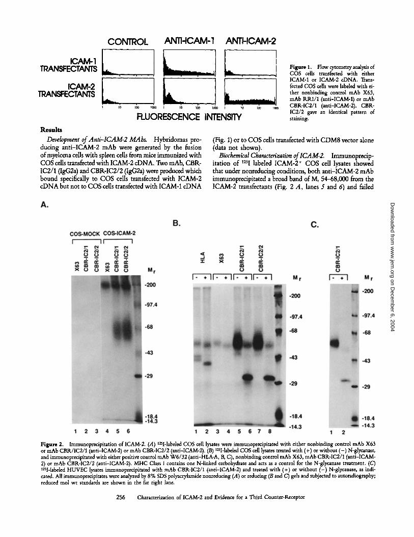
CONTROL ANTI-ICAM-1 ANTI-ICAM-2
Development ofAnti-ICAM-2 MAbs.
Hybridomas pro-ducing anti-ICAM-2 mAb were generated by the fusionofmyeloma cells with spleen cells from mice immunized withCOS cells transfected with ICAM-2 cDNA. Two mAb, CBR-IC2/1(IgG2a) and CBRIC2/2 (Ig02a) were produced whichbound specifically to COS cells transfected with ICAM-2cDNA but not to COS cells transfected with ICAM-1 cDNA
FLUORESCENCE INTENSITY
256
Characterization of ICAM-2 and Evidence for a Third Counter-Receptor
Figure 1 .
Flow cytometry analysis ofCOS cells transfected with eitherICAM-1 or ICAM-2 cDNA. Trans-fected COS cells were labeled with ei-ther nonbinding control mAb X63,mAb RRI/1 (anti-ICAM-1) or mAbCBR-IC2/1 (anti-ICAM-2) . CBR-IC2/2 gave an identical pattern ofstaining .
(Fig. 1) or to COS cells transfected with CDM8 vector alone(data not shown) .
Biochemical Characterization ofICAM-2.
Immunoprecip-itation of tall labeled ICAM-2+ COS cell lysates showedthat under nonreducing conditions, both anti-ICAM-2 mAbimmunoprecipitated a broad band ofM 54-68,000 from theICAM-2 transfectants (Fig . 2 A, lanes 5 and 6) and failed
Figure 2 .
Immunoprecipitation of ICAM-2 . (A) 1251-labeled COS cell lysates were immunoprecipitated with either nonbinding control mAb X63or mAb CBRAC2/1 (anti-ICAM-2) or mAb CBR-IC2/2 (anti-ICAM-2) . (B) 1251-labeled COS cell lysates treated with (+) or without (-) N-glycanase,and immunoprecipitated with either positive control mAb W6/32 (anti-HLAA, B, C), nonbinding control mAb X63, mAb CBR-IC2/1 (anti-ICAM-2) or mAb CBR-IC2/2 (anti-ICAM-2) . MHC Class I contains one Winked carbohydrate and acts as a control for the N-glycanase treatment . (C)1251-labeled HUVEC lysates immunoprecipitated with mAb CBR-IC2/1 (anti-ICAM-2) and treated with (+) or without (-) N-glycanase, as indi-cated. All immunoprecipitates were analyzed by 8% SDS polyacrylamide nonreducing (A) or reducing (B and C) gels and subjected to autoradiography;reduced mol wt standards are shown in the far right lane .
on Decem
ber 6, 2004 w
ww
.jem.org
Dow
nloaded from

Table 1 .
Distribution of ICAM-1 and ICAM-2 on NormalHuman Tissues"
Tissue
ICAM-1 ICAM-2
257
de Fougerolles et al .
to immunoprecipitate from mock transfectants (Fig. 2 A, lanes2 and 3) . Under reducing conditions, ICAM-2 migrates witha Mr of 55-65,000 (Fig. 2 B, lanes S and 7) . To evaluatethe contribution of glycosylation to this Mr, samples weretreated with N-glycanase, thereby removing all Winkedoligosaccharides (33) . Treatment with N-glycanase resultedin reduction of the ICAM-2 band to an approximate 31,000ML (Fig . 2 B, lanes 6 and 8), which corresponds closely tothe predicted mature peptide backbone of 28,393 Mr. There-fore, ICAM-2, like ICAM-1, is a heavily N-glycosylatedprotein .
Immunoprecipitation of ICAM-2 from various cell lysatesconfirmed the results obtained with the COS cell transfec-tants . Cultured HUVEC represent the most abundant sourceof ICAM-2 as determined by mRNA expression (17) or bycell surface expression (Table 2) . Immunoprecipitation ofICAM-2 from HUVEC lysates revealed a species migratingwith an Mr of 55-65,000 under reducing conditions (Fig .2 C, lane 1) . Deglycosylated ICAM-2 from HUVEC (Fig .2 C, lane 2) was the same Mr as from transfected COS cells .Unlike ICAM-1, little variation in MI among different celltypes was seen for ICAM-2, as immunoprecipitations fromboth SKW3 and JY lysates yielded identical results to thoseseen with HUVEC lysates (data not shown) .
Tissue Distribution of ICAM-2 .
Immunohistochemicalstaining offrozen tissue sections revealed ICAM-1 and ICAM-2to have distinct patterns of distribution (Table 1, Fig . 3) .Normal fetal (21 wk) and adult tissues showed a pattern ofICAM-1 staining similar to that previously reported (34, 35) .In contrast to ICAM-1, the distribution of ICAM-2 was re-stricted to the endothelium and some lymphoid cells. ICAM-2was expressed on all blood vessel endothelium, including highendothelial venules (Fig. 3 D), and expression was consis-tently stronger than that seen for ICAM-1 . Examination ofgerminal centers of lymphoid tissue revealed ICAM-2 to beabsent on most cells (Fig. 3 B and D) . In the spleen, althoughICAM-2 was virtually absent on all lymphocytes in the whitepulp, strong reactivity was seen on the sinus lining cells ofthe red pulp (Fig. 3 B) . In addition to the marginal reactivityof follicular mantle cells, ICAM-2 was found to be stronglyexpressed on small clusters of lymphocytes within germinalcenters of both spleen and tonsil (Fig. 3 D). As expected,anti-ICAM-1 mAb showed intense reactivity with the ger-minal centers oflymphoid follicles, most likely representingreactivity with B cells and follicular dendritic cells, while thefollicular mantle and marginal zones of the follicle showedweaker reactivity (Fig. 3 A and C). ICAM-1 was highly ex-pressed on tonsil surface and crypt epithelium (Fig. 3 C),
Organs studied : lymph node, tonsil, spleen, thymus, heart, brain,peripheral nerve tissue, skin, lungs, stomach, small and large intestine,liver, kidney, ovary, uterus, mammary gland, adrenal gland, and thyroidgland . Strong expression is denoted as "+" and lack of expression is de-noted as " - " ; "( + )" indicates a partial reactivity . Where not indicated,tissues were negative for both ICAM-1 and ICAM-2 .t Marginal reactivity for ICAM-2 was observed for follicular mantle cellsin the spleen .5 Small foci of germinal center lymphocytes were positive for ICAM-2 .
All vascular endothelium +Lymph Node/Tonsil/Spleen
Follicle mantle cells +Germinal center cells +Interfollicular/paracortical T cells -Follicular dendritic cells (FDC) +Interdigitating cells (IDC) -Starry sky macrophages +High endothelial venules
(lymph node, tonsil) +Tonsil
Oral mucosa +Crypt epithelia +GranulocytesFibrocytes/fibroblasts
SpleenMarginal zone cells +Sinus lining cells +
ThymusHassall's corpusclesCortical thymocytesMedullary thymocytes
SkinEpidermal cells, Langerhans' cells,
Melanocytes, Sweat gland cellsFibrocytes/fibroblasts
LungsAlveolar lining cells, pneumocytes
type I and II, alveolar macrophages +Capillary endothelial cells +
LiverHepatocytes +Kupffer's cells +Sinus lining cells +Bile duct cells -
KidneyEndothelial cells of the glomerulum +Cells of Bowman's capsule, basalmembrane of the glomerulumcapillaries, tubular cells -
Intertubular spindle cells +Small Intestine
Peyer's patches +Lymphocytes of the mucosal stroma +
on Decem
ber 6, 2004 w
ww
.jem.org
Dow
nloaded from

258
Characterization of ICAM-2 and Evidence for a Third Counter-Receptor
on Decem
ber 6, 2004 w
ww
.jem.org
Dow
nloaded from

whereas ICAM-2 was not . Fetal thymus expressed high levelsof ICAM-1 in the medulla and only low levels in the cortex,while ICAM-2 reactivity was confined to vessel endothelium.In the cortical region ICAM-1 expression was primarily focalon dendritic and epithelial cells, whereas high levels ofICAM-1in the medulla was localized to both the dendritic/macro-phage cells and the thymocytes .
In nonlymphoid organs, several differences in expressionwere observed between ICAM-1 and ICAM-2 (Table 1) . Whilemacrophages, fibroblast-like cells, and dendritic cells expressedhigh levels of ICAM-1 in most tissues studied, ICAM-2 ex-pression in tissues was restricted to blood vessel endothelium .Additional ICAM-2 expression was seen only in the liver andthe kidney. In the liver, ICAM-2 was weakly expressed onKupffer cells and some sinusoidal lining cells (Fig. 3 F) .ICAM-1 by comparison was strongly expressed by Kupffercells and sinusoidal lining cells (Fig. 3 E) . In kidney, the glo-merular capillary endothelium expressed ICAM-2, as did theintertubular spindle cells (Fig, 3 M. The ICAM-2 expres-sion in kidney, although weaker than ICAM-1, was similarin its distribution (Fig . 3 G) . These exceptions aside, ICAM-2was not present on any other tissues studied, including smalland large intestine, striated and smooth muscle, brain, thy-roid gland, and skin .
Immunofuorescence Flow Cytometry of ICAM-2 MembraneExpression. Flow cytometric analysis of human tumor celllines and PBLs supported the results obtained in frozen tissuesections (Table 2) . Resting lymphocytes and purified T cellsexpressed both ICAM-1 and ICAM-2 at low levels, althoughICAM-2 was always significantly higher in expression thanICAM-1 . The pattern of expression for ICAM-2 differedfrom ICAM-1 in that nearly all resting cells were ICAM-2positive and expression increased only slightly upon PHAactivation . Two-color flow cytometry revealed resting T(CD3+) and B (CD20+) lymphocytes to express equivalentamounts of ICAM-2 on their cell surface (data not shown) .In contrast to resting lymphocytes, freshly purified mono-cytes showed greater ICAM expression, with ICAM-1 andICAM-2 being generally equivalent . In vitro culturing ofmonocytes has been used as a method of obtaining macro-phage-like CD16 + cells (24) . Upon culturing of monocytesto induce macrophage differentiation, ICAM-2 expression re-
mained unchanged, whereas the inducibility of ICAM-1 wasconsistent with published data (34) .
Expression of ICAM-2 on cell lines was coordinate withNorthern blotting analysis. Lines which had been previouslyshown to express ICAM-2 mRNA(HUVEC, SKW3, Jurkat,BBN, Ramos, U937) or lack ICAM-2 mRNA (HeLa, FS1,2,3)showed corresponding patterns of cell surface expression (Table2) . All T and B lymphoblastoid lines examined expressed con-siderable ICAM-2, while other cell lines exhibited a broadrange of expression . Treatment ofseveral cell lines (HUVEC,JY, and SKW3) with phosphoinositol-phospholipase C (PI-PLC), revealed no PI-linked form of ICAM-2 as cell surfaceexpression remained unchanged (data not shown). Mouse lym-phoblastoid and fibroblastic cell lines did not react with theanti-human ICAM-2 mAb, implying that these mAb did .not crossreact to murine ICAM-2.ICAM-2 Expression on Cultured Endothelial Cells.
Of allcell types examined, ICAM-2 was most highly expressed onendothelial cells . We examined the relative levels of ICAMexpression on resting and stimulated human umbilical veinendothelial cells (Fig. 4) . Resting endothelial cells showedlow basal level expression of ICAM-1 and approximately 10-fold higher ICAM-2 expression . The level of ICAM-2 ex-pression was also significantly higher than that seen for ei-ther LFA-3 or HLAA, B, C . Upon stimulation ofendothelialcells with TNF-ce, ICAM-1 expression increased after 4 h,becoming maximal after 24 h stimulation, while ICAM-2expression remained unchanged. LFA3 was largely unaffectedby cytokine stimulation, whereas HLAA,B,C showed slightlyincreased expression, as previously reported (15, 36, 37) . Iden-tical findings were obtained with a variety of other stimuliincluding Ilrl0, LPS, and IFN--y (data not shown) . ICAM-2therefore was found to be constitutively expressed at highlevels on HUVEC and, unlike ICAM-1, was not increasedby stimulation with inflammatory mediators .
Functional Characterization ofICAM-2 MAh
To determinewhether CBR-IC2/1 and CBR/IC2/2 could inhibit ICAM-2-LFA-1 interaction, binding of ICAM-2+ COS cells topurified LFA-1 was performed in the presence ofthese mAb(Fig. 5) . A high percentage of transfected cells bound topurified LFA-1 in the presence of control mAb W6/32, andthis was totally inhibitable by an anti-LFA-1 mAb. Of the
Figure 3 .
Photomicrograph ofimmunohistochemical alkaline phosphatase anti-alkaline phosphatase (APAAP) staining of frozen tissues withanti-ICAM-1(RR1/1) and anti-ICAM-2 (CBR-IC2/1) mAb (magnification x 200) . (A) Staining of spleen with anti-ICAM-1 mAb . Note that the germinal center(GC) cells, follicular mantle (FM) cells and marginal zone (MZ) cells are positive for ICAM-1 . Endothelium of small vessels is also positive (arrow) .(B) Staining of spleen with anti-ICAM-2 mAb . With the exception of very faint staining of the follicular mantle (FM) lymphocytes, no staining ofthe white pulp is observed including germinal center (GC) and marginal zone (MZ) cells. The red pulp (RP) shows intense staining of the sinus liningcells . Small vessel endothelium is stained (arrows) . (C) Normal tonsil stained with anti-ICAM-1 mAb. The germinal center (GC) cells show strongreactivity, whereas the follicular mantle (FM) cells are only weakly reactive . Virtually all vascular endothelium, including high endothelial venules,are positive (arrows) . ICAM-1 is also present on the surface and crypt epithelium (E) of tonsil . (D) Normal tonsil stained with anti-ICAM-2 mAb .Note that virtually all vascular endothelium is positive, including high endothelial venules (HEV) . Small clusters ofgerminal center (GC) lymphocytesare also positive for ICAM-2 (arrow) . The follicular mantle (FM) cells show negative to faint reactivity. Epithelium is negative (not shown) . (E) Normalliver stained with anti-ICAM-1 mAb. Kupffer cells and sinusoidal endothelium are stained (arrows) . The endothelium of the vessels (VS) in the portalfields are also positive. Liver epithelium and bile ducts (BD) are negative. (F) Normal liver stained with anti-ICAM-2 mAb. Sinuoidal endotheliumis largely negative for ICAM-2 and only weak reactivity is found for Kupffer cells (arrow) . Positive staining is found for the endothelium of the vessels(VS) in the portal fields. Liver epithelium and bile duct (BD) epithelium are negative . (G) Normal kidney stained with anti-ICAM-1 mAb. Notethat the endothelium of all glomerular capillaries is positive. Some reactivity is also observed with the intertubular spindle cells. (M Normal kidneystained with anti-ICAM-2 mAb. Note the pronounced staining of all glomerular capillaries . Reactivity to intertubular spindle cells is also seen .
259
de Fougerolles et al.
on Decem
ber 6, 2004 w
ww
.jem.org
Dow
nloaded from

Table 2.
Relative Surface Antigen Expression of ICAMs by
two anti-ICAM-2 mAb, CBR-IC2/1 caused slight inhibi-Immunofluorescence Flow Cytometry
tion, whereas CBR-IC2/2 completely blocked adherence tolevels seen in the presence of anti-LFA 1 mAb. A combina-tion of both anti-ICAM-2 mAb gave no additional inhibi-tion to that seen when using CBRIC2/2 alone. Both mAbwere used at saturating concentrations, and this inhibitoryeffect could be diluted out . Similar results were obtained usingmouse L cells stably expressing human ICAM-2, demon-strating the ability of mAb CBRIC2/2 to completely in-hibit ICAM-2 interaction was purified LFA1(data not shown) .CBR-IC2/1 and CBRAC2/2 were determined to recognizenearby but distinct epitopes on ICAM-2, because althoughCBRIC2/2 would completely block CBR-IC2/1 binding,the latter could only partially block the former (data notshown) .
Effect ofAnti-ICAM-2 MAbs on Homotypic Aggregation.We examined the ability of anti-ICAM-1 and anti-ICAM-2mAb to inhibit LFA1-dependent homotypic aggregation ofseveral cell lines (Table 3 and Fig . 6) . PMAinduced aggrega-tion ofJY, an EBVtransformed B lymphoblastoid cell line,was previously found to be inhibitable almost completely bythe blocking anti-ICAM-1 mAb, RR1/1 (10) . Alone, anti-ICAM-1 mAb was found to inhibit most aggregation (Fig.6 C), but when used in conjunction with anti-ICAM-2 mAbaggregation was inhibited completely (Fig. 6 E) ; to the sameextent as with anti-LFA-1 mAb (Fig. 6 F) . Consistent withthe functional characterization of both CBRIC2/1 andCBR-IC2/2, when the anti-ICAM-2 mAb were used sep-arately with RR1/1, only CBRIC2/2 inhibited aggregation .Aggregation was not inhibited by the anti-ICAM-2 mAbalone (Fig . 6 D) ; this is due to the presence of ICAM-1 andthe overriding role it plays in LFA1-dependent aggregate for-mation of this cell line . PMA treatment of cells resulted inlittle change in surface expression ofeither ICAM-1, ICAM-2,or LFA-1 during the course of the assay (data not shown) .After 12-24 h however, ICAM-1 expression increased andICAM-2 showed a slight decrease .PMAinduced aggregation ofSKW3 was shown to be LFA
1-dependent, but ICAM-1-independent (Table 3) . This isconsistent with previously published reports (10) . Anti-ICAM-2 mAb had no effect on SKW3 aggregation eitheralone or in combination with RR1/1 (Table 3) . Given theability of RR1/1 and CBR-IC2/2 to inhibit ICAM-1 andICAM-2-dependent binding by other cells, these results sug-gest the presence of a-third ligand for LFA1 on SKW3.
Efect ofAnti-ICAM-2 MAbs on Cell Line Binding to LFA-1 .To better define this putative third LFA1 ligand, binding of
Membrane expression determined by immunofluorescence flow cytom-etry with RRl/1 for ICAM-1, CBR-IC2/1 for ICAM-2, and W6/32for HLA-A,B,C . Values are determinative of at least two experiments .Fluorescent beads were used to calibrate the cytometer such that one unitwas approximately 103 fluorescein equivalents (Counter Diagnostics).t Miscellaneous cell lines include : Human umbilical vein endothelial cells,HUVEC; human breast carcinoma, ZR-75-1 ; human hepatocellular car-cinoma, Hep G2 ; human epitheloid carcinoma cell line, HeLa ; humanrhabdomyosarcoma, RD 3/5; human fibrosarcoma, FS 1,2,3 ; human glio-blastoma, A-172 ; and the Reed-Sternberg line, L428 .
260
Characterization of ICAM-2 and Evidence for a Third Counter-Receptor
Cell line/type
Specific
ICAM-1
linear fluorescenceintensity'
ICAM-2 HLA
Lymphocytes 13 45 840T lymphocytes 14 40 9201 day PHA-blasts 64 40 1,1004 day PHA-blasts 200 55 1,6007 day PHA-blasts 70 32 1,100Monocytes 72 87 990Cultured monocytes 293 80 1,200Neutrophils 5 0 275NK cells 12 32 1,050Erythrocytes 0 0 0
T lymphoblastoidSKW3 0 114 670Jurkat 21 181 200Sup T 57 178 180Molt 4 28 240 253
B lymphoblastoidJY 125 130 1,600Ramos 190 260 670Raji 282 72 468Daudi 260 135 260ER-LCL 150 110 705BBN 100 60 215RPMI 8866 146 117 618
MonocyticU937 31 29 220HL60 0 23 268
MelanomasBK 181 8 265RPMI 7591 194 0 710
ErythroleukemicK562 155 95 6HEL 58 118 950
MiscellaneoustHUVEC 41 470 260ZR-75-1 105 30 105Hep G2 142 17 140HeLa 225 0 155RD3/5 0 11 208FS1,2,3 225 0 645A-172 0 0 385L428 835 169 16
on Decem
ber 6, 2004 w
ww
.jem.org
Dow
nloaded from

s0m
dN
0
MHC CLASS I
LFA-3
CAM-l-
ICAM-2
t to tm
Table 3.
Inhibition of PMA-induced Aggregation with LFA-1, ICAM-1 and ICAM-2 rnAb"
Cell type
Control
«LFA-1
«ICAM-1
«ICAM-2
JY
4
0
1
4SKW3
4
0
4
4
" FreshJY or SKW3 cells were preincubated at 4°C for 45 min with either no mAb (control) or 1/200 dilution of mAb containing ascites : TS1/22(anti-LFA-la), RRl/1 (anti-ICAM-1), CBR-IC2/1+CBR-IC2/2 (anti-ICAM-2), RR1/1+CBR-IC2/1+CBR-IC2/2 (anti-ICAM-1+2), or W6/32(anti-HLA-A,B,C) . Cells were then stimulated with 50 ng/ml of PMA, and aggregation scored visually after either 2 h (TY) or 5 h (SKW3).t Aggregation scored as described in Materials and Methods. Scores : 0, no cells clustered ; 1, <10% of cells aggregating ; 2, 10-50% of cells ag-gregating ; 3, 50-100% of cells aggregating ; 4, nearly 100% of cells in loose aggregates; 5, 100% of cells in very compact aggregates .
FLUORESCENCE INTENSITY
Figure 5.
Effect of ICAM-2 mAb on binding of ICAM-2-expressingCOS cell transfectants to purified LFA-1. Binding of s1Cr-labeled ICAM-2*COS cell transfectants was measured by incubating cells on LFA-1-coatedmicrotiter wells for 60 min at room temperature and then washing fourtimesby aspiration . Site density ofLFA-1 as determined byradioimmuno-assay was 1,100 sites/P,,,2.Cells were incubated with saturating concen-trations of control mAb W6/32 (anti-HLA-A,B,C) ; mAb CBRIC2/1(anti-ICAM-2); mAbCBRIC2/2 (anti-ICAM-2); or mAbCBR-IC2/1andmAbCBRIC2/2 (anti-ICAM-2) . Alternatively, the absorbed purifiedLFA-1 was pretreated with mAb TSl/22 (anti-LFA-la). One representa-tive experiment of five is shown and error bars indicate one standarddeviation.
261
de Fougerolles et al.
Aggregation indext
UNSi1MULATED
4 HOURS
24 HOURS
Figure 4.
Immunofluorescence flowcytometry analysis of HUVECbeforeand after TNF-a stimulation. HUVECwere stimulated for the indicated lengthof time with 20 ng/mL TNF-a andthen labeled with either mAb W6/32(anti-HLA-A,AC), mAbTS2/9 (anti-LFA-3), mAbRRl/1 (anti-ICAM-1),mAb CBI-IC2/1 (anti-ICAM-2), ornonbinding control mAb X63 (thinlines), and then followed by FITCanti-mouse Ig .
«ICAM-1+2
«HLA
cell lines to purified LFA-1 was performed (Fig. 7) . Consis-tent binding of all cell lines to purified LFA1 was observedin the presence of control X63 mAb. Similar binding wasachieved if no mAbwas added or ifother control mAbs wereused (W6/32, TS2/9, TS2/16 ; data not shown) . Parallelingthe aggregation results, JY binding to purified LFA1 waslargely inhibitable with anti-ICAM-1 mAb alone, and whencombined with anti-ICAM-2mAb, binding was further in-hibitable to the level seen in the presence ofanti-LFA1mAb.Little inhibition was seen when anti-ICAM-2mAbwere usedalone. SKW3 binding to LFA1 was only slightly inhibitedwith a combination ofmAbto ICAM-1 and ICAM-2, whereasbinding was abolished with LFA-1 mAb. This third mecha-nism of adhesion to LFA1 was also present at suboptimalLFA1 sites densities and was resistant to stringent washingconditions (21-gauge needle aspiration) (data not shown) .Numerous other cell lines were tested and several Ourkat,Sup T, Ramos, Molt 4) also expressed an ICAM-1, ICAM-2-independent pathway of adhesion to LFA1(data not shown) .Binding of unstimulated HUVEC to purified LFA1 was foundto be entirely ICAM-1 and ICAM-2-dependent. MAb to ei-ther ICAM-1 or ICAM-2 had little effect alone, whereas thecombination of mAb to ICAM-1 and ICAM-2 eliminatedbinding as effectively as LFA1 mAb.B Lymphoblastoid Cell Adhesion to HUVEC.
The adhe-sion of the JY B lymphoblastoid cell line to HUVEC is anideal in vitro system in which to study LFA1-dependentbinding of lymphocytes to endothelial cells (Fig. 8) . Severalmechanisms of lymphocyte-endothelial adhesion exist, of
on Decem
ber 6, 2004 w
ww
.jem.org
Dow
nloaded from

Figure 6 .
JY aggregation is completely inhibited by mAb to ICAM-1 and ICAM-2 . Photomicrographs ofJY cells aggregating in the presence ofPMA, with : (A) control mAb X63; (B) anti-HLA framework mAb (W6/32); (C) anti-ICAM-1 mAb (RR1/1) ; (D) anti-ICAM-2 mAb (CBR-IC2/1and CBR-IC2/2) ; (E) both anti-ICAM-1 and anti-ICAM-2 mAb; (F) anti-LFA-la chain mAb (TS1/22) .
which LFA-1-ICAM and VLA4VCAM-1 are the most im-portant and intensively studied (1, 38, 39) . SinceJY cells lack01 expression (A . de Fougerolles and T.A . Springer, unpub-lished observations), adhesion of these cells to endotheliumallows the LFA-1-ICAM interactions to be studied indepen-dently (15) of any /31-dependent interactions, such as VLA-4VCAM-1. The contribution of CD2-LFA-3 interactionsto initial lymphocyte-endothelial cell adhesion has previously
been shown to be minimal (15), and this is reflected in theinability of LFA-3 mAb to inhibit JY binding to HUVEC(Fig . 8) . Previously, it was shown that binding of JY cellsto HUVEC occurred via two LFA-1-dependent pathways,an inducible ICAM-1-dependent pathway and an uninducibleICAM-1-independent pathway (15) . To examine ifthis ICAM-1-independent pathway of adhesion was due to ICAM-2, JYadhesion to HUVEC was assayed in the presence ofICAM-2
262
Characterization of ICAM-2 and Evidence for a Third Counter-Receptor
on Decem
ber 6, 2004 w
ww
.jem.org
Dow
nloaded from

Figure 7 .
Adhesion of cell lines to purified LFA-1 reveals the presenceof a novel LFA-1 ligand. Binding of BCECF4abeled cell lines was mea-sured by incubating cells on LFA-1-coated microtiter wells for 60 minat 37°C and then washing six times by aspiration. Site density of LFA-1as determined by radioimmunoassay was 1100 sites/p,mz . Cells were in-cubated in the presence of control mAb X63 ; Mab RR 1/1 (anti-ICAM-1) ; mAb CBRIC2/1 and mAb CBR-IC2/2 (anti-ICAM-2) ; mAb RRIA,mAb CBR-IC2/1 and mAb CBR-IC2/2 (anti-ICAM-1+2), or mAbTSI/22 (anti-LFA-la) . One ofeight representative experiments is shownand error bars indicate one SD.
mAb (Fig. 8) . ICAM-2 mAb inhibited binding ofJY cellsto unstimulated endothelium more strongly than ICAM-1mAb. Treatment ofHUVEC with TNF-a for 24 h, resultedin a significant increase in JY adhesion (Fig. 8) . This adhe-sion was inhibited moreby ICAM-1 mAb than ICAM-2 mAb,and the combination of ICAM mAb inhibited similarly toLFA1 mAb. The ICAM-1 component of adhesion was in-ducible, whereas the amount of binding seen in the presenceof ICAM-1 mAb, in other words the ICAM-2 component,was constant . Studies with 24 h LPS stimulated HUVECand 4 h TNF-cx stimulated HUVEC yielded similar results,however, less ICAM-1-dependent binding was seen with en-dothelium stimulated for 4 h than 24 h (data not shown)as expected from flow cytometry data on the induction ofICAM-1 . These results demonstrate that ICAM-2 is the pre-dominant LFA1 ligand on resting endothelial cells whereasICAM-1 is the predominant ligand on stimulated endothelialcells .
DiscussionICAM-2 was initially described and characterized as a cDNA
clone that encoded a counter receptor for LFA-1 (17) . In thisstudy we have reported the production of two murine mAbto ICAM-2 which allow the characterization ofthe ICAM-2molecule. ICAM-2 was found to be a broad band by SDS-PAGE o£ Mr 55-65,000 under reducing conditions. UnlikeICAM-1 (34), little variation in size was seen for ICAM-2immunoprecipitated from different cell lines . Based on cDNAsequence, ICAM-2 has a polypeptide backbone of Mr 28,393and six potential Winked glycosylation sites (17) . Glycosy-lation of ICAM-2 accounted for an increase in M, of30,000-35,000, about 5,000 Mr/N-linked site. Compara-tively, ICAM-1 has 8 Winked glycosylation sites, whichaccount for about 41,000 Mr of the apparent Mr in SDS-PAGE. An increase of about 3,000 Mr/N-linked glycosyla-
263
de Fougerolles et al.
Figure 8 .
JYbinding to HUVEC is solely ICAM-1 and ICAM-2-de-pendent . Confluent monolayers of HUVEC in 96-well plates were eitheruntreated or stimulated for 24 h before assay with 20 ng/ml recombinantTNF-a. BCECF-labeled JY cells were bound to the endothelial cellmonolayer for 60 min at 37°C and then washed by aspiration six times .Before binding, JY cells were incubated with saturating concentrationsofcontrol mAb X63; mAb'17S2/9 (anti-LFA3) ; mAb RRI/1 (anti-ICAM-1) ; mAb . CBR-IC2/1 and mAb CBR-IC2/2 (anti-ICAM-2); or mAbRR1/1, mAb CBRIC2/1 and mAb CBRIC2/2 (anti-ICAM-1+2). Al-ternatively, HUVEC were pretreated with mAb TSI/22 (anti-LEA-la) .One representative experiment of four is shown and error bars indicateone SD.
tion site is more typical of glycoproteins ; therefore, theWinked carbohydrates of ICAM-1 and ICAM-2 appear un-usually large . There are six and three Winked glycosylationsites in domains 1 and 2 of ICAM-2 and ICAM-1, respec-tively. The ligand binding region of ICAM-1 has been local-ized to domain 1 (14) ; the comparable region in ICAM-2is more heavily glycosylated.The overall tissue distribution ofICAM-2 is more restricted
than that of ICAM-1 . ICAM-2 is restricted largely to en-dothelium and certain interstitial cells . Aside from vascularstaining, ICAM-2 is not found at all in the thymus, whilediscrete clusters of ICAM-2 positive cells are seen in lym-phoid tissue germinal centers. As it is difficult to distinguishbetween the tightly clustered follicular dendritic cells (FDC)and the surrounding germinal center B cells, this discretestaining pattern could reflect reactivity to either FDCs orB cells or a subpopulation thereof. Previous reports havedemonstrated that FDCs display a unique antigenic pheno-type, including expression of many adhesion molecules suchas ICAM-1 (40) . The expression of ICAM-2 on these cellsmay contribute in antigen presentation by interacting withLFA-1 on circulating lymphocytes . The distribution ofICAM-1 is consistent with previous reports (34, 35) andparallels closely that ofHLADR. ICAM-1 is present on non-lymphoid cells, including vascular endothelium, thymic andmucosal epithelial cells, as well as B cells and follicular den-dritic cells in the germinal centers of lymphoid follicles.The distributions of ICAM-1 and ICAM-2 on cell lines
parallel closely the immunohistology results . Expression ofICAM-2 was most pronounced on HUVECs, where its levelon resting endothelial cells was consistently 10-15-fold higherthan that for ICAM-1. ICAM-2 expression on resting lym-
on Decem
ber 6, 2004 w
ww
.jem.org
Dow
nloaded from

phocytes was several-fold higher than that seen with ICAM-1,while monocytes expressed equivalent levels of ICAM-1 andICAM-2 . ICAM-1 is strongly expressed on melanoma andcarcinoma cells (41, 42), whereas ICAM-2 is not . The weakexpression of ICAM-2 observed on leukocytes by immuno-fluorescence flow cytometry was undetectable by immuno-histochemical analysis of tissue sections, presumably due tothe lower sensitivity of this technique.The inducibility of ICAM-1 and constitutive expression
of ICAM-2 have important implications for their role ininflammatory and immune responses . Previous studies haveshown that although ICAM-1 was expressed at very low basallevels on endothelial cells, it was readily inducible by exposureof HUVEC to recombinant IMot, IIrl,3, IFN- . y, TNF-a,and LPS (15, 36, 37) . A second noninducible ligand on en-dothelial cells for LFA-1 was described (15) and was postu-lated to be ICAM-2 (17) . Indeed, our studies on ICAM-2confirmed these predictions . ICAM-2 surface expression onendothelial cells is unaffected by a variety of inflammatorycytokines. Similarly, while ICAM-1 was upregulated uponstimulation ofresting lymphocytes, ICAM-2 expression wasunchanged . These results point towards ICAM-1 being themajor ligand for LFA-1 during inflammatory or immune re-sponses, while ICAM-2 is of more relative importance in theunstimulated resting state or early on during a response be-fore ICAM-1 expression is increased .ICAM-2 is the predominant LFA1 ligand on resting en-
dothelium, and therefore this pathway of adhesion betweenlymphocytes and resting endothelium may have importantconsequences for normal recirculation of lymphocytes throughtissue endothelium . The importance of LFA-1 in recircula-tion is demonstrated by the 40 to 60% reduction in normallymphocyte migration into lymph nodes and Peyer's patchesthat is seen following in vivo treatment with LFA1 mAb(43) . Naive and memory T cells show distinct pathways ofrecirculation, as memory T cells selectively exit from bloodthrough peripheral tissue endothelium, whereas naive T cellsexit through lymph node high endothelial venules (44) .ICAM-2 is an attractive candidate ligand to facilitate memoryT cell recirculation as it is basally expressed at high levelson resting endothelium and memory T cells have increasedLFA-1 expression (45) . Similarly, resting T lymphocytes ex-press little or no ICAM-1, and as such ICAM-2 may be im-portant in initial T cell adhesion with antigen presenting cellsthat bear LFA-1 (46, 47) . Indeed, in both allogeneic and au-tologous mixed lymphocyte reaction a role is suggested forLFA-1 ligand(s) other than ICAM-1 (48) . Another immunereaction where cell-to-cell contact is required is direct cyto-toxicity. Lysis of certain targets by T cells appear to occurin an ICAM-1-independent, LFA-1-dependent manner (16) .It will be of interest to see what role ICAM-2 plays in thesephenomena .A mAb that blocks binding ofICAM-2 to LFA-1 was used
to investigate several phenomena that were known to beLFA1-dependent, yet ICAM-1-independent (10, 15) . Onesuch case involves homotypic aggregation of JY, an EBVtransformed B lymphoblastoid cell line. PMA-induced aggre-gation of this cell line, while completely LFA-1-dependent,
was only partially inhibitable with mAb to ICAM-1 (10) .Our results confirm these findings and extend them to showthat ICAM-2 accounts for the remaining aggregates . Whilethe ICAM-2 mAb in combination with ICAM-1 mAb caninhibit all aggregation, the ICAM-2 mAb alone has no in-hibitory effect on aggregation . This observation highlightsone important difference between ICAM-1 and ICAM-2,namely their relative avidity for LFA1. By immunofluores-cence ICAM-1 and ICAM-2 are expressed at similar levelson JY, yet by far the major adhesive component in homo-typic aggregation and binding to purified LFA-1 is due toICAM-1. Similar findings are seen with HUVEC bindingto LFA1 andJY adhesion to HUVEC, where although restingendothelial cells express 10-fold more ICAM-2 than ICAM-1,the effect of the anti-ICAM-2 mAb is roughly equivalentto that seen with the anti-ICAM-1 mAb. Even after 4 hTNF-a stimulation of endothelial cells, when ICAM-2 sur-face expression is still several-fold greater than ICAM-1, JYbinding to HUVEC is largely inhibitable by ICAM-1 mAb(data not shown). Lastly, when comparing adhesion to purifiedLFA-1 of transfected COS cells expressing equivalent levelsof ICAM-1 and ICAM-2, the ICAM-1 expressing cells weremore resistant to increased washing shear force than werethe ICAM-2 expressing COS cells (17) . All of these experi-ments point towards ICAM-2 being the lower affinity ligandfor LFA-1 . At the present time the exact reason for the loweraffinity of LFA1 for ICAM-2, as compared to ICAM-1, isnot known, although differences in LFA-1 binding sites, anddecreased accessibility ofLFA1 for ICAM-2, due to its shortertwo domain structure and increased level of glycosylation,are all plausible explanations.
Another important distinction between ICAM-1 andICAM-2 is the spectrum of integrins with which they in-teract . Although ICAM-1 has been shown to interact withanother leukocyte integrin, Mac-1 (49), ICAM-2 shows nodetectable binding to Mac-1 . Presently, LFA-1 is the onlyknown counter-receptor for ICAM-2 .
Aside from purely adhesive interactions, there may wellbe qualitative differences in how ICAM-1 and ICAM-2 in-teract with LFA-1 . Previously, it had been shown that restingT cells could be actived through combination of immobi-lized anti-CD3 antibodies and purified ICAM-1 (50, 51) . Itwill thus be interesting to see if ICAM-2 can exert similareffects, and ascertain if signal transduction via LFA-1 is thesame when using ICAM-2 as ligand . Similarly, activation ofT cells through CD3 causes a change in LFA-1 avidity fromlow to high, resulting in increased binding ofT cells to purifiedICAM-1 (30) . By examining if this avidity change also ex-tends to ICAM-2 binding, it would be possible to furtherdissect differences between the ICAMs. Another area whereICAM-1 and ICAM-2 could potentially differ is in their as-sociation with cytoplasmic proteins . Preferential interactionwith either ICAM-1 or ICAM-2, perhaps dictated in focaladhesions by the contact distance between cells, could thenresult in different cytoskeletal changes affecting the overallstructure and organization of the cell .
Certain LFA-1-dependent ICAM-1-independent phenom-ena were found not to be accountable for by ICAM-2, thus
264
Characterization of ICAM-2 and Evidence for a Third Counter-Receptor
on Decem
ber 6, 2004 w
ww
.jem.org
Dow
nloaded from

suggesting the possibility of additional LFA1 ligand(s) . Athird ligand was found to be largely responsible for SKW3PMA-induced homotypic aggregation, and several cell lines,including SKW3, were found to bind to LFA1-coated plasticin an ICAM-1, ICAM-2-independent manner. Thestrength
References
of interaction between LFA-1 and this novel ligand appearsto be intermediate between that of ICAM-1 and ICAM-2,although the relative contributions of ligand density versusaffinity await the isolation and characterization of the ligand .
We thank Mr. M. Diamond for helpful discussions, Dr. D. Staunton for providing ICAM-2 cDNA, Dr.O. Carpen for natural killer cell preparation, and Mr. E. Luther for flow cytometric analysis .
This work was supported by National Institutes of Health grant CA-31798. A. de Fougerolles was a recip-ient of Baxter Foundation and Ryan Fellowships.
Address correspondence to Timothy A. Springer, The Center for Blood Research and Department ofPathology, Harvard Medical School, 800 Huntington Avenue, Boston, MA 02115.
Received for publication 8 January 1991 and in revised form 18 March 1991 .
Note added in proof: Nortamo et al. (52) recently reported a more limited characterization of ICAM-2using a mAb to a fusion protein. Although the reported molecular weights are similar there are majordiscrepancies with our reported cell distribution and previous Northern analysis (17) . Nortamo et al . (52)find little or no expression of ICAM-2 on peripheral blood lymphocytes, monocytes, and the MOLT4,SKW3, and Jurkat cell lines. We find much less ICAM-1 than ICAM-2 on these cell lines whereas Nor-tamo et al . (52) find the reverse. In further contrast, Nortamo et al. (52) report that only 39% ofunstimu-lated umbilical vein endothelial cells are positive for ICAM-2 and that ICAM-2 is expressed more weaklythan ICAM-1 .
265
de Fougerolles et al .
1986 . Ahuman intercellular adhesion molecule (ICAM-1) dis-tinct from LFA-1. J. Immunol. 137:1270.Marlin, S.D., and TA. Springer. 1987 . Purified intercellularadhesion molecule-1 (ICAM-1) is a ligand for lymphocytefunction-associated antigen 1 (LFA-1). Cell. 51 :813 .Simmons, D., M.W. Makgoba, andB. Seed . 1988. ICAM, anadhesion ligand of LFA-1, is homologous to the neural celladhesion molecule NCAM. Nature (Lon4 331:624.Staunton, D.E ., S.D. Marlin, C. Stratowa, M.L. Dustin, andTA. Springer. 1988 . Primary structure of intercellular adhe-sion molecule 1 (ICAM-1) demonstrates interaction betweenmembers ofthe immunoglobulin and integrin supergene fam-ilies . Cell. 52:925 .Staunton, D.E ., M.L . Dustin, H.P. Erickson, and TA.Springer. 1990 . The arrangement ofthe immunoglobulin-likedomains of ICAM-1 and the binding sites for LFA-1 andrhinovirus. Cell. 61:243 .Dustin, M.L ., and TA. Springer. 1988 . Lymphocyte functionassociated antigen-1 (LFA1) interaction with intercellular adhe-sion molecule-1 (ICAM-1) is one ofat least three mechanismsfor lymphocyte adhesion to cultured endothelial cells.J. CellBiol. 107:321.Makgoba, MW, M.E . Sanders, G.E . Ginther Luce, E.A .Gugel, M.L . Dustin, TA. Springer, and S. Shaw. 1988 . Func-tional evidence that intercellular adhesion molecule-1 (ICAM-1) is a ligand for LFA-1 in cytotoxic T cell recognition. Eur.J Immunol. 18 :637 .Staunton, D.E ., M.L . Dustin, andTA. Springer. 1989 . Func-tional cloning of ICAM-2, a cell adhesion ligand for LFA-1homologous to ICAM-1 . Nature (Lond.). 339:61 .
1 . Springer, T.A . 1990. Adhesion receptors of theimmune system.Nature (Lond.). 346:425 .
2. Hemler, M.E. 1990 . VLAproteins in the integrin family: Struc- 11 .tures, functions, and their role on leukocytes . Annu. Rev. Im-munol. 8:365 .
3. Hynes, R.O. 1987 . Integrins: A family of cell surface receptors. 12 .Cell. 48:549 .
4. Larson, R.S., andTA. Springer. 1990 . Structure and functionof leukocyte integrins . Immunological Rev. 114:181 . 13 .
5. Kishimoto, TK., R.S . Larson, A.L . Corbi, M.L . Dustin, D.E .Staunton, and TA. Springer. 1989 . The leukocyte integrins:LFA-1, Mac-1, and p150,95. Adv. Immunol. 46 :149 .
6 . Sanchez-Madrid, F., J. Nagy, E. Robbins, P. Simon, andTA.Springer. 1983 . A human leukocyte differentiation antigen 14 .family with distinct alpha subunits and a common beta subunit :the lymphocyte function-associated antigen (LFA-1), the Obicomplement receptor (OKM1/Mac-1), and the p150,95 mole-cule . J. Exla Med. 158:1785. 15 .
7. Anderson,D.C., andTA. Springer. 1987 . Leukocyte adhesiondeficiency: An inherited defect in the Mac-1, LFA1, andp150,95glycoproteins . Annu. Rev. Med. 38:175 .
8. Wardlaw, A.J., M.L . Hibbs, S.A . Stacker, andTA. Springer.1990 . Distinct mutations in two patients with leukocyte adhe- 16 .sion deficiency and their functional correlates . J Exp. Med.172:335 .
9. Springer, T.A ., M.L . Dustin, T.K. Kishimoto, and S.D. Marlin.1987 . The lymphocyte function-associated LFA-1, CD2, andLFA3 molecules: cell adhesion receptors ofthe immune system . 17 .Annu . Rev. Immunol. 5:223 .
10 . Rothlein, R., M.L . Dustin, S.D. Marlin, and TA. Springer.
on Decem
ber 6, 2004 w
ww
.jem.org
Dow
nloaded from

18 . Sanchez-Madrid, F., A.M . Krensky, C.F. Ware, E. Robbins,J.L . Strominger, S.J . Burakoff, andTA. Springer. 1982 . Threedistinct antigens associated with human T lymphocytemediated cytolysis: LFA-1, LFA-2, and LFA-3. Proc. Natl. Acad.Sci. USA. 79:7489.
19 . Barnstable, C.J ., W.F. Bodmer, G. Brown, G. Galfre, C. Mil-stein, A.F. Williams, andA. Ziegler. 1978 . Production ofmono-clonal antibodies to group A erythrocytes, HLA and otherhuman cell surface antigens : new tools for genetic analysis.Cell. 14 :9 .
20 . Kearney, J.F., A. Radbruch, B. Liesegang, and K. Rajewsky.1979 . A new mouse myeloma cell line that has lost immuno-globulin expression but permits the construction of antibody-secreting hybrid cell lines . j. Immunol. 123:1548.
21 . Julius, M.H ., E. Simpson, andL.A. Herzenberg. 1973 . Arapidmethod for the isolation offunctional thymus-derived murinelymphocytes . Eur. J. Immunol. 3:645 .
22 . Timonen, T, C.W. Reynolds, J.R . Ortaldo, and R.B. Her-berman . 1982. Isolation of human and rat natural killer cells.J. Immunol. Methods. 51:269 .
23 . Cantrell, D.A., and K.A . Smith. 1983 . Transient expressionofinterleukin 2 receptors: consequences for T cell growth .J.Exp. Med. 158:1895.
24 . Clarkson, S.B., and P.A . Ory. 1988 . CD16 developmentallyregulated IgG Fc receptors on cultured human monocytes.J.Exp. Med. 167:408 .
25 . Aruffo, A., and B. Seed. 1987 . Molecular cloning of a CD28cDNA by a high efficiency COS cell expression system . Proc.Natl. Acad. Sci. USA . 84:8573 .
26 . Gefter, M.L ., D.H . Margulies, and M.D. Scharff. 1977 . Asimple method for polyethylene glycol-promoted hybridizationof mouse myeloma cells. Som . Cell Gen . 3:231 .
27 . Cordell, J., B. Falini, O.N . Erber, A.K . Ghosh, Z. Abdulaziz,S. Macdonald, K. Polford, H. Stein, and D.Y. Mason. 1984 .Immunoenzymatic labeling of monoclonal antibodies usingimmune complexes of alkaline phosphatase and monoclonalanti-alkaline phosphatase (APAAP complexes) . J. Histochem .Cytochem . 31 :219 .
28 . Kishimoto, TK., K. O'Connor, andTA. Springer. 1989 . Leu-kocyte adhesion deficiency: Aberrant splicing of a conservedintegrin sequence causes a moderate deficiency phenotype. J.Biol. Chem . 264:3588.
29 . Laemmli, U.K . 1970 . Cleavage of structural proteins duringthe assembly of the head ofbacteriophage T4 . Nature (Lond.).227:680 .
30 . Dustin, M.L ., andTA. Springer. 1989 . T cell receptor cross-linking transiently stimulates adhesiveness through LFA-1. Na-ture (Lond.). 341:619 .
31 . Dustin, M.L ., J.G. Aguilar, M.L . Hibbs, R.S . Larson, S.A .Stacker, D.E . Staunton,Aj. Wardlaw, andT.A . Springer.1989.Structure and regulation of the leukocyte adhesion receptorLFA-1 and its counter-receptors, ICAM-1 and ICAM-2 . ColdSpring Harbor Symp Quant. Biol. 54:753 .
32 . Rothlein, R., andTA. Springer. 1986 . The requirement forlymphocyte function-associated antigen 1 in homotypic leu-kocyte adhesion stimulated by phorbol ester. J. Exp. Med.163:1132 .
33 . Tarentino, A., C. Gomez, andT. Plummer. 1985 . Deglycosy-lation of asparagine-linked glycans by peptide:N-glycosidaseF. Biochemistry. 24:4665 .
34 . Dustin, M.L ., R. Rothlein, A.K . Bhan, C.A . Dinarello, andT.A. Springer. 1986 . Induction by I1,1 and interferon, tissuedistribution, biochemistry, and function of a natural adher-
266
ence molecule (ICAM-1) . J. Immunol. 137:245 .35 . Boyd, A., I . Wicks, D. Wilkinson, J. Novotny, I. Campbell,
S. Wawryk, L. Harrison, and G. Bums. 1989 . Intercellularadhesion molecule 1 (ICAM-1) : regulation and role in cellcontact-mediated lymphocyte function. In Leucocyte TypingIV WKnapp, B. Dorken, W Gilks, E. Rieber, R. Schmidt,H. Stein, and A. von dem Borne, editors . Oxford UniversityPress, Oxford. pp. 684.
36 . Pober, J.S., M.A. Gimbrone, Jr., L.A . Lapierre, D.L . Mendrick,W Fiers, R. Rothlein, andTA. Springer. 1986 . Overlappingpatterns ofactivation ofhuman endothelial cells by interleukin1, tumor necrosis factor and immune interferon . J. Immunol.137:1893.
37 . Wawryk, S.O., J.R . Novotny, I.P. Wicks, D. Wilkinson, D.Maher, E. Salvaris, K. Welch, J. Fecondo, andA.W. Boyd.1989.The role of the LFA1/ICAM-1 interaction in human leuko-cyte homing and adhesion . Immunol. Rev. 108:135 .
38 . Rice, G., J. Munro, and M. Bevilacqua . 1990 . Inducible celladhesion molecule 110 (INCAM-110) is an endothelialreceptorfor lymphocytes : A CDII/CD18-independent adhesion mech-anism. J. Exp Med. 171:1369.
39 . Carlos, T., B. Schwartz, N. Kovach, E. Yee, M. Rosso, L. Os-born, G. Chi-Rosso, B. Newman, R. Lobb, and J. Harlan.1990. Vascular cell adhesion molecule-1 (VCAM-1) mediateslymphocyte adherence to cytokine-activated cultured humanendothelial cells . Blood. 76:965 .
40 . Schriever, F., A.S . Freedman, G. Freeman, E. Messner, G. Lee,J. Daley, and L.M. Nadler. 1989 . Isolated human follicular den-dritic cells display a unique antigenic phenotype.J. Expt Med.169:2043.
41 . Johnson, J .P., B.G . Stade, B. Holzmann, W. Schwable, andG. Riethmuller. 1989 . De novo expression of intercellular-adhesion molecule 1 in melanoma correlates with increased riskof metastasis . Proc Nad. Acad. Sci. USA . 86:641 .
42 . Stade, B., G. Riethmuller, andJ. Johnson. 1989 . Potential roleof ICAM-1 in metastasis formation in human malignant mela-noma . In Leucocyte Typing IV. W. Knapp, B. Dorken, W.Gilks, E. Rieber, R. Schmidt, H. Stein, andA. von dem Borne,editors. Oxford University Press, Oxford . pp . 693.
43 . Hamann, A., D.J . Westrich, A. Duijevstijn, E.C. Butcher, H .Baisch, R. Harder, and H.G . Thiele . 1988 . Evidence for anaccessory role of LFA-1 in lymphocyte-high endothelium in-teraction during homing. J. Immunol. 140:693 .
44 . Mackay, C.R., W.L . Marston, andL. Dudler. 1990 . Naive andmemory T cells show distinct pathways of lymphocyte recir-culation . J. Exp. Med. 171:810 .
45 . Sanders, M.E, M.W. Makgoba, S.O. Sharrow, D. Stephany,T.A . Springer, H.A . Young, and S. Shaw. 1988 . Humanmemory T lymphocytes express increased levels of three celladhesion molecules (LFA3, CD3, LFA1) and three other mol-ecules (UCHL1, CDw29, and Pgp-1) and have enhancedgamma interferon production . J. Immunol. 140:1401.
46 . Dransfield, I., A. Buckle, and N. Hogg. 1990 . Early eventsof the immune response mediated by leukocyte integrins. Im-munol. Rev. 114:29.
47 . Makgoba, M.W., M.E . Sanders, and S. Shaw. 1989 . TheCD2-LFA3 and LFA1-ICAM pathways: relevance to T-cellrecognition. Immunol. Today. 10:417 .
48 . Bagnasco, M., G. Pesce, C. Pronzato, and G. Canonica . 1990 .Functional involvement oftheLEA-1/ICAM-1 adhesion systemin the autologous mixed lymphocyte reaction . Cell Immunol.128:362 .
49 . Diamond, M.S., D.E . Staunton, A.R. de Fougerolles, S.A .
Characterization of ICAM-2 and Evidence for a Third Counter-Receptor
on Decem
ber 6, 2004 w
ww
.jem.org
Dow
nloaded from

Stacker,J. Garcia-Aguilar,M.L. Hibbs, and TA. Springer. 1990 .ICAM-1(CD54)-A counter-receptor forMac-1(CDllb/CD18) .J. Cell Biol. 111:3129.
50 . van Noesel, C., F. Miedema, M. Brouwer, M.A. deRie, L.A .Aarden, and R.A.W. Van Lier. 1988 . Regulatory propertiesofLFA-1 alpha and beta chains in human T-lymphocyte activa-tion . Nature (Land.). 333:850.
51 . van Seventer, G., Y Shimizu, K. Horgan, and S. Shaw. 1990 .
267
de Fougerolles et al .
The LFA-1 ligand ICAM-1 provides an important costimula-tory signal for T cell receptor-mediated activation of restingT cells. J Immunol. 144:4579.
52 . Nortamo, P., R. Salcedo, TTimonen,M. Patarroyo, and C.G.Gahmberg . 1991 . A monoclonal antibody to the human leu-kocyte adhesion molecule intercellular adhesion molecule-2 :Cellular distribution and molecular characterization ofthe an-tigen. J. Immunol. 146:2530.
on Decem
ber 6, 2004 w
ww
.jem.org
Dow
nloaded from