characterization of the neurons of the mouse hypogastric ganglion: morphology and electrophysiology
TRANSCRIPT

Journal of the Autonomic Nervous System, 29 (1990) 255-270 255 Elsevier
JANS 01034
Characterization of the neurons of the mouse hypogastric ganglion: morphology and electrophysiology
Helen Rogers *, Charles Kennedy and Graeme Henderson Department of Pharmacology, Unioersity of Cambridge, Cambridge, U.K.
(Received 31 August 1989) (Revision received and accepted 30 November 1989)
Key words: Hypogastric ganglion; Ganglion cell; Potassium current; Synaptic potential
Abstract
The somata of mouse hypogastric ganglion cells injected with Lucifer yellow were ovoid in shape, lacked dendritic processes but gave rise to a single axonal process. Antidromic activation demonstrated that some of the cells contained in this ganglion innervated the vas deferens. The passive and active membrane properties of the ganglion cells were determined in current clamp experiments. Cells fired tetrodotoxin-sensitive action potentials in response to intracellularly-applied depolarizing current. In voltage clamp evidence was obtained for both a persistent inward calcium current at potentials between - 30 and - 40 mV and a transient calcium current evoked by step depolarizations to around -20mV. In current damp, however, cells did not fire calcium action potentials in the presence of tetrodotoxin. Three potassium currents, I M (blocked by 1 mM barium and by 30 gM bethanechol), 1 A (blocked by 2 mM 4-aminopyridine) and IK(Ca) fast (blocked by 100 gM cadmium and by 5 mM tetraethylammonium) were characterized in these neurons. In addition, IK(Ca) slow was observed in a small proportion of cells. Fast, all-or-nothing, excitatory synaptic potentials were recorded in response to single stimuli applied to the afferent fibres running to the ganglion. In most cells the excitatory synaptic potentials were suprathreshold for action potential initiation and were markedly reduced or abolished by 100 #M mecamylamine, 1 mM hexamethonium and following desensitization to 100 gM nicotine. Excitatory synaptic potentials arose from stimulation of a single presynaptic nerve process and are typical of strong synaptic inputs.
Introduction
Whi l s t s y m p a t h e t i c g a n g l i o n cells gene ra l l y ex-
h ib i t m o r e c o m p l e x d e n d r i t i c g e o m e t r y t h a n the i r
p a r a s y m p a t h e t i c c o u n t e r p a r t s ( fo r r ev i ew see [23])
the re a p p e a r to be d i f f e rences in d e n d r i t i c a rch i -
t ec tu re w i t h i n a s ingle g a n g l i o n across species a n d
in d i f f e ren t gang l i a w i t h i n the s a m e species. F o r
Correspondence: G. Henderson, Department of Pharmacology, University of Cambridge, Tennis Court Road, Cambridge CB2 1QJ, U.K. * Present address: Department of Neuropharmacology, Glaxo
Group Research Ltd, Ware, Hefts SG12 0DJ, U.K.
e x a m p l e , t he cel ls o f t he s u p e r i o r ce rv ica l a n d
s u b m a n d i b u l a r g a n g l i a o f the m o u s e g ive r ise to
f ewer d e n d r i t i c p roces se s t h a n the i r c o u n t e r p a r t s
in the rat , g u i n e a - p i g a n d h a m s t e r [24,26]. T h e
lesser d e g r e e o f d e n d r i t i c a r b o r i z a t i o n m a y ind i -
ca t e a d i f f e r e n c e in cel l f u n c t i o n o r s imp ly a
c o n s e r v a t i o n o f space . T h e f o r m e r a p p e a r s m o r e
l ike ly s ince a c o r r e l a t i o n exists b e t w e e n the deg ree
o f d e n d r i t i c a r b o r i z a t i o n a n d the n u m b e r o f ind i -
v i d u a l s y n a p t i c i n p u t s w h i c h a g a n g l i o n cel l re-
ce ives [23,24,26]. G a n g l i o n cel ls r ece iv ing o n l y a
s ingle i n p u t m a y ac t as s imp le re lays w h e r e a s
t hose r ece iv ing m u l t i p l e i n p u t s m a y serve in teg ra - t ive func t ions . P r e v i o u s e l e c t r o p h y s i o l o g i c a l s tud-
0165-1838/90/$03.50 © 1990 Elsevier Science Publishers B.V. (Biomedical Division)

256
ies in what were termed the hypogastric ganglion of the guinea-pig [3,15] and the pelvic ganglion of the rat [27] observed that stimulation of the pre- ganglionic fibres with a single stimulus could give rise to a large, all-or-nothing, suprathreshold exci- tatory postsynaptic potential (EPSP). In the guinea-pig hypogastric ganglion this all-or-nothing EPSP results from the activation of a single, 'strong' synaptic input. The situation is slightly more complex in the adult rat lower lumbar sym- pathetic chain [14]. There, ganglion cells have an extensive dendritic arborization and appear to re- ceive on to the dendrites both subthreshold syn- aptic inputs and suprathreshold strong synaptic inputs. It was suggested that the strong inputs activate a regenerative dendritic calcium conduc- tance which augments the signal recorded in the cell soma. In addition to calcium conductances [2,10,14,21], sympathetic neurons also exhibit several voltage-dependent potassium conduc- tances [1,2,4-7,11,19-21], which can modulate the firing of action potentials.
In the present paper we report the morphologi- cal and electrophysiological characteristics of adult mouse hypogastric ganglion cells. What we have termed the hypogastric ganglion is the cluster of cells lying close to and innervating the vas de- ferens. This ganglion has not been described pre- viously in the mouse. The neuroanatomy of the autonomic innervation of the pelvic organs is not consistent across species and further confusion arises from the use of different terminology in different research reports. The mouse hypogastric ganglion as described here appears to bear some relation to the accessory ganglia of the rat [22] and the anterior pelvic ganglia of the guinea-pig [8]. Some of the results contained in the present paper were communicated to the Physiological Society [121.
Materials and Methods
Dissection Mature male mice (DBA/1 strain) weighing 20
to 25 g were killed by cervical dislocation and the abdominal cavity was opened to expose the organs of the pelvic region. On each side of the lower
abdominal cavity a large area of connective tissue containing the hypogastric ganglia was excised along with the vasa deferentia, the ventral and dorsal prostate, coagulating glands and the sec- tions of the urethra and ureters which lie adjacent to the ganglia. One ganglion preparation was pinned out in the base of a shallow Perspex bath (volume 0.4 ml) lined with Sylgard and superfused with an oxygenated Krebs solution (for composi- tion see below). The urethra and ureter were re- moved from the ventral surface and the coagulat- ing gland was removed from the dorsal surface to reveal the ganglion lying adjacent to the prostatic end of the vas deferens (Fig. 1). Blood vessels and connective tissue overlying the ganglion were re- moved. In experiments in which ganglion cells were to be antidromically activated, the entire vas deferens was left attached to the ganglion, whilst in some other experiments the vas deferens was dissected away.
Electrophysiological recording Intracellular recordings were made from indi-
vidual ganglion cells using glass micropipettes filled with 3 M KC1 (tip resistance 30-80 MI2). Current clamp recordings were made using a con- ventional bridge circuit amplifier which permitted simultaneous current injection and voltage record- ing. Voltage clamp recordings were made using a single electrode voltage clamp amplifier with a 25% duty cycle and 2.5 to 3.5 kHz switching frequency. The headstage voltage was monitored constantly to ensure adequate settling of the clamp. Voltage and current records were captured on a digital storage oscilloscope and played out on a chart recorder.
In order to study antidromic activation of the ganglion cells most of the vas deferens attached to the ganglion was drawn into a second chamber through a small (< 0.5 mm) hole. Two platinum ring electrodes were placed around the epididymal end of the vas deferens, square wave current pulses (0.5 ms duration) were applied and the antidromic response was recorded in the soma of the ganglion cell. To stimulate afferent fibres to the hypogastric ganglion, a fine tipped ( < 10 /~m) Krebs-filled micropipette was placed on a fibre tract connected to the central rather than vas deferens side of the
ganglion and brief (0.1 ms) current pulses were applied. Stimulus intensity was adjusted to ensure that it was sufficient to evoke a synaptic response.
Dye injection and fluorescence microscopy In these experiments the recording electrode
contained a 1% solution of the fluorescent dye, Lucifer yellow (Aldrich). Dye was injected into individual ganglion cells by passing negative cur- rent pulses (1 nA, 25 ms, 10 Hz) and constant negative current up to 1 nA down a dye-filled microelectrode for 5-20 min. The preparation was fixed overnight in a 4% solution of formaldehyde in 0.1 M phosphate buffer (pH 7.0), and then cleared using ethanol. The morphology of cells was examined using a fluorescence microscope at magnifications up to 1000 × . The excitation and emission wavelengths were 450-490 nm and 520 nm, respectively.
Drugs and solutions The preparation was superfused at 2-3 ml
min-1 with a modified Krebs solution of the fol- lowing composition (mM): NaC1, 126; KCI, 2.5; NaH2PO4, 1.2; MgClz, 1.3; CaC1 z, 2.4; NaHCO3, 26; glucose, 10; saturated with 95% 02, 5% CO 2. Current clamp experiments were performed at 37 ° C. Single electrode voltage clamp experiments were performed at 30°C in order to reduce the rates of activation and inactivation of rapid cur- rents (e.g. see ref [7]). Drugs were dissolved in the Krebs solution and, apart from acetylcholine, ap- plied to the ganglion in known concentrations in the superfusate. Acetylcholine (1 M) was applied by pressure ejection from a micropipette (tip di- ameter 5-10 #m) placed above the ganglion close to the impaled cell. Pressure pulses, 2 -6 psi (1 psi = 6.8 kPa), 50-800 ms duration were applied at 4 min intervals. Cadmium and barium were added to the Krebs solution without changing the calcium concen t ra t ion . Drugs used were acetylcholine chloride, 4-aminopyridine, atropine m e t h y l b r o m i d e , b e t h a n e c h o l c h l o r i d e , hexamethonium bromide, mecamylamine hydro- chloride, nicotine hydrogen tartrate, tetrodotoxin, tubocurarine chloride (all from Sigma), cadmium chloride (BDH Chemicals) and barium chloride, tetraethylammonium chloride (Aldrich).
257
Where appropriate, results are presented as the mean + standard error of the mean (SEM).
Results
The results reported in this paper were ob- tained from 168 cells recorded under current clamp, 27 cells recorded under voltage clamp and 20 cells injected with Lucifer yellow.
Morphology
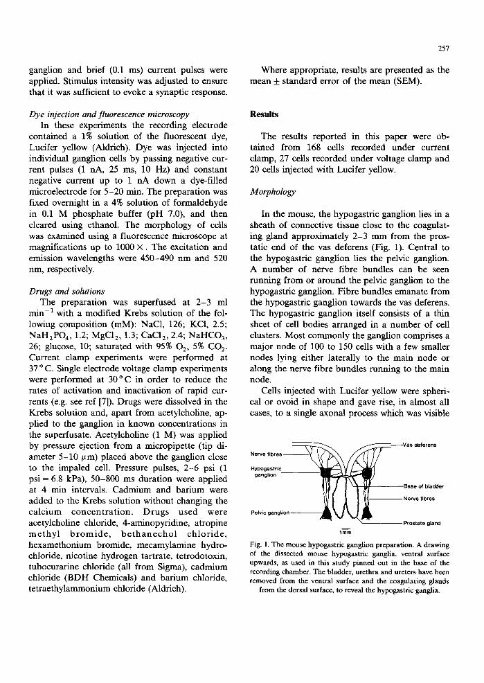
In the mouse, the hypogastric ganglion lies in a sheath of connective tissue close to the coagulat- ing gland approximately 2-3 mm from the pros- tatic end of the vas deferens (Fig. 1). Central to the hypogastric ganglion lies the pelvic ganglion. A number of nerve fibre bundles can be seen running from or around the pelvic ganglion to the hypogastric ganglion. Fibre bundles emanate from the hypogastric ganglion towards the vas deferens. The hypogastric ganglion itself consists of a thin sheet of cell bodies arranged in a number of cell clusters. Most commonly the ganglion comprises a major node of 100 to 150 cells with a few smaller nodes lying either laterally to the main node or along the nerve fibre bundles running to the main node.
Cells injected with Lucifer yellow were spheri- cal or ovoid in shape and gave rise, in almost all cases, to a single axonal process which was visible
Nerve fibres ~ ~ ~ -Vas deferens
Hypogastric ganglion
9ase of bladder
Nerve fibres
Pelvic ganglion Prostate gland
1ram
Fig. 1. The mouse hypogastric ganglion preparation. A drawing of the dissected mouse hypogastric ganglia, ventral surface upwards, as used in this study pinned out in the base of the recording chamber. The bladder, urethra and ureters have been removed from the ventral surface and the coagulating glands
from the dorsal surface, to reveal the hypogastric ganglia.

258
1Olin
C - 1.0 -0 .8 - 0 . 8 - 0 . 4 - 0 . 2
V(mV) n n n I I
"-7 J I T M
2.lime
0 0
- 1 0
-gO
- I O
-4O
- l iO
-eO
l(nA)
1
2
_J I 2.1ms
Fig. 2. Morphology and membrane properties of hypogastric ganglion cells. A. A representative mouse hypogastric ganglion cell injected with Lucifer yellow and photographed in whole mount. Portions of the axonal process not in the plane of focus have been omitted. B. Membrane potential change recorded in a cell (upper trace) in response to a rectangular hyperpolarizing current pulse (lower trace). The cell had a resting membrane potential of -41 inV. C. Voltage-current relationship for another cell. The maximum voltage deflection is plotted against the amplitude of the injected current. The cell had an input resistance of 51 M~2 at its resting membrane potential of - 4 8 mV. D. A single action potential (1, upper trace) evoked by injection of a depolarizing current pulse (lower trace). The depolarization evoked by this current pulse did not always reach threshold for action potential initiation (middle trace); the maximum depolarization was not sustained for the duration of the pulse (2) and a rebound hyperpolarization was
apparent on cessation of the pulse.
for a d i s t ance of 2 0 - 3 0 × the d i a m e t e r o f the cell
s o m a (Fig. 2A). In 15 o u t of 20 f i l led cells the
p rocess ran f r o m the gang l ion t owards the vas
deferens . In one cell the p rocess r an b a c k towards
the pelv ic gang l ion and in the r e m a i n d e r the p ro -
cess a p p e a r e d to run towards pe lv ic o rgans o t h e r
t han the vas deferens . W e have n o t i nves t iga t ed
the i n n e r v a t i o n of o the r pe lv ic o rgans by the cells
of this gangl ion . T h e d i m e n s i o n s o f the cell s o m a
were 35 + 2 / ~ m (n = 10) a long the l onge r axis and
28 + 1 /~m (n = 10) a long the shor t e r axis. N o
e v i d e n c e was o b t a i n e d for d e n d r i t i c p rocesses aris-
ing f r o m the cell soma.
Electrophysiological properties of ganglion cells
Current clamp Membrane properties. A f t e r i m p a l e m e n t , gan-
g l ion cells m a i n t a i n e d a c o n s t a n t res t ing m e m -
b r a n e p o t e n t i a l for p e r i o d s of up to 6 h. In m a n y
cel ls an in i t ia l h y p e r p o l a r i z a t i o n to a r o u n d - 9 0
m V as soc i a t ed wi th a fal l in i npu t res i s tance was

TABLE I
Electrophysiological properties of mouse hypogastric ganglion cells
259
Mean + SEM (n) Range
Cells exhibiting a short afterhyperpolarization Resting membrane potential (mV) Membrane time constant (ms) Input resistance (MI2) Threshold depolarization for action potential generation (mV) Action potential amplitude (mV) measured from resting potential Action potential duration (ms) measured at threshold Afterhyperpolarization amplitude (mV) measured from threshold Afterhyperpolarization duration (t l /2, ms)
Cells exhibiting a large, prolonged afterhyperpolarization Resting membrane potential (mV) Membrane time constant (ms) Input resistance (M~2) Threshold depolarization for action potential generation (mV) Action potential amplitude (mV) measured from resting potential Action potential duration (ms) measured at threshold Afterhyperpolarization amplitude (mV) measured from threshold Afterhyperpolarization duration (tl/2, ms)
- 48.6 + 0.6 (129) - 38 to - 65 2.4 + 0.2 (54) 1.0 to 6.7
45.6 + 2.6 (75) 20 to 150 17.6 + 0.3 (148) 9 to 28 80.1 + 0.8 (148) 55 to 106
1.6 + 0.1 (157) 1.1 to 2.3 35.0 + 1.8 (10) 22 to 42
4.7 + 0.4 (10) 2.5 to 11.3
-52 .6 + 2.3 (10) - 4 4 to - 7 2
67.3 + 8.7 (6) 35 to 100 17.5 + 1.2 (11) 10 to 24 86.8 + 2.8 (10) 71 to 96
1.9 + 0.1 (11) 1.8 to 2.3 38.0 + 3.3 (5) 26 to 44
221 +19 (5) 160 to 285
seen just following impalement but this declined over 5-10 rain to a stable level which was main- tained for the remainder of the recording. This hyperpolarization may have resulted from calcium entry, caused by the impalement, activating a potassium conductance [17].
The passive and active electrophysiological properties of ganglion cells which maintained a stable resting membrane potential after impale- ment for more than 30 min are given in Table I and illustrated in Fig. 2. The values for resting membrane potential, input resistance and mem- brane time constant were continuous throughout the range. In approximately 5% of cells listed separately in Table I, the action potential wave- form was of a different shape. This small sub- population of cells had a noticeably larger and longer after-hyperpolarization following the action potential as well as a longer action potential dura- tion but did not differ markedly from the pre- dominant group of cells in the other membrane properties examined (Table I). All action poten- tials were abolished by tetrodotoxin (q'TX; 1/~M).
Small hyperpolarizing current pulses (0.1-0.3 nA, 2.5 ms) applied at the resting membrane potential displayed time-dependent anomalous
rectification (Fig. 2B). On termination of the pulse the membrane potential rebounded above the rest- ing membrane potential and occasionally triggered 'off-spikes'. Small depolarizing pulses, sub- threshold to initiate an action potential, evoked graded depolarizations which were not maintained for the duration of the pulse (Fig. 2D).
Antidromic stimulation. Antidromic action potentials evoked by stimuli applied through elec- trodes placed around the vas deferens were recorded from the somata of 14 out of 25 cells tested. The threshold voltage required to evoke an antidromic response varied from cell to cell. Anti- dromic action potentials were all-or-nothing, arose at a fixed latency after the stimulus and had a slight inflection on the rising phase approximately 20 mV above the resting membrane potential. The mean amplitude of the action potential measured from resting membrane potential was 76.0 + 3.6 mV (n = 10) and the mean duration also measured at resting membrane potential was 2.2 ___ 0.1 ms (n = 10). The action potential was followed by a brief after-hyperpolarization of about 20 mV in amplitude from resting membrane potential and a half time of decay of 5.5 +0.5 ms. When the stimulating electrodes were positioned approxi-

260
05 I nA
-37
e.
- - 1 , m 1 I
1 0 0 m s e c
1 . 5 m
2 . 0 - -
2 . 5 -
-,oo -io to -Io/'-to 0 - -
0 . 5 - -
1 . 0 -
V (mY)
c 60--
I l . . . . . . f . . . . .
4 0 -
1
2 0 - - 0J I I I I
2.5 5 10 20
EXTRACELLULAR K+CONCENTRATION (raM)
Fig. 3. Electrophysiological characteristics of l M. A. Current (upper) and voltage (lower) traces are shown for a single cell voltage-clamped at -37 mV and stepped to more negative potentials for 350 ms. Hyperpolarization induced an instantaneous current step followed by a slow, relaxing current. The data obtained from these experiments are plotted in B. Crosses represent the instantaneous current seen at onset of the hyperpolarizing pulse and filled circles represent the steady-state current obtained immediately before returning to the holding potential. The point where the lines cross (-95 mV here) represents the reversal potential for I M. The variation of this reversal potential with the extracellular K + concentration in the same cell is shown in C. TTX
(300 nM) was present throughout.
mately 20 mm from the recording electrode, act ion potentials arose at a mean latency after the st imu- lus of 22.6 _+ 0.8 ms (n = 10) indicat ing a conduc- t ion velocity for the post-ganglionic nerve of ap- proximately 1 ms - l , which is characteristic of
unmyel ina ted fibres.
Voltage clamp In the presence of TTX (300 n M - 1 /~M) the
pharmacological and biophysical properties of three potassium currents have been characterized.
(i) M-current (IM). To facilitate the study of I M cells were voltage clamped at a holding poten- tial of - 4 0 mV (the mean holding current was 0.222_+0.097 nA, range - 0 . 3 5 0 to +1.150 hA,
n = 13). Small hyperpolar izing voltage steps of 300-500 ms dura t ion elicited an ins tan taneous inward current followed by a slow, inward current relaxation (Fig. 3A). On re turn ing to the holding potent ial an ins tan taneous outward current was
again seen, followed by a slow, outward current relaxation. The slow, inward current relaxat ion is associated with a decrease in m e m b r a n e conduc- tance since the ins tan taneous current step seen at the onset of the voltage pulse was greater than that seen at the offset of the voltage pulse. As the ampli tude of the voltage-step was increased the inward current relaxation first increased in ampli- tude then decreased and finally changed direct ion to outwards (Fig. 3A, B). The reversal potent ia l

(Erev) for this current was - 90.0 + 3.6 mV (n = 7). In two cells the change in Ere v for a 10-fold change in external potassium concentration was 54 and 52 mV (Fig. 3C). These values are close to that predicted by the Nernst equation for a cur- rent carried by potassium ions.
Both the inward and the outward current re- laxations displayed a time course which was ap- proximately exponential. The time constant of decay had a maximum value of 100 to 125 ms and decreased with increasing hyperpolarization. The conductance underlying the relaxations was also voltage-dependent, being activated at potentials positive to - 6 0 mV. However, the maximum con- ductance could not be measured as I M, uncon- taminated by other currents, could not be isolated at less negative potentials. The maximum conduc- tance obtained varied between 5.1 and 7.7 nS at step potentials of - 3 1 to - 2 7 mV.
Cd 2÷ (100 ~M) produced only a 20% decrease in the slow current relaxations (n = 2), suggesting that only a small proportion of the slow current relaxations is due to the inactivation/activation of a calcium-dependent, potassium conductance. Similarly, tetraethyl ammonium (TEA) (5 mM) inhibited the slow current relaxations by only 14% (n = 2). Tubocurarine (100 #M) either slightly in- creased or had no effect on the amplitude of the current relaxations (n = 2). The effects of both TEA and tubocurarine were reversible on washout. In contrast, Ba 2÷ (1 mM) and bethanechol (30 ~M; Fig. 4) rapidly elicited a steady-state inward current (mean 584 pA and 975 pA, respectively; n = 2 for each) and depressed the slow current relaxations (mean decrease 59% and 85%, respec- tively; n = 2 for each). These effects were rapidly reversible on washout. In one cell the effects of bethanechol were reversed by atropine (1/~M).
(ii) A-current (IA). With increasing amplitude of hyperpolarizing voltage steps from - 40 mV the slow, outward current relaxation seen on re- polarizing to - 4 0 mV became progressively ob- scured by a rapid transient outward current (IA; see Fig. 3A, two rightmost current traces). The characteristics of this current were studied by using more negative holding potentials and shorter (100 ms) voltage commands, in order to reduce the contribution of I M to the evoked current. I A was
Ba 2"I" 10 -3 M
261
nA
-61-J
1 0 0 msee
f 100 sec
Bethanecho l 3 x 1 0 - 5 M
-40
100 m s e c 1 0 0 sec
Fig. 4. Inhibition of I M by bar ium and bethanechol. Current (upper) and voltage (lower) traces for two cells held at - 4 0 mV and repeatedly (0.1 Hz) stepped to - 6 1 mV for 400 ms in A and - 5 8 mV for 350 ms in B (downward deflections). Individual responses are shown on an expanded timescale.
TTX (300 nM) was present throughout.
totally inactivated at a holding potential of - 5 0 mV (Fig. 5A, C). Removal of the inactivation was obtained by stepping to voltages more negative than - 6 0 mV and on returning to the holding potential a fast, rapidly decaying outward current was seen (Fig. 5A). The time constant of decay varied between 3.3 and 8.5 ms. Stepping to more negative potentials increased removal of inactiva- tion in a sigmoidal manner and the maximum effect was seen at around - 1 3 0 mV (Fig. 5C). Half-maximal removal of inactivation was seen at - 9 5 . 0 + 1.5 mV (n = 3).
Activation of I A was studied in two cells which were held at - 9 0 mV and stepped to less negative

262
A L _ V H ---
-50mY m
- 58
(mY) C 2OOO
1500
-- 1000
500
0 -160
V H
(mY)
5 m $
-140 -120 -100 -80 ,60 -40 Membrane potential (mV}
Fig. 5. Electrophysiological characteristics of 1A. A and B show current traces obtained form the same cell. In A removal of inactivation was studied. The cell was held at - 5 0 mV and stepped for 100 ms to the more negative potentials indicated. On returning to - 5 0 mV, fast, rapidly decaying outward currents were seen. The amplitude of these currents is repre- sented by the filled circles in C. In B activation of I A was studied. The cell was held at - 90 mV and stepped for 100 ms to the less negative potentials indicated. Again, fast, rapidly decaying outward currents were seen, the amplitude of which is represented by the open circles in C. The curves in C were
fitted by eye. TI 'X (300 nM) was present throughout.
potentials. Threshold for activation was around - 7 0 mV and a fast, outward current was seen which rapidly decayed during the voltage step (Fig. 5B, C). I A could not be studied at further, less negative potentials due to the appearance of other outward currents. In two cells 4-aminopyri- dine (2 mM) inhibited by 82% the I A which was evoked by stepping from - 9 0 mV to - 5 0 mV (data not shown).
(iii) Calcium-dependent potassium currents. V o l t a g e s t e p s f r o m - 4 0 m V to p o t e n t i a l s i n t h e
r e g i o n o f - 2 3 to - 1 6 m V f o r 10 m s e v o k e d a n
i n i t i a l c u r r e n t r e s p o n s e w h i c h c o u l d n o t b e c o m -
p l e t e l y c l a m p e d ( s e e F i g . 6 A ) , f o l l o w e d b y a n
o u t w a r d c u r r e n t d u r i n g t h e r e m a i n d e r o f t h e v o l t -
a g e s t e p . O n r e t u r n i n g t o t h e h o l d i n g p o t e n t i a l
r a p i d l y d e c a y i n g o u t w a r d t a i l c u r r e n t w a s s e e n
(amplitude 610 + 90 pA, n = 6; time constant for decay 2.9 ___ 0.4 ms, n = 6) (Fig. 6A, B). Both TEA (5mM) and Cd 2+ (100/~M) markedly reduced the outward current and the tail current (78 + 12% reduction in tail current by TEA, n = 3 and 94% reduction in tail current by Cd 2÷, n = 2) (Fig. 6A and B); Cd 2÷ but not TEA also abolished the partially clamped response seen during the voltage step. Thus, the partially clamped current appears
control
- 4 0 ~ ' ~ ~v mV
TEA (5raM)
control
rnV
. • 0.3nA 40mV
5ms
Cd ( 100 I.IM)
- -~ t,
Fig. 6. Characteristics of the calcium-dependent current. Cur- rent (upper) and voltage (lower) traces are shown for a single cell voltage clamped at - 4 0 mV and stepped to - 2 0 mV for 10 ms. Tail currents seen on repolarization are indicated by an arrow. Each trace represents the average of four responses and apparent irregularities in the voltage traces in the first ms following onset of the step command are due to the averaging process. In addition, the inward (calcium) transient during the voltage step was not completely clamped (indicated by a star on the voltage trace in A and B). A. the tail currents were inhibited by TEA (5 mM). B. on washout of TEA the current amplitude returned to control values. Subsequently, the tail currents were blocked by Cd 2+ (100 /,M). TTX (1 btM) was
present throughout.

263
A (I)
(,I) ! (iv)
--4 ~ - . - ~ J 2OmV
2.Sme
B
C
Control
Contro l
mecamylamine (301AM)
nicotine (IO01JM)
mecamylamlne (IO01JdM)
w a s h
. J 20mY 2.Sins
__] lOmV 2.saw
Fig. 7. Evoked synaptic responses. Synaptic responses were evoked by single stimuli applied to the preganglionic fibres. The stimulus artefact has been attenuated for clarity. A (i)-(iv) shows the typical, suprathreshold synaptic responses (i.e. EPSP plus action potential) recorded in four different ganglion cells. The most frequently observed synaptic responses were those illustrated in (iii) and (iv). B. Mecamylamine (30 and 100 /~M) reduced the EPSP in a concentration-dependent manner. C. Desensitization induced by application of nicotine (100 #M) for 13 rain abolished the synaptic response. Nicotine initially depolarized the cell but this effect had
declined to pre-drug level by 13 min.
to be carried by calcium ions whilst the outward current and the tail current appear to result f rom activation and inact ivat ion of a rapidly decaying ca lc ium-dependent potass ium conductance. I n two
cells out of six studied, a small ( < 100 pA), slowly decaying c o m p o n e n t of the tail current was ap- parent . Tubocura r ine (100 #M) abolished this slow c ompone n t wi thout affecting the more rapidly de-

264
A
-9~
-118 ~
* I N --
( m v ) ~
-08 ~:' - : " - ~
~ J 10mV -106 2.6ms
..,_] 20mV
c 26
r =0.86
6
-1
membrane potentlel (m¥) E rev -e.lm¥
, o
Fig. 8. Effect of membrane hyperpolarization on synaptic responses. A. In this cell hyperpolarization of the membrane potential by up to 82 mV from resting membrane potential ( -46 mV) failed to render the synaptic response subthreshold for action potential initiation. B. In another cell, the EPSP was subthreshold for action potential initiation at resting membrane potential ( 45 mV). Hypetpolarization to the potentials indicated on the left hand side of each trace increased the amplitude of the EPSP. C. From the same cell as in B, a plot of EPSP amplitude against membrane holding potential gave a linear relationship and a value for reversal
potential of - 9.6 mV.
caying tail current. We have not studied the slow current further.
In addi t ion to the effects described above, Cd 2÷ also caused an inward current in cells c lamped at - 4 0 mV (235 + 115 pA, n = 4). In two cells held at - 3 5 mV the mean inward current following applicat ion of Cd 2÷ (100/~M) was greater (mean 569 pA) than that observed at - 4 0 mV. T E A (5 mM) also elicited an inward current (mean 164 pA, n = 2) whereas tubocurar ine (100/~M, n = 2) had no effect on the holding current. These results suggest that at potentials between - 3 0 and - 4 0 mV there exists a mainta ined, vol tage-dependent
calcium-influx, which activates a calcium-depen- dent outward current.
Synaptic potentials
In addi t ion to the m e m b r a n e properties of gan- glion cells the na ture of fast synaptic t ransmission through the ganglion was examined. A single st imulus applied to pregangl ionic fibres evoked a fast excitatory postsynapt ic potent ia l (EPSP) in almost all cells tested. EPSPs were also observed in cells which exhibited a p ronounced after-hyper- polar izat ion following the action potential . In the

majority of cells, EPSPs were suprathreshold for action potential initiation (Fig. 7). The mean am- plitude of the synaptically-evoked action potential measured from resting membrane potential was 75.4 + 1.5 mV (n = 62). Fig. 7 shows examples of the types of EPSP/act ion potential complex ob- served. The most frequent response observed (Fig. 7A(iv)) was characterized by an action potential which rose from resting membrane potential without an inflection on the rising phase at the threshold for action potential initiation; the action potential was followed by a pronounced after- depolarization. In these cells hyperpolarization of the somatic membrane (up to 100 mV from resting membrane potential) by current injection failed to render the EPSP subthreshold (Fig. 8A). This is in contrast to the remainder of cells exhibiting a suprathreshold EPSP in which hyperpolarization could render the EPSP subthreshold. Approxi- mately 5% of cells showed EPSPs which were subthreshold (Fig. 8B) when evoked at resting membrane potential. Although in an individual cell these subthreshold EPSPs varied slightly in amplitude, they were evoked in an all-or-nothing manner. In cells with a subthreshold EPSP the decay phase of the EPSP evoked at resting mem- brane potential conformed to a single exponential with a time constant of 4.9 + 0.8 ms (n = 6).
Synaptic responses were blocked by TTX (1 /~M) (n = 2) or by raising the magnesium con- centration of the superfusing solution to 20 mM (n = 4). The nicotinic antagonists mecamylamine (10-100 /~M; n = 8) and hexamethonium (0.1-1 mM; n = 26) markedly depressed and in some cases abolished the EPSP (Fig. 7B). Mecamyla- mine was generally more effective than hexameth- onium in blocking the EPSP. In the presence of the antagonists the EPSPs became subthreshold for action potential initiation. EPSPs of the type shown in Fig. 7A(iv) were the most difficult to block completely with antagonists; concentrations of hexamethonium up to 1 mM being required to render the EPSP subthreshold. At these concentra- tions hexamethonium did not exert any significant local anaesthetic action since it did not alter the shape of the action potential evoked in the soma by current injection through the recording elec- trode. In five cells, desensitization of nicotinic
265
receptors by prolonged exposure (10-15 min) to nicotine (100-300 /~M) abolished the EPSP (Fig. 7C) without affecting the shape of the action potential evoked in the same cell by current injec- tion. The effect of nicotine reversed on washout of the drug.
All-or-nothing nature of the EPSP As the intensity of the stimulus applied to the
presynaptic fibre bundle was increased the EPSP appeared in an all-or-nothing manner. The ampli- tude of the EPSP did not increase as the intensity of the stimulus was increased above the threshold level (Fig. 9). Only very occasionally did raising the stimulus intensity or placing the stimulating electrode on another fibre bundle reveal a second synaptic input which occurred at a different syn- aptic latency to the first.
Whilst focal stimulation of the fibres running to the ganghon evoked an EPSP action potential complex, depolarization of the cell by current pulses injected into the cell through the recording electrode evoked an action potential which was not followed by an afterdepolarization (see Fig. 2B). In the presence of TTX (1 /~M) depolariza- tion of the cell by up to 30 mV from resting membrane potential by current pulses injected into the cell through the recording electrode did not give rise to regenerative potentials.
To investigate whether the EPSP resulted from the stimulation of a single presynaptic axon (i.e. a strong input) the position of the stimulating elec- trode was moved across a fibre bundle in ap- proximately 5/~m steps (Fig. 10). Although chang- ing the position altered the stimulus strength re- quired to evoke an EPSP (the stimulus intensity required increased on either side of a 'hot spot') the amplitude of the EPSP did not vary. Further- more, when the EPSP was rendered subthreshold for action potential initiation in the presence of hexamethonium the remaining EPSP appeared in an all-or-nothing manner as the stimulus intensity was increased (Fig. 9).
In six cells in which the EPSP was subthreshold for action potential initiation or became sub- threshold on hyperpolarizing the cell the ampli- tude of the EPSP increased linearly as the mem- brane potential was made more negative (see Fig.

266
2ev ~
Current ( ~ )
10,
8 .
4
2ev---t " - - / ~ 2
~ 0 , , i , 20V ~ 20 4 0 O0 80
8tlmu~* (V)
s o v ~
4ov ~ ~ 7ov ~
.ov- .ov - - - J lOmV 2.Sins
Fig. 9. All-or-nothing nature of the EPSP. The EPSP evoked in response to preganglionic nerve fibre stimulation was recorded over a range of stimulus intensities (the voltage applied is indicated on the left of each trace and the insert shows the relationship between stimulus voltage and current flow through the electrode measured at the end of the experiment). The bathing solution contained hexamethonium (1 mM) to render the synaptic response subthreshold for action potential ini- tiation. The EPSP was evoked in an all-or-nothing manner and did not increase in amplitude with increasing stimulus inten- sity. The stimulus artefact which increased with increasing
stimulus intensity has been attenuated for clarity.
8C). T h e m e a n e x t r a p o l a t e d reversa l po t en t i a l was
- 15.6 _+ 4.2 mV.
Response to exogenous acetylcholine
Loca l a p p l i c a t i o n of ace ty lcho l ine , by p ressu re
e j ec t ion f r o m a m i c r o p i p e t t e p o s i t i o n e d c lose to
the i m p a l e d cell, e v o k e d a c o m p l e x d e p o l a r i z i n g
r e sponse wi th an ini t ia l phase b l o c k e d by hexa-
m e t h o n i u m and a la te phase b l o c k e d by a t r o p i n e
(Fig. l l A ) . In the p r e sence o f a t r o p i n e (1 btM) and
h e x a m e t h o n i u m (0 .3 -1 m M ) the r e sponse to
ace ty l cho l ine was a lmos t c o m p l e t e l y abo l i shed .
U n l i k e the E P S P the a m p l i t u d e o f the n i c o t i n i c
r e sponse to a ce ty l cho l i ne was g r aded ; i nc r ea s ing
pe r iods o f e j ec t ion ( f r o m 2 0 0 - 8 0 0 ms) e v o k e d
la rger r e sponses (Fig. 11B). T h e a p p l i c a t i o n o f
a ce ty l cho l i ne d id no t give rise i m m e d i a t e l y to an
ac t ion p o t e n t i a l (cf. the s t r o n g i n p u t synap t i c
responses i l lus t ra ted in Fig. 6). T h e tl/2 for decay
eTv i
1 2 0 V ~ 1 2 0 V
,~2vL.
to~
92V .-,,
120V ~" __.J 10mV
Fig. 10. Activation of a single presynaptic axon evokes an all-or-nothing synaptic response. In the upper diagram the position of the tip of the stimulating electrode on the pregan- glionic fibre running to the ganglion is represented by the open circles. The minimum voltage at which a synaptic response was evoked is indicated. 120 V was the maximum possible stimulus and failed to evoke a synaptic response at the lateral electrode positions. Each of the lower traces shows the synaptic response (action potentials have been attenuated in these recordings) evoked at the different stimulating electrode positions. The voltages indicated on the left of each trace relate to the
threshold voltages indicated in the upper diagram.

267
Control
Hexemethonlum 3001JM
B
200rns 5psi
t 4psi 25m•
400ms
Atropine IlJM
600ms
Hexemethonlum 3001JM
"1" Atropine llJkl mlks~.<~? 71SL~._2%,, ,~
1' . ~ 8mV - - ] 5 m V
800ms 1 • S•
Fig. 11. Responses to exogenously applied acetylcholine. Acetylcholine was applied by pressure application from a micropipette positioned close to the impaled cell. The duration and amplitude of the pressure pulses are indicated. A. The response to acetylcholine had a fast (1) and a slow (2) component. The fast component was abolished by hexamethonium, whereas the slow component was abolished by atropine. B. In the presence of atropine (1 #M), the response to acetylcholine was dose-dependent in
that it increased with increasing duration of pressure application. Note the difference in time scale in A and B.
of the nicotinic response to applied acetylcholine was 2.3 + 1.5 s (n = 9).
Discussion
Our results describe the first characterization of the neurons contained in the mouse hypogastric ganglion. Some of the cells in this ganglion appear to innervate the smooth muscle of the vas de- ferens, since electrical stimulation via ring elec- trodes placed around the the epididymal portion of the vas deferens could antidromically activate ganglion cells. We have therefore termed this gan- glion the hypogastric ganglion since through it is transmitted the motor, sympathetic outflow to the
vas deferens. In immunohistochemical studies, tyrosine hydroxylase-like and neuropeptide Y-like immunoreactivity could be observed within the cell bodies contained in this ganglion (M. Costa, J.B. Furness and G. Henderson, unpublished ob- servations).
The small, ovoid morphology of these cells is similar both in dimensions and lack of dendritic processes to that of ganglion cells in the analogous pelvic ganglion of the rat [27]. Guinea-pig pelvic ganglion cells, although being devoid of dendrites, do have short tufts arising from the cell surface onto which synaptic inputs are received [3]. Cat pelvic ganglion cells on the other hand have exten- sive dendritic processes and receive multiple syn- aptic inputs [see 27]. The majority of mouse hypo-

268
gastric ganglion cells had basic electrophysiologi- cal properties similar to those reported previously for cells in other ganglia in the pelvic region of the rat [27], guinea-pig [3,15] and rabbit [16; Rogers and Henderson, unpublished observations].
At present we have insufficient data to specu- late on the functions of the small number of cells which exhibited a pronounced afterhyperpolariza- tion following the action potential. The small number recorded in this study could reflect either their low density in this preparation or a greater degree of difficulty in obtaining stable impale- ments from this group of cells. Sensory AH cells of the enteric plexi of the guinea-pig exhibit a pronounced afterhyperpolarization but unlike the cells in the mouse hypogastric ganglion, they do not receive a fast synaptic input [13].
The results obtained in this study under volt- age-clamp show that a calcium conductance and several potassium conductances are present in the soma of neurons within the mouse hypogastric ganglion, similar to other sympathetic ganglia. Calcium currents have previously been studied in the rat superior cervical ganglion [2,10]. There also appears to be a maintained calcium influx at holding potentials of - 3 5 and - 4 0 mV in mouse hypogastric ganglion cells as Cd 2+ evoked an in- ward current. This is consistent with observations made in frog sympathetic ganglia [21] where, using fura-2 to measure intracellular calcium levels, it was found that the resting calcium concentration was sensitive to the membrane holding potential, increasing at potentials positive to - 4 5 mV. Rat vesical pelvic ganglion cells have been reported to show spontaneous hyperpolarizations resulting from the activation of a calcium-dependent potas- sium conductance [19]. In contrast, mouse hypo- gastric ganglion cells did not exhibit spontaneous conductance changes but maintained a stable rest- ing membrane potential or holding current.
The influx of calcium evoked by depolarizing steps appeared to activate two calcium-dependent potassium currents in mouse hypogastric ganglion cells, as following repolarization two Cd:+-sensi - tive components to the tail current were seen. In all cells the more prominent of the two currents was the rapidly-inactivating, TEA-sensitive cur- rent similar to I c seen in bullfrog sympathetic
ganglion cells [1,20] and the rat superior cervical ganglion [2,11]. In two cells a much smaller, slowly-decaying tail current was also seen, which was abolished by tubocurarine, suggesting that it was due to decay of IAHP [5,20]. The relative amplitude of I c and IAHP here is consistent with a significant role for Ic, but not IAHp, in repolari- zation following an action potential. IAHp may be responsible for the prolonged afterhyperpolariza- tion which was observed in the current clamp experiments in a small proportion of cells.
Long hyperpolarizing pulses (300-500 ms) re- vealed a BaZ+-sensitive, slowly activating and in- activating potassium current similar to I M previ- ously reported in other sympathetic ganglia [4,5,7]. 1 M has a 'clamping' effect on membrane potential and its activation and inactivation may modulate the efficiency of neurotransmission through this ganglion. Superimposed on the outwards I M evoked by large hyperpolarizing voltage steps was a rapidly activating and inactivating outward cur- rent. It is similar electrophysiologically and pharmacologically t o I A [6], which has also been described in rat superior cervical ganglion cells [11] and guinea-pig inferior mesenteric ganglion cells [5]. Since 1A is totally inactivated at the resting potential of mouse hypogastric ganglion cells it can play no role in the repolarization following an action potential. However, at the more hyperpolarized potentials seen during the afterhyperpolarization a degree of removal of I A- inactivation may occur, allowing I A to act in an inhibitory manner on the further firing of action potentials.
Preganglionic nerve stimulation evoked a suprathreshold EPSP in most cells, the ovoid na- ture of the cells, the lack of a dendritic tree and the smaller amplitude of the synaptically activated action potential compared to that evoked by cur- rent injection demonstrate that these cells receive their synaptic input on to the somatic membrane. We have provided considerable pharmacological evidence to support the view that the depolariza- tion following the action potential is synaptically mediated. In addition, antidromically-activated action potentials did not give rise to after-de- polarizations. Therefore, it is unlikely that the action potential component of the EPSP/act ion

poten t ia l complex results f rom direct or co l la te ra l ac t iva t ion of the gangl ion cells. In those cells in which the synapt ic response gave rise immed ia t e ly to an ac t ion po ten t ia l the synapt ic inpu t m a y be concen t ra t ed at the axon hi l lock region, an area of enhanced m e m b r a n e exci tabi l i ty . A l t e rna t ive ly the quan ta l conten t of ace ty lchol ine release f rom pre- gangl ionic fibres m a y be very large.
The EPSP recorded in mouse hypogas t r i c gan- g l ion cells appea red in an a l l -or -noth ing m a n n e r wi th increasing s t imulus intensi ty. Our f indings of a s t imula t ion ' h o t spot ' on the afferent f ibre bun - dle and the cons tan t ampl i t ude of the evoked EPSP with increas ing s t imulus in tens i ty ind ica te that the cells receive a single, s t rong input . In this respect the mouse hypogas t r ic gangl ion cells ap- pea r similar to those of the guinea-pig hypogas t r i c gangl ion [3,15] and hams te r s u b m a n d i b u l a r gangl- ion [26] and to some of the cells of the ra t pelvic gangl ion [27]. Since mouse hypogas t r i c gangl ion cells lack a dendr i t ic tree and since in the presence of TTX depola r iz ing pulses up to 30 mV posi t ive to rest ing m e m b r a n e po ten t ia l d id not evoke ei ther a regenerat ive ca lc ium spike or an inward ca lc ium current it is unl ikely that the a l l -o r -no th ing na tu re of the EPSP results f rom graded synap t ic de- po la r iza t ion summat ing to act ivate a regenera t ive ca lc ium conductance . The reversal po ten t i a l ( - 15 mV) of the EPSP is s imilar to that p rev ious ly r epor ted for nicot inic EPSPs in o ther gangl ion p repa ra t ions [9,18,25]. Fur the rmore , dur ing the onset of the b l o c k a d e by h e x a m e t h o n i u m or m e cam ylamine the ampl i tude of the EPSP was reduced in a graded, t ime-dependen t manner .
The electr ical ly quiescent na ture of mouse hy- pogas t r ic gangl ion cells, the single, s t rong f ibre i npu t and the a l l -o r -no th ing na tu re of the supra th resho ld synapt ic po ten t ia l wou ld ind ica te that phys io logica l ly these cells serve p r imar i ly a s imple relay funct ion and do no t in i t ia te or in- tegrate in format ion . Hypogas t r i c gangl ion cells do possess somat ic muscar in ic receptors as ind ica ted by the a t ropine-sens i t ive c o m p o n e n t of the re- sponse to app l i ed acety lchol ine and the inh ib i t ion of I M p roduced by bethanechol . Fu r the r invest i- ga t ion of the synapt ic responses to t ra ins of pre- gangl ionic s t imula t ion m a y reveal the funct ion of these receptors.
269
Acknowledgements
S u p p o r t e d by a grant f rom the M R C ; H.R. he ld an M R C Resea rch Studentsh ip . W e are grate- ful to Professor M. Cos ta for discussions on the n e u r o a n a t o m y of the pelvic region.
References
1 Adams, P.R., Constanti, A., Brown, D.A. and Clark, R.B., Intracellular Ca 2+ activates a fast voltage-sensitive K- current in vertebrate sympathetic neurones, Nature, 296 (1982) 746-749.
2 Belluzzi, O., Sacchi, O. and Wanke, E., Identification of delayed potassium and calcium currents in the rat sym- pathetic neurone under voltage clamp, J. Physiol. (Lond.)., 358 (1985) 109-129.
3 Blackman, J.G., Crowcroft, P.J., Devine, C.E., Holman, M.E. and Yonemura, K., Transmission from preganglionic fibres in the hypogastric nerve to peripheral ganglia of male guinea-pigs, J. Physiol. (Lond.), 201 (1969) 723-743.
4 Brown, D.A. and Adams, P.R., Muscarinic suppression of a novel voltage-sensitive K ÷ current in a vertebrate neurone, Nature, 283 (1980) 673-676.
5 Cassell, J.F. and McLachlan, E.M., Muscarinic agonists block five different potassium conductances in guinea-pig sympathetic neurones, Br. J. Pharmacol., 91 (1987) 259-261.
6 Connor, J.A. and Stevens, C.F., Voltage clamp studies of a transient outward membrane current in gastropod neural somata, J. Physiol. (Lond.), 213 (1971) 21-30.
7 Constanti, A. and Brown, D.A., M-currents in voltage- clamped mammalian sympathetic neurones, Neurosci. Lett., 24 (1981) 289-294.
8 Costa, M. and Furness, J.B., Observations on the anatomy and amine histochemistry of the nerves and ganglia which supply the pelvic viscera and on the associated chromaffin tissue in the guinea-pig, Z. Anat. Entwickl. Gesch., 140 (1973) 85-108.
9 Dennis, M.J. Harris, A.J. and Kuffler, S.W., Synaptic trans- mission and its duplication of focally applied acetylcholine in parasympathetic neurones in the heart of the frog. Proc. R. Soc. B, 177 (1971) 509-539.
10 Galvan, M. and Adams, P.R., Control of calcium current in rat sympathetic neurons by norepinephrine, Brain Res., 244 (1982) 135-144.
11 Galvan, M. and Sedlmeir, C., Outward currents in voltage- clamped rat sympathetic neurones, J. Physiol. (Lond), 356 (1984) 115-133.
12 Henderson, G. and Rogers, H., Opioid depression of syn- aptic transmission in the mouse hypogastric ganglion in vitro, J. Physiol. (Lond.), 390 (1987) 53P.
13 Hirst, G.D.S., Holman, M.E. and Spence, I., Two types of neurones in the myenteric plexus of duodenum in the guinea-pig, J. Physiol. (Lond.), 236 (1974) 303-326.

270
14 Hirst, G.D.S. and McLachlan, E.M., Development of den- dritic calcium currents in ganglion cells of the rat lower lumbar sympathetic chain, J. Physiol. (Lond.), 377 (1986) 349-368.
15 Holman, M.E., Muir, T.C., Szurszewski, J.H. and Yonemura, K., Effect of iontophoretic application of cholinergic agonists and their antagonists to guinea-pig pelvic ganglia, Br. J. Pharmacol., 41 (1971) 26-40.
16 Lacey, M.G. and Henderson, G., Electrophysiological anal- ysis of kappa opiate receptor activation in the rabbit vas deferens, Neuropeptides, 5 (1984) 257-260.
17 Nield, T.O., Slowly developing depolarization of neurones in the guinea-pig inferior mesenteric ganglion following repetitive stimulation of the preganglionic nerves, Brain Res., 140 (1978) 231-239.
18 Nishi, S. and Koketsu, K., Electrical properties and activi- ties of single sympathetic neurones of frogs, J. Cell. Comp. Physiol., 55 (1960) 15-30.
19 Nishimura, T., Tokimasa, T. and Akasu, T., Calcium-de- pendent potassium conductance in neurones of rabbit vesi- cal pelvic ganglia, J. Auton. Nero. Syst., 24 (1988) 133-145.
20 Pennefather, P., Lancaster, B., Adams, P.R. and Nicoll, R.A., Two distinct Ca-dependent K currents in bullfrog sympathetic ganglion cells, Proc. Natl. Acad. Sci. USA, 82 (1985) 3040-3044.
21 Pfaffinger, P.J., Leibowitz, M.D., Subers, E.M., Nathanson, N.M., Almers, W. and Hille, B., Agonists that suppress M-current elicit phosphoinositide turnover and Ca 2+ tran- sients, but these events do not explain M-current suppres- sion, Neuron, 1 (1988) 477-484.
22 Purinton, P.T., Fletcher, T.F. and Bradley, W.E., Gross and light microscopic features of the pelvic plexus in the rat, Anat. Rec., 175 (1973) 697-706.
23 Purves, D., Snider, W.D. and Voyvodic, J.T., Trophic regu- lation of nerve cell morphology and innervation in the autonomic nervous system, Nature, 336 (1988) 123-128.
24 Purves, D. and Lichtman, J.W., Geometrical differences among homologous neurons in mammals, Science, 228 (1985) 298-302.
25 Selyanko, A.A., Derkach, V.A. and Skok, I.V., Fast excita- tory postsynaptic currents in voltage clamped mammalian sympathetic ganglion neurons, J. Auton. Nero. Syst., 1 (1979) 127-137.
26 Snider, W.D., The dendritic complexity and innervation of submandibular neurons in five species of mammals, J. Neurosci., 7 (1987) 1760-1768.
27 Tabatabal, M., Booth, A.M. and De Groat, W.C., Morpho- logical and electrophysiological properties of pelvic gangl- ion cells in the rat, Brain Res., 382 (1986) 61-70.