characterization of tet and idh gene expression in chronic
TRANSCRIPT

RESEARCH Open Access
Characterization of TET and IDH geneexpression in chronic lymphocyticleukemia: comparison with normal B cellsand prognostic significanceMichaël Van Damme1*, Emerence Crompot1, Nathalie Meuleman2, Marie Maerevoet2, Philippe Mineur3,Dominique Bron2, Laurence Lagneaux1 and Basile Stamatopoulos1
Abstract
Background: Chronic lymphocytic leukemia (CLL) is the most common hematological malignancy in westerncountries, characterized by a heterogeneous clinical course. Although genetic studies have identified chromosomalaberrations or specific mutations, epigenetic changes have been poorly characterized in CLL.
Methods: We assessed ten-eleven translocations (TET) 1, 2, and 3, isocitrate dehydrogenase (IDH) 1, and 2messenger RNA (mRNA) expression using real-time PCR on purified leukemic B cells from 214 CLL patients (medianfollow-up = 75 months, range 1–380), normal peripheral blood B cells (n = 20), and umbilical cord blood B cells(n = 21). The microenvironment influence was assessed after 24 h co-culture of CLL cells with bone marrowmesenchymal stromal cells (BMSC). Finally, 5-hydroxymethylcytosine level (%5-hmC) was assessed by ELISA in CLLcells alone or with microenvironment stimuli.
Results: TET 1 and 3 and IDH2 were decreased in CLL cells compared with healthy B cells (P = 0.0221, 0.0013,<0.0001, respectively), while IDH1 was overexpressed (P = 0.0037). TET2 and IDH1 were significantly correlated withtreatment-free survival (TFS); patients with high TET2/IDH1 expression had a higher median TFS (111 months) thanpatients with low expression (78 months, P = 0.0071/0.0123). Moreover, TET1 expression decreased (P = 0.0371), whileTET3 and IDH2 expression increased (P = 0.0273/0.0039) in co-cultures. However, %5-hmC was not correlated withclinical data and was unchanged following microenvironment stimuli.
Conclusions: Despite a slight deregulation in CLL cells compared with normal B cells, we identified a significantassociation between TET/IDH gene expression and prognosis, suggesting that epigenetic changes could potentially beassociated with disease progression. Moreover, despite an identical %5-hmC, TET gene expression was influenced bycontact with BMSC confirming the crucial role of the microenvironment in CLL pathogenesis.
Keywords: Chronic lymphocytic leukemia, TET, IDH, 5-Hydroxymethylcytosine, Prognosis
* Correspondence: [email protected] of Clinical Cell Therapy, ULB Cancer Research Center (U-CRC),Institut Jules Bordet, Université Libre de Bruxelles (ULB), Route de Lennik,808, 1070 Brussels, BelgiumFull list of author information is available at the end of the article
© The Author(s). 2016 Open Access This article is distributed under the terms of the Creative Commons Attribution 4.0International License (http://creativecommons.org/licenses/by/4.0/), which permits unrestricted use, distribution, andreproduction in any medium, provided you give appropriate credit to the original author(s) and the source, provide a link tothe Creative Commons license, and indicate if changes were made. The Creative Commons Public Domain Dedication waiver(http://creativecommons.org/publicdomain/zero/1.0/) applies to the data made available in this article, unless otherwise stated.
Van Damme et al. Clinical Epigenetics (2016) 8:132 DOI 10.1186/s13148-016-0298-y

BackgroundChronic lymphocytic leukemia (CLL) is the most com-mon hematological malignancy in the west and is char-acterized by a heterogeneous clinical course [1]; somepatients will live several decades without any symptoms,while others will rapidly require a treatment and will havea decreased overall survival (OS). Clinical and molecularfactors, such as Binet stage, lymphocyte doubling time(LDT), mutational status of the immunoglobulin heavy-chain variable-region (IgHV), zeta-chain-associated pro-tein kinase 70 (ZAP70), lipoprotein lipase (LPL) or CD38expression, and serum levels of soluble CD23 (sCD23)and beta-2-microglobulin (B2M), can be used to classifypatients into different prognostic subgroups [2]. Moreover,increasing evidence suggests a role for the microenviron-ment in CLL pathogenesis. Our group previously demon-strated that bone marrow mesenchymal stromal cells(BMSC) protect CLL but not normal B cells from apop-tosis through direct contact [3].While genetic lesions, such as chromosomal aberrations
[4] or specific mutations, [5–8] are involved in CLL phys-iopathology, in recent years, growing evidence has sug-gested that epigenetic characteristics are key factors inleukemic processes. Recent studies have investigated epi-genetic features and demonstrated the importance ofDNA methylation [9] or histone post-translational modifi-cations in prognosis, oncogene regulation, or therapeutictargeting [10–12]. We demonstrated in previous papersthat histone deacetylase (HDAC) mRNA expression wasassociated with poor (HDAC7, HDAC10, and SIRT5) orgood prognosis (HDAC6, SIRT3, and SIRT6) [13] in CLLpatients. Moreover, global HDAC enzymatic activity is astrong predicator of poor prognosis in CLL which can re-fine well-known prognostic factors [14].In 2009, Tahiliani and colleagues discovered 5-
hydroxymethylcytosine (5-hmC) as the sixth base of theDNA in mammalian cells [15]. Ten-eleven translocationproteins (TET) are the dioxygenases responsible for theoxidation of 5-methylcytosine (5-mC) to form 5-hmC.There are three known TET isoenzymes (TET1, 2, and 3),and they require oxygen, Fe(II), and 2-oxoglutarate fortheir activity. This last cofactor is produced in the Krebscycle by the isocitrate dehydrogenases (IDH) 1 and 2.Other subsequent studies suggested that the 5-hmCmarker could be a step in the demethylation process[16–21] and/or a pattern allowing specific enzymes tobind hydroxymethylated regions of the genome [22–26].Hydroxymethylation enzyme defects have previously
been associated with hematological malignancies; mu-tations in TET2 were found in acute myeloidleukemia (AML) or chronic myelomonocytic leukemia(CMML) and induced loss of hydroxymethylation andwere linked with poor prognosis [27–30]. However,reports on TET2 mutations in B cell neoplasms are
rare [31], and little is known about DNA hydroxy-methylation in CLL.In the present study, we measured the mRNA expression
of TET1, 2, and 3 and IDH1 and 2 by quantitative real-timePCR (qPCR) in highly purified CD19+ B cells from a largecohort of CLL patients (n = 214) and correlated these datawith clinical outcome. We also investigated the potentialassociation between the global DNA 5-hmC rate andmicroenvironment stimuli, especially BMSC.
MethodsPatients and samplesThis study was approved by the Bordet Institute EthicsCommittee and conducted according to the principlesexpressed in the Declaration of Helsinki. All sampleswere collected at the time of diagnosis before any treat-ment, and after, written informed consent was obtainedfrom 214 CLL patients who presented with a typicalCD19+CD5+CD23+ phenotype and a Catovsky score of4 or 5/5. Treatment-free survival (TFS) and OS were cal-culated from the time of diagnosis until the date of firsttreatment and the date of death, respectively. All deathswere CLL-related. Control samples were obtained fromthe peripheral blood of 20 age-matched healthy volun-teers (PBHV) (mean 69 years old, range 54–90) afterwritten informed consent was obtained and from 21 um-bilical cord blood (UCB) samples after full-term deliveryand written informed consent of the mothers was ob-tained. Leukemic B cells were isolated from mono-nuclear cells with magnetic beads targeting CD19+phenotype (MidiMACS, Miltenyi Biotec). Briefly, pelletof 10 million mononuclear cells were resuspended in80 μl of PBS-05% BSA and 20 μl of CD19 microbeads.After 15 min of incubation at 4 °C and washing, cell sus-pension was applied onto a column placed in a magneticfield. Three washes were performed to discard unlabeledcells and then the column was removed to the separatorand placed on a collector tube where magnetically la-beled cells were flushed out. For experiments which re-quire culture, cells were negatively isolated by anindirect magnetic labeling system (B-CLL Cell IsolationKit from MidiMACS, Miltenyi Biotec) to avail any cellactivation or apoptosis. The isolation process is per-formed in two steps: a first labeling with a cocktail ofbiotin-conjugated monoclonal anti-human antibodiesand then, after incubation, an adding of anti-biotinmicrobeads. CD19+ cells are directly collected duringthe elution of the column.
BMSC isolation and conditioned medium preparationBMSC were harvested from the sternum or iliac crest ofhealthy volunteers and were isolated by the classical adhe-sion method, as previously described [32]. Conditionedmedia were prepared from 24 h cultures of BMSC alone
Van Damme et al. Clinical Epigenetics (2016) 8:132 Page 2 of 11

or CLL B cells + BMSC with and without contact (sepa-rated by a 0.4-μm pore-size filter). Cultures of CLL B cellswith BMSCs or different conditioned media were per-formed for 24 h.
Assessment of classic prognostic factorsZAP70 and LPL were measured by qPCR as previouslydescribed [33]. CD38 expression was assessed by flowcytometry, sCD23 and B2M were measured by ELISA,and IgHV gene mutational analysis was performed usingthe IGH Somatic Hypermutation Assay v2.0 (Invivo-scribe–Ref. 5-101-0031). LDT was assessed according toMontserrat et al. [34]. Classical cytogenetics by standardkaryotype analysis and additional interphase FISH wereperformed to screen for the most common aberrationsusing the Chromoprobe Multiprobe® CLL System (Cyto-cell, Amplitech–Ref. PMP 018/017/016/020). Patientswere then classified according to the recommendationsof Döhner and Cuneo et al. [4, 35]. Additional detailscan be found in Additional file 1: Text 1. All of thesefactors, except B2M for OS, were shown to be significantpredictors of TFS and OS, indicating that our cohort isrepresentative of a CLL population (Additional file 1:Figure S1 and Table S1).
RNA and DNA extraction and expression quantificationTotal RNA was extracted from purified CD19+ cells in asingle step using TriPure Isolation Reagent (Roche LifeScience–Ref. 11 667 165 001). Complementary DNA(cDNA) was generated from 500 ng of RNA using qScriptcDNA SuperMix (Quanta Biosciences–Ref. 95048-100)according to the manufacturer’s protocol. TET1, 2, and 3and IDH1 and 2 mRNA expression was quantified byreal-time PCR using SYBR Green technology (AppliedBiosystems–Ref. 4367659). Gene expression was normal-ized to the cyclophilin A (PPIA) gene as an endogenouscontrol as previously described [13] and calibrated bysubtracting 10 (chosen arbitrarily) from the ΔCt. Thecomparative ΔΔCt method was then used for data ana-lysis, and the fold changes were subsequently calculated(fold change = 2−ΔΔCt). All primer sequences are availablein the Additional file 1: Text 1 (Life Technologies).Genomic DNA (gDNA) was extracted from purifiedCD19+ cells with a QIAamp DNA Blood Mini Kit(Qiagen–Ref. 51104).
Assessment of 5-hmC levelsWe measured the 5-hydroxymethylcytosine percentage(%5-hmC) using a “Quest 5-hmC DNA ELISA Kit”(Zymo Research–Ref. D5425). Briefly, 150 ng of dena-tured DNA was added to wells coated with 200 ng ofanti-5-hydroxymethylcytosine polyclonal antibodies. Thesignal was detected at 405 nm after washing and addingthe anti-DNA HRP (horseradish peroxidase) antibodies.
Optic density was converted to %5-hmC using a standardcurve based on five controls (with values of 0, 0.03, 0.12,0.23, and 0.55%) provided in the kit. To quantify %5-hmC,we normalized the ELISA results with data obtained fromthe quantification of three genes (glyceraldehyde-3-phos-phate dehydrogenase (GAPDH), actin, and PPIA) in eachgDNA sample. Because these three endogenous controlsproduced similar results (indicating that an equivalentDNA quantity was loaded), we only used GAPDH andexpressed the results as %5-hmC/GAPDH.
Statistical analysisThe patients were stratified according to low and highTET or IDH expression with a cut-off value set by recur-sive partitioning maximizing the concordance with TFSas previously described for other prognostic factors [14].All median comparisons were performed with a Mann–Whitney test or a Wilcoxon test for paired comparisons.TFS and OS analyses were performed with the Kaplan–Meier curves, and differences between groups in termsof prognosis were assessed with a log-rank test. All testswere two-sided. Differences were considered statisticallysignificant at P < 0.05. All analyses were performed usingthe GraphPad Prism 5.0 (GraphPad Software) or SPSS18.0.0 software.
ResultsPopulationThe median age of the population was 63 years (range34–86). The median treatment-free survival (TFS) was88.07 months (range 0.33–251.43), and the median OSwas 241.87 months (range 0.40–380.20). The medianfollow-up was 74.53 months (range 0.40–380.20). In thispopulation, prognostic factors, such as IgHV mutationalstatus, ZAP70, CD38, Binet stage, sCD23, B2M, LDT,and cytogenetic profile (cytog. profile), were significantlycorrelated with TFS and OS (P < 0.05), except B2M forOS (Additional file 1: Figure S1). Complete prognosticdata were not available for all patients due to a lack ofbiological material. ZAP70, LPL expression, and CD38 ex-pression and clinical data were available for all 214 patientsincluded in this study. Incomplete data were available forIgHV mutational status (97.20%), LDT (84.11%), B2M(87.38%), sCD23 (81.78%), and cytog. profile (74.30%). Allprognostic factors (except B2M for OS) were shown to beassociated with prognosis, indicating that these 214 CLLpatients comprise a classical CLL cohort.
TET1 and 3 and IDH1 and 2 are deregulated in CLLTET1, 2, and 3 and IDH1 and IDH2 were measured inCD19+ CLL samples (mean purity 99.2%, range 97.5–99.9) and compared with the normal CD19+ cells. Weused two types of normal counterparts: CD19+ purifiedB cells from the peripheral blood of healthy volunteers
Van Damme et al. Clinical Epigenetics (2016) 8:132 Page 3 of 11
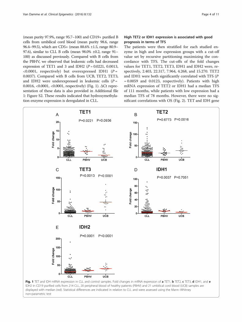
(mean purity 97.9%, range 95.7–100) and CD19+ purified Bcells from umbilical cord blood (mean purity 98.6, range96.4–99.5), which are CD5+ (mean 88.6% ±1.5, range 80.9–97.6), similar to CLL B cells (mean 98.0% ±0.2, range 91–100) as discussed previously. Compared with B cells fromthe PBHV, we observed that leukemic cells had decreasedexpression of TET1 and 3 and IDH2 (P = 0.0221, 0.0013,<0.0001, respectively) but overexpressed IDH1 (P =0.0037). Compared with B cells from UCB, TET2, TET3,and IDH2 were underexpressed in leukemic cells (P =0.0016, <0.0001, <0.0001, respectively) (Fig. 1). ΔCt repre-sentation of these data is also provided in Additional file1: Figure S2. These results indicated that hydroxymethyla-tion enzyme expression is deregulated in CLL.
High TET2 or IDH1 expression is associated with goodprognosis in terms of TFSThe patients were then stratified for each studied en-zyme in high and low expression groups with a cut-offvalue set by recursive partitioning maximizing the con-cordance with TFS. The cut-offs of the fold changesvalues for TET1, TET2, TET3, IDH1 and IDH2 were, re-spectively, 2.403, 22.317, 7.964, 4.268, and 15.270. TET2and IDH1 were both significantly correlated with TFS (P= 0.0059 and 0.0123, respectively). Patients with highmRNA expression of TET2 or IDH1 had a median TFSof 111 months, while patients with low expression had amedian TFS of 78 months. However, there were no sig-nificant correlations with OS (Fig. 2). TET and IDH gene
Fig. 1 TET and IDH mRNA expression in CLL and control samples. Fold changes in mRNA expression of a TET1, b TET2, c TET3, d IDH1, and eIDH2 in CD19 purified cells from 214 CLL, 20 peripheral blood of healthy patients (PBHV) and 21 umbilical cord blood (UCB) samples aredisplayed with median (red). Statistical differences are indicated in relation to CLL and were assessed using the Mann–Whitneynon-parametric test
Van Damme et al. Clinical Epigenetics (2016) 8:132 Page 4 of 11

Fig. 2 Prognostic power of TET and IDH gene mRNA expression. Fold changes in enzyme mRNA expression were measured by qPCR, andcut-offs were set by recursive partitioning. TFS and OS Kaplan–Meier curves are displayed for a, b TET1, c, d TET2, e, f TET3, g, h IDH1 and, i, jIDH2. Statistical differences between the curves were calculated using the log-rank test
Van Damme et al. Clinical Epigenetics (2016) 8:132 Page 5 of 11

expression was also compared between the groups basedon well-known prognostic factors (IgHV mutational sta-tus, ZAP70, CD38, Binet stage, sCD23, B2M, LDT, andcytogenetic profile). No obvious and important signifi-cant differences were observed (Additional file 1: TablesS2 and S3).
TET1, TE3, and IDH2 expression is influenced bymicroenvironment stimuli but global %5-hmC isunchangedBecause microenvironment stimuli are known to influ-ence the cellular physiology of CLL B cells, we measuredTET and IDH expression in leukemic cells before andafter contact with BMSC (Fig. 3a–e). We observed adownregulation of TET1 (10.31 vs 8.18, P = 0.0371) andan upregulation of TET3 (26.73 vs 36.40, P = 0.0273)
and IDH2 (48.78 vs 84.32, P = 0.0039). However, thesemodifications in mRNA expression were not associatedwith an alteration in global %5mC (Fig. 3f ), as it wassimilar in both conditions (0.0900 vs 0.0990, P = 0.4768).To ensure that BMSC co-culturing did not contaminatethe subsequent DNA extraction, we cultured CLL B cellsin the conditioned medium of BMSC alone or BMSC +CLL B cells (with and without contact). None of theseconditions showed significant differences in terms of %5-hmC as shown in Fig. 4.
%5-hmC has no prognostic significance in CLLWe performed a comparison of %5-hmC between prog-nostic subgroups based on classical prognostic factors(ZAP70, LPL, and CD38), but we did not observed anysignificant correlations (P = 0.3837, 0.5467, and 0.8671,
Fig. 3 CLL B cells and BMSC co-cultures impact on TET/IDH mRNA expression and %5-hmC. Fold changes in enzyme mRNA expression of a TET1,b TET2, c TET3, d IDH1, and e IDH2 were plotted for ten CLL patients for samples cultured alone or with BMSC. f The %5-hmC was measured byELISA in nine CLL B cell samples cultured alone and after contact with BMSC. Significance was assessed using the Wilcoxon signed rank test
Van Damme et al. Clinical Epigenetics (2016) 8:132 Page 6 of 11

respectively; Fig. 5a–c). A log-rank test was also per-formed following Kaplan–Meier analysis of patients dis-playing low %5-hmC (n = 15) vs high %5-hmC (n = 15).Using the median as the positive threshold, we did notobserve a statistical difference (median TFS of 84 vs49.57 months, respectively, P = 0.7934; Fig. 5d). Otherthresholds (based on recursive partitioning or ROCcurves maximizing the concordance with the mutationalstatus [14]) gave similar results. OS analysis was not per-formed due to the absence of deaths among the patients
during the study. Similar results were obtained if wenormalized %5-hmC to actin or PPIA (peptidyl prolylisomerase A or cyclophilin A).
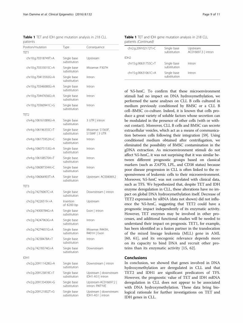
Mutation in TET/IDH genes are rare and non-recurrent in CLLIn order to investigate the mutational profile of TET andIDH genes, we used publically available whole exomeand RNAseq data previously published by Quesada et al.[36] and Ferreira et al. [37]. Within the CLL-ES project(accessible on https://dcc.icgc.org/projects/CLLE-ES), sim-ple somatic mutations were analyzed for a total of 218 pa-tients. Six patients (2.8%) presented a mutation for TET1,5 (2.3%) for TET2, 7 (3.2%) for TET3, 5 (2.3%) for IDH1and 2 (0.9%) for IDH2 for a total of 25 patients with 27mutations (Table 1). However, only 3 patients (1.4%) pre-sented a missense mutation respectively in TET1, TET2,and TET3 genes. All the other mutations in these fivegenes were found in intron, UTR (untranslated region),downstream or upstream the gene indicating that mu-tations in TET/IDH genes are rare and non-recurrentevents compared to what has been observed in otherhematological malignancies [27–30].
DiscussionIn the present paper, we performed a comprehensiveand complete expression profile of the known proteinsinvolved in DNA hydroxymethylation. To our know-ledge, it is the first time that such a study has been per-formed on a large cohort of CLL patients. In 2013,Hernandez-Sanchez and colleagues observed that TET2was overexpressed in CLL cells compared with cellsfrom healthy donors [38]. The normal counterpart ofCLL cells is not clearly defined in the literature and re-mains controversial [39]. We therefore decided to usetwo different controls of normal B cells: first, the periph-eral normal B cell from healthy donors since they repre-sent intuitively the normal counterpart in the healthydonors; second, the B cells from the umbilical cordblood since they express the CD5 similarly to CLL [40]and because Saunders et al. demonstrated that they dis-play a similar proteic profile to CLL [41]. In our results,TET2 decreased in CLL cells compared with normal Bcells from UCB, but the expression did not differ fromthat in the PBHV. These discrepancies could be ex-plained by at least three reasons: our cohort was largerthan the one used in the previous study (214 vs 23) aswell as the control group of PBHV (20 vs 5); the qPCRcontrol in the previous study was GAPDH, while weused PPIA, which is more stable in CLL; and finally,based on the reverse TET2 primer (the forward has beenmistaken for the GAPDH reverse primer), their ampli-con was in the 3' region (nucleotide 8941 to 8960), whileours was further upstream (nucleotide 4367 to 4389)which is a region less sensible to RNA degradation.
Fig. 4 Normalized 5-hmC levels in CLL B cells cultured alone in RPMIor with conditioned medium (CM). The different conditioned mediawere obtained from supernatants of 24 h cultures of a BMSC, b CLLB cells + BMSC with contact, and c CLL B cells + BMSC withoutcontact (separated by a 0.4-μm pore-size filter). Statistical differenceswere assessed using the paired Wilcoxon non-parametric test
Van Damme et al. Clinical Epigenetics (2016) 8:132 Page 7 of 11

In terms of prognosis, we observed that TET2 andIDH1 were associated with TFS, while the other investi-gated genes were not; patients expressing a high level ofthese enzymes had a longer TFS than the low expressiongroup. TET2 is a major factor in hematopoiesis, and sev-eral hematological malignancies, such as myelodysplasticsyndrome [42], acute myeloid leukemia [43], or chronicmyelomonocytic leukemia [44], have been linked to TET2mutations. IDH1, meanwhile, is a cytosolic enzyme, andthe mutated version produces 2-hydroxyglutarate, a trun-cated form of 2-oxoglutarate that impairs the function ofenzymes such as TET [45]. Mutations of TET2 are rare inlymphoid malignancies compared in myeloid malignan-cies, and based on previous studies, we confirmed in thepresent work that no relevant mutations in TET or IDHenzymes has been found in CLL cells. However, few muta-tions of TET2 have been observed in other B cell neo-plasms and more in T cell neoplasms [46, 47]. Severalstudies have also reported a skew toward the myeloidlineage when the TET2 gene is altered [48, 49].DNA hydroxymethylation, TET enzyme expression,
and prognosis have been investigated in other cancers.In acute myeloid leukemia, %5-hmC varied among pa-tients but was correlated with poor OS [50]. In anotherstudy, the hypomethylation agent decitabine was used totreat elderly AML patients and increased %5-hmC [51].
TET2 was decreased in cervical squamous cell carcin-oma along with %5-hmC, and low levels in patients wereshown to be associated with a poor prognosis [52]. Incontrast to our results, another team observed that highexpression of IDH1 is associated with shorter overall sur-vival in cytogenetically normal acute myeloid leukemia[53]. In epithelial ovarian cancer, TET2 and %5-hmC de-creased compared with normal controls, and this low ex-pression was associated with poor overall survival of thepatients [54]. The percentage of 5-hmC was decreased inchildhood refractory cytopenia as well as TET2 expres-sion. This decreased expression was not associated withthe presence of mutations but was correlated with an in-crease in microRNA-22, which may regulate the enzyme[55]. Another study suggested that loss of %5-hmC maybe due to simple nuclear exclusion of the oxidases [56].For breast cancer, reduced %5-hmC was assessed as a bio-marker of tumor development [57], and TET1 decreaseswere linked to poor prognosis [58]. To our knowledge, noother team has performed a prognostic study of TET en-zymes in CLL.We previously demonstrated that microenvironment
stimuli influenced HDAC activity by assessing B cell recep-tor (BCR) stimulation using anti-IgM antibodies [14]. Here,we observed the effects of BMSC on TET1, TET3, andIDH2 mRNA expression without the global modification
Fig. 5 Prognosis significance of 5-hydroxymethylcystosine levels. Normalized %5-hmC was measured by ELISA in B cells from a 15 ZAP70− and15 ZAP70+ patients or b 14 LPL− and 16 LPL+ patients or c 17 CD38− and 13 CD38+ patients. Median percentages are shown in red. Statisticaldifferences were assessed using the Mann–Whitney non-parametric test. d Kaplan–Meier curves for TFS are shown for 15 patients with high (red) and15 low (green) %5-hmC. The median value was used as the cut-off. Statistical differences between the curves were calculated using the log-rank test
Van Damme et al. Clinical Epigenetics (2016) 8:132 Page 8 of 11
of %5-hmC. To confirm that these microenvironmentstimuli had no impact on DNA hydroxymethylation, weperformed the same analyses on CLL B cells cultured inmedium previously conditioned by BMSC or a CLL Bcell–BMSC co-culture. Indeed, it is known that cells pro-duce a great variety of soluble factors whose secretion canbe modulated in the presence of other cells (with or with-out contact). Moreover, CLL B cells and BMSC can secreteextracellular vesicles, which act as a means of communica-tion between cells following their integration [59]. Usingconditioned medium obtained after centrifugation, weeliminated the possibility of BMSC contamination in thegDNA extraction. As microenvironment stimuli do notaffect %5-hmC, it was not surprising that it was similar be-tween different prognostic groups based on classicalmarkers (such as ZAP70, LPL, and CD38 status) becausepoor disease progression in CLL is often linked to the re-sponsiveness of leukemic cells to their microenvironment.Moreover, %5-hmC was not correlated with clinical data,such as TFS. We hypothesized that, despite TET and IDHenzyme deregulation in CLL, these alterations have no im-pact on global DNA hydroxymethylation itself. Decrease ofTET2 expression by siRNA (data not shown) did not influ-ence the %5-hmC, suggesting that TET2 could have aprognostic impact independently of its enzymatic activity.However, TET enzymes may be involved in other pro-cesses, and additional functional studies will be needed tounderstand their impact on prognosis. TET1, for example,has been identified as a fusion partner in the translocationof the mixed lineage leukemia (MLL) gene in AML[60, 61], and its oncogenic relevance depends moreon its capacity to bind DNA and recruit other pro-teins than its enzymatic activity [15, 62].
ConclusionsIn conclusion, we showed that genes involved in DNAhydroxymethylation are deregulated in CLL and thatTET2 and IDH1 are significant predicators of TFS.However, the prognostic value of TET and IDH mRNAderegulation in CLL does not appear to be associatedwith DNA hydroxymethylation. These data bring bio-logical rationale for further investigations on TET andIDH genes in CLL.
Table 1 TET and IDH gene mutation analysis in 218 CLLpatients
Position/mutation Type Consequence
TET1
chr10:g.70318749T>A Single basesubstitution
Upstream
chr10:g.70333015C>A Single basesubstitution
Missense: P307H
chr10:g.70413592G>A Single basesubstitution
Intron
chr10:g.70346080G>A Single basesubstitution
Intron
chr10:g.70447656G>A Single basesubstitution
Intron
chr10:g.70360941C>G Single basesubstitution
Intron
TET2
chr4:g.106161089G>A Single basesubstitution
3 UTR | intron
chr4:g.106196355C>T Single basesubstitution
Missense: S1563F,S1584F | 3 UTR
chr4:g.106175952A>C Single basesubstitution
Intron
chr4:g.106075153G>A Single basesubstitution
Intron
chr4:g.106108570A>T Single basesubstitution
Intron
chr4:g.106087334A>C Single basesubstitution
Intron
chr4:g.106064903T>A Single basesubstitution
Upstream: AC004069.2
TET3
chr2:g.74276067C>A Single basesubstitution
Downstream | intron
chr2:g.74226519->A Insertionof ≤200 bp
Upstream
chr2:g.74300784G>A Single basesubstitution
Exon | intron
chr2:g.74247963G>A Single basesubstitution
Intron
chr2:g.74274651G>A Single basesubstitution
Missense: R443H,R401H | Exon
chr2:g.74238478A>T Single basesubstitution
Intron
chr2:g.74278374G>A Single basesubstitution
Downstream
IDH1
chr2:g.209111428G>A Single basesubstitution
Downstream | intron
chr2:g.209123819C>T Single basesubstitution
Upstream | downstreamIDH1-AS1| intron
chr2:g.209135430A>G Single basesubstitution
Upstream-AC016697.2 |intron: PIKFYVE
chr2:g.209121002T>G Single basesubstitution
Upstream | downstream:IDH1-AS1 | intron
Table 1 TET and IDH gene mutation analysis in 218 CLLpatients (Continued)
chr2:g.209102172T>C Single basesubstitution
Upstream:AC016697.3 | intron
IDH2
chr15:g.90631755C>T Single basesubstitution
Intron
chr15:g.90631061C>A Single basesubstitution
Intron
Van Damme et al. Clinical Epigenetics (2016) 8:132 Page 9 of 11

Additional file
Additional file 1: Text 1, Figure S1, Figure S2, Table S1 Tables S2 andTable S3. (DOCX 1600 kb)
Abbreviations5-hmC: 5-Hydroxymethylcytosine; 5-mC: 5-Methylcytosine; AML: Acutemyeloid leukemia; B2M: Beta-2-microglobulin; BCR: B cell receptor;BMSC: Bone marrow mesenchymal stromal cells; CLL: Chronic lymphocyticleukemia; CMML: Chronic myelomonocytic leukemia; cytog.profile: Cytogenetic profile; GAPDH: Glyceraldehyde-3-phosphatedehydrogenase; HDAC: Histone deacetylase; HRP: Horseradish peroxidase;IDH: Isocitrate dehydrogenases; IgHV: Mutational status of theimmunoglobulin heavy-chain variable-region; LDT: Lymphocyte doublingtime; LPL: Lipoprotein lipase; OS: Overall survival; PBHV: Peripheral B cellsfrom healthy volunteers; PPIA: Peptidyl prolyl isomerase A or cyclophilin A;qPCR: Quantitative real-time PCR; sCD23: Soluble CD23; SIRT: Sirtuin; TET:Ten-eleven translocation; TFS: Treatment-free survival; UCB: Umbilical cordblood; ZAP70: Zeta-chain-associated protein kinase 70
AcknowledgementsNot applicable.
FundingThis study was supported by research funding from the “Fonds IRIS-Recherche,”the “Télévie Fund,” the “David and Alice Van Burren Fund,” the “Plan NationalCancer” of the Belgian Ministry of Health, and the “les Amis de l’Institut Bordet.”
Availability of data and materialsThe datasets supporting the conclusions of this article are included withinthe article (and its Additional file 1).
Authors’ contributionsMVD performed the research, analyzed the data, generated the figures andtables, and wrote the manuscript. EC participated in the revision of themanuscript. NM, DB, and PM provided the patient samples and data. LL andBS performed, supervised, and designed the research and revised themanuscript. All authors read and approved the final manuscript.
Competing interestsThe authors declare that they have no competing interests.
Consent for publicationNot applicable.
Ethics approval and consent to participateThis study was approved by the Bordet Institute Ethics Committee andconducted according to the principles expressed in the Declaration of Helsinki.All samples were collected at the time of diagnosis before any treatment, andafter, written informed consent was obtained from 214 CLL patients.
Author details1Laboratory of Clinical Cell Therapy, ULB Cancer Research Center (U-CRC),Institut Jules Bordet, Université Libre de Bruxelles (ULB), Route de Lennik,808, 1070 Brussels, Belgium. 2Department of Hematology (U-CRC), InstitutJules Bordet, Université Libre de Bruxelles (ULB), Brussels, Belgium.3Department of Hemato-Oncology, Grand Hôpital de Charleroi, Gilly,Belgium.
Received: 7 July 2016 Accepted: 24 November 2016
References1. Hamblin T. Chronic lymphocytic leukaemia: one disease or two? Ann
Hematol. 2002;81(6):299–303.2. Van Bockstaele F, Verhasselt B, Philippe J. Prognostic markers in chronic
lymphocytic leukemia: a comprehensive review. Blood Rev. 2009;23(1):25–47.3. Lagneaux L, Delforge A, Bron D, De Bruyn C, Stryckmans P. Chronic
lymphocytic leukemic B cells but not normal B cells are rescued from
apoptosis by contact with normal bone marrow stromal cells. Blood. 1998;91(7):2387–96.
4. Dohner H, Stilgenbauer S, Benner A, Leupolt E, Krober A, Bullinger L, et al.Genomic aberrations and survival in chronic lymphocytic leukemia. N EnglJ Med. 2000;343(26):1910–6.
5. Rossi D, Rasi S, Fabbri G, Spina V, Fangazio M, Forconi F, et al. Mutations ofNOTCH1 are an independent predictor of survival in chronic lymphocyticleukemia. Blood. 2012;119(2):521–9.
6. Wang L, Lawrence MS, Wan Y, Stojanov P, Sougnez C, Stevenson K, et al.SF3B1 and other novel cancer genes in chronic lymphocytic leukemia. NEngl J Med. 2011;365(26):2497–506.
7. Rossi D, Fangazio M, Rasi S, Vaisitti T, Monti S, Cresta S, et al. Disruption ofBIRC3 associates with fludarabine chemorefractoriness in TP53 wild-typechronic lymphocytic leukemia. Blood. 2012;119(12):2854–62.
8. Puente XS, Pinyol M, Quesada V, Conde L, Ordonez GR, Villamor N,et al. Whole-genome sequencing identifies recurrent mutations in chroniclymphocytic leukaemia. Nature. 2011;475(7354):101–5.
9. Cahill N, Rosenquist R. Uncovering the DNA methylome in chroniclymphocytic leukemia. Epigenetics. 2013;8(2):138–48.
10. Bokelmann I, Mahlknecht U. Valproic acid sensitizes chronic lymphocyticleukemia cells to apoptosis and restores the balance between pro- andantiapoptotic proteins. Mol Med. 2008;14(1-2):20–7.
11. Inoue S, Walewska R, Dyer MJ, Cohen GM. Downregulation of Mcl-1 potentiatesHDACi-mediated apoptosis in leukemic cells. Leukemia. 2008;22(4):819–25.
12. Stamatopoulos B, Meuleman N, De Bruyn C, Mineur P, Martiat P, Bron D,et al. Antileukemic activity of valproic acid in chronic lymphocytic leukemiaB cells defined by microarray analysis. Leukemia. 2009;23(12):2281–9.
13. Van Damme M, Crompot E, Meuleman N, Mineur P, Bron D, Lagneaux L, et al.HDAC isoenzyme expression is deregulated in chronic lymphocytic leukemiaB-cells and has a complex prognostic significance. Epigenetics. 2012;7(12):1403–12.
14. Van Damme M, Crompot E, Meuleman N, Mineur P, Dessars B, El Housni H,et al. Global histone deacetylase enzymatic activity is an independentprognostic marker associated with a shorter overall survival in chroniclymphocytic leukemia patients. Epigenetics. 2014;9(10):1374–81.
15. Tahiliani M, Koh KP, Shen Y, Pastor WA, Bandukwala H, Brudno Y, et al.Conversion of 5-methylcytosine to 5-hydroxymethylcytosine in mammalianDNA by MLL partner TET1. Science. 2009;324(5929):930–5.
16. Valinluck V, Sowers LC. Endogenous cytosine damage products alter the siteselectivity of human DNA maintenance methyltransferase DNMT1. CancerRes. 2007;67(3):946–50.
17. Hashimoto H, Liu Y, Upadhyay AK, Chang Y, Howerton SB, Vertino PM, et al.Recognition and potential mechanisms for replication and erasure ofcytosine hydroxymethylation. Nucleic Acids Res. 2012;40(11):4841–9.
18. Chen CC, Wang KY, Shen CK. The mammalian de novo DNA methyltransferasesDNMT3A and DNMT3B are also DNA 5-hydroxymethylcytosinedehydroxymethylases. J Biol Chem. 2012;287(40):33116–21.
19. Cortellino S, Xu J, Sannai M, Moore R, Caretti E, Cigliano A, et al. ThymineDNA glycosylase is essential for active DNA demethylation by linkeddeamination-base excision repair. Cell. 2011;146(1):67–79.
20. Hashimoto H, Hong S, Bhagwat AS, Zhang X, Cheng X. Excision of5-hydroxymethyluracil and 5-carboxylcytosine by the thymine DNAglycosylase domain: its structural basis and implications for active DNAdemethylation. Nucleic Acids Res. 2012;40(20):10203–14.
21. He YF, Li BZ, Li Z, Liu P, Wang Y, Tang Q, et al. Tet-mediated formation of5-carboxylcytosine and its excision by TDG in mammalian DNA. Science.2011;333(6047):1303–7.
22. Jin SG, Kadam S, Pfeifer GP. Examination of the specificity of DNAmethylation profiling techniques towards 5-methylcytosine and5-hydroxymethylcytosine. Nucleic Acids Res. 2010;38(11):e125.
23. Frauer C, Hoffmann T, Bultmann S, Casa V, Cardoso MC, Antes I, et al.Recognition of 5-hydroxymethylcytosine by the Uhrf1 SRA domain. PLoSOne. 2011;6(6):e21306.
24. Yildirim O, Li R, Hung JH, Chen PB, Dong X, Ee LS, et al. Mbd3/NURDcomplex regulates expression of 5-hydroxymethylcytosine marked genes inembryonic stem cells. Cell. 2011;147(7):1498–510.
25. Mellen M, Ayata P, Dewell S, Kriaucionis S, Heintz N. MeCP2 binds to 5hmCenriched within active genes and accessible chromatin in the nervoussystem. Cell. 2012;151(7):1417–30.
26. Spruijt CG, Gnerlich F, Smits AH, Pfaffeneder T, Jansen PW, Bauer C, et al.Dynamic readers for 5-(hydroxy)methylcytosine and its oxidized derivatives.Cell. 2013;152(5):1146–59.
Van Damme et al. Clinical Epigenetics (2016) 8:132 Page 10 of 11

27. Tefferi A, Lim KH, Abdel-Wahab O, Lasho TL, Patel J, Patnaik MM, et al.Detection of mutant TET2 in myeloid malignancies other than myeloproliferativeneoplasms: CMML, MDS, MDS/MPN and AML. Leukemia. 2009;23(7):1343–5.
28. Nibourel O, Kosmider O, Cheok M, Boissel N, Renneville A, Philippe N, et al.Incidence and prognostic value of TET2 alterations in de novo acutemyeloid leukemia achieving complete remission. Blood. 2010;116(7):1132–5.
29. Weissmann S, Alpermann T, Grossmann V, Kowarsch A, Nadarajah N, Eder C,et al. Landscape of TET2 mutations in acute myeloid leukemia. Leukemia.2012;26(5):934–42.
30. Ko M, Huang Y, Jankowska AM, Pape UJ, Tahiliani M, Bandukwala HS, et al.Impaired hydroxylation of 5-methylcytosine in myeloid cancers with mutantTET2. Nature. 2010;468(7325):839–43.
31. Asmar F, Punj V, Christensen J, Pedersen MT, Pedersen A, Nielsen AB, et al.Genome-wide profiling identifies a DNA methylation signature thatassociates with TET2 mutations in diffuse large B-cell lymphoma.Haematologica. 2013;98(12):1912–20.
32. Najar M, Rouas R, Raicevic G, Boufker HI, Lewalle P, Meuleman N, et al.Mesenchymal stromal cells promote or suppress the proliferation of Tlymphocytes from cord blood and peripheral blood: the importance of lowcell ratio and role of interleukin-6. Cytotherapy. 2009;11(5):570–83.
33. Stamatopoulos B, Meuleman N, Haibe-Kains B, Duvillier H, Massy M, MartiatP, et al. Quantification of ZAP70 mRNA in B cells by real-time PCR is apowerful prognostic factor in chronic lymphocytic leukemia. Clin Chem.2007;53(10):1757–66.
34. Montserrat E, Sanchez-Bisono J, Vinolas N, Rozman C. Lymphocyte doublingtime in chronic lymphocytic leukaemia: analysis of its prognosticsignificance. Br J Haematol. 1986;62(3):567–75.
35. Cuneo A, Rigolin GM, Bigoni R, de Angeli C, Veronese A, Cavazzini F, et al.Chronic lymphocytic leukemia with 6q- shows distinct hematologicalfeatures and intermediate prognosis. Leukemia. 2004;18(3):476–83.
36. Quesada V, Conde L, Villamor N, Ordonez GR, Jares P, Bassaganyas L, et al.Exome sequencing identifies recurrent mutations of the splicing factorSF3B1 gene in chronic lymphocytic leukemia. Nat Genet. 2012;44(1):47–52.
37. Ferreira PG, Jares P, Rico D, Gomez-Lopez G, Martinez-Trillos A, Villamor N,et al. Transcriptome characterization by RNA sequencing identifies a majormolecular and clinical subdivision in chronic lymphocytic leukemia.Genome Res. 2014;24(2):212–26.
38. Hernandez-Sanchez M, Rodriguez AE, Kohlmann A, Benito R, Garcia JL,Risueno A, et al. TET2 overexpression in chronic lymphocytic leukemia isunrelated to the presence of TET2 variations. Biomed Res Int. 2014;2014:814294.
39. Caligaris-Cappio F, Ghia P. The normal counterpart to the chronic lymphocyticleukemia B cell. Best Pract Res Clin Haematol. 2007;20(3):385–97.
40. Gary-Gouy H, Sainz-Perez A, Marteau JB, Marfaing-Koka A, Delic J, Merle-BeralH, et al. Natural phosphorylation of CD5 in chronic lymphocytic leukemia Bcells and analysis of CD5-regulated genes in a B cell line suggest a role for CD5in malignant phenotype. J Immunol. 2007;179(7):4335–44.
41. Saunders FK, Lawry J, Winfield DA, Goepel JR, Hancock BW, Sharrard RM,et al. Comparison of protein synthesis profiles in chronic lymphocyticleukaemia cells and B-lymphocytes from peripheral blood, cord blood andtonsil. Experientia. 1994;50(5):493–6.
42. Langemeijer SM, Kuiper RP, Berends M, Knops R, Aslanyan MG, Massop M,et al. Acquired mutations in TET2 are common in myelodysplasticsyndromes. Nat Genet. 2009;41(7):838–42.
43. Delhommeau F, Dupont S, Della Valle V, James C, Trannoy S, Masse A, et al.Mutation in TET2 in myeloid cancers. N Engl J Med. 2009;360(22):2289–301.
44. Bacher U, Haferlach C, Schnittger S, Kohlmann A, Kern W, Haferlach T.Mutations of the TET2 and CBL genes: novel molecular markers in myeloidmalignancies. Ann Hematol. 2010;89(7):643–52.
45. Figueroa ME, Abdel-Wahab O, Lu C, Ward PS, Patel J, Shih A, et al. LeukemicIDH1 and IDH2 mutations result in a hypermethylation phenotype, disrupt TET2function, and impair hematopoietic differentiation. Cancer Cell. 2010;18(6):553–67.
46. Quivoron C, Couronne L, Della Valle V, Lopez CK, Plo I, Wagner-Ballon O,et al. TET2 inactivation results in pleiotropic hematopoietic abnormalities inmouse and is a recurrent event during human lymphomagenesis. CancerCell. 2011;20(1):25–38.
47. Nakajima H, Kunimoto H. TET2 as an epigenetic master regulator for normaland malignant hematopoiesis. Cancer Sci. 2014;105(9):1093–9.
48. Pronier E, Almire C, Mokrani H, Vasanthakumar A, Simon A, da CostaReis Monte Mor, et al. Inhibition of TET2-mediated conversion of5-methylcytosine to 5-hydroxymethylcytosine disturbs erythroid and
granulomonocytic differentiation of human hematopoietic progenitors.Blood. 2011;118(9):2551–5.
49. Ko M, Bandukwala HS, An J, Lamperti ED, Thompson EC, Hastie R, et al.Ten-Eleven-Translocation 2 (TET2) negatively regulates homeostasis anddifferentiation of hematopoietic stem cells in mice. Proc Natl Acad Sci U SA. 2011;108(35):14566–71.
50. Kroeze LI, Aslanyan MG, van Rooij A, Koorenhof-Scheele TN, Massop M,Carell T, et al. Characterization of acute myeloid leukemia based on levels ofglobal hydroxymethylation. Blood. 2014;124(7):1110–8.
51. Chowdhury B, McGovern A, Cui Y, Choudhury SR, Cho IH, Cooper B, et al.The hypomethylating agent Decitabine causes a paradoxical increase in5-hydroxymethylcytosine in human leukemia cells. Sci Rep. 2015;5:9281.
52. Zhang LY, Han CS, Li PL, Zhang XC. 5-Hydroxymethylcytosineexpression is associated with poor survival in cervical squamous cellcarcinoma. Jpn J Clin Oncol. 2016;46(5):427–34.
53. Ma QL, Wang JH, Wang YG, Hu C, Mu QT, Yu MX, et al. High IDH1expression is associated with a poor prognosis in cytogenetically normalacute myeloid leukemia. Int J Cancer. 2015;137(5):1058–65.
54. Zhang LY, Li PL, Wang TZ, Zhang XC. Prognostic values of 5-hmC, 5-mC andTET2 in epithelial ovarian cancer. Arch Gynecol Obstet. 2015;292(4):891–7.
55. Coutinho DF, Monte-Mor BC, Vianna DT, Rouxinol ST, Batalha AB, Bueno AP,et al. TET2 expression level and 5-hydroxymethylcytosine are decreased inrefractory cytopenia of childhood. Leuk Res. 2015;39(10):1103–8.
56. Muller T, Gessi M, Waha A, Isselstein LJ, Luxen D, Freihoff D, et al. Nuclearexclusion of TET1 is associated with loss of 5-hydroxymethylcytosine inIDH1 wild-type gliomas. Am J Pathol. 2012;181(2):675–83.
57. Tsai KW, Li GC, Chen CH, Yeh MH, Huang JS, Tseng HH, et al. Reduction ofglobal 5-hydroxymethylcytosine is a poor prognostic factor in breast cancerpatients, especially for an ER/PR-negative subtype. Breast Cancer Res Treat.2015;153(1):219–34.
58. Yang L, Yu SJ, Hong Q, Yang Y, Shao ZM. Reduced expression of TET1, TET2,TET3 and TDG mRNAs are associated with poor prognosis of patients withearly breast cancer. PLoS One. 2015;10(7):e0133896.
59. Tkach M, Thery C. Communication by extracellular vesicles: where we areand where we need to go. Cell. 2016;164(6):1226–32.
60. Ono R, Taki T, Taketani T, Taniwaki M, Kobayashi H, Hayashi Y. LCX,leukemia-associated protein with a CXXC domain, is fused to MLL in acutemyeloid leukemia with trilineage dysplasia having t(10;11)(q22;q23). CancerRes. 2002;62(14):4075–80.
61. Lorsbach RB, Moore J, Mathew S, Raimondi SC, Mukatira ST, Downing JR.TET1, a member of a novel protein family, is fused to MLL in acute myeloidleukemia containing the t(10;11)(q22;q23). Leukemia. 2003;17(3):637–41.
62. Huang H, Jiang X, Li Z, Li Y, Song CX, He C, et al. TET1 plays an essentialoncogenic role in MLL-rearranged leukemia. Proc Natl Acad Sci U S A.2013;110(29):11994–9.
• We accept pre-submission inquiries
• Our selector tool helps you to find the most relevant journal
• We provide round the clock customer support
• Convenient online submission
• Thorough peer review
• Inclusion in PubMed and all major indexing services
• Maximum visibility for your research
Submit your manuscript atwww.biomedcentral.com/submit
Submit your next manuscript to BioMed Central and we will help you at every step:
Van Damme et al. Clinical Epigenetics (2016) 8:132 Page 11 of 11