characterization and encapsulation of probiotic - ecommons
TRANSCRIPT

Characterization and encapsulation of probiotic bacteria
using a pea-protein alginate matrix
A Thesis Submitted to the College of Graduate Studies and Research
in Partial Fulfillment of the Requirements
for the Degree of Master of Science
in the Department of Food and Bioproduct Sciences
University of Saskatchewan
Saskatoon, Saskatchewan, Canada
By
Bhagya Lakshmi Kotikalapudi
July 2009

i
PERMISSION TO USE
In presenting this thesis in partial fulfillment of the requirements for a Postgraduate degree
from the University of Saskatchewan, I agree that the Libraries of this University may make it freely
available for inspection. I further agree that permission for copying of this thesis in any manner, in
whole or in part, for scholarly purposes may be granted by the professor or professors who
supervised my thesis work or, in their absence, by the Head of the Department or the Dean of the
College in which my thesis work was done. It is understood that any copying or publication or use of
this thesis or parts thereof for financial gain shall not be allowed without my written permission. It is
also understood that due recognition shall be given to me and to the University of Saskatchewan in
any scholarly use which may be made of any material in my thesis.
Requests for permission to copy or to make other use of material in this thesis in whole or
part should be addressed to:
Head of the Department of Food and Bioproduct Sciences
University of Saskatchewan
Saskatoon, Saskatchewan
Canada, S7N 5A8

ii
ABSTRACT
Research was undertaken to examine different in vitro characteristics of probiotic
bacteria, including Lactobacillus acidophilus ATCC® 11975™, Bifidobacterium infantis ATCC
15697D, Bifidobacterium catenulatum ATCC® 27675 and Bifidobacterium adolescentis ATCC®
15703™ in order to identify suitable strain(s) for encapsulation. Under simulated gastric
conditions (pH 2.0), L. acidophilus was the most acid-tolerant strain (D-value 10.2 ± 0.8 min),
and was able to survive for 30 min; whereas, the other tested probiotics underwent a rapid
(within the first 5 min at pH 2.0) 4-5 log cfu/mL loss in viability. All probiotics tested were able
to survive 5 h exposure to 0.3% Oxgall bile at pH 5.8. The relative ranking of probiotic
adherence to Caco-2 cells was determined to be: L. acidophilus >B. catenulatum >B.
adolescentis >B. infantis, which correlated with 4.5 104, 3.1 103, 2.6 101, and 1.5 101
cfu/mL associated with Caco-2 cell monolayers, respectively. The most hydrophobic probiotics
included L. acidophilus (46.5 ± 6.1%) and B. catenulatum (65.5 ± 5.2%); their hydrophobicity
were positively correlated with auto-aggregation ability. Addition of divalent cations, EDTA,
and bile salts were found to affect hydrophobicity as well; for example, 0.5 mM MgCl2 resulted
in a 20% increase in cell surface hydrophobicity of L. acidophilus from baseline levels; whereas,
the addition of 0.1 and 0.5% bile salts decreased L. acidophilus hydrophobicity from control
levels by 60 and 90%, respectively. Cell free culture supernatant of L. acidophilus effectively
inhibited the growth of Escherichia coli O157:H7, and Clostridium sordelli. Bactericidal activity
of L. acidophilus cell-free supernatant (the lethal factor was determined to be both heat and
trypsin-resistant) against Escherichia coli O157:H7 and Clostridium sordelli ATCC 9714 over
24 h resulted in reductions of 5.5 and 3.5 log cfu/mL, respectively. Further examination of
probiotics revealed varying degrees of resistance to the antimicrobial agents ciprofloxacin (4

iii
µg/mL), naladixic acid (32 µg/mL), kanamycin (64 µg/mL) and sulfisoxazone (256 µg/mL).
Determination of carbon source utilization patterns indicated that B. catenulatum utilized a
number of carbohydrates including -methyl-D-glucoside, D-xylose, D-cellobiose, and -D-
lactose; whereas, L. acidophilus, B. infantis, and B. adolescentis utilized D-xylose. L.
acidophilus was ultimately selected for encapsulation in a 3 mm diameter pea protein-alginate
matrix followed by in vitro challenge to simulated gastric conditions (pH 2.0). Encapsulation of
L. acidophilus demonstrated a significant (P < 0.05) protective effect during the 2 h exposure to
simulated acidic stomach conditions; within capsules, there was approximately 1 log cfu/mL loss
in cell viability, whereas unprotected cells experienced >6 log/mL loss in cell viability over the
same period.

iv
ACKNOWLEDGEMENTS
I express my sincere appreciation to my supervisor Dr. Darren R. Korber for his
mentorship and financial support during my M. Sc. program and for his critical comments and
reviews on my publications and this dissertation. I thank members of my advisory committee,
Drs. Low, Nickerson, and Shand for their invaluable suggestions and critical comments on this
thesis work. This research was financially supported by the Advanced Foods and Materials
Network of Excellence.
I wish to express my appreciation to my colleagues in the laboratory, especially Darren
Rowatt for his technical help. The timely assistance provided by the staff members of the
department is gratefully acknowledged.

v
DEDICATION
This dissertation is dedicated to my beloved parents Seetarama Murty and Annapurna,
husband K. C. Mouli and daughter Srinidhi for their understanding, love and moral support
throughout the course of the program.

vi
TABLE OF CONTENTS PAGE #
PERMISSION TO USE .............................................................................................................. i
ABSTRACT .............................................................................................................................. ii
ACKNOWLEDGEMENTS........................................................................................................iv
DEDICATION............................................................................................................................v
TABLE OF CONTENTS ...........................................................................................................vi
LIST OF FIGURES ....................................................................................................................x
LIST OF TABLES.................................................................................................................. xiii
1 General introduction............................................................................................................1
1.1 Hypotheses ..................................................................................................................2
1.2 Technical objectives ....................................................................................................3
2 Literature review .................................................................................................................3
2.1 Historical perspective of probiotics ..............................................................................4
2.1.1 Definition of probiotics ............................................................................................4
2.2 The gastrointestinal ecosystem.....................................................................................5
2.2.1 Gastrointestinal strains of human origin .................................................................10
2.3 Probiotic bacteria .......................................................................................................11
2.3.1 Acid resistance of probiotics ..................................................................................17
2.3.2 Bile resistance of probiotics ...................................................................................20
2.3.3 Probiotic adhesion to human intestinal cells ...........................................................22
2.3.4 Antagonistic activities of probiotics against pathogens...........................................25
2.3.4.1 Competitive exclusion (CE) ...........................................................................25
2.3.4.2 Co-aggregation of probiotics ..........................................................................26
2.3.4.3 Probiotics modulating the immune response...................................................27
2.3.4.4 Production of antimicrobial compounds by probiotics ....................................28
2.3.4.5 Acylated homoserine lactones ........................................................................33

vii
2.4 Clinical evidence of probiotic efficacy.......................................................................34
2.5 Prebiotics...................................................................................................................37
2.5.1 Inulin and fructo-oligosaccharides (FOSs) as prebiotics .........................................38
2.5.2 Health benefits of prebiotics...................................................................................39
2.6 Viability of probiotic microorganisms........................................................................41
2.6.1 Influence of cryoprotectants...................................................................................42
2.6.2 Encapsulation ........................................................................................................42
2.6.2.1 Extrusion technique........................................................................................44
2.6.2.2 Emulsion technique ........................................................................................45
2.7 Synbiots.....................................................................................................................45
3 In vitro characterization of probiotic survival, adherence and antimicrobial resistance:
candidate selection for encapsulation in a pea protein isolate-alginate delivery system ..............47
Abstract.................................................................................................................................47
3.1 Introduction ...............................................................................................................48
3.2 Materials and methods ...............................................................................................50
3.2.1 Bacteria and culture conditions ..............................................................................50
3.2.2 Probiotic resistance to simulated gastric juice (SGJ)...............................................51
3.2.3 Probiotic resistance to bile salts..............................................................................51
3.2.3.1 Microscopy ....................................................................................................52
3.2.4 Probiotic adherence to Caco-2 cell lines.................................................................52
3.2.4.1 Cell culture ....................................................................................................52
3.2.4.2 Adhesion assay...............................................................................................52
3.2.5 Probiotic susceptibility to antibiotics......................................................................53
3.2.6 Probiotic carbon source utilization profiles ............................................................54
3.2.7 Growth experiments...............................................................................................54
3.2.8 Encapsulation of L. acidophilus .............................................................................55
3.2.9 Survival of encapsulated L. acidophilus in SGJ ......................................................56
3.2.10 Statistical analysis ..............................................................................................56
3.3 Results.......................................................................................................................56

viii
3.3.1 Probiotic resistance to SGJ.....................................................................................56
3.3.2 Probiotic resistance to bile .....................................................................................59
3.3.3 Probiotic adherence to Caco-2 cells........................................................................62
3.3.4 Probiotic susceptibility to antibiotics......................................................................63
3.3.5 Carbon source utilization of probiotics ...................................................................64
3.3.6 Survival of encapsulated L. acidophilus in SGJ ......................................................66
3.4 Discussion .................................................................................................................68
4 In vitro growth control of Escherichia coli O157:H7 and Clostridium sordelli ATCC 9714
by the probiotic Lactobacillus acidophilus ATCC 11975...........................................................75
Abstract.................................................................................................................................75
4.1 Introduction ...............................................................................................................76
4.2 Materials and methods ...............................................................................................78
4.2.1 Bacteria and culture conditions ..............................................................................78
4.2.2 Detection of antimicrobial activity .........................................................................79
4.2.3 BATH (Bacterial adhesion to hydrocarbons) test....................................................79
4.2.4 Auto-aggregation analysis......................................................................................80
4.2.5 Co-aggregation analysis .........................................................................................80
4.2.6 Detection of antimicrobial activity of the probiotic supernatant ..............................81
4.2.7 Growth inhibition assay .........................................................................................82
4.3 Results.......................................................................................................................83
4.3.1 Agar-spot test.........................................................................................................83
4.3.2 Bacterial adhesion to hydrocarbons........................................................................85
4.3.3 Bacterial auto-aggregation and co-aggregation analysis .........................................87
4.3.4 Detection of antimicrobial activity of probiotic supernatant ...................................89
4.4 Discussion .................................................................................................................91
5 General discussion and conclusions ...................................................................................94
6 Recommendations .............................................................................................................96
7 References.........................................................................................................................97

ix
APPENDIX A.........................................................................................................................118
A.1 Screening for probiotic co-aggregation with other intestinal flora .................................119
A.1.1 Bacterial adhesion to hydrocarbons............................................................................119
A.1.2 Bacterial auto-aggregation analysis ............................................................................121
A.1.3 Visual co-aggregation analysis...................................................................................122
x
LIST OF FIGURES PAGE #
Figure 2.3.1 Consensus tree, based on comparative sequence analysis of 16S rRNA,
showing major phylogenetic groups of lactic acid bacteria with low mol%
guanine plus cytosine in the DNA and the non-related gram-positive
genera Bifidobacterium and Propionibacterium (Adapted from Holzapfel
et al., 2001). ........................................................................................................16
Figure 2.3.2 Examination by scanning electron microscopy of adherence of
Lactobacillus acidophilus strain 1 onto the differentiated human intestinal
epithelial cells Caco-2. (A) Low magnification of Caco-2 monolayer
covered by L. acidophilus bacteria; (B) High magnification of L.
acidophilus whole cells (Bernet et al., 1994). ......................................................24
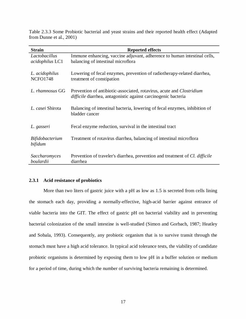
Figure 2.3.3 Electron photomicrographs of the effect of L. salivarius subsp. salivarius
CRL 1328 bacteriocin-like substance on E. faecalis. The Enterococcus
control cell (A), vesiculization of protoplasm (B), vesiculization of
protoplasm and a damaged cell wall (C), pore formation in the cell wall
(D), a disintegrated cell with loss of the protoplasmic material through a
cell wall pore (E), and a disintegrated cell (F) (Ocana et al., 1999) ......................30
Figure 2.3.4 Mode of action of organic acids (RCOOH) on, A) non pH-sensitive
bacteria (Lactic acid bacteria, Bifidobacteria), B) pH-sensitive bacteria
(Coliforms, Clostridia, Salmonella, Listeria spp.) (Adapted from
Gauthier, 2002) ...................................................................................................32
Figure 2.6.1 Flow diagram of encapsulation of bacteria by the extrusion and emulsion
techniques (Reproduced from Krasaekoopt et al., 2003). .....................................44
Figure 3.3.1 Survival of L. acidophilus (A), B. adolescentis (B), B. catenulatum (C),
and B. infantis (D) in SGJ pH 2.0 (●) and pH 6.0 (○). The data are
expressed as mean ± one standard deviation (n=3)...............................................57
Figure 3.3.2 Survival of L. acidophilus in SGJ pH 4.0 (●) and pH 5.0 (○). The data are
expressed as the mean ± one standard deviation (n=3). ........................................59

xi
Figure 3.3.3 Survival of L. acidophilus (A), B. adolescentis (B), B. catenulatum (C),
and B. infantis (D) in media containing 0.3% (w/v) Oxgall bile (○) and
without added bile (●). The data are expressed as the mean ± one standard
deviation (n=3)....................................................................................................60
Figure 3.3.4 Growth of probiotics in A) MRS and B) 0.3% (w/v) Oxgall bile
containing MRS media. The data are expressed as the mean ± one
standard deviation (n=3)......................................................................................61
Figure 3.3.5 Light micrographs (magnification 400X NA 0.22) of crystals formed by
test probiotic strains cultivated in MRS-broth media supplemented with
0.3% (w/v) Oxgall bile for 24 h incubation at 37ºC under anaerobic
conditions. a) L. acidophilus, b) B. adolescentis, c) B. catenulatum and d)
B. infantis............................................................................................................61
Figure 3.3.6 Adhesion of probiotic bacteria to Caco-2 cells. Data are expressed as the
mean ± one standard deviation (n=3). ..................................................................62
Figure 3.3.7 Maximum optical densities (O.D. 600) attained by probiotics during
growth on different carbon sources. Data are expressed as the mean ± one
standard deviation (n=2)......................................................................................66
Figure 3.3.8 Light micrograph of pea protein-alginate beads prepared by extrusion. ................67
Figure 3.3.9 Survival of free (solid symbols) and encapsulated (open symbols) L.
acidophilus in SGJ of pH 2.0 (●, ○) and pH 6.0 (▼, Δ). The data are
expressed as the mean ± one standard deviation (n=3). ........................................67
Figure 4.3.1 Antimicrobial activities of probiotics against E. coli O157. Agar spot test
showing zone of inhibition around probiotic spot. Spots represent: A) B.
infantis; B) B. adolescentis; C) L. acidophilus; and D) B. catenulatum................83
Figure 4.3.2 Agar-spot test showing antibacterial activity of L. acidophilus on MRS
agar medium overlaid with Luria-Bertani (LB) soft agar seeded with E.
coli O157:H7. A) MRS (control), and B) MRS containing 0.2% (w/v)
sodium-bicarbonate .............................................................................................85
Figure 4.3.3 Visual determination of co-aggregation of L. acidophilus ATCC 11975
with E. coli O157:H7. Tubes 1 and 2 represents co-aggregation before (0
h), and after (24 h) incubation, respectively .........................................................88

xii
Figure 4.3.4 Dark-field (10X NA 0.22) photomicrographs, showing co-aggregates of
L. acidophilus and E. coli O157. A) Before incubation, and B) Co-
aggregates after incubation at 37ºC for 24 h. .......................................................89
Figure 4.3.5 Antimicrobial activity of L. acidophilus supernatant on the growth of A)
E. coli and, B) Cl. sordelli ..................................................................................90

xiii
LIST OF TABLES PAGE #
Table 2.2.1 LAB typically associated with the human host (Goktepe et al., 2006) ...................11
Table 2.3.1 Microorganisms considered as probiotics (Adapted from Holzapfel et al.,
2001)....................................................................................................................13
Table 2.3.2 Key to differentiating lactic acid bacteria and a comparison with current
taxonomic classification (Adapted from Holzapfel et al., 2001) ............................14
Table 2.3.3 Some Probiotic bacterial and yeast strains and their reported health effect
(Adapted from Dunne et al., 2001) .......................................................................17
Table 2.3.4 Lactobacilli with an adhesion index of at least one bacterium per Caco-2
cell (Adapted from Morelli, 2000) ........................................................................25
Table 2.3.5 Classification of LAB bacteriocins (Adapted from Drider et al., 2006)..................29
Table 2.4.1 Various special therapeutic or prophylactic properties of specific
probiotics (Adapted from Parvez et al., 2006).......................................................35
Table 2.5.1 Examples of ingredients that are commonly regarded as human prebiotics
(Wang, 2009) .......................................................................................................38
Table 2.5.2 Microbiological changes reported in human feeding studies with prebiotics
(Adapted from Macfarlane et al., 2008) ................................................................40
Table 3.3.1 D-values (min) of free and encapsulated probiotics exposed to SGJ (pH
2.0). Data are expressed as the mean ± one standard deviation (n=3) ....................58
Table 3.3.2 Antibiotic resistance profiles of the tested probiotic strains. ..................................63
Table 3.3.3 Optical densities at 590 nm attained after 48 h by probiotics during growth
on different sole carbon sources. The data are expressed as the mean ± one
standard deviation (n=3) .......................................................................................65
Table 4.3.1 Inhibition of selected pathogenic bacteria by probiotics as determined by
agar spot test. .......................................................................................................84

xiv
Table 4.3.2 Antibacterial activities of L. acidophilus against selected pathogenic
bacteria.................................................................................................................84
Table 4.3.3 Percent adhesion to hydrocarbons of enteric pathogens and probiotic
strains...................................................................................................................86
Table 4.3.4 Adhesion to hydrocarbons as measured using the BATH test and % auto-
aggregation of probiotic and pathogenic strains ....................................................87
Table 4.3.5 Co-aggregation percentages of pathogen and probiotic strains after
incubation at 37ºC, anaerobically..........................................................................88

1
1 General introduction
Probiotics are microorganisms introduced orally in the gastrointestinal tract (GIT) that
are able to contribute positively to the activity of intestinal microflora and therefore, to the health
of its host. Most probiotic bacteria belong to the group of lactic acid bacteria (LAB) and among
them lactobacilli and bifidobacteria reportedly play a significant role in maintaining the intestinal
ecosystem and in stimulating the immune system of the host (Saarela et al., 2002). Many in vitro
properties, such as adhesion, resistance to pH, etc., are usually investigated to determine if a
specific selected strain would be suitable as a probiotic (Collins et al., 1998).
Co-aggregation is a process by which genetically-distinct bacteria adhere to one another
via specific molecules. Cumulative evidence suggests that such adhesion influences the
development of complex multispecies biofilms (Ricard et al., 2003). Bacterial aggregation
between microorganisms of the same strain (auto-aggregation) or between genetically divergent
strains (co-aggregation) is of considerable importance in several ecological niches, especially
inH the human gut, where probiotics are to be active (Collado et al., 2007a). A relationship
between auto-aggregation and adhesion ability has been reported for some bifidobacterial spp.
(Del Re et al., 2000; Collado et al., 2007b). A correlation between adhesion ability and
hydrophobicity, as measured by microbial adhesion to hydrocarbons, has also been observed in
some lactobacilli (Del Re et al., 2000). Furthermore, it has been suggested (Collado et al., 2007b)
that inhibitor- or bacteriocin-producing LAB, which co-aggregates with pathogens, may
constitute an important host defence mechanism against infection. Co-aggregation with potential
gut pathogens could therefore contribute to the probiotic properties ascribed to specific LAB.
The determination of antimicrobial susceptibility of a bacterial strain is an important
prerequisite for its approval as a probiotic (Moubareck et al., 2005). The resistance gene

2
reservoir hypothesis (Ammor et al., 2007) suggests that beneficial and commensal bacterial
populations play a role in the transfer of antibiotic resistance to pathogenic and opportunistic
bacteria. At present, there is great concern that commensal bacterial populations from food and
the GIT of humans and animals, such as LAB and bifidobacteria, could act as a reservoir for
antibiotic resistance genes. Resistance factors could ultimately be transferred to human
pathogenic and opportunistic bacteria, thereby hampering the treatment of infections and general
spread of antimicrobial resistance. LAB spp. have traditionally been used as starter cultures in
the production of fermented feed and foodstuffs. Further, LAB and bifidobacteria are normal
inhabitants of the GIT where they are known to exert health-promoting effects, and these
selected strains are currently been used as probiotics. Antibiotic resistance genes carried by LAB
and bifidobacteria could be transferred to human pathogenic bacteria either during food
manufacture or during passage through the GIT (Ammor et al., 2007).
The overall goal of this research is to screen four probiotic bacteria, including
Lactobacillus acidophilus ATCC 11975, Bifidobacterium infantis ATCC 15697, B. catenulatum
ATCC 27675 and B. adolescentis ATCC 15703, for characteristics that make them effective
candidates for encapsulation.
1.1 Hypotheses
The following hypothesis will be tested during this research:
a) Biochemical characterization of probiotic bacteria will identify candidates suitable for
evaluation of probiotic encapsulation technology.
b) Probiotic encapsulation using pea protein-alginate beads will enhance probiotic survival
in simulated gastric juice.
3
1.2 Technical objectives
The following objective will be investigated:
a) To examine a panel of probiotic bacteria for their ability to survive and or grow in low
pH environments, as well as in the presence of bile salts;
b) To quantify the adherence of probiotic bacteria to intestinal epithelial surfaces, as well as
their ability to co-aggregate with other enteric flora;
c) To determine the antimicrobial potential of probiotic bacteria against a panel of selected
enteric pathogens;
d) To characterize probiotic bacteria using BIOLOG sole carbon source utilization, and
antimicrobial profiling, to assist in strain-specific discrimination of the probiotic
organism from other bacteria that colonize the gut environment; and
e) To utilize protein-alginate capsules to determine the effect of encapsulation of probiotic
bacteria on survival in a model acidic (pH 2.0) gastric system.
2 Literature review
Foods are no longer considered by consumers only in terms of taste and immediate
nutritional needs, but also in terms of their ability to provide specific health benefits beyond their
basic nutritional value. Currently, the largest segment of the functional food market is provided
by the foods targeted towards improving the balance and activity of the intestinal microflora
(Saarela et al., 2002). Consumption of foods containing live bacteria is the oldest and still most
widely used way to increase the numbers of advantageous bacteria in the intestinal tract. Such
bacteria are called ‘Probiotics’ and have been predominantly selected from the genera
Lactobacillus and Bifidobacterium, both of which have been extensively studied and established
as valuable native inhabitants of the GIT (Fuller, 1989; Salminen et al., 1998, Capela et al.,

4
2006). Various microorganisms, particularly species of Lactobacillus and Streptococcus, have
traditionally been used in fermented dairy products to promote human health as well as food
functionality and flavor.
2.1 Historical perspective of probiotics
Escherich described the microbiota of the infant GIT and suggested benefits of their
colonization in digestion. Around the same time, Doderlein postulated the beneficial association
of vaginal bacteria in inhibiting the growth of pathogenic bacteria by producing lactic acid
(Goktepe et al., 2006). Studies by Moro in 1900 and by Beijerinck in 1901 reported the
beneficial association of LAB with human host (Goktepe et al., 2006). The longevity of
Caucasians was related to the high intake of fermented milk products (Metschnikoff, 1907), as
elucidated in his bestselling book The Prolongation of Life. LAB belong to a group of Gram-
positive, non-sporulating, non-respiring cocci or rods, which produce lactic acid as a major
metabolic end product during the fermentation of carbohydrates (Salminen et al., 1998).
Although phylogenetically different, bifidobacteria are another group of lactic acid producing
bacteria which are commonly accepted as LAB. Bifidobacteria were found to be typically
associated with the feces of breast-fed infants and a lower incidence of intestinal upset was
observed for breast-fed infants, when compared with formula-fed infants (Goktepe et al., 2006).
2.1.1 Definition of probiotics
The word probiotic is derived from the Greek meaning “for life”. Probiotics were first
defined by Kollath in 1953 to denote all organic and inorganic food complexes in contrast to
harmful antibiotics. Lilly and Stillwell (1965) defined probiotics as “microorganisms promoting
the growth of other microorganisms”. Although numerous definitions have been proposed since

5
then, most have failed to be completely satisfactory because they lack statements such as
“stabilization of the gut flora” (Goktepe et al., 2006). Havenaar and Veld (1992) have defined
probiotics as “mono- or mixed cultures of live microorganisms which, when applied to animal or
human, beneficially affect the host by improving the properties of the indigenous microflora”.
When these probiotic bacteria are present in yogurt and other fermented foods, they may
beneficially alter the normal gut flora (Metchnikoff, 1907). Probiotics have also been defined by
the European Union (EU) Expert Group on Functional Foods in Europe (FUFOSE) to be “viable
preparations in foods or dietary supplements to improve the health of humans and animals”
(FUFOSE working group, 1999). More recently, probiotics have been referred to as “live
microorganisms which when administered in adequate amounts confer a health benefit on the
host” (FAO/WHO, 2001).
2.2 The gastrointestinal ecosystem
The GIT of the human body is a complex ecosystem with a diverse and concentrated
microbial population that mediates numerous interactions with the chemical environment, such
as digestion, adhesion and colonization in the GIT. The mucosal surface area increases by:
circular folding which contributes to about a 3-fold increase, through the production of villi, for a
7- to 10-fold increase, and by the formation of intestinal microvilli, which results in a 15- to 40-
fold increase (Holzapfel et al., 1998). Varying numbers of bacteria are found throughout the GIT,
ranging from 101-103 cfu/mL or g in the stomach contents; 107 cfu/mL in the jejunum, up to 109
cfu/g in the terminal ileum and approximately 5 × 10¹¹ cfu/g in the distal colon contents
(Goktepe et al., 2006).
The bacteria detected in feces reflect the bacteria present in the distal colon, thus studies
of the human GIT microflora usually involve analysis of fecal samples (Moore et al., 1978).

6
Traditional culture methods have been used to analyze and characterise microbial communities
in a natural ecosystem to obtain a complete diversity picture. In contrast to the microaerophillic
lactobacilli, the study of anaerobic bifidobacteria and eubacteria was made possible by the
development of anaerobic techniques in the early 1970s (Goktepe et al., 2006). However,
cultivation techniques have major limitations, as many microbes in different ecosystems cannot
be cultivated by standard culture methods (Ward et al., 1990). Classical culture-independent
techniques include direct microscopic analysis and monitoring specific enzymes or metabolites,
and have provided valuable insight into the real numbers of microflora in faecal samples.
Microscopic analysis of faecal samples by Langendijk et al. (1995) revealed approximately 1011
to 1012 organisms per g of wet feces. It is noteworthy that these techniques are very limited in
their ability to give any in-depth characterisation of specific organisms present or community
diversity. Fluorescence microscopy, confocal laser scanning microscopy and flow cytometry
have been used to detect viable populations through the use of fluorescent probes (Lipski et al.,
2001). If epifluorescence microscopy and/or confocal laser scanning microscopy are applied, the
method is usually referred to as fluorescence in situ hybridization (FISH). FISH has been used to
study the composition of GIT microbial system (Tannock et al., 2000). Various studies reported
that the microscopic technique itself is not perfect and may significantly under-report the true
numbers (Ward et al., 1990).
Various short chain fatty acids (SCFA), such as acetate, propionate and butyrate, are end
products of anaerobic bacterial fermentation. Thus, measurement of these acids in feces can be
correlated with specific bacterial metabolism in the intestine (Rowland, 1989). For example,
Lactobacillus casei GG fed to children with an intestinal infection significantly increased the
total SCFA concentration (Siigur et al., 1996). Increases or decreases in specific enzymes for

7
example, β-glucuronidase and β-galactosidase, in feces can also point to the metabolic activities
of certain groups of bacteria. Reduction in β-glucuronidase levels was reported in humans during
ingestion of L. casei GG (Ling et al. 1994). Also, a significant correlation has been observed
between the levels of faecal β-galactosidase and numbers of bifidobacteria (Favier et al., 1997).
While many faecal enzymes, such as azoreductase and nitroreductase are mainly produced by the
species Bacteroides, Eubacterium and Clostridium, more studies are needed to accurately
correlate specific faecal enzymes with specific groups of bacteria (Rowland, 1989).
Cell viability can also be inferred from enzymatic activities such as esterase conversion
of carboxyfluorescein diacetate (cFDA). The reduction of tetrazolium salts, or dyes such as
propidium iodide, TOTO-1, SYTO 9, carboxyfluorescein and oxonol, have been used as viability
indicators (Goktepe et al., 2006). Bunthof et al. (2001) have combined culture plating technique
with dyes, and reported that cFDA labels the culturable subpopulation; whereas, TOTO-1 lables
the non-culturable population. Determination of percentages of guanine+cytosine (G+C) content
is one of the few methods depicting the total bacterial community of the GIT without any
previous knowledge of component bacteria or their DNA sequences (Apajalahti et al., 1998).
This approach has been applied by Apajalahti et al. (2003) to determine the total bifidobacteria
community in human feces.
DNA-based methods for the detection of probiotic microorganisms are mainly based on
restriction enzyme analysis or PCR (Polymerase Chain Reaction), or both. Various methods
including amplified fragment length polymorphism (AFLP), pulsed field gel electrophoresis
(PFGE), Random amplification of polymorphic DNA (RAPD) including multiplex PCR,
arbitrary primed PCR (AP-PCR) and tiplicate arbitrary-primed PCR (TAP-PCR) has been used
for identification and tracking of individual probiotic strains (Gardiner et al., 2002). AFLP is a

8
combination of PCR and restriction enzyme analysis, where genomic DNA is digested with two
different types of restriction endonucleases. Strain specific identification using AFLP method has
been used to differentiate Lactobacillus plantarum, Lactobacillus pentosus and Lactobacillus
paraplantarum (Torriani et al., 2001). PFGE has been used for typing Lactobacillus casei,
bifidobacteria and Lactobacillus rhamnosus (Goktepe et al., 2006).
RAPD is a PCR-based method in which a pattern of amplicons is produced through the
simultaneous amplification of many chromosomal sequences mediated by annealing of short
oligonucleotide primers. RAPD PCR analysis has been reported to be capable of differentiating
between L. acidophilus group strains (Pleiss et al., 1995). This technique has also been found to
be useful for monitoring introduced and indigenous lactobacilli in the intestinal tract (Gardiner et
al., 2002). RAPD PCR analysis of yeast isolates from feta cheese provided reliable identification
at species level and good discrimination at the strain level (Psomas et al., 2001). In AP-PCR,
reactions are performed by using specific primer targeting a highly conserved region within the
16S rRNA gene. TAP-PCR is a variation of AP-PCR, where three different annealing
temperatures are used in triplicate reactions, and has been used by Cusick and O’Sullivan (2000)
to type isolates from major genera of LAB and bifidobacteria. The primers used in the
amplification reaction in Rep-PCR and ERIC-PCR techniques targets the species specific Rep
(Repetitive extragenic palindromic) elements and ERIC (enterobacterial repetitive intergenic
consensus) sequences, which are conserved regions dispersed on the genomic DNA of
microorganisms. The profiles obtained due to the amplification of inter-Rep and inter-ERIC
distances are species and sometimes strain specific (Goktepe et al., 2006).
Other culture-independent techniques which can mediate the identification of individual
bacterial species or strains include denaturing gradient gel electrophoresis (DGGE), which has

9
been used to detect lactobacillus and other species in the human GIT (Walter et al., 2001).
Accurate typing of unknown isolates is now achieved through sequence analysis of 16S
ribosomal RNA (rRNA) or the corresponding rDNA amplicons following PCR. Matsuki et al.
(2002) has investigated the microbial population of six healthy human volunteers by applying
16S rRNA-gene-targeted group-specific-oligonucleotide primers for the Bacteroides fragilis
group, Bifidobacterium, the Clostridium coccoides group, and Prevotella, and identified 74% of
the predominant bacteria in the feces. Another method called amplified ribosomal DNA
restriction analysis (ARDRA) has been developed on the basis of 16s rRNA gene amplification
followed by restriction analysis, and was used by Ventura et al. (2000) to identify different
species of Lactobacillus isolated from human feces and vagina.
Andersson et al. (2008) has developed a method based on 454-pyrosequencing for
monitoring of microbial communities in throat, stomach and fecal samples. Pyrosequencing is a
method of DNA sequencing based on the sequencing by synthesis, which involves taking a
single strand of the DNA to be sequenced and then synthesizing its complementary strand
enzymatically. A highly variable region of the 16S rRNA gene is amplified using primers that
target adjacent conserved regions, followed by direct sequencing of individual PCR products.
The cpn60 gene (encoding the universally conserved 60 kDa chaperonin), has been established
as a useful target for molecular phylogenetics, characterization of complex microbial
communities and to differentiate between closely related bacterial isolates by hybridization or
sequence analysis (Dumonceaux et al. 2006). An approximately 555-bp segment of the gene
corresponding to nucleotides 274–828 of the Escherichia coli cpn60 sequence (the cpn60
universal target, or cpn60 UT) can be amplified from virtually any genome using universal,
degenerate PCR primers. The cpn60 based identification of fecal microflora of cats has revealed

10
diverse populations dominated by Actinobacteria (particularly bifidobacteria) and Firmicutes
(particularly lactobacilli) (Desai et al. 2009).
2.2.1 Gastrointestinal strains of human origin
In spite of increased research on gut microbial ecology, only a small number of
approximately 400 species of different genera have been cultivated and studied with regard to
their physiology, metabolic interactions, and taxonomy (Goktepe et al., 2006). Table 2.2.1
presents the LAB found likely to be associated with the human host (Goktepe et al., 2006). The
large intestine is densely populated by Bacteriodes and the Gram-positive, anaerobic genera
Eubacterium and Bifidobacterium. Lactobacilli are the predominant species in the vagina and are
also normally present in the oral cavity (103-104 cfu/g), the ileum (103-107 cfu/g), and colon
(104-108 cfu/g), where they play an important role in maintenance of a stable gut mucosa
(Lidbeck et al., 1993). Long and Swenson (1977) showed that bifidobacteria and lactobacilli are
the dominant bacterial species found to be present in the feces of breast-fed infants. There is a
lack of research evidence that has demonstrated a single dominant species in the human GIT.
However, L. acidophilus (commonly referred as simply “acidophilus”) has been recovered in
relatively high numbers from the GIT (Molin et al., 1993). Strains of acidophilus have been
isolated from the intestinal tract of humans as well as animals such as rodents and birds.
LABs are gram-positive, non-spore forming, catalase-negative organisms that are devoid
of cytochromes and anaerobic but aerotolerant. They are fastidious, acid-tolerant, and strictly
fermentative (either homo- or hetero); lactic acid is the major end product of sugar fermentation
(Axelsson, 1998). However, some species can form catalase or cytochromes on media containing
hematin or related compounds and some lactobacilli can also produce non-heme catalase, called
pseudocatalase, which cause confusion for LAB identification (Holzapfel et al., 2001).

11
Table 2.2.1 LAB typically associated with the human host (Goktepe et al., 2006)
Lactobacilli Other LAB Intestinal Bacteria
Lactobacillus acidophilus group Bifidobacterium adolescentis
L. acidophilus senso strictu B. angulatum L. animalis B. bifidum L. brevis B. breve L. buchneri B. cantenulatum L.crispatus B. dentium L. curvatus B. infantis L. delrueckii B. longum L. fermentum B. pseudocantenulatum L. gasseri Enterococcus fecalis L. johnsonii E. faecium L. paracasei Leuc. Mesenteroides L. plantarum Pedicoccus pentosaceus L. reuteri Weissella confusa L. rhamnosus L. ruminis L. sakei Vaginal Bacteria Lactobacillus acidophilus Bifidobacterium bifidum L. fermentum B. longum L. casei B. infantis L. rhamnosus B. breve L. cellobiosus B. catenulatum L. plantarum B. dentium L. brevis L. delbrueckii L. salivarious L. jensenii L. vaginalis L. gasseri L. crispatus
2.3 Probiotic bacteria
Strains of LAB, such as Lactobacillus, Bifidobacterium, Eubacterium and Streptococcus,
have traditionally been used in the manufacture of fermented dairy products and are generally
regarded as safe (GRAS) (O’Sullivan et al., 1992). In addition, these bacteria are desirable
members of the intestinal microflora (Berg, 1998). Table 2.3.1 shows a list of microorganisms

12
including both LAB and non-lactics which are generally considered as probiotics. Lack of
pathogenicity, tolerance to gastrointestinal conditions (acid and bile), ability to adhere to the
gastrointestinal mucosa and competitive exclusion of pathogens (Collins et al., 1998; Ouwehand
et al., 2002) are some of the general criteria that have been used for the selection of probiotics.
L. casei strain “Shirota” has been reported to have the longest history of safe use as a probiotic in
food with proven health benefits (Goktepe et al., 2006). Lactobacillus acidophilus and
Bifidobacterium bifidum were used to make mildly acidified yogurts called “bio-yogurts” in
Germany during late 1960s (Goktepe et al., 2006). Viable probiotic strains with beneficial
functional properties are supplied in the market as fermented food products, mainly “yogurt”-
type, or in lyophilized form, both as food supplements and as pharmaceutical preparations. For
many years, pharmaceutical preparations containing live microorganisms in capsules, also
known as “biotherapeutics”, were used for the restoration of the GIT population, e.g., after or
during antibiotic treatment (Goktepe et al., 2006).
The prevalence of lactobacillus and bifidobacterial spp. in the intestinal tract of humans is
not known accurately. Lactobacillus crispatus, L. gasseri, L. salivarius, and L. reuteri have been
reported as the major species of the Lactobacillus microflora (Mitsuoka et al., 1990). Whereas,
Lactobacillus johnsonii, Lactobacillus ruminis, Lactobacillus casei, and Lactobacillus brevis
have been detected occasionally. Bifidobacterium longum has been found predomonently in adult
human GIT, while Bifidobacterium bifidum was detected occasionally. In contrast,
Bifidobacterium infantis and Bifidobacterium breve were detected predominantly in infant feces,
while B. longum and B. bifidum detected occasionally (Biavati et al., 1984).

13
Table 2.3.1 Microorganisms considered as probiotics (Adapted from Holzapfel et al., 2001) Lactobacillus spp. Bifidobacterium spp. Other LAB "Non-lactics" a
L. acidophilus B. adolescentis Enterococcus faecalis a Bacillus cereus L. amylovorus B. animalis Ent. faecium ("toyoi") a,c L. casei B. bifidum Sporolactobacillus Escherichia colia L. crispatus B. breve inulinusc Propionibacterium L. delbrueckii subsp. B. infantis freudenreichii a,c bulgaricus a B. lactis b Saccharomyces L. gallinarum c B. longum cerevisiae L. gasseri ("boulardii") a L. johnsonii L. paracasei L. plantarum L. reuteri L. rhamnosus a Mainly in pharmaceutical preparations; b Synonym of B. animalis; c Mainly for animals.
On the basis of their morphologic and phenotypic features, the LAB are subdivided
(Table 2.3.2) into the genera Betabacterium, Thermobacterium, Streptobacterium, Streptococcus,
Betacoccus, Tetracoccus, and Microbacterium (Holzapfel et al., 2001). Enterococcus,
Lactococcus, and Vagococcus have been separated from the original genus, Streptococcus
(Holzapfel et al., 2001). The genus Streptococcus represents mainly pathogenic bacteria, except
Streptococcus thermophilus; whereas, some strains of Enterococcus spp. may be involved in
opportunistic infections, and some are considered to play role in food fermentations, and are also
found as commensals in the GIT. Lactococcus spp. are generally considered to be non-
pathogenic and safe.
Probiotic Bifidobacterium spp. are generally strict anaerobes. Fermentation of the sugars
and sugar alcohols like L-arabinose, D-xylose, D-mannose, salicin, D-mannitol, D-sorbitol, and
D-melezitose serve as key characteristics to identify the most important species of bifidobacteria
(Klein et al., 1998).

14
Table 2.3.2 Key to differentiating lactic acid bacteria and a comparison with current taxonomic classification (Adapted from Holzapfel et al., 2001)
Genus Shape Catalase Nitrite reduction Fermentation Current genera Betabacterium Rod − − Hetero- Lactobacillus
Weissella Thermobacterium Rod − − Homo- Lactobacillus Streptobacterium Rod − − Homo- and Hetero Lactobacillus
Carnobacterium Streptococcus Coccus − − Homo- Streptococcus
Enterococcus Lactococcus Vagococcus
Betacoccus Coccus − − Hetero- Leuconostoc Oenococcus Weissella Brochothrix
Microbacterium Rod + + Homo- Pediococcus Tetracoccus Coccus + + Homo- Tetragenoccus
Analysis of the cell wall peptidoglycan composition was also found suitable for the
identification of some species like Bifidobacterium longum, Bifidobacterium infantis, and
Bifidobacterium suis (Bonaparte, 1997). Practically all organisms used in probiotic foods or food
supplements are representatives of the genera Lactobacillus, Enterococcus, or Bifidobacterium.
The genus Bifidobacterium shares some phenotypic features with typical LAB and is considered
to form part of the LAB. Phylogenetically-distinct, bifidobacteria exhibit a relatively high G + C
content of 55–67 mol% in the DNA and form part of the Actinomycetes branch. The “true” LAB
form part of Clostridium branch, which is characterized by a G + C content of <55 mol% in the
DNA. Genes encoding rRNA, comprising conserved and variable domains, are typically chosen
for phylogenetic work as they are present in all microorganisms. Analysis of the 16s rRNA gene
is considered to be the most powerful and accurate technique for determining the degree of
phylogenetic relation of microorganisms.

15
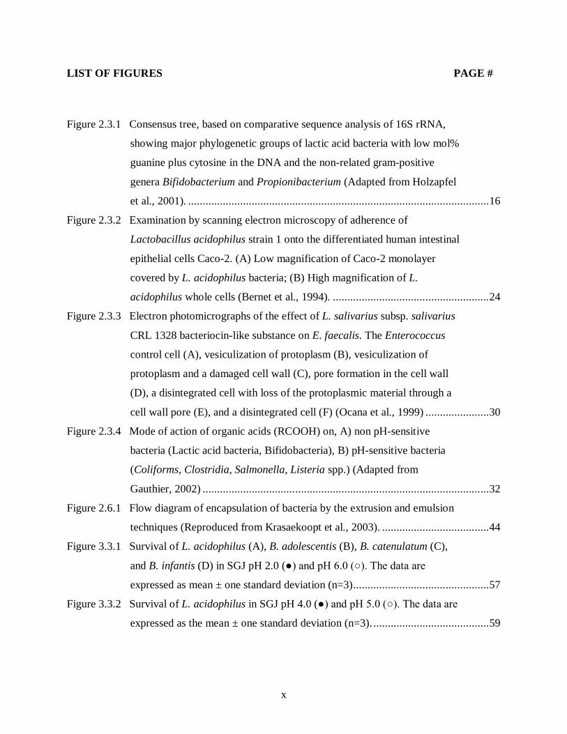
The phylogenetic relation of the different genera of “true” LAB is shown in Figure 2.3.1
and is based on the comparison of 16S rRNA sequences. Carnobacterium, Enterococcus,
Vagococcus, Aerococcus, Tetragenococcus, and Lactosphaera are more closely related to each
other than to any other LAB. Lactobacilli are phylogenetically diverse, whereas Lactococcus and
Streptococcus are closely related. Lactobacillus and Pediococcus are mixed, phylogenetically, as
shown by the 16S rRNA sequencing data with 5 species of a Pediococcus clustering with 32
homo- and hetero-fermentative Lactobacillus spp. in the so-called Casei and Pediococcus group
(Collins et al., 1991). In 16S rRNA sequence data of pediococci and lactobacilli, the taxa
generated do not correspond with the phylogenetic branching. Therefore, certain species of LAB
may have to be reclassified.
Performing a preliminary in vitro assessment is a prerequisite to assess the properties of
probiotic bacterial strains (FAO/WHO, 2002). Various papers (Dunne et al., 2001; Morelli,
2007) have suggested that a probiotic bacterial strain should be assessed according to the
following (or a very similar) criteria: human origin, nonpathogenic behavior, resistance to
technologic processes (i.e., viability and activity in delivery vehicles), resistance to gastric
acidity and bile toxicity, adhesion to gut epithelial tissue, ability to persist within the
gastrointestinal tract, production of antimicrobial substances, ability to modulate immune
responses, and ability to influence metabolic activities (e.g., cholesterol assimilation, lactase
activity, and vitamin production). It has also been suggested that the demonstration of probiotic
activity of a certain strain involve well-designed, double-blind, placebo-controlled human studies
(Dunne et al., 2001).

16
Figure 2.3.1 Consensus tree, based on comparative sequence analysis of 16S rRNA, showing major phylogenetic groups of lactic acid bacteria with low mol% guanine plus cytosine in the DNA and the non-related gram-positive genera Bifidobacterium and Propionibacterium (Adapted from Holzapfel et al., 2001)
Health benefits associated with the ingestion of probiotic bacteria includes: reduction in
colon irritation, constipation, traveler’s diarrhea, inhibition of the adhesion of pathogenic genera
including Escherichia, Clostridium, Salmonella and Campylobacter to the intestinal lumen,
synthesis of B vitamins, lowering of blood ammonia levels, cholesterol absorption and inhibition
of tumor formation (Ziemer and Gibson, 1998). Some reported health benefits of probiotic
bacteria are listed in Table 2.3.3.
17
Table 2.3.3 Some Probiotic bacterial and yeast strains and their reported health effect (Adapted from Dunne et al., 2001) Strain Reported effects Lactobacillus acidophilus LC1
Immune enhancing, vaccine adjuvant, adherence to human intestinal cells, balancing of intestinal microflora
L. acidophilus NCFO1748
Lowering of fecal enzymes, prevention of radiotherapy-related diarrhea, treatment of constipation
L. rhamnosus GG Prevention of antibiotic-associated, rotavirus, acute and Clostridium
difficile diarrhea, antagonistic against carcinogenic bacteria L. casei Shirota Balancing of intestinal bacteria, lowering of fecal enzymes, inhibition of
bladder cancer L. gasseri Fecal enzyme reduction, survival in the intestinal tract Bifidobacterium bifidum
Treatment of rotavirus diarrhea, balancing of intestinal microflora
Saccharomyces boulardii
Prevention of traveler's diarrhea, prevention and treatment of Cl. difficile diarrhea
2.3.1 Acid resistance of probiotics
More than two liters of gastric juice with a pH as low as 1.5 is secreted from cells lining
the stomach each day, providing a normally-effective, high-acid barrier against entrance of
viable bacteria into the GIT. The effect of gastric pH on bacterial viability and in preventing
bacterial colonization of the small intestine is well-studied (Simon and Gorbach, 1987; Heatley
and Sobala, 1993). Consequently, any probiotic organism that is to survive transit through the
stomach must have a high acid tolerance. In typical acid tolerance tests, the viability of candidate
probiotic organisms is determined by exposing them to low pH in a buffer solution or medium
for a period of time, during which the number of surviving bacteria remaining is determined.

18
Minekus et al. (1995) has developed a dynamic computer-controlled model, which
reflects the in vivo conditions of the stomach and small intestine. This model permits an accurate
simulation of the factors influencing the survival of probiotic microorganisms, such as pH, bile
concentration and transit through the different parts of the GIT. Studies by Dunne et al. (2001)
suggested that lactobacilli isolated from human ileal samples could successfully transit the
human stomach conditions and function effectively. In these studies, bifidobacteria were found
to be less resistant to stomach conditions like low pH etc. than lactobacilli. Studies by Conway et
al. (1987) showed that yogurt-producing species of lactobacilli were more sensitive to gastric
juice while enteric species were more resistant. The best-performing among the two
L. acidophilus strains (strain ADH) used in the study were reclassified as Lactobacillus gasseri,
which is a homofermentative lactobacilli (Morelli, 2000). Lactobacillus rhamnosus GG was
unable to survive at pH 1.0, but remained viable at pH 3.0 and higher (Goldin et al., 1992). In
vitro studies (Hood and Zottola, 1988; Charteris et al., 1998a) showed that enteric lactobacilli
had a lower pH tolerance limit of 2.0 for several min. Eight meat starter cultures including
Lactobacillus and Pediococcus strains were exposed to low pH (pH 1.0 to 5.0) conditions of
stomach for 1 h. The number of surviving bacteria was decreased from the inoculated level of
7.4-7.6 log cfu/mL to < 4 log cfu/mL at pH 1.0 and pH 2.0, whereas pH 4.0 and 5.0 did not affect
the viability (Erkkila and Petaja, 2000). Only 51 out of 312 pre-selected LAB strains, including
Lactobacillus, Pediococcus, Enterococcus, isolated from Iberian dry fermented sausages, human
and pig feces were able to survive after 1.5 h of exposure at pH 2.5, where the number of final
surviving bacteria ranges between 5.4 to 8.9 log cfu/g (Ruiz-Moyano et al., 2008). The bile
resistant isolates of Bifidobacterium strains displayed considerably higher survival at 90 min of
exposure at pH 2.0, with a concentration of final surviving bacteria ~ 6.5 log cfu/mL, than their

19
corresponding strains of origin (Noriega et al., 2004). Survival of Bifidobacterium animalis
strains BLC-1, Bb-12, and Bo, Lactobacillus acidophilus strains LAC-1 and Ki, Lactobacillus
paracasei subsp. paracasei strain LCS-1 and Lactobacillus brevis strain LMG 6906 inoculated
into whey cheese was assessed by Madureira et al. (2005). Except L. paracasei subsp. paracasei
LCS-1 and B. animalis Bb-12, all bacteria were resistant to the action of artificial gastric juice
(pH 2.5–3.0) and maintained their initial viable cell numbers (~ 8 log cfu/mL) after both 60 and
120 min of exposure.
In order to evaluate the survival of lactobacilli in the low pH conditions of the human
stomach, five Lactobacillus strains were compared in simulated gastric juice (SGJ, pH 2.0) for
90 min (Corcoran et al., 2005). Lactobacillus rhamnosus GG had the highest survival rate and
maintained their initial viable cell numbers (~ 9 log cfu/mL), while the poorest survivor was
L. paracasei NFBC 338, whose concentration declined to undetectable levels after only 30 min
of exposure. These studies also showed that glucose (19.4 mM) was responsible for the enhanced
survival of L. rhamnosus GG in simulated gastric juice. The level of surviving bacteria was
reduced by approximately 5.6 log cfu/mL upon removal of glucose (Corcoran et al., 2005).
Furthermore, five Lactobacillus strains were examined for their acid tolerance (pH 2.5).
Lactobacillus acidophilus NCFM was found to be the least acid tolerant, with a final surviving
population of 6.0 × 10⁴ cfu/mL, whereas the population of L. acidophilus 30SC and ATCC
43121 remained relatively constant (~ 6 log cfu/mL) (Oh et al., 2000).
Probiotics have been incorporated into a range of dairy products, including yoghurts,
soft-, semi-hard and hard cheeses, ice cream, milk powders and frozen dairy desserts. However,
there are still several problems with respect to the low viability of probiotic bacteria in GIT and
food environments. Probiotics of intestinal origin are difficult to propagate and high survival is

20
important for both economic reasons and health effects. Consequently, there is a demand for new
technologies such as encapsulation to enhance probiotic viability.
2.3.2 Bile resistance of probiotics
Bile is an aqueous solution made up of bile acids, cholesterol, phospholipids, and the
pigment biliverdin, which gives the bile its yellow-green color. About 500-700 mL/day of bile
acids are synthesized in the liver from cholesterol and are secreted from the gall bladder into the
duodenum, after food intake by an individual (Hofmann and Roda, 1984). Bile plays an essential
role in lipid digestion; it emulsifys and solubilizes lipids and functions as biological detergent.
Prior to secretion into the duodenum, bile acids are conjugated either with glycine
(glycoconjugated) or taurine (tauroconjugated) (Begley et al., 2006). In the colon conjugated bile
undergoes various chemical changes including deconjugation, dehydroxylation,
dehydrogenation, and deglucuronidation, almost solely by microbial activity (Begley et al.,
2006). The antimicrobial nature of bile is mainly because of its detergent property, which
dissolves bacterial membranes. Bile salt hydrolases (BSHs) are generally intracellular, oxygen-
insensitive enzymes that catalyze the hydrolysis of bile salts. Hydrolysis of bile salts is mediated
by various genera of the intestinal microflora, including Clostridium (Gopal et al., 1996),
Bacteroides (Kawamoto et al., 1989), Lactobacillus (Lundeen and Savage, 1990; Christiaens et
al., 1992), Bifidobacterium (Grill et al., 2000a) and Enterococcus (Franz et al., 2001). A number
of BSHs have been identified and characterized in probiotic bacteria, and the ability of probiotic
strains has often been included among the criteria for probiotic strain selection (Begley et al.,
2006). Bile tolerance of probiotic bacteria can be investigated by incubating them for 24 hrs in a
milk-yeast medium containing different concentrations of bile extracts and monitoring cell
viability and pH before and after incubation (Goktepe et al., 2006). This assay was used by

21
several authors to assess the bile resistance of potential or already commercialized probiotic
lactobacilli. All these studies reported a growth delay of lactobacilli in the presence of oxgall that
was strain- and not species-dependent. It has been hypothesized that deconjugation of bile salts is
a detoxification mechanism and BSH enzymes play a role in bile tolerance of probiotic
organisms in the GIT (Savage, 1992). Both conjugated and deconjugated bile acids have been
determined to inhibit the growth of Klebsiella spp., Enterococcus spp. and Escherichia coli
strains in vitro. However, deconjugated forms of bile acids were found to be more inhibitory
against Gram-positive than Gram-negative bacteria (Stewart et al., 1986). Studies by Smet et al.
(1995) suggested that deconjugation of bile acids decreases their solubility and thus diminishes
the detergent’s activity and makes it less toxic to bacteria in the intestine. It was assumed that the
conjugated form of the bile salts exhibits toxicity by causing intracellular acidification through
the same mechanism as organic acid. In contrast, Tannock et al. (1989) stated that deconjugated
bile salts are more inhibitory than conjugated bile salts to anaerobes including lactobacilli.
Similarly, deconjugated bile was reported to be involved in growth inhibition of Bifidobacterium
spp. including B. breve, B. longum, and B. coryneforme, where the viable counts were reduced
by approximately 6, 7 and 2 log cfu/mL respectively, after 2 h incubation in the presence of 1
mM deconjugated bile (Grill et al., 2000b). Another hypothesis states that certain Clostridium
spp. utilize the amino acid taurine as an electron acceptor and have demonstrated improved
growth rates in the presence of taurine and taurine-conjugated bile salts (Moser and Savage,
2001). However, taurine or taurine conjugates did not affect the growth of Lactobacillus spp.
tested (Tannock et al. 1989). Cholic acid was found to accumulate in lactobacillus cells by means
of a transmembrane proton gradient (Kurdi et al., 2000). Whereas studies by Boever et al. (2000)
reported that cholic acid was highly deleterious for the viability of lactobacilli. It has also been

22
suggested that the BSH enzymes are detergent shock proteins (Adamowicz et al., 1991) that
protects the lactobacilli from its toxic effects and may have a competitive advantage over the
non-BSH producing bacteria. However, studies of Moser and Savage (2001) reported that
deconjugation and resistance are unrelated activities. Lastly, studies done by Gopal et al. (1996)
showed no relationship between the ability of 6 strains of L. acidophilus and 8 strains of
Bifidobacterium spp. to grow in bile (0.3% oxgall) and their ability to hydrolyze bile salts
(glycocholic acid or taurocholic acid).
A link between bile salt hydrolysis and bile tolerance has been provided by the studies
conducted on wild-type and bsh mutant pairs of Lactobacillus plantarum, Lactobacillus
amylovorus and Listeria monocytogenes. Results showed that mutant cells were significantly
more sensitive to bile and bile salts and displayed decreased growth rates in the presence of bile
salts (Begly et al., 2006).
2.3.3 Probiotic adhesion to human intestinal cells
It is generally agreed that LAB must adhere to intestinal mucus or epithelial cells in order
to persist in the gut. The ability of LAB to adhere to mucosal surfaces prevents their rapid
removal by gut contraction and subsequent peristaltic flow of digesta, and could also confer a
competitive advantage. A large body of research has been conducted to screen probiotic bacteria
for their ability to attach to intestinal cells (Goktepe et al., 2006). In vitro experimentation shows
that some strains of Lactobacillus adhere to intestinal tissue cultured cells in a species-dependent
way (Fuller, 1975). However, other studies concluded that the capacity to adhere to the surface is
undoubtedly insufficient by itself to ensure that the microorganisms can colonize the epithelial
habitat (Savage, 1984). Cultured human intestinal cell line models, which express various
specific characteristics of cell phenotypes of intestinal epithelium, have been used to study

23
probiotic adhesion. Clones of the HT-29 and Caco-2 cell lines such as absorptive Caco-2BB2,
Caco-2/TC7 cells and HT29-19A cells, and the mucin-secreting HT29-C1.16E cells have been
established (Servin, 2004). These cell lines have been shown to undergo morphological and
functional differentiation in vitro, a characteristic feature of mature enterocytes of the small
intestine. Moreover, these cell line models form junctional complexes, and so constitute a
monolayer that mimics the intestinal epithelial barrier (Cereijido et al., 1998). Chauviere et al.
(1992a) observed that bacterial adhesiveness is a strain-specific property. He found that among
twenty-five strains of lactobacilli, seven adhered to enterocyte-like Caco-2 cells, whereas only
three of them, including L. acidophilus, possessed calcium-independent adhesiveness.
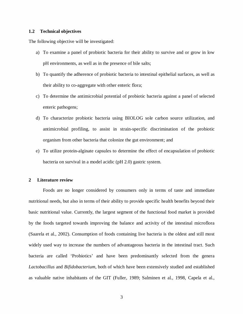
The L. acidophilus LA 1 strain exhibited a high calcium independent adhesion onto
human enterocyte like Caco-2 cells (Figure 2.3.2) and also bound strongly to the mucus secreted
by the homogeneous cultured human goblet cell line HT29-MTX (Bernet et al., 1994). Heat-
killed L. acidophilus LB was also found to adhere to Caco-2 and HT-29 cell line models
(Coconnier et al., 1993). Several Bifidobacterium spp., including B. adolescentis, B. angulatum,
B. bifidum, B. breve, B. catenulatum, B. infantis, B. longum, B. pseudocatenulatum, B. breve 4,
B. infantis 1 and B. lactis DR10 also adhered to Caco-2 and HT29-MTX cells (Gopal et al.,
2001). Furthermore, the in vitro adhesion index system (Table 2.3.4) has proven to be extremely
sensitive to factors such as pH, the presence of calcium ions, the number of lactobacilli, the
presence of culture supernatant, and the growth phase in which the bacteria were harvested
(Tuomola and Salminen, 1998; Blum and Reniero, 2000).

24
Figure 2.3.2 Examination by scanning electron microscopy of adherence of Lactobacillus acidophilus strain 1 onto the differentiated human intestinal epithelial cells Caco-2. (A) Low magnification of Caco-2 monolayer covered by L. acidophilus bacteria; (B) High magnification of L. acidophilus whole cells (Bernet et al., 1994)

25
Table 2.3.4 Lactobacilli with an adhesion index of at least one bacterium per Caco-2 cell (Adapted from Morelli, 2000) Strain Source Adhesion index
L. acidophilus BG2FO4 Human 2.3 L. johnsonii LA1 Human 1.55 L. acidophilus LB Human 2.1 L. rhamnosus GG Human 1.25 L. acidophilus C7 Chicken 1.5 L. helveticus CNRZ239 Dairy 1.4 L. helveticus CNRZ 240 Dairy 2.1 L. delbrukii subsp. lactis CNRZ 239 Dairy 1.9 L. delbrukii subsp. lactis ATCC 7830 Unknown 2.3 L. delbrukii subsp. lactis LY Yogurt 1.5
2.3.4 Antagonistic activities of probiotics against pathogens
LAB has a number of properties which make them highly suitable for probiotic
therapeutics that are of pharmaceutical interest. Several mechanisms have been ascribed to
probiotic action such as competitive exclusion, production of antimicrobial compounds,
modulation of immune response, alternation of intestinal bacterial metabolic activity, alteration
of microecology of the human intestine, and inhibition of bacterial translocation.
2.3.4.1 Competitive exclusion (CE)
CE can be defined as the principle that if two species try to occupy the same ecological
niche, a superior species will eventually emerge to replace the inferior one (Vine et al., 2004).
Various studies reported that adhesive probiotic bacteria can prevent the attachment of pathogens
and remove them from the intestinal tract (Benno and Mitsuoka, 1992; Vine et al., 2004). CE of
uropathogenic Enterococcus faecalis by Lactobacillus isolates were reported by Velraeds et al.
(1996) in their in vivo studies done in a rat model of urinary infection. Heat-killed L. acidophilis

26
strain LB that adheres to the Caco-2 cell lines, was shown to inhibit the adhesion of
diarrheagenic E. coli in a concentration-dependent manner (Chauviere et al., 1992b). The
mechanism of CE in this study was explained to involve steric hindrance. Two Bifidobacterium
strains including B. bifidum M6 and B. bifidum A1 were assessed for their ability to inhibit the
adhesion and the displacement of enteropathogens including Clostridium difficile ATCC 9689,
Enterobacter sakazakii ATCC 29544, Salmonella enterica serovar typhimurium ATCC 29631,
Escherichia coli NCTC 8603 and Listeria monocytogenes ATCC 15313 (Gueimonde et al.,
2007). The levels of displacement varied between 15 and 70% depending on the strains used.
Higher affinity of Bifidobacterium strains for the receptors present in the mucus was explained as
a mechanism of pathogen displacement in this study.
2.3.4.2 Co-aggregation of probiotics
Aggregation between microorganisms of the same strain (auto-aggregation), or between
different species and strains (co-aggregation) for example with pathogens, as well as their ability
to displace pathogens is an important property of probiotic organisms and may have greater
advantage over non-co-aggregating organisms which are easily removed from GIT environment.
The interaction of probiotic organisms with the natural gut flora is key to the potential success of
the organism in terms of colonization and long-term persistence. Co-aggregation of probiotic
bacterial strains has been suggested to enable them to form a physical-chemical barrier that
prevents colonization by pathogenic bacteria (Collado et al., 2007b). Lactobacilli have been
found to co-aggregate with some uropathogenic bacteria and inhibit their growth (Redondo-
Lopez et al. 1990). Co-aggregation of Lactobacillus acidophilus, Lactobacillus gasseri, and
Lactobacillus jensenii with pathogens like Candida albicans, E. coli, and Gardnerella vaginalis
was observed by Boris et al. (1998). Furthermore, self-aggregation or clumping may

27
substantially increase the colonization potential of lactobacilli in environments with short
residence times such as GIT. It has been hypothesized that combinations of probiotic bacterial
strains may improve the health benefits compared to the strains alone (Collado et al., 2007b).
2.3.4.3 Probiotics modulating the immune response
The GIT is a complex ecosystem which contains up to 1 × 10¹⁴ cfu of bacterial cells of
various phenotypes lining the epithelial wall and expressing complex metabolic activities
(Zboril, 2002). The mechanism(s) of the immune response of the intestinal microbiota have been
explained (Nicaise et al., 1999) by examining the regulation of interleukin-1 (IL-1), IL-6, tumor
necrosis factor-α (TNF-α) and IL-12 production in macrophages from germ-free and from
flora-associated mice, and germ-free mice colonized with E. coli and found that IL-12
production in the spleen was enhanced by intestinal flora. Interleukins are implicated in
determining the relative levels of T-helper 1 (Th1) and T-helper 2 (Th2) responses, and play an
important role in defending the host against intracellular microorganisms. Immune regulation
involves homeostasis between Th1 and Th2 activity, with Th1 cells driving the type-1 pathway
(cellular immunity) and Th2 cells driving the type-2 pathway (humoral immunity). The most
important function of the resident intestinal microbiota is to act as a microbial barrier against
pathogens by influencing humoral and cellular mucosal immune responses during the neonatal
phase of life, and thereafter to maintain a physiologically-normal steady-state condition of
inflammation throughout life (Cebra, 1999). In innate mucosal immunity, the host defense
mechanisms are triggered as a result of specific recognition of pathogen-associated molecular
patterns (PAMPs). Whereas, all endogenous bacterial species of the microbiota share microbe-
associated molecular patterns (MAMPs). Epithelial and monocytic cells can sense the

28
environment of the GIT by means of pattern-recognition receptors (PRR) (Didierlaurent et al.,
2002).
2.3.4.4 Production of antimicrobial compounds by probiotics
Several antimicrobial substances have been found to be produced by LAB that have
considerable advantages in competition with pathogens and other harmful bacteria (Soomro et
al., 2002). These substances include fatty acids, organic acids, hydrogen peroxide, and diacetyl,
acetoin and the best studied small, heat-stable inhibitory peptides called ‘bacteriocins’ (Simova
et al., 2009). The term bacteriocin was first introduced by Jacobs and coworkers in 1953, and
defined as protein antibiotics of relative high molecular weight mainly working against the same,
or closely related, species by adsorption to receptors on the target cells (Salminen and Wright,
1998a). Bacteriocins produced by LAB were divided into three classes: 1) lantibiotics; 2) small
hydrophobic heat-stable peptides, and 3) large heat-labile proteins (Drider et al., 2006; see table
2.3.5).
The most common Lactobacillus spp. known to produce bacteriocins are Lactobacillus
sakei and Lactobacillus curvatus. Lactobacillus sakei has been shown to possess antimicrobial
activity against Listeria monocytogenes due to the production of the bacteriocins sakacin A, M,
P, 674, K, and T (Schillinger and Lucke, 1989). The only purified bacteriocin approved for use
in products for human consumption is nisin, which is produced by Lactococcus lactis subsp.
lactis strains (Jack et al., 1995). Escherichia coli participates in antibacterial defense by
producing colicins that function by forming pores in the cell membrane rendering the cells
permeable to nuclease activity (Riley and Wertz, 2002).

29
Table 2.3.5 Classification of LAB bacteriocins (Adapted from Drider et al., 2006) Class Characteristics Subcategory Examples
I Lantibiotics (containing lanthionine and β-lanthionine)
Type A (elongated molecules; molecular mass, < 4 kDa)
Nisin A Nisin Z Subtilin Epidermin
Type B (globular molecules; molecular mass, 1.8 to 2.1 kDa)
Mersacidin Actagardin Mutacin II
II Non-modified heat-stable
bacteriocins containing peptides
Subclass IIa (antilisterial pediocin-like bacteriocins)
Sakacin P, G, A etc.
with molecular masses of <10 kDa
Subclass IIb (two-peptide bacteriocins)
Plantaricin EF Plantaricin JK
Subclass IIc (other peptide bacteriocins)
Lactococcin 972
III Protein bacteriocins with
molecular masses of >30kDa Helveticin J Millericin B
The antimicrobial action of bacteriocins involves increased permeability of the
cytoplasmic membrane of the target cells for a broad range of monovalent cations, such as Na+,
K+, Li+, Cs+, Rb+ and choline, which leads to the destruction of proton motive force by
dissipation of the transmembrane pH gradient and eventually to cell death (Oppegard et al.,
2007; Simova et al., 2009). Bactericidal activity (Figure 2.3.3) of a bacteriocin-like substance
produced by vaginal Lactobacillus salivarius subsp. salivarius CRL 1328 with activity against
Enterococcus faecalis, E. faecium, and Neisseria gonorrhoeae has also been reported (Ocana et
al., 1999).
30
Corr et al. (2007) demonstrated the in vivo antimicrobial effects of L. salivarius
UCC118 against L. monocytogenes primarily depended on their capacity to produce the
bacteriocin, Abp118. Lactobacillus spp. strain GG, isolated from the feces of a normal person,
was reported to produce a bacteriocin with inhibitory activity in the pH range between 3 and 5
against a wide variety of bacterial species including Gram-positive and Gram-negative
organisms (Silva et al., 1987).
Figure 2.3.3 Electron photomicrographs of the effect of L. salivarius subsp. salivarius CRL 1328 bacteriocin-like substance on E. faecalis. The Enterococcus control cell (A), vesiculization of protoplasm (B), vesiculization of protoplasm and a damaged cell wall (C), pore formation in the cell wall (D), a disintegrated cell with loss of the protoplasmic material through a cell wall pore (E), and a disintegrated cell (F) (Ocana et al., 1999)
LAB produces lactic acid as the major metabolic end-product of sugar
fermentation. Besides exerting its activity through lowering the pH and through its undissociated

31
form, lactic acid is also known to function as a permeabilizer of the Gram-negative bacterial
outer membrane (Alakomi et al., 2000), allowing other compounds to act synergistically with
lactic acid (Niku-Paavola et al., 1999).
The mode of action of organic acids on bacteria is explained in figure 2.3.4. Non-
dissociated, or free, organic acids (RCOOH) can penetrate the bacterial cell wall, and thus enter
the bacterial cytoplasm where they are exposed to a pH value near to neutrality (7.85 ± 0.05 for
E. coli) and subsequently dissociate (Gauthier, 2002), releasing H+ and anions (A-). As a result of
accumulating protons, the internal pH decreases. In case of non-pH sensitive bacteria (Figure
2.3.4) under low internal pH conditions, the organic acids remain in the free acid form and thus
exit the bacteria. This process forms equilibrium between internal and external pH and protects
the bacteria from detrimental effects of pH fluctuations (Lambert and Stratford, 1999). A specific
mechanism (H+ -ATPase pump) in pH-sensitive bacteria acts to bring the pH inside the bacteria
back to a normal level, as they can not tolerate a large spread between the internal and the
external pH. This process consumes energy and eventually stops the growth of the bacteria or
even kills it. A lower cytoplasmic pH inhibits glycolysis, prevents active transport, and also
interferes with signal transduction (Lambert and Stratford, 1999). Furthermore, the anionic (A-)
part of the acid cannot diffuse freely through the cell wall, and thus accumulates inside the
bacterial cell. Accumulation of anions leads to internal osmotic problems for the bacteria.
Complete inhibition of S. typhimurium SL1344 growth was observed due to a
pH-lowering effect produced by L. acidophilus LAP5 strain (Fayol-Messaoudi et al., 2005).
Makras et al. (2006) demonstrated an inhibitory effect of lactic acid produced by probiotic
lactobacilli upon invasion of Salmonella into Caco-2/TC7 cells. Viability of Salmonella was

32
found to be decreased rapidly after 1 h (~2 logs) and dramatically after 3 h (~8 logs) in the
presence of L. rhamnosus GG supernatant containing lactic acid (De Keersmaecker et al., 2006).
Figure 2.3.4 Mode of action of organic acids (RCOOH) on, A) non pH-sensitive bacteria (Lactic acid bacteria, Bifidobacteria), B) pH-sensitive bacteria (Coliforms, Clostridia, Salmonella, Listeria spp.) (Adapted from Gauthier, 2002)
Some LAB produce hydrogen peroxide (H2O2) under aerobic growth conditions and
because of the lack of cellular catalase, pseudocatalase, or peroxidase, they release it into the
environment to protect themselves from its antimicrobial action. It is a strong oxidizing agent
and can oxidize the –SH group of membrane proteins of Gram-negative bacteria, which are
especially susceptible (Ray, 2004). Lactobacillus gasseri CRL1421 and L. gasseri CRL1412

33
were found to produce H2O2 and lactic acid in vitro, which reportedly reduced the viability of
Staphylococcus aureus by 4 log units (P < 0.05), after 6 h of incubation (Otero and Macias,
2006).
2.3.4.5 Acylated homoserine lactones
Quorum sensing (QS) is a mechanism by which bacteria organize the expression of
certain genes in response to their population density by producing, detecting and releasing small
signal molecules. Quorum sensing was first observed in the marine bacterium, Vibrio fischeri
(Nealson et al., 1970), an organism which undergoes bioluminescence under high cell densities.
It has been demonstrated that inactivation of the QS system in pathogens can result in a
significant decrease in virulence factor expression (Swift et al., 1997). Disruption of QS has
become a new anti-infective strategy (Defoirdt et al., 2004). Acylated homoserine lactones
(AHLs) are important intercellular signalling molecules used by many bacteria to monitor their
population density in QS control of gene expression. These signalling compounds are the
biochemical products of the LuxI family of proteins. AHLs acts as QS signal molecules in
Gram-negative bacteria (Miller and Bassler, 2001) and small (oligo) peptides play this role in
Gram-positive bacteria (Defoirdt et al., 2004). The family of autoinducers known as AI-2
operate in both Gram-negative and Gram-positive bacteria. Recently, AI-2-like molecules have
also been detected in the cell-free culture fluid of several LABs, including Lactobacillus
rhamnosus GG (Dirix et al., 2004). Besides the autoinduction, the most common mechanism of
QS in Gram-positive bacteria consists of an ATP-binding cassette (ABC) transporter for
secretion of the peptide, and a two-component system for sensing of the autoinducer
concentration. Whereas, QS in Gram-positive Staphylococcus aureus and Eenterococcus faecalis
are the exceptions of the ABC mechanism, which use a non-ABC transporter to secrete the

34
autoinducer. The QS system in Bacillus subtilis further involves genetic competence and
sporulation, in which the peptide, after a Sec-dependent secretion and extracellular processing, is
imported into the cytoplasm by an oligopeptide transport system where it interacts with
intracellular receptors (Dirix et al., 2004). Studies done by McClean et al. (1997) showed that QS
pigment production in Gram-negative Chromobacterium violaceum was stimulated by naturally-
occurring AHL molecules. The acyl side chain has been found to be linked to the stimulatory and
inhibitory effect of the AHLs. AHLs with an acyl side chain containing up to 8 carbon atoms
were found to have a stimulatory effect and acted as QS agonists; whereas, acyl side chains
containing 10 or more carbon atoms showed an inhibitory effect and acted as QS antagonists. A
study done by Dong et al. (2002) suggested that enzymatic AHL inactivation could be used as a
biocontrol strategy. In this study, expression of the AiiA (AHL-inactivating activity) enzyme in
transformed Erwinia carotovora decreased the production of cell wall-degrading enzymes by the
pathogen to about 10% and inhibited soft rot disease symptoms in susceptible plants almost
completely.
A probiotic isolate, Lactobacillus fermentum RC-14, was reported to inhibit acute
Staphylococcus aureus infection (Gan et al., 2002). The probiotic was found to secrete cell-
surface extracellular matrix-binding proteins and a biosurfactant which prevented pathogen
adherence to surgical implants. These findings illustrate different potential microbial antagonistic
mechanisms other than antibiotic production, such as signal interference, for the control and
prevention of biofilm formation by pathogenic bacteria.
2.4 Clinical evidence of probiotic efficacy
Metchnikoff (1907) was perhaps the first researcher to propose that fermented dairy
products have beneficial properties. Various nutritional and therapeutic benefits (Table 2.4.1) to

35
the consumer were found to be associated with the consumption of lactic cultures and their
fermented products (Danone, 2001; Parvez et al., 2006). The disorders hypolactasia, lactose
malabsorption or maldigestion, and lactose intolerance, are characterized by a low concentration
of the lactose cleaving enzyme lactase, or β-galactosidase, in the brush-border membrane of
small intestinal enterocytes.
Table 2.4.1 Various special therapeutic or prophylactic properties of specific probiotics (Adapted from Parvez et al., 2006)
Microflora Associated actions Bifidobacteria spp. Reduced incidence of enterocolotis
Enterococcus faecium Decreased duration of acute diarrhoea Lactobacillus strains Efective in ameliorating pouchitis, decreased diarrhoea and
symptoms of intolerence in lactose intolerent individuals, improved mucosal immune function, mucin secretion and prevention of disease
L. acidophilus Significant decrease of diarrhoea in patients receiving pelvic irradiation, prevented uropatogens Escherichia coli, Klebsiella pneumoniae, Pseudomonas aeruginosa, lowered serum cholesterol evels
L. plantarum Reduced incidence of diarrhoea and inflammation in inflammatory bowel, reduced bloating, pain and flatulence in irritable bowel syndrome, positive effect on immunity in HIV positive children
L. reuteri Shortened the duration of acute gastroenteritidis and diarrhoea L. rhamnosus Enhanced cellular immunity in healthy adults in controlled trial L. salivarius Suppressed and eradicated Helicobacter pyroli in vitro and in vivo Bacteroides spp. Increased bacterial urease activity in chronic arthritis Saccharomyces boulardii
Reduced Cl. difficile diarrhoea and antibiotic associated diarrhoea, shortened the duration of acute gastroenteritis
A significant proportion of the human population has some degree of intolerance to
lactose. With the exception of the population of Northern and Central Europe, approximately
85% of adults worldwide are lactose malabsorbers (Goktepe et al., 2006). The LAB has an

36
excellent capacity to metabolize lactose. Studies by Kolars et al. (1984) showed that, in lactase-
deficient subjects, lactose was absorbed significantly better from yogurt than from milk. Two
different mechanisms of action have been put forward to explain this finding. Yogurt and
probiotic LAB contain high levels of lactase, which is released within the intestinal lumen when
these bacteria are lysed by bile secretions. Lactase then acts on the ingested lactose, thus
relieving maldigestion symptoms. The reduced intestinal transit time of yogurt might also allow
slower digestion of lactose. Several reviews (Rolfe, 2000; Kopp-Hoolihan, 2001; de Vrese et al.,
2001) have described that some probiotics could improve lactose digestion and eliminate the
symptoms of intolerance. While mechanisms by which these probiotics exert their effects are not
fully understood yet; they may involve modifying gut pH, expressing β-galactosidase, exerting
positive effects on intestinal functions and colonic microbiota. In a systematic review by Levri et
al. (2005), in contrast, it was concluded that probiotic supplementation in general did not
alleviate the symptoms of lactose intolerance in adults. The lactose contained in yogurts can be
considered as a prebiotic for people with lactose maldigestion (Szilagyi, 2004). Regular
consumption of lactose influences their colonic microbiota and reduces lactose intolerance
(Hertzler and Savaiano, 1996).
One particular strain, Lactobacillus casei DN-114001, has been reported to reduce
diarrhoeal morbidity by 40% in children (Agarwal and Bhasin, 2002). Study on pregnant women
and newborns suggested that consumption of probiotic L. rhamnosus GG reduced the rate of
newborns having atopic dermatitis (Kalliomaki et al., 2001).
In an Australian study, 178 newborns of women with allergies who received either
L. acidophilus LAVRI-A1 or placebo daily for the first 6 months of life showed no difference in
atopic dermatitis (probiotic, 23/89 versus placebo, 20/88; P = 0.629). However, at 12 months, the

37
rate of sensitization was significantly higher in the probiotic group (P = 0.030). These results
suggested that the probiotic treatment has increased the risk of subsequent cow’s milk
sensitization (P=0.012) (Taylor et al., 2007). Similarly, a double-blind, randomized, placebo-
controlled trial study was conducted by Abrahamsson et al., (2007) on 188 subjects with allergic
disease, in which the mothers received Lactobacillus reuteri ATCC 55730 daily from gestational
week 36 until delivery, and their babies continued with the probiotic until 12 months. Probiotic
supplemented babies showed less IgE-associated eczema during the second year.
2.5 Prebiotics
Association of colonic microflora with beneficial health effects is now well established.
Consequently, the use of prebiotics as functional food ingredients to manipulate the composition
of colonic microflora to improve health is currently a growing interest (Wang, 2009). According
to Solange et al., (2007) prebiotics are non-digestible carbohydrates (oligosaccharides) of low
molecular weight and intermediate in nature between simple sugars and polysaccharides.
Resistant short-chain carbohydrates (SCCs) are also referred to as non-digestible
oligosaccharides or low digestible carbohydrates (LDCs) (Cummings et al., 2001). Prebiotics are
defined as “selectively fermented ingredients that allow specific changes, both in the
composition and/or activity in the gastrointestinal microbiota that confers benefits upon host
well-being and health” (Wang, 2009). Prebiotics resist hydrolysis and absorption in the upper
parts of the GIT and may selectively be metabolized by at least one type of probiotic
microorganism in the colon, leading to growth and/or metabolic activation of the probiotic
microorganisms, improvement of the colonic flora, and beneficial luminal or systemic effects for
the health of the host (Gibson and Roberfroid, 1995). Bifidobacteria and lactobacilli are the most

38
important bacterial genera targeted for selective stimulation by prebiotics (Teitelbaum and
Walker, 2002). The “bifidogenic” effects of SCC or LDCs make it possible for their inclusion
into conventional food products (Holzapel et al., 1998). Oligosaccharides that have been
considered as prebiotics include galacto-oligosaccharides, isomalto-oligosaccharides, soybean-
oligosaccharides, lactosucrose, and xylo-oligosaccharides (Rastall and Gibson, 2002). For the
comparison of prebiotic effects of dietary oligosaccharides, a quantitative prebiotic index (PI)
equation has been developed by Palframan et al. (2003). This equation is based on the changes in
key bacterial groups like bifidobacteria, lactobacilli, clostridia and bacteroides during
fermentation. The changes in bacterial groups from previous studies were entered into the
equation in order to determine a quantitative PI score. Table 2.5.1 shows examples of prebiotics
used for human consumption (Wang, 2009).
Table 2.5.1 Examples of ingredients that are commonly regarded as human prebiotics (Wang, 2009) Prebiotics Inulin-type fructans Trans-galactooligosaccharides (TOS) Lactulose Isomalto-oligosaccharides Lactosucrose Xylo-oligosaccharides Soybean-oligosaccharides Gluco-oligosaccharides Fructo-oligosaccharides (FOS) Galacto-oligosaccharides (GOS)
2.5.1 Inulin and fructo-oligosaccharides (FOSs) as prebiotics
Inulin and FOSs are considered as typical “bifidogenic factors” and are probably the most
commonly-used prebiotics in the market (Bouhnik et al., 1999). Growth of L. casei was
significantly (P ≤ 0.05) higher when inulin of different chain lengths was incorporated at a
concentration of 1.5 g/100 g yogurt mix (Aryana et al. 2007). Similarly a study by Akalin et al.

39
(2004) reported higher counts of Bifidobacterium longum (106 cfu/g) and Bifidobacterium
animalis (3.59-2.25 × 10⁷ cfu/g) in yogurts containing FOS during 28 days storage at 4°C;
whereas, this level was maintained for only 7 days in yoghurt without any prebiotic. Scientific
studies done in Japan showed that the consumption of FOS in the diet increases the population of
bifidobacteria and other beneficial micro-organisms, even in the absence of probiotics (Losada
and Olleros, 2002). Similarly, increased numbers of lactobacilli and bifidobacteria were found
following the consumption of FOS in a study by Mitsuoka (1990). To assess the effects of
lactulose administration on faecal bifidobacteria, a controlled, randomised, double-blind, and
parallel group trial study was conducted on 16 healthy volunteers by Bouhnik et al. (2004).
Participants ingested lactulose or placebo (sucrose) at a dose of 5 g twice a day for 6 weeks.
Results showed that ingestion of 10 g of lactulose per day increased fecal bifidobacterial counts
from day 0 to day 42 (8.25 ± 0.53 to 9.54 ± 0.28 log cfu/g wet weight, respectively). Shin et al.
(2000) reported a decrease in mean doubling time and increase in viability of Bifidobacterium
spp. (Bf -1 and Bf -6) in skim milk during 4 weeks of storage at 4°C, containing inulin, fructo-
oligosaccharides (FOS) and galacto-oligosaccharides (GOS) at a concentration of 50 g/L. A 67%
viability of Bf-1 and 45% viability of Bf-6 were retained in the presence of FOS. When grown in
the presence of GOS, 52 and 39% retention of viability of Bf-1 and Bf-6 was observed,
respectively. Inulin was the least effective (P < 0.05) in retaining viability of either strain.
2.5.2 Health benefits of prebiotics
A number of beneficial effects are associated with prebiotics, including the favorable
influence on the small bowel by improved sugar digestion and absorption, glucose and lipid
metabolism, and protection against known risk factors of cardiovascular diseases (Bruzzese et
al., 2006) as shown in Table 2.5.2.

40
Table 2.5.2 Microbiological changes reported in human feeding studies with prebiotics (Adapted from Macfarlane et al., 2008)
Substrate Type of study Delivery Effect on microbiota GOS Placebo controlled
study involving 12 subjects
10 g prebiotic fed daily for 8 weeks
Faecal excretion of bifidobacteria and lactobacilli increased
Feeding study with
12 subjects 2.5 g prebiotic given per day
for 3 weeks Increased bifidobacteria in faeces,
reductions in numbers of clostridia and bacteroides
TOS Feeding study with 8
volunteers Subjects given 10 g TOS for
3 weeks Significant increases in faecal bifidobacteria, enterobacteria
unaffected Parallel study
involving 40 healthy subjects
7.5 or 15 g prebiotic fed per day for 3 weeks
Lactobacilli increased in 15 g/day, no effects on clostridia, small reductions in enterobacteria
GOS ⁄ FOS Placebo-controlled
study with 90 term infants
Infant formula supplemented with either 4
or 8 g/l low molecular weight GOS and high
molecular weight FOS for 28 days
Significantly increased lactobacilli and bifidobacteria in
the two GOS feeding groups.
GOS Placebo-controlled
study involving 30 subjects
Volunteers given 8.1 g GOS syrup, 8.1 g GOS plus 3 X 10¹⁰ Bifidobacterium lactis Bb-12, or 3 X 10¹⁰ B. lactis without GOS for 3 weeks
Little change in faecal bifidobacteria seen with GOS
alone. GOS plus B. lactis and B. lactis on its own resulted in faecal
excretion of the organism and reduced numbers of B. longum.
GOS ⁄ FOS(9 :1 ratio) Double-blinded,
randomized controlled trial
(DBRCT) involving 20 infants aged 28–
90 days
Infant formula supplemented with 0.8 g /100 ml GOS ⁄ FOS for 6
weeks
Increased total bifidobacteria counts in stools. Reduced B.
adolescentis compared to standard infant formula controls.
Bifidobacteria spp. composition in prebiotic infants similar to that
found in breast-fed babies, with B. infantis, B. breve and B. longum
predominating
GOS ⁄ FOS (9:1 ratio) Feeding study involving 42 preterm infants, 15 placebos, and a reference group
given fortified mothers milk
Formula food supplemented with 10 g/l prebiotic mixture
Bifidobacteria numbers greatly increased from initially low
levels, compared to un-supplemented controls. No
significant effects on bacteroides, clostridia, enterobacteria or yeasts
41
Various in vivo and in vitro studies shows health benefits of prebiotics such as FOS and GOS
alone, or in combination with inulin (Macfarlane et al., 2008). Production of short chain fatty
acids (SCFAs) is considered to be a major beneficial feature related to the primary prevention of
colorectal cancer (Scheppach et al., 2001). SCFAs and lactic acid are produced as a result of
metabolism of FOS by fermentative bacteria, leading to a drop in the pH of the large intestine.
Low pH conditions were reported to be ideal for growth of bifidogenic flora. A study done by
Liu et al. (2004) on 55 cirrhotic patients, were randomized to receive a probiotic plus prebiotic
(synbiotic) preparation containing 4 freeze-dried probiotics namely Pediacoccus pentoseceus,
Leuconostoc mesenteroides, L. paracasei subsp. paracasei and L. plantarum. Each probiotic was
given at a dose of 1010 cfu/sachet, along with 10 g of bioactive, fermentable fiber (beta glucan,
2.5 g; inulin, 2.5 g; pectin, 2.5 g; resistant starch, 2.5 g) (n = 20), fermentable fiber alone (n =
20), or placebo (n = 15) for 30 days. The controls had significant fecal overgrowth of potentially
pathogenic E. coli and Staphylococcus spp., while the synbiotic treated subjects had significantly
increased fecal content of Lactobacillus strains.
2.6 Viability of probiotic microorganisms
Fermented dairy products such as milk and yogurt are the most accepted food carriers for
live probiotic delivery to the human GIT. A recommended intake of probiotics is 108-109 viable
live cells daily (Lourens-Hattingh and Viljoen, 2001) in order to get the beneficial health effects.
Standards have been made in many countries for the numbers of viable probiotic bacteria that are
present in commercial fermented products. For example, in Japan, fermented milks and LAB
containing beverages must contain a minimum of 107 cfu/mL or gram of product (Robinson,
1987). Despite the importance of viability of these beneficial bifidobacteria, surveys have shown
poor viability of bifidobacteria in yoghurt preparations (Akalin et al., 2004). Several factors, like

42
acidity of the product, post acidification (acid produced during storage), level of oxygen in the
products, sensitivity to antimicrobial substances produced by bacteria, temperature of storage
during manufacture and storage of yoghurt, have been found to reduce the viability of probiotics
(Lankaputhra and Shah, 1995; Dave and Shah, 1997). Thus, maintaining viability of
bifidobacteria until the products are consumed in order to ensure the delivery of live organisms
has been of much interest.
2.6.1 Influence of cryoprotectants
Freeze-drying is a preservation process that has been employed to improve the survival of
probiotic microorganisms (Rybka and Kailaspathy, 1995). Survival and rapid growth after
freezing is one of the most important characteristics for the selection of probiotics because they
are most often delivered in the form of frozen dairy desserts. Addition of compatible
cryoprotectants into the media or yogurt mix prior to fermentation has been shown to improve
the viability of probiotic microorganisms (Kets et al., 1996). Improvement in the viability of
probiotics may occur as the cryoprotectants inhibit the formation of intracellular or extracellular
ice by binding to the water (Capela et al., 2006). The cryoprotectant UnipectineTM RS 150 was
found to improve the number of viable L. acidophilus 33200, L. casei 279, B. longum 536 and
L. rhamnosus (GG) by 1 log during storage in fermented dairy desserts (Shah and Ravula, 2000).
In another study, an 80% improvement in the survival of B. longum 536 was found by using
UnipectineTM RS 150 (Capela et al., 2006).
2.6.2 Encapsulation
Encapsulation is a process whereby cells are retained within an wall material to reduce
cell injury. Encapsulation in hydrocolloid beads has been investigated as a means to protect and

43
improve viability of probiotic microorganisms in food products and in the intestinal tract (Rao et
al., 1989). Other benefits of encapsulation includes: protection of probiotics from bacteriophage
(Steenson et al., 1987), increased survival during freeze-drying and freezing (Kim et al., 1995),
and greater stability during storage (Kebary et al., 1998). Hou et al. (2003) demonstrated that
encapsulation of Lactobacillus delbrueckii spp. bulgaricus increased their bile tolerance, and
viability was elevated by approximately four log units after encapsulation within artificial
sesame oil emulsions. Capela et al. (2006) found improved viability of probiotic organisms
encapsulated in 3% v/w sodium alginate in freeze-dried yogurt after 6 months of storage at
4 (~8 log cfu/g) and 21°C (~6 log cfu/g). Spray drying, freeze drying, fluidized bed drying are
techniques which are used for converting probiotic cultures into a concentrated powdered form.
However, these techniques do not protect the bacteria from the product environment or during
their passage through the GIT because they release the bacteria completely into the product. The
viability of Bifidobacterium bifidum BB-12 and L. acidophilus LA-5 encapsulated in Na-alginate
by either an extrusion or an emulsion technique and used in white-brined cheese was monitored
(Ozer et al., 2009). Both encapsulation techniques were found to be effective in keeping the
numbers of probiotic bacteria higher than the level of the therapeutic minimum (>107 cfu/g).
While the counts of non-encapsulated probiotic bacteria decreased approximately by 3 logs, the
decrease was more limited in the cheeses containing microencapsulated cells (approximately
1 log).
Depending on the method used to form the beads, the encapsulation techniques applied
for probiotics for use in fermented milk products or biomass production can be classified as
either the: i) extrusion or droplet method, and ii) emulsion or two phase systems (Figure 2.6.1).

44
Both techniques have been found to increase the survival of probiotic bacteria by up to 80-95%
(Audet et al., 1988; Rao et al. 1989).
2.6.2.1 Extrusion technique
In extrusion encapsulation, microorganisms are added into a hydrocolloid solution
(alginate) and then the cell suspension is extruded through a syringe needle to form droplets,
which free-fall into a hardening solution or setting bath (Figure 2.6.1).
MIX
Emulsification in vegetable oil
Sodium alginate
Extrusion
EmulsionCell suspension
CaCl2
Cell suspension
Microbial cell
Liquid core
CaCl2 solution
Calcium alginate bead
Figure 2.6.1 Flow diagram of encapsulation of bacteria by the extrusion and emulsion techniques (Reproduced from Krasaekoopt et al., 2003)
The size and shape of the beads depends on the diameter of the needle and the distance of
free-fall (Kim, 1995). Alginate is used commonly as supporting material in this technique. The
concentrations of alginate used to form the gel varies from a very low concentration of 0.6% to

45
form a gel with 0.3M CaCl2 to 1–2% alginate and 0.05–1.5M CaCl2 (Krasaekoopt et al., 2003).
The size and sphericity of the bead also depends on the viscosity of the sodium alginate solution,
the distance between the syringe and the calcium chloride collecting solution and the extruder
orifice diameter. As the concentration of sodium alginate increases, the size of the beads
decreases. Using a 0.27 mm syringe resulted in a bead size of 2–3 mm. The composition of the
alginate also influences bead size; small beads result from ‘‘low guluronic’’ alginates
(Krasaekoopt et al., 2003).
2.6.2.2 Emulsion technique
In the emulsion technique, a small volume of the cell-polymer suspension (the
discontinuous phase) is added to a large volume of a vegetable oil (e.g. canola or corn oil). The
mixture is homogenized to form a water-in-oil emulsion. Once formed, the water-soluble
polymer must be insolubilized (cross-linked) to form tiny gel particles within the oil phase
(Figure 2.6.1). The smaller the internal phase particle size of the emulsion, the smaller the final
microparticles will be. The supporting material used in this technique may include κ-carageenan
and locust bean gum (Audet et al., 1988), cellulose acetate phthalate (Rao et al., 1989), alginate
(Sheu and Marshall, 1991), chitosan and gelatin (Groboillot et al., 1993).
2.7 Synbiots
The combination of a probiotic and prebiotic is referred to as a synbiot. Synbiots have
been shown to confer health benefits beyond those of either on its own. Rowland et al. (1998)
showed the enhanced reduction in the number of colonic aberrant crypt foci, as well as colon
carcinogenesis in rats (Gallaher and Khil, 1999). Antibiotic-associated diarrhea was found to be
prevented by the combined application of Lactobacillus sporogenes and FOS in children (Rosa

46
et al., 2003). Synbiotic combination of L. paracasei and maltodextrin was found to decrease the
colonization of E. coli in the jejunum of piglets. This symbiotic combination led to an increase in
lactobacilli and bifidobacteria and decreased clostridia and enterobacteria (Bomba et al., 2002).
On the other hand, Anderson et al. (2004) did not find any improvement of gut barrier function
in surgical patients following synbiotic therapy.
Therefore, further investigation is needed to continue, and extend our understanding of
probiotics, along with the efficacy once encapsulated. The present research investigates the
ability of probiotic bacteria to survive in simulated gastric conditions of the GIT, as well as their
ability to become established on the surface of the intestinal epithelium or to other resident
microflora. An attempt was made to determine the antimicrobial potential of the probiotic
bacteria against a panel of selected enteric pathogens. The probiotic bacteria were also
characterized using biochemical techniques to aid in their identification during follow-up
research. Model HCl/bile-based model gastric systems were then be used to compare the effect
of encapsulation on the survival of the probiotic bacteria.
47
3 In vitro characterization of probiotic survival, adherence and antimicrobial resistance: candidate selection for encapsulation in a pea protein isolate-alginate delivery system
Abstract
A panel of probiotic strains, including Lactobacillus acidophilus ATCC 11975,
Bifidobacterium infantis ATCC 15697D, B. catenulatum ATCC 27675 and B. adolescentis
ATCC 15703, were screened for a number of traits, including their resistance to acid and bile and
adherence to Caco-2 cells. Under simulated gastric conditions (pH 2.0), L. acidophilus was the
most acid-tolerant strain (D-value 10.2 ± 0.8 min), and was able to survive for 30 min; whereas,
the other probiotics tested underwent a rapid (within the first 5 min at pH 2.0) 4-5 log cfu/mL
loss in viability. Notably, the number of viable L. acidophilus decreased by less than 1 log
cfu/mL over the same interval. All probiotics tested were able to survive 5 h of exposure to 0.3%
(w/v) Oxgall bile at pH 5.8. The relative ranking of probiotic adherence to Caco-2 cells was
determined to be: L. acidophilus > B. catenulatum > B. adolescentis > B. infantis, which
correlated with 4.5 104, 3.1 103, 2.6 101, and 1.5 101 cfu/mL associated with Caco-2 cell
monolayers, respectively. The probiotics also revealed varying degrees of resistance for the
antimicrobial agents: ciprofloxacin, naladixic acid, kanamycin and sulfisoxazone. Determination
of carbon source utilization patterns indicated that all strains grew to the highest culture optical
densities on D-xylose. L. acidophilus was determined to be the best overall in vitro performer,
and hence was selected for encapsulation within a ~3 mm diameter pea protein-alginate matrix.
Upon exposure of encapsulated L. acidophilus to simulated gastric conditions (pH 2.0) there was
only a ~1 log cfu/mL loss of cell viability observed over a 2 h period; whereas, unprotected L.
acidophilus cells experienced a reduction in cell viability of greater than 6 log cfu/mL over the
same period. The present study indicates that the encapsulated delivery of probiotics is a feasible
means for enhancing probiotic survival during passage through the upper gastrointestinal tract.

48
3.1 Introduction
Probiotics are microorganisms that are consumed orally and that reach the GIT in a viable
state, where they subsequently contribute to the beneficial activity of intestinal microflora and,
thus, to the health of its host. Most probiotic bacteria are LAB and among them lactobacilli and
bifidobacteria reportedly play a significant role in maintaining the intestinal microbial ecosystem
and in stimulating the immune system of the host (Saarela et al., 2002). Finegold et al. (1977)
demonstrated that while lactobacilli were not the predominant species among the intestinal
microflora, they were commonly isolated from the GITs of healthy humans. Others have shown
that typical numbers of endogenous faecal lactobacilli per gram of faeces ranged from 105 to 108
cfu (Kimura et al., 1997; Holzapfel et al., 1998).
It has been shown that the functional and technical properties of probiotics are strain-
specific, and thus it is necessary to screen various isolates for a variety of in vitro properties to
obtain new probiotic candidates is of utmost important (Collins et al., 1998; Ouwehand et al.,
2002). These include general aspects (e.g., origin, identity, resistance to mutations, resistance to
environmental stress prevailing in the GIT such as low pH, bile acid and pancreatic juice),
technical considerations (e.g., in vitro growth and processing properties), functional and
beneficial properties (e.g., adhesion and colonization to intestinal mucosa, competitiveness,
antagonism against pathogens, and stimulation to immune response) and safety concerns (e.g., no
invasive potential, no transferable resistance against therapeutic antibiotics and no virulence
factors) (Holzapfel et al., 2002). Lactobacilli fulfill these criteria (Dunne et al., 2001) and their
use is now being supported by clinical benefits (Reid et al., 2003). Cellular stress on probiotics
begins in the stomach, where pH values as low as 1.5 are typically encountered (Lankaputhra
and Shah, 1995). Following passage through the stomach, probiotics come in contact with bile in
the upper intestinal tract, the concentration of which is variable and difficult to predict at any

49
given time (Northfield and McColl, 1973). After traveling through these relatively harsh
conditions, the probiotic should ideally adhere to the lower intestinal tract epithelia and
microflora where it would proliferate as a stable member (Conway et al., 1987), so that they are
able to exert health benefits to the host for prolonged periods. It is noteworthy that some
conjecture presently exists as to whether all probiotics must actually colonize the epithelial
membranes of the intestine for a probiotic effect to be realized.
While there continues to be a lack of clear information to the biologically-effective
probiotic dose, it is clear that increasing the number of viable probiotic that reach the upper GIT
is important. Improved survival of probiotics exposed to the low pH gastric environment (Rao et
al., 1989; Wenrong and Griffiths, 2000) and bile salts (Lee and Heo, 2000), molecular oxygen in
the case of obligatory anaerobic microorganisms, bacteriophages (Steenson et al., 1987),
chemical antimicrobials (Sultana et al., 2000), and increased survival during freezing and freeze
drying (Shah and Rarula, 2000) have all been reported following probiotic microencapsulation.
Additional advantages of probiotic encapsulation over free cells include an increase of sensory
properties, stability (Gomes and Malcata, 1999), and immobilization of the cells for their
homogeneous distribution throughout the product (Krasaekoopt et al., 2003). A variety of
substrates have been employed as encapsulating materials for entrapping live microorganisms,
including alginate, whey protein, pea protein, gelatin, methylcellulose, etc. (Sheu et al., 1993;
Prevost and Divies, 1988; Gombotz and Wee, 1998; Pierucci et al., 2007). Among these,
alginate-gel beads have frequently been used for the immobilization of LAB (Sheu et al., 1993),
as it is an accepted food additive and has the benefit of being nontoxic to the cells being
immobilized (Prevost and Divies, 1988).

50
The objective of present study was to conduct in vitro screening of a panel of probiotic
organisms, using acid resistance, bile toxicity and ability to attach to the human intestinal cells,
as test variables. Probiotics were also screened for resistance to commercial antibiotics and their
ability to utilize different sole carbon sources. Based on the above tests, a candidate probiotic
was selected for encapsulation in a pea protein-alginate matrix and then challenged with
simulated gastric conditions.
3.2 Materials and methods
3.2.1 Bacteria and culture conditions
The probiotic strains Lactobacillus acidophilus ATCC 11975, Bifidobacterium infantis
ATCC 15697D, B. catenulatum ATCC 27675 and B. adolescentis ATCC 15703 were stored at
-80ºC using 10% glycerol amended in culture broth as a cryoprotectant. DeMan Rogosa and
Sharpe (MRS) broth (VWR, Mississauga, ON, Canada) was used as a media for routine
cultivation of lactobacilli; whereas, MRS supplemented with 0.05% (w/v) L-cysteine HCl was
used for cultivation of bifidobacteria. Prior to each experiment, stock cultures were propagated
twice in sterile broth under anaerobic conditions (10% H2, 10% CO2, and 80% N2) at 37°C for 24
h to provide inocula for subsequent experiments. Inocula of each strain were harvested at the
early stationary phase of growth (~ 20 h; data not shown) by centrifugation at 3,250 × g (Model
5810 R with swing-bucket rotor A-4-81; Eppendorf, Hamburg, Germany) for 10 min at 4C.
Cells were then washed twice with phosphate-buffered saline (PBS; 4.3 mM Na2HPO4.7H20,
137.0 mM NaCl, 2.7 mM KCl, 1.4 mM KH2PO4, pH 7.2) and resuspended in peptone saline
(PS; 8.5 g/l NaCl, 1.0 g/l peptone, pH 7.0).

51
3.2.2 Probiotic resistance to simulated gastric juice (SGJ)
Probiotic resistance to SGJ was performed according to the following experimental
design; washed cell suspensions (~105 cfu/mL) were inoculated in triplicate into SGJ without
pepsin (0.08 M HCl containing 0.2% w/v NaCl) with pH adjusted to 2.0 and 6.0 (control) using
either HCl or NaOH. Samples were mixed well, incubated at 37C and aliquots removed every 5
min for 2 h and serially diluted in PS. Surviving bacteria were enumerated by pour plating. D-
values for each probiotic were calculated for the 0 to 5 min. SGJ exposure period using the
equation, D = t / n where n = (log N0-log Nt) = 1 log10 reduction of the cell number, D =
Decimal reduction time (min) at pH 2.0, N0 = bacteria at 0 time; Nt = surviving bacteria after an
exposure time, t (min). The best surviving probiotic at pH 2.0 will be tested for its survival at pH
4.0 and 5.0.
3.2.3 Probiotic resistance to bile salts
The method of Gilliland et al. (1984) was used to evaluate the growth potential of the
four probiotic strains in broth media with and without Oxgall bile salts (Sigma-Aldrich Inc.,
St. Louis, MO, USA). Accordingly, MRS broth without (control) and with 0.3% bile (w/v) was
inoculated with 100 µL (~106 cfu/mL) of each overnight culture, and then incubated under
anaerobic conditions for 24 h at 37ºC and 100 rpm shaking. Growth was monitored hourly by
measuring absorbance at 600 nm using a spectrophotometer. Experiments were conducted in
triplicate.
Survival of the different probiotic strains in the presence of 0.3% Oxgall was monitored
by the standard plate count method. Overnight cultures were inoculated into flasks containing
sterile MRS (control) and MRS-bile, and incubated at 37C for 5 h, anaerobically. Aliquots were

52
removed every hour and serially diluted in PS. Surviving bacteria were enumerated by spread
plating.
3.2.3.1 Microscopy
An inverted light microscope (Nikon Eclipse, TS100; 400 X NA 0.55 lens) was used to
take photographs of crystals formed by probiotic bacteria in the MRS broth containing 0.3% bile
and MRS broth without any added bile (control).
3.2.4 Probiotic adherence to Caco-2 cell lines
3.2.4.1 Cell culture
Enterocyte-like Caco-2 cells were kindly provided by Dr. Wolfgang Köster (Vaccine
Infectious Disease Organization (VIDO), Saskatoon, Canada). Caco-2 cells were routinely
cultured at 37ºC in 5% CO2-95% air atmosphere in Dulbecco Modified Eagle’s Minimal
Essential Medium (DMEM; Aldrich Sigma, ON) supplemented with 10% heat-inactivated fetal
bovine serum (Invitrogen Canada Inc., Burlington, ON). Caco-2 cell monolayers were used for
the probiotic adherence assay at the post-confluence stage (after 10 d). The concentration of
Caco-2 cells in the monolayer was determined by trypsinizing the cells using Trypsin-EDTA for
10 min at 37ºC followed by counting in a hemocytometer. Caco-2 cells (50 to 52 passages) were
seeded at a concentration of 3 104 cells/mL in sterile 24-well tissue culture plates. The culture
medium was changed every 2 d.
3.2.4.2 Adhesion assay
Adhesion assay was conducted as outlined by Jacobsen et al. (1999) and Pennacchia et al.
(2006), as briefly outlined below. Post-confluence Caco-2 monolayers were gently washed two
times with Hank’s Balanced Salt (HBS) solution (Invitrogen Canada Inc., Burlington, ON).

53
Probiotic bacteria in their early stationary phase of growth (~20 h; data not shown) were
harvested and washed twice with PBS by centrifugation at 3,250 × g (Model 5810 R equipped
with a swing-bucket rotor A-4-81; Eppendorf) for 10 min at 4C and diluted (10x) with DMEM
to provide a bacterial concentration of ~5 105 cfu/mL. A 2 mL aliquot of each bacterial culture
was used to inoculate the tissue culture plate wells containing Caco-2 cells, followed by
incubation at 37ºC in 5% CO2-95% air atmosphere for 2 h. Control wells contained
un-inoculated DMEM. The viability of probiotic strains was also monitored in 5% CO2-95% air
atmosphere for 2 h. Following incubation, Caco-2 cell monolayers were washed 5 times with
HBS solution to release unbound bacteria. Washed Caco-2 cell monolayers were trypsinized
using Trypsin-EDTA for 5 min at 37C to detach the bacterial cells from Caco-2 cells. The
number of viable adhering bacteria was determined by spread plating serial 10-fold dilutions on
MRS agar and incubating at 37C for 48 h under anaerobic conditions.
3.2.5 Probiotic susceptibility to antibiotics
Antibiotic resistance of each probiotic strain was determined using the Sensititre
CMV1AGNF plates (Trek Diagnostic Systems, Inc., Westlake, OH, USA) that contained
17 antimicrobial agents incorporated in dehydrated form into the wells of a plastic microtiter
plate in either single or paired concentrations, as specified by the NARMS (National
Antimicrobial Resistance Monitoring System) protocol of the CDC. Inocula for the antibiotic
sensitivity assays was prepared by suspending isolated colonies of each probiotic (picked from
fresh cultures on MRS agar plates incubated anaerobically for 48 h at 37°C) in a tube with 5 mL
of sterile distilled water to an optical density of 0.5 McFarland standard, and subsequently
diluting 1:100 in MRS broth. A 50 µL aliquot of this MRS broth suspension (containing ~106
cfu/mL) was then transferred to each microtiter plate well. The plates were sealed and incubated

54
anaerobically at 37°C for 48 h (Klare et al., 2005; Doern et al., 1985), and then examined
manually for the presence of turbidity. A ‘susceptibility category’ test result was assigned to
each antimicrobial, as outlined below. With antimicrobials present in paired concentrations,
turbidity in both wells was considered ‘resistant’, turbidity only in the well containing the lower
concentration was considered ‘intermediate’, and absence of turbidity in either well was
considered ‘susceptible’. For antimicrobials present in individual concentrations, turbidity was
considered ‘resistant’ and absence of turbidity was considered ‘susceptible’. The minimum
inhibitory concentration (MIC) breakpoints were determined according to the national committee
for clinical laboratory standards (NCCLS, 2002a; and NCCLS, 2002b).
3.2.6 Probiotic carbon source utilization profiles
Biolog Ecoplates (Biolog, Inc., Hayward, CA, USA) contain 31 different sole carbon
sources and can be used to screen substrate preference amongst different pure cultures. Cultures
grown on solid MRS agar media for 48 h were swabbed and resuspended in sterile distilled
water. The turbidity of these culture suspensions were then compared with the Biolog GP
microplate turbidity standard (~108 cfu/mL). Biolog Ecoplate wells were subsequently inoculated
with 150 µl of each cell suspension. Following an initial (time 0) reading, Biolog plates were
incubated anaerobically at 37ºC for 48 h. The substrate utilization patterns were analyzed with a
microplate reader at a wavelength of 590 nm.
3.2.7 Growth experiments
Test was conducted according to Vernazza et al., 2006. Basal medium was prepared as follows:
in g/L peptone water (Sigma chemicals company, Poole, UK) 2.0, yeast extract (Oxoid Ltd,
Hampshire, UK) 2.0, NaCl 0.1, K2HPO4 (Oxoid Ltd, Hampshire, UK) 0.04, KH2PO4 (Oxoid

55
Ltd, Hampshire, UK) 0.04, MgSO4.7H2O 0.01, CaCl2 2H2O 0.01, NaHCO3 2.0, Tween 80 (BDH
chemicals, Poole, UK) 2.0 mL/L, hemin (Sigma chemicals company, Poole, UK) 0.005, vitamin
K1 10 µL/L, Cysteine-HCl 0.5, bile salts (Oxoid Ltd, Hampshire, UK) 0.5, pH 7.0. Test
carbohydrate solutions including D-lactose (BDH chemicals, Poole, UK), methyl-D-glucoside
(NB Co. Cleveland, Ohio), D-xylose (Sigma chemicals company, Poole, UK) D-cellobiose
(Sigma chemicals company, Poole, UK) and D-glucose (BDH chemicals, Poole, UK) were
prepared (100 g/l). Test carbohydrate solution (1 ml) was added in 9 ml sterilized basal media.
Bacteria were grown on MRS agar plates at 37°C for 48 h, and a single colony was added in
basal media containing different carbohydrates. Each tested bacteria were subcultured twice in
identical medium. Growth of test cultures was monitored for 48 h by measuring absorbance at
600 nm using a spectrophotometer.
3.2.8 Encapsulation of L. acidophilus
Pea protein isolate (4.0% w/v; PPI) + alginate (0.5% w/v; AL) capsule was prepared by
dissolving 40.0 g/L PPI in 0.1 M NaOH under constant mechanical stirring, and heating at 80ºC
for 30 min before cooling to 40ºC in a water bath. Once cooled, the solution was then neutralized
to pH 7.0 with 1.0 M HCl and 5.0 g/L AL powder was then dispersed into the solution. The
biopolymer mixture was then heated at 80ºC for another 30 min, before being cooled to 40ºC in a
water bath. The weights of the solutions were corrected for water loss during heating. A
suspension containing 1.0 g washed L. acidophilus (~108 cfu/mL) was mixed with 18.0 g of the
4.0% PPI + 0.5% AL solution held at 40ºC and injected through a 20 G needle into 30 mL of
filter sterilized 5.0% CaCl2 + 1.0% (w/v) Tween 80 crosslinking bath. The resultant capsules
were allowed to harden in the cross-linking solution for 30 min, and then washed three times
with PS.

56
3.2.9 Survival of encapsulated L. acidophilus in SGJ
The survival of free and encapsulated L. acidophilus cells in SGJ (0.08 M HCl with 0.2
% (w/v) NaCl) were studied at pH 6.0 and 2.0, as described below. Aliquots of 0.5 mL of free
and encapsulated L. acidophilus cells in PS were added to 9.5 mL of SGJ at 37ºC and incubated
anaerobically. Samples containing SGJ at pH 6.0 were removed at 20 min intervals over the 2 h
assay period; for samples containing SGJ at pH 2.0, tubes were removed at 5 min time intervals
over the first 30 min, and thereafter at 30 min intervals until 2 h. Tubes containing SGJ at pH 2.0
were neutralized with 1.0 M NaOH upon removal from incubation. The contents of the tubes
containing the encapsulated cells were homogenised (ES, Omni International Inc., Marietta, GA,
USA) at a setting of 4.5 for 30s, serially diluted in PS and surviving bacteria enumerated by pour
plating using MRS media. The plates were incubated under anaerobic conditions at 37ºC for 48
h. Experiments were conducted in triplicate.
3.2.10 Statistical analysis
Statistical analysis was carried out to see the differences between the experiments with a
confidence interval of 95% by using analysis of variance (ANOVA). Comparison of means was
carried out using Tukey’s HSD test. All the experiments are repeated in triplicates (n=3).
3.3 Results
3.3.1 Probiotic resistance to SGJ
To determine the effect of prolonged exposure of probiotics to the acidic pH of the
stomach, a 2 h in vitro SGJ survival assay was employed (Figure 3.3.1).

57
Figure 3.3.1 Survival of L. acidophilus (A), B. adolescentis (B), B. catenulatum (C), and B. infantis (D) in SGJ pH 2.0 (●) and pH 6.0 (○). The data are expressed as mean ± one standard deviation (n=3)
Results from these experiments demonstrated that L. acidophilus ATCC 11975 was the
most resistant (D-value 10.2 ± 0.8 min) to SGJ (pH 2.0) (Table 3.3.1), but could not survive
longer than 30 min of exposure under these conditions. The D-value of L. acidophilus was
significantly (P < 0.05) more than other tested strains. Whereas, the viability of L. acidophilus
was not significantly affected (P > 0.05) at pH 4.0 and 5.0 and remained viable even after 3 h of

58
exposure to SGJ (Figure 3.3.2). Plate count data revealed that none of the three Bifidobacterium
spp. tested could survive more than 15 min of exposure to pH 2.0 SGJ, with viable cell numbers
decreasing to undetectable levels in this period (Table 3.3.1).
Table 3.3.1 D-values (min) of free and encapsulated probiotics exposed to SGJ (pH 2.0). Data are expressed as the mean ± one standard deviation (n=3)
Bacteria D-value (min)
Survival studies
L. acidophilus* 10.17 ± 0.80a
B. catenulatum* 1.26 ± 0.02b
B. infantis* 1.15 ± 0.04b
B. adolescentis* 1.12 ± 0.03b
Encapsulation studies
L. acidophilus (Free) 0-30 min. 10.05 ± 0.19a
L. acidophilus (Free) 30-120 min. 61.77 ± 4.25b
L. acidophilus (Encapsulated) 0-30 min. 59.06 ± 4.91a
L. acidophilus (Encapsulated) 30-120 min. 138.04 ± 8.04b
a,b Means in columns within a category with unlike superscripts differ (P < 0.05). Statistical analysis was done separately for free and encapsulated cells. *D-values were determined over the first 5 min due to the rapid depletion of viable free cells.

59
Figure 3.3.2 Survival of L. acidophilus in SGJ pH 4.0 (●) and pH 5.0 (○). The data are expressed as the mean ± one standard deviation (n=3)
3.3.2 Probiotic resistance to bile
The effect of Oxgall bile on the viability of the four probiotics is shown in figure 3.3.3;
all test strains were able to survive for 5 h in MRS media supplemented with 0.3% bile. Optical
density data showed that slow growth of all test strains occurred over a 24 h period in the
presence of 0.3% bile-containing MRS (Figure 3.3.4). Following growth in bile-containing
media, all tested probiotic strains produced unique crystalline structures, whereas no crystalline
structures were observed when probiotics were added in control (Figure 3.3.5).

60
Figure 3.3.3 Survival of L. acidophilus (A), B. adolescentis (B), B. catenulatum (C), and B. infantis (D) in media containing 0.3% (w/v) Oxgall bile (○) and without added bile (●). The data are expressed as the mean ± one standard deviation (n=3)
Optical density results (Figure 3.3.4) suggest that slow growth of all test strains occurred
in the presence of 0.3% bile containing MRS media, as compared to the control (MRS broth
without added bile).

61
Figure 3.3.4 Growth of probiotics in A) MRS and B) 0.3% (w/v) Oxgall bile containing MRS media. The data are expressed as the mean ± one standard deviation (n=3)
Figure 3.3.5 Light micrographs (magnification 400X NA 0.22) of crystals formed by test probiotic strains cultivated in MRS-broth media supplemented with 0.3% (w/v) Oxgall bile for 24 h incubation at 37ºC under anaerobic conditions. a) L. acidophilus, b) B. adolescentis, c) B. catenulatum and d) B. infantis
d c
a b
25µm

62
3.3.3 Probiotic adherence to Caco-2 cells
The in vitro adherence of probiotic bacteria to Caco-2 cells was determined, using a 2 h
assay, to be species-specific (Figure 3.3.6). The initial number of bacterial cells added to each
well was 5.4 105 cfu/mL. By the assay endpoint, L. acidophilus was shown to adhere to Caco-2
cells in the highest numbers (4.5 104 cfu/mL or ~10% of the added cells). In contrast, about
10-fold fewer B. catenulatum cells attached (3.1 103 cfu/mL), and B. adolescentis and
B. infantis showed relatively poor adhesion with the number of adhering cells at the end of the
assay being 2.6 101 and 1.5 101 cfu/mL, respectively. The viability of all tested probiotics
was not effected in 5% CO2-95% air atmosphere significantly (P < 0.05, data not shown).
Figure 3.3.6 Adhesion of probiotic bacteria to Caco-2 cells. Data are expressed as the mean ± one standard deviation (n=3)

63
3.3.4 Probiotic susceptibility to antibiotics
It was determined that the patterns of antimicrobial susceptibility of the probiotic strains
were quite variable (Table 3.3.2). L. acidophilus was found to be resistant to ciprofloxacin
(4 µg/mL), nalidixic acid (32 µg/mL) and sulfisoxazole (256 µg/mL), and showed intermediate
resistance to cefoxitin (4 µg/mL), amikacin (8 µg/mL), ceftriaxone (8 µg/mL), gentamicin
(4 µg/mL), trimethoprim/sulphamethoxazole (1/19 µg/mL), and kanamycin (8 µg/mL).
B. catenulatum was resistant to nalidixic acid (32 µg/mL) and kanamycin (64 µg/mL) and
intermediately resistant to amikacin (4 µg/mL), ciprofloxacin (0.5 µg/mL), gentamicin
(8 µg/mL), sulfisoxazole (64 µg/mL), trimethoprim/sulphamethoxazole (0.25/4.75 µg/mL), and
streptomycin (32 µg/mL).
Table 3.3.2 Antibiotic resistance profiles of the tested probiotic strains Antibiotic Conc. range,
µg/mL L.
acidophilus B.
catenulatum B.
infantis B.
adolescentis Cefoxitin 32-0.5 I (4.0) S S S
Amikacin 64-0.5 I (8.0) I (4.0) I (4.0) S
Chloramphenicol 32-2 S S S S Tetracycline 32-4 S S S S Ceftriaxone 64-0.25 I (8.0) S I (0.25) S Amoxillin/Clavulanic-Acid 32/16-1/0.5 S S S S Ciprofloxacin 4-0.015 R I (0.5) I (0.25) S Gentamicin 16-0.25 I (4.0) I (8.0) I (2.0) S Nalidixic acid 32-0.5 R R R I (0.5) Ceftiofur 8-0.12 S S S S
Sulfisoxazole 256-16 R I (64.0) I (16.0) S Trimethoprim/Sulphamethoxazole 4/76-0.12/2.38 I (1.0/19.0) I (0.25/4.75) I 0.25/4.75) S Kanamycin 64-8 I (8.0) R I (8.0) R Ampicillin 32-1 S S S S Streptomycin 64-32 S I (32.0) R S
I, Intermediate (conc. µg/mL); S, Sensitive (No growth in any of the tested conc. range); R, Resistant (Growth in highest tested conc.)

64
B. infantis was resistant to streptomycin (64 µg/mL) and nalidixic acid (32 µg/mL) and
showed intermediate resistance to amikacin (4 µg/mL), ceftriaxone (0.25 µg/mL), ciprofloxacin
(0.25 µg/mL), gentamicin (2 µg/mL), trimethoprim/sulphamethoxazole (0.25/4.75 µg/mL), and
kanamycin (8 µg/mL). Lastly, B. adolescentis was resistant to kanamycin (64 µg/mL) and
showed intermediate resistance to nalidixic acid (0.5 µg/mL). None of the tested probiotics were
resistant to chloramphenicol, tetracycline, amoxillin/clavulanic-acid, ceftiofur and ampicillin.
3.3.5 Carbon source utilization of probiotics
Growth of probiotics on different carbohydrates was tested by adding different
carbohydrates in basal media (Figure 3.3.7). Carbon source utilization profiles of L. acidophilus,
B. catenulatum, B. infantis and B. adolescentis are shown in Table 3.3.3, respectively.
All probiotics were able to utilize glucose (control) and lactose as sole carbon sources.
Xylose was utilized by B. adolescentis and B. catenulatum. Cellobiose was utilized by
B. catenulatum and L. acidophilus, where poor growth was observed by the later strain. Slow
growth of B. adolescentis and B. infantis was observed with methyl-D-glucoside, with a longer
lag phase (~ 20 h). Analysis of Ecoplate results indicated that all strains grew to the highest
culture optical densities on D-xylose. B. catenulatum utilized a number of carbohydrates
including -methyl-D-glucoside, D-cellobiose, and -D-lactose.

65
Table 3.3.3 Optical densities at 590 nm attained after 48 h by probiotics during growth on different sole carbon sources. The data are expressed as the mean ± one standard deviation (n=3)
Carbon name B. infantis L.
acidophilus B.
adolescentis B.
catenulatum 2-hydroxybenzoic acid 0.38 ± 0.03a 0.34 ± 0.03ab 0.29 ± 0.00b 0.38 ± 0.00b 4-hydroxybenzoic acid 0.38 ± 0.02a 0.34 ± 0.02ab 0.27 ± 0.04b 0.39 ± 0.03b D, L-α-glycerol phosphate 0.28 ± 0.01a 0.30 ± 0.09a 0.28 ± 0.00a 0.37 ± 0.02a D-cellobiose 0.32 ± 0.04a 0.37 ± 0.07a 0.35 ± 0.02a 0.69 ± 0.04b D-galactonic acid γ-lactone 0.31 ± 0.04a 0.38 ± 0.00b 0.27 ± 0.03a 0.37 ± 0.02b D-galacturonic acid 0.34 ± 0.04a 0.40 ± 0.03ab 0.31 ± 0.01b 0.40 ± 0.02b D-glucosaminic acid 0.30 ± 0.02a 0.37 ± 0.02ab 0.28 ± 0.07ab 0.40 ± 0.01b D-malic acid 0.38 ± 0.14a 0.37 ± 0.02a 0.33 ± 0.04a 0.37 ± 0.01a D-mannitol 0.38 ± 0.04a 0.36 ± 0.05a 0.32 ± 0.01a 0.40 ± 0.02a D-xylose 1.28 ± 0.16a 0.92 ± 0.02a 0.98 ± 0.10ab 1.21 ± 0.12b Glucose-1-phosphate 0.37 ± 0.06a 0.34 ± 0.03a 0.31 ± 0.02a 0.39 ± 0.02a Glycogen 0.32 ± 0.02a 0.34 ± 0.02ab 0.31 ± 0.02ab 0.37 ± 0.03b Glycyl-L-glutamic acid 0.33 ± 0.00a 0.37 ± 0.02b 0.30 ± 0.01c 0.38 ± 0.01c i-erythritol 0.38 ± 0.03a 0.40 ± 0.09a 0.28 ± 0.04a 0.39 ± 0.01a Itaconic acid 0.35 ± 0.03a 0.38 ± 0.04ab 0.30 ± 0.04ab 0.39 ± 0.00b L-arginine 0.32 ± 0.07a 0.39 ± 0.02a 0.28 ± 0.02a 0.35 ± 0.03a L-asparagine 0.32 ± 0.03a 0.36 ± 0.04ab 0.29 ± 0.01bc 0.40 ± 0.02c L-phenylalanine 0.46 ± 0.04a 0.37 ± 0.05ab 0.34 ± 0.01bc 0.47 ± 0.04c L-serine 0.40 ± 0.01a 0.38 ± 0.06ab 0.29 ± 0.05ab 0.39 ± 0.00b L-threonine 0.36 ± 0.03a 0.38 ± 0.05a 0.33 ± 0.05a 0.41 ± 0.01a N-acetyl-d-glucosamine 0.38 ± 0.03a 0.52 ± 0.06a 0.30 ± 0.02ab 0.41 ± 0.05b Phenylethlamine 0.35 ± 0.01a 0.34 ± 0.01b 0.29 ± 0.01bc 0.37 ± 0.01c Putrescine 0.35 ± 0.08a 0.34 ± 0.02a 0.29 ± 0.02a 0.31 ± 0.10a Pyruvic acid methyl ester 0.30 ± 0.01a 0.47 ± 0.03ab 0.22 ± 0.09bc 0.36 ± 0.01c Tween 40 0.32 ± 0.06a 0.27 ± 0.09a 0.28 ± 0.06a 0.37 ± 0.17a Tween 80 0.27 ± 0.07a 0.29 ± 0.10a 0.28 ± 0.05a 0.45 ± 0.09a water 0.30 ± 0.03a 0.35 ± 0.03a 0.27 ± 0.03a 0.32 ± 0.09a α-cyclodextrin 0.32 ± 0.04a 0.34 ± 0.02ab 0.29 ± 0.01ab 0.38 ± 0.05b α-D-lactose 0.37 ± 0.13a 0.40 ± 0.07a 0.44 ± 0.01a 0.97 ± 0.07b α-Ketobutyric acid 0.36 ± 0.01a 0.39 ± 0.05a 0.29 ± 0.05a 0.37 ± 0.03a β-methyl-D-glucoside 0.33 ± 0.04a 0.47 ± 0.03b 0.37 ± 0.08ab 0.82 ± 0.01c γ-hydroxybutyric acid 0.44 ± 0.02a 0.42 ± 0.08a 0.35 ± 0.00a 0.44 ± 0.03a
a,b,c Means in a row within a category with unlike superscripts differ (P < 0.05).

66
Figure 3.3.7 Maximum optical densities (O.D. 600) attained by probiotics during growth on different carbon sources. Data are expressed as the mean ± one standard deviation (n=2)
3.3.6 Survival of encapsulated L. acidophilus in SGJ
L. acidophilus was selected for encapsulation and subsequent challenge, in vitro, to
simulated gastric conditions (pH 2.0). The PPI-AL microcapsules, prepared by extrusion, were
teardrop in shape and uniform in size (~3 mm) (Figure 3.3.8). The survival of L. acidophilus in
acidic condition is shown in Figure 3.3.9, and clearly demonstrates a protective effect of
encapsulation during the 2 h exposure to simulated gastric conditions with only a ~1 log cfu/mL
loss in cell viability. Whereas, unprotected L. acidophilus cells underwent a >6 log cfu/mL loss
in cell viability over the same period (P < 0.05). Comparing D-values of the encapsulated and
un-encapsulated cells (Table 3.3.1) revealed that the PPI-AL matrix improved survival by almost
6 times (59.0 ± 5.0 min versus 10.1 ± 0.2 min; D-value for the free cells calculated over the first
30 min).
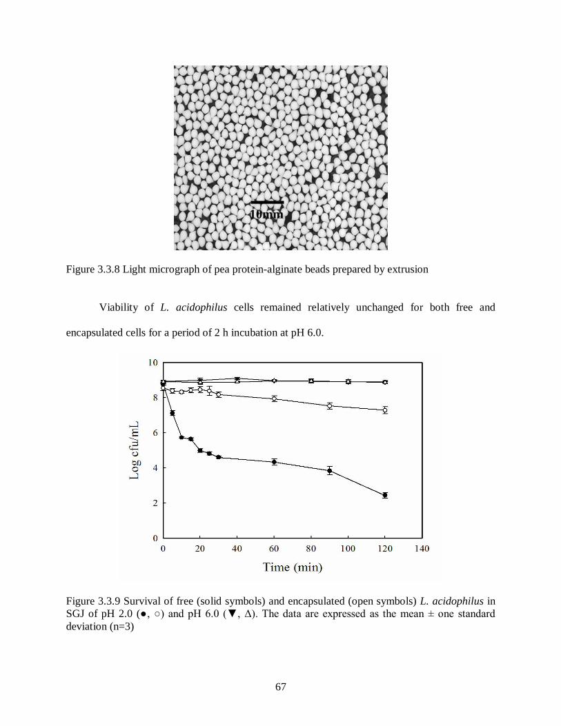
67
Figure 3.3.8 Light micrograph of pea protein-alginate beads prepared by extrusion
Viability of L. acidophilus cells remained relatively unchanged for both free and
encapsulated cells for a period of 2 h incubation at pH 6.0.
Figure 3.3.9 Survival of free (solid symbols) and encapsulated (open symbols) L. acidophilus in SGJ of pH 2.0 (●, ○) and pH 6.0 (▼, Δ). The data are expressed as the mean ± one standard deviation (n=3)
10 mm
10mm

68
3.4 Discussion
The human GIT is a complex microbial community consisting of ~1010-1014 cfu of
bacteria per gram of faecal material. Given such high numbers and diversity, it is not surprising
that the composition and numbers of these organisms should play a significant role in human
health and disease. Within this community, probiotics (lactobacilli and bifidobacteria) have been
studied for their health promoting and otherwise beneficial effects (Dunne et al., 2001; Goktepe
et al., 2006). However, for probiotics to exert beneficial effects in the GI tract, they must survive
transit through the stomach. Each day, the human stomach secretes about 3 L of gastric juice
with a pH of around 2.0. The transit time of food in the stomach is between 2 and 4 h (Smith,
1995); liquids take about 20 min to pass through the stomach (GastroNet Australia, 2001).
Hence, it is necessary for probiotic microorganisms, if unaided or unprotected, to tolerate the
acidic conditions of the stomach for periods of up to 4 hrs so that they may reach the colon in a
viable state. Lactobacilli of intestinal origin are considered intrinsically-resistant to acidic
environments and are often employed in fermented foods as probiotics. In order to improve the
numbers of surviving probiotics during gastric transit, components such as milk (Conway et al.,
1987), milk proteins (Charteris et al., 1998a), cheese and yogurt (Gardiner et al., 1999),
reconstituted skim milk with gum acacia (Desmond et al., 2002), and cereal extracts have been
used (Charalampopoulos et al., 2003). However, actual in vivo data demonstrating this enhanced
survival is lacking. Figure 3.3.1 shows that simulated gastric conditions with a pH of 2.0 were
lethal in as little as 10 min for all unprotected probiotics tested, with the exception of
L. acidophilus which survived for 30 min. Thus, L. acidophilus was most likely to survive
passage through the stomach compared with the other probiotics tested in this study. For
Bifidobacteria spp., transit through the stomach gastric conditions would only be possible if the
pH was raised to above 4.0 if, for example, the organism was ingested along with a meal.

69
Raising the pH to 4.0 or 5.0 also significantly improved the survival kinetics of L. acidophilus
strains (Figure 3.3.2); this would likely be a viable strategy for improving survival of probiotics
through the stomach environment.
Bile tolerance by probiotics has been shown to be strain- and bile type-dependent, with
resistance levels ranging from bile concentrations of 0.125 to 2.0% (Lian et al., 2003; Margolles
et al., 2003). While all strains involved in this study could tolerant 0.3% Oxgall bile (Figure
3.3.3), there were some minor differences. In general, the required concentration of bile salts
considered necessary to screen for resistant strains for human use is 0.3%.
In the current study, crystalline floccules were found in broth media containing bile (Figure
3.3.4) and were species specific. Zavaglia et al. (2002) suggested that when cultures are exposed
to bile, changes take place in the cell membrane causing auto-aggregation and subsequently,
flocculation. Metabolic actions of the live cells or the work of enzymes released from lysed dead
cells in the culture medium could induce a chemical reaction which makes the bile component of
the medium insoluble. The significance of the different shapes of crystals formed by the tested
probiotic panel in current study is not yet clear and to my knowledge has not previously been
described.
The adhesion of probiotic strains to intestinal epithelial cells is considered as a
prerequisite feature for attachment and proliferation in the intestinal environment (Kailasapathy
and Chin, 2000). Adhesion has also been cited as important for transient colonization (Alander et
al., 1999), enhanced healing of the damaged gastric mucosa (Elliott et al., 1998), modulation of
the immune system (Perdigon et al., 2002) and antagonism against pathogens (Jin et al., 2000).
Caco-2 cell lines have seen extensive use as an in vitro model of the human intestinal epithelium
for screening probiotic adherence (Gopal et al., 2001), and culture plating has been employed to
70
count the total adhering bacteria to Caco-2 cells (Forestier et al., 2001; Matijasic et al., 2003;
Bertazzoni-Minelli et al., 2004; Pennacchia et al., 2006). Previously, the adhesion of L. casei
rhamnosus Lcr35 was studied using an initial bacterial inoculum of 109 cfu/well, where the final
number of viable adhering bacterial cells was a little less than 105 cfu/well (Forestier et al.,
2001). In a second study, adhesion percentages of six L. casei strains were calculated, in which
four had an adhesion percentage ranging from 0.3 to 0.74 (Bertazzoni-Minelli et al., 2004). Two
L. gasseri strains of human origin, K7 and LF221 showed good adhesion with Caco-2 cells,
when added with an initial concentration of 109 cfu/well, yielding final numbers of viable
adhering bacterial cells of 107 cfu/well (Matijasic et al., 2003). Finally, the eight Lactobacillus
strains belonging to the L. plantarum-group were analysed by Pennacchia et al. (2006). The
number of final adhering bacteria was ~107 cfu/mL when 109 cfu/well was added initially. In the
present study, the probiotic strains examined were shown to attach to the Caco-2 cells in vitro
with varying success, as shown in figure 3.3.6. Clearly, L. acidophilus showed higher or similar
adhesion levels to those results described above (Forestier et al., 2001; Matijasic et al., 2003;
Bertazzoni-Minelli et al., 2004; Pennacchia et al., 2006). Among the Bifidobacterium strains
tested B. catenulatum attached considerably more strongly to Caco-2 cells in comparison with
B. adolescentis and B. infantis, suggesting the latter strains may have limited use as probiotics
when viewed from the adhesion perspective.
It has also been reported that some lactobacillus strains can strongly adhere to the gut
mucosa, where they subsequently interfere with the adhesion of pathogenic bacteria to intestinal
cells (Servin, 2004). Adherence to the intestinal epithelial cells is also an important requirement
for colonization by micro-organisms, and for bacterial pathogens, which is a pivotal step for
virulence (Westerlund and Korhonen, 1993). The ability of some lactobacillus strains to

71
co-aggregate with intestinal pathogens in intestinal lumen might prevent pathogens from
reaching the intestinal mucosa. At the same time, production of antimicrobial compounds by
probiotics helps to kill enteric pathogens and ultimately reduce their population in the GIT. As
adhesion to intestinal tissue has been considered to be one of the cornerstones of probiotic
function, it was considered in the present work as one of the main criteria for probiotic selection
for encapsulation; thus, L. acidophilus, which attached at a rate of ~10% of the cells used in the
assay, was the most successful probiotic of the tested panel. However, there is a lack data
indicating that probiotic gut colonization occurs, or that it is even necessary for a probiotic effect
to be realized (Tappenden and Deutsch, 2007).
There is limited information on the resistance of probiotic bacteria to antimicrobials of
clinical importance and even a more limited understanding of how these resistance factors
influence probiotic reproductive success. Antibiotic resistance of probiotics might be a desirable
feature, as it could potentially aid in their survival in the GIT, especially when used after
antibiotic therapy or in co-administration with antibiotics to restore intestinal health (Salminen et
al., 1998a). Alternatively, resistance to these agents could increase the spread of resistance
within the dense, diverse anaerobic microbial population (including pathogens) inhabiting the
GIT of humans and animals (Scott, 2002). The antibiotic susceptibility system used in this study
is a microtiter plate-based version of the classic broth dilution method for testing for antibiotic
susceptibility, and provided both qualitative and quantitative susceptibility results. All tested
probiotics were resistant to Gram-negative spectrum-antibiotic nalidixic acid (conc. 32 g/mL),
whereas, B. adolescentis showed intermediate resistance (Table 3.3.2). Charteris et al. (1998b)
reported resistance of B. adolescentis and B. infantis to nalidixic acid (30 µg/mL). Kanamycin
resistance (30 µg/disk) and sensitivity to the broad-spectrum antibiotics chloramphenicol

72
(32 µg/disk) and tetracycline (30 µg/disk) of tested Lactobacilli and Bifidobacteria strains were
reported by Zhou et al. (2005). Similarly, all probiotics tested in the present study were sensitive
to chloramphenicol (32 µg/mL) and tetracycline (32 µg/mL), B. catenulatum and B. adolescentis
were resistant to kanamycin conc. 64 g/mL, whereas, L. acidophilus and B. infantis showed
intermediate resistance to kanamycin. L. acidophilus, B. catenulatum and B. infantis showed
intermediate resistance to the combination of trimethoprim/sulphamethoxazole. Studies by
Masco et al. (2006) showed reduced resistance of bifidobacteria against a therapeutic
combination of trimethoprim and sulphamethoxazole due to their synergistic inhibitory effect on
thymidine synthesis.
At any given time, the human colon and particularly the proximal bowel contain many
different types of carbohydrates. Consequently, bacteria which can adapt rapidly to the changing
substrate availability and grow on a variety of carbon sources will have a competitive advantage
in the GIT ecosystem (Macfarlane et al., 2008). Carbon substrate utilization was highly variable
among tested probiotics and considerable inter-species and inter-strain differences existed, as
shown in Table 3.3.3. While individual probiotic species exhibited unique carbohydrate substrate
preferences, several trends in carbon source utilization emerged from this study. For example,
the highest culture optical densities were recorded by B. catenulatum (Table 3.3.3) for a number
of carbohydrates, including -methyl-D-glucoside, D-cellobiose, D-xylose and -D-lactose.
Similarly, rapid growth of B. catenulatum was observed in the presence of lactose and xylose
(Hopkins et al. 1998). Fermentation of cellobiose by B. catenulatum strains was previously
reported by Salminen et al. (1998a); whereas, B. adolescentis and B. infantis did not ferment
cellobiose (Roy and Ward, 1990). Low utilization of Tween-40 and Tween-80 were recorded for
L. acidophilus and B. infantis. Similarly, maximum specific growth rates of 0.25 and 0.29 were
73
recorded by B. catenulatum and B. adolescentis strains with xylose; whereas, B. infantis did not
show appreciable growth in the presence of xylose (Hopkins et al., 1998). In the present study,
the highest optical densities for all the probiotics tested were observed when D-xylose was
provided as the sole carbons source. The differences in carbon source utilization patterns
observed in this study may have particular interest from the viewpoint of future prebiotic
development. Use of such compounds to target specific organisms in the large intestine would be
of great value for defined health-promoting purposes in future.
Buffering capacities, carbohydrate and protein constituents, and encapsulation are some
of the technologies that have been used previously for increasing probiotic survival in acidic
environments (Corcoran et al., 2005). Encapsulation is currently seeing intense focus as a
physical means for protecting probiotics from the harsh effects of the gastric environment
(Mortazavian et al., 2007). Microencapsulation using cellulose acetate phthalate protected
bifidobacteria quite notably during spray drying and during prolonged exposure to simulated
gastric conditions (Favaro-Trindale and Grosso, 2002). Emulsification, gelation and film
formation are some of the properties which supports the technological utilisation of pea proteins
(Choi and Han 2002; Rangel et al. 2003). Pea protein has also been utilised for
microencapsulation of α-tocopherol (Pierucci et al., 2007) but to my knowledge has not been
used for entrapping live microorganisms. Transformation towards strictly plant-based protein
materials would be advantageous, as probiotics ingredients could be then incorporated into
non-dairy products, or have greater acceptance in materials that restrict animal-derived materials
(eg; gelatin, whey). While a number of capsule matrices have been explored, the cross-linked
alginate matrix system has most frequently seen application for LAB encapsulation (Sheu et al.,
1993). However, these systems appear to be subject to a more rapid degradation and release of

74
active ingredients at low pH (Gombotz and Wee, 1998). Lee and Heo (2000) showed that
Bifidobacterium longum encapsulated in calcium alginate containing 2.0, 3.0, and 4.0% sodium
alginate survived exposure to SGJ (pH 1.5) significantly better than free cells. The death rate of
the cells in the beads decreased proportionally with an increase in both the alginate gel
concentration and bead size. However, survival of probiotics in alginate starch microcapsules
was improved during refrigerated storage in yoghurt but not after exposure to acid and bile
solutions (Sultana et al., 2000). Microencapsulated L. acidophilus strains showed 2-3 log
improvement in viability after 3h of incubation at pH 2.0, when compared to free cells
(Chandramouli et al. 2004). Similarly, non-encapsulated cells were completely destroyed when
exposed to artificial gastric juice of pH 1.2 and 1.5 for 3 h, while the treatment reduced the
viable count of encapsulated cells only by 3 log units (Kim et al. 2008). In the present study
(Figure 3.3.9), encapsulation of L. acidophilus within pea protein – alginate capsules resulted in
an approximate 6-fold increase in the survival of bacteria in pH 2.0 SGJ.
3.5 Connection to the next study
Preliminary screening of co-aggregation potential may be useful to identify potential probiotic
strains suitable for food, human or animal use. Co-aggregation of probiotics with pathogens
could potentially interfere with pathogen colonization of the GIT, survival (through various
antimicrobial mechanisms) and subsequent manifestation of illness. In addition, co-aggregation
of probiotics with other established commensal GIT microflora may aid in the attachment and
colonization of probiotics to mucosal surfaces. Thus, the next study was performed to elucidate
the aggregation and antibacterial properties of tested probiotic strains.

75
4 In vitro growth control of Escherichia coli O157:H7 and Clostridium sordelli ATCC 9714 by the probiotic Lactobacillus acidophilus ATCC 11975
Abstract
A panel of probiotic strains, including Lactobacillus acidophilus ATCC 11975,
Bifidobacterium infantis ATCC 15697D, Bifidobacterium catenulatum ATCC 27675 and
Bifidobacterium adolescentis ATCC 15703, were screened, using the agar spot test for their in
vitro antibacterial activity against Escherichia coli O157:H7 and Clostridium sordelli ATCC
9714. Based on preliminary findings, L. acidophilus was then examined in more detail for its in
vitro co-aggregation and antimicrobial activity against E. coli O157:H7 and Cl. sordelli. Cell
surface properties of probiotic and pathogen strains were characterized in terms of adhesion to
hydrocarbons (BATH), and their relative auto- and co-aggregation abilities. Cl. sordelli showed
the highest 66.0 ± 5.5 % hydrophobicity and was also the most successful auto-aggregating
strain, showing 63.6 ± 1.4 % auto-aggregation after 24 h incubation at 37°C under anaerobic
conditions. L. acidophilus was able to co-aggregate with both tested pathogens and showed 47.9
3.6 % and 51.8 4.2 % co-aggregation with E. coli and Cl. sordelli, respectively. Bactericidal
activity of probiotic supernatant (cell-free) was observed, as the number of viable bacteria was
reduced over 24 h by approximately 4 log and 2 log cfu/mL for E. coli and Cl. sordelli,
respectively. In general, L. acidophilus-mediated inhibition of E. coli was greater than that of Cl.
sordelli. Antagonistic activity of the probiotic strain appeared to be partially pH-independent, as
the use of buffered MRS (containing 0.2% sodium bicarbonate) agar media did not completely
eliminate the inhibitory activity. Antagonistic activity was shown to be related to heat-stable
non-proteinaceous compound(s), which were resistant to heating at 100C for 10 min and to
trypsin (1 mg/mL).

76
4.1 Introduction
The use of LAB as probiotics for the therapeutic treatment of a broad number of
gastrointestinal conditions and infectious diseases (e.g., rotavirus diarrhea, influenza virus and
Helicobacter pylori) has increased over the last few years (Goktepe et al., 2006). Lactobacilli
and bifidobacteria, the most commonly-cited probiotic genera, reportedly play a significant role
in maintaining the intestinal microflora and stimulating the immune system of its host (Saarela et
al., 2002). When present in sufficient numbers (107 cfu/g or mL), probiotics are believed to
create a healthy equilibrium between beneficial and potentially harmful microflora in the gut
(Suskovic et al., 2001). Lactobacillus spp., such as L. acidophilus, L. bulgaricus, L. lactis along
with Bifidobacterium bifidum and Streptococcus faecium constitute an integral part of the healthy
gastro-intestinal microecology (Fernandes et al., 1987).
Antimicrobial substances produced by probiotics, either singly or in combination, may
have bactericidal or bacteriostatic effects on other microbial populations. These antimicrobial
effects can alter the proliferation of undesired pathogens by influencing the outcome of
competition for chemicals or available energy (Fredrickson and Stephanopoulos, 1981).
Production of antibacterial substances such as antibiotics (Shahani et al., 1976), bacteriocins
(Barefoot and Klaenhammer, 1983; Millette et al., 2007), diacetyl and ammonia (Vandenbergh,
1993), hydrogen peroxide (Dahiya and Speck, 1967; Reid, 2000) and organic acids such as
acetic, propionic and lactic acid (Naaber et al., 2004) by certain L. acidophilus strains have been
reported. The in vitro production of inhibitory compounds toward known pathogens by various
probiotic species has often been used in the selection of candidate probiotic strains.
In vitro inhibitory activity against a wide range of bacteria, including Clostridium spp.,
Bacteroides spp., Bifidobacterium spp., Enterobacteriaceae spp., Pseudomonas spp.
Staphylococcus spp., and Streptococcus spp., has been shown by Lactobacillus spp. strain GG,

77
isolated from the human GI tract (Silva et al., 1987). Lactobacillus gasseri, which is considered
to be a dominant inhabitant of the human intestine, was found to produce a bacteriocin that
exhibited a wide spectrum of bactericidal activity against enteric pathogens. Early studies
explored the use of oral preparations of viable Lactobacillus acidophilus in the treatment of
functional gastrointestinal disturbances (Rafsky and Rafsky, 1955). Subsequently, Vincent et al.
(1959) found that L. acidophilus produced lactocidin, a substance with broad antibacterial
properties.
Co-aggregation is a process by which genetically-distinct bacteria become attached to
one another via specific lectin-like adhesins and receptor molecules. Only a limited number of
studies have examined probiotic-pathogen co-aggregation, on the basis that it might be an
important protective mechanism against pathogen colonization in the gastrointestinal tract (GIT)
(Reid et al., 1988; Schellenberg et al., 2006; Collado et al., 2008). Cumulative evidence,
especially from oral microbiology work, suggests that aggregation influences the development of
complex multispecies biofilms (Rickard et al., 2003). Bacterial aggregation between
microorganisms of the same strain (auto-aggregation) or between genetically-divergent strains
(co-aggregation) is likely of considerable importance in the human GIT, where mechanisms for
colonizing the epithelial lining, and the organisms that inhabit it, would likely be necessary for
any subsequent probiotic effect (Collado et al., 2008). A relationship between auto-aggregation
and adhesion ability has been reported for some bifidobacterial spp., and a correlation between
adhesion ability and hydrophobicity has been observed in some lactobacilli (Del Re et al., 2000).
Furthermore, it has been suggested (Collado et al., 2007) that co-aggregation of bacteriocin-
producing LAB with pathogens, may constitute an important host-defence mechanism against

78
infection. Co-aggregation with potential gut pathogens could therefore contribute to the probiotic
properties ascribed to specific LAB.
The objective of the present study was to determine cell surface hydrophobicity, auto-
aggregation and pathogen co-aggregation properties of probiotic strain L. acidophilus ATCC
11975. Antibacterial properties of this probiotic strain were then evaluated against selected
pathogens.
4.2 Materials and methods
4.2.1 Bacteria and culture conditions
The probiotic strains Lactobacillus acidophilus ATCC 11975, Bifidobacterium infantis
ATCC 15697D, Bifidobacterium catenulatum ATCC 27675 and Bifidobacterium adolescentis
ATCC 15703 were purchased from the American Type Culture Collection (ATCC, Manassas,
VA), and stored at -80°C using 10% glycerol amended in culture broth as a cryoprotectant.
Lactobacilli De Man Rogosa and Sharpe (MRS) broth (VWR International Ltd., Mississauga,
ON) was used as a culture media for lactobacilli; whereas, MRS supplemented with 0.05% w/v
L-cysteine HCl was used for bifidobacteria. Prior to each assay, stock cultures were propagated
twice in fresh MRS broth and then incubated anaerobically (10% H2, 10% CO2, and 80% N2) at
37°C for 24 h. Seed culture was harvested at the end of the exponential phase of growth
(approximately 20 h) by centrifugation at 3,250 × g (Model 5810 R equipped with a swing-
bucket rotor A-4-81; Eppendorf, Hamburg, Germany) for 10 min at 4°C and washed twice with
phosphate-buffered saline (PBS, 4.3 mM Na2HPO4.7H20, 137 mM NaCl, 2.7 mM KCl, 1.4 mM
KH2PO4, pH 7.2) and resuspended in peptone saline (PS, 8.5 g/L NaCl, 1.0 g/L peptone, pH 7.0).
Escherichia coli O157:H7 strain B1 was grown in Luria-Bertani medium (LB, Sigma-Aldrich
Canada Ltd., Oakville, ON) and incubated aerobically at 37C. Clostridium sordelli ATCC 9714

79
was grown in Reinforced Clostridial Medium (RCM, Oxoid Ltd., Hampshire, UK) and incubated
anaerobically. All anaerobic culture work was conducted using an anaerobic chamber (Forma
Scientific, Inc., Model 1025, Marietta, OH) maintained at 37C.
4.2.2 Detection of antimicrobial activity
This assay was a modification of the agar spot test described by Jacobsen et al. (1999).
Test probiotic cultures were spotted (~ 2 µL) on the surface of MRS agar and on MRS agar
containing 2 g/L NaHO3 (Fisher Scientific Co., Fair Lawn, NJ) to neutralize any acid effect
(Toure et al., 2003). Plates were incubated anaerobically at 37°C for 24 h to develop the colony
spots. Anaerobic conditions were used to minimize the formation of hydrogen peroxide and
acetic acid. The inhibitory effect of MRS was tested as a negative control on each plate. A 100
µL volume of an overnight culture of the indicator (pathogen) bacteria was then mixed with
7 mL of soft agar (0.7% agar), using Luria-Bertani (LB) agar for E. coli O157:H7, and RCM for
Clostridia spp., and poured over the probiotic spot. The plates were incubated anaerobically (for
Clostridia spp.) or aerobically (for E. coli O157:H7) at 37ºC for an additional 24 h, after which
inhibition zones were quantified in centimeters. Each test was performed in triplicate.
4.2.3 BATH (Bacterial adhesion to hydrocarbons) test
The bacterial adhesion to hydrocarbons (BATH) test was performed according to the
method of Collado et al. (2008) with minor modifications. Overnight cultures were harvested by
centrifugation at 3,250 × g (Model 5810 R with swing-bucket rotor A-4-81; Eppendorf,
Hamburg, Germany) for 15 min at 4°C and washed once with PBS and resuspended in the same
buffer, after which the absorbance (A600) of the cell suspension was measured. Equal proportions
of viable bacterial culture and solvent (xylene) were then mixed by vortexing for 2 min. A 2-
80
phase system developed and the aqueous phase was removed for determination of absorbance
(A600) after 30 min at room temperature. Affinity to hydrocarbons (hydrophobicity) was reported
as the average percentage of 3 replicates, according to the formula BATH% = [(A0-A)/A0] × 100,
where A0 and A are the absorbance before and after mixing with xylene, respectively. The effect
of CaCl2, MgCl2, EDTA and Oxgall bile on cell wall hydrophobicity of probiotics was also
determined by growing them in MRS media containing CaCl2 (Conc. 0.5, 5.0 and 10.0 mM),
MgCl2 (Conc. 0.5, 5.0 and 10.0 mM), EDTA (0.1, 0.3 and 0.5 mM) and Oxgall bile (0.1, 0.3 and
0.5 mM) overnight.
4.2.4 Auto-aggregation analysis
Aggregation of the bacterial cultures was screened using spectrophotometric assays, as
described by Collado et al., (2008). Overnight cultures were centrifuged and washed twice with
PBS buffer and suspended in the same buffer. Equal volumes of each of the microbial
suspensions were combined in sterile test tubes and mixed for 10s on a vortex mixer, and then
incubated at 37ºC for 24 h without agitation, anaerobically. The percent aggregation was
calculated using the formula 1 – (ATime /Ainitial) × 100, where ATime represents the absorbance of
the culture after incubation.
4.2.5 Co-aggregation analysis
The co-aggregation test was performed using bacterial suspensions prepared as described
for the auto-aggregation analysis, as follows. Absorbance (A600) of upper suspension was
measured after 0, 2, 4, and 24 h of incubation. The co-aggregation (%) was calculated according
to the following equation: [(Apat + Aprobio) - (Amix)/(Apat + Aprobio)] × 100, where, Apat and Aprobio

81
represent A600 of the pathogen (pat) and probiotics (probio) suspensions at time 0 min and Amix
represents A600 of the mixed bacterial suspension at different times.
Aggregation (auto- and co-aggregation) abilities of micro-organisms were screened by
visual observation, and the degree of aggregation was recorded on a scale 0 to 4+ as follows: a
score of 0 for no visible aggregates in the cell suspension, 1+ for small uniform aggregates in the
suspension, 2+ for aggregates that were easily seen but did not settle, 3+ for large aggregates
which settled and left some turbidity in the supernatant fluid, and 4+ for large aggregates which
settled and left clear supernatant fluid. Co-aggregation was considered to occur when the score in
the reaction mixtures was greater than the auto-aggregation score of either strain in monoculture.
Unstained co-aggregates were also observed under microscope using a 10X dark-field
objective lens (0.22 numerical aperture, Carl Zeiss MicroImaging GmbH, Jena, Germany).
4.2.6 Detection of antimicrobial activity of the probiotic supernatant
Extracellular inhibitory substances produced by L. acidophilus and present in the culture
medium were studied by a modified agar well-diffusion technique (Toure et al., 2003). An
overnight MRS culture of L. acidophilus grown under standard conditions (section 4.2.1) was
centrifuged at 10,000 × g (Sorvall superspeed RC2-B) for 30 min at 4°C and the supernatant was
collected. Supernatant was tested for heat stability (100°C for 10 min), and sensitivity to trypsin
(EC 3.4.21.4; Sigma Chemical Co., St. Louis, MO) at a final concentration of 1 mg/ml (pH 8.0)
at 37°C for 1 h. A 7 mL aliquot of LB or RCM soft agar (0.7% agar) was seeded with 100 µL
volume of an overnight culture of the pathogen indicator strain at a final concentration of ~107
cfu/mL, and poured on the top of LB or RCM agar plates and allowed to solidify at room
temperature. Wells (7 mm) aseptically cut in the solidified agar were filled with either 80 L of
treated or untreated supernatant. The plates were left at 5°C for 2 h to allow diffusion of the

82
tested supernatant and then incubated aerobically (for E. coli O157:H7) or anaerobically (for Cl.
sordelli) for 24 h at 37°C, after which zones of inhibition were measured.
4.2.7 Growth inhibition assay
Growth inhibition of pathogens by the supernatant of the probiotic was tested using the
assay described by Forestier et al. (2001). Briefly, overnight L. acidophilus cultures were
pelleted at 10,000 × g (Sorvall superspeed RC2-B) for 30 min at 4°C, and the supernatant fluid
collected and filtered using a 0.2 µm pore size syringe filter (Fisher Scientific Co., Fair Lawn,
NJ) to remove any remaining bacteria. Pathogenic strains to be tested were cultured overnight in
their respective media (section 4.2.1). Pathogenic bacteria were harvested by centrifuging at
3,250 × g (Model 5810 R with swing-bucket rotor A-4-81; Eppendorf, Hamburg, Germany) for
10 min at 4°C. The supernatant was discarded and the cells were washed once and resuspended
using PBS. The growth inhibition assay was performed by incubating an equal volume of the
pathogen culture (containing approximately 1.1 108 cfu/mL) along with the probiotic
supernatant at 37°C for 24 h. Experiments with Cl. sordelli were conducted under anaerobic
conditions. MRS broth (pH 6.8 ± 0.2) was used as a control. For both pathogens, aliquots were
removed after every 2 h and serially diluted and plated on the appropriate media to quantify the
number of surviving bacterial. Control experiments were performed by incubating the same
number of pathogens with MRS broth instead of probiotic supernatant. All experiments were
conducted in triplicate.

83
4.3 Results
4.3.1 Agar-spot test
Strong antagonistic activity against the tested pathogen strains was detected by the
probiotic strains using the agar-spot method (Figure 4.3.1). All probiotics tested demonstrated
inhibition against the enteric pathogens tested. However, L. acidophilus showed the most
effective inhibition of E. coli O157 and Cl. sordelli with a zone diameter of 0.8 ± 0.3 cm and 1.4
± 0.2 cm (Table 4.3.1), respectively, and was selected for further tests to determine its cell
surface hydrophobicity, auto-aggregation, co-aggregation and antimicrobial activity with tested
pathogen strains.
Figure 4.3.1 Antimicrobial activities of probiotics against E. coli O157. Agar spot test showing zone of inhibition around probiotic spot. Spots represent: A) B. infantis; B) B. adolescentis; C) L. acidophilus; and D) B. catenulatum

84
Table 4.3.1 Inhibition of selected pathogenic bacteria by probiotics as determined by agar spot test
Probiotics
Pathogens B. infantis B. adolescentis L. acidophilus B. catenulatum
E. coli O157a 0.7 ± 0.2 0.6 ± 0.2 0.8 ± 0.3 0.4 ± 0.2
Cl. sordellia 0.4 ± 0.2 0.5 ± 0.1 1.4 ± 0.2 1.0 ± 0.3
a Zones of inhibition are expressed as the diameter in cm. The data are expressed as mean ± one standard deviation (n=3).
The diameter of zones of inhibition of L. acidophilus was found to be reduced by
approximately 50% when 0.2% sodium-bicarbonate (acid control) was added to the MRS agar
media (Table 4.3.2 and Figure 4.3.2). The control treatment (MRS media alone) showed no
inhibitory effect against the pathogens tested (data not shown).
Table 4.3.2 Antibacterial activities of L. acidophilus against selected pathogenic bacteria
Pathogens Diameter of zones (cm) on MRS
Diameter of zones (cm) on MRS (0.2% Na-carbonate)
E. coli O157a 0.8 0.3 0.5 ± 0.2
Cl. sordellia 1.4 ± 0.2 0.7 ± 0.1 aThe data are expressed as mean ± one standard deviation (n=3).

85
Figure 4.3.2 Agar-spot test showing antibacterial activity of L. acidophilus on MRS agar medium overlaid with Luria-Bertani (LB) soft agar seeded with E. coli O157:H7. A) MRS (control), and B) MRS containing 0.2% (w/v) sodium-bicarbonate
4.3.2 Bacterial adhesion to hydrocarbons
The percentage of cells adhering to the hydrocarbon xylene was used to provide an index
of cell surface hydrophobicity. Percent adhesions of probiotic and pathogen strains to xylene are
shown in Table 4.3.3. The most hydrophobic tested pathogen strains were Aeromonas hydrophila
ATCC 7965, Citrobacter freundii ATCC 8090, Clostridium sordelli ATCC 9714, Listeria
monocytogenes ATCC 43256, Salmonella enteridis ATCC 4931 and Yersinia enterocolitica
ATCC 35669. The most hydrophobic probiotic strains were Lactobacillus acidophilus ATCC
11975, Escherichia coli ATCC 14763, Escherichia coli ATCC 4351, Lactobacillus lactis ATCC
11954, and Bifidobacterium catenulatum ATCC 27675 and Saccharomyces cerevisiae ATCC
24859. In general, LAB strains showed a lower hydrophobicity than pathogens. Based on
preliminary findings (Table 4.3.1), L. acidophilus was further studied for its auto- and co-
aggregation properties with E. coli O157:H7 and Cl. sordelli.
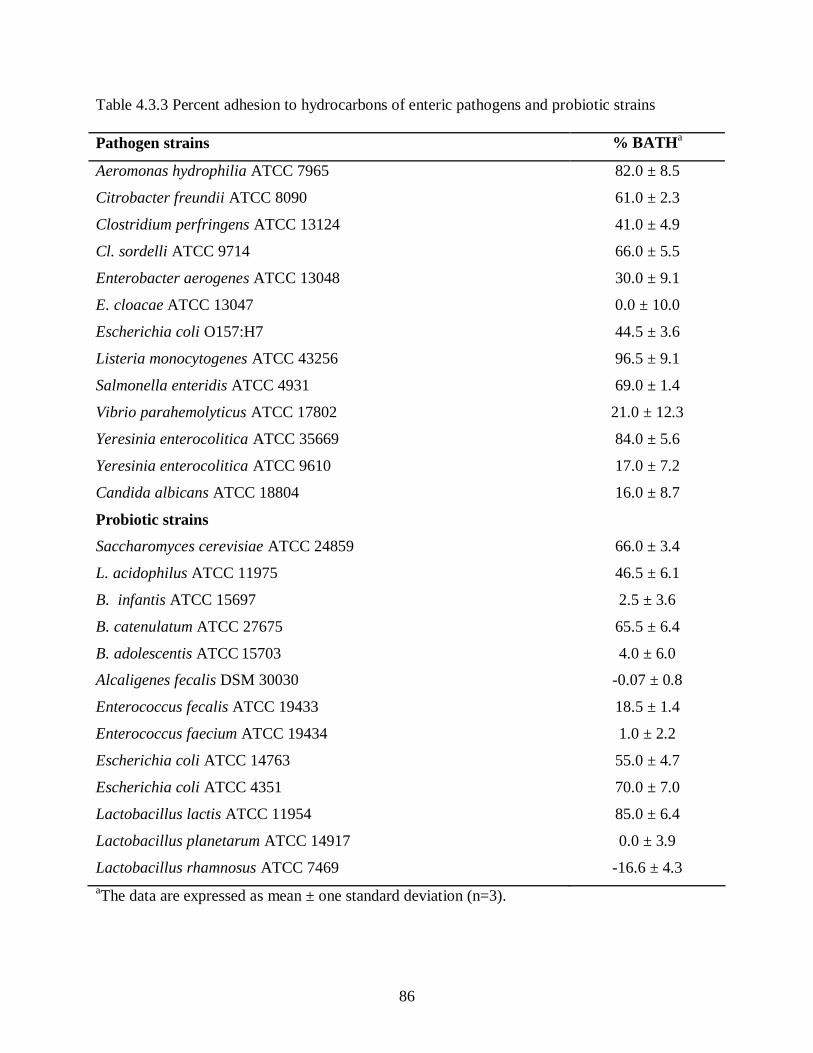
86
Table 4.3.3 Percent adhesion to hydrocarbons of enteric pathogens and probiotic strains Pathogen strains % BATHa
Aeromonas hydrophilia ATCC 7965 82.0 ± 8.5
Citrobacter freundii ATCC 8090 61.0 ± 2.3
Clostridium perfringens ATCC 13124 41.0 ± 4.9
Cl. sordelli ATCC 9714 66.0 ± 5.5
Enterobacter aerogenes ATCC 13048 30.0 ± 9.1
E. cloacae ATCC 13047 0.0 ± 10.0
Escherichia coli O157:H7 44.5 ± 3.6
Listeria monocytogenes ATCC 43256 96.5 ± 9.1
Salmonella enteridis ATCC 4931 69.0 ± 1.4
Vibrio parahemolyticus ATCC 17802 21.0 ± 12.3
Yeresinia enterocolitica ATCC 35669 84.0 ± 5.6
Yeresinia enterocolitica ATCC 9610 17.0 ± 7.2
Candida albicans ATCC 18804 16.0 ± 8.7
Probiotic strains
Saccharomyces cerevisiae ATCC 24859 66.0 ± 3.4
L. acidophilus ATCC 11975 46.5 ± 6.1
B. infantis ATCC 15697 2.5 ± 3.6
B. catenulatum ATCC 27675 65.5 ± 6.4
B. adolescentis ATCC 15703 4.0 ± 6.0
Alcaligenes fecalis DSM 30030 -0.07 ± 0.8
Enterococcus fecalis ATCC 19433 18.5 ± 1.4
Enterococcus faecium ATCC 19434 1.0 ± 2.2
Escherichia coli ATCC 14763 55.0 ± 4.7
Escherichia coli ATCC 4351 70.0 ± 7.0
Lactobacillus lactis ATCC 11954 85.0 ± 6.4
Lactobacillus planetarum ATCC 14917 0.0 ± 3.9
Lactobacillus rhamnosus ATCC 7469 -16.6 ± 4.3 aThe data are expressed as mean ± one standard deviation (n=3).

87
4.3.3 Bacterial auto-aggregation and co-aggregation analysis
The auto-aggregation properties of L. acidophilus and pathogen strains were measured
over 24 h at 37°C using absorbance measurements at 600 nm. Results of percentage auto-
aggregation are shown in Table 4.3.4. In general, the auto-aggregation abilities of all tested
strains increased over a period of 24 h incubation at 37°C. Clostridium sordelli was the best
auto-aggregating strain among those tested, resulting in 63.6 ± 1.4% auto-aggregation after 24 h
incubation.
Table 4.3.4 Adhesion to hydrocarbons as measured using the BATH test and % auto-aggregation of probiotic and pathogenic strains
% Auto-aggregationa Bacterial strains
2h 4h 24h
L. acidophilus ATCC 11975 7.3 ± 1.0 16.9 ± 2.1 36.7 ± 4.5
E. coli O157:H7 5.2 ± 0.8 15.0 ± 1.0 45.3 ± 7.0
Cl. sordelli ATCC 9714 15.0 ± 6.3 34.0 ± 5.2 63.6 ± 1.4
aMean ± standard deviation of results from three separate experiments.
These results were confirmed by visual aggregation determination; a score of 3+ was
given to Cl. sordelli and L. acidophilus and 2+ was given to E. coli auto-aggregates.
Lactobacillus acidophilus was able to co-aggregate with both tested pathogens, the results of
which are shown in Table 4.3.5.

88
Table 4.3.5 Co-aggregation percentages of pathogen and probiotic strains after incubation at 37ºC, anaerobically
% Co-aggregation Co-aggregation with L. acidophilus
2h 4h 24h
E. coli O157:H7a 16.0 2.4 43.6 1.1 47.9 3.6
Cl. sordelli ATCC 9714a 23.2 3.2 43.7 1.5 51.8 4.2
aMean ± standard deviation of results from three separate experiments.
Percent co-aggregation tended to increase over the 24 h incubation period. The percent of
co-aggregation of L. acidophilus with E. coli O157:H7 was 43.6 1.1% after 4 h incubation and
47.9 3.6 % after 24 h. Whereas, Cl. sordelli showed 43.7 1.5% co-aggregation after 4 h and
51.8 4.2% after 24 h incubation at 37°C. These results were also confirmed by visual
determination (Figure 4.3.3) and microscopic examination (Figure 4.3.4) of co-aggregates. A
visual score of 3+ was given to both E. coli O157:H7- and Cl. sordelli-L. acidophilus
co-aggregates.
Figure 4.3.3 Visual determination of co-aggregation of L. acidophilus ATCC 11975 with E. coli O157:H7. Tubes 1 and 2 represents co-aggregation before (0 h), and after (24 h) incubation, respectively
1 2

89
Figure 4.3.4 Dark-field (10X NA 0.22) photomicrographs, showing co-aggregates of L. acidophilus and E. coli O157. A) Before incubation, and B) Co-aggregates after incubation at 37ºC for 24 h
4.3.4 Detection of antimicrobial activity of probiotic supernatant
Antagonistic activity of L. acidophilus supernatant was further evaluated by an agar-well
diffusion method. No zones of inhibition were observed with cell-free probiotic supernatant. The
antibacterial activity of probiotic cell-free supernatant (pH 4.5 ± 0.1) was examined in broth
culture using the Gram-negative aerobe, E. coli O157:H7, and then Gram-positive anaerobic
pathogen, Cl. sordelli. MRS broth (pH 6.7 ± 0.3) was used as control. Standard plate counts were
used to determine the viability of the two pathogen strains over a 24 h period (Figure 4.3.5).
A B
100 µm 100 µm

90
Figure 4.3.5 Antimicrobial activity of L. acidophilus supernatant on the growth of A) E. coli and, B) Cl. sordelli
Results showed that pathogen growth was inhibited, and that none of the pathogen strains
grew in the presence of cell-free probiotic supernatant over the first 10 h. After 24 h incubation,
the number of viable bacteria was reduced approximately by 4 log and 2 log cfu/ml for E. coli
O157:H7 and Cl. sordelli, respectively, compared to the pH 6.7 ± 0.3 MRS control (no probiotic
supernatant; Figure 4.3.5). The treatment of supernatant by trypsin at final concentration of
1mg/ml and heat (100º C for 10 min) did not affect the inhibitory activity (data not shown).

91
4.4 Discussion
Cell surface hydrophobicity assays do not measure the intrinsic microbial cell surface
hydrophobicity, but rather the bacterial partitioning to a certain hydrophobic substrate (i.e.,
xylene). The BATH test has been extensively used for measuring cell surface hydrophobicity in
LAB (Vinderola et al. 2004; Collado et al. 2008). Many studies showed that the presence of
(glycol-) proteinaceous material at the cell surface resulted in higher hydrophobicity; whereas,
hydrophilic surfaces were associated with the presence of polysaccharides (Rojas et al. 1996). In
the present study, the cell surface hydrophobicity and percent auto-aggregation of common
enteric pathogens like Salmonella enteridis and Escherichia coli O157:H7, commensal
microflora of GIT including Escherichia coli ATCC 14763, Escherichia coli ATCC 4351,
Enterococcus fecalis ATCC 19433, Enterococcus faecium ATCC 19434, and tested probiotics
were determined and are shown in figure A.1 and A.3 (Appendix A). The effect of different
cations including MgCl2, CaCl2, EDTA and Oxgall bile on cell surface hydrophobicity of
probiotics is evaluated (Figure A.2, Appendix A). Cell surface hydrophobicity was found to be
related to auto-aggregation properties as most of the strains with higher adhesion to
hydrocarbons showed high auto-aggregation abilities.
Aggregation properties of probiotics with pathogens may be of importance for both food
preservation and therapeutic impact on intestinal microbiota. Preliminary screening of
co-aggregation potential may be useful to identify potential probiotic strains suitable for food,
human or animal use. The present study was in agreement with the findings of Collado et al.
(2008) which showed that bacterial strains with higher adhesion to hydrocarbon (xylene) showed
high auto-aggregation abilities (Table 4.3.4; Figure A.1 and A.3, Appendix A). In vitro analysis
of the probiotic L. acidophilus demonstrated co-aggregation with both Gram-negative E. coli
O157:H7 and Gram-positive Cl. sordelli (Table 4.3.5). Another important finding of the present

92
study was that incubation of two probiotic strains with a single pathogen strain resulted in greater
co-aggregation and more rapid and complete clearing of liquid in the culture tube (Table A.1 and
Figure A.4, Appendix A).
This finding supports the idea that co-aggregation of probiotics with pathogens could
potentially interfere with pathogen colonization of the GIT, survival (through various
antimicrobial mechanisms) and subsequent manifestation of illness. One hypothesis is that
co-aggregation of probiotics with other established commensal GIT microflora may aid in the
attachment and colonization of probiotics to mucosal surfaces. The probiotic L. acidophilus
ATCC 11975 tested in this study showed significant antibacterial activity against both Gram-
positive Cl. sordelli ATCC 9714 and Escherichia coli O157:H7 (Figure 4.3.5). This activity was
possibly due to the antibacterial substances produced by this probiotic strain. It has been reported
by several investigators that lactobacilli are able to produce antimicrobial substances when
grown in specific media; a L. acidophilus strain produced lactacin (Barefoot et al., 1983), while a
L. plantarum isolate produced plantaricin (Anderson et al., 1988). Corr et al. (2007) has
demonstrated that the in vivo antimicrobial effects of L. salivarius UCC118 depended primarily
on their capacity to produce the bacteriocin Abp118.
Bacteria generally compete with each other by secreting antagonistic compounds, which
may affect cell viability directly or indirectly by modifying the surrounding environment. In the
present thesis research, strong probiotic antagonism against the tested pathogen strains was
detected using the agar-spot method (Table 4.3.1). Interestingly, the probiotic strain did not
exhibit the same antibacterial activity when cell free culture supernatant was tested using the
agar-well diffusion method. Similar conflicting results have been reported by Harris et al. (1989),
when seven LAB strains produced inhibitory zones against Listeria monocytogenes on solid

93
media, but only three of them produced inhibitory zones via the agar-well diffusion method. The
inability to detect antibacterial activity in the probiotic supernatant in the present study may be
due to the lower production of this substance; only in close proximity to the producing cells is a
threshold concentration achieved necessary to exert an antimicrobial effect. Another possible
explanation for the absence of antibacterial activity in the well-diffusion test is that cell-cell
contact is necessary between probiotic and pathogen strains for the factor to be produced,
something that would be absent when the culture supernatant was used. Accordingly, physical
contact between both producer organism (probiotic) and indicator (pathogens) in the same
environment might actually be necessary for the induction and synthesis of the antimicrobial.
Studies done by Toure et al. (2003) showed that the antibacterial activity of some bifidobacterial
strains was recovered by concentrating the spent cultures by speed-vac. The fact that probiotic
antagonism against the pathogens did not completely disappear when 0.2% sodium bicarbonate
was added to the culture medium (Table 4.3.2; Figure 4.3.2) is strongly suggestive that other
antibacterial factors, other than the effect of organic acids, were operative.
Jacobsen et al. (1999) reported that the inhibitory activity of lactobacilli is variable even
within the same species. In the present study, the mechanism(s) of pathogen inhibition was
seemingly not due to the production of a proteinaceous compound like a bacteriocin, which
exhibit antagonistic activity against taxonomically closely-related bacteria (Klaenhammer, 1993;
Jack et al., 1995). Previous studies have demonstrated the production of broad-spectrum
antibacterial substances, referred to as ’bacteriocin-like’ compounds, by different species of
lactobacilli (Silva et al., 1987; Camard et al., 1997). Moreover, treatment of probiotic
supernatant with either heat (100C for 10 min) or trypsin (1 mg/mL for 1 h) failed to eliminate
the antagonistic activity of probiotic supernatant during growth inhibition assay (data not

94
shown). This suggests that the probiotic-dependent inhibitory activity observed in the current
study was from the result of a heat stable non-proteinaceous substance. Niku-Paavola et al.
(1999) identified various antimicrobial compounds in the culture filtrate of Lactobacillus
plantarum, including benzoic acid, methylhydantoin and mevalonolactone, that were able to
withstand heat treatment at 120°C for 15 min as well as trypsin (1mg/mL for 1 h at 37°C)
treatment. However, it was also apparent that production of organic acid, which lowered the pH
(4.5 ± 0.1) of the probiotic supernatant, was partially responsible for the observed antagonistic
effect. Moreover, the fact that depletion of nutrients in probiotic supernatant could be a possible
explanation for growth inhibition of pathogens (figure 4.3.5). The dominance of inhibitor-
producing lactobacilli in the gastrointestinal tract and the ability of these organisms to interact
closely with enteric pathogens seemingly constitute an important host defense mechanism
against infection. Further experiments identify the chemical nature of the antibacterial
compound(s), and to establish the inhibition spectrum of the probiotic against a larger panel of
pathogenic organisms are necessary. Current findings from this research suggest that L.
acidophilus ATCC 11975 has the potential for interfering with disease-causing pathogens. Use
of GIT model systems to evaluate this inhibitory potential, in vivo, would help establish this
potential more clearly.
5 General discussion and conclusions
The greatest hurdles that probiotic bacteria must overcome during food processing,
storage and in the upper GIT are acid and the presence of bile. A probiotic L. acidophilus strain
exhibiting acid and bile tolerance has previously been isolated and evaluated. The present
characterization studies indicated that Lactobacillus acidophilus ATCC 11975 was the most
acid-tolerant strain (D-value 10.2 ± 0.80 min), able to survive 30 min at pH 2.0, and also bile-

95
tolerant. Furthermore, the highest adhesion to Caco-2 cells was observed using L. acidophilus
(4.5104 cfu/mL). L. acidophilus has the potential for interfering with disease-causing pathogens
including Escherichia coli O157:H7 and Clostridium sordelli ATCC 9714. Co- and auto-
aggregation of L. acidophilus with enteric pathogens was also apparent in this study. Use of GIT
model systems would help to evaluate the inhibitory potential of L. acidophilus more clearly.
Knowledge of antimicrobial resistance patterns may also provide important information for
consumption of naturally-resistant probiotics during antibiotic therapy, thereby protecting the
intestinal mucosa from colonization of deleterious pathogenic strains. Screening for strains that
can efficiently utilize certain prebiotics will provide competitive advantage for the probiotics in
nutrient deficient large intestine. It is thought that the sole carbon source utilization profiles will
provide useful information about the metabolic potential of the different probiotic
microorganisms. Results showed that all probiotic strains grew to the highest culture optical
densities on D-xylose. Further examination of substrate preferences of probiotics would be
necessary for co-delivery of probiotics and prebiotics. In the present study, encapsulation of L.
acidophilus strains with pea protein-alginate matrix was found to give 6 times more protection
under acid stress. Thus future probiotic-prebiotic (synbiotic) combinational developments may
be facilitated by screening the potential probiotic strain for desirable characteristics such as
probiotic substrate preferences and antibiotic resistance profiling. As a physical delivery tool,
delivery of probiotics, with or without prebiotics, within a capsule is suggested to improve the
survival and subsequently enhance the growth potential of these beneficial bacteria in the GIT.

96
6 Recommendations
Further identification and characterization of probiotic cell-wall properties is
recommended to understand their role in adhesion to hydrocarbons, auto-aggregation, and
relation to co-aggregation mechanisms. Selection of new probiotic combinations should be
conducted for specific target pathogens or pathogen-associated microbiota aberrancies.
Molecular techniques like multiplex RAPD-PCR, could be used to reveal the complete metabolic
potential of each of the probiotic strain which opens the possibility to engineer new combinations
of pre- and probiotics.

97
7 References
Abrahamsson, T. R., Jakobsson, T., Bottcher, M. F., Fredrikson, M., Jenmalm, M. C., and Bjorksten, B., 2007. Probiotics in prevention of IgE-associated eczema: a double-blind, randomized, placebo-controlled trial. J. Allergy Clin. Immunol., 119: 1174-1180.
Adamowicz, M. P., Kelley, M., and Nickerson, K. W., 1991. Detergent (sodium dodecyl sulfate) shock proteins in Escherichia coli. J. Bacteriol., 173: 229-233.
Agarwal, K. N. and Bhasin, S. K., 2002. Feasibility studies to control acute diarrhoea in children by feeding fermented milk preparations, Actimel and Indian Dahi. Eur. J. Clin. Nutr., 56: S56-S59.
Akalin, A. S., Fenderya, S., and Akbulut, N., 2004. Viability and activity of bifidobacteria in yoghurt containing fructooligosaccharide during refrigerated storage. Int. J. Food Sci. Technol., 39: 613-621.
Alakomi, H. L., Skytta, E., Saarela, M., Mattila-Sandholm, T., Latva-Kala, K., and Helander I. M., 2000. Lactic acid permeabilizes Gram negative bacteria by disrupting the outer membrane. Appl. Environ. Microbiol., 66: 2001-2005.
Alander, M., Satokari, R., Korpela, R., Saxelin, M., Vilpponen-Salmela, T., Mattila-Sandholm, T., and Wright, V. A., 1999. Persistence of colonisation of human colonic mucosa by a probiotic strain, Lactobacillus rhamnosus GG after oral consumption. Appl. Environ. Microbiol., 65: 351–354.
Ammor, M. S., Florez, A. B., Mayo, B., 2007. Antibiotic resistance in non-enterococcal lactic acid bacteria and bifidobacteria. Food Microbiol., 24: 559-570.
Anderson, R. E., Daeschel, M. A., and Hassan, H. M., 1988. Antibacterial activity of plantaricin SIK-83, a bacteriocin produced by Lactobacillus plantarum. Biochem., 70: 381-390.
Anderson, A. D., McNaught, C. E., Jain, P. K. and MacFie, J., 2004. Randomised clinical trial of symbiotic therapy in elective surgical patients. Gut, 53: 241-245.
Andersson, A. F., Lindberg, M., Jakobsson, H., Backhed, F., Nyren, P., and Engstrand, L., 2008. Comparative analysis of human gut microbiota by barcoded pyrosequencing. PLoS ONE, 3: 1-8.
Apajalahti, J. H. A., Sarkilahti, L. K., Maki, B. R. E., Heikkinen, J. P., Nurminen, P. H., and Holben, W. E., 1998. Effective recovery of bacterial DNA and percent-guanine-plus-cytosine-based analysis of community structure in the gastrointestinal tract of broiler chickens. Appl. Environ. Microbiol., 64: 4084-4088.

98
Apajalahti, J. H. A., Kettunen, A., Nurminen, P. H., Jatila, H., and Holben, W. E., 2003. Selective plating underestimates abundance and shows differential recovery of bifidobacterial species from human feces. Appl. Environ. Microbiol., 69: 5731-5735.
Aryana, K., Plauche, J. S., Rao, R. M., Mcgrew, P. and Shah, N. P., 2007. Fat-free plain yogurt manufactured with inulins of various chain lengths and Lactobacillus acidophilus. J. food Sci., 72: M79-M84.
Audet, P., Paquin, C., and Lacroix, C., 1988. Immobilized growing lactic acid bacteria with k-carrageenan-locust bean gum gel. Appl. Microbiol. Biotechnol., 29: 11-18.
Axelsson, L. V., 1998. Lactic acid bacteria: classification and physiology. In: Salminen S, Von Wright A, eds. Lactic acid bacteria: microbiology and functional aspects. 2nd ed. Marcel Dekker Inc., New York, 1-72.
Barefoot, S. F., and Klaenhammer, T. R., 1983. Detection and activity of lactacin B, a bacteriocin produced by Lactobacillus acidophilus. Appl. Environ. Microbiol., 45: 1808-1815.
Begley, M., Hill, C., and Gahan, C. G. M., 2006. Bile Salt Hydrolase Activity in Probiotics. Appl. Environ. Microbiol., 72: 1729-1738.
Beijerinck, M. W., 1901. Lactic acid bacteria of the industry. Arch. Neerl. Sci. Exact. Nat., 6: 212-243.
Benno, I., and Mitsuoka, T., 1992. Impact of Bifidobacterium longum on human fecal microflora. Microbiol. Immun., 36: 683-694.
Berg, R. D., 1998. Probiotics, prebiotics or “conbiotics.” Trends Microbiol., 6: 89-92.
Bernet, M. F., Brassart, D., Neeser, J. R., and Servin, A. L., 1994. Lactobacillus acidophilus LA 1 binds to cultured human intestinal cell lines and inhibits cell attachment and cell invasion by enterovirulent bacteria. Gut, 35: 483-489.
Bertazzoni-Minelli, E., Benini, A., Marzotto, M., Sbarbati, A., Ruzzenente, O., Ferrario, R., Hendriks, H., and Dellaglio, F., 2004. Assessment of novel probiotic Lactobacillus casei strains for the production of functional dairy foods. Int. Dairy J., 14: 723–736.
Biavati, B., Castagnoli, P., Crociani, F., and Trovatelli, L. D., 1984. Species of the Bifidobacterium in the feces of infants. Microbiologica, 7: 341-345.
Blum, S., and Reniero, R., 2000. Industrial panel statements: adhesion of selected Lactobacillus strains to enterocyte-like Caco-2 cells in vitro: a critical evaluation of the reliability of in vitro adhesion assays. Proceedings of at the 4th workshop of the PROBEMO-FAIR CT 96-1028 project “Functional Foods for EU health in 2000”.

99
Boever, D. P., Wouters, R., Verschaeve, L., Berckmans, P., Schoeters, G., and Verstraete, W., 2000. Protective effect of the bile salt hydrolase-active Lactobacillus reuteri against bile salt cytotoxicity. Appl. Microbiol. Biotecnol., 53: 709-714.
Bomba, A., Nemcova, R., Gancarcikova, S., Herich, R., Guba, P., and Mudronova, D., 2002. Improvement of the probiotic effect of micro-organisms by their combination with maltodextrins, fructo-oligosaccharides and polyunsaturated fatty acids. Br. J. Nutri., 88: S95-S99.
Bonaparte, C., 1997. Selective isolation and taxonomic position of bifidobacteria isolated from commercial fermented dairy products in central Europe. Dissertation. Technische Universitat, Berlin.
Boris, S., Suarez, J. E., Vazquez, F., and Barbes, C., 1998. Adherence of human vaginal Lactobacilli to vaginal epithelial cells and interaction with uropathogens. Infect. Immun., 66: 1985-1989.
Bouhnik, Y., Attar, A., Joly, F. A., Riottot, M., Dyard, F., and Flourie, B., 2004. Lactulose ingestion increases faecal bifidobacterial counts: A randomised double-blind study in healthy humans. Euro. J. Clin. Nutri., 58: 462-466.
Bouhnik, Y., Kvahedi, L., Achour, Attar, A., Salfati, J., Pochart, P., Marteau, P., Flourie, B., Bornet, F., and Rambaud, J. C., 1999. Short chain fructo-oligosaccharide administration dose-dependently increases fecal bifidobacteria in healthy humans. J. Nutr., 129: 113-116.
Bruzzese, E., Volpicelli, M., Squaglia, M., Tartaglione, A., and Guarino, A., 2006. Impact of prebiotics on human health. Digest. Liver Dis., 38: S283-S287.
Bunthof, C. J., Bloemen, K., Breeuwer, P., Rombouts, F. M., and Abee, T., 2001. Flow Cytometric Assessment of Viability of Lactic Acid Bacteria. Appl. Environ. Microbiol., 67: 2326-2335.
Camard, M. F. B., Brassart, V. L. B., Neeser, J. R., Servin, A. L., and Hudault, S., 1997. The human Lactobacillus acidophilus Strain LA1 secretes a non-bacteriocin antibacterial substance(s) active in vitro and in vivo. Appl. Environ. Microbiol., 63: 2747-2753.
Capela, P., Hay, T. K. C., and Shah, N. P., 2006. Effect of cryoproyectants, prebiotics and microencapsulation on survival of probiotic organisms in yoghurt and freeze-dried yoghurt. Food Res. Int., 39: 203-211.
Cebra, J. J., 1999. Influences of microbiota on intestinal immune system development. Am. J. Clin. Nutr., 69: 1046S-1051S.
Cereijido, M., Valdes, J., Shoshani, L. and Contreras, R. G., 1998. Role of tight junctions in establishing and maintaining cell polarity. Annu. Rev. Physiol. 60: 161-177.

100
Chandramouli, V., Kailasapathy, K., Peiris, P., and Jones, M., 2004. An improved method of microencapsulation and its evaluation to protect Lactobacillus spp. in simulated gastric conditions. J. Microbiol. Meth., 56: 27– 35.
Charalampopoulos, D., Pandiella, S. S. and Webb, C., 2003. Evaluation of the effect of malt, wheat and barley extracts on the viability of potentially probiotic lactic acid bacteria under acidic conditions. Int. J. Food Microbiol., 82: 133–141.
Charteris, W. P., Kelly, P. M., Morelli, L., and Collins, J. K., 1998a. Development and application of an in vitro methodology to determine the transit tolerance of potentially probiotic Lactobacillus and Bifidobacterium species in the upper human gastrointestinal tract. J. Appl. Microbiol. 84: 759-768.
Charteris, W. P., Kelly, P. M., Morelli, L. and Collins, J. K., 1998b. Antibiotic susceptibility of potentially probiotic Bifidobacterium isolates from the human gastrointestinal tract. Lett. Appl. Microbiol., 26: 333–337.
Chauviere, G., Coconnier, M. H., Kerneis, S., Fournait, J. and Servin, A. L., 1992a. Adhesion of human Lactobacillus acidophilus strain LB to human enterocyte-like Caco-2 cells. J. Gen. Microbiol., 138: 1689-1696.
Chauviere, G., Coconnier, M. H., Kerneis, S., Michaud, A. D., Joly, B., and Servin, A. L., 1992b. Competitive exclusion of diarrheagenic Escherichia coli (ETEC) from human enterocyte-like Caco-2 cells by heat-killed Lactobacillus. FEMS Microbiol. Lett., 70: 213-217.
Choi, W. S. and Han J. A., 2002. Film-forming mechanism and heat denaturation effects on the physical and chemical properties of pea-protein-isolate edible films. J. Food Sci., 67:1399-1406.
Christiaens, H., Leer, R. J., Pouwels, P. H., and Verstraete, W., 1992. Cloning and expression of a conjugated bile acid hydrolase gene from Lactobacillus plantarum by using a direct plate assay. Appl. Environ. Microbiol., 58: 3792-3798.
Coconnier, M. H., Bernet, M. F., Chauviere, G., and Servin, A. L., 1993. Adhering heat-killed human Lactobacillus acidophilus, strain LB inhibits the process of pathogenicity of diarrhoeagenic bacteria in cultured human intestinal cells. J. Diarrhoeal Dis. Res., 11: 235-242.
Collado, M. C., Meriluoto, J., and Salminen, S., 2007a. Development of new probiotics by strain combinations: Is it possible to improve the adhesion to intestinal mucus? J. Dairy Sci., 90: 2710-2716.
Collado, M. C., Surono, I., Meriluoto, J., and Salminen, S., 2007b. Indigenous dadih lactic acid bacteria: Cell-surface properties and interactions with pathogens. J. Food Sci., 72: M89-M93.
Collado, M. C., Meriluoto, J., and Salminen, S., 2008. Adhesion and aggregation properties of probiotic and pathogen strains. Eur. Food Res. Technol., 226: 1065-1073.
101
Collins, M. D., Rodrigues, U., and Ash, C., 1991. Phylogenetic analysis of the genus Lactobacillus and related lactic acid bacteria as determined by reverse transcriptase sequencing of 16S rRNA. FEMS Microbiol. Lett.; 77: 5-12.
Collins, J. K., Thornton, G., and Sullivan, G. O., 1998. Selection of probiotic strains for human application. Int. Dairy J., 8: 487-490.
Conway, P. L., Gorbach, S. L., and Golden, B. R., 1987. Survival of lactic acid bacteria in the human stomach and adhesion to intestinal cells. J. Dairy Sci., 70: 1-12.
Corcoran, B. M., Stanton, C., Fitzgerald, G. F., and Ross R. P., 2005. Survival of probiotic Lactobacilli in acidic environments is enhanced in the presence of metabolizable sugars. Appl. Environ. Microbiol., 71: 3060-3067.
Corr, S. C., Li, Y., Riedel, C. U., Toole, P., Hill, C., and Gahan, C. G. M., 2007. Bacteriocin production as a mechanism for the anti-infective activity of Lactobacillus salivarius UCC118. PNAS, 104: 7617–7621.
Cummings, J. H., Christie, S., and Cole, T. J., 2001. A study of fructo-oligosaccharides in the prevention of traveller’s diarrhoea. Aliment. Pharmacol. Ther., 15: 1139-1145.
Cusick, S. M., and O’Sullivan, D. J., 2000. Use of a single, triplicate arbitrarily primed-PCR procedure for molecular fingerprinting of lactic acid bacteria. Appl. Environ. Microbiol., 66: 2227-2231.
Dahiya, R. S., and Speck, M. L., 1967. Hydrogen peroxide formation by Lactobacilli and its effect on Staphylococcus aureus. J. Dairy Sci., 51: 1568p-1572.
Danone, 2001. Fermented Foods and Healthy Digestive Functions. France: Danone Publications, John Libbey Eurotext.
Dave, N., and Shah, P., 1997. Viability of yogurt and probiotic bacteria in yogurts made from commercial starter culture. Int. Dairy J., 731-741.
De Keersmaecker, S. C., Verhoeven, T. L., Desair, J., Marchal, K., Vanderleyden, J. and Nagy, I., 2006. Strong antimicrobial activity of Lactobacillus rhamnosus GG against Salmonella typhimurium is due to accumulation of lactic acid. FEMS Microbiol. Lett., 259: 89-96.
De Vrese, M., Stegelmann, A., Richter, B., Fenselau, S., Laue, C., and Schrezenmeir, J., 2001. Probiotics - compensation for lactase insufficiency. Am. J. Clin. Nutr., 73: 421S-429S.
Defoirdt, T., Boon, N., Bossier, P., and Verstraete, W., 2004. Disruption of bacterial quorum sensing: an unexplored strategy to fight infections in aquaculture. Aqua., 240: 69-88.
Del Re, B., Sgorbati, B., Miglioli, M., and Palenzona, D., 2000. Adhesion, autoaggregation and hydrophobicity of 13 strains of Bifidobacterium longum. Lett. Appl. Microbiol. 31: 438-442.

102
Desai, A. R., Musil, K. M., Carr, A. P., and Hill, J. E., 2009. Characterization and quantification of feline fecal microbiota using cpn60 sequence-based methods and investigation of animal-to-animal variation in microbial population structure. Vet. Microbiol., 137: 1-9.
Desmond, C., Ross, R. P., OCallaghan, E., Fitzgerald, G. and Stanton, C., 2002. Improved survival of Lactobacillus paracasei NFBC 338 in spray-dried powders containing gum acacia. J. Appl. Microbiol., 93: 1003-1011.
Didierlaurent, A., Sirard, J. C., Kraehenbuhl, J. P., and Neutra, M. R., 2002. How the gut senses its content? Cell. Microbiol., 4: 61-72.
Dirix, G., Monsieurs, P., Dombrecht, B., Daniels, R., Marchal, K., Vanderleydena, J., and Michiels, J., 2004. Peptide signal molecules and bacteriocins in Gram-negative bacteria: a genome-wide in silico screening for peptides containing a double-glycine leader sequence and their cognate transporters. Peptides, 25: 1425-1440.
Doderlein, A., 1892. The vaginal transudate and its significance for childbed fever. Centralbl. Bakteriol., 11: 699-700.
Doern, G. V., Dascal, A., and Keville, M., 1985. Susceptibility testing with the sensititer breakpoint broth microdilution system. Diagn. Microbiol. Infect. Dis., 3: 185-191.
Dong, Y. H., Gusti, A. R., Zhang, Q., Xu, J. L., and Zhang, L. H., 2002. Identification of quorum-quenching N-acyl homoserine lactonases from Bacillus species. Appl. Environ. Microbiol., 68: 1754-1759.
Drider, D., Fimland, G., Chard, Y., McMullen, L. M., and Prevost, H., 2006. The continuing story of class IIa bacteriocins. Microbiol. Mol. Biol. Rev., 70: 564–582.
Dumonceaux, T. J., Hill, J. E., Briggs, S. A., Amoako, K. K., Hemmingsen, S. M., and Van Kessel, A. G., 2006. Enumeration of specific bacterial populations in complex intestinal communities using quantitative PCR based on the chaperonin-60 target. J. Microbiol. Meth., 64: 46-62.
Dunne, C., OMahony, L., Murphy, L., Thornton, G., Morrissey, D., OHalloran, S., Feeney, M., Flynn, S., Fitzgerald, G., Daly, C., Kiely, B., O’Sullivan, G. C., Shanahan, F., and Collins, J. K., 2001. In vitro selection criteria for probiotic bacteria of human origin: correlation with in vivo findings. Am. J. Clin. Nutr., 73: 386S-392S.
Elliott, S. E., Buret, A., McKnight, W., Miller, M. J. S., and Wallace, J. L., 1998. Bacteria rapidly colonize and modulate healing of gastric ulcers in rats. Am. J. Physiol., 275: G425-G432.
Erkkila, S., and Petaja, E., 2000. Screening of commercial meat starter cultures at low pH and in the presence of bile salts for potential probiotic use. Meat Sci., 55: 297-300.
Escherich, T., 1885. Die darmbakterien des neugeborenen und sauglings. Fortschr. Med., 3: 515-522.

103
Favaro-Trindale, C. S. and Grosso, C. R. F., 2002. Microencapsulation of L. acidophilus (La-05) and B. lactis (Bb-12) and evaluation of their survival at the pH values of the stomach and in bile. J. Microencap., 19: 485-494.
Favier, C., Neut, C., Mizon, C., Cortot, A., Colombel, J. F., and Mizon, J., 1997. Faecal β-D-galactosidase production and bifidobacteria are decreased in Crohn’s-disease. Digest. Dis. Sci., 42: 817-822.
Fayol-Messaoudi, D., Berger, C. N., Coconnier-Polter, M. H., Lievin-Le Moal, V. and Servin, A. L., 2005. pH-, lactic acid-, and non-lactic acid-dependent activities of probiotic lactobacilli against Salmonella enterica serovar typhimurium. Appl. Environ. Microbiol., 71: 6008-6013.
Fernandes, C. F., Shahani, K. M., and Amer, M. A., 1987. Therapeutic role of dietary lactobacilli and lactobacillic fermented dairy products. FEMS Microbiol. Rev., 46: 343–356.
Finegold, S. M., Sutter, V. L., Sugihara, P. L., Elder, H. A., Lehmann, S. M., and Phillips, R. L., 1977. Fecal microbial flora in seventh day adventist populations and control subjects. Am. J. Clin. Nutr., 30: 1781-1792.
Food and Agriculture Organization and World Health Organization (FAO/WHO), 2001. Health and nutritional properties of probiotics in food including powder milk with live lactic acid bacteria. FAO and WHO Joint Expert Committee Report.
Food and agricultural organization and World health Organization (FAO/WHO), 2002. Joint working group report on drafting guidelines for the evaluation of probiotics in food. London, Ontario, April 30 and May 1.
Forestier, C., Champs, C. D., Vatoux, C., and Joly, B., 2001. Probiotic activities of Lactobacillus casei rhamnosus: in vitro adherence to intestinal cells and antimicrobial properties. Res. Microbiol., 152: 167-173.
Franz, C. M. A. P., Specht, I., haberer, P. and Holzapfel, W. H., 2001. Bile salt hydrolase activity of Enterococci isolated from food: screening and quantitative determination. J. Food Prot., 64: 725-729.
Fredrickson, A. G. and Stephanopoulos, G., 1981. Microbial competition. Science, 213: 972–979.
FUFOSE working group, 1999. Scientific concepts of functional foods in Europe- Consensus document. Br. J. Nutr., 81: S1-S27.
Fuller, R., 1975. Nature of the determinant responsible for the adhesion of lactobacilli to chicken crop epithelial cells. J. Gen. Microbiol., 87: 245-250.
Fuller, R., 1989. Probiotics in man and animals. J. Appl. Bacteriol., 66: 365–378.
Gallaher, D. D., and Khil, J., 1999. The effect of synbiotics on colon carcinogenesis in rats. J. Nutr., 129: 1483S-1487S.

104
Gan, B. S., Kim, J., Reid, G., Cadieux, P., and Howard, J. C., 2002. Lactobacillus fermentum RC-14 inhibits Staphylococcus aureus infection of surgical implants in rats. J. Infect. Dis., 185: 1369-1372.
Gardiner, G. E., Ross, R. P., Wallace, J. M., Scanlan, F. P., Jagers, P. P., Fitzgerald, F. P., Collins, J. K., and Stanton, C., 1999. Influence of a probiotic adjunct culture of Enterococcus faecium on the quality of cheddar cheese. J. Agric. Food Chem., 47: 4907-4916.
Gardiner, G. E, Heinemann, C., Baroja, M. L., Bruce, A. W., Beuerman, D., Madrenas, J., Reid, G., 2002. Oral administration of the probiotic combination Lactobacillus rhamnosus GR-1 and L. fermentum for human intestinal applications. Int. Dairy J., 12: 191-196.
GastroNet Australia, 2001. Your digestive system, downloaded from http://www.gastro.net.au/frame_digestive.html on 14.06.2001.
Gauthier, R., 2002. Intestinal health, the key to productivity (The case of organic acids). Precongreso Cientifico Avicola IASA, XXVII Convencion ANECA-WPDC, Puerto Vallarta, Jal. Mexico, 1-12.
Gibson, G. R., and Roberfroid, M. R., 1995. Dietary modulation of the human colonic microbial: introducing the concept of prebiotics. J. Nutr., 125: 1401-12.
Gilliland, S. E., Staley, T. E., and Bush, L. J., 1984. Importance in bile tolerance of Lactobacillus acidophilus used as a dietary adjunct. J. Dairy Sci., 67: 3045-3051.
Goktepe, I., Juneja, V. K. and Ahmedna, M., 2006. Probiotics in food safety and human health. Boca Raton, FL: Taylor and Francis group.
Goldin, B. R., Gorbach, S. L., Saxelin, M., Barakat, S., Gualtieri, L., and Salminen, S., 1992. Survival of Lactobacillus species (strain GG) in human gastrointestinal tract. Dig. Dis. Sci., 173: 121-128.
Gombotz, W. R., and Wee, S. F., 1998. Protein release from alginate matrices. Adv. Drug Deliv. Rev., 31: 276-285.
Gomes, A. M. P., and Malcata, F. X., 1999. Bifidobacterium species and Lactobacillus acidophilus: biological, biochemical, technological and therapeutical properties relevant for use as probiotics. Trends Food Sci. Technol., 10: 139-157.
Gopal, A., Shah, N. P. and Roginski, H., 1996. Bile tolerance, taurocholate deconjugation and cholesterol removal by Lactobacillus acidophilus and Bifidobacterium spp. Milchwissenschaft., 51: 619-623.
Gopal, P. K., Prasad, J., Smart, J., and Gill, H. S., 2001. In vitro adherence properties of Lactobacillus rhamnosus DR20 and Bifidobacterium lactis DR10 strains and their antagonistic activity against an enterotoxigenic Escherichia coli. Int. J. Food Microbiol., 67: 207-216.

105
Grill, J. P., Perrin, S., and Schneider, F., 2000a. Bile salt toxicity to some Bifidobacteria strains: role of conjugated bile salt hydrolase and pH. Can. J. Microbiol., 46: 878-884.
Grill, J. P., Cayuela C., Antonie, J. M., and Schneider, F., 2000b. Isolation and characterization of a Lactobacillus amylovorus mutant depleted in conjugated bile salt hydrolase activity: relation between activity and bile salt resistance. J. Appl. Microbiol., 89: 553-563.
Groboillot, A. F., Champagne, C. P., Darling, G. D., and Poncelet, D., 1993. Membrane formation by interfacial cross-linking of chitosan for microencapsulation of Lactococcus lactis. Biotechnol. Bioeng., 42: 1157-1163.
Gueimonde, M., Margolles, A., Reyes-Gavilan, C. G., and Salminen, S., 2007. Competitive exclusion of enteropathogens from human intestinal mucus by Bifidobacterium strains with acquired resistance to bile: a preliminary study. Int. J. Food Microbiol., 113: 228-232.
Harris, L. J., Daeschel, M. A., Stiles, M. E., and Klaenhammer, T. R., 1989. Antimicrobial activity of lactic acid bacteria against Listeria monocytogenes. J. Food Protect., 52: 384-387.
Havenaar, R., and Veld, J. H. J., 1992. Probiotics, a general view. In: B. B. Wood (Ed.) The lactic acid bacteria in health and disease. Elsevier, London, 1: 209-224.
Heatley, R. V., and G. M. Sobala. 1993. Acid suppression and gastric flora. Baillere’s Clin. Gastroenterol., 7: 167-181.
Hertzler, S. R., and Savaiano, D. A., 1996. Colonic adaptation to daily lactose feeding in lactose maldigesters reduces lactose intolerance. Am. J. Clin. Nutr., 64: 232-236.
Hofmann, A. F., and Roda, A., 1984. Physicochemical properties of bile acids and their relationship to biological properties: an overview of the problem. J. Lipid Res., 25: 1477–1489.
Holzapfel, W. H., Haberer, P., Snel, J., Schillinger, U. and Huis in’t Veld, J. H., 1998. Overview of gut flora and probiotics. Int. J. Food Microbiol., 41: 85-101.
Holzapfel, W. H., Haberer, P., Geisen, R., Bjorkroth, J., and Schillinger, U., 2001. Taxonomy and important features of probiotic microorganisms in food and nutrition. Am. J. Clin. Nutr., 73: 365S-73S.
Holzapfel, W. H., and Schillinger, U., 2002. Introduction to pre- and probiotics. Food Res. Int., H35: 109-116.
Hood, S. K., and Zottola, A., 1988. Effect of low pH on the ability of Lactobacillus acidophilus to survive and adhere to human intestinal cells. J. Food. Sci., 53: 1514-1516.
Hopkins, M. J., Cummings, J. H. and Macfarlane, G. T., 1998. Inter-species differences in maximum specific growth rates and cell yields of bifidobacteria cultured on oligosaccharides and other simple carbohydrate sources. J. Appl. Microbiol., 85: 381-386.

106
Hou, R.C., Lin M. Y., Wang, M. M. and Tzen, J. T., 2003. Increase of viability of entrapped cells of Lactobacillus delbrueckii spp. Bulgaricus in artificial sesame oil emulsions. J. Dairy Sci., 86: 424-428.
Jack, R. W., Tagg, J. R., and Ray, B., 1995. Bacteriocins of Gram-positive bacteria. Microbiol. Rev., 59: 171-200.
Jacobs, M. F., Tynkkynen, S., and Sibakov, M., 1995. Highly bioluminescent Streptococcus thermophilus strain for the detection of dairy relevant antibiotics in milk. Appl. Microbiol. Biotecnol., 44: 405.
Jacobsen, C. N., Nielsen, V. R., Hayford, A. E., Moller, P. L., Michaelsen, K. F., Paerregaard, A., Sandstrom, B., Tvede, M. and Jakosen, M., 1999. Screening of probiotic activities of forty-seven strains of Lactobacillus spp. by in vitro techniques and evaluation of the colonization ability of five selected strains in humans. Appl. Environ. Microbiol., 65: 4949-4956.
Jin, L. Z., Marquardt, R. R. and Baidoo, S. K., 2000. Inhibition of enterotoxigenic Escherichia coli K88, K99 and 987P by the Lactobacillus isolates from porcine intestine. J. Sci. Food Agric., 80: 619-624.
Kailasapathy, K. and Chin, J., 2000. Survival and therapeutic potential of probiotic organisms with reference to Lactobacillus acidophilus and Bifidobacterium spp. Immunol. Cell Biol., 78: 80-88.
Kalliomaki, M., Salminen, S., Arvilommi, H., Kero, P., Koskinen, P., and Isolauri, E., 2001. Probiotics in primary prevention of atopic disease: a randomised placebo-controlled trial. Lancet, 357: 1076-1079.
Kawamoto, K., Horibe, I., and Uchida, K., 1989. Purification and characterization of a new hydrolase for conjugated bile acids, chenodeoxy- cholyltaurine hydrolase, from Bacteroides vulgatus. J. Biochem., 106: 1049-1053.
Kebary, K. M. K., Hussein, S. A., and Badawi, R. M., 1998. Improving viability of Bifidobacteria and their effect on frozen ice milk. Egypt. J. Dairy Sci., 26: 319-337.
Kets, E. P. W., Teunissen, P. J. M., and Bont, J. A. M., 1996. Effect of compatible solutes on survival of lactic acid bacteria subjected to drying. App. Environ. Microbiol., 62: 259-291.
Kim, K. I., and Yoon, Y. H., 1995. A study on the preparation of direct vat lactic acid bacterial starter. Korea. J. Dairy Sci., 17: 129-134.
Kim, S., Cho, S. Y., Kim, S. H., Song, O., Shind, S., Cha, D., and Parke, H. J., 2008. Effect of microencapsulation on viability and other characteristics in Lactobacillus acidophilus ATCC 43121. LWT, 41: 493-500.
Kimura, K., McCartney, A. L., McConnell, M. A. and Tannock, G. T., 1997. Analysis of fecal populations of bifidobacteria and lactobacilli and investigation of the immunological

107
responses of their human hosts to the predominant strains. Appl. Environ. Microbiol., 63: 3394-3398.
Klaenhammer, T. R., 1993. Genetics of bacteriocins produced by lactic acid bacteria. FEMS Microbiol. Rev., 12: 39–85.
Klare, I., Konstabel, C., Bertling, S. M., Reissbrodt, R., Huys, G., Vancanneyt, M., Swings, J., Goossens, H., and Witte, W., 2005. Evaluation of new broth media for microdilution antibiotic susceptibility testing of Lactobacilli, Pediococci, Lactococci, and Bifidobacteria. Appl. Environ. Microbiol., 71: 8982-8986.
Klein, G., Pack, A., Bonaparte, C., and Reuter, G., 1998. Taxonomy and physiology of probiotic lactic acid bacteria. Int. J. Food. Microbiol., 41: 103-125.
Kolars, J. C., Levitt, M. D., Aouji, M., and Savaiano, D. A., 1984. Yoghurt- an auto digesting source of lactose. N. Engl. J. Med., 310: 1-3.
Kollath, W., 1953. Ernahrung und Zahnsystem. Deutsche Zahnarztliche Zeitschrift. 8: 7-16.
Kopp-Hoolihan, L., 2001. Prophylactic and therapeutic uses of probiotics: a review. J. Am. Diet Assoc., 101: 229-238.
Krasaekoopt, W., Bhandari, B., and Deeth, H. 2003. Evaluation of encapsulation techniques of probiotics for yoghurt. Int. Dairy J., 13: 3-13.
Kurdi, P., Veen, H. W. V., Tanaka, H., Mierau, I., Konings, W. N., Tannock, G. W., Tomita, F., and Yokota, A., 2000. Cholic acid is accumulated spontaneously, driven by membrane delta pH, in many lactobacilli. J. Bacteriol., 182: 6525-6528.
Lambert, R. J., and Stratford, M., 1999. Weak-acid preservatives: modelling microbial inhibition and response. J. Appl. Microbiol., 86: 157–164.
Langendijk, P. S., Schut, F., Jansen, G. J., Raangs, G. C., and Kamphuis, G. R., Wilkinson, M. H. and Welling, G. W., 1995. Quantitative fluorescence in situ hybridization of Bifidobacterium spp. with genus-specific 16S rRNA-targeted probes and its application in faecal samples. Appl. Environ. Microbiol., 61: 3069-3075.
Lankaputhra, W. E. V., and Shah, N. P., 1995. Survival of Lactobacillus acidophilus and Bifidobacterium spp in the presence of acid and bile salts. Cult. Dairy Prod. J., 30: 2-7.
Lee, K. I., and Heo, T. R., 2000. Survival of Bifidobacterium longum immobilized in calcium alginate beads in simulated gastric juices and bile salt solution. Appl. Environ. Microbiol., 66: 869-973.
Levri, K. M., Ketvertis, K., Deramo, M., Merenstein, J. H., and D'Amico F., 2005. Do probiotics reduce adult lactose intolerance? A systematic review. J. Fam. Pract., 54: 613-620.

108
Lian, W. C., Hsiao, H. C., and Chou, C. C., 2003. Viability of microencapsulated bifidobacteria in simulated gastric juice and bile solution. Int. J. Food Microbiol., 86: 293-301.
Lidbeck, A., and Nord, C. E., 1993. Lactobacilli and the normal human anaerobic microflora. Int. J. Tissue React., 13: 115-122.
Lilly, D. M., and Stillwell, R. H., 1965. Probiotics: growth promoting factors produced by microorganisms. Science, 147: 747-748.
Ling, W. H., Korpela, R., Mykkanen, H., Salminen, S. and Hanninen, O., 1994. Lactobacillus strain GG supplementation decreases colonic hydrolytic and reductive enzyme activities in healthy female adults. J. Nutr., 124: 18-23.
Lipski, A., Friedrich, U., and Altendorf, K., 2001. Application of rRNA-targeted oligonucleotide probes in biotechnology. Appl. Microbiol. Biotechnol., 56: 40-57.
Liu, Q., Duan, Z. P., Ha da, K., Bengmark, S., Kurtovic, J., and Riordan, S. M., 2004. Synbiotic modulation of gut flora: effect on minimal hepatic encephalopathy in patients with cirrhosis. Hepatol., 39: 1441-1449.
Long, S. S., and Swenson, R. M., 1977. Development of anaerobic fecal flora in healthy new-born infants. J. Pediatr., 91: 298–301.
Losada, M. A., and Olleros, T., 2002. Towards a healthier diet for the colon: the influence of fructooligosaccharides and lactobacilli on intestinal health. Nutri. Res., 22: 71-84.
Lourens-Hattingh, A., and Viljoen, B. C., 2001. Review: Yoghurt as probiotic carrier food. Int. Dairy J., 11: 1-17.
Lundeen, S. G., and Savage, D. C., 1990. Characterization and purification of bile salt hydrolase from Lactobacillus spp. strain 100-100. J. Bacteriol., 172: 4171-4177.
Macfarlane, G. T., Steed, H., and Macfarlane, S., 2008. Bacterial metabolism and health-related effects of galacto-oligosaccharides and other prebiotics. J. Appl. Microbiol., 104: 305-344.
Madureira, A. R., Pereiraa, C. I., Truszkowskab, Gomesa, A. M., Pintadoa, M. E., and Malcataa, F. X., 2005. Survival of probiotic bacteria in a whey cheese vector submitted to environmental conditions prevailing in the gastrointestinal tract. Int. Dairy J., 15: 921-927.
Makras, L., Triantafyllou, V., Fayol-Messaoudi, D., Adriany, T., Zoumpopoulou, G. and Tsakalidou, E., 2006. Kinetic analysis of the antibacterial activity of probiotic lactobacilli towards Salmonella enterica serovar typhimurium reveals a role for lactic acid and other inhibitory compounds. Res. Microbiol., 157: 241-247.
Margolles, A., Garcia, L., Sanchez, B., Gueimonde, M., and Gavilan, C. G., 2003. Characterization of a Bifidobacterium strain with acquired resistance to cholate - A preliminary study. Int. J. Food Microbiol., 82: 191-198.

109
Masco, L., Hoorde1, K. V., Brandt, E. D., Swings, J., and Huys, G., 2006. Antimicrobial susceptibility of Bifidobacterium strains from humans, animals and probiotic products. J. Antimicrob. Chemother., 58: 85-94.
Matijasic, B. B., Narat, M., and Zoric, M., 2003. Adhesion of two Lactobacillus gasseri probiotic strains on Caco-2 cells. Food Tech. Biotechnol., 41: 83-88.
Matsuki, T., Watanabe, K., Fujimoto, J., Miyamoto, Y., Takada, T., Matsumoto, K., Oyaizu, H., and Tanaka, R., 2002. Development of 16S rRNA-gene-targeted group-specific primers for the detection and identification of predominant bacteria in human feces. Appl. Environ. Microbiol., 68: 5445-5451.
McClean, K. H., Winson, M. K., Fish, L., Taylor, A., Chhabra, S. R., Camara, M., Daykin, M., Lamb, J. H., Swift, S., Bycroft, B. W., Stewart, G. S. A. B., and Williams, P., 1997. Quorum sensing and Chromobacterium violaceum: exploitation of violacein production and inhibition for the detection of N-acylhomoserine lactones. Microbiol., 143: 3703- 3711.
Metchnikoff, E., 1907. The prolongation of life. Optimistic studies. William Heinemann, London. United Kingdom.
Miller, M. B., Bassler, B. L., 2001. Quorum sensing in bacteria. Annu. Rev. Microbiol., 55: 165- 199.
Millette, M., Luquet, F. M. and Lacroix, M. 2007. In vitro growth control of selected pathogens by Lactobacillus acidophilus and Lactobacillus casei-fermented milk. Lett. Appl. Microbiol., 44: 314–319.
Minekus, M., Marteau, P., Havenaar R, and Huisintveld, J. H. J., 1995. A multicompartmental dynamic computer-controlled model simulating the stomach and small-intestine. ATLA Alternat. Lab Animals. 23: 197-209.
Mitsuoka, T., 1990. Bifidobacteria and their role in human health. J. Ind. Microbiol., 6: 263-268.
Molin, G., Jeppsson, B., and Johansson, M. L., 1993. Numerical taxonomy of Lactobacillus spp. associated with healthy and diseased mucosa of the human intestines. J. Appl. Bacteriol., 74: 314-23.
Moore, W. E. C., Cato, E. P., and Holdeman, L. V., 1978. Some current concepts in intestinal bacteriology. Am. J. Clin. Nutr., 31: S33-S42.
Morelli, L., 2000. In vitro selection of probiotic lactobacilli: a critical appraisal. Curr. Iss. Intest. Microbiol., 1: 59-67.
Morelli, L., 2007. In vitro assessment of probiotic bacteria: From survival to functionality. Int. Dairy J., 17: 1278-1283.

110
Moro, E., 1900. Bacillus acidophilus n. spec., A contribution to the knowledge of the normal intestinal bacteria of infants. Jahr. Kinderheilkunde, 52: 38-55.
Mortazavian, A., Razavi, S. H., Ehsani, M. R., and Sohrabvandi, S., 2007. Principles and methods of microencapsulation of probiotic microorganisms. Iran. J. Biotech., 5: 1-18.
Moser, S. A., and Savage, D. C., 2001. Bile salt hydrolase activity and resistance to toxicity of conjugated bile salts are unrelated properties in Lactobacilli. Appl. Environ. Microbiol., 67: 3476-3480.
Moubareck, C., Gavini, F., Vaugien, L., Butel, M. J. and Doucet-Populaire, F., 2005. Antimicrobial susceptibility of bifidobacteria. J. Antimicrob. Chemother., 55: 38-44.
Naaber, P., Smidt, I., Stsepetova, J., Brilene, T., Annuk, H. and Mikelsaar, M., 2004. Inhibition of Clostridium difficile strains by intestinal Lactobacillus species. J. Med. Microbiol., 53: 551–554.
National Committee for Clinical Laboratory Standards. 2002. Performance standards for antimicrobial susceptibility testing: Twelfth informational supplement M100-S12. National committee for clinical laboratory standards, Villanova.
National Committee for Clinical Laboratory Standards. 2002. Performance standards for antimicrobial disk and dilution susceptibility tests for bacteria isolated from animals; approved standard M31-A2. National committee for clinical laboratory standards, Villanova.
Nealson, K. H., Platt, T., and Hastings, J. W., 1970. Cellular control of the synthesis and activity of the bacterial luminescence system. J. Bacteriol., 104: 313- 322.
Nicaise, P., Gleizes, A., Sandre, C., Kergot, R., Lebrec, H., Forestier, F. and Labarre, C., 1999. The intestinal microflora regulates cytokine production positively in spleen-derived macrophages but negatively in bone marrow-derived macrophages. Eur. Cytokine Netw. 10: 365-372.
Niku-Paavola, M. L., Laitila, A., Mattila-Sandholm, T. and Haikara, A., 1999. New types of antimicrobial compounds produced by Lactobacillus plantarum. J. Appl. Microbiol., 86: 29-35.
Noriega, L., Gueimonde, M., Sanchez, B., Margolles, A., Clara, G., and Reyes-Gavilan, D. L., 2004. Effect of the adaptation to high bile salts concentrations on glycosidic activity, survival at low PH and cross-resistance to bile salts in Bifidobacterium., Int. J. Food Microbiol., 94: 79-86.
Northfield, T. C., and McColl, I., 1973. Postprandial concentrations of free and conjugated bile acids down the length of the normal human small intestine. Gut, 14: 513-518.
O’Sullivan, M. G., Thornton, G. M., O’Sullivan, G. C., and Collins, J. K., 1992. Probiotic bacteria: myth or reality? Trends Food Sci. Technol., 3: 309-14.

111
Ocana, V. S. Holgado, A. P. R., and Macias, M. E. N., 1999. Characterization of a bacteriocin-like substance produced by a vaginal Lactobacillus salivarius strain. Appl. Environ. Microbiol., 65: 5631-5635.
Oh, S., Kim, S. H., and Worobo, R. W., 2000. Characterization and purification of a bacteriocin produced by a potential probiotic culture, Lactobacillus acidophilus 30SC. J Dairy Sci., 83: 2747–2752.
Oppegard, C., Fimland, G., Thorbaek, L., and Meyer, J. N., 2007. Analysis of the two-peptide bacteriocins Lactococcin G and Enterocin 1071 by site-directed mutagenesis. Appl. Environ. Microbiol., 73: 2931–2938.
Otero, M. C., and Macias, M. E. N., 2006. Inhibition of Staphylococcus aureus by H2O2-producing Lactobacillus gasseri isolated from the vaginal tract of cattle. Anim. Reprod. Sci., 96: 35-46.
Ouwehand, A., Salminen, S., and Isolauri, E. 2002. Probiotics: an overview of beneficial effects. Antonie van Leeuwenhoek, 82: 279-289.
Ozer, B., Kirmaci, H. A., Senel, E., Atamer, M., and Hayaloglu. A., 2009. Improving the viability of Bifidobacterium bifidum BB-12 and Lactobacillus acidophilus LA-5 in white-brined cheese by microencapsulation., Int. Dairy J., 19: 22-29
Palframan, R., Gibson, G. R., Rastall, R. A., 2003. Development of a quantitative tool for the comparison of the prebiotic effect of dietary oligosaccharides. Lett. Appl. Microbiol., 37: 281-284.
Parvez, S., Malik, K. A., Kang, S. A., and Kim, H. Y., 2006. Probiotics and their fermented food products are beneficial for health. J. Appl. Microbiol., 100: 1171–1185.
Pennacchia, C., Vaughan, and Villani, F., 2006. Potential probiotic Lactobacillus strains from fermented sausages: Further investigations on their probiotic properties. Meat Sci., 73: 90-101.
Perdigon, G., Galdeano, M. C., Valdez, J. C., Medici, M., 2002. Interaction of lactic acid bacteria with the gut immune system. Eur. J. Clin. Nutr., 4: S21-26.
Pierucci, A. P. T. R., Andrade, L. R., Farina, M., Pedrosa, C., and Rocha-Leao M. H. M., 2007. Comparison of α-tocopherol microparticles produced with different wall materials: pea protein a new interesting alternative. J. Microencap., 24: 201-213.
Pleiss, E. M., D., and Dicks, L. M. T., 1995. Evaluation of random amplified polymorphic DNA (RAPD)-PCR as a method to differentiate Lactobacillus acidophilus, Lactobacillus crispatus, Lactobacillus amylovorus, Lactobacillus gallinarum, Lactobacillus gasseri , and Lactobacillus johnsonii. Curr. Microbiol., 31: 114-118.
Prevost, H., and Divies, C., 1988. Continuous pre-fermentation of milk by entrapped yoghurt bacteria: development of the process. Milchwissenschaft 43: 621-625.

112
Psomas, E., Andrghetto, C., Litopoulou-Tzanetaki, E., Lombardi, A., Tzanetakis, N., 2001. Some probiotic properties of yeast isolates from infant feces and feta cheese. Int. J. Food Microbiol., 69: 125-133.
Rafsky, H. A., and Rafsky, J. C., 1955. Clinical and bacteriological studies of a new Lactobacillus concentrate in functional disturbances. Am. J. Gastroenterol., 24: 87–92.
Rangel, A., Domont, G. D., Pedrosa, C., and Ferreira, S. T., 2003. Functional properties of purified vicilins from cowpea (Vigna unguiculata) and pea (Pisum sativum) and cowpea protein isolate. J. Agric. Food Chem., 51: 5792-5797.
Rao, A. V., Shiwnarain, N. and Maharaj, J., 1989. Survival of microencapsulated Bifidobacterium pseudolongum in simulated gastric and intestinal juices. Can. Inst. Food. Sci. Technol. J., 22: 345-349.
Rastall, R. A., Gibson, G. R., 2002. Prebiotic oligosaccharides: evaluation of biological activities and potential future developments. Caister Academic Press, London, 107-148.
Ray, B., 2004. Fundamental food microbiology, third edition. Boca Raton: CRC press
Redondo-Lopez, V., Cook, R. L., and Sobel, J. D., 1990. Emerging role of lactobacilli in the control and maintenance of the vaginal bacterial microflora. Rev. Infect. Dis. 12: 856–872.
Reid, G., 2000. In vitro testing of Lactobacillus acidophilus NCFMTM as a possible probiotic for the urogenital tract. Int. Dairy J. 10: 415-419.
Reid, G., Jass, J., Sebulsky, M. T., and McCormick, J. K., 2003. Potential uses of probiotics in clinical practice. Clin. Microbiol. Rev., 16: 658-672.
Reid, G., McGroarty, J. A., Angotti, R. and Cook, R. L., 1988. Lactobacillus inhibitor production against Escherichia coli and coaggregation ability with uropathogens. Can. J. Microbiol., 34: 344–351.
Rickard, A. H., Gilbert, P., High, N. J., Kolenbrander, P. E., and Handley, P. S., 2003. Bacterial coaggregation: an integral process in the development of multispecies biofilms. Trends Microbiol., 11: 94–100.
Riley, M. A., and Wertz, J. E., 2002. Bacteriocin diversity: ecological and evolutionary perspectives. Biochimie., 84: 357-364.
Robinson, R. K., 1987. Survival of Lactobacillus acidophilus in fermented products. S. Afr. J. Dairy Sci., 19: 25-27.
Rojas, M., and Conway, P. L., 1996. Colonization by lactobacilli of piglet small intestinal mucus. J. Appl. Bacteriol., 81: 474-480.
Rolfe, R. D., 2000. The role of probiotic cultures in the control of gastrointestinal health. J. Nutr., 130: 396S-402S.

113
Rosa, M. L., Bottaro, G., Gulino, N., Gambuzza, F., Forti, F. D., Ini, G., and Tornambe, E., 2003. Prevention of antibiotic-associated diarrhea with Lactobacillus sporogenes and fructo-oligosaccharides in children. A multicentric double-blind vs placebo study (Italian). Minerva Pediatr., 55: 447-452.
Rowland, I. R., 1989. Metabolic profiles of intestinal floras. In Hattori, T., Ishida, Y., Maruyama, Y., Morita, R. Y., and Uchida, A., (ed.), Recent advances in microbial ecology. Japan Scientific Societies Press, Tokyo, Japan, 510-514.
Rowland, J. R., Rumney, C. J., Coytts, J. T., Lievense, L. C., 1998. Effect of Bifidobacterium longum and insulin on gut bacterial metabolism and carcinogen-induced aberrant crypt foci in rats. Carcinogenesis, 19: 281-285.
Roy, D. and Ward, P., 1990. Evaluation of rapid methods for differentiation of Bifidobacterium species. J. Appl. Bacteriol., 69: 139-149.
Ruiz-Moyano, S., Martin, A., Benito, M. J., Nevado, F. P., and Cordoba, M. G., 2008. Screening of lactic acid bacteria and bifidobacteria for potential probiotic use in Iberian dry fermented sausages. Meat Sci., 80: 715-721.
Rybka, S., and Kailaspathy, K., 1995. The survival of culture bacteria in fresh and freeze-dried AB yogurt. Aust. J. Dairy Tech., 50: 51-57.
Saarela, M., Lathenmaki, L., Crittenden, R., Salminen, S., and Mattilasandholm, T., 2002. Gut bacteria and health foods - the European perspective. Int. J. Food Microbiol., 78: 99-117.
Salminen, S., and Wright, V. A., 1998a. Lactic acid bacteria: microbiology and functional aspects, second edition, revised and expanded. New York: Marcel Dekker, Inc.
Salminen, S., Wright, V. A., Morelli, L., Marteau, P., Brassart, D., Vos de, W. M., Fonde’n, R., Saxelin, M., Collins, K., Mogensen, G., Birkeland, S. E., and Sandholm, T. M., 1998b. Demonstration of safety of probiotics-a review. Int. J. Food Microbiol., 44: 93-106.
Savage, D. C., 1984. Adherence of the normal flora. In attachment of organisms to the gut mucosa. Vol. I. Beedeker, E. C. ed., Boca Raton CRC Press. p. 3-10.
Savage, D. C., 1992. Gastrointestinal microbial ecology; possible modes of action of direct-fed microbials in animal productiona review of the literature. In Direct-fd Microbials in Animal Production ed. National Feed Ingredients Association. West Des Meines, USA, 11-81.
Schellenberg, J., Smoragiewicz, W., and Karska-Wysocki, B., 2006. A rapid method combining immunofluorescence and flow cytometry for improved understanding of competitive interactions between lactic acid bacteria (LAB) and methicillin-resistant S. aureus (MRSA) in mixed culture. J. Microbiol. Meth., 65: 1-9.
Scheppach, W., Leuhrs, H., Menzel, T., 2001. Beneficial health effects of low-digestible carbohydrate consumption. Br. J. Nutr., 85: 823-930.

114
Schillinger, U., and Lucke, F. K., 1989. Antibacterial activity of Lactobacillus sake isolated from meat. Appl. Environ. Microbiol., 55: 1901-1906.
Scott, K. P., 2002. The role of conjugative transposons in spreading antibiotic resistance between bacteria that inhabit the gastrointestinal tract. Cell. Mol. Life Sci., 59: 2071-2082.
Servin, A. L., 2004. Antagonistic activities of Lactobacilli and Bifidobacteria against microbial pathogens. FEMS Microbiol. Reviews, 28: 405-440.
Shah, N. P., and Rarula, R. R., 2000. Microencapsulation of probiotic bacteria and their survival in frozen fermented dairy desserts. Aust. J. Dairy Technol., 55: 139-144.
Shahani, K. M., Vakil, J. R., and Kilara, A., 1976. Natural antibiotic activity of Lactobacillus acidophilus and bulgaricus. II. Isolation of acidophilin from L. acidophilus. Cult. Dairy Prod. J., 12: 8-11.
Sheu, T. Y., and Marshall, R. T., 1991. Improving culture viability in frozen dairy desserts by microencapsulation. J. Dairy Sci., 74: 107.
Sheu, T. Y., and Marshall, R.T., 1993. Microentrapment of lactobacilli in calcium alginate gels. J. Food Sci., 54: 557-561.
Shin, H. S., Lee, J. H., Pestka, J. J., and Ustunol, Z., 2000. Growth and viability of commercial Bifidobacterium spp. in skim milk containing oligosaccharides and inulin. J. Food Sci., 65: 884-887.
Siigur, U., Tamm, E., Torm, S., Lutsar, I., Salminen, S., and Midtvedt, T., 1996. Effect of bacterial infection and administration of a probiotic on faecal short-chain fatty acids. Microbial Ecol. Health Dis., 9: 271-277.
Silva, M., Jacobus, N. V., Deneke, C. and Gorbach, S. L., 1987. Antimicrobial substance from a human Lactobacillus strain. Antimicrob. Agents Chemother., 31: 1231-1233.
Simon, G. L., and Gorbach, S. L., 1987. Intestinal flora and gastrointestinal function. In Physiology of Gastrointestinal Tract. 2nd ed. L. R. Johnson, ed. Raven Press, New York, NY. 2: 1729.
Simova, E. D., Beshkova, D. B., and Dimitrov, Z. P., 2009. Characterization and antimicrobial spectrum of bacteriocins produced by lactic acid bacteria isolated from traditional Bulgarian dairy products. J. Appl. Microbiol., 106: 692-701.
Smet, D., Hoorde, L.V., Woestyne, M. V., Christiaens, H., and Verstraete, V., 1995. Significance of bile salt hydrolytic activities of lactobacilli. J. Appl. Bacteriol., 79: 292-301.
Smith, T., 1995. The digestive system. The Human Body. Ken Fin Books, Collingwood, 150-173.

115
Solange, I., Ismael, M., and Mancilha, M., 2007. Non-digestible oligosaccharides: A review. Carbohydr. Polym., 68: 587-597.
Soomro, A. H, Masud T., and Anwaar, K., 2002. Role of lactic acid bacteria (LAB) in food reservation and Human Health- A Review. Pakistan J. Nutr., 1: 20-24.
Steenson, L. R., Klaenhammer, T. R., and Swaisgood, H. E., 1987. Calcium alginate-immobilized cultures of lactic streptococci are protected from attack by lytic bacteriophage. J. Dairy Sci., 70: 1121-1127.
Stewart, L., Pellegrini, C. A., Way, L. W., 1986. Antibacterial activity of bile acids against common biliary tract organisms. Surg. Forum, 37: 157-159.
Sultana, K., Godward, G., Reynolds, N., Arumugaswamy, R., Peiris, P., and Kailasapathy, K., 2000. Encapsulation of probiotic bacteria with alginate-starch and evaluation of survival in simulated gastrointestinal conditions and in yoghurt. Int. J. Food Microbiol., 62: 47-55.
Suskovic, J., Kos, B., Goreta, J. and Matosic, S. 2001. Role of lactic acid bacteria and bifidobacteria in synbiotic effect. Food Technol. Biotechnol., 39: 227-235.
Swift, S., Karlyshev, A.V., Fish, L., Durant, E. L., Winson, M.K., Chhabra, S. R., Williams, P., Macintyre, S., and Stewart, G. S. A. B., 1997. Quorum sensing in Aeromonas hydrophila and Aeromonas salmonicida: identification of the LuxRI homologs AhyRI and AsaRI and their cognate N-acylhomoserine lactone signal molecules. J. Bacteriol. 179: 5271-5281.
Szilagyi, A., 2004. Redefining lactose as a conditional prebiotic. Can. J. Gastroenterol., 218: 163-167.
Tannock, G. W., Dashkevicz, M. P. and Feighner, S. D., 1989. Lactobacilli and bile salt hydrolase in the murine intestinal tract. Appl. Environ. Microbiol., 55: 1848-1851.
Tannock, G. W., Munro, K., Harmsen, H. J. M., Welling, G. W., Smart, J., and Gopal, P. K., 2000. Analysis of the fecal microflora of human subjects consuming a probiotic product containing Lactobacillus rhamnosus DR20. Appl. Environ. Microbiol., 66: 2578-2588.
Tappenden, K. A. and A. S. Deutsch, 2007. The physiological relevance of the intestinal microbiota - contributions to human health. J. Am. Coll. Nutr., 26: 679S-683S.
Taylor, A. L., Dunstan, J. A., and Prescott, S. L., 2007. Probiotic supplementation for the first 6 months of life fails to reduce the risk of atopic dermatitis and increases the risk of allergen sensitization in high-risk children: a randomized controlled trial. J. Allergy Clin. Immunol., 119: 184-191.
Teitelbaum, J. E., and Walker, W. A., 2002. Nutritional impact of pre- and probiotics as protective gastrointestinal organisms. Ann. Rev. Nutr., 22: 107-138.
116
Torriani, S., Felis, G. E., and Dellaglio, F., 2001. Differentiation of Lactobacillus plantarum, L. pentosus, and L. paraplantarum by recA gene sequence analysis and multiplex PCR assay with recA gene-derived primers. Appl. Environ. Microbiol., 67: 3450-3454.
Toure, R., Kheadr, E., Lacroix, C., Moroni, O., and Fliss, I. 2003. Production of antibacterial substances by bifidobacterial isolates from infant stool active against Listeria monocytogenes. J. Appl. Microbiol., 95: 1058-1069.
Tuomola, E. M., and Salminen, S. J., 1998. Adhesion of some probiotic and dairy Lactobacillus strains to Caco-2 cell cultures. Int. J. Food Microbiol., 41: 45-51.
Vandenbergh, P., 1993. Lactic acid bacteria, their metabolic products and interference with microbial growth. FEMS Microbiol. Rev., 12: 221-238.
Velraeds, M. M. C., Mei, H. C. V. D., Reid, G., and Busscher, H. J., 1996. Inhibition of initial adhesion of uropathogenic Enterococcus faecalis by biosurfactants from Lactobacillus isolates. Appl. Environ. Microbiol., 62: 1958–1963.
Ventura, M., Casas, I. A., Morelli, L., and Callegari, M. L., 2000. Rapid amplified ribosomal DNA restriction analysis (ARDRA) identification of Lactobacillus spp. isolated from fecal and vaginal samples. Syst. Appl. Microbiol., 23: 504-509.
Vernazza, C. L., Gibson, G. R., and Rastall, R. A., 2006. Carbohydrate preference, acid tolerance, and bile tolerance in five strains of Bifidobacterium. J. Appl. Microbiol., 100: 846-853.
Vincent, J. G., Veomett, R. C., and Riley, R. F., 1959. Antibacterial activity associated with Lactobacillus acidophilus. J. Bacteriol., 78: 477– 484.
Vinderola, C. G., Medici, M., and Perdig, G., 2004. Relationship between interaction sites in the gut, hydrophobicity, mucosal immunomodulating capacities and cell wall protein profiles in indigenous and exogenous bacteria. J. Appl. Microbiol., 93: 794–799.
Vine, N. G., Leukes, W. D., Kaiser, H., Daya, S., Baxter, J., and Hecht, T., 2004. Competition for attachment of aquaculture candidate probiotic and pathogenic bacteria on fish intestinal mucus. J. Fish Dis., 27: 319-326.
Walter, J., Hertel, C. Tannock, G. W., Lis, C. M., Munro, K., and Hammes, W. P., 2001. Detection of Lactobacillus, Pediococcus, Leuconostoc, and Weissella species in human feces by using group-specific PCR primers and denaturing gradient gel electrophoresis. Appl. Environ. Microbiol., 67: 2578-2585.
Wang, Y., 2009. Prebiotics: Present and future in food science and technology. Food Res. Int., 42: 8-12.
Ward, D. M., Weller, R., and Bateson, M. M., 1990. 16S rRNA sequences reveal numerous uncultured microorganisms in a natural community. Nature, 345: 63-65.

117
Wenrong, S. and Griffiths, M. W., 2000. Survival of bifidobacteria in yogurt and simulated gastric juice following immobilization in gellan-xanthan beads. Int. J. Food Microbiol., 61: 17-26.
Westerlund, B. and Korhonen, T. K., 1993. Bacterial proteins binding to the mammalian extracellular matrix. Mol. Microbiol., 9: 687-694.
Zavaglia, A. G., Kociubinski, G., Perez, P., Disalvo, E., and Antoni, D. G., 2002. Effect of bile on the lipid composition and surface properties of bifidobacteria. J. Appl. Microbiol., 93: 794-799.
Zboril, V., 2002. Physiology of microflora in the digestive tract. Vnitr. Lek., 48: 17–21.
Zhou, J. S., Pillidgec, C. J., Gopal P. K., and Gill, H. S., 2005. Antibiotic susceptibility profiles of new probiotic Lactobacillus and Bifidobacterium strains. Int. J. Food Microbiol., 98: 211-217.
Ziemer, C. J., and Gibson, G. R., 1998. An overview of probiotics, prebiotics and synbiotics in the functional food concept: perspectives and future strategies. Int. Dairy J., 8: 473-479.

118
APPENDIX A

119
A.1 Screening for probiotic co-aggregation with other intestinal flora
A.1.1 Bacterial adhesion to hydrocarbons
The percent adhesions of common enteric pathogens like Salmonella enteridis and
Escherichia coli O157:H7, commensal microflora of GIT including Escherichia coli ATCC
14763, Escherichia coli ATCC 4351, Enterococcus fecalis ATCC 19433, Enterococcus faecium
ATCC 19434, and tested probiotics are shown in Figure A.1.
Figure A.1 Hydrophobicity of probiotic and pathogen strains expressed as percentages. The data are expressed as the mean ± one standard deviation (n=3)
The effect of MgCl2, CaCl2, EDTA and oxgall bile on cell surface hydrophobicity of
probiotics is shown in Figure A.2. Cell surface hydrophobicity of all tested probiotics was found
to be reduced as the concentration of MgCl2 was increased from 0.5-5 mM, and EDTA
concentration from 100-500 µM. Addition of CaCl2 (0.5-10 mM) in MRS media was found to

120
increase the cell surface hydrophobicity of all tested probiotics. Interestingly, the percent
hydrophobicity decreased with the addition of Oxgall bile. Increase in bile concentration from
0.1-0.5% (w/v) caused a further decrease in hydrophobicity.
Figure A.2 Effect of different concentrations of A) MgCl2, B) CaCl2, C) EDTA and D) 0.3% Oxgall bile (w/v) on cell surface hydrophobicity of L. acidophilus (●), B. catenulatum (○), B. adolescentis (▼) and B. infantis (Δ). The data are expressed as the mean ± one standard deviation (n=3)
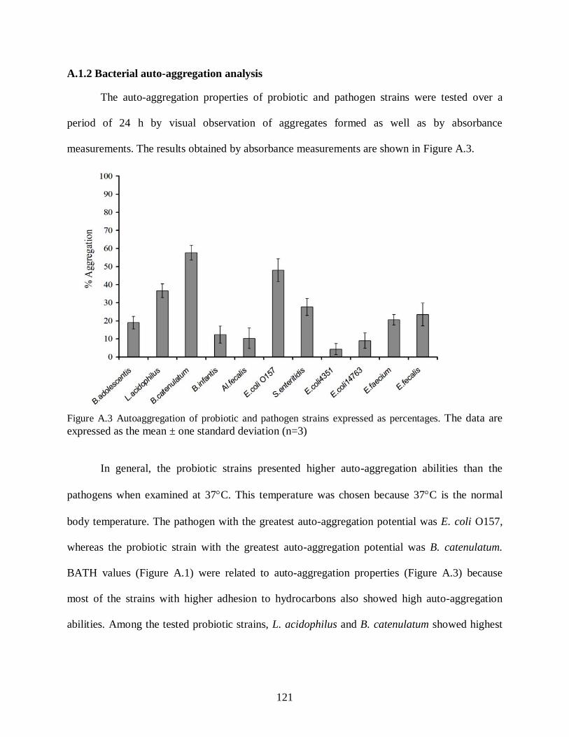
121
A.1.2 Bacterial auto-aggregation analysis
The auto-aggregation properties of probiotic and pathogen strains were tested over a
period of 24 h by visual observation of aggregates formed as well as by absorbance
measurements. The results obtained by absorbance measurements are shown in Figure A.3.
Figure A.3 Autoaggregation of probiotic and pathogen strains expressed as percentages. The data are expressed as the mean ± one standard deviation (n=3) In general, the probiotic strains presented higher auto-aggregation abilities than the
pathogens when examined at 37C. This temperature was chosen because 37C is the normal
body temperature. The pathogen with the greatest auto-aggregation potential was E. coli O157,
whereas the probiotic strain with the greatest auto-aggregation potential was B. catenulatum.
BATH values (Figure A.1) were related to auto-aggregation properties (Figure A.3) because
most of the strains with higher adhesion to hydrocarbons also showed high auto-aggregation
abilities. Among the tested probiotic strains, L. acidophilus and B. catenulatum showed highest

122
percent adherence to hydrocarbon (% BATH) and also highest auto-aggregation abilities. B.
adolescentis and B. infantis showed a lower % BATH as well as lower autoaggregation.
A.1.3 Visual co-aggregation analysis
Visual co-aggregation of two probiotic strains in combination with other intestinal flora,
including E. coli 4351, E. coli 14763, Alcaligens fecalis, E. faecium and E. fecalis and pathogen
strains is shown in Table A.1. All tested probiotics showed co-aggregation with some of the
commensal and pathogenic bacteria in the test panel. It was interesting to note that incubation of
two probiotic strains with a single pathogen strain resulted in greater co-aggregation and more
rapid and complete clearing of liquid in the culture tube. Figure A.4 shows photomicrographs of
the precipitated floccules.
Table A.1 Coaggregation of different combinations of probiotic bacteria with enteric pathogens and commensal bacteria
Probiotic combinations Bacteria LA+BA LA+BC LA+BI BA+BC BA+BI BC+BI
E. coli 4351 +++ ++++ +++ + ++++ ++++ E. coli 14763 +++ +++ ++ ++ ++++ ++++ E. coli O157 ++ +++ ++ ++++ ++ ++++ S. enteritidis ++++ ++++ ++ ++++ ++ ++++ Al. fecalis ++++ ++++ +++ ++++ ++++ ++++ E. faecium ++++ ++++ +++ ++++ ++++ ++++ E. fecalis +++ ++++ +++ ++++ ++++ ++++
LA, L. acidophilus; BA, B. adolescentis; BC, B. catenulatum; BI, B. infantis (+) for small uniform aggregates in the suspension, (++) for aggregates that are easily seen but do not settle, (+++) for large aggregates which settle and leave some turbidity in the supernatant fluid, and (++++) for large aggregates which settle and leave clear supernatant fluid.
The formation of aggregates was dependant on the strains tested (probiotic and pathogen
strains) and also the concentration and time of co-incubation. Formation of aggregates was found

123
to be dependant on the concentration of pathogen and probiotic strains. It was noted that when
the concentration of pathogen was lower than the probiotic, complete clearing of liquid in the
culture tube containing combination of probiotics and pathogen was observed. This finding
supports the idea that given the low infective dose of certain pathogens, co-aggregation with
commensal or probiotic microbiota could potentially help wash them from the GIT, thus
preventing their colonization and manifestation of illness.
Figure A.4 Photomicrographic images of co-aggregates of L. acidophilus, B. adolescentis and S. enteritidis in combination; A) Unstained bacteria before incubation, B) Unstained co-aggregates after incubation at 37ºC for 24 h, and C) Fluorescently-stained (BacLightTM Live/Dead stain) co-aggregates
A 40 µm 40 µm 40 µm
B C