characterization and bacterial anti-adherent effect on modified … · bacterial adhesion to...
TRANSCRIPT

The Journal of Advanced Prosthodontics 207
Characterization and bacterial anti-adherent effect on modified PMMA denture acrylic resin containing platinum nanoparticles
Ki-Young Nam* Department of Dentistry, Dongsan Medical Center, School of Medicine, Keimyung University, Daegu, Republic of Korea
PURPOSE. This study characterized the synthesis of a modified PMMA (Polymethyl methacrylate) denture acrylic loading platinum nanoparticles (PtN) and assessed its bacterial inhibitory efficacy to produce novel antimicrobial denture base material. MATERIALS AND METHODS. Polymerized PMMA denture acrylic disc (20 mm x 2 mm) specimens containing 0 (control), 10, 50, 100 and 200 mg/L of PtN were fabricated respectively. The obtained platinum-PMMA nanocomposite (PtNC) was characterized by TEM (transmission electron microscopy), SEM/EDX (scanning electron microscope/energy dispersive X-ray spectroscopy), thermogravimetric and atomic absorption spectrophotometer analysis. In antimicrobial assay, specimens were placed on the cell culture plate, and 100 μL of microbial suspensions of S. mutans (Streptococcus mutans) and S. sobrinus (Streptococcus sobrinus) were inoculated then incubated at 37oC for 24 hours. The bacterial attachment was tested by FACS (fluorescence-activated cell sorting) analysis after staining with fluorescent probe. RESULTS. PtN were successfully loaded and uniformly immobilized into PMMA denture acrylic with a proper thermal stability and similar surface morphology as compared to control. PtNC expressed significant bacterial anti-adherent effect rather than bactericidal effect above 50 mg/L PtN loaded when compared to pristine PMMA (P=.01) with no or extremely small amounts of Pt ion eluted. CONCLUSION. This is the first report on the synthesis and its antibacterial activity of Pt-PMMA nanocomposite. PMMA denture acrylic loading PtN could be a possible intrinsic antimicrobial denture material with proper mechanical characteristics, meeting those specified for denture bases. For clinical application, future studies including biocompatibility, color stability and warranting the long-term effect were still required. [ J Adv Prosthodont 2014;6:207-14]
KEY WORDS: Platinum nanoparticles; Denture acrylic resin; Nanocomposite; Anti-adherent effect
http://dx.doi.org/10.4047/jap.2014.6.3.207http://jap.or.kr J Adv Prosthodont 2014;6:207-14
INTRODUCTION
A denture base seated on oral cavity leads to changes in weakening the natural hygienic effect of tongue and salivary
flow, especially, by inducing formation and deposit of bio-films on both prosthetics and adjacent mucosa.1 Though bacterial cells could be washed out by saliva and swallowed unless they adhere and replicate, once formed, biofilms are notoriously difficult to remove. Bacterial adhesion to bioma-terial, inert polymer such as denture acrylic base, and the ability of many microorganisms to form biofilms on foreign bodies are well-known steps in the pathogenesis of oral infections.1,2 The insertion of denture tends to create a new available surface for plaque formation and therefore to increase the level of microorganisms in the oral cavity, In addition, roughness voids of denture surface could acceler-ate initial microbial adhesion as well. To overcome these complications, performative and latent antibacterial denture base material that can kill or strongly resist against bacteria, or reduce its adhesion for preventing biofilms formation.3,4
Corresponding author: Ki-Young NamDepartment of Dentistry, Dongsan Medical Center, School of Medicine, Keimyung University, 56 Dalseong-ro, Jung-gu, Daegu, 700-712, Republic of KoreaTel. 82532507807: e-mail, [email protected] December 1, 2013 / Last Revision March 3, 2014 / Accepted March 11, 2014
© 2014 The Korean Academy of ProsthodonticsThis is an Open Access article distributed under the terms of the Creative Commons Attribution Non-Commercial License (http://creativecommons.org/licenses/by-nc/3.0) which permits unrestricted non-commercial use, distribution, and reproduction in any medium, provided the original work is properly cited.
pISSN 2005-7806, eISSN 2005-7814
208
Traditional chemical-based oral disinfectants, though they could be efficient against pathogenic microbes, their volatile ingredients and byproducts could be toxic and harmful to oral mucosa or supporting tissue.5 Occasionally, even den-ture cleansing itself might be compromised to some aged or hospitalized patients due to mentally or physically handi-capped conditions.6,7 Systemic or local antibiotic prescrip-tions have been made for reducing the bacterial population, however, the emergences of more resistant and virulent strains of microorganisms become great clinical challenges.
Some metal nanoparticles (NP) have been known that they act as antibacterial agents to interact directly with microorganisms.8,9 Among them, Pt, as a low-allergy and non-genotoxic noble metal for the organism,10 has been widely used as a catalyst in diverse applications. The antibac-terial activity of Pt has been known since the work of Rosenberg et al.,11 who reported its inhibitory activity on Escherichia coli division. Significantly, Pt nanoparticles (PtN), cluster of Pt atoms with sizes ranging from 1 to 100 nm, are of great interest owing to their highly catalytic activi-ty and are currently being evaluated for the ability to reduce inflammation.10,12 Contact between PtN and bacteria pro-motes chemical interactions that cause bacterial cell to be disintegrated.13 PtN can scavenge reactive oxygen species (ROS),13 and free radicals from antioxidant responses can trigger chain reactions that damage bacteria. Polymeric mate-rials such as PMMA denture acrylic resin have high structur-al tailorability and flexibility with a distinct potential to pre-vent aggregation of NP,14,15 thus, they could be an excellent candidate for the formulation of the nanocomposites based on the inclusion of NP.
Metal-polymer nanocomposites, a polymer matrix NP combined as the additives, have been developed to improve mechanical properties of polymers.16 As for dental applica-tions, the adhesive of 4-methacryloyloxyethyl trimellitic anhydride (4-META)/methyl methacrylate (MMA) in combi-nation with PtN increased dentin bond strength, probably due to enhanced polymerization.17,18 and the addition of PtN to resin-based materials may improve the biocompatibility as an antioxidant.19 Nevertheless, to the author’s knowledge, no study has ever reported to explore the denture acrylic resin containing PtN for its antibacterial activity. Currently, den-ture base materials that could resist the adhesion of microor-ganisms are still unavailable, thus, a simple, effective and latent antimicrobial denture acrylic should be required. The aim of the present research was firstly to characterize a PMMA denture acrylic containing PtN, assessing its mechan-ical characters through thermal, SEM/EDX and ionic elu-tion analysis, secondly to evaluate its antibacterial effect via FACS (fluorescence-activated cell sorting) analysis as an anti-bacterial nanocomposite. The hypothesis was that Pt-PMMA nanocomposites (PtNC) would decrease bacterial adherence and result in a proper mechanical characters for possibility of clinical use.
MATERIALS AND METHODS
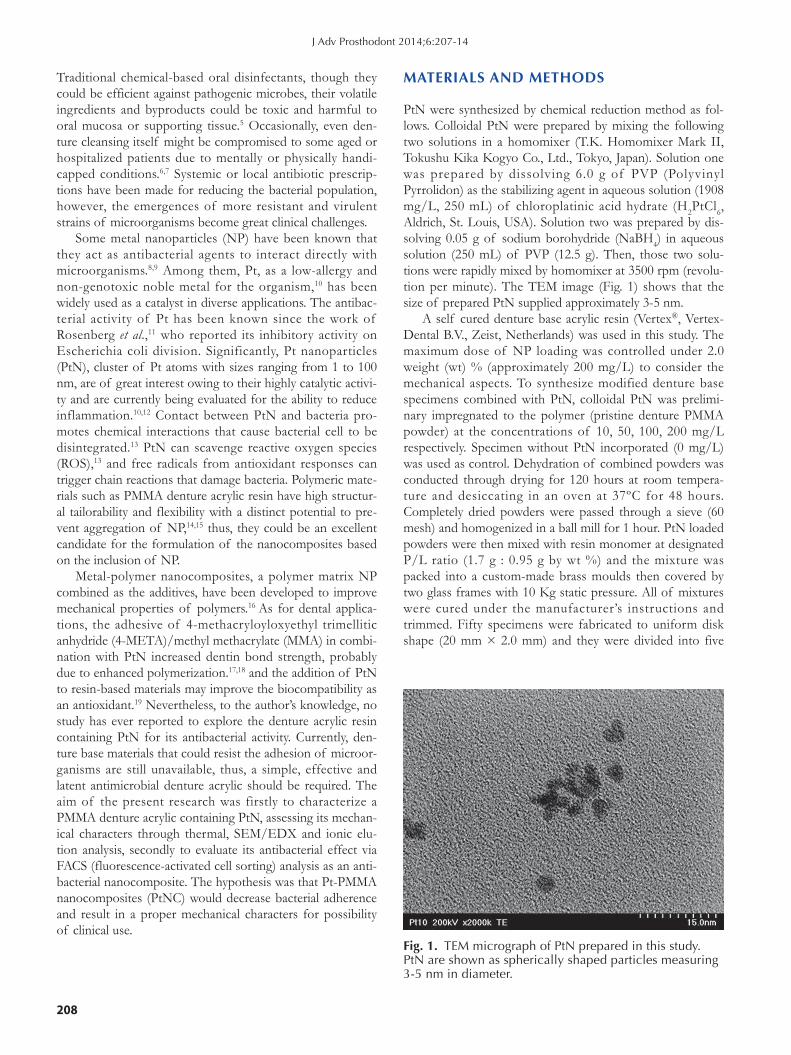
PtN were synthesized by chemical reduction method as fol-lows. Colloidal PtN were prepared by mixing the following two solutions in a homomixer (T.K. Homomixer Mark II, Tokushu Kika Kogyo Co., Ltd., Tokyo, Japan). Solution one was prepared by dissolving 6.0 g of PVP (Polyvinyl Pyrrolidon) as the stabilizing agent in aqueous solution (1908 mg/L, 250 mL) of chloroplatinic acid hydrate (H2PtCl6, Aldrich, St. Louis, USA). Solution two was prepared by dis-solving 0.05 g of sodium borohydride (NaBH4) in aqueous solution (250 mL) of PVP (12.5 g). Then, those two solu-tions were rapidly mixed by homomixer at 3500 rpm (revolu-tion per minute). The TEM image (Fig. 1) shows that the size of prepared PtN supplied approximately 3-5 nm.
A self cured denture base acrylic resin (Vertex®, Vertex-Dental B.V., Zeist, Netherlands) was used in this study. The maximum dose of NP loading was controlled under 2.0 weight (wt) % (approximately 200 mg/L) to consider the mechanical aspects. To synthesize modified denture base specimens combined with PtN, colloidal PtN was prelimi-nary impregnated to the polymer (pristine denture PMMA powder) at the concentrations of 10, 50, 100, 200 mg/L respectively. Specimen without PtN incorporated (0 mg/L) was used as control. Dehydration of combined powders was conducted through drying for 120 hours at room tempera-ture and desiccating in an oven at 37ºC for 48 hours. Completely dried powders were passed through a sieve (60 mesh) and homogenized in a ball mill for 1 hour. PtN loaded powders were then mixed with resin monomer at designated P/L ratio (1.7 g : 0.95 g by wt %) and the mixture was packed into a custom-made brass moulds then covered by two glass frames with 10 Kg static pressure. All of mixtures were cured under the manufacturer’s instructions and trimmed. Fifty specimens were fabricated to uniform disk shape (20 mm × 2.0 mm) and they were divided into five
Fig. 1. TEM micrograph of PtN prepared in this study. PtN are shown as spherically shaped particles measuring 3-5 nm in diameter.
J Adv Prosthodont 2014;6:207-14

The Journal of Advanced Prosthodontics 209
groups (n = 10) according to the concentrations of PtN incorporated.
The microstructure of obtained PtNC was performed to identify the PtN in samples successfully loaded by a field emission electron microscope attached to SEM/EDX (Hitachi S-4100 FE-SEM/EDS, Tokyo, Japan) at an acceler-ated voltage of 20 KeV. The PtNC samples were gold sput-tered under high vacuum before the analysis.
Thermogravimetric (TG) analyses and differential scan-ning calorimetry (DSC) were carried out simultaneously using a TG-DTA 92 (Setaram, Caluire, France) instrument with a heating rate of 10ºC/min from 30 to 600ºC under nitrogen atmosphere.
An atomic absorption spectrophotometer (Analyst 100, Perkin-Elmer, Krakow, WI, USA) and shaking incubator (SI-600R, JEIO TECH, Seoul, Korea) were used. Each disc specimen (20 mm × 2.0 mm, n = 15) was put into 100 mL of sterile distilled water and stored at 37ºC under agitation. The values of eluted Pt ion were determined at 24 and 120 hours with daily replacing distilled water. The quantity of elution was scored as the amounts of Pt ion in the solution per unit of surface area of the disc (cm2) and the measure-ments were performed in three independent tests.
Bacteria tested in this study were S. mutans (Streptococcus mutans, ATCC 25175) and S. sobrinus (Streptococcus sobri-nus, ATCC 27607). Streptococci were maintained on BHI (brain heart infusion) and they were grown under aerobic conditions. Before antimicrobial assay, specimens were stored in sterilized distilled water for 2 weeks to leach excess residual monomer then ultrasonically cleaned (Branson 2200; Branson, Danbury, CT, USA) for 1 hour. To secure the ste-rility of specimens, sterilization with ethylene oxide gas for 24 hours was also conducted then specimens were coated with synthetic saliva (Taliva, Hanlim Pharm. Co., Seoul, Korea) for 1 hour before contacting to bacteria to mimic the oral cavity. Bacterial species were inoculated in BHI broth and incubated for 6 hours, to the point when growth is con-sidered to be in the logarithmic phase. Followed by inoculat-ing 1% (v/v) of the bacterial seed suspension to 1.5 mL medium in 12-well plate (Costa, Corning Co., Corning, NY, USA) containing specimens, they were cultured for 24 hours at 37ºC condition. After incubation, all of experimental specimens were washed out 3-times with PBS (Phosphate Buffered Saline), the adherent bacteria on each samples were then detached into 1 mL PBS by sonication for four 30 sec-ond-pulses with three 30 seconds intermittent cooling. All of samples were centrifuged at 10,000 g for 3 minutes to pellet the cells. Bacterial counting of attached streptococci were performed by FACS analysis after staining with bacterial via-bility and counting kit (Molecular Probes, Eugene, OR, USA). Briefly, bacterial pellets were resuspended in 500 µL of PBS which contain 0.1 µL of 3.34 mM SYTO 9 nucleic acid stain (for live and dead bacteria staining) and 0.1 µL of 30 mM propidium iodide (PI, for dead bacteria-specific staining). Cells were incubated for 15 minutes at room tem-perature protected from light. For the final step, added 1 µL of the microsphere suspension to the stained cell sample as
a reference standard for sample volume then mixed well and analyzed by flow cytometry analysis (Accuri Flow Cytometer, BD Biosciences, San Jose, CA, USA). The populations of live and dead bacteria were calculated from either the fluo-rescence versus side scatter cytogram or the green fluores-cence versus red fluorescence cytogram. To confirm the dif-ferentiation between live and dead bacteria by fluorescent staining, stained bacterial sample images were analyzed under fluorescent microscope (Olympus BX 51, Olympus, Tokyo, Japan).
The bacterial inhibitory effects of PtNC were evaluated by one-way ANOVA, followed by Student’s t-test for post-Hoc test. A significance level of 0.01 was used for statistical tests. All statistical analyses were performed using SPSS 18.0 for Windows (SPSS Inc., Chicago, IL, USA).
RESULT
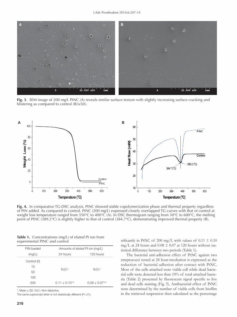
SEM/EDX analysis identified that Pt peak is clearly shown which indicates that PtN were successfully loaded and immobilized into PMMA (Fig. 2). The SEM images of sur-face of 200 mg/L PtNC (Fig. 3A) exhibited similar surface texture to that of control (Fig. 3B) with slightly increased cracking and blistering observed. Thermal analysis of 200 mg/L of PtNC showed stable polymerization phase and improved thermal stability by TG/DSC curves in compari-son with control (Fig. 4). TG curve of PtNC showed closely similar aspect to that of control with a sharp weight reduc-tion observed from 350ºC to 400ºC (Fig. 4A). DSC curve for PtNC also expressed a similar thermogram from 30ºC to 600ºC with the slightly increased endothermic peak at 389.2°C as compared to 384.7ºC in that of control (Fig. 4B). The eluted Pt ion were not calculated from 10 to 100 mg/L Pt loaded groups at two designated times, only revealed sig-
Fig. 2. The SEM/EDX pattern of PtNC electrode. The spectrum shows the characteristic peaks of Pt representing successful loading of PtN into PMMA. Unassigned peaks originate from polymer or external contaminants.
Characterization and bacterial anti-adherent effect on modified PMMA denture acrylic resin containing platinum nanoparticles
210
nificantly in PtNC of 200 mg/L with values of 0.11 ± 0.10 mg/L at 24 hours and 0.08 ± 0.07 at 120 hours without sta-tistical difference between two periods (Table 1).
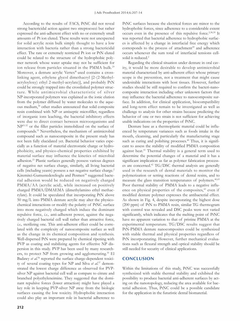
The bacterial anti-adhesion effect of PtNC against two streptococci tested at 24 hour-incubation is expressed as the reduction of bacterial adhesion after contact with PtNC. Most of the cells attached were viable cell while dead bacte-rial cells were detected less than 10% of total attached bacte-ria (Table 2) presented by fluorescent signal specific to live and dead cells staining (Fig. 5). Antibacterial effect of PtNC were determined by the number of viable cells from biofilm in the retrieved suspension then calculated as the percentage
Fig. 3. SEM image of 200 mg/L PtNC (A) reveals similar surface texture with slightly increasing surface cracking and blistering as compared to control (B)(x50).
BA
Table 1. Concentrations (mg/L) of eluted Pt ion from experimental PtNC and control
PtN loaded Amounts of eluted Pt ion (mg/L)
(mg/L) 24 hours 120 hours
Control (0)
N.D.a N.D.a10
50
100
200 0.11 ± 0.10*,b 0.08 ± 0.07*,b
*; Mean ± SD, N.D.; Non-detective.The same superscript letter is not statistically different (P=.01).
Fig. 4. In comparative TG-DSC analysis, PtNC showed stable copolymerization phase and thermal property regardless of PtN added. As compared to control, PtNC (200 mg/L) expressed closely overlapped TG curves with that of control at weight loss temperature ranged from 350°C to 400°C (A). In DSC thermogram ranging from 30°C to 600°C, the melting point of PtNC (389.2°C) is slightly higher to that of control (384.7°C), demonstrating improved thermal property (B).
A B
J Adv Prosthodont 2014;6:207-14
The Journal of Advanced Prosthodontics 211
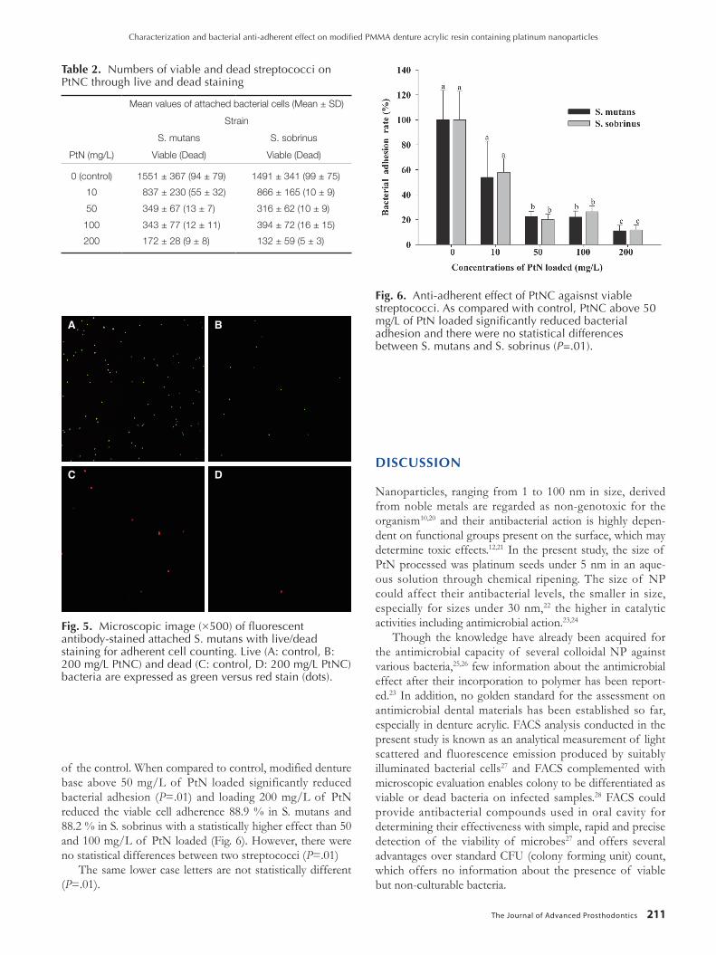
of the control. When compared to control, modified denture base above 50 mg/L of PtN loaded significantly reduced bacterial adhesion (P=.01) and loading 200 mg/L of PtN reduced the viable cell adherence 88.9 % in S. mutans and 88.2 % in S. sobrinus with a statistically higher effect than 50 and 100 mg/L of PtN loaded (Fig. 6). However, there were no statistical differences between two streptococci (P=.01)
The same lower case letters are not statistically different (P=.01).
DISCUSSION
Nanoparticles, ranging from 1 to 100 nm in size, derived from noble metals are regarded as non-genotoxic for the organism10,20 and their antibacterial action is highly depen-dent on functional groups present on the surface, which may determine toxic effects.12,21 In the present study, the size of PtN processed was platinum seeds under 5 nm in an aque-ous solution through chemical ripening. The size of NP could affect their antibacterial levels, the smaller in size, especially for sizes under 30 nm,22 the higher in catalytic activities including antimicrobial action.23,24
Though the knowledge have already been acquired for the antimicrobial capacity of several colloidal NP against various bacteria,25,26 few information about the antimicrobial effect after their incorporation to polymer has been report-ed.23 In addition, no golden standard for the assessment on antimicrobial dental materials has been established so far, especially in denture acrylic. FACS analysis conducted in the present study is known as an analytical measurement of light scattered and fluorescence emission produced by suitably illuminated bacterial cells27 and FACS complemented with microscopic evaluation enables colony to be differentiated as viable or dead bacteria on infected samples.28 FACS could provide antibacterial compounds used in oral cavity for determining their effectiveness with simple, rapid and precise detection of the viability of microbes27 and offers several advantages over standard CFU (colony forming unit) count, which offers no information about the presence of viable but non-culturable bacteria.
A B
C D
Fig. 5. Microscopic image (×500) of fluorescent antibody-stained attached S. mutans with live/dead staining for adherent cell counting. Live (A: control, B: 200 mg/L PtNC) and dead (C: control, D: 200 mg/L PtNC) bacteria are expressed as green versus red stain (dots).
Fig. 6. Anti-adherent effect of PtNC agaisnst viable streptococci. As compared with control, PtNC above 50 mg/L of PtN loaded significantly reduced bacterial adhesion and there were no statistical differences between S. mutans and S. sobrinus (P=.01).
Table 2. Numbers of viable and dead streptococci on PtNC through live and dead staining
Mean values of attached bacterial cells (Mean ± SD)
Strain
S. mutans S. sobrinus
PtN (mg/L) Viable (Dead) Viable (Dead)
0 (control) 1551 ± 367 (94 ± 79) 1491 ± 341 (99 ± 75)
10 837 ± 230 (55 ± 32) 866 ± 165 (10 ± 9)
50 349 ± 67 (13 ± 7) 316 ± 62 (10 ± 9)
100 343 ± 77 (12 ± 11) 394 ± 72 (16 ± 15)
200 172 ± 28 (9 ± 8) 132 ± 59 (5 ± 3)
Characterization and bacterial anti-adherent effect on modified PMMA denture acrylic resin containing platinum nanoparticles
212
According to the results of FACS, PtNC did not reveal strong bactericidal action against two streptococci but rather expressed the anti-adherent effect with no or extremely small amounts of Pt ion eluted. These results were not unexpected for solid acrylic resin bulk simply thought to have a low interaction with bacteria rather than a strong bactericidal effect. The rare or extremely restricted Pt ion or PtN eluted could be related to the structure of the hydrophobic poly-mer network whose water uptake may not be sufficient for ion release from particles situated in the PMMA bulk.29 Moreover, a denture acrylic Vertex® used contains a cross-linking agent, ethylene glycol dimethacryl [2-(2-Methyl-acryloyloxy) ethyl 2-methyl-acrylate)], and probably PtN could be strongly trapped into the crosslinked polymer struc-ture. Whi le ant imicrobia l character i s t ic of s i lver NP-incorporated polymers was explained as the ionic elution from the polymer diffused by water molecules to the aque-ous medium,30 other studies announced that solid composite resin combined with NP or antiseptic microfiller, regardless of inorganic ions leaching, the bacterial inhibitory effects were due to direct contact between microorganisms and NP31.32 or the filler particles immobilized on the surface of compounds.33 Nevertheless, the mechanism of antimicrobial compound such as nanocomposite in the present study has not been fully elucidated yet. Bacterial behavior varies espe-cially as a function of material electrostatic charge or hydro-phobicity, and physico-chemical properties exhibited by material surface may influence the kinetics of microbial adhesion.23 Plastic surfaces generally possess various degrees of negative net surface charge, similarly, all living bacterial cells (including yeasts) possess a net negative surface charge.2
Kiremitci-Gumustederelioglu and Pesmen34 suggested bacte-rial adhesion would be reduced on negatively charged PMMA/AA (acrylic acid), while increased on positively charged PMMA/DMAEMA (dimethylamino ethyl methac-rylate). It could be speculated that incorporating PtN above 50 mg/L into PMMA denture acrylic may alter the physico-chemical interactions or modify the polarity of PtNC surface into more negatively charged which induce the dominant repulsive force, i.e., anti-adherent power, against the nega-tively charged bacterial cell wall rather than attractive force, i.e., sterilizing one. This anti-adherent effect could be corre-lated with the complexity of nanocomposite surface as well as the change in its chemical composition and synthesis. Well-dispersed PtN were prepared by chemical ripening with PVP as coating and stabilizing agents for effective NP dis-persion in this study. PVP has been used by many research-ers, to protect NP from growing and agglomerating.35 El Badawy et al.36 reported the surface charge-dependent toxici-ty of several coating types for NP and Silva et al.37 demon-strated the lowest charge difference as observed for PVP-silver NP against bacterial cell wall as compare to citrate and branched polyethyleneimine. They suggested that the domi-nant repulsive forces (lower attraction) might have played a key role in keeping PVP-silver NP away from the biologic surfaces causing the low toxicity. Hydrophobic interaction could also play an important role in bacterial adherence to
PtNC surfaces because the electrical forces are minor to the hydrophobic forces, since adherence to a considerable extent occurs even in the presence of this repulsive force.2,38,39 It was reported that bacterial adherence to hydrophobic surfac-es is affected by a change in interfacial free energy which corresponds to the process of attachment39 and adherence occurs whenever the sum total of interfacial tensions cell-solid is reduced.2
Regarding the clinical situation under denture in oral cav-ity, it would be more desirable to develop antimicrobial material characterized by anti-adherent effect whose primary scope is the prevention, not a treatment that might cause undesirable interactions with host tissues. However, further studies should be still required to confirm the bacteri-nano-composite interaction including other unknown factors that may influence the bacterial adherence to nanocomposite sur-face. In addition, for clinical application, biocompatibility and long-term effect remain to be investigated as well as challenge to analysis for other strains because predicting the behavior of one or two strain is not sufficient for achieving usable indications on the properties of PtNC.
Denture base as a thermoplastic material could be influ-enced by temperature variances such as foods intake in the mouth, cleansing, and particularly the manufacturing stage such as curing and polishing processes.40 Thus, it is signifi-cant to assess the stability of modified PMMA composition against heat.41 Thermal stability is a general term used to determine the potential changes of a material and it has a significant implication as far as polymer fabrication process-es are concerned.42 TG/DSC thermal analysis are generally used in the research of dental materials to monitor the polymerization or setting reactions of dental resins, and to measure the glass transition temperatures of polymers.40,43 Poor thermal stability of PMMA leads to a negative influ-ence on physical properties of the composites,43 even if modified denture polymer expresses the antibacterial effect. As shown in Fig. 4, despite incorporating the highest dose (200 ppm) of PtN to PMMA resin, similar TG thermogram with control was revealed and DSC peaks were not varied significantly, which indicates that the melting point of PtNC have no apparent variation to that of pristine PMMA at the experimental temperature. TG/DSC results suggest that PtN-PMMA denture nanocomposites could be synthesized with stable thermal and physical properties regardless of PtN incorporatating. However, further mechanical evalua-tions such as flexural strength and optical stability should be still needed for security of clinical applications.
CONCLUSION
Within the limitations of this study, PtNC was successfully synthesized with stable thermal stability and exhibited the possibility to produce bacterial anti-adherent surfaces by act-ing on the nanotopology, reducing the area available for bac-terial adhesion. Thus, PtNC could be a possible candidate for the application in the futuristic dental biomaterial.
J Adv Prosthodont 2014;6:207-14
The Journal of Advanced Prosthodontics 213
REFERENCES
1. Yildir im MS, Hasanreisoglu U, Hasirci N, Sultan N. Adherence of Candida albicans to glow-discharge modified acrylic denture base polymers. J Oral Rehabil 2005;32:518-25.
2. Klotz SA, Drutz DJ, Zajic JE. Factors governing adherence of Candida species to plastic surfaces. Infect Immun 1985; 50:97-101.
3. Loesche WJ. Role of Streptococcus mutans in human dental decay. Microbiol Rev 1986;50:353-80.
4. Saito T, Takatsuka T, Kato T, Ishihara K, Okuda K. Adherence of oral streptococci to an immobilized antimicro-bial agent. Arch Oral Biol 1997;42:539-45.
5. Murdoch-Kinch CA, Mallatt ME, Miles DA. Oral mucosal injury caused by denture cleanser tablets: a case report. Oral Surg Oral Med Oral Pathol Oral Radiol Endod 1995;80:756-8.
6. Stone C, Sabes WR. Denture cleaner chemical burn. Gen Dent 1995;43:554-5.
7. De Visschere LM, Grooten L, Theuniers G, Vanobbergen JN. Oral hygiene of elderly people in long-term care institu-tions-a cross-sectional study. Gerodontology 2006;23:195-204.
8. Panácek A, Kolár M, Vecerová R, Prucek R, Soukupová J, Krystof V, Hamal P, Zboril R, Kvítek L. Antifungal activity of silver nanoparticles against Candida spp. Biomaterials 2009;30:6333-40.
9. Rai M, Yadav A, Gade A. Silver nanoparticles as a new gener-ation of antimicrobials. Biotechnol Adv 2009;27:76-83.
10. Sawosz E, Chwalibog A, Szeliga J, Sawosz F, Grodzik M, Rupiewicz M, Niemiec T, Kacprzyk K. Visualization of gold and platinum nanoparticles interacting with Salmonella enter-itidis and Listeria monocytogenes. Int J Nanomedicine 2010; 5:631-7.
11. Rosenberg B, Vancamp L, Krigas T. Inhibition of cell divi-sion in Escherichia coli by electrolysis products from a plati-num electrode. Nature 1965;205:698-9.
12. Chwalibog A, Sawosz E, Hotowy A, Szeliga J, Mitura S, Mi tura K, Grodz ik M, Or lowski P, Sokolowska A. Visualization of interaction between inorganic nanoparticles and bacteria or fungi. Int J Nanomedicine 2010;5:1085-94.
13. Onizawa S, Aoshiba K, Kajita M, Miyamoto Y, Nagai A. Platinum nanoparticle antioxidants inhibit pulmonary inflam-mation in mice exposed to cigarette smoke. Pulm Pharmacol Ther 2009;22:340-9.
14. Sur I, Cam D, Kahraman M, Baysal A, Culha M. Interaction of multi-functional silver nanoparticles with living cells. Nanotechnology 2010;21:175104.
15. Wang Y, Bansal V, Zelikin AN, Caruso F. Templated synthesis of single-component polymer capsules and their application in drug delivery. Nano Lett 2008;8:1741-5.
16. Boomi P, Prabu HG, Mathiyarasu J. Synthesis and character-ization of polyaniline/Ag-Pt nanocomposite for improved antibacterial activity. Colloids Surf B Biointerfaces 2013;103: 9-14.
17. Hoshika S, Nagano F, Tanaka T, Ikeda T, Wada T, Asakura K, Koshiro K, Selimovic D, Miyamoto Y, Sidhu SK, Sano H.
Effect of application time of colloidal platinum nanoparti-cles on the microtensile bond strength to dentin. Dent Mater J 2010;29:682-9.
18. Hoshika S, Nagano F, Tanaka T, Wada T, Asakura K, Koshiro K, Selimovic D, Miyamoto Y, Sidhu SK, Sano H. Expansion of nanotechnology for dentistry: effect of colloi-dal platinum nanoparticles on dentin adhesion mediated by 4-META/MMA-TBB. J Adhes Dent 2011;13:411-6.
19. Ma S, Izutani N, Imazato S, Chen JH, Kiba W, Yoshikawa R, Takeda K, Kitagawa H, Ebisu S. Assessment of bactericidal effects of quaternary ammonium-based antibacterial mono-mers in combination with colloidal platinum nanoparticles. Dent Mater J 2012;31:150-6.
20. Akin D, Sturgis J, Ragheb K, Sherman D, Burkholder K, Robinson JP, Bhunia AK, Mohammed S, Bashir R. Bacteria-mediated delivery of nanoparticles and cargo into cells. Nat Nanotechnol 2007;2:441-9.
21. Goodman CM, McCusker CD, Yilmaz T, Rotello VM. Toxicity of gold nanoparticles functionalized with cationic and anionic side chains. Bioconjug Chem 2004;15:897-900.
22. Park MV, Neigh AM, Vermeulen JP, de la Fonteyne LJ, Verharen HW, Briedé JJ, van Loveren H, de Jong WH. The effect of particle size on the cytotoxicity, inflammation, de-velopmental toxicity and genotoxicity of silver nanoparticles. Biomaterials 2011;32:9810-7.
23. Campoccia D, Montanaro L, Arciola CR. A review of the biomaterials technologies for infection-resistant surfaces. Biomaterials 2013;34:8533-54.
24. Kajita M, Hikosaka K, Iitsuka M, Kanayama A, Toshima N, Miyamoto Y. Platinum nanoparticle is a useful scavenger of superoxide anion and hydrogen peroxide. Free Radic Res 2007;41:615-26.
25. Sondi I, Salopek-Sondi B. Silver nanoparticles as antimicrobi-al agent: a case study on E. coli as a model for Gram-negative bacteria. J Colloid Interface Sci 2004;275:177-82.
26. Lima E, Guerra R, Lara V, Guzmán A. Gold nanoparticles as efficient antimicrobial agents for Escherichia coli and Salmonella typhi. Chem Cent J 2013;7:11.
27. Alvarez-Barrientos A, Arroyo J, Cantón R, Nombela C, Sánchez-Pérez M. Applications of flow cytometry to clinical microbiology. Clin Microbiol Rev 2000;13:167-95.
28. Pils S, Schmitter T, Neske F, Hauck CR. Quantification of bacterial invasion into adherent cells by flow cytometry. J Microbiol Methods 2006;65:301-10.
29. Damm C, Münstedt H, Rösch A. Long-term antimicrobial polyamide 6/silver-nanocomposites. J Mater Sci 2007;42: 6067-73.
30. Kumar R, Münstedt H. Silver ion release from antimicrobial polyamide/silver composites. Biomaterials 2005;26:2081-8.
31. Ahn SJ, Lee SJ, Kook JK, Lim BS. Experimental antimicrobi-al orthodontic adhesives using nanofillers and silver nanopar-ticles. Dent Mater 2009;25:206-13.
32. Yoshida K, Tanagawa M, Atsuta M. Characterization and in-hibitory effect of antibacterial dental resin composites incor-porating silver-supported materials. J Biomed Mater Res 1999;47:516-22.
33. Imazato S, Ebi N, Takahashi Y, Kaneko T, Ebisu S, Russell
Characterization and bacterial anti-adherent effect on modified PMMA denture acrylic resin containing platinum nanoparticles
214
RR. Antibacterial activity of bactericide-immobilized filler for resin-based restoratives. Biomaterials 2003;24:3605-9.
34. Kiremitci-Gumusderelioglu M, Pesmen A. Microbial adhesion to ionogenic PHEMA, PU and PP implants. Biomaterials 1996;17:443-9.
35. Wang H, Qiao X, Chen J, Wang X, Ding S. Mechanisms of PVP in the preparation of silver nanoparticles. Mater Chem Phys 2005;94:449-53.
36. El Badawy AM, Silva RG, Morris B, Scheckel KG, Suidan MT, Tolaymat TM. Surface charge-dependent toxicity of sil-ver nanoparticles. Environ Sci Technol 2011;45:283-7.
37. Silva T, Pokhrel LR, Dubey B, Tolaymat TM, Maier KJ, Liu X. Particle size, surface charge and concentration dependent ecotoxicity of three organo-coated silver nanoparticles: com-parison between general linear model-predicted and observed toxicity. Sci Total Environ 2014;468-469:968-76.
38. Fletcher M, Loeb GI. Influence of substratum characteristics on the attachment of a marine pseudomonad to solid surfac-es. Appl Environ Microbiol 1979;37:67-72.
39. Liu J, Hurt RH. Ion release kinetics and particle persistence in aqueous nano-silver colloids. Environ Sci Technol 2010;44: 2169-75.
40. Soygun K, Bolayir G, Boztug A. Mechanical and thermal properties of polyamide versus reinforced PMMA denture base materials. J Adv Prosthodont 2013;5:153-60.
41. Jerolimov V, Jagger RG, Milward PJ. Effect of the curing cy-cle on acrylic denture base glass transition temperatures. J Dent 1991;19:245-8.
42. Davy KW, Anseau MR, Berry C. Iodinated methacrylate co-polymers as X-ray opaque denture base acrylics. J Dent 1997; 25:499-505.
43. Aydogan Ayaz E, Durkan R, Bagis B. The effect of acryl-amide incorporation on the thermal and physical properties of denture resins. J Adv Prosthodont 2013;5:110-7.
J Adv Prosthodont 2014;6:207-14