characterisation of psoriasis susceptibility locus 6
TRANSCRIPT
Characterisation of psoriasis susceptibility locus 6(PSORS6) in patients with early onset psoriasis andevidence for interaction with PSORS1
U Huffmeier,1 J Lascorz,1 T Becker,2 F Schurmeier-Horst,3 A Magener,4 A B Ekici,1
S Endele,1 C T Thiel,1 S Thoma-Uszynski,5 R Mossner,6 K Reich,7 W Kurrat,8
T F Wienker,2 H Traupe,5 A Reis1
c Additional tables and figureare published online only athttp://jmg.bmj.com/content/vol46/issue11
1 Institute of Human Genetics,University Hospital Erlangen,University Erlangen-Nuremberg,Germany; 2 Institute of MedicalBiometry, Informatics andEpidemiology, University ofBonn, Germany; 3 Department ofDermatology, University ofMunster, Germany;4 Department of Pathology,University of Erlangen, Germany;5 Department of Dermatology,University of Erlangen, Germany;6 Department of Dermatology,University of Gottingen,Germany; 7 DermatologikumHamburg, Hamburg, Germany;8 Asklepios Nordseeklinik,Westerland/Sylt, Germany
Correspondence to:Professor A Reis, Institute ofHuman Genetics, UniversityHospital Erlangen, University ofErlangen-Nuremberg, 91054Erlangen, Germany;[email protected]
Received 20 November 2008Revised 20 February 2009Accepted 23 March 2009Published Online First11 June 2009
This paper is freely availableonline under the BMJ Journalsunlocked scheme, see http://jmg.bmj.com/info/unlocked.dtl
ABSTRACTBackground: Psoriasis is a genetically complex, chronicinflammatory skin disease. The authors have previouslyidentified a susceptibility locus on chromosome 19p13(PSORS6).Methods and results: In a follow-up linkage disequili-brium (LD) study in an independent family based cohort,the authors found evidence for association to a newlydiscovered microsatellite at this locus (D19SPS21,p,5.361025). An LD based association scan in 300 triosrevealed association to several single, single nucleotidepolymorphisms (SNPs) in one LD block. When the authorsstratified this cohort for carrying the PSORS1 risk allele atthe HLA-C locus, evidence for association became muchstronger at single SNP and haplotype levels (p valuesbetween 1.061024 and 8.061024). In a replication studyof 1114 patients and 937 control individuals, evidence forassociation was also observed after stratification to thePSORS1 risk allele. In both study groups, logisticregression showed evidence for interaction between therisk alleles at PSORS1 and PSORS6. Best p values forrs12459358 in both study groups remained significantafter correction for multiple testing. The associated LDblock did not comprise any known genes. Interestingly, anadjacent gene, MUC16, coding for a large glycosylatedprotein expressed in epithelia and of unknown function,could be shown to be also expressed in tissues relevantfor pathogenesis of psoriasis such as skin and thymus.Immunohistochemical analyses of skin revealed focalstaining for MUC16 in suprabasal epidermal cells. Furtherfunctional studies are required to clarify its potential rolein psoriasis and identify the causal variant(s) at this locus.Conclusion: The data establish PSORS6 as a confirmedpsoriasis susceptibility locus showing interaction withPSORS1.
Psoriasis is a chronic inflammatory disorder of theskin. The disease has a complex aetiology andaffects about 0.5–4.0% of the European andNorthern American population.1 2 Even thoughthere is considerable evidence for genetic factorsfrom family and twin studies—for example, con-cordance rate of up to 65% in monozygotictwins—only a few susceptibility factors have beendiscovered to date. While positional cloning stra-tegies using genome wide linkage analysis (GWLA)have allowed identification of disease causingvariants in numerous monogenic diseases, theadaptation of this systematic approach to complexdiseases3 has been successful in only a few instances.In psoriasis vulgaris (PsV), the most evidentiary
linkage region is at psoriasis susceptibility locus1 ( = PSORS1) on chromosome 6p21.3,4 especially infamilies with PsV manifesting at younger age((40 years, type I psoriasis5). At this locus, themost consistently associated allele is the *Cw0602allele of HLA-C gene, although the remarkably highlinkage disequilibrium (LD) at this locus hashampered definite identification of the causativevariant.6 In addition, more than nine psoriasissusceptibility loci have been identified (PSORS1-9and one further for psoriatic arthritis, PSORAS1).Only at three loci, replicated association to candidategenes (RAPTOR and SLC12A8) has been reported sofar (PSORS2 and PSORS5)7–10 or several genes—forexample, LOR, LCE1C, PGLYRP, SPRR genes,PRR9 genes and IVL—have been proposed to accountfor psoriasis susceptibility (PSORS4).11–14 In contrast,genome-wide association studies (GWAS) haveturned out to be a very successful approachfor many complex diseases. Recently, variants intwo genes of the IL-23R pathway were identified asPsV susceptibility factors by two independentgroups.15 16 Like in other HLA related complex traits,the relative risk associated with these variants ismarkedly lower than that for HLA-C associated allele.It is expected that many more susceptibility alleleswith small effects contribute to the aetiology of PsV.
In this study, we describe our positional cloningefforts at PSORS6 located on chromosome 19p13,which we previously identified in a genome-widelinkage scan of extended multiplex PsV families fromGermany.17 Subsequently, we detected genetic asso-ciation in an independent family based cohort totwo newly discovered microsatellite markers belowthe linkage peak. After exploring the LD structure atthis previously poorly characterised genomic region,we performed association studies based on the LDstructure and could narrow a PsV susceptibility alleleto a 50 kb intergenic LD block. This finding wasreplicated in a further case–control cohort. In bothgroups, association was stronger in patients carryingthe PSORS1 risk allele, suggesting interactionbetween both loci. Strong LD hampered furtherrefinement of the locus, which contains no knowngene. One of the neighbouring genes (MUC16),though, showed expression in several tissues rele-vant for PsV and the product of this gene showedimmunostaining in epidermal cells. We speculatethat this locus contains yet unidentified regulatoryelements influencing other genes in this genomicregion.
Original article
736 J Med Genet 2009;46:736–744. doi:10.1136/jmg.2008.065029
on October 21, 2021 by guest. P
rotected by copyright.http://jm
g.bmj.com
/J M
ed Genet: first published as 10.1136/jm
g.2008.065029 on 11 June 2009. Dow
nloaded from

METHODS
PatientsThe 210 nuclear families with and without 90 additional trios,respectively, were the ones described previously.18 19 In brief, allindex cases had an early onset form of psoriasis vulgaris withmedium (SD) age of onset of 16.2 (9.1) years; 53.3% of indexpatients were male. In 61% of trios, the father and/or themother were also affected by psoriasis.
The case cohort consisted of 1114 single patients with PsVand was recruited through dermatology clinics at three psoriasisrehabilitation hospitals and at three university hospitals.20 Anearly onset form of psoriasis (type I) was diagnosed in all but 75patients. The medium age of onset was 23.2 (11.9) years, and21.4 (9.5) years in type I patients. The majority of patientssuffered from plaque type of PsV. We excluded all patients withsigns of psoriatic arthritis until the time of recruitment whenmedium age was 48.2 (13.4) years; 62% of patients were male.
The 937 control probands had no PsV and no history or signsof inflammatory joint disease at the time of recruitment, whentheir mean age was 31.6 (10) years. All were German (white)healthy blood donors; 58% of probands were male.
The studies were approved by the ethical committees of theUniversity of Erlangen-Nuremberg and of the University ofMunster. Written informed consent was obtained from eachpatient and control proband before enrolment. The investiga-tions were conducted according to Declaration of Helsinkiprinciples.
Microsatellite analysesTo identify new microsatellites within the linkage region, weused the program Tandem Repeat Finder (Benson21; http://tandem.bu.edu/trf/trf.html (accessed April 2002)). In order tohave markers with fewer alleles, we chose a set of 24 evenlyspaced microsatellites that were mostly tetra- or penta-nucleo-tides or had in the case of di-nucleotides ,15 repeats in thedatabase sequence. Primers were chosen with the program Primer3 (http://frodo.wi.mit.edu/cgi-bin/primer3/primer3_www.cgi(accessed April 2002)); all forward primers were labelled withone of three fluorescent dyes (FAM, HEX, TET). The micro-satellites were arranged in two different panels according tomagnitude and labelling of the polymerase chain reaction (PCR)product. PCRs were performed as previously described.18 For eightmicrosatellites, we obtained either no PCR product or thecorresponding microsatellite was not polymorphic. Therefore 16microsatellites (supplemental table 1) remained that could begenotyped in 210 trios; these markers cover a genomic region of,1.3 Mb around the previously described microsatellites D19S922,D19S865 and D19S916.17 PCR products were pooled and sizefractionated on an ABI3100 DNA analysis system (AppliedBiosystems, Foster City, California, USA) along with a TAMRAlabelled standard. Electrophoresis files were analysed withGenotyper and/or Genemapper software (Applied Biosystems).Consistency for correct allele sizes was ensured by simultaneousgenotyping of previously defined DNA controls. The overallgenotyping rate was 93.3%. Genotypes were checked forMendelian inheritance, while the transmission disequilibrium test(TDT) as implemented in the family based association test (FBAT)software22 was used to test for association under default options.
Sequencing, LD structure and SNP genotypingIn the genomic region of D19SPS20 and D19SPS21, about225 kb of sequence were analysed for conservation with mousesequence and the sequences of other species (as identified with
the feature of University of California Santa Cruz (UCSC)genome browser at http://genome.ucsc.edu/ (accessedNovember 2005)). Those sequences exhibiting evidence forconservation were selected; primers for coverage of thesesequences were chosen with Primer 3. In 32 independentpsoriasis patients chosen from affected individuals of the trios,105 PCR products were amplified and sequenced as previouslydescribed.9 In order to uncover the LD structure of this region,we filtered for single nucleotide polymorphisms (SNPs) with aminor allele frequency (MAF) of .0.10.
Based on the LD structure, we chose a first set of 44haplotype tagging SNPs (htSNPs) and genotyped them asTaqMan assays in the 300 trios. After first evidence forassociation of psoriasis to several single SNPs, we chose 24independent individuals of this study group that were eitherhomozygous (n = 19) or heterozygous (n = 5) for a haplotypeconsisting of several associated SNP alleles within the LD block.We sequenced the probands for a further 29 PCR productswithin/neighbouring this LD block to identify further possiblerisk variants. In the meantime the first HapMap data had beenmade publicly available. We combined information from bothsources to choose an appropriate and redundant set of 30further htSNPs with different programs such as SNPtagger,23
Ldmax24 and Haploview.25 These SNPs were also genotyped in300 trios with two different methods—SNPlex or TaqMan(Applied Biosystems)—as recommended by the manufacturer.
For data analysis, only SNPs with a MAF .5%, no deviationfrom Hardy–Weinberg equilibrium (HWE) (p values .0.01) andno excess of Mendelian errors were accepted. For the remaining63 SNPs (supplemental table 2) overall genotyping rate was96.0%. For two SNPs that were genotyped with both methods(SNPlex and TaqMan) the concordance rate was .99.5%. Foreach SNP a subset of genotypes was confirmed through directsequencing in 24–32 randomly chosen probands. Genotyping ofthe same DNA of a control individual was part of every singleexperiment and resulted in consistent genotypes for all SNPsincluded in this study.
The case control study—1114 PsV patients and 937 controlprobands—were genotyped for nine SNPs in the associated LDblock (rs8100377, rs6511831, rs12459358, rs8109594, rs8102472,rs7249334, rs10413384, rs2591618 and rs28699225) either withSNPlex or TaqMan. Similarly, no SNP showed significantdeviation from HWE. Overall genotyping rate was 97.6%. SNPsof two genes of the IL-23R pathway, namely IL12B and IL23R,were genotyped as recently described.20
LD analysis, statistical and haplotype analysisLD of tagging SNPs was determined with the softwareHaploview.25 LD blocks were basically defined according to themodel ‘‘Solid Spine’’. In order to be able to include several sibs ofone family, we used the TDT as implemented in the associationmethod for single SNPs and haplotypes as described in Becker andKnapp.26 This method can be viewed as a generalisation of theTDT.27 Haplotypes were calculated within the predefined LDblocks. In the case–control study, association with single SNPswas tested with Armitage’s trend test.28 Odds ratios (ORs) andtheir confidence intervals were determined for single SNP alleleswith final p values of ,0.05. Haplotypes within the blocks werecalculated with the same method as in trios.26
In order to compare results from the family study (TDTstatistics) with those of case–control studies, we used the modelof Kazeem and Farrall29 and estimated ORs for SNP rs1249358from the TDT data and calculated a combined OR of bothstudies, family based and case–control.
Original article
J Med Genet 2009;46:736–744. doi:10.1136/jmg.2008.065029 737
on October 21, 2021 by guest. P
rotected by copyright.http://jm
g.bmj.com
/J M
ed Genet: first published as 10.1136/jm
g.2008.065029 on 11 June 2009. Dow
nloaded from
Stratification for PSORS1 and interaction with other psoriasissusceptibility allelesFor stratification for the PSORS1 risk allele, we used an estimateas previously described.20 In order to test for interaction in thecase–control study between known psoriasis susceptibility lociand the risk alleles identified at PSORS6, we used logisticregression as suggested by Cordell and Clayton.30 We compareda model of three degrees of freedom with an interactionparameter and one parameter for each, the PSORS1 riskhaplotype and the SNP rs1245938 at PSORS6, to a model oftwo degrees of freedom with just the parameters for themarkers. In the nuclear families, we compared the number oftransmissions/non-transmissions of rs1245938 at PSORS6 afterstratification of index cases for carrying the PSORS1 riskhaplotype.
In order to test for possible interaction of susceptibilityfactors of the IL-23R pathway and the PSORS6 variantrs1249358 in the family cohort, we used the variant of IL-23Rpathway that was most highly associated in the trios(rs6887695) and tested for interaction with the programUNPHASED.31
Correction for multiple testingIn the family based cohort, we performed an explorative studywith microsatellites with few alleles (mostly two to threefrequent ones) in order to exclude/confirm a role of PSORS6 inpsoriasis. Due to the explorative character of this study, we didnot correct for the number of markers/alleles tested. In order tocorrect for the number of SNPs tested in the second associationstudy, also accounting for LD, we applied a Monte Carlosimulation procedure for the most strongly associated SNP.26
Taking into account that we considered the complete sample aswell as the samples stratified according to PSORS1 and theIL12B variant, we applied a further Bonferroni correction by afactor of 3. To account for the fact that we used differentmethods to do analysis stratified for PSORS1 on the one handand for IL12B on the other hand (also we did apply only onekind of analysis in each situation), we applied a furtherBonferroni correction by a factor of 2.
In order to find out whether the strongly associated SNPsmight be the disease-causing variant(s) or whether there isevidence for other not yet identified variants, we performedhaplotype analyses. This analysis again was regarded asexplorative.
In silico analysesWe performed in silico analyses to identify potential unknowngenes within the associated region. Several gene predictionprograms were used: GrailEXP, Genscan and Augustus (32 33
http://augustus.gobics.de/ (accessed November 2005)).Furthermore, we analysed human expressed sequence tags(ESTs) within this interval that were annotated at the UCSCgenome browser (http://genome.ucsc.edu/ (accessed November2005)) and performed blast analysis (http://www.ncbi.nlm.nih.gov/blast/Blast.cgi (accessed November 2005)) to confirm theirlocation within this region. Human and mouse sequences werecompared with the Pipmaker software.34 In addition, we usedtwo further features annotated in the UCSC genome browser:‘‘mouse chain alignments’’ and ‘‘mouse alignment net’’.35 Finally,the regions that showed evolutionary conservation were screenedfor protein sequences with protein blast (blastx, http://www.ncbi.nlm.nih.gov/blast/Blast.cgi (accessed November 2005)).
Transcription analysesGenomic intervals that contained in silico gene predictions andshowed evolutionary conservation were considered for reversetranscriptase PCR (RT-PCR) based transcription screening. Weused a set of primers within these intervals and performedsystematic RT-PCRs from cDNA of four different tissuesrelevant for psoriasis and/or other autoimmune diseases: twocDNAs from skin, blood leucocytes and thymus, respectively,and one from thyroid. Furthermore, we chose forward primersfor different RT-PCRs covering the 59 region of a differentialtranscript predicted by Genscan. This transcript was longer inthe 59 region than the longest annotated mRNA of MUC16, andreached into our associated genomic interval. In combinationwith reverse primers within the 59 region of the mRNA ofMUC16, we performed again exon overlapping RT-PCRs.Moreover, exon-overlapping primers derived from 59 exons ofthe MUC16 RefSeq sequence were selected.
One positive (GAPDH) and one negative control (an intronicfragment of PTPN22) were included in each experiment. RT-PCRs were performed with invitrogen taq polymerase(Invitrogen, Carlsbad, California, USA) under standard condi-tions with a touch-down PCR program; each experiment wasperformed with or without betain. Successfully amplified PCRproducts of the expected size were sequenced.
Analysis of copy number variation using quantitative PCRIn order to analyse the genomic region of BAC RP11-79F15, abacterial artificial chromosome (BAC) that was previouslyreported to show copy number variation (CNV; Database ofGenomic Variation,36), we developed two different quantitativePCRs (qPCR), one within the gene MBD3L1, the other onewithin the non-repetitive 59 part of MUC16 gene. Genomiccopies of the target regions were compared to the housekeepinggene albumin. qPCRs were set up as previously described.37
Sequences of primers and probes are available upon request.Ninety-four psoriasis patients and 94 control individuals weretested for CNV at MBD3L1, and 94 control individuals for CNVat MUC16. We obtained reliable genotypes (defined as SD,0.15) in 97.9% or 98.9% of individuals, respectively.
ImmunohistochemistryImmunohistochemical analyses were performed with antibodyOC125 (MUC16) purchased from Dako (Carpinteria, California,USA) according to the modified ABC method (avidin biotinperoxidase complex) as recommended by the manufacturer.Twelve skin biopsies—10 of psoriasis vulgaris patients and twoof control probands—were analysed.
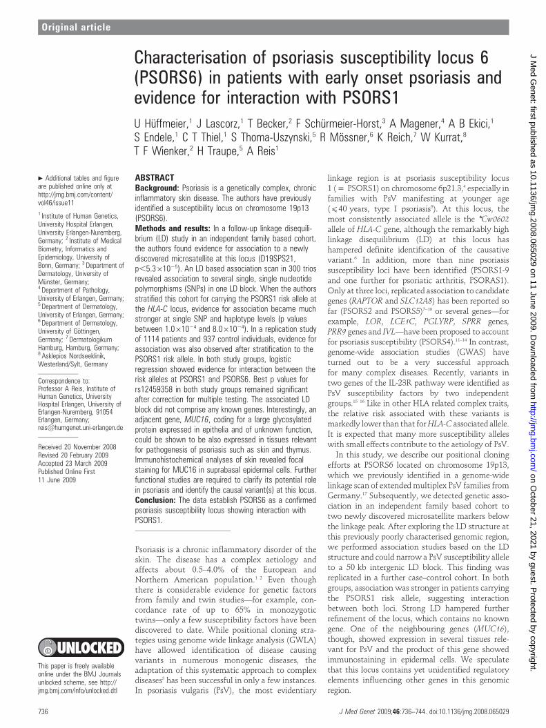
RESULTSTen out of 16 microsatellites had two frequent alleles (.5%), afurther four markers had three, and two microsatellites had fouror five frequent alleles, respectively. FBAT statistics showedstrong association with one allele of D19SPS21 in the 210 trios(p = 5.361025, fig 1). Of the neighbouring microsatellites,D19SPS20 was less strongly associated (p,2.761022).
Sequencing of the 32 individuals in an interval of about225 kb surrounding D19SPS20 and D19SPS21 identified 87frequent SNPs with a MAF of .0.10, corresponding to a densityof one SNP every 2.6 kb. Haploview identified nine larger LDblocks (supplemental fig 1). One area located between blocks IVand V exhibited remarkably reduced LD and also a lower SNPdensity. To cover this region better we sequenced additionalPCR fragments in a further set of 24 independent PsV patients,
Original article
738 J Med Genet 2009;46:736–744. doi:10.1136/jmg.2008.065029
on October 21, 2021 by guest. P
rotected by copyright.http://jm
g.bmj.com
/J M
ed Genet: first published as 10.1136/jm
g.2008.065029 on 11 June 2009. Dow
nloaded from
either homozygous or heterozygous for the associated haplo-type (see below) and combined these data with that fromHapMap published in the meantime. This resulted in a densecoverage of 63 successfully genotyped haplotype tagging SNPs,one every 3.3 kb.
In the 300 trios, association to several single SNPs in thecentre of the interval—SNPs 24 to 41, corresponding to blockV—was the main finding (fig 2, supplemental table 2). Also atthe haplotype level, association was observed in blocks IV to VI(fig 3). However, in comparison with findings in the micro-satellite scan, evidence for association was less significant(p,0.006). After stratification of index patients of all trios tothe PSORS1 risk allele, association findings were substantiallystronger in the trios with index patients carrying the PSORS1risk allele ( = PSORS1 positive trios; fig 2 and 3), while noevidence for association was identified in PSORS1 negativetrios. Four single SNPs in LD block V (rs6511838, rs12459358,rs8102472, rs7249334) gave the strongest association signal; thecorresponding transmission ratios were 88:46, 92:47, 80:41 and80:42 (T:U), respectively (p values between 1.061024 and8.061024 (fig 2)). Similarly, a frequent haplotype (32.0%)within this LD block, which was a combination of mostlyassociated alleles (TAGCCGTACGACTCGGCG), showed asimilarly skewed transmission ratio of 37:12 (p = 1.061024
(fig 3)). None of the further variants of this haplotype identifiedthrough sequencing showed association indicating that (one of)the four variants or a yet unidentified one(s) that are in strongLD with them are disease-causing. After correction for thenumber of SNPs tested, the best single marker association pvalue of p = 0.000132 for rs12459358 increased to 0.0046. Thefurther Bonferroni correction in order to correct for the differentstratification strategies resulted in a p value of 0.0276 in thefamily sample.
In order to replicate our association finding, we analysed anindependent case–control study of 2051 individuals for ninesingle SNPs within LD block V. Initially, we did not observeevidence for association of PsV to any of the single SNPs or thecorresponding haplotypes (tables 1 and 2). After stratification tothe PSORS1 risk allele, though, three SNPs (rs12459358,rs8102472 and rs7249334) showed large allele frequencydifferences of about 8% resulting in significant p values of
5.061023, 0.013 and 0.027 in carriers of the PSORS1 risk allele(table 1). The corresponding haplotype TAGGTAGCG withinblock V had a frequency of 31.8% in these latter patients and of24.7% in the corresponding control individuals (p = 0.012; OR1.42, 95% CI 1.05–1.93) (table 2). Since the case–control studywas regarded as a replication study, we initially did not correctfor the number of tests performed. However, the best p value of0.005 (for rs12459358) would withstand MC based correctionfor the number of SNPs tested (p = 0.034; same method as forthe family sample) as well as Bonferroni correction (p = 0.045).
We chose SNP rs12459358 to compare association effects ofboth studies by the model of Kazeem and Farrall.29 While theOR for rs12459358 in the subset of PSORS1 positive trios wasestimated to be 2.6¡0.20 and therefore higher than the one ofthe case–control study, the combined OR was estimated to be1.78¡0.11. Although we detected evidence for significance ofthis latter value (x2 = 25.37, p,4.761027), test of homogeneityof ORs indicated significant heterogeneity.
Our interaction analysis in the case–control study with regardto risk alleles of PSORS1 and PSORS6 revealed that theinteraction parameter significantly improved the model fit(p = 0.044). We have thus significant deviation from a multi-plicative two marker model. In addition, the skewed transmis-sion ratio in the independent family based cohort was evenmore significant (p = 1.6561023), indicating evidence for inter-action in both, the family and the case–control groups. Incontrast, no evidence for interaction between the PSORS6 riskallele and variant rs6887695 of the IL12B gene was detected(p = 0.41).
The analyses with gene prediction programs did not provideinteresting candidate genes/regions. Genscan predicted sixhypothetical genes plus additional 59 exons of the annotatedgene MUC16. These six gene suggestions were unlikely to bereal since almost none of the exons overlapped evolutionaryconserved regions. Further in silico analyses of annotated ESTsshowed that only 13 of the 44 ESTs revealed a satisfyingalignment with the region of interest and these mostlyoverlapped with one of the many repetitive elements.
Alignment of corresponding human and mouse genomicsequence showed 20 regions of good conservation withsimilarities to known genes from unlinked regions.
Figure 1 Results of family basedassociation test (FBAT) statistics for 16microsatellites. Line with rhombirepresents results. Stars indicate relativelocalisation of previously analysedmicrosatellites (Lee et al17; *D19S922,**D19S916, ***D19S865 and****D19S221).
Original article
J Med Genet 2009;46:736–744. doi:10.1136/jmg.2008.065029 739
on October 21, 2021 by guest. P
rotected by copyright.http://jm
g.bmj.com
/J M
ed Genet: first published as 10.1136/jm
g.2008.065029 on 11 June 2009. Dow
nloaded from

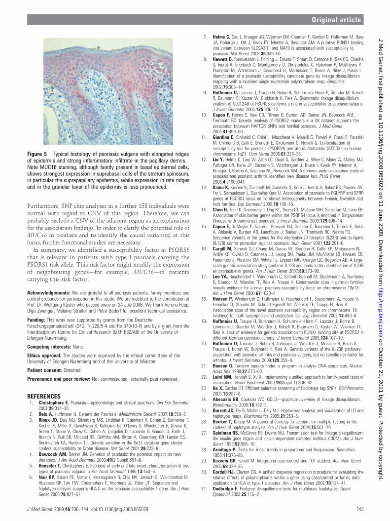
Nevertheless, we were unable to amplify any RT-PCR productsfor any of the regions from skin, thyroid, thymus and/or bloodleucocytes. Also the predicted additional 59 exons of theannotated MUC16 mRNA were undetectable in cDNA fromthese tissues. In contrast, we detected transcription of MUC16mRNA in all tissues but leucocytes (fig 4). These results wereconfirmed in independent experiments using different primers(data not shown). Accordingly, immunohistochemical analysesusing an antibody against MUC16 showed immunostaining inepidermal cells of all 12 skin biopsies investigated. In addition,several individuals—four patients and one control individual—showed focally more intensive staining in several areas of basaland suprabasal epidermal cells (fig 5).
Finally, we were able to develop two robust assays fordetecting variation in copy number in the region of BAC RP11-79F15, overlapping the 39 region of MUC16 telomeric to theassociated region. This region was previously reported to bepolymorphic for copy number in the European population.However, we did not find any loss or gain of copy number inany of the analysed 184 and 93 individuals, respectively.Additional data from Affymetrix 6.0 SNP chips of 158individuals analysed with the Affymetrix Console 2.1 did notshow CNV of this region (data not shown). We conclude thatthe region of this BAC shows no significant variation in copynumber, at least in the German population.
DISCUSSIONThe adaptation of the classical linkage analysis paradigm,initially developed for gene identification of Mendelian dis-orders, to complex traits is based on an initial genome-widelinkage scan followed by LD studies in regions showing evidencefor linkage. This strategy has been successful for several diseasessuch as Crohn’s,3 38 asthma39 and sarcoidosis40 to name a few.We followed this strategy with an initial genome-wide linkagescan of 32 extended PsV families with multiple affected
individuals. This study identified two major linked regions,one at the HLA locus on chromosome 6 (PSORS1) and one onchromosome 19p13 (PSORS6) (Lee et al17). PSORS1 is the majorPsV susceptibility locus and has been replicated in all linkagestudies to date, while the PSORS6 has only been replicated in astudy from the UK.41
We performed a systematic LD study in the region of thelinkage peak at psoriasis susceptibility locus 6 (PSORS6). Whenscanning a 1.3 Mb region from the core linkage interval using 16informative microsatellites with reduced heterozygosity, wewere able to identify a strong association to one allele ofmicrosatellite D19SPS21. Since the neighbouring markerD19SPS20 was also associated, we focused our efforts on thisregion of about 250 kb. At the time of discovery, the LDstructure of this region was poorly characterised. We thereforeidentified SNPs in 32 patients, determined the LD structure, andconfirmed it by genotyping tagging SNPs in 600 independentparents of the trio cohort. The structure proved to beconcordant with that later reported by HapMap.42 In theTDT analysis in 300 trios we found association with severalSNPs from the same 50 kb haplotype block. Similarly, thehaplotype encompassing the same alleles was also associated.The association signals increased after stratification for thePSORS1 risk allele on chromosome 6p, suggesting that both riskfactors interact.
To verify these findings we genotyped tagging SNPs for thishaplotype in an independent case control cohort of 1114patients and 937 controls. After stratification for PSORS1 theassociation was again significant, although less strongly.Nevertheless, we consider this a successful replication. Whilewe found evidence for interaction with the PSORS1 risk allele, asimilar effect of variants of the IL-23R pathway was notdetectable. The combined OR of both study groups for one SNPat PSORS6 was estimated to be 1.78¡0.11. This magnitude ofsusceptibility is in the range observed in non-HLA loci in
Figure 2 Results of transmission disequilibrium test (TDT) statistics for the 63 haplotype tagging single nucleotide polymorphisms (SNPs). (a)Relative locations of SNPs between bp 8904347 and 9112258 on chromosome 19 (hg18). Stars indicate the location of the two associatedmicrosatellites (*D19SPS20, **D19SPS21). (b) The blue line displays negative logarithms of p values ,0.2 in all trios, the magenta line the ones intrios with index patients carrying the PSORS1 risk allele. The green line indicates the significance level of p,0.05, curly braces affiliation to a haplotypeblock as shown in fig 3 and supplemental table 2.
Original article
740 J Med Genet 2009;46:736–744. doi:10.1136/jmg.2008.065029
on October 21, 2021 by guest. P
rotected by copyright.http://jm
g.bmj.com
/J M
ed Genet: first published as 10.1136/jm
g.2008.065029 on 11 June 2009. Dow
nloaded from

psoriasis and many other complex diseases. For example, therecently described and well replicated variants in genes of the IL-23R pathway15 16 showed a similar effect size in our Germancase control cohort (OR 1.50).20
Interaction between PSORS1 and one of the further psoriasissusceptibility loci has been described previously for PSORS4 onchromosome 1q21.43 In our study, stratification according toPSORS1 indicated that PSORS6 was only relevant in PSORS1carriers. Indeed, we did not observe association of psoriasis withPOSRS6 in non-carriers of the PSORS1 risk allele. Thus, we canspeak of epistasis, and our model is neither multiplicative noradditive. Our family based sample contained some families withmore than one affected child. For this configuration, aderivation of the exact disease model remains a challenge.
Although chromosome 19 has about twice the gene density ofthe genome average,44 our results did not map to any knowncoding region. Many non-coding regions, though, harbour
regulatory elements regulating expression of adjacent genes.Interestingly, we showed that one of the neighbouring genes,mucin 16 (MUC16), of unknown function is expressed in severaltissues that are relevant for psoriasis and/or other autoimmunediseases such as skin, thymus, and thyroid. Expression in otherepithelia such as ocular surfaces (cornea, conjunctiva), respira-tory tract and vagina has been described.45 46 The highlyglycosylated MUC16 has long been known as a tumour markerindicating recurrence of ovarian cancer and preceding cancergenesis47 and is often referred to as antigen CA125.48 Ourimmunohistochemical analysis revealed weak general stainingof the epidermis with focally increased signals in suprabasal cellsof the suprapapillary epidermis. This location coincides with themain hyperproliferative zone in psoriatic skin, while in normalepidermis proliferation is restricted to basal cells.49 Ourresults show a discrepancy with previously performed micro-array analyses that did not show differential expression.50–52
Figure 3 Genomic region of interest, linkage disequilibrium (LD) structure in trios, and results of transmission disequilibrium test (TDT) statistics forhaplotypes within seven haplotype blocks. (a) Name, exon structure and orientation of RefSeq genes. (b) Pairwise LD plot for the 63 single nucleotidepolymorphisms (SNPs) based on data of trios with index patients carrying the PSORS1 risk allele. Each square plots the level of LD between a pair ofsites in the region; comparisons between neighbouring sites lie along the upper most line. Red colouring indicates strong LD, light red less strong LD, ashadow of red intermediate LD, and white indicates weak LD. The areas limited by a black line show the seven haplotype blocks basically defined bythe model ‘‘Solid Spine’’. (c) Results of FAMHAP statistics for haplotypes with .30 informative transmissions in all trios; shown are results within theseven haplotype blocks in all trios (blue) and in trios with index patients carrying the PSORS1 risk allele (magenta). The green line indicates thesignificance level of p,0.05, stars the location of the two associated microsatellites (*D19SPS20, **D19SPS21). For p values .0.15, a negativelogarithm of 0.15 is shown.
Original article
J Med Genet 2009;46:736–744. doi:10.1136/jmg.2008.065029 741
on October 21, 2021 by guest. P
rotected by copyright.http://jm
g.bmj.com
/J M
ed Genet: first published as 10.1136/jm
g.2008.065029 on 11 June 2009. Dow
nloaded from

This might be explained by a restricted expression of MUC16 inthe suprabasal layer.
Interestingly, retinoids and glucocorticoids, which are effec-tive therapeutic agents in psoriasis, enhance MUC16 expressionin eye epithelia.53–55 Furthermore, a role of MUC16 in protectionagainst pathogen adherence56 renders it an even more interestinggene for psoriasis with regard to the HLA-C risk allele. EspeciallyPSORS1 positive patients are prone to develop the guttate formof psoriasis,57 a form that has been strongly associated withthroat infections with b-haemolytic streptococci.58 Recently avariant in the 59 region of another member of the mucin family,MUC19, has been newly identified to be relevant in thepathogenesis of Crohn’s disease.59 This might indicate a moregeneral role of these epithelial proteins in the pathogenesis ofdiseases with compromised barrier function. Nevertheless, wecannot exclude that expression of other genes in this genomicregion might also be affected. To analyse this aspect exhaust-ingly, a combination of several functional analyses such asqPCR, expression studies of further positional candidates, forexample, would be necessary.
Besides SNPs, copy number variation (CNV) has beenrecognised as a new class of potential risk factors in complexdiseases. In psoriasis, a CNV comprising the b-defensin gene
cluster was reported as a risk factor in various European cohorts,including our own study group.60 Iafrate et al36 reported afrequent CNV of the adjacent telomeric region encompassingthe 39 parts of MUC16 gene as well as MBD3L1 and ZNF558genes. Since LD of the associated SNPs with this CNV could notbe excluded, we developed two qPCR assays using an establishedapproach37 and genotyped part of the case–control cohort.However, we could not detect this CNV in our population.
Table 1 Allele frequencies of the associated tag SNP in (A) 1114 psoriasis patients and 937 controls and (B) carriers of the PSORS1 risk allele (633psoriasis patients and 131 control individuals) and results of x2 statistics
SNP rs-ID Allele
(A) (B)
ControlsPsoriasisvulgaris
p Value
ControlsPsoriasisvulgaris
p Value OR (95% CI)n (%) n (%) n (%) n (%)
rs8100377 T 1630 (88.6) 1944 (88.2) NS 226 (89.7) 1109 (88.6) NS NA
C 210 (11.4) 260 (11.8) 26 (10.3) 143 (11.4)
rs6511831 A 831 (45.0) 1051 (48.1) NS 112 (43.8) 594 (48.0) NS NA
G 1015 (55.0) 1135 (51.9) 144 (56.3) 644 (52.0)
rs8109594 G 1470 (79.5) 1714 (81.4) NS 206 (79.8) 964 (80.2) NS NA
A 378 (20.5) 392 (18.6) 52 (20.2) 238 (19.8)
rs12459358 G 1114 (61.1) 1368 (62.8) NS 141 (55.1) 793 (64.4) 0.005 1.47 (1.12 to 1.94)
A 708 (38.9) 810 (37.2) 115 (44.9) 439 (35.6)
rs8102472 T 537 (30.9) 700 (33.4) NS 63 (26.0) 405 (34.2) 0.013 1.48 (1.08 to 2.02)
C 1199 (69.1) 1398 (66.6) 179 (74.0) 779 (65.8)
rs7249334 A 652 (35.2) 779 (37.0) NS 80 (30.8) 461 (38.2) 0.027 1.39 (1.04 to 1.85)
G 1198 (64.8) 1325 (63.0) 180 (69.2) 747 (61.8)
rs10413384 G 1458 (79.1) 1732 (78.4) NS 198 (76.2) 992 (78.9) NS NA
A 386 (20.9) 478 (21.6) 62 (23.8) 266 (21.1)
rs2591618 C 1080 (58.1) 1307 (58.9) NS 144 (55.0) 759 (60.2) NS NA
T 778 (41.9) 913 (41.1) 118 (45.0) 501 (39.8)
rs28699225 G 1461 (79.7) 1739 (79.0) NS 194 (77.6) 996 (79.9) NS NA
A 371 (20.3) 461 (21.0) 56 (22.4) 250 (20.1)
CI, confidence interval; NA, not applicable; NS, not significant; OR odds ratio; SNP, single nucleotide polymorphism.
Table 2 Allele frequencies of the risk haplotype in (A) 1114 psoriasispatients and 937 controls and (B) carriers of the PSORS1 risk allele (633psoriasis patients and 131 control individuals) and results of x2 statistics
Haplotype(s)
Controls Psoriasisvulgaris
p Value OR (95% CI)n (%) n (%)
(A)
TAGGTAGCG 552.6 (29.5) 698.7 (31.4) NS NA
S non-risk 1320.6 (70.5) 1526.5 (68.6)
(B)
TAGGTAGCG 64.7 (24.7) 403 (31.8) 0.012 1.42 (1.05 to 1.93)
S non-risk 197.2 (75.3) 864.3 (68.2)
CI, confidence interval; NA, not applicable; NS, not significant; OR, odds ratio.
Figure 4 Electrophoresis of conventional reverse transcriptasepolymerase chain reaction (RT-PCR) products demonstrating expectedamplicon sizes of a product for each (a) GAPDH and (b) MUC16 (primersin exons 4+5, PCR product of 363 bp), amplified from cDNAs of differenttissues: lane 1: size standard, 2: skin, 3: thymus, 4: thyroid, 5: bloodleucocytes, 6: non-template control.
Original article
742 J Med Genet 2009;46:736–744. doi:10.1136/jmg.2008.065029
on October 21, 2021 by guest. P
rotected by copyright.http://jm
g.bmj.com
/J M
ed Genet: first published as 10.1136/jm
g.2008.065029 on 11 June 2009. Dow
nloaded from
Furthermore, SNP chip analyses in a further 158 individuals werenormal with regard to CNV of this region. Therefore, we canprobably exclude a CNV of the adjacent region as an explanationfor the association findings. In order to clarify the potential role ofMUC16 in psoriasis and to identify the causal variant(s) at thislocus, further functional studies are necessary.
In summary, we identified a susceptibility factor at PSORS6that is relevant in patients with type I psoriasis carrying thePSORS1 risk allele. This risk factor might modify the expressionof neighbouring genes—for example, MUC16—in patientscarrying this risk factor.
Acknowledgements: We are grateful to all psoriasis patients, family members andcontrol probands for participation in this study. We are indebted to the contribution ofProf. Dr. Wolfgang Kuster who passed away on 24 July 2006. We thank Verena Popp,Olga Zwenger, Melanie Streiter and Petra Badorf for excellent technical assistance.
Funding: This work was supported by grants from the DeutscheForschungsgemeinschaft (DFG, Tr 228/5-4 and Re 679/10-4) and by a grant from theInterdisciplinary Centre for Clinical Research (IZKF B32/A8) of the University ofErlangen-Nuremberg.
Competing interests: None.
Ethics approval: The studies were approved by the ethical committees of theUniversity of Erlangen-Nuremberg and of the University of Munster
Patient consent: Obtained.
Provenance and peer review: Not commissioned; externally peer reviewed.
REFERENCES1. Christophers E. Psoriasis—epidemiology and clinical spectrum. Clin Exp Dermatol
2001;26:314–20.2. Reis A, Huffmeier U. Genetik der Psoriasis. Medizinische Genetik 2007;19:350–5.3. Rioux JD, Daly MJ, Silverberg MS, Lindblad K, Steinhart H, Cohen Z, Delmonte T,
Kocher K, Miller K, Guschwan S, Kulbokas EJ, O’Leary S, Winchester E, Dewar K,Green T, Stone V, Chow C, Cohen A, Langelier D, Lapointe G, Gaudet D, Faith J,Branco N, Bull SB, McLeod RS, Griffiths AM, Bitton A, Greenberg GR, Lander ES,Siminovitch KA, Hudson TJ. Genetic variation in the 5q31 cytokine gene clusterconfers susceptibility to Crohn disease. Nat Genet 2001;29:223–8.
4. Bowcock AM, Barker JN. Genetics of psoriasis: the potential impact on newtherapies. J Am Acad Dermatol 2003;49(2 Suppl):S51–6.
5. Henseler T, Christophers E. Psoriasis of early and late onset: characterization of twotypes of psoriasis vulgaris. J Am Acad Dermatol 1985;13:450–6.
6. Nair RP, Stuart PE, Nistor I, Hiremagalore R, Chia NV, Jenisch S, Weichenthal M,Abecasis GR, Lim HW, Christophers E, Voorhees JJ, Elder JT. Sequence andhaplotype analysis supports HLA-C as the psoriasis susceptibility 1 gene. Am J HumGenet 2006;78:827–51.
7. Helms C, Cao L, Krueger JG, Wijsman EM, Chamian F, Gordon D, Heffernan M, DawJA, Robarge J, Ott J, Kwok PY, Menter A, Bowcock AM. A putative RUNX1 bindingsite variant between SLC9A3R1 and NAT9 is associated with susceptibility topsoriasis. Nat Genet 2003;35:349–56.
8. Hewett D, Samuelsson L, Polding J, Enlund F, Smart D, Cantone K, See CG, ChadhaS, Inerot A, Enerback C, Montgomery D, Christodolou C, Robinson P, Matthews P,Plumpton M, Wahlstrom J, Swanbeck G, Martinsson T, Roses A, Riley J, Purvis I.Identification of a psoriasis susceptibility candidate gene by linkage disequilibriummapping with a localized single nucleotide polymorphism map. Genomics2002;79:305–14.
9. Huffmeier U, Lascorz J, Traupe H, Bohm B, Schurmeier-Horst F, Stander M, KelschR, Baumann C, Kuster W, Burkhardt H, Reis A. Systematic linkage disequilibriumanalysis of SLC12A8 at PSORS5 confirms a role in susceptibility to psoriasis vulgaris.J Invest Dermatol 2005;125:906–12.
10. Capon F, Helms C, Veal CD, Tillman D, Burden AD, Barker JN, Bowcock AM,Trembath RC. Genetic analysis of PSORS2 markers in a UK dataset supports theassociation between RAPTOR SNPs and familial psoriasis. J Med Genet2004;41:459–60.
11. Giardina E, Sinibaldi C, Chini L, Moschese V, Marulli G, Provini A, Rossi P, ParadisiM, Chimenti S, Galli E, Brunetti E, Girolomoni G, Novelli G. Co-localization ofsusceptibility loci for psoriasis (PSORS4) and atopic dermatitis (ATOD2) on humanchromosome 1q21. Hum Hered 2006;61:229–36.
12. Liu Y, Helms C, Liao W, Zaba LC, Duan S, Gardner J, Wise C, Miner A, Malloy MJ,Pullinger CR, Kane JP, Saccone S, Worthington J, Bruce I, Kwok PY, Menter A,Krueger J, Barton A, Saccone NL, Bowcock AM. A genome-wide association study ofpsoriasis and psoriatic arthritis identifies new disease loci. PLoS Genet2008;4:e1000041.
13. Kainu K, Kivinen K, Zucchelli M, Suomela S, Kere J, Inerot A, Baker BS, Powles AV,Fry L, Samuelsson L, Saarialho-Kere U. Association of psoriasis to PGLYRP and SPRRgenes at PSORS4 locus on 1q shows heterogeneity between Finnish, Swedish andIrish families. Exp Dermatol 2009;18:109–15.
14. Chen H, Toh TK, Szeverenyi I, Ong RT, Theng CT, McLean WH, Seielstad M, Lane EB.Association of skin barrier genes within the PSORS4 locus is enriched in SingaporeanChinese with early-onset psoriasis. J Invest Dermatol 2009;129:606–14.
15. Capon F, Di Meglio P, Szaub J, Prescott NJ, Dunster C, Baumber L, Timms K, GutinA, Abkevic V, Burden AD, Lanchbury J, Barker JN, Trembath RC, Nestle FO.Sequence variants in the genes for the interleukin-23 receptor (IL23R) and its ligand(IL12B) confer protection against psoriasis. Hum Genet 2007;122:201–6.
16. Cargill M, Schrodi SJ, Chang M, Garcia VE, Brandon R, Callis KP, Matsunami N,Ardlie KG, Civello D, Catanese JJ, Leong DU, Panko JM, McAllister LB, Hansen CB,Papenfuss J, Prescott SM, White TJ, Leppert MF, Krueger GG, Begovich AB. A large-scale genetic association study confirms IL12B and leads to the identification of IL23Ras psoriasis-risk genes. Am J Hum Genet 2007;80:273–90.
17. Lee YA, Ruschendorf F, Windemuth C, Schmitt-Egenolf M, Stadelmann A, NurnbergG, Stander M, Wienker TF, Reis A, Traupe H. Genomewide scan in german familiesreveals evidence for a novel psoriasis-susceptibility locus on chromosome 19p13.Am J Hum Genet 2000;67:1020–4.
18. Hensen P, Windemuth C, Huffmeier U, Ruschendorf F, Stadelmann A, Hoppe V,Fenneker D, Stander M, Schmitt-Egenolf M, Wienker TF, Traupe H, Reis A.Association scan of the novel psoriasis susceptibility region on chromosome 19:evidence for both susceptible and protective loci. Exp Dermatol 2003;12:490–6.
19. Huffmeier U, Traupe H, Burkhardt H, Schurmeier-Horst F, Lascorz J, Bohm B,Lohmann J, Stander M, Wendler J, Kelsch R, Baumann C, Kuster W, Wienker TF,Reis A. Lack of evidence for genetic association to RUNX1 binding site at PSORS2 indifferent German psoriasis cohorts. J Invest Dermatol 2005;124:107–10.
20. Huffmeier U, Lascorz J, Bohm B, Lohmann J, Wendler J, Mossner R, Reich K,Traupe H, Kurrat W, Burkhardt H, Reis A. Genetic variants of the IL-23R pathway:association with psoriatic arthritis and psoriasis vulgaris, but no specific risk factor forarthritis. J Invest Dermatol 2009;129:355–8.
21. Benson G. Tandem repeats finder: a program to analyze DNA sequences. NucleicAcids Res 1999;27:573–80.
22. Laird NM, Horvath S, Xu X. Implementing a unified approach to family-based tests ofassociation. Genet Epidemiol 2000;19(Suppl 1):S36–42.
23. Ke X, Cardon LR. Efficient selective screening of haplotype tag SNPs. Bioinformatics2003;19:287–8.
24. Abecasis GR, Cookson WO. GOLD—graphical overview of linkage disequilibrium.Bioinformatics 2000;16:182–3.
25. Barrett JC, Fry B, Maller J, Daly MJ. Haploview: analysis and visualization of LD andhaplotype maps. Bioinformatics 2005;21:263–5.
26. Becker T, Knapp M. A powerful strategy to account for multiple testing in thecontext of haplotype analysis. Am J Hum Genet 2004;75:561–70.
27. Spielman RS, McGinnis RE, Ewens WJ. Transmission test for linkage disequilibrium:the insulin gene region and insulin-dependent diabetes mellitus (IDDM). Am J HumGenet 1993;52:506–16.
28. Armitage P. Tests for linear trends in proportions and frequencies. Biometrics1955;11:375–86.
29. Kazeem GR, Farrall M. Integrating case-control and TDT studies. Ann Hum Genet2005;69:329–35.
30. Cordell HJ, Clayton DG. A unified stepwise regression procedure for evaluating therelative effects of polymorphisms within a gene using case/control or family data:application to HLA in type 1 diabetes. Am J Hum Genet 2002;70:124–41.
31. Dudbridge F. Pedigree disequilibrium tests for multilocus haplotypes. GenetEpidemiol 2003;25:115–21.
Figure 5 Typical histology of psoriasis vulgaris with elongated ridgesof epidermis and strong inflammatory infiltrate in the papillary dermis.Note MUC16 staining, although faintly present in basal epidermal cells,shows strongest expression in suprabasal cells of the stratum spinosum,in particular the suprapapillary epidermis, while expression in rete ridgesand in the granular layer of the epidermis is less pronounced.
Original article
J Med Genet 2009;46:736–744. doi:10.1136/jmg.2008.065029 743
on October 21, 2021 by guest. P
rotected by copyright.http://jm
g.bmj.com
/J M
ed Genet: first published as 10.1136/jm
g.2008.065029 on 11 June 2009. Dow
nloaded from

32. Hyatt D, Snoddy J, Schmoyer D, Chen G, Fischer K, Parang M, Vokler I, Petrov S,Locascio P, Olman V, Land M, Shah M, Uberbacher E. Improved analysis andannotation tools for whole-genome computational annotation and analysis: GRAIL-EXP genome analysis toolkit and related analysis tools. Genome Sequencing & BiologyMeeting, 2000.
33. Burge C, Karlin S. Prediction of complete gene structures in human genomic DNA.J Mol Biol 1997;268:78–94.
34. Schwartz S, Zhang Z, Frazer KA, Smit A, Riemer C, Bouck J, Gibbs R, Hardison R,Miller W. PipMaker—a web server for aligning two genomic DNA sequences.Genome Res 2000;10:577–86.
35. Schwartz S, Kent WJ, Smit A, Zhang Z, Baertsch R, Hardison RC, Haussler D, MillerW. Human-mouse alignments with BLASTZ. Genome Res 2003;13:103–7.
36. Iafrate AJ, Feuk L, Rivera MN, Listewnik ML, Donahoe PK, Qi Y, Scherer SW, Lee C.Detection of large-scale variation in the human genome. Nat Genet 2004;36:949–51.
37. Thiel CT, Kraus C, Rauch A, Ekici AB, Rautenstrauss B, Reis A. A new quantitativePCR multiplex assay for rapid analysis of chromosome 17p11.2-12 duplications anddeletions leading to HMSN/HNPP. Eur J Hum Genet 2003;11:170–8.
38. Hugot JP, Chamaillard M, Zouali H, Lesage S, Cezard JP, Belaiche J, Almer S, TyskC, O’Morain CA, Gassull M, Binder V, Finkel Y, Cortot A, Modigliani R, Laurent-Puig P,Gower-Rousseau C, Macry J, Colombel JF, Sahbatou M, Thomas G. Association ofNOD2 leucine-rich repeat variants with susceptibility to Crohn’s disease. Nature2001;411:599–603.
39. Laitinen T, Polvi A, Rydman P, Vendelin J, Pulkkinen V, Salmikangas P, Makela S,Rehn M, Pirskanen A, Rautanen A, Zucchelli M, Gullsten H, Leino M, Alenius H,Petays T, Haahtela T, Laitinen A, Laprise C, Hudson TJ, Laitinen LA, Kere J.Characterization of a common susceptibility locus for asthma-related traits. Science2004;304:300–4.
40. Valentonyte R, Hampe J, Huse K, Rosenstiel P, Albrecht M, Stenzel A, Nagy M,Gaede KI, Franke A, Haesler R, Koch A, Lengauer T, Seegert D, Reiling N, Ehlers S,Schwinger E, Platzer M, Krawczak M, Muller-Quernheim J, Schurmann M, SchreiberS. Sarcoidosis is associated with a truncating splice site mutation in BTNL2. NatGenet 2005;37:357–64.
41. Veal CD, Clough RL, Barber RC, Mason S, Tillman D, Ferry B, Jones AB, Ameen M,Balendran N, Powis SH, Burden AD, Barker JN, Trembath RC. Identification of a novelpsoriasis susceptibility locus at 1p and evidence of epistasis between PSORS1 andcandidate loci. J Med Genet 2001;38:7–13.
42. Frazer KA, Ballinger DG, Cox DR, Hinds DA, Stuve LL, Gibbs RA, Belmont JW,Boudreau A, Hardenbol P, Leal SM, Pasternak S, Wheeler DA, Willis TD, Yu F, Yang H,Zeng C, Gao Y, Hu H, Hu W, Li C, Lin W, Liu S, Pan H, Tang X, Wang J, Wang W, YuJ, Zhang B, Zhang Q, Zhao H, Zhao H, Zhou J, Gabriel SB, Barry R, Blumenstiel B,Camargo A, Defelice M, Faggart M, Goyette M, Gupta S, Moore J, Nguyen H, OnofrioRC, Parkin M, Roy J, Stahl E, Winchester E, Ziaugra L, Altshuler D, Shen Y, Yao Z,Huang W, Chu X, He Y, Jin L, Liu Y, Shen Y, Sun W, Wang H, Wang Y, Wang Y, XiongX, Xu L, Waye MM, Tsui SK, Xue H, Wong JT, Galver LM, Fan JB, Gunderson K,Murray SS, Oliphant AR, Chee MS, Montpetit A, Chagnon F, Ferretti V, Leboeuf M,Olivier JF, Phillips MS, Roumy S, Sallee C, Verner A, Hudson TJ, Kwok PY, Cai D,Koboldt DC, Miller RD, Pawlikowska L, Taillon-Miller P, Xiao M, Tsui LC, Mak W, SongYQ, Tam PK, Nakamura Y, Kawaguchi T, Kitamoto T, Morizono T, Nagashima A,Ohnishi Y, Sekine A, Tanaka T, Tsunoda T, Deloukas P, Bird CP, Delgado M,Dermitzakis ET, Gwilliam R, Hunt S, Morrison J, Powell D, Stranger BE, Whittaker P,Bentley DR, Daly MJ, de Bakker PI, Barrett J, Chretien YR, Maller J, McCarroll S,Patterson N, Pe’er I, Price A, Purcell S, Richter DJ, Sabeti P, Saxena R, Schaffner SF,Sham PC, Varilly P, Altshuler D, Stein LD, Krishnan L, Smith AV, Tello-Ruiz MK,Thorisson GA, Chakravarti A, Chen PE, Cutler DJ, Kashuk CS, Lin S, Abecasis GR,Guan W, Li Y, Munro HM, Qin ZS, Thomas DJ, McVean G, Auton A, Bottolo L, CardinN, Eyheramendy S, Freeman C, Marchini J, Myers S, Spencer C, Stephens M,Donnelly P, Cardon LR, Clarke G, Evans DM, Morris AP, Weir BS, Tsunoda T, MullikinJC, Sherry ST, Feolo M, Skol A, Zhang H, Zeng C, Zhao H, Matsuda I, Fukushima Y,Macer DR, Suda E, Rotimi CN, Adebamowo CA, Ajayi I, Aniagwu T, Marshall PA,Nkwodimmah C, Royal CD, Leppert MF, Dixon M, Peiffer A, Qiu R, Kent A, Kato K,Niikawa N, Adewole IF, Knoppers BM, Foster MW, Clayton EW, Watkin J, Gibbs RA,Belmont JW, Muzny D, Nazareth L, Sodergren E, Weinstock GM, Wheeler DA, YakubI, Gabriel SB, Onofrio RC, Richter DJ, Ziaugra L, Birren BW, Daly MJ, Altshuler D,Wilson RK, Fulton LL, Rogers J, Burton J, Carter NP, Clee CM, Griffiths M, Jones MC,McLay K, Plumb RW, Ross MT, Sims SK, Willey DL, Chen Z, Han H, Kang L, GodboutM, Wallenburg JC, L’Archeveque P, Bellemare G, Saeki K, Wang H, An D, Fu H, Li Q,Wang Z, Wang R, Holden AL, Brooks LD, McEwen JE, Guyer MS, Wang VO, PetersonJL, Shi M, Spiegel J, Sung LM, Zacharia LF, Collins FS, Kennedy K, Jamieson R,Stewart J. A second generation human haplotype map of over 3.1 million SNPs.Nature 2007;449:851–61.
43. Capon F, Semprini S, Dallapiccola B, Novelli G. Evidence for interaction betweenpsoriasis-susceptibility loci on chromosomes 6p21 and 1q21. Am J Hum Genet1999;65:1798–800.
44. Grimwood J, Gordon LA, Olsen A, Terry A, Schmutz J, Lamerdin J, Hellsten U,Goodstein D, Couronne O, Tran-Gyamfi M, Aerts A, Altherr M, Ashworth L, Bajorek E,Black S, Branscomb E, Caenepeel S, Carrano A, Caoile C, Chan YM, Christensen M,Cleland CA, Copeland A, Dalin E, Dehal P, Denys M, Detter JC, Escobar J, Flowers D,Fotopulos D, Garcia C, Georgescu AM, Glavina T, Gomez M, Gonzales E, Groza M,Hammon N, Hawkins T, Haydu L, Ho I, Huang W, Israni S, Jett J, Kadner K, Kimball H,Kobayashi A, Larionov V, Leem SH, Lopez F, Lou Y, Lowry S, Malfatti S, Martinez D,McCready P, Medina C, Morgan J, Nelson K, Nolan M, Ovcharenko I, Pitluck S,Pollard M, Popkie AP, Predki P, Quan G, Ramirez L, Rash S, Retterer J, Rodriguez A,Rogers S, Salamov A, Salazar A, She X, Smith D, Slezak T, Solovyev V, Thayer N, TiceH, Tsai M, Ustaszewska A, Vo N, Wagner M, Wheeler J, Wu K, Xie G, Yang J,Dubchak I, Furey TS, DeJong P, Dickson M, Gordon D, Eichler EE, Pennacchio LA,Richardson P, Stubbs L, Rokhsar DS, Myers RM, Rubin EM, Lucas SM. The DNAsequence and biology of human chromosome 19. Nature 2004;428:529–35.
45. Argueso P, Sumiyoshi M. Characterization of a carbohydrate epitope defined by themonoclonal antibody H185: sialic acid O-acetylation on epithelial cell-surface mucins.Glycobiology 2006;16:1219–28.
46. Hattrup CL, Gendler SJ. Structure and function of the cell surface (tethered) mucins.Annu Rev Physiol 2008;70:431–57.
47. Palmer C, Duan X, Hawley S, Scholler N, Thorpe JD, Sahota RA, Wong MQ, Wray A,Bergan LA, Drescher CW, McIntosh MW, Brown PO, Nelson BH, Urban N. Systematicevaluation of candidate blood markers for detecting ovarian cancer. PLoS ONE2008;3:e2633.
48. Rump A, Morikawa Y, Tanaka M, Minami S, Umesaki N, Takeuchi M, Miyajima A.Binding of ovarian cancer antigen CA125/MUC16 to mesothelin mediates celladhesion. J Biol Chem 2004;279:9190–8.
49. Leigh IM, Pulford KA, Ramaekers FC, Lane EB. Psoriasis: maintenance of an intactmonolayer basal cell differentiation compartment in spite of hyperproliferation.Br J Dermatol 1985;113:53–64.
50. Koczan D, Guthke R, Thiesen HJ, Ibrahim SM, Kundt G, Krentz H, Gross G, Kunz M.Gene expression profiling of peripheral blood mononuclear leukocytes from psoriasispatients identifies new immune regulatory molecules. Eur J Dermatol 2005;15:251–7.
51. Reischl J, Schwenke S, Beekman JM, Mrowietz U, Sturzebecher S, Heubach JF.Increased expression of Wnt5a in psoriatic plaques. J Invest Dermatol2007;127:163–9.
52. Zhou X, Krueger JG, Kao MC, Lee E, Du F, Menter A, Wong WH, Bowcock AM.Novel mechanisms of T-cell and dendritic cell activation revealed by profiling ofpsoriasis on the 63,100-element oligonucleotide array. Physiol Genomics2003;13:69–78.
53. Hori Y, Spurr-Michaud S, Russo CL, Argueso P, Gipson IK. Differential regulation ofmembrane-associated mucins in the human ocular surface epithelium. InvestOphthalmol Vis Sci 2004;45:114–22.
54. Hori Y, Spurr-Michaud SJ, Russo CL, Argueso P, Gipson IK. Effect of retinoic acidon gene expression in human conjunctival epithelium: secretory phospholipase A2mediates retinoic acid induction of MUC16. Invest Ophthalmol Vis Sci2005;46:4050–61.
55. Seo KY, Chung SH, Lee JH, Park MY, Kim EK. Regulation of membrane-associatedmucins in the human corneal epithelial cells by dexamethasone. Cornea2007;26:709–14.
56. Blalock TD, Spurr-Michaud SJ, Tisdale AS, Heimer SR, Gilmore MS, Ramesh V,Gipson IK. Functions of MUC16 in corneal epithelial cells. Invest Ophthalmol Vis Sci2007;48:4509–18.
57. Gudjonsson JE, Karason A, Antonsdottir AA, Runarsdottir EH, Gulcher JR,Stefansson K, Valdimarsson H. HLA-Cw6-positive and HLA-Cw6-negative patientswith psoriasis vulgaris have distinct clinical features. J Invest Dermatol2002;118:362–5.
58. Telfer NR, Chalmers RJ, Whale K, Colman G. The role of streptococcal infection inthe initiation of guttate psoriasis. Arch Dermatol 1992;128:39–42.
59. Barrett JC, Hansoul S, Nicolae DL, Cho JH, Duerr RH, Rioux JD, Brant SR, SilverbergMS, Taylor KD, Barmada MM, Bitton A, Dassopoulos T, Datta LW, Green T, GriffithsAM, Kistner EO, Murtha MT, Regueiro MD, Rotter JI, Schumm LP, Steinhart AH,Targan SR, Xavier RJ, Libioulle C, Sandor C, Lathrop M, Belaiche J, Dewit O, Gut I,Heath S, Laukens D, Mni M, Rutgeerts P, Van Gossum A, Zelenika D, Franchimont D,Hugot JP, de Vos M, Vermeire S, Louis E, Cardon LR, Anderson CA, Drummond H,Nimmo E, Ahmad T, Prescott NJ, Onnie CM, Fisher SA, Marchini J, Ghori J,Bumpstead S, Gwilliam R, Tremelling M, Deloukas P, Mansfield J, Jewell D, Satsangi J,Mathew CG, Parkes M, Georges M, Daly MJ. Genome-wide association definesmore than 30 distinct susceptibility loci for Crohn’s disease. Nat Genet2008;40:955–62.
60. Hollox EJ, Huffmeier U, Zeeuwen PL, Palla R, Lascorz J, Rodijk-Olthuis D, van deKerkhof PC, Traupe H, de Jongh G, den Heijer M, Reis A, Armour JA, Schalkwijk J.Psoriasis is associated with increased beta-defensin genomic copy number. NatGenet 2008;40:23–5.
Original article
744 J Med Genet 2009;46:736–744. doi:10.1136/jmg.2008.065029
on October 21, 2021 by guest. P
rotected by copyright.http://jm
g.bmj.com
/J M
ed Genet: first published as 10.1136/jm
g.2008.065029 on 11 June 2009. Dow
nloaded from