characterisation of p2x receptors within the anterior ... · characterisation of p2x receptors...
TRANSCRIPT
1 | P a g e
Characterisation of P2X receptors within the anterior pituitary and their role in modulating
pituitary function
A thesis submitted for the degree of Doctor of Philosophy
From Imperial College London
Makeda R Agada
Section of Investigative Medicine
Division of Diabetes, Endocrinology and Metabolism
Faculty of Medicine
2 | P a g e
Abstract
Extracellular ATP acts in an autocrine/paracrine manner influencing cellular responses via
two classes of purinoreceptors: metabotropic P2Y and inotropic P2X receptors. Recent
observations have demonstrated a role for P2X receptors (P2X1-7) in calcium mediated
hormone secretion from the pituitary. In the present study we used immortalised cell lines
and primary pituitary cultures to investigate the effects of P2X receptors within the anterior
pituitary. We used Reverse Transcriptase Polymerase Chain reaction (RT-PCR) to
determine the expression of P2XR sub-types within pituitary cell lines and rat pituitary tissue.
Our studies demonstrated similar expression of P2X receptors between rat pituitary tissue
and cell lines. The expression of P2X1-7 receptors was identified in rat primary pituitary
tissue. In the lactotroph cell lines, MMQ and GH3 we found P2X2, 3, 4, 5, 7 and P2X3, 4, 7
receptors, respectively. In LβT2 gonadotroph type cells, P2X2, 3, 4, 7 were identified
whereas the corticotroph cell line AtT20:D16 expressed P2X1,2. Finally the murine cell line
TtT/GF, which represent an in vitro model of the non-endocrine folliculostellate were profiled
for P2X receptor expression. To our knowledge this is the first time this particular cell type
has been examined for P2X receptor expression. Weak expression for P2X2, 4 was
identified. The distribution pattern of P2X1-7 receptors within the rat pituitary was determined
using double labelling immunoflourecscence and western blot. Protein expression for P2X4
and P2X7 was confirmed by Western blot of rat pituitary lysates, but no signal for P2X3
could be determined. Double immunofluorescence found P2X4 to be localised in
corticotrophs, gonadotrophs, sommaotrophs, lactotrophs and thyrotrophs within a small
group of cells. P2X7 was only found localised in a small group of somaotrophs.
We then undertook a more thorough characterisation of the functional significance of P2X
receptor expression within the different mammalian pituitary subtypes. We began our
investigation by establishing a rat primary pituitary culture model and utilised multiplexing
technology to assess the effects of ATP on the secretion of multiple pituitary hormones
(Adrenocorticotropic hormone (ACTH), Growth Hormone (GH), Prolactin (PRL) and Thyroid

3 | P a g e
Stimulating Hormone (TSH) ) simultaneously. ACTH and PRL were the most responsive to
ATP stimulation. TSH and GH showed little response to ATP stimulation. We then went on to
examine the effects of ATP on hormone secretion within our various cell lines. In AtT20:D16
cells, ATP had a significant effect on ACTH release when applied alone and in combination
with CRH following incubation for 7 and 12 hours. Using the lactotroph model GH3, we
assessed the effects of ATP and various ATP analogues on PRL release in vitro. All
concentrations of ATP tested (0.001-1000µM) had a significant effect of PRL release from
cells. On the other hand both analogues, the non- hydrolysable form of ATP adenosine 5'-
O-(3-thio)triphosphate (ATP--S) and the P2X selective analogue, 2',3’-O-(4-benzoyl-
benzoyl) (BzATP) had no significant effect on PRL release. We then attempted to
characterise P2XR function in the pituitary using highly specific P2 and P2XR antagonists.
The P2 receptor antagonist pyridoxal-phosphate-6-azophenyl-2',4'-disulfonate (PPADS)
significantly decreased both basal release of PRL and stimulated PRL secretion in response
to TRH (1nM), ATP, (1 and 100µM) and TRH and ATP in combination. A similar profile was
observed after treatment with the selective P2X7R inhibitor, A438079. The selective P2X3R
inhibitor failed to influence basal PRL secretion but did significantly attenuate the effect of
TRH and ATP stimulated PRL release. Our results confirm previous investigations that ATP
interactions with purinergic receptors expressed within the pituitary may play a role in
modulating pituitary function. Our data has also shown through the use of specific receptor
antagonists, that P2X receptors, particularly P2X3 and P2X7 may be involved in regulating
hormone secretion from lactotrophs.
4 | P a g e
Declaration
The copyright of this thesis rests with the author and is made available under a Creative
Commons Attribution Non-Commercial No Derivatives licence. Researchers are free to copy,
distribute or transmit the thesis on the condition that they attribute it, that they do not use it
for commercial purposes and that they do not alter, transform or build upon it. For any reuse
or redistribution, researchers must make clear to others the licence terms of this work’
All of the work in this thesis was performed by the author. All collaboration and assistance is
described below:
Chapter 3
Sprague Dawley rat pituitaries used for immunofluorescence studies were extracted,
prepared and mounted on to slides by Rebecca Fish, of Pfizer Neusentis, Cambridge
Chapter 4
Radiolabelled ligands for pituitary hormone radioimmunoassays were provided by Professor
Mohammad Ghatei and iodinated by Professor Ghatei or Dr Christopher John of Imperial
College London

5 | P a g e
Acknowledgements I would like to thank Professor Julia Buckingham for giving me the opportunity to carry out
this work and for her supervision.
Special thanks to Dr Christopher John for his supervision, guidance and advice. There are
many people I would also like to thank and acknowledge from the different sites I have
worked at during the course of my PhD. Thanks to staff in the Division of Neuroscience, both
past and present, for their help and advice. I would particularly like to thank Dr Simon
McAuthur, Dr Praveen Paul and Dr Margareta Nikolic for their constant words of
encouragement and their expertise which got me through my first year. I would also like to
thank Dr Rob Fawkes and Dr Pamela Mellon for their help in sourcing pituitary cell lines
used in my thesis. I would like to thank everyone within the Section of Investigative
Medicine for the friendly chats in the coffee room and tissue culture lab (my home from
home) and for making my many weekend visits to the 6th floor of the Commonwealth
building more pleasurable. They helped make my transition to a new department less
stressful and made the 6th floor a great place to work. I would also like to thank all the
members of the SAS lab who helped me in some way with my numerous
radioimmunoassays.
Thanks also go to the members of Pfizer Neusentis, Cambridge, for their help in the lab and
for making me feel so welcome. In addition, thanks to Mike Rigby and Rebecca Fish for
giving me the opportunity to work at Pfizer. In particular the support and guidance of Becky
has been invaluable throughout the course of my work.
Away from the lab, I would like to thank all my friends and family who have supported me
throughout the course of my PhD and beyond. My parents have been my foundation and my
inspiration during my PhD and my gratitude for their support will never properly be
articulated. My mother was a source of comfort and strength, my rock during the tougher

6 | P a g e
parts of my PhD. I wish she could read this as I know it would have made her so proud. So
instead I dedicate this to her, Joan Baynes.
To do this work, I received a Pfizer CASE studentship.
7 | P a g e
Abbreviations Acid–sensing ion channel family ASIC
Adrenocorticotropic hormone ACTH
Adenosine 5’diphosphate ADP
Adenosine 5’-monophosphate AMP
Analyses of variance ANOVA
Adenosine 5’-triphosphate ATP Adenosine 5'-O-(3-thio)triphosphate ATP--S Brilliant blue G BBG Albumin from Bovine Serum BSA 2',3’-O-(4-benzoyl-benzoyl) BzATP Calcium Ca2+ Central nervous system CNS
Corticotropin-releasing factor CRF
cyslic adenosine monophosphate cAMP
4',6-diamidino-2-phenylindole DAPI
complementary Deoxyribonucleic acid cDNA
Deoxyribonucleic acid DNA Deoxyribonuclease I DNase I
Dimethyl sulfoxide DMSO
Dispersal media DM
Dithiothreitol DTT
Dopamine DA
Dulbecco's modified eagle medium DMEM
Dulbecco's Modified Eagle Medium:Nutrient Mixture F-12 DMEM / F-12
4-(3-pentylamino)-2,7-dimethyl-8-(2-methyl-4-methoxyphenyl)-pyrazolo--pyrimidine
DMP904
Molar concentration of agonist that produces 50% of Maximal effect EC50
8 | P a g e
Ectonucleotidases EN
Normal Donkey serum NDS
Ethylenediaminetetraacetic acid EDTA
Ethylene Glycol Tetraacetic Acid EGTA
Ethidium bromide EtBr
Fetal calf serum FCS
Follicle-stimulating hormone FSH
Folliculo-stellate FS
Gram(s) g
Normal Goat Serum NGS
Gonadotropin-releasing Hormone GnRH
G-protein coupled metabotrophic receptors GPCR
Growth Hormone GH
Growth Hormone releasing Hormone GHRH
Hydrogen H+
Hydrogen Peroxide H2O2
4-(2-hydroxyethyl)-1- piperazineethanesulfonic acid HEPES
Molar concentration of antagonist that produces 50% of maximal effect IC50
Immunoglobulin G IgG
Inositol 1,4,5-trisphosphate IP3
Insulin-Transferrin-Selenium ITS
Interleukin IL
Ivermectin IVM
Potassium K+
Lipopolysaccharide LPS
Lutenizing Hormone LH
Messenger RNA mRNA
Minute(s) mins
Micromolar μM
9 | P a g e
Millimolar mM
Nanomolar nM
Non-adrenergic non-cholinergic NANC
Non-essential amino-acids NEAA
Sodium Na2+
Peripheral nervous system PNS
Phosphate Buffered Saline PBS
Phospholipase A2 PLA2
Phospholipase-C PLC
polymerase chain reaction PCR
Prolactin PRL
Purinergic 1/Adenosine Receptors P1/AR
Purinergic 2X Receptors P2X
Purinergic 2Y Receptors P2YR
Pyridoxal-phosphate-6-azophenyl-2', 4'-disulfonate PPADS
Probability value pValue
Ribonucleic acid RNA
Radioimmunoprecipitation Buffer RIPA
Reverse transcriptase polymerase chain reaction RT-PCR
Room temperature RT
Standard error of the mean S.E.M
Sodium Dodecyl Sulfate SDS
Somatostain SS
Temperature Temp
Tri-iodo-L-thyronine sodium salt T3
Tris-Borate-EDTA buffer TBE
Thyrotropin-releasing hormone TRH
Thyroid-stimulating hormone TSH
2′,3′-O-(2,4,6-Trinitrophenyl) adenosine 5′-triphosphate TNP-ATP
Tris(hydroxymethyl)aminomethane hydrochloride Tris-HCl

10 | P a g e
Transmembrane domain TM
Ultraviolet UV
Uridine-disphosphate UDP
Uridine-triphosphate UTP
Zinc Zn2+
3-((5-(2,3-dichlorophenyl)-1H-tetrazol-1-yl)methyl A-438079
pyridine)
5-[5-iodo-4-methoxy-2-(1-methylethyl)phenoxy] -2,4- AF-353 pyrimidinediamine hydrochloride
11 | P a g e
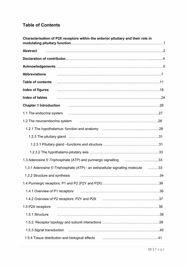
Table of Contents
Characterisation of P2X receptors within the anterior pituitary and their role in modulating pituitary function…………………..…………………………………………………1
Abstract ………………………………………………………………………………………...2
Declaration of contributor.…………………………………………………………….................4
Acknowledgements ………………………………………………………………………………..5
Abbreviations ………………………………………………………………………………7
Table of contents ……………………………………………………………………………..11
Index of figures ……………………………………………………………………………..18
Index of tables ………………………………………………………………………………24
Chapter 1 Introduction …………………………………………………………………….26
1.1 The endocrine system ……………………………………………………………………27
1.2 The neuroendocrine system …………………………………………………………..28
1.2.1 The hypothalamus- function and anatomy ………………………………………….28
1.2.3 The pituitary gland ……………………………………………………………………31
1.2.3.1 Pituitary gland –functions and structure …………………………………………31
1.2.3.2 The hypothalamo-pituitary axis ……………………………………………………33
1.3 Adenosine 5’-Triphosphate (ATP) and purinergic signalling ………………………..33
1.3.1 Adenosine 5’-Triphosphate (ATP) - an extracellular signalling molecule ………33
1.3.2 Structure and synthesis ……………………………………………………………34
1.4 Purinergic receptors: P1 and P2 (P2Y and P2X) ………………………………………….36
1.4.1 Overview of P1 receptors ……………………………………………………………36
1.4.2 Overview of P2 receptors: P2Y and P2X ………………………………………….37
1.5 P2X receptors …………………………………………………………………………… 38
1.5.1 Structure ……………………………………………………………………………..38
1.5.2. Receptor topology and subunit interactions ………………………………………….39
1.5.3 Signal transduction …………………………………………………………………….40
1.5.4 Tissue distribution and biological effects …………………………………………41

12 | P a g e
1.6 Purinergic signalling in the neuroendocrine system …………………………………42
1.6.1 P2X signalling in the hypothalamus …………………………………………………..43
1.6.2 Purinergic signalling in the pituitary …………………………………………………..44
1.6.2.1 Overview of P2X mediated hormone release …………………………………….45
1.6.3. P2XRs and pituitary subtypes …………………………………………………..45
1.6.3.1 Corticotrophs …………………………………………………………………………46
1.6.3.2 Gonadotrophs …………………………………………………………………………46
1.6.3.3 Lactotrophs ……………………………………………………………………………46
1.7 Summary of aims of thesis………………………………………………………………….. 49
Chapter 2 Material and Methods …………………………………………………………….50
2.1 Materials ………………………………………………………………………………………51
2.1.1. Chemicals and reagents ……………………………………………………………51
2.1.2. Buffers and solutions …………………………………………………………………….53
2.1.3. Antibodies ……………………………………………………………………………..54
2.1.4. Kits ………………………………………………………………………………………55
2.1.5. Cell culture Media …………………………………………………………………….55
2.1.6. Cell lines ……………………………………………………………………………..56
2.1.6.1 AtT20 D16:16 cells …………………………………………………………………..56
2.1.6.2 TtT/GF cells …………………………………………………………………………..56
2.1.6.3 GH3 cells ………………………………………………………………………………57
2.1.6.4 MMQ cells ……………………………………………………………………………...57
2.1.6.5 LβT2 cells ………………………………………………………………………………57
2.1.6.6 αT3-1cells………………………………………………………………………………57
2.2 Methods ……………………………………………………………………………………….58
2.2.1 Maintenance of cell lines …………………………………………………………….58
2.2.2 Passage of cells ……………………………………………………………………..58
2.2.3 Determination of the functional expression of P2X receptors in pituitary cell lines….59
2.2.3.1 Treatment of cell line (AtT:20 :D16) with native secretagogue…………………….. 59
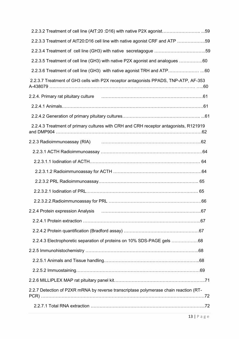
13 | P a g e
2.2.3.2 Treatment of cell line (AtT:20 :D16) with native P2X agonist…………………….. ...59
2.2.3.3 Treatment of AtT20:D16 cell line with native agonist CRF and ATP ……………….59
2.2.3.4 Treatment of cell line (GH3) with native secretagogue ……………………………..59
2.2.3.5 Treatment of cell line (GH3) with native P2X agonist and analogues …………….60
2.2.3.6 Treatment of cell line (GH3) with native agonist TRH and ATP………………… …60
2.2.3.7 Treatment of GH3 cells with P2X receptor antagonists PPADS, TNP-ATP, AF-353 A-438079 ……………………………………………………………………………………… …..60
2.2.4. Primary rat pituitary culture ……………………………………………………………61
2.2.4.1 Animals……………………………………………………………………………………61
2.2.4.2 Generation of primary pituitary cultures…………………………………………….. ...61
2.2.4.3 Treatment of primary cultures with CRH and CRH receptor antagonists, R121919 and DMP904 ………………………………………………………………………………………62
2.2.3 Radioimmunoassay (RIA) …………………………………………………………..62
2.2.3.1 ACTH Radioimmunoassay ……………………………………………………………64
2.2.3.1.1 Iodination of ACTH………………………………………………………………… 64
2.2.3.1.2 Radioimmunoassay for ACTH ……………………………………………………64
2.2.3.2 PRL Radioimmunoassay………………………………………………………….. 65
2.2.3.2.1 Iodination of PRL…………………………………………………………………. 65
2.2.3.2.2.Radioimmunoassay for PRL ………………………………………………………66
2.2.4 Protein expression Analysis …………………………………………………………..67
2.2.4.1 Protein extraction ……………………………………………………………………..67
2.2.4.2 Protein quantification (Bradford assay) ……………………………………………67
2.2.4.3 Electrophoretic separation of proteins on 10% SDS-PAGE gels ………………68
2.2.5 Immunohistochemistry …………………………………………………………………..68
2.2.5.1 Animals and Tissue handling………………………………………………………..68
2.2.5.2 Immuostaining…………………………………………………………………………69
2.2.6 MILLIPLEX MAP rat pituitary panel kit……………………………………………………..71
2.2.7 Detection of P2XR mRNA by reverse transcriptase polymerase chain reaction (RT-PCR) …………………………………………………………………………………………………72
2.2.7.1 Total RNA extraction …………………………………………………………………...72
14 | P a g e
2.2.7.2 Reverse transcription …………………………………………………………………..73
2.2.7.3 PCR of cDNA …………………………………………………………………………….74
2.7.4 Visualisation of PCR product by agarose gel electrophoresis……………….................74
2.8 Statistical Analysis ……………………………………………………………………75
CHAPTER THREE: P2X receptor expression within the anterior pituitary gland......................................................................................................................................76
3.1 INTRODUCTION ………………………………………………………………………………77
3.1.1 An investigation of P2XR expression in whole pituitary and pituitary …………………77
3.1.2 Aims ……………………………………………………………………………………….78
3.3 RESULTS ……………………………………………………………………………………….79
3.3.1 Expression of P2X receptor transcripts in rat pituitary by RT PCR …………………79
3.3.2 Expression of P2X receptor transcripts in pituitary cell lines …………………………81
3.3.2.3 Expression of P2X receptor transcripts in a folliculostellate (TtT/GF) cell line ……81
3.3.2.4 Expression of P2X receptor transcripts in a corticotroph (AtT-20 D16:16) cell line 81
3.3.2.5 Expression of P2X receptor transcripts in lactotroph (GH3 and MMQ) cell lines …83
3.3.2.6 Expression of P2X receptor transcripts in a gonadotroph (LβT2) cell line …………86
3.3.2 Characterisation of purinergic receptor expression in whole pituitary tissue using Western blot ……………………………………………………………………………………….88
3.3.3 Optimisation of immunoflourescent techniques for the detection and characterisation of purinergic receptors in whole pituitary tissue: Coexpressionn of P2X receptors with pituitary hormones. ……………………………………………………………………………………….90
3.3.3.1 Coexpression of P2X4 receptor with ACTH, FSH, GH, PRL and TSH ………………90
3.3.3.2 Coexpression of P2X7 receptor with ACTH, FSH, GH, PRL and TSH ………………96
3.4 Discussion ……………………………………………………………………………………..102
3.5 Conclusions …………………………………………………………………………….107
Chapter FOUR : Investigation of P2X receptor antagonists: Effects on pituitary function in vitro...………………………………………………………………………………..109
4.1 Introduction …………………………………………………………………………….110
4.1.1 Investigation of P2X receptor pharmacology in vitro …………………………………110
4.2 Aims ……………………………………………………………………………………..113
15 | P a g e
4.3 Results ……………………………………………………………………………………..114
4.3.1 Validation of rat primary pituitary culture by RIA …………………………………114
4.3.1.1 Effects of the CRF inhibitor R121919 on ACTH release from primary pituitary culture………………………………………………………………………………………………115 4.3.1.2 Effects of CRF inhibitor DMP904 on ACTH release from primary pituitary culture……………………………………………………………………………………………….1164.3.2 Investigation into the effects of purinergic receptor stimulation on hormone release from rat primary pituitary cells …………………………………………………………...118
4.3.2 .1 The effects of ATP on ACTH secretion from primary pituitary cells …………….118
4.3.2 .2 The effects of ATP on GH from primary pituitary cells ……………………………127
4.3.2 .3 The effects of ATP on PRL secretion from primary pituitary cells……………… 136
4.3.2 .4 The effects of ATP on TSH from primary pituitary cells ………………………….145
4.3.3 Investigating the effect of purinergic receptor activation on secretagogue-induced hormone release from pituitary cell lines………………………………………………………154
4.3.3.1 AtT20 D16:16 corticotroph cells – response to secretagogue (corticotrophin releasing factor) release from pituitary cell lines ………………………………………. 154
4.3.3.2 AtT20 D16:16 corticotroph – response to ATP…………………………………….. 156
4.3.3.4 GH3 lactotroph cell line - response to secretagogue (thyrotrophin releasing hormone)………………………………………………………………………………………….. 157
4.3.3.5 GH3 lactotroph cell line - response to ATP …………………………………………163
4.3.1.3 GH3 lactotroph cell line - response to ATP analogues: adenosine 5′-[γ-thio]triphosphate tetralithium salt (ATPΥs) ……………………………………………………..174
4.3.1.4 GH3 lactotroph cell line - response to 3'-O-[4-benzoyl]benzoyl adenosine 5'-triphosphate (BzATP), a selective P2X receptor agonist. ……………………………………183
4.3.4 An investigation into the effects of purinergic receptor antagonism on hormone secretion from GH3 cells ……………………………………………………………………192
4.3.4.2 The effects of pre-treating cultured GH3 cells with graded concentrations of PPADS (1-1000µM) on TRH (1nM) induced PRL secretion …………………………………193
4.3.4.3 The effects of pre-treating cultured GH3 cells with graded concentrations of PPADS (1-1000µM) on ATP (1µM and 100µM) induced PRL secretion…………………………… 195
4.3.4.4 The effects of pre-treating cultured GH3 cells with graded concentrations of PPADS (1-1000µM) on ATP (1µM) and TRH (1nM) induced PRL secretion ………………………..198
4.3.4.5 The effects of pre-treating cultured GH3 cells with graded concentrations of PPADS (1-1000µM) on ATP (100µM) and TRH (1nM) induced PRL secretion ……………………200

16 | P a g e
4.3.5.1 An investigation into the effects of TNP-ATP on prolactin secretion from cultured GH3 cells………………………………………………………………………………………….. 203
4.3.5.2 The effects of pre-treating cultured GH3 cells with graded concentrations of TNP-ATP (1-5000nM) on TRH (1nM) induced PRL secretion………………………………….. …203
4.3.5.3 The effects of pre-treating cultured GH3 cells with graded concentrations of TNP-ATP (1-5000nM) on ATP (1µM and 100µM) induced PRL release ………………………….205
4.3.5.4 The effects of pre-treating cultured GH3 cells with graded concentrations of TNP-ATP (1-5000nM) on ATP (1µM) and TRH (1nM) induced PRL secretion …………………..208
4.3.5.5 The effects of pre-treating cultured GH3 cells with graded concentrations of TNP-ATP (1-5000nM) on ATP (100µM) and TRH (1nM) induced PRL release…………………. 210
4.3.5.4 An investigation into the effects of P2X3 inhibitor AF-353 on prolactin release from cultured GH3 cells ………………………………………………………………………………...213
4.3.5.4.1 The effects of pre-treating cultured GH3 cells with graded concentrations of AF-353 (1-5000nM) on TRH (1nM) induced PRL secretion. ………………………………213
4.3.5.4.2 The effects of pre-treating cultured GH3 cells with graded concentrations of AF-353 (1-5000nM) on ATP (1µM and 100µM) induced PRL secretion ……………………215
4.3.5.4.3 The effects of pre-treating cultured GH3 cells with various concentrations of AF-353 (1-5000nM) on ATP (1µM) and TRH (1nM) induced PRL release………………. 218
4.3.5.4.4 The effects of pre-treating cultured GH3 cells with graded concentrations of AF-353(1-5000nM) on ATP (100µM) and TRH (1nM) induced PRL release ……………220
4.3.5.5 An investigation into the effects of the P2X7 inhibitor A-438079 on prolactin release from cultured GH3 cells …………………………………………………………………………223
4.3.5.5 .1 The effects of pre-treating cultured GH3 cells with various concentrations of A-438079 (1-5000nM) on TRH (1nM) induced PRL release. ………………………………..223
4.3.3.5.2 The effects of pre-treating cultured GH3 cells with graded concentrations of A-438079 (1-5000nM) on ATP (1µM and 100µM) induced PRL release …………………225
4.3.3.5.3 The effects of pre-treating cultured GH3 cells with graded concentrations of A-438079 (1-5000nM) on ATP (1µM) and TRH (1nM) induced PRL release ………………228
4.3.3.5.4 The effects of pre-treating cultured GH3 cells with various concentrations of A-438079 (1-5000nM) on ATP (100µM) and TRH (1nM) induced PRL release ………….230
4.4 Discussion …………………………………………………………………………………….233
4.5 Conclusion …………………………………………………………………………….243
5. General discussion ………………………………………………………………….. 244
5.1 Conclusions …………………………………………………………………………….245
5.2 Future work directions ……………………………………………………………………245

17 | P a g e
5.2.1 What is significance of other P2X receptors expressed in the pituitary? : P2X4 ……245
5.2.2 What is the significance of P2X receptors expressed in human pituitary tissue?.......248
5.2.3 What are the signalling pathways involved in P2X receptor function and activation?.................................................................................................................. 248
Appendices 1 ……………………………………………………………………………………..251
Appendices 2 ……………………………………………………………………………………..254
References………………………………………………………………………………………255
18 | P a g e
Index of figures Figure1.1 Role of hypothalamic hormones in controlling peptide hormone secretion from the anterior pituitary gland Hypothalamic neurones release six hormones……………………….30
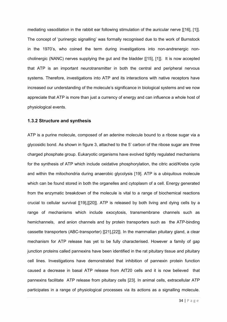
Figure1.2 Structure of ATP molecule and it metabolites. ……………………………………...35
Figure1.3 Schematic of ATP interactions with P1 and P2 receptors at the plasma membrane. ……………………………………………………………………………………….37
Figure1. 4 Schematic of P2X topology in the plasma membrane …………………………40
Figure1. 5 Adapted from Stojilkovic, 2009. The Hypothalamo-Pituitary Axis and P2X Expression in the Anterior Pituitary ……………………………………………………………….44
Figure 3.1 A-B Identification of P2X receptor transcripts : Rat pituitary tissue ………80
Figure 3.2 A-B Identification of P2X receptor transcripts in pituitary cell lines: TtTGF and AtT20 D16:16………………………………………………………………………………………..82
Figure 3.2 C-D Identification of P2X receptor transcripts in pituitary cell lines: GH3 and MMQ………………………………………………………………………………………………….84
Figure 3.2 E Identification of P2X receptor transcripts in pituitary cell lines : LbT2………….86
Figure 3.3 Expression of P2XRs in Rat pituitary :Immunoblotting of P2X receptor protein using control tissue (Brain) and rat pituitary……………………………………………………...89
Figure 3.4 A -B: Double staining to investigate colocalisation (yellow) of P2X4 receptor immunoreactivity (green) with ACTH (red) in the cells of the rat pituitary gland………......91
Figure 3.4 C-D Double staining to investigate colocalisation (yellow) of P2X4 receptor immunoreactivity (green) with FSH (red) in the rat pituitary gland) ………………………….92
Figure 3.4 E-F Double staining to show colocalisation (yellow) of P2X4 receptor immunoreactivity (green) with G H (red) in the cells of the rat pituitary gland with G H......93
Figure 3.4 G-H Double staining to show colocalisation (yellow) of P2X4 receptor immunoreactivity (green) with PRL (red) in the rat pituitary gland ………………………….94
Figure 3.4 I-J Double staining to show colocalisation (yellow) of P2X4 receptor immunoreactivity (green) with TSH (red) in the rat pituitary gland …………………………..95
Figure 3.5 A –B Double staining to investigate localisation of P2X7 receptor immunoreactivity (green) in the cells of the rat pituitary gland. With ACTH ………………97
Figure 3.5 C-D Double staining to show colocalisation (yellow) of P2X7 receptor immunoreactivity (green) with GH (red) in the rat pituitary gland …………………………..98
Figure 3.5 E-F Double staining to show colocalisation (yellow) of P2X7 receptor immunoreactivity (green) with PRL (red) E and TSH (red) (F) in the rat pituitary gland……99
Figure 3.6 Negative controls used in immunostain of rat pituitary .Absence of P2X antibody and no pituitary hormone………………………………………………………………………..100
19 | P a g e
Figure 4.1 Effects of CRF receptor antagonist R121919 on CRF stimulated ACTH release from primary anterior pituitary cultures………………………………………………………..116
Figure 4.2 Effects of CRF receptor antagonist DMP904 on CRF stimulated ACTH release from primary anterior pituitary cultures…………………………………………………………117
Figure 4.3 A Effect of 0.001 µM ATP on ACTH release from primary pituitary…………….119
Figure 4.3 B Effect of 0.01µM ATP on ACTH release from cultured primary pituitary representative graph shown. …………………………………………………………………..120
Figure 4.3 C Effect of 0.1µM ATP on ACTH from cultured primary pituitary cells ………121
Figure 4.3 D Effect of 1µM ATP on ACTH release from cultured primary pituitary cells…122
Figure 4.3 E Effect of 10µM ATP on ACTH release from cultured primary pituitary cells………………………………………………………………………………………………… 123
Figure4.3 F Effect of 100µM ATP ACTH from primary pituitary cells ……………………….124
Figure4.3 G Effect of 1000µM ATP on ACTH from primary pituitary cells…………………..125
Figure 4.4 A Effect of 0.001µM ATP GH release from cultured primary pituitary ………128
Figure 4.4 B Effect of 0.01µM ATP for GH from cultured primary pituitary ………………129
Figure 4.4 C Effect of 0.1µM ATP for GH from cultured primary pituitary ………………..130
Figure 4.4 D Effect of 1µM ATP on GH from cultured primary pituitary cells…………….131
Figure 4.4E Effect of 10µM ATP for GH from cultured primary pituitary cells……………132
Figure 4.4 F Effect of 100µM ATP for GH from cultured primary pituitary cells…………133
Figure 4.4 G Effect of 1000µM ATP for GH from cultured primary pituitary ………………134
Figure 4.5 A Effect of 0.001µM ATP for PRL from cultured primary pituitary cells ……….137
Figure 4.5 B Effect of 0.01µM ATP for PRL from cultured primary pituitary cells ……….138
Figure 4.5 C Effect of 0.1µM ATP for PRL from cultured primary pituitary cells………….139
Figure 4.5 D Effect of 1µM ATP for PRL from cultured primary pituitary …………………..140
Figure 4.5 E Effect of 10µM ATP for PRL from cultured primary pituitary cells……………141
Figure 4.5 F Effect of 100µM ATP for PRL from cultured primary pituitary cells …………142
Figure 4.5 G Effect of 1000µM ATP for PRL from cultured primary pituitary cells ………143
Figure 4.6 A Effect of 0.001µM ATP for TSH from cultured primary pituitary cells………... 146
Figure 4.6 B Effect of 0.01µM ATP for TSH from cultured primary pituitary cells………… 147
Figure 4.6 C Effect of 0.1µM ATP for TSH from cultured primary pituitary cells………….. 148
20 | P a g e
Figure 4.6 D Effect of 1µM ATP for TSH from cultured primary pituitary cells……………... 149
Figure 4.6 E Effect of 10µM ATP for TSH from cultured primary pituitary cells……………. 150
Figure 4.6 Effect of 100µM ATP for TSH from cultured primary pituitary cells…………….. 151
Figure 4.6 G Effect of 1000µM ATP or TSH from cultured primary pituitary cells………… 152
Figure 4.7 The dose dependent effects of CRF (1-100nM) upon ACTH release from AtT20 D16:16 cells over 4 hrs……………………………………………………………………………155
Figure 4.8 Effects of CRF and ATP on ACTH secretion from AtT20 D16:D16 AtT20 cells were treated with ATP (100uM) alone or in combination with CRF (100nM) for 1(A), 4(B), 7(C) and 12(D) hrs……………………………………………………………………………..157
Figure 4..9 A Effects of 1nM of PRL secretagogue TRH on PRL release from cultured GH3 cells. Cultures (2x10⁵ cells/well) treated with TRH alone with 1nM, for 0-90 minutes. …159
Figure 4.9 B Effects of 10nM of PRL secretagogue TRH on PRL release from cultured GH3 cells. Cultures (2x10⁵ cells/well) treated with TRH alone with 10nM, for 0-90 minutes. 160
Figure 4.9 C Effects of 100nM PRL secretagogue TRH on PRL release from cultured GH3 cells. Cultures (2x10⁵ cells/well) treated with TRH alone with 100nM for 0-90 minutes…. 161
Figure 4.9 D Effects of 1000nM of PRL secretagogue TRH on PRL release from GH3 cells. Cultures (2x10⁵ cells/well) treated with TRH alone with 1000nM for 0-90 minutes. ……162
Figure4.10 A Dose and time graphs for PRL from cultured GH3 cells treated with 0.001µM ATP…………………………………………………………………………………………………165
Figure4.10 B Dose and time graphs for PRL from cultured GH3 cells treated with 0.01µM ATP………………………………………………………………………………………………..166
Figure4.10 Dose and time graphs for PRL from cultured GH3 cells treated with 0.1µM ATP Cultures ……………………………………………………………………………………………167
Figure4.10 D Dose and time graphs for PRL from cultured GH3 cells treated with 1µM ATP. ………………………………………………………………………………………………………168
Figure4.10. E Dose and time graphs for PRL from cultured GH3 cells treated with 10µM ATP………………………………………………………………………………………………….169
Figure 4.10 F Dose and time graphs for PRL from cultured GH3 cells treated with 100µM ATP………………………………………………………………………………………………..170
Figure 4.10 G Dose and time graphs for PRL from cultured GH3 cells treated with 100µM ATP……………………………………………………………………………………………171
Figure 4.10 H & I representative dose and response graphs for PRL release from cultured GH3 cells treated with ATP…………………………………………………………………..172
Figure 4.11. A Dose and time graphs for PRL from cultured GH3 cells treated with 0.001µM ATP s…………………………………………………………………………………175
21 | P a g e
Figure4.11. B Dose and time graphs for PRL from cultured GH3 cells treated with 0.01µM ATP……………………………………………………………………………………………….176
Figure4.11 C Dose and time graphs for PRL from cultured GH3 cells treated with 0.1µM ATP ……………………………………………………………………………………………..177
Figure4.11 D Dose and time graphs for PRL from cultured GH3 cells treated with 1µM ATP ………………………………………………………………………………………………178
Figure 4.11E Dose and time graphs for PRL from cultured GH3 cells treated with 10µM ATP…………………………………………………………………………………………………179
Figure4.11 F Dose and time graphs for PRL from cultured GH3 cells treated with 100µM ATP…………………………………………………………………………………………………180
Figure 4.11.G Dose and time graphs for PRL from cultured GH3 cells treated with 1000µM ATP ……………………………………………………………………………………………… 181
Figure4.12 A Dose and time graphs for PRL from cultured GH3 cells treated with 0.001µM BzATP………………………………………………………………………………………………184
Figure4.12 B Dose and time graphs for PRL from cultured GH3 cells treated with 0.01µM BzATP ………………………………………………………………………………………………185
Figure4.12 C Dose and time graphs for PRL from cultured GH3 cells treated with 0.1µM BzATP ………………………………………………………………………………………………186
Figure4.12D Dose and time graphs for PRL from cultured GH3 cells treated with 1µM BzATP ………………………………………………………………………………………………187
Figure 4.12E Dose and time graphs for PRL from cultured GH3 cells treated with 10µM BzATP ………………………………………………………………………………………………188
Figure4.12 F Dose and graphs for PRL from cultured GH3 cells treated with 100µM BzATP …………………………………………………………………………………………….189
Figure4. 12 G Dose and time graphs for PRL from cultured GH3 cells treated with 1000µM BzATP ……………………………………………………………………………………………..190
Figure 4.13 A The effects of graded concentrations of P2 receptor antagonist PPADS (1-1000µM) on PRL secretion from cultured GH3 cells in the presence of 1nM TRH……..194
Figure4.13 B The effects of graded concentrations of P2 receptor antagonist PPADS (1-1000µM) on PRL secretion from cultured GH3 cells in the presence of 1µM ATP……...196
Figure4.13 C The effects of graded concentrations of P2 receptor antagonist PPADS (1-1000µM) on PRL secretion from cultured GH3 cells in the presence of 100µM ATP…..197
Figure 4.13 D The effects of graded concentrations of P2 receptor antagonist PPADS (1-1000µM) on PRL secretion from cultured GH3 cells in the presence of 1nM TRH and 1µM ATP………………………………………………………………………………………………….199
22 | P a g e
Figure. 4.13 E The effects of graded concentrations of P2 receptor antagonist PPADS (1-1000µM) on PRL secretion from cultured GH3 cells in the presence of 1nM TRH and 100µM ATP…………………………………………………………………………………………201
Figure4.14.A The effects of graded concentrations of P2X receptor antagonist TNP-ATP (1-5000nM) on PRL secretion from cultured GH3 cells in the presence of 1nM TRH Cultures (2X10⁵ cells/well) treated with TRH (1nM) alone 30 or pre-treated with TNP-ATP for 15mins followed by TRH. ………………………………………………………………………………….204
Figure 4.14. B The effects of graded concentrations of P2X receptor antagonist TNP-ATP (1-5000nM) on PRL secretion from cultured GH3 cells in the presence of 1µM ATP……...206
Figure.4..14C The effects of graded concentrations of P2X receptor antagonist TNP-ATP (1-5000nM) on PRL secretion from cultured GH3 cells in the presence of 100µM……….. 207
Figure. 4..14D The effects of graded concentrations of P2x receptor antagonist TNP-ATP (1-5000nM) on PRL secretion from cultured GH3 cells in the presence of 1µM ATP 1µM ATP and 1nM TRH………………………………………………………………………………..209
Figure. 4.14 E The effects of graded concentrations of P2X receptor antagonist TNP-ATP (1-5000nM) on PRL secretion from cultured GH3 cells in the presence of 100µM ATP and 1nM ………………………………………………………………………………………………..211
Figure. 4.15A The effects of graded concentrations of P2X3 receptor antagonist AF-353 (1-5000nM) on PRL secretion from cultured GH3 cells in the presence of 1nM TRH 214
Figure. 4.15B The effects of graded concentrations of P2X3 receptor antagonist AF-353 (1-5000nM) on PRL secretion from cultured GH3 cells in the presence of 1µM ATPt 216
Figure. 4.15 C The effects of graded concentrations of P2X3 receptor antagonist AF-353 (1-5000nM) on PRL secretion from cultured GH3 cells in the presence of 100µM ATP 217
Figure.4.15D The effects of graded concentrations of P2X3 receptor antagonist AF-353 (1-5000nM) on PRL secretion from cultured GH3 cells in the presence of 1nM TRH and 1 µM ATP ………………………………………………………………………………………………219
Figure. 4.15 E The effects of graded concentrations of P2X3 receptor antagonist AF-353 (1-5000nM) on PRL secretion from cultured GH3 cells in the presence of 1 nM TRH and 100µM ATP ……………………………………………………………………………………….221
Figure. 4.14A The effects of graded concentrations of P2X7 receptor antagonist A-438079(1-5000nM) on PRL secretion from cultured GH3 cells in the presence of 1nM TRH ………………………………………………………………………………………… 224
Figure.4.16B The effects of graded concentrations of P2X7 receptor antagonist A-438079(1-5000nM) on PRL secretion from cultured GH3 cells in the presence of 1µM ATP……...226
Figure. 4.16 C The effects of graded concentrations of P2X7 receptor antagonist A-438079(1-5000nM) on PRL secretion from cultured GH3 cells in the presence of 100µM ATP …………………………………………………………………………………………………227

23 | P a g e
Figure. 4.16 D The effects of graded concentrations of P2X7 receptor antagonist A-438079(1-5000nM) on PRL secretion from cultured GH3 cells in the presence of 1nM TRH and 1µM ATP. ……………………………………………………………………………229
Figure. 4.16 E Dose-response graph for PRL from cultured GH3 cells pre- treated with P2X7 receptor antagonist A-438079 (1-5000nM) 1nM TRH and 100 µM ATP……… .. 231
24 | P a g e
Index of tables Table.1 .1 Pituitary cell sub-types and their associated hormones…………………………..32
Table1.2 Widespread physiological expression of P2X receptors in mammalian systems 42
Table 1.3: The distribution of purinoceptors within the pituitary gland………………………48
Table 2.1 Chemicals and reagents……………………………………………………………51
Table 2.2 Solutions and Buffers…………………………………………………………………53
Table 2.3. Antibodies…………………………………………………………………………….. 54
Table 2.4 Kits ………………………………………………………………………………………55
Table 2.5 Cell culture medium …………………………………………………………………….56
Table 3.1 Summary of cell lines profiled by RT-PCR to P2XR1-7 expression……………….78
Table3.2. Summary of P2X transcripts detected in rat pituitary tissue and pituitary cell lines…………………………………………………………………………………………………...87
Table 3.3 Summary of P2X immunorectivity detected in rat pituitary tissue and brain tissue………………………………………………………………………………………………….90
Table 3.4.coexistence between P2X receptors and pituitary hormones…………………….101
Table 4.1 List of ATP analogues EC50 (µM) ………………………………………………….111
Table 4.2 List of P2X receptor antagonists with IC50 (µM) …………………………………112
Table 4.3 Effects of ATP (0.001-1000uM) on ACTH (pg/ml) release from rat primary pituitary cells in vitro…………………………………………………………………………………………126
Table 4.4 Effects of ATP on GH (pg/ml) release in primary pituitary cells ………………..135
Table 4.5 Effects of ATP on PRL (pg/ml) release in primary pituitary cells ………………..144
Table 4.6 Effects of ATP on TSH (pg/ml) release in primary pituitary cells…………………153
Table 4.7 Summary of ATP analogues and time points assessed in vitro………………….163
Table 4.8 Effects of ATP on PRL (ng/ml) release in GH3 cells………………………………173
Table 4.9 Effects of ATPs on PRL (ng/ml) release in GH3 cells………………………….182
Table 4.10 Effects of BzATP on PRL (ng/ml) from GH3 cells………………………………..191
Table 4.11 List of purinergic compounds to be investigated in vitro systems………………193
Table 4.12 Summary of the effects of PPADS on PRL (ng/ml) release from GH3 cells in vitro……………………………………………………………………………………………..202
25 | P a g e
Table 4 13 Summary of the effects of TNP-ATP on PRL (ng/ml) release from GH3 cells in vitro……………………………………………………………………………………………….212
Table 4 14 summary of the effects of AF-353 on PRL (ng/ml) release from GH3 cells in vitro………………………………………………………………………………………………..222
Table 4 15 summary of the effects of A-438079 on PRL (ng/ml) release from GH3 cells in vitro ……………………………………………………………………………………………….232

26 | P a g e
Chapter 1 Introduction
27 | P a g e
1.1 The endocrine system
In humans and other mammals, regulation of the internal environment and responses to the
external environment are coordinated by signals derived from both the nervous and
endocrine systems. The nervous system exerts its influence through a system of specialised
cells or neurons, which convey messages from the brain directly to specific organs. In
vertebrates, the endocrine system is composed of a collections of glands (e.g. thyroid,
adrenal and pituitary), which function to produce chemical messengers which are secreted
into the bloodstream and subsequently act to modify or control organ function. There are
over 50 different types of “chemical messengers” or hormones secreted within the endocrine
system. These hormones can be grouped into three classes, and the germinal epithelium
from which the gland is derived from, will determine the class of hormone secreted. Glands
derived from the ectoderm and endodermal layer will produce amine hormones which are
derived from tyrosine residues, and peptide hormones which are composed of short chain
amino acids. Glands made up of mesodermal tissue produce steroid hormones, which all
share a common precursor – cholesterol.
Protein hormone synthesis occurs in the rough endoplasmic reticulum and golgi body of the
secretory cells that compose the endocrine gland and are secreted into the blood, where
they bind to receptors expressed on the surface or within the target organ. Receptor binding
is coupled to an internal signalling mechanism which allows transduction of the signal to be
initiated by the hormone. Hormones can act locally at the site of production (autocrine), on
neighbouring cells (paracrine) or distally at sites located away from the gland transported via
the circulation (endocrine), with hormone effects lasting from hours up to weeks. Variations
in the amounts of hormone secreted coupled with alterations in the pattern of hormone
secretion allow for fine control of numerous physiological functions including metabolism,
reproduction and sexual differentiation, growth and development, sleep , mood and
28 | P a g e
arguably most important, the regulation of the internal environment (homeostasis). Co-
ordination of inputs from both the nervous system and endocrine system in order to maintain
homeostasis is best exemplified by the regulation of the master gland or pituitary gland [[1],
[2]].
1.2 The neuroendocrine system
The neuroendocrine system describes the interactions between the brain (especially the
hypothalamus) and the glands of the endocrine system, and includes the numerous
neuroendocrine cells which receive direct neuronal input and subsequently release
hormones into the blood. Together, their actions serve to regulate several major
physiological functions within the body. Proper coordination between inputs from both the
brain and the peripheral nervous system (PNS) are required to maintain normal homeostasis
which is controlled by the hypothalamus, which I will describe in more detail in section 1.2.1.
Hypothalamic control of the pituitary gland is also vital to ensure the system functions
correctly as hormones released from here ultimately control all aspects of human physiology
such as food intake, blood pressure and responses to stress or infection. The pituitary will be
discussed in section 1.2.2 [2].
1.2.1 The hypothalamus- function and anatomy
Homeostasis is a dynamic balance that must be maintained by all multi-cellular organisms in
spite of changes to its external and internal environment. The hypothalamus plays a
significant role in initiating an array of behavioural and physiological responses to maintain
this equilibrium. The hypothalamus forms the connection between the central nervous
system (CNS) and the rest of the body [1].
The hypothalamus is connected to the brain via the midbrain and telencephalon regions of
the brain. It is composed of three parts; the periventricular, medial and lateral hypothalamus.
Afferent and efferent neurones covey signals to and from the brain and inputs from distal
29 | P a g e
endocrine glands via the peripheral nervous system (PNS). Hypothalamic neurones connect
with the brain in groups located in the third ventricle of the brain. Neurones which emanate
from the medial layer of the hypothalamus control pituitary gland function, forming a
continuous link called the hypophyseal nerve tract. Neurones sprouting from the lateral
portion are involved in control of autonomic function. Blood supplied by the hypophyseal
inferior and superior arteries facilitate control of the posterior and anterior pituitary
respectively. Hypothalamic nuclei secrete hormones directly into a capillary portal system
that transports the hypothalamic hormones directly to the endocrine cells that constitute the
pituitary gland. There are six hypothalamic hormones which regulate pituitary function (see
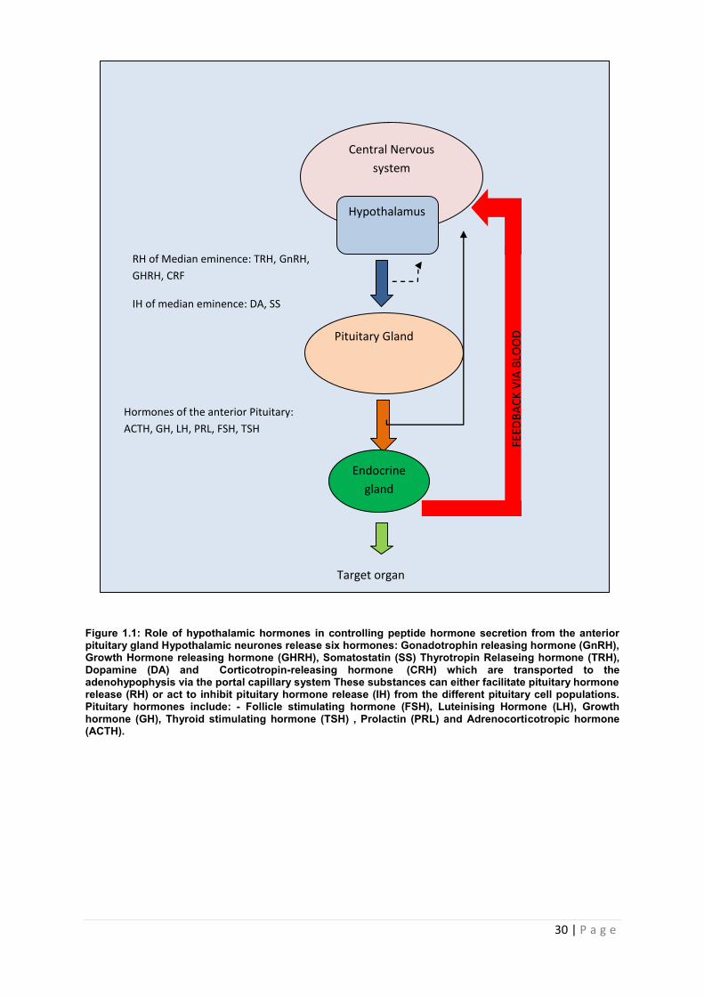
figure 1.1 and table 1). These hormones possess either a stimulatory or inhibitory effect on
hormones release from the anterior pituitary. Pituitary hormones can control their own
release via negative feedback. This feedback mechanism is illustrated in figure 1.1 [3].
30 | P a g e
Figure 1.1: Role of hypothalamic hormones in controlling peptide hormone secretion from the anterior pituitary gland Hypothalamic neurones release six hormones: Gonadotrophin releasing hormone (GnRH), Growth Hormone releasing hormone (GHRH), Somatostatin (SS) Thyrotropin Relaseing hormone (TRH), Dopamine (DA) and Corticotropin-releasing hormone (CRH) which are transported to the adenohypophysis via the portal capillary system These substances can either facilitate pituitary hormone release (RH) or act to inhibit pituitary hormone release (IH) from the different pituitary cell populations. Pituitary hormones include: - Follicle stimulating hormone (FSH), Luteinising Hormone (LH), Growth hormone (GH), Thyroid stimulating hormone (TSH) , Prolactin (PRL) and Adrenocorticotropic hormone (ACTH).
Pituitary Gland
Target organ
RH of Median eminence: TRH, GnRH,
GHRH, CRF
IH of median eminence: DA, SS
Hormones of the anterior Pituitary:
ACTH, GH, LH, PRL, FSH, TSH
Endocrine
gland
Central Nervous
system
Hypothalamus
FEED
BA
CK
VIA
BLO
OD
31 | P a g e
1.2.3 The pituitary gland
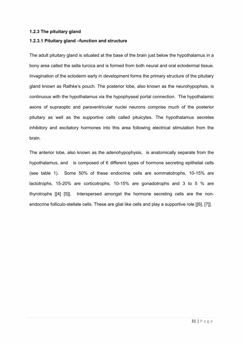
1.2.3.1 Pituitary gland –function and structure
The adult pituitary gland is situated at the base of the brain just below the hypothalamus in a
bony area called the sella turcica and is formed from both neural and oral ectodermal tissue.
Invagination of the ectoderm early in development forms the primary structure of the pituitary
gland known as Rathke’s pouch. The posterior lobe, also known as the neurohypophsis, is
continuous with the hypothalamus via the hypophyseal portal connection. The hypothalamic
axons of supraoptic and paraventricular nuclei neurons comprise much of the posterior
pituitary as well as the supportive cells called pituicytes. The hypothalamus secretes
inhibitory and excitatory hormones into this area following electrical stimulation from the
brain.
The anterior lobe, also known as the adenohypophysis, is anatomically separate from the
hypothalamus, and is composed of 6 different types of hormone secreting epithelial cells
(see table 1). Some 50% of these endocrine cells are sommatotrophs, 10-15% are
lactotrophs, 15-20% are corticotrophs, 10-15% are gonadotrophs and 3 to 5 % are
thyrotrophs [[4] [5]]. Interspersed amongst the hormone secreting cells are the non-
endocrine folliculo-stellate cells. These are glial like cells and play a supportive role [[6], [7]].
32 | P a g e
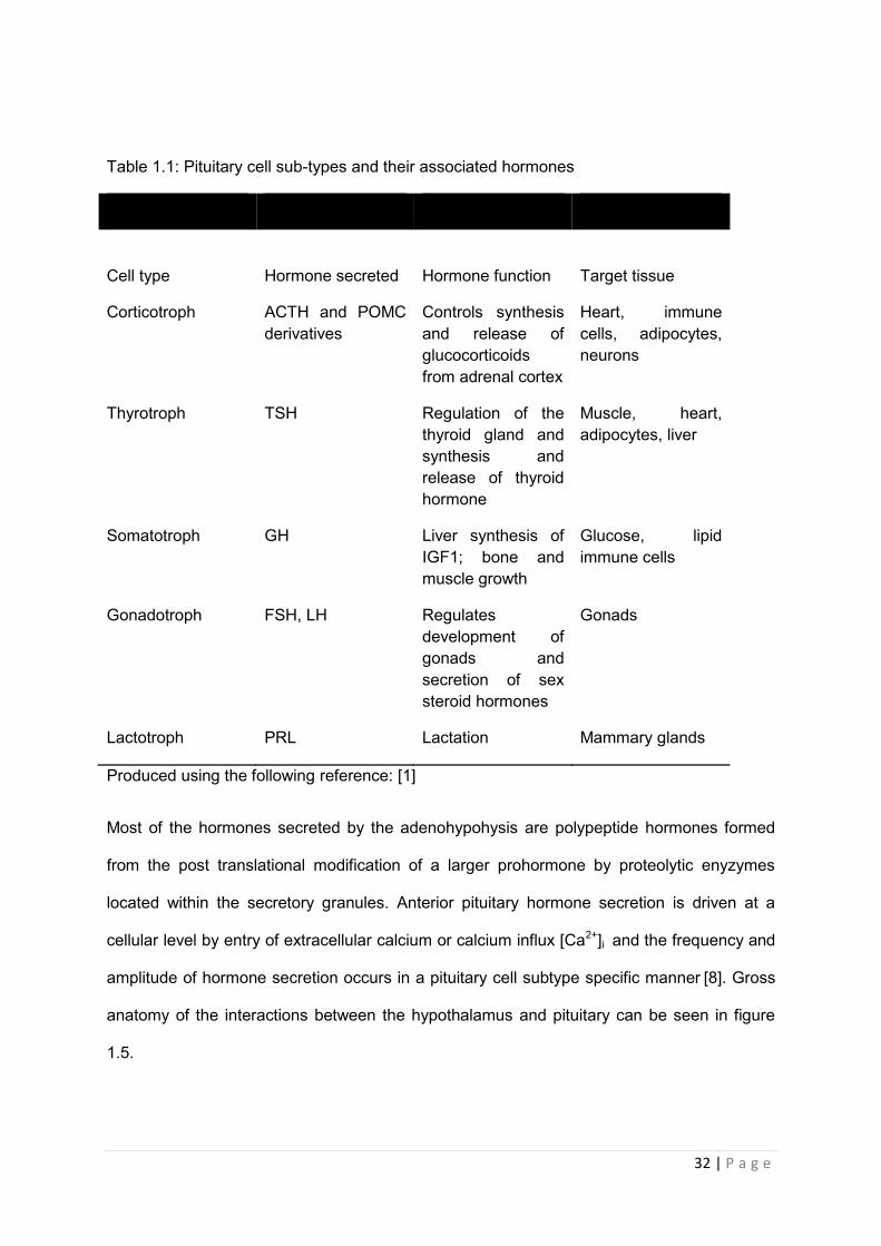
Table 1.1: Pituitary cell sub-types and their associated hormones
Cell type
Hormone secreted
Hormone function
Target tissue
Corticotroph ACTH and POMC derivatives
Controls synthesis and release of glucocorticoids from adrenal cortex
Heart, immune cells, adipocytes, neurons
Thyrotroph TSH Regulation of the thyroid gland and synthesis and release of thyroid hormone
Muscle, heart, adipocytes, liver
Somatotroph GH Liver synthesis of IGF1; bone and muscle growth
Glucose, lipid immune cells
Gonadotroph FSH, LH Regulates development of gonads and secretion of sex steroid hormones
Gonads
Lactotroph PRL Lactation Mammary glands
Produced using the following reference: [1]
Most of the hormones secreted by the adenohypohysis are polypeptide hormones formed
from the post translational modification of a larger prohormone by proteolytic enyzymes
located within the secretory granules. Anterior pituitary hormone secretion is driven at a
cellular level by entry of extracellular calcium or calcium influx [Ca2+]i and the frequency and
amplitude of hormone secretion occurs in a pituitary cell subtype specific manner [8]. Gross
anatomy of the interactions between the hypothalamus and pituitary can be seen in figure
1.5.
33 | P a g e
1.2.3.2 The hypothalamo-pituitary axis
Established at week 20 of gestation in humans, the hypothalamo-pituitary axis is vital for
monitoring and maintenance of the internal environment and communicating any changes
back to the central nervous system (CNS). Pituitary function is also augmented by paracrine
factors such as IL-6, insulin-like growth factor and acetylcholine [[9], [10], [7] [11]]. More
recent observations have demonstrated that adenosine 5’-triphosphate (ATP) can be added
to list of molecules that augment pituitary function [[12], [13], [14]].
1.3 Adenosine 5’-Triphosphate (ATP) and purinergic signalling
The ability of adenosine 5’-triphosphate (ATP) to act as an extracellular signalling molecule
was first observed over 80 years ago. Since then, investigations have increased our
understanding into ATP behaviour as a paracrine and autocrine messenger. These effects
are conveyed by interactions with its native receptors. We now appreciate that ATP is more
than just a currency of energy and can influence a whole host of physiological events. The
aim of this following section is to give a brief history of ATP as a signalling molecule, then to
introduce the concept of purinergic signalling and the physiological role of P2X receptors
(P2XR), and then the importance of purinergic signalling in modulating pituitary function.
1.3.1 Adenosine 5’-Triphosphate (ATP) - an extracellular signalling molecule
In 1929 the pioneering work of Dury and Szent-GyÖrgyi first identified a role for extracellular
purines, in this case adenosine and adenosine 5’-monophosphate (AMP), in regulating the
cardiovascular system [15]. Gillespie, in 1934, demonstrated that ATP could stimulate
contraction of both guinea pig ileum and uterus and was more potent than adenosine and
AMP in this regard. During the 1940’s the role of ATP as an extracellular signalling molecule
was confirmed in numerous tissues, and in 1948, Emmelin and Feldberg, working on cats,
observed that ATP produced effects both within the peripheral and central nervous systems
[15]. Holton and Holton’s work in the 1950’s identified ATP’s role as a neurotransmitter in
34 | P a g e
mediating vasodilation in the rabbit ear following stimulation of the auricular nerve [[16], [1]].
The concept of ‘purinergic signalling’ was formally recognised due to the work of Burnstock
in the 1970’s, who coined the term during investigations into non-andrenergic non-
cholinergic (NANC) nerves supplying the gut and the bladder [[15], [1]]. It is now accepted
that ATP is an important neurotransmitter in both the central and peripheral nervous
systems. Therefore, investigations into ATP and its interactions with native receptors have
increased our understanding of the molecule’s significance in biological systems and we now
appreciate that ATP is more than just a currency of energy and can influence a whole host of
physiological events.
1.3.2 Structure and synthesis
ATP is a purine molecule, composed of an adenine molecule bound to a ribose sugar via a
glycosidic bond. As shown in figure 3, attached to the 5’ carbon of the ribose sugar are three
charged phosphate group. Eukaryotic organisms have evolved tightly regulated mechanisms
for the synthesis of ATP which include oxidative phosphorylation, the citric acid/Krebs cycle
and within the mitochondria during anaerobic glycolysis [19]. ATP is a ubiquitous molecule
which can be found stored in both the organelles and cytoplasm of a cell. Energy generated
from the enzymatic breakdown of the molecule is vital to a range of biochemical reactions
crucial to cellular survival [[19],[[20]]. ATP is released by both living and dying cells by a
range of mechanisms which include exocytosis, transmembrane channels such as
hemichannels, and anion channels and by protein transporters such as the ATP-binding
cassette transporters (ABC-transporter) [[21],[22]]. In the mammalian pituitary gland, a clear
mechanism for ATP release has yet to be fully characterised. However a family of gap
junction proteins called pannexins have been identified in the rat pituitary tissue and pituitary
cell lines. Investigations have demonstrated that inhibition of pannexin protein function
caused a decrease in basal ATP release from AtT20 cells and it is now believed that
pannexins facilitate ATP release from pituitary cells [23]. In animal cells, extracellular ATP
participates in a range of physiological processes via its actions as a signalling molecule.
35 | P a g e
NH2
H
H H
O
O
H H
N C
N C
N C
C N
C H
O
OH H
O CH2 P
OH
O
OH
O P O HO P
OH OH
H
36 | P a g e
AMP, as well as uracil triphosphate (UTP) and uracil diphosphate (UDP). Purinergic
receptors can be found distributed in a variety of tissues and more than one receptor type
can be found in any given organ or biological system [[25], [26], ([27], [27]]. Figure 1.3
provides a schematic overview of how ATP and it derivatives interact with P1 and P2
receptors.
1.4.1 Overview of P1 receptors
Adenosine (P1) receptors are G-coupled protein receptors. To date four mammalian P1
receptors have been cloned: A1R, A2AR, A2BR and A3R [[15], [26], ([27], [27]]. ATP released
from both excitatory and non-excitatory cells is subject to degradation by ectonucleotidases
which break down ATP into its precursors, thus providing a source of adenosine which can
then bind to P1 receptors located on the plasma membrane of cells. Ligand binding of
adenosine to A1R (& potentially A3R) has been shown to have an inhibitory effect on target
cells. This occurs primarily via the down regulation of adenylate cylclase and subsequent
reduction in activity of the cyclic adenosine monophosphate (cAMP) dependent protein
kinase [15]. Other signalling mechanisms associated with A1R include both an increase in
Ca2+ mobilization due to activation of phospholipase C (PLC), as well as down regulation of
calcium (Ca2+) mediated signalling primarily via the activation of potassium (K+) channels. In
contrast, A2AR and A2BR are positively linked to adenylate cylclase (via Gs and Gq pathway)
and activation of these receptors also causes modulation of Ca 2+ channels. Activation of the
mitogen activated protein (MAP) kinase pathway is associated with all P1 receptors [[28]
[29][30]].

37 | P a g e
-
EN EN EN
38 | P a g e
The binding of ATP to P2X receptors (P2XRs) causes a change in conformation which
favours the passage of cations (Na2+, K+ and Ca2+) through the protein pore. There are 7
receptors (P2X1-7) which can be found expressed homo- and hetero-metrically in a variety of
organisms [[26],[31]]. An overview of the molecular structure and agonist selectivity of
purinergic receptors is shown in Figure 1.3.
1.5 P2X receptors
1.5.1 Structure
P2XRs represent a novel class of ligand gated channels which in response to ATP, non-
selectively facilitate the passage of divalent cations such as Ca2+ and monovalent ions such
as K+ [[32],[33]]. P2XR are structurally distinct from other members of the ligand gated ion
channel family, which include glutamate receptors (e.g. N-methyl-D-aspartate receptor) and
the nicotinic acetylcholine receptor [[29],[34],[35]]. P2XRs are described as calcium
channels since they show a preference for the passage of Ca2+ allowing a greater influx of
Ca2+ compared to other family members. P2X receptors are most similar in structure to the
acid–sensing ion channel family (ASIC), however studies have found there to be no amino
acid sequence homology between the two families. Following cloning of P2 receptor
encoding genes in 1994, seven human P2XR have been identified to date. Receptors can
be found expressed in eukaryotic organisms in either the homo- or hetero-meric form.
Recombinant expression of murine and rat derived P2X subunits have found. P2X6 is
exceptional as it does not form functional homomeric receptors and can only be found as
part of a functional heteromer with P2X2 and P2X4. P2X7 on the other hand does not form
functional heteromers with other P2XRs and can only be found expressed as a homomer
[33]. 11 different heteromeric receptors may be assembled from the 7 P2X subunits,
however far fewer than this have been documented in vivo. Currently P2X2/3, P2X2/6,
P2X4/6, and P2X1/5 are the only functional heteromultimers to have been identified [36].
Therefore comprehensive comparative pharmacology data on heteromeric receptors and
homomeric receptors is not currently available [[34] ,[37],[36]].
39 | P a g e
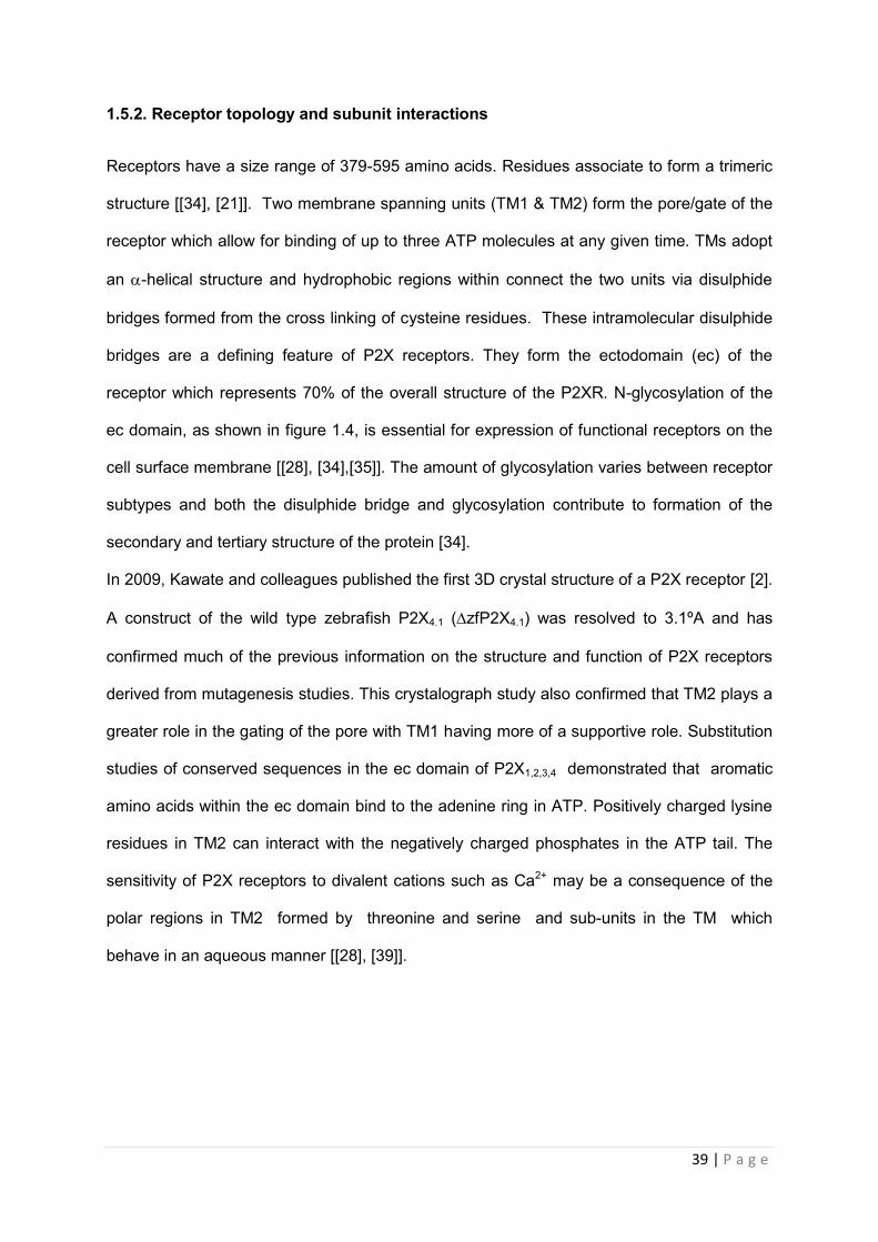
1.5.2. Receptor topology and subunit interactions
Receptors have a size range of 379-595 amino acids. Residues associate to form a trimeric
structure [[34], [21]]. Two membrane spanning units (TM1 & TM2) form the pore/gate of the
receptor which allow for binding of up to three ATP molecules at any given time. TMs adopt
an -helical structure and hydrophobic regions within connect the two units via disulphide
bridges formed from the cross linking of cysteine residues. These intramolecular disulphide
bridges are a defining feature of P2X receptors. They form the ectodomain (ec) of the
receptor which represents 70% of the overall structure of the P2XR. N-glycosylation of the
ec domain, as shown in figure 1.4, is essential for expression of functional receptors on the
cell surface membrane [[28], [34],[35]]. The amount of glycosylation varies between receptor
subtypes and both the disulphide bridge and glycosylation contribute to formation of the
secondary and tertiary structure of the protein [34].
In 2009, Kawate and colleagues published the first 3D crystal structure of a P2X receptor [2].
A construct of the wild type zebrafish P2X4.1 (zfP2X4.1) was resolved to 3.1ºA and has
confirmed much of the previous information on the structure and function of P2X receptors
derived from mutagenesis studies. This crystalograph study also confirmed that TM2 plays a
greater role in the gating of the pore with TM1 having more of a supportive role. Substitution
studies of conserved sequences in the ec domain of P2X1,2,3,4 demonstrated that aromatic
amino acids within the ec domain bind to the adenine ring in ATP. Positively charged lysine
residues in TM2 can interact with the negatively charged phosphates in the ATP tail. The
sensitivity of P2X receptors to divalent cations such as Ca2+ may be a consequence of the
polar regions in TM2 formed by threonine and serine and sub-units in the TM which
behave in an aqueous manner [[28], [39]].
40 | P a g e
Figure 1.4 Schematic of P2X topology in the plasma membrane. This diagram shows essential internal interactions (formation of disulphide bridge and glycosylation sites) in the ec domain required for proper receptor expression. The two TM units span the lipid membrane while the amino and carboxy groups are located intracellularly.
1.5.3 Signal transduction
The selective filtration of cations by P2XRs is a dynamic process. The longer that ATP is
docked to the receptor, the greater the dilation of the pore and the larger the size of cation
that can pass through the pore. Studies have shown this process can occur whilst
simultaneously inhibiting the entrance of anions [34]. Pharmacological characterisation of
both human and rat receptors have been conducted using recombinant protein expressed in
both mammalian (such as the human astrocytoma cell line 1321N1) and non-mammalian
systems (Xenopus oocytes). Combined with mutagenesis data, these studies have provided
a wealth of information on receptor sensitivity to agonist, antagonist and rates of
desensitisation. Although there are some discrepancies depending on the expression
TM2 TM1
NH+3
COO-
Ectodomain
Transmembrane domain
Cytosolic domain
N-linked glycosylation
Disulphide bonds
41 | P a g e
system used, particularly with regard to ATP analogues, such as α,β-meATP and BzATP, it
is generally the case that P2XR kinetics are primarily dependant on the subtype structure
and differ for hetero- and homo- polymerized receptors [[22], [40],[41]].
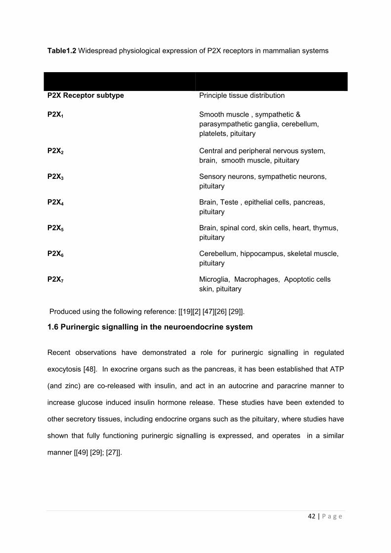
1.5.4 Tissue distribution and biological effects
As a consequence of the diverse expression of P2XR within mammalian cellular
microenvironments, it is essential that receptor expression is tightly regulated in order to
ensure that receptor mediated responses occur in a coordinated manner [[30], [42]]. A list of
the different tissues which express P2X channels are shown in table 1.2. Constitutive cycling
of inotropic P2XRs is one of the mechanisms by which these receptor mediated responses
can be regulated in order to maintain homeostasis [43]. For example, P2X4R are up-
regulated on the surface of activated macrophages as required from a readily available pool
of receptors in the lysosomal compartment and P2XR density can also be readily altered in
presynaptic neurons within the olfactory bulb [44].
In general, P2XR appear to act as “cellular sensors” within the different microenvironments
that the receptors have been observed within [[21] [30]]. The large extracellular domain
significantly contributes to the modulation of local tissue responses. The receptor is sensitive
to subtle changes in the microenvironment including changes in the levels of proton ions
(H+), trace metal ions (Zn2+, Cu2+), calcium ions and, of course, ATP. Receptors convey
these fluctuations by rapidly increasing or decreasing the passage of Ca2+ down its
electrochemical gradient which then controls the activity of calcium dependent secondary
messengers within the cell and therefore the overall response of the cells [[21] [45],[2]].
42 | P a g e
Table1.2 Widespread physiological expression of P2X receptors in mammalian systems
Produced using the following reference: [[19][2] [47][26] [29]].
1.6 Purinergic signalling in the neuroendocrine system
Recent observations have demonstrated a role for purinergic signalling in regulated
exocytosis [48]. In exocrine organs such as the pancreas, it has been established that ATP
(and zinc) are co-released with insulin, and act in an autocrine and paracrine manner to
increase glucose induced insulin hormone release. These studies have been extended to
other secretory tissues, including endocrine organs such as the pituitary, where studies have
shown that fully functioning purinergic signalling is expressed, and operates in a similar
manner [[49] [29]; [27]].
P2X Receptor subtype Principle tissue distribution
P2X1 Smooth muscle , sympathetic & parasympathetic ganglia, cerebellum, platelets, pituitary
P2X2 Central and peripheral nervous system, brain, smooth muscle, pituitary
P2X3 Sensory neurons, sympathetic neurons, pituitary
P2X4 Brain, Teste , epithelial cells, pancreas, pituitary
P2X5 Brain, spinal cord, skin cells, heart, thymus, pituitary
P2X6 Cerebellum, hippocampus, skeletal muscle, pituitary
P2X7 Microglia, Macrophages, Apoptotic cells skin, pituitary
43 | P a g e
1.6.1 P2X signalling in the hypothalamus
All nuclei of the neuronal fibres which stem from the hypothalamus and terminate at the
pituitary express more than one type of purinergic receptor. P2X2 receptors expressed in
these nuclei aid in the controlled release of vasopressin from the posterior pituitary [50].
Recently, investigation of purinergic signalling in the hypothalamus has shown that ATP
stimulates increased levels of intracellular calcium within hypothalamic neurons in the rat
[47]. In vitro, the pulsatile release of luteinising hormone releasing hormone (LHRH) from
cultured LHRH neurons is augmented by ATP activation of P2XR2 and P2XR4 and Ca2+ [51].
Purinergic receptors are also expressed in the supportive cells of the hypothalamus
including microglial cells and astrocytes [52]. Although studies have indicated that functional
homo- and heteromeric P2X2 receptors play a role in CRH release, their potential role in the
HPA axis and localised responses to stress require further investigation.
44 | P a g e
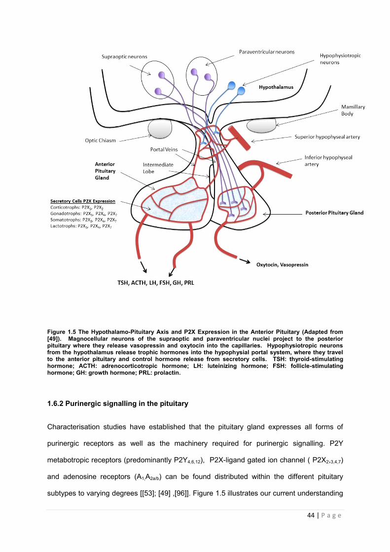
Figure 1.5 The Hypothalamo-Pituitary Axis and P2X Expression in the Anterior Pituitary (Adapted from [49]). Magnocellular neurons of the supraoptic and paraventricular nuclei project to the posterior pituitary where they release vasopressin and oxytocin into the capillaries. Hypophysiotropic neurons from the hypothalamus release trophic hormones into the hypophysial portal system, where they travel to the anterior pituitary and control hormone release from secretory cells. TSH: thyroid-stimulating hormone; ACTH: adrenocorticotropic hormone; LH: luteinizing hormone; FSH: follicle-stimulating hormone; GH: growth hormone; PRL: prolactin.
1.6.2 Purinergic signalling in the pituitary
Characterisation studies have established that the pituitary gland expresses all forms of
purinergic receptors as well as the machinery required for purinergic signalling. P2Y
metabotropic receptors (predominantly P2Y4,6,12), P2X-ligand gated ion channel ( P2X2,3,4,7)
and adenosine receptors (A1,A2a/b) can be found distributed within the different pituitary
subtypes to varying degrees [[53]; [49] ,[96]]. Figure 1.5 illustrates our current understanding
45 | P a g e
of expression of P2X receptors amongst the pituitary subpopulations. Exploitation of
mammalian expression systems such as HEK cells using membrane expressed luciferase
have shown internal concentrations of ATP to reach concentrations of up to 200uM at the
plasma membrane [[54] [33]]. This involves incubating cells in Krebs Ringer buffer at room
temperature and assaying samples for ATP release using a luminometer [2].
Pharmacological studies have shown that concentrations within this range are sufficient to
elicit a response at purinergic receptors. Enzymes to control the activity of ATP are also
expressed in the plasma membrane of pituitary cells with mRNA for the ecto-nucleotideases,
ectonucleoside triphosphate diphosphohydrolases 1, 2 and 3 (E-NTPDases) detected in
whole pituitary [[33, 55]].
1.6.2.1 Overview of P2X mediated hormone release
In the pituitary, binding of ATP to P2X allows the passage of calcium down its
electrochemical gradient inside the cell. This causes a depolarisation of the plasma
membrane and stimulation of action potentials. These action potentials generate large
influxes of intracellular calcium [Ca]I which penetrate all areas of the cell [56]. This global
signalling allows calcium to span the gap between hormone loaded vesicles and calcium
channels, which is vital for regulated hormone secretion [[57], [1]].
1.6.3. P2XRs and pituitary subtypes
Our current understanding concerning the influence of purinergic signalling within specific
pituitary cell populations has been derived from a combination of electrophysiological and
molecular based investigations, using both pituitary cell lines and primary tissue.

46 | P a g e
1.6.3.1 Corticotrophs
Our current understanding of purinergic signalling within corticotrophs is limited. In 1989,
work from two separate laboratories demonstrated adenosine signalling played a functional
role in regulating ACTH secretion from pituitary corticotrophs. More recently Zhao and co-
workers have identified that both P2 receptor families are expressed within the mouse
corticotroph cell line, AtT-20s. The same study demonstrated that ATP stimulation of the
cells increased the production of POMC by a mechanism which did not involve protein
kinase A (PKA). Although this pituitary subtype appears more sensitive to adenosine, it has
been suggested that both P2Y and P2X receptors may be involved in the production of
POMC [[59], [60], [61]]
1.6.3.2 Gonadotrophs
Data from studies utilising embryonic, neonatal and adult rat pituitary tissue have suggested
that ATP and P2X interactions are responsible for the spontaneous changes in membrane
potential observed within gonadotrophs. P2X receptors are believed to control basal release
of hormone from these cells and therefore act as pacemaking channels in gonadotrophs.
Zemkova and colleagues demonstrated in primary gonadotroph cultures that ATP acting on
P2X2 receptors induce depolarising currents (via the influx of calcium through these
channels) and initiate spontaneous action potentials in cells. A combination of patch clamp
studies and RT-PCR have demonstrated that these channels serve to synchronise electrical
activity across the membrane and to amplify calcium mobilising activity initiated by
hypothalamic gonadotropin-releasing hormone and the release of LH [[62]; [34]].
1.6.3.3 Lactotrophs
Investigations have demonstrated that approximately 90% of pituitary lactotrophs are
responsive to ATP. He and colleagues demonstrated that lactotrophs express functional
receptors that are able to generate depolarising currents when treated with ATP. Patch
clamp analysis of cells pretreated with thasigargin (which blocked internal calcium
47 | P a g e
mobilisation by P2Y channels) and subsequently treated with P2XR agonists demonstrated
an increased calcium influx through these channels of sufficient magnitude to stimulate
prolactin release. Pharmacological profiling with various P2XR ligands and subsequent RT-
PCR analysis suggested that P2X4 was the most likely candidate responsible for the calcium
mediated release in these cells. Further work utilising electrophysiology and pharmacological
profiling of TRH responsive cells confirmed these findings that direct interactions between
ATP and P2X4 were responsible for prolactin release from cells. These cells were also
sensitive to the P2X4 modulator ivermectin (IVM), which increases both pore dilation and
sensitivity of receptors for ATP [[63]; [64],[65], [13]. A summary of purinergic receptor
expression within the different pituitary subtypes is shown in table1. 3.
48 | P a g e
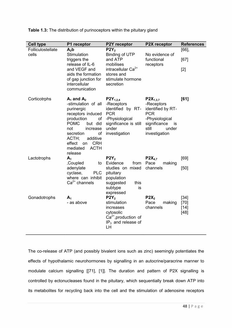
Table 1.3: The distribution of purinoceptors within the pituitary gland
Cell type P1 receptor P2Y receptor P2X receptor References Folliculostellate cells
A2b Stimulation triggers the release of IL-6 and VEGF and aids the formation of gap junction for intercellular communication
P2Y2 Binding of UTP and ATP mobilises intracellular Ca2+ stores and stimulate hormone secretion
No evidence of functional receptors
[66], [67] [2]
Corticotrphs A1 and A2 -stimulation of all purinergic receptors induced production of POMC but did not increase secretion of ACTH; additive effect on CRH mediated ACTH release
P2Y1,2,4 -Receptors identified by RT-PCR -Physiological significance is still under investigation
P2X1,3,7 -Receptors identified by RT-PCR -Physiological significance is still under investigation
[61]
Lactotrophs A1
-Coupled to adenylate cyclase, PLC where can inhibit Ca2+ channels
P2Y2 Evidence from studies on mixed pituitary population suggested this subtype is expressed
P2X4,7 Pace making channels
[69] [50]
Gonadotrophs A1 - as above
P2Y2 stimulation increases cytosolic Ca2+,production of IP3 and release of LH
P2X2 Pace making channels
[34] [70] [14] [48]
The co-release of ATP (and possibly bivalent ions such as zinc) seemingly potentiates the
effects of hypothalamic neurohormones by signalling in an autocrine/paracrine manner to
modulate calcium signalling [[71], [1]]. The duration and pattern of P2X signalling is
controlled by ectonucleases found in the pituitary, which sequentially break down ATP into
its metabolites for recycling back into the cell and the stimulation of adenosine receptors
49 | P a g e
which are generally negatively coupled to calcium mobilising G-protein coupled receptors
[[73] [2]; [75] ]
1.7 Summary of aims of thesis
Purinergic signalling is evolutionarily highly conserved and has been demonstrated to play a
role post-development in a range of organs, including the mammalian pituitary gland.
Previous investigations have demonstrated a role for ATP interactions with P2X1-7 family to
play a role in hormone release from the pituitary gland. However investigations into the role
to which individual receptors contribute to pituitary function has been inhibited by the limited
range of specific ATP analogues and/or antagonist.
The overall concept behind the work presented in this thesis was to further examine the role
of purinergic signalling in the propagation and amplification of pituitary hormone release, and
also the mechanism by which individual P2X receptors contribute to hormone release from
the different pituitary cells. To do this, highly specific P2X antagonists were used to
determine the contribution made by each family member, along with a range pituitary cell
lines so that each cell type can be assed in isolation.
With detailed histological characterisation available for P2Y expression in the rat pituitary
gland [76], emphasis was also placed on determining the distribution patterns of the P2X
receptors expressed in the pituitary . Although functional expression of the P2X receptors
has been demonstrated using electrophysiology and Q-PCR, confirmation of which receptors
reside in the pituitary subtypes by immunostaining has yet to be achieved. This branch of the
investigation would provide evidence of co-localisation between individual P2X receptors
and pituicytes.

50 | P a g e
Chapter 2 Material and Methods
51 | P a g e
2.1 Materials
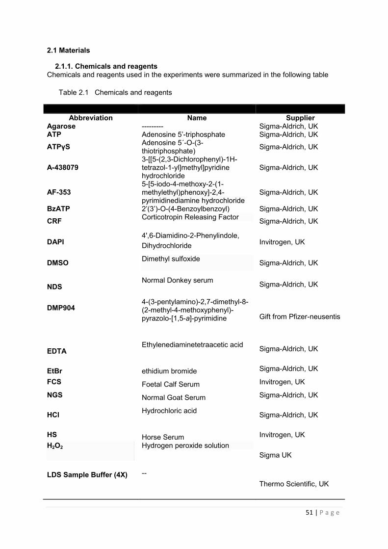
2.1.1. Chemicals and reagents Chemicals and reagents used in the experiments were summarized in the following table
Table 2.1 Chemicals and reagents
Abbreviation Name Supplier
Agarose --------- Sigma-Aldrich, UK ATP Adenosine 5’-triphosphate Sigma-Aldrich, UK
ATPγS Adenosine 5´-O-(3-thiotriphosphate) Sigma-Aldrich, UK
A-438079 3-[[5-(2,3-Dichlorophenyl)-1H-tetrazol-1-yl]methyl]pyridine hydrochloride
Sigma-Aldrich, UK
AF-353 5-[5-iodo-4-methoxy-2-(1-methylethyl)phenoxy]-2,4-pyrimidinediamine hydrochloride
Sigma-Aldrich, UK
BzATP 2’(3’)-O-(4-Benzoylbenzoyl) Sigma-Aldrich, UK
CRF Corticotropin Releasing Factor Sigma-Aldrich, UK
DAPI 4',6-Diamidino-2-Phenylindole, Dihydrochloride Invitrogen, UK
DMSO Dimethyl sulfoxide Sigma-Aldrich, UK
NDS Normal Donkey serum
Sigma-Aldrich, UK
DMP904
4-(3-pentylamino)-2,7-dimethyl-8-(2-methyl-4-methoxyphenyl)-pyrazolo-[1,5-a]-pyrimidine
Gift from Pfizer-neusentis
EDTA Ethylenediaminetetraacetic acid
Sigma-Aldrich, UK
EtBr ethidium bromide Sigma-Aldrich, UK
FCS Foetal Calf Serum Invitrogen, UK
NGS Normal Goat Serum Sigma-Aldrich, UK
HCl Hydrochloric acid
Sigma-Aldrich, UK
HS Horse Serum Invitrogen, UK H2O2
Hydrogen peroxide solution
Sigma UK
LDS Sample Buffer (4X)
-- Thermo Scientific, UK
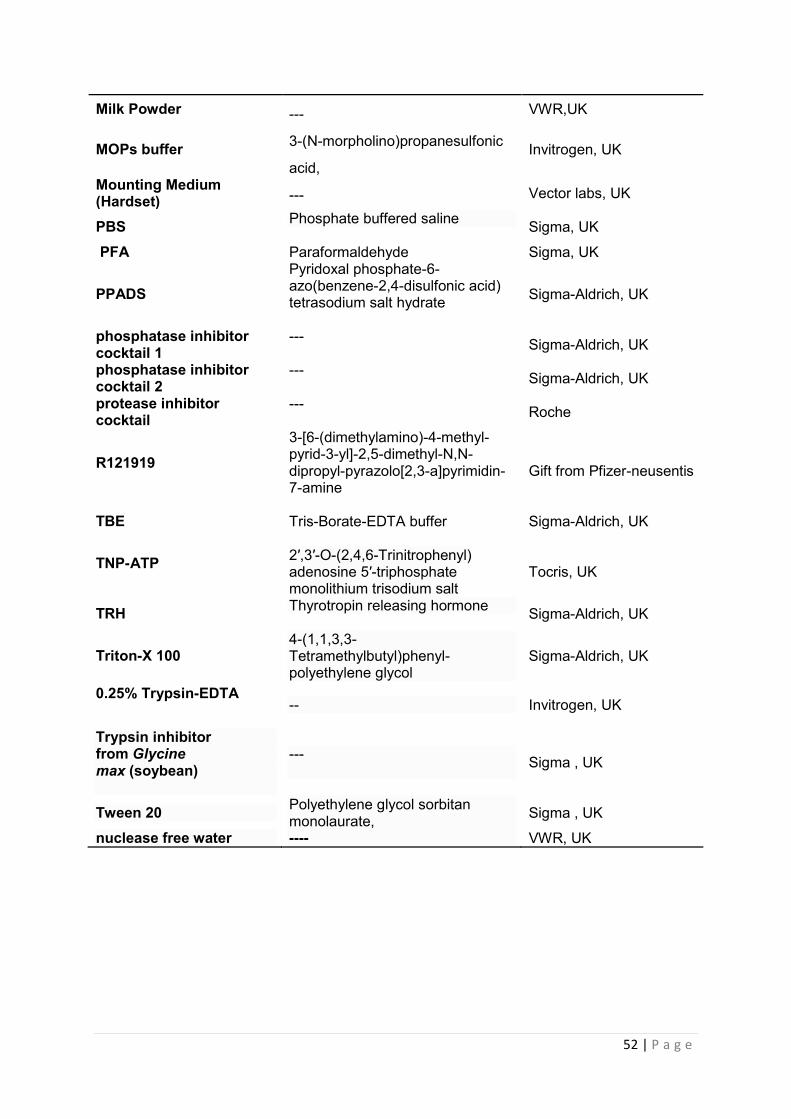
52 | P a g e
Milk Powder --- VWR,UK
MOPs buffer 3-(N-morpholino)propanesulfonic
acid, Invitrogen, UK
Mounting Medium (Hardset) --- Vector labs, UK
PBS Phosphate buffered saline Sigma, UK
PFA Paraformaldehyde Sigma, UK
PPADS Pyridoxal phosphate-6-azo(benzene-2,4-disulfonic acid) tetrasodium salt hydrate
Sigma-Aldrich, UK
phosphatase inhibitor cocktail 1
--- Sigma-Aldrich, UK
phosphatase inhibitor cocktail 2
--- Sigma-Aldrich, UK
protease inhibitor cocktail
--- Roche
R121919
3-[6-(dimethylamino)-4-methyl-pyrid-3-yl]-2,5-dimethyl-N,N-dipropyl-pyrazolo[2,3-a]pyrimidin-7-amine
Gift from Pfizer-neusentis
TBE Tris-Borate-EDTA buffer Sigma-Aldrich, UK
TNP-ATP 2′,3′-O-(2,4,6-Trinitrophenyl) adenosine 5′-triphosphate monolithium trisodium salt
Tocris, UK
TRH Thyrotropin releasing hormone Sigma-Aldrich, UK
Triton-X 100 4-(1,1,3,3-Tetramethylbutyl)phenyl-polyethylene glycol
Sigma-Aldrich, UK
0.25% Trypsin-EDTA
-- Invitrogen, UK
Trypsin inhibitor from Glycine max (soybean)
--- Sigma , UK
Tween 20 Polyethylene glycol sorbitan monolaurate, Sigma , UK
nuclease free water ---- VWR, UK
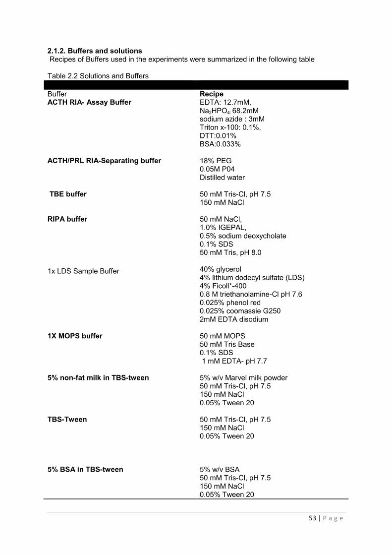
53 | P a g e
2.1.2. Buffers and solutions Recipes of Buffers used in the experiments were summarized in the following table Table 2.2 Solutions and Buffers Buffer Recipe ACTH RIA- Assay Buffer EDTA: 12.7mM,
Na2HPO4: 68.2mM sodium azide : 3mM Triton x-100: 0.1%, DTT:0.01% BSA:0.033%
ACTH/PRL RIA-Separating buffer 18% PEG 0.05M P04 Distilled water
TBE buffer 50 mM Tris-Cl, pH 7.5 150 mM NaCl
RIPA buffer 50 mM NaCl, 1.0% IGEPAL, 0.5% sodium deoxycholate 0.1% SDS 50 mM Tris, pH 8.0
1x LDS Sample Buffer
40% glycerol 4% lithium dodecyl sulfate (LDS) 4% Ficoll*-400 0.8 M triethanolamine-Cl pH 7.6 0.025% phenol red 0.025% coomassie G250 2mM EDTA disodium
1X MOPS buffer 50 mM MOPS 50 mM Tris Base 0.1% SDS 1 mM EDTA- pH 7.7
5% non-fat milk in TBS-tween 5% w/v Marvel milk powder 50 mM Tris-Cl, pH 7.5 150 mM NaCl 0.05% Tween 20
TBS-Tween 50 mM Tris-Cl, pH 7.5 150 mM NaCl 0.05% Tween 20
5% BSA in TBS-tween
5% w/v BSA 50 mM Tris-Cl, pH 7.5 150 mM NaCl 0.05% Tween 20
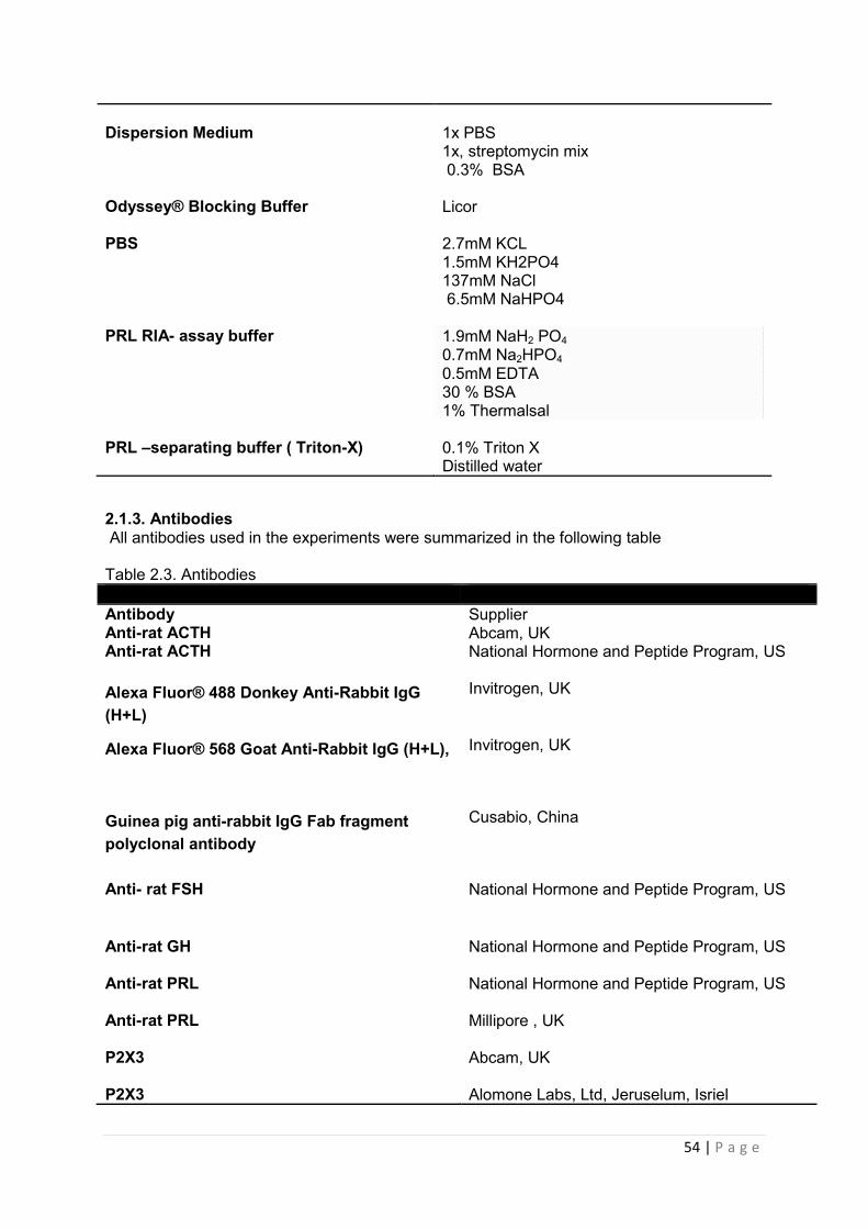
54 | P a g e
Dispersion Medium 1x PBS
1x, streptomycin mix 0.3% BSA
Odyssey® Blocking Buffer Licor PBS
2.7mM KCL 1.5mM KH2PO4 137mM NaCl 6.5mM NaHPO4
PRL RIA- assay buffer 1.9mM NaH2 PO4 0.7mM Na2HPO4 0.5mM EDTA 30 % BSA 1% Thermalsal
PRL –separating buffer ( Triton-X) 0.1% Triton X
Distilled water
2.1.3. Antibodies All antibodies used in the experiments were summarized in the following table Table 2.3. Antibodies Antibody Supplier Anti-rat ACTH Abcam, UK Anti-rat ACTH National Hormone and Peptide Program, US
Alexa Fluor® 488 Donkey Anti-Rabbit IgG (H+L)
Invitrogen, UK
Alexa Fluor® 568 Goat Anti-Rabbit IgG (H+L),
Invitrogen, UK
Guinea pig anti-rabbit IgG Fab fragment polyclonal antibody
Cusabio, China
Anti- rat FSH National Hormone and Peptide Program, US
Anti-rat GH National Hormone and Peptide Program, US
Anti-rat PRL National Hormone and Peptide Program, US
Anti-rat PRL Millipore , UK
P2X3 Abcam, UK
P2X3 Alomone Labs, Ltd, Jeruselum, Isriel
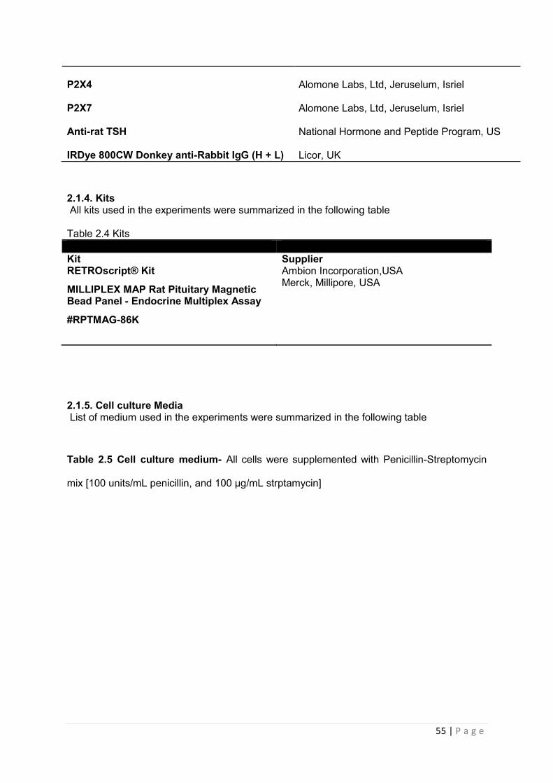
55 | P a g e
P2X4
Alomone Labs, Ltd, Jeruselum, Isriel
P2X7
Alomone Labs, Ltd, Jeruselum, Isriel
Anti-rat TSH National Hormone and Peptide Program, US
IRDye 800CW Donkey anti-Rabbit IgG (H + L) Licor, UK
2.1.4. Kits All kits used in the experiments were summarized in the following table Table 2.4 Kits Kit Supplier RETROscript® Kit Ambion Incorporation,USA
MILLIPLEX MAP Rat Pituitary Magnetic Bead Panel - Endocrine Multiplex Assay
#RPTMAG-86K
Merck, Millipore, USA
2.1.5. Cell culture Media List of medium used in the experiments were summarized in the following table
Table 2.5 Cell culture medium- All cells were supplemented with Penicillin-Streptomycin
mix [100 units/mL penicillin, and 100 µg/mL strptamycin]
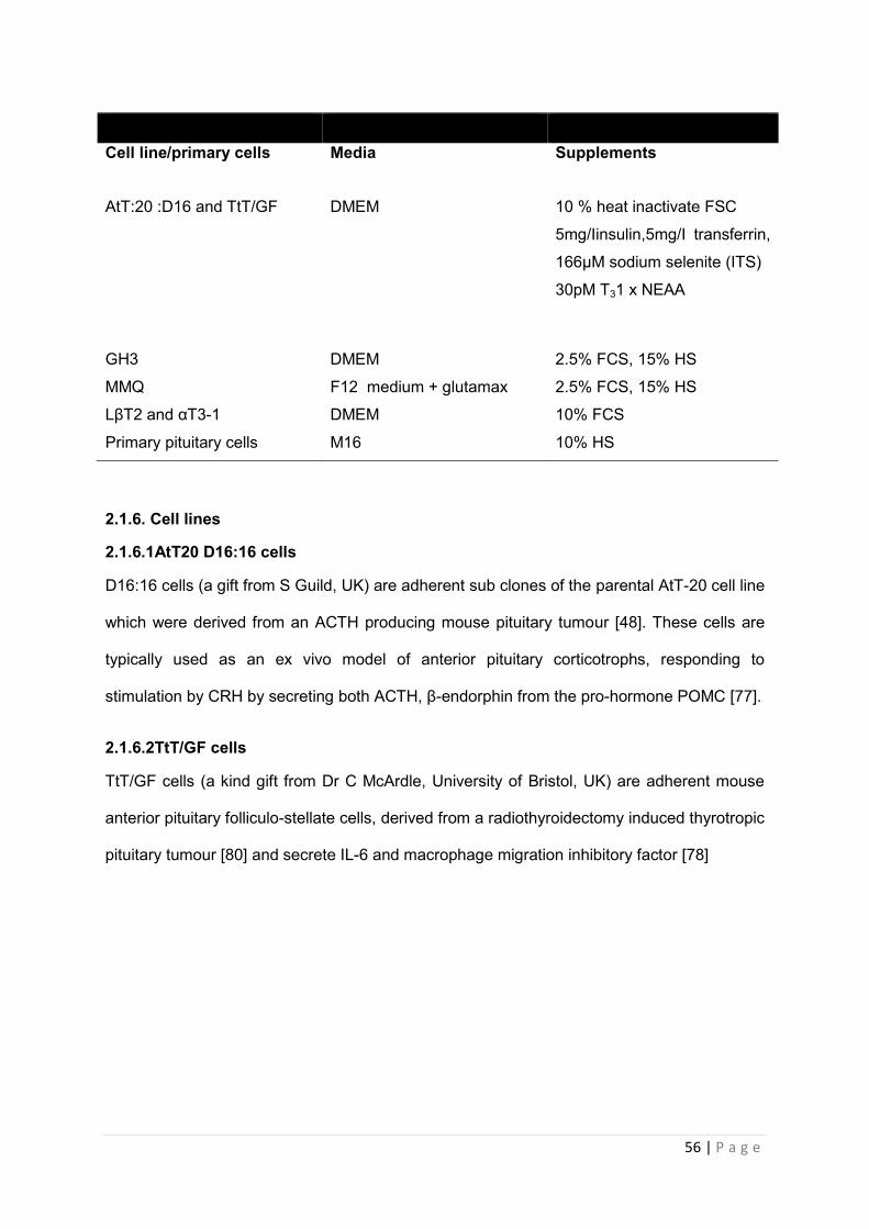
56 | P a g e
2.1.6. Cell lines
2.1.6.1AtT20 D16:16 cells
D16:16 cells (a gift from S Guild, UK) are adherent sub clones of the parental AtT-20 cell line
which were derived from an ACTH producing mouse pituitary tumour [48]. These cells are
typically used as an ex vivo model of anterior pituitary corticotrophs, responding to
stimulation by CRH by secreting both ACTH, β-endorphin from the pro-hormone POMC [77].
2.1.6.2TtT/GF cells
TtT/GF cells (a kind gift from Dr C McArdle, University of Bristol, UK) are adherent mouse
anterior pituitary folliculo-stellate cells, derived from a radiothyroidectomy induced thyrotropic
pituitary tumour [80] and secrete IL-6 and macrophage migration inhibitory factor [78]
Cell line/primary cells Media Supplements
AtT:20 :D16 and TtT/GF DMEM 10 % heat inactivate FSC
5mg/Iinsulin,5mg/I transferrin,
166µM sodium selenite (ITS)
30pM T31 x NEAA
GH3 DMEM 2.5% FCS, 15% HS
MMQ F12 medium + glutamax 2.5% FCS, 15% HS
LβT2 and αT3-1 DMEM 10% FCS
Primary pituitary cells M16 10% HS
57 | P a g e
2.1.6.3 GH3 cells
GH3 cells are a non-adherent sub-clone of the parental GH1 cell line which was derived
from the rat MtT/W5 pituitary tumour .Cells have the ability to secret both PRL and GH in
response to stimulation with TRH, which has led to them begin referred to as a
‘somatolactotrope’, in terms of their cellular phenotype [1].
2.1.6.4 MMQ cells
MMQ cells (a kind gift from Dr Jack Ham, University of Cardiff, UK) are a non-adherent
model of pituitary lactotrophs which were derived from the rat pituitary tumour 7315a. They
possess functional dopamine and oxytocin receptors and secrete PRL [[80], [81]]
2.1.6.5 LβT2 cells
LβT2 cells (a kind gift from Dr Pamela Mellon, University of California, USA via Dr Rob
Fowkes, The Royal Vet College, London, UK) are an adherent model of mature pituitary
gonadotrophs which were derived from tumour tissue within transgenic mice possessing an
altered LHb-subunit promoter [1]. These cells express GnRH receptors and secrete LH and
FSH in response to stimulation with GnRH [[83],[84]]
2.1.6.6 αT3-1 cells
αT3-1 cells (a kind gift from Dr Pamela Mellon, The Salk Institute, USA via Dr Rob Fowkes,
The Royal Vet College, London, UK) are an immortalised adherent mouse cell line derived
from a tumour produced within a transgenic mouse line using oncogene SV40 T antigen and
the human glycoprotien α-subunit gene promoter [1]. Cells express GnRH receptors, but
secrete immature gonadotrophins in response to GnRH. Hence cells are regarded as having
a phenotype akin to immature precursor pituitary cells [[86],[87]].

58 | P a g e
2.2 Methods
2.2.1 Maintenance of cell lines
All cells were grown in 75ml ventilated flasks (Cellstar , Griner Bio-one GmGH, Germany) at
37ºC and 5% CO2 (Binder incubator GmbH, Tuttlingen, Germany). Media used (included
required supplements) for individual cell lines can be found in table 6. All cells were
supplemented with Penicillin-Streptomycin mix [100 units/mL penicillin, and 100 µg/mL
strptamycin]
2.2.2 Passage of cells
Upon reaching confluency (~80-90%) adherent cells (AtT20 D16:16, TtT/GF, LβT2, αT3-1)
were first washed with Dubbecco’s phosphate –buffered saline (PBS) containing 2.7mM
KCL, 1.5mM KH2PO4, 137mM NaCl and 6.5mM NaHPO4 (Invitrogen Ltd,UK) and then cells
were subjected to trypsinisation via incubation with 0.25% trypsin (Invitrogen Ltd,UK) for 5
minutes at 37ºC and 5% CO2. Trypsin was neutralised by addition of an equal volume of cell
media and cells were resuspended into T175 flasks with fresh media. For RIA, cells were
seeded at a density of 2x10⁻⁵ cells per well in 48 well plates (Cellstar, Greiner Bio-One,
UK)(see section 3.4.3) or for western blot cells were seeded at a density of 2x10⁶ cells per
well (see section 3.3.2). Cell suspensions (GH3, &MMQ) were pelleted from medium by
spinning at 1100g for 5 minutes at room temperature. Medium was then aspirated and cells
were washed in PBS and spun for a further 5 minutes. PBS was then aspirated and
replaced with 10ml of fresh medium to resuspend cells. This was either transferred to a fresh
T175 with additional medium or cell counts were performed and cells seeded into 24 well
plates for RIA or western blot in the same manner as described above for adherent cells.
59 | P a g e
2.2.3 Determination of the functional expression of P2X receptors in pituitary cell lines
2.2.3.1 Treatment of cell line (AtT:20 :D16) with native secretagogue
Cells were seeded (2x10⁵ well-1) into 48 well plates (Cellstar, Greiner Bio-One, UK) and
incubated with a range of doses ( 0.1,1,10,100nM) of native pituitary agonists, CRF (Sigma,
Poole, UK) for various lengths of time (1,4,7 and 12 hours). Each concentration was tested
n= 6 wells. Errors shown are ± SEM. Medium was collected on ice and stored at -80ºC until
assayed for hormone release by radioimmunoassay (RIA).
2.2.3.2 Treatment of cell line (AtT:20 :D16) with native P2X agonist
Cells were seeded (2x10⁵ well-1) into 48 well plates (Cellstar, Greiner Bio-One, UK) and
incubated with a single dose of ATP (100µM) (Sigma, Poole, UK) for various lengths of time
(1,4,7 and 12 hours). Concentration was tested n=6 replicates/well. Errors shown are ±
SEM. Medium was collected on ice and stored at -20ºC until assayed for hormone release
by RIA.
2.2.3.3 Treatment of cell line (AtT:20 :D16) with native agonist CRF and ATP
AtT20:D16 (2x1051.well-1) cells were incubated with a single dose of CRH [100nM](Sigma,
Poole, UK) for 1, 4, 7 and 12 hours, alone and in the presence of ATP
[100µM](Sigma,Poole,UK). Medium was collected on ice and stored at -80ºC until assayed
for hormone release by RIA.
2.2.3.4 Treatment of cell line (GH3) with native secretagogue
GH3 cells were seeded (2x10⁵ well-1) into 48 well plates (Cellstar, Greiner Bio-One, UK) and
incubated with a range of doses (1,10,100, and 1000nM) of native pituitary agonists, TRH
(Sigma, Poole, UK) for various lengths of time (5,10,15,20,30,60 minutes). Each
concentration was tested n=6 replicates/wells. Errors shown are ± SEM. Medium was
collected on ice and stored at -80ºC until assayed for hormone release by RIA.
60 | P a g e
2.2.3.5 Treatment of cell line (GH3) with native P2X agonist and analogues
GH3 cells were seeded (2x10⁵ well-1) into 48 well plates (Cellstar, Greiner Bio-One, UK) and
incubated with a range of doses (0.001,0.01,0.1,1,10,100,1000µM) of native pituitary
agonists, ATP or analogues ATP--S and BzATP (Sigma, Poole, UK) for various lengths of
time (5,10,15,20,30,60 minutes). Each concentration was tested n=6 replicates/wells.
Errors shown are ± SEM. Medium was collected on ice and stored at -80ºC until assayed for
hormone release by RIA.
2.2.3.6 Treatment of cell line (GH3) with native agonist TRH and ATP
GH3 (2x1051.well-1) cells were incubated with a single dose of TRH [1nM](Sigma, Poole, UK)
for 30 minutes alone and in the presence of ATP [1µM or 100µM](Sigma,Poole,UK). Medium
was collected on ice and stored at -80ºC until assayed for hormone release by RIA.
2.2.3.7 Treatment of GH3 cells with P2X receptor antagonists PPADS, TNP-ATP,AF-353 A-438079
Using conditions previously described for GH3 cells (see table 2.6 for details), cells were
seeded into 48 well plates (Cellstar , Griner Bio-one GmGH, Germany) at a density of
2x105.ml-1.well-1. After 48h cultures were serum starved [F12 medium containing 0.1% FCS,
0.1%HS and streptomycin mix:] for a further 24h. Cells were then treated with 1nM TRH
(Sigma-Aldrich, UK), 1µM or 100µM ATP (Sigma-Aldrich, UK), either alone or in combination
with ATP. Cells were also treated with TRH and/or ATP in combination with P2X receptor
antagonists PPADS (1, 5, 10, 50,100,500,1000µM), TNP-ATP, AF-353 and A-438079
(1,5,10,50,100,500,1000,5000nM). Medium was collected after 30 minutes and stored at -
80ºC for measurement of PRL release. Table 2.1 contains the list of PX2 receptor
antagonists used.
61 | P a g e
2.2.4. Primary rat pituitary culture
2.2.4.1 Animals
All experiments were carried out in accordance with the United Kingdom Animals (Scientific
Procedures) Act 1986 under the project licence PPL 70/5593. Male Sprague –Dawley rats
were obtained from a commercial supplier (Charles River Laboratories, Inc., Margate, UK)
and maintained in standard, wire top cages in the Comparative Biology Services unit at
Hammersmith Hospital (Faculty of Medicine, Imperial College London). Animals were
housed 5 rats per cage in a room with controlled lighting (lights were on between 0.800h &
20.00h) temperature (21ºC-23 ºC) and humidity (60-65%) with standard rat chow provided
ad libitum. Rats were handled daily for a week prior to experiments.
2.2.4.2 Generation of primary pituitary cultures
Primary rat pituitary cultures were carried out according to previous studies [(Tomić, Jobin et
al. 1996), ([96]Tomić et al. 2000)]. Pituitaries were excised from male Sprague–Dawley rats
and immediately transferred to Dispersal media (DM) [1x PBS supplemented with 1x,
streptomycin mix, 0.3% BSA]. Tissue was then washed in medium to remove blood and cut
into 0.5mm blocks. Tissues were then centrifuged in 10ml of DM at 700g at 4ºC and the cell
pellet retained. The pituitary cells were incubated in T75 flasks containing 30ml of 0.25%
trypsin for 60 min at 37ºC on a shaking incubator (Inferign Ltd, UK) followed by 30ml of DM
supplemented with 0.1mg/ml trypsin inhibitor (Sigma,,UK) and 0.01% DNase (Sigma,,UK) for
10 minutes at 37ºC. The cell suspension was then centrifuged (700g, 5 minutes at 4ºC), the
supernatant aspirated and the cells filtered through a nylon mesh (Becton, UK) to obtain a
single cell suspension. The cells underwent repeated centrifugation in fresh medium (4x
700g, 4ºC, 5 min) following which cells were resuspended and a cell count was taken. Cells
were then plated into 48 well plate at the appropriated density in M199 medium containing
10% horse serum and pen/strep 100u/mL.
62 | P a g e
2.2.4.3 Treatment of primary cultures with CRH and CRH receptor antagonists, R121919 and DMP904
Using conditions previously described for AtT-20 cells (see table 1 for details), primary
pituitary cells were seeded into 24 well plates (Cellstar, Griner Bio-one GmGH, Germany) at
a density of 1x105.ml-1.well-1. After 48h cultures were serum starved [DMEM containing 0.5%
FCS, T3: and streptomycin mix:] for a further 24h. Cells were then treated with 100nM CRH
(Sigma, UK) either alone or in combination with CRH receptor antagonists, R121919 or
DMP904 (1, 5, 10, 50 or 100nM). Medium was collected after 4h and stored at -20ºC for
measurement of ACTH release.
2.2.3 RadioimmunoAssay (RIA)
The radioimmunoassay was first developed in the 1950’s by Solomon A Berson and Rosalyn
Yalow. This in vitro binding assay exploits the immune reaction between an antibody (Ab)
and an antigen (Ag) to measure/quantify the amount of substance in a biological sample. Ag
is labelled with radioactivity (I125) and incubated with a fixed amount of antibody and
unlabelled Ag, which is provided by the sample under investigation. This sets up a
competition between radioactive labelled Ag and unlabelled Ag for limited amount of binding
sites on the Ab. To allow enough time for Ag-Ab complexes to occurs, the reaction is allowed
to incubate and go to completion.
*Ag + Ab + Ag ↔x *AgAb + AgAb
Ag = unlabelled antigen
*Ag = radiolabelled antigen
Ab = antibody
63 | P a g e
Unlabelled Ag in the form of Standards are also incubated alongside the reaction. During
incubation period the amount of radiolabelled Ag will be displaced by the unlabelled Ag,
in unknown amounts, in the sample being measured. Unbound antigen is removed by
incubating samples with 2° ab, directed to the species which the primary ab was raised
in, and centrifugation to precipitate out the Ab-Ag complex. The pellet formed will contain
Ag-ab complexes and the free Ag will be in the supernatant. Both the bound and the free
Antigens are read on a γ –counter. The intensity of the radioactivity measure following
separation of ab bound Ag from free, unbound Ag, is inversely proportional to the
amount of Ag in the sample under investigation. The results from the standards can be
used to construct a standard curve from which the unknown samples can be interpolated
from. Inter-assay variation can be calculated by assaying aliquots of the same sample in
each assay performed and comparing the concentrations obtained in each. To measure
and correct for baseline drift, tubes with no sample (‘zero’ tubes) are placed at regular
intervals throughout the assay and standard curves are performed at the beginning and
end of each assay. A table to show the general set up of an RIA can be found in
appendices 1.

64 | P a g e
2.2.3.1 ACTH Radioimmunoassay
2.2.3.1.1 Iodination of ACTH
All iodinations of ACTH peptide were carried out by Professor MA Ghatei (Professor of
Regulatory Peptides, Metabolic Medicine, Faculty of Medicine, Imperial College) according
to the following method. Synthetic ACTH was iodinated using the iodogen method [88]. The
oxidising agent, 1,3,4,6,-tetrachloro-3, 6-diphenylglycoluril (iodogen reagent) is evaporated
onto the walls of a polypropelene tube from a solution in methylene chloride. The peptide to be
iodinated and 125I are then added. The reaction is terminated when the mixture is removed from
the tube, thus negating the need for a reducing agent.
To iodinate ACTH, 10μg of peptide in 10μl of 0.2 M phosphate buffer pH 7.2 were reacted with
37 MBq of Na 125I (Perkin Elmer) and 23 nmol (10 μg) of iodogen reagent (Pierce Chemical
Co.) for 4 minutes at 22oC. The 125I-peptide was purified by reversed-phase high performance
liquid chromatography using an ACN/ H2O /0.05 % TFA gradient of 20 % to 25 % over 50 min.
The fractions were collected using a Frac-100 fraction collector (Pharmacia). The specific
activity (SA) of an iodinated peptide is the amount of radioactivity per mole of hormone. It is
measured in Bequerel (Bq) which is an SI unit of radioactivity and is equivalent to 1
disintegration per second. The purified label was found to have a SA of 60.6 Bq/fmol as
measured by self-displacement in the assay.
2.2.3.1.2 Radioimmunoassay for ACTH
The concentration of hormones released into the medium of primary pituitary cultures and
pituitary cell lines was determined using RIA with reagents supplied by the National Institute
of Diabetes and Digestive and Kidney Disease (NIDDK, California). Details of antibodies can
be found in table 2.3. Reference standards were prepared for each hormone in triplicate, via
serial dilution of stock standard (ACTH 2µg/ml) in assay buffer [see table 2.2, section 2.1.2].
This produced standards ranging from 10,000pg/ml to 10pg/ml. 200µl of peptide specific
antibody (diluted 1:60,000) was added to 100µl of sample in duplicate. Control tubes were
65 | P a g e
included in triplicate consisting of iodinated/labelled peptide (100µl) and assay buffer (300µl)
to determine non-specific binding and separate tubes with label (100ul), antibody (200ul) and
buffer (300ul) to determine specific binding. Standard and sample tubes were each
administered 100ul of radio labelled peptide (which had been diluted to give 2000 counts per
minute) and 200ul antibody and all assay tubes were then mixed and left to incubate for 4
days at 4ºC.
On the fourth day, separation of bound and free peptide began with the addition of 200ul
horse serum: 0.9% saline (1:1 mix) to all tubes at room temperature for 30minutes. 600ul of
separating buffer [see table 2] were then added to the tubes and centrifuged at 3000 rpm at
8ºC for 30 minutes. The supernatant was aspirated and the bound fraction counted on an
automatic gamma counter (Perkin-Elmer LAS Ltd, Beaconfield, UK) for 120 seconds.
Hormone concentrations within assayed samples were derived from the respective standard
curves. The detection limit of assay is 40pg/ml.
2.2.3.2 PRL Radioimmunoassay
2.2.3.2.1 Iodination of PRL
All iodination of PRL peptides was carried out by Dr Chris John according to the following
method. 2µg/20µl of PRL peptide in 0.05M phosphate buffer, pH 7.4. was reacted with
27.75MBq of Na 125I (Amersham) and 10µl of N-Chloro-p-toluenesulfonamide sodium salt
hydrate (chloramine-T;1.5mg/ml in 0.05M phosphate buffer, pH 7.4)(Sigma-Aldrich, UK) for
30s at room temperature after which time the reaction was stopped by addition of 10µl
sodium metabisulphate (2.5mg/ml in 0.05M phosphate buffer). The resulting mixture was
immediately pipetted on to the surface of a sephadex G50 column which had been
previously equilibrated with 0.05M phosphate buffer containing 0.5% w/v bovine serum
albumin (BSA, Sigma, UK). The mixture was then eluted through the column using the
phosphate buffer containing 0.5% BSA and 1ml fractions were collected, stoppered and the
radioactive content of each was determined. The first fractions containing the highest counts
66 | P a g e
of radioactivity (usually fractions 4- 6) contained the 125I-labelled. The 125I-peptide was
assessed for use in RIA by constructing a standard curve and zero tubes and the
percentage binding determined. Labelled peptide that gave at least 60% binding in zero
were used in PRL-RIAs.
2.2.3.2.2. Radioimmunoassay for PRL
PRL was determined in duplicate by radioimmunoassay using reagents supplied by the
National Hormone and Pituitary Programme (Bethesda, USA). The PRL antibody (NIDDK-
anti-rPRL-S-9) had negligible cross reactivity with rat-GH, -TSH, -FSH and -LH and was
stored at -80°C. Solutions of the prolactin standard (NIDDK-rat-PRL-RP-3) were prepared by
serial dilution of a stock standard (10µg/ml in distilled water stored at -80°C) in assay buffer
(see table 2.2), to produce solutions ranging from 0.049-50ng/ml. Tubes containing 50µl
aliquots of standard or test solution and 100µl PRL antibody (diluted 1 in 3200) were
prepared in duplicate. Tubes containing 150µl assay buffer or 100µl PRL antibody and 50µl
assay buffer were also set up to determine non-specific and specific binding, respectively. In
addition, 250µl of the tracer 125I-labelled PRL, diluted in assay buffer to give approximately
10000 counts/min/250µl was added to each tube. The tubes were then vortexed and
incubated for 2 days at room temperature. Separation of the bound and free peptide was
achieved using a mixture of anti-rabbit precipitating serum raised in donkey (IDS,
Washington, England), diluted 1 in 24 v/v and non-immune rabbit serum, diluted 1 in 200 v/v,
both in assay buffer. 100µl of this mixture was added to each tube (except total count tubes)
which were then vortexed and incubated for 2h at room temperature, after which time 500µl
4% w/v polyethylene glycol (PEG, BDH Chemicals, Dagenham, UK) was added to each
tube. The tubes were then centrifuged at 4000g at 4°C for 30 minutes. Following
centrifugation, the supernatant was then aspirated and the radioactivity of the remaining
pellet was counted on Bound and free label were separated and both the pellet and

67 | P a g e
supernatant counted for 180 seconds in a γ-counter (model NE1600, Thermo Electron
Corporation).
2.2.4 Protein expression Analysis
2.2.4.1 Protein extraction
Protein was extracted from cells and rat tissue (brain and pituitary) according to a previously
published protocol (Zemkova et al. 2010). Cells were washed twice in cold PBS and then
resuspended in 100µl of RIPA buffer (Sigma,UK) which was supplemented with
phosphatase inhibitor cocktail 1 and 2 (Sigma Poole) and a protease inhibitor cocktail
(Roche). Samples were lysed on ice for 60 minutes following which cells were then
centrifuged at 13,000 rpm for 60 minutes at 4ºC. Rat tissue was washed twice in cold PBS to
remove any residual blood, and then homogenised in RIPA buffer using a micropestle
(VWR, International Ltd, Leicester, UK). Tissue was then incubated in buffer for 60min on ice
and then centrifuged at 14,000 rpm at 4ºC for 1hr. The supernatant was retained and stored
at-20ºC.
2.2.4.2 Protein quantification (Bradford assay)
This colorimetric assay was used to assess the protein content in samples using coomassie
blue dye in solution. The binding of the coomassie dye to protein within samples causes a
shift in absorbance from 465nm to 595nm which can be detected using a
spectrophotometer. This is measured against a standard curve of bovine serum albumin
(0.1-1mg/ml). Samples were diluted (tissue-1:100) and both samples and standards were
added in duplicate to 96 well plates and incubated with G250/coomassie solution [100mg
G250 (Sigma, Poole, UK) 95% ethanol, 85% orthophosphoric acid] for 2 minutes. The
amount of protein in each sample was determined by reading the absorbance (Anthos
Labtec, UK) at 595nm. Concentration of protein was then estimated by linear regression of
the standard curve.
68 | P a g e
2.2.4.3 Electrophoretic separation of proteins on 10% SDS-PAGE gels
Protein was resuspended in 1x sample buffer (Invitrogen Ltd, UK) and protein denatured at
100ºC for 5 minutes. 100µg of protein was separated by electrophoresis following application
to pre-cast, polyacrylamide 10% Bis-Tris Gels (Invitrogen, UK). Non denaturing conditions
were used for separation, using 1x MOPs buffer (Invitrogen Ltd, UK), running at 200v for
40min (Invitrogen, Ltd, UK).
Following separation, gels were transferred onto PVDF membrane iBlot® western blotting
system (Invitrogen, UK) for 7 minutes. The membrane was transferred to 5% non-fat milk
powder (Marvel,Stafford, UK) in Tris–buffered saline [50mM Tris HCL pH 7.4,150mM Nacl]
supplemented with 0.05% Tween (Sigma, Poole, UK) (TBS-T) for 1h. Following 3 x 10min
washing cycles with 50ml TBS-T, blots were incubated overnight at 4°C with P2XR
antibodies in 5% BSA in Tris–buffered saline. The following day the blot was washed 1 x
30min and 2 x 15min with TBS-T. Deposition of the antibody was visualised by
using Odyssey Imaging System (Licor, UK). Firstly the blot was incubated with IRDye
antibody, a fluorescently-labelled secondary antibody in Odyssey Blocking Buffer for 1h
(1:10000;Licor, UK) at room temperature. The membrane was then washed 4 times in TBS-
T for 15 min. Specific antibody binding to protein was visualised and documented on the
Odyssey Imaging System, by excitation of the fluorescent signal on the membrane in the
appropriate channel according to the manufacturers guidelines.
2.2.5 Immunohistochemistry
2.2.5.1 Animals and Tissue handling
All experimental procedures, used in immunohistochemistry were performed in accordance
with the UK Animal (Scientific Procedures) Act 1986. Male Sprague–Dawley rats (200-250g)
(Charles River, UK) were sacrificed by non-schedule 1 decapitation. The pituitaries were
dissected out and fixed in 4% paraformaldehyde in 0.1 mol/l phosphate buffer pH 7.4 for 1 h,
then transferred to 5, 15 and then finally 30%- sucrose in PBS and kept in the solution until
69 | P a g e
they sank to the bottom. Tissue was then removed from sucrose and emended in OCT
embedding media (Thermo scientific, UK) and frozen at -80oC. Sections were cut (10 μM in
thickness) using a Leica cryostat (CM3050) and mounted onto superfast slides (Thermo
scientific, UK). Slides were stored at -80oC until further use.
2.2.5.2 Immuostaining
To determine the expression of P2X protein within the rat pituitary tissue, staining of
receptors using fluorescently labelled P2X antibodies was undertaken. Antibodies raised for
different pituitary hormones (ACTH, FSH, GH, PRL and TSH) were also used to identify the
different pituitary subpopulations, and thus allowed co-localisation of both target proteins to
be assessed. Antibodies for both P2X receptors and the pituitary hormones were raised in
the same species and required a sequential, double staining method which had been
applied previously [[76], [89], [90],[91]]. Following thawing of slides for 2 hours at room
temperature (temp), Slides were then washed twice in PBS (Sigma, UK) for 5 minutes.
Slides were then permeabilised in PBS with 0.1% triton x-100 for 20 minutes. Slides were
washed in PBS (2X 5 minutes) and then blocked in IHC buffer with 5% donkey serum
(Sigma,UK) for 30 minutes ( see table for recipe of IHC buffer) to reduce nonspecific binding
of the fluorescent secondary donkey antibody. Block was removed and rabbit polyclonal
P2X antibodies (Alomone, Israel) added in 5% NDS in IHC buffer at the following
concentration: P2X3 (1:10,000), P2X4 (1:8,000) and P2X7 (1:1000). Slides were allowed to
incubate in a humidified chamber at 4°C overnight. The next day slides were washed in PBS
with 0.1% Tween-20 (3 x 15 minutes). To enhance the signal of P2X antibodies, a biotin-
conjugated tyramide signal amplification (TSA) kit (Invitrogen, UK) was utilised in a manner
previously described [[76], [89],[91]]. This was allowed to incubate with tissue for 10 minutes
and excess removed by PBS washes (3 x 5 minutes). Then tissue was incubated with Alexa
Fluor® 488 Donkey Anti-Rabbit IgG (H+L) (Invitrogen, UK) (1:400 dilution ) and the nuclear
stain DAPI (1:1000) (Invitrogen,UK) for 30 minutes at room temperature and then washed in
PBS with 0.1% tween 20 ( 3X5 mins).
70 | P a g e
To facilitated combination with second primary antibodies, which were also raised in same
species, slides were then incubated with polyclonal guinea pig anti-rabbit IgG Fab fragment
to block any any unbound rabbit binding sites on the 1st primary antibody (1:100 in PBS for 2
hours). Excess Fab fragments were removed through PBS washes (3 X2 minutes). Tissue
was then blocked in IHC buffer with 5% NGS (Sigma,UK) for 30 minutes to reduce
nonspecific binding of the fluorescent secondary goat antibody. Block was removed and
rabbit polyclonal pituitary hormone antibodies (see table for details of manufactures) were
added in 5% NGS in IHC buffer at the following concentration:ACTH (1:2000), FSH
(1:10,000), GH (1:1000), PRL (1:1000) and TSH (1:10,000). Slides were allowed to incubate
in a humidified chamber at room temp. for 1 hour. Slides were washed in PBS with 0.1%
Tween-20 (3 x 15 minutes). Then tissue was incubated with Alexa Fluor® 568 Goat Anti-
Rabbit IgG (H+L), (Invitrogen, UK) (1:400 dilution) and the nuclear stain DAPI (1:1000)
(Invitrogen,UK) for 30 minutes at room temperature and then washed in PBS with 0.1%
tween 20 ( 3X5 mins). After a final wash was (3 X 5 minutes in PBS), sections were
mounted in fluromount (Thermo scientific, UK) and stored in dark until visualisation.
Negative controls were performed by substituting primary antibodies for IHC block in both
the 1st and 2nd part of immunostain. To check that no cross reactivity occurred between
consecutive stains with the two rabbit antisera, the results of single and double staining was
compared. Pictures were obtained using a confocal scanning microscope (Zeiss, Microscope
Axio Observer.Z1) which allowed image reconstruction (AxioVision 4 Documentation). All
images taken at 20X magnification. Only cells that demonstrated the same morphology and
position when viewed under the two different filters (in the same focal plane) were deemed
to co-localise both P2X receptor and pituitary hormones. These slides were then taken at
40x magnification.
71 | P a g e
2.2.6 MILLIPLEX MAP rat pituitary panel kit
The Millipore MILLIPLEX MAP rat pituitary panel kit was used for the simultaneous
quantification of the following rat pituitary hormones: ACTH, PRL, TSH and GH. Samples of
supernatant were collected on ice from primary culture experiments and stored at-80°C. On
the day of assay, samples were thawed on ice and then centrifuged 10, 000 rpm for 10 mins
at 4°C. Samples were then diluted in media (1:10 dilution) and subjected to high through
analysis in according to the manufactures guidelines. This involved incubating hormone
standards of varying concentrations, two controls and samples in a 96 well format with
polystyrene microspheres (antibody immobilised beads) coated which two different
fluorescent dye. Each hormone had its own set of beads and is allowed to bind with analyte
in samples overnight at 4° C with continuous shaking (min rpm). The following day, the plate
is washed using the Bio-Plex Pro Wash Station (Biorad,UK) 3 x with 200µL wash buffer.
Samples were then incubated with detection antibody (50µl) and the reporter molecule,
Streptavidin- phycoerythrin (PE) conjugate (50µl) (Merck-Millipore, UK) at room temperature
for a total of 1 hour. Further washing of the plate occurs before the plate is then filled with
100µL sheath fluid to resuspend beads and then read on the Bio-Plex® MAGPIX™ Multiplex
Reader (Biorad, UK). Lasers in the plate reader excite an internal dye in the bead and the
PE, thus allowing identification of each individual bead, creating a profile for each hormone.
Data was captured in Bio-Plex Data Pro software manger (Biorad, UK) and the exported to
Prism 5.01 program (GraphPad Software Inc. San Diego, USA) for statistical analysis.
72 | P a g e
2.2.7 Detection of P2XR mRNA by reverse transcriptase polymerase
chain reaction (RT-PCR)
2.2.7.1 Total RNA extraction
Total cellular RNA was extracted from all cell lines for RT- PCR for detection of P2X receptor
1-7 and GAPDH (internal control). Cells were cultured in T75 flasks and removed from
media. Cells were counted under a light microscope using a haemocytometer (Sigma, Poole
UK). Approximately 1x 10⁷ cells were used for extraction using the Qiagen RNeasy mini kit
(Qiagen, West Sussex, and UK). RNA was also extracted from whole tissue. Rats were
killed by decapitation and brain, pituitary, kidney and eye were removed. Brain and kidney
were cut into 0.5cm pieces, and then all tissue was placed in 3-5ml of RNAlater RNA
stabilisation reagent (Qiagen, West Sussex, UK) to protect the RNA. Tissues were then
incubated in reagent at 4ºC overnight, and then kept at -20ºC until required.
Approximately 30mg of tissue (pituitary, brain and kidney) was placed in liquid nitrogen and
disrupted using a pestle and motor. RNA was then extracted using the Qiagen RNeasy mini
kit. Tissue/cells were resuspended in 600µl of RTL® buffer which was supplemented with
100mM β-mercaptoethanol and placed in the Qiagen shredder column and centrifuged at full
speed for 3 minutes. Supernatant was mixed with 1ml 70% ethanol (Sigma Poole, UK).
Successive aliquots of sample were spun through an RNeasy spin column at 10,000rpm for
15s. 700µl of RW1® buffer was then added to the samples, which were then spun at
10,000rpm for 15s. An on-column DNase digestion was then performed using an RNase–
Free DNase set (Qiagen, West Sussex, UK). Samples were incubated with 1U DNase I in
buffer RDD® for 15 minutes at room temperature.
Following this, the column was subjected to two washes with 500µl of buffer RPE® by
centrifuging at 10,000rpm for 15s, and then 2 minutes. The column was then transferred to
a 1.5ml collection tube and RNA was eluted from the spin column via addition of 50µl of
nuclease free water (VWR International Ltd, Leicester, UK) and spun at 10,000 rpm for 1

73 | P a g e
minute. This step was repeated to ensure all RNA was eluted from the column. Neat aliquots
of purified RNA (1µl) were quantified using spectrophotomery (NanoDrop Technologies,Inc,
Wilmington,USA) by measuring the optical density at absorbance 260/280nm for
measurement of the purity of RNA. A second reading at 260/230nm was also used to
determine purity of nucleic acid and the concentration of samples was determined. RNA
samples yielding a ratio value below 1.8 for both readings were not analysed.
2.2.7.2 Reverse transcription
Extracted RNA was reversed transcribed using Moloney Murine Leukemia Virus (MMLV-
RT) as provided in the RETROscript® Kit (Ambion Incorporation, Texas, USA) to generate
cDNA as follows. 1µg of RNA was denatured by incubation for 3 minutes at 85ºC with
random decamers (at a final concentration 5µM) and nuclease free water (at final volume
12ul) on a thermal cycler (Omigene thermal cycler Hybaid Ltd, Middlesex, UK), and
subsequently placed on ice. To this 12µl mixture , 1X RT buffer [50 mM Tris-HCl, pH 8.3,75
mM KCl, 3 mM MgCl, 5mM DTT], dNTP mix [0.2mM of dATP,dCTP,dGTP,dTTP], RNase
inhibitor [1 U/μL] and 5U/µl MMLV-RT were added. This was made up to 20µl with nuclease
free water, mixed and incubated at 44ºC for 1hr and then 92ºC for 10mins to inactivate
MMLV. Samples were then stored at -20ºC until further use.

74 | P a g e
2.2.7.3 PCR of cDNA
PCR was conducted on cDNA for the detection of P2X receptors using previously published
primer sequences and cycling conditions for P2X1, 2, 3, 6 and GAPDH [92] and P2X4,5,7
[2]. For a list of primer sequences see appendix 2.
5µl of RT reaction was added to 1x PCR buffer [10mM Tris-HCl, pH 8.3,5 0mM KCl, 1.5mM
MgCl2], 0.2µM dNTP mix, 2mM MgCl (Invitrogen, Ltd,UK) , 0.04U/µl of Platinum® Taq DNA
polymerase (Invitrogen Ltd, UK). Primers [sense and antisense at a final concentration of
0.2µM each] (Invitrogen Ltd, UK) specific for each reaction were added and volume made up
to 50µl with nuclease free water. For detection and amplification of receptors 1, 2, 3, 6, PCR
mixture was subjected to denaturation at 96 ºC for 5 minutes, then subjected to 48 cycles of
94ºC for 45s, 58ºC for 45s and 72ºC for 90s followed by a final extension for 10 minutes at
72ºC. For the amplification of receptors 4, 5, 7, PCR mixture was subjected to denaturation
at 94ºC for 2 minutes, then subjected to 35 cycles of 94ºC for 30s, 58ºC for 30s and 72ºC for
60s followed by a final extension for 7 minutes at 72ºC. Controls were included with the
omission of MMLV-RT and RNA in the 1st round and the omission of Taq and cDNA in the
second round. PCR product was stored at 4ºC.
2.7.4 Visualisation of PCR product by agarose gel electrophoresis
10µl of amplicon was mixed with 2.5µl of loading buffer (Ambion, Texas, USA) and
separated on a 1.5% agarose gel (Sigma Poole, UK) stained with ~ 0.02% ethidium
bromide (Sigma, Poole, UK). To determine that the correct sequence was amplified, the
size of product was determined by comparison with molecular weight bands (5µl loaded)
which correspond to 100, 200, 300, 400, 500, 600, 700, 800, 900, 1000 and 1500 base pairs
(BDH laboratories, Poole, UK). Electrophoresis was carried out for 90 minutes at 100V in
Tris/Borate/EDTA (TBE) buffer (Sigma Poole, UK) following which product was visualized
under UV light (Syngene, Cambridge, UK) and the image analysed and documented
(Syngene, Cambridge, UK).

75 | P a g e
2.8 Statistical Analysis
All data are expressed as the mean value ± S.E.M.. Rat pituitary peptide hormone levels
were compared by one-way ANOVA followed by post-hoc Tukey’s correction. Prism Version
5.01 (GraphPad Software Inc. San Diego, USA) was used for statistical analysis. In all
cases, values of p<0.05 were considered statically significant.
76 | P a g e
CHAPTER THREE
P2X receptor expression within the anterior pituitary gland

77 | P a g e
3.1 INTRODUCTION
3.1.1 An investigation of P2XR expression in whole pituitary and pituitary cell lines
The endocrine function of the anterior pituitary consists primarily of regulated hormone
secretion from multiple secretory cell types, with nonendocrine folliculo-stellate cells
providing a supportive role [[94, 95]]. Characterisation studies have established that the
pituitary gland expresses all forms of purinergic receptors as well as the machinery required
for purinergic signalling. P2Y metabotropic receptors (predominantly P2Y4,6,12), P2X-ligand
gated ion channel( P2X2,3,4,7) and adenosine receptors (A1,A2a/b) can be found distributed
within the different pituitary subtypes to varying degrees [[96][49]]. Enzymes to control the
activity of ATP are also expressed in the plasma membrane of pituitary cells with mRNA for
the ecto-nucleotideases, ectonucleoside triphosphate diphosphohydrolases 1, 2 and 3 (E-
NTPDases) detected in whole pituitary tissue [[96], [97] ].
The significance of ATP and the role of individual P2X receptors in the pituitary gland is not
fully understood, Therefore, a study to determine expression of P2X receptors in relation to
each other and subsequent location within pituitary subtypes is of importance as it will
provide further information with regards to P2X receptor function, specifically with regard to
the regulation of the pituitary under physiological, and potentially pathophysiological
conditions.
This chapter confirms the distribution of P2XRs expressed in rat anterior pituitary tissue, as
determined by RT-PCR. Molecular characterisation was also carried out on a range
immortalised pituitary cell lines (see table 3.1), with each in vitro model analysed for P2XR
transcript, representing a subtype of the pituitary. This work supported subsequent
investigations aimed at assessing the functional significance of blocking individual P2X
receptors found expressed in the pituitary (See chapter 4). Double staining of the anterior
pituitary gland has proved to be a useful tool for determining the pattern of expression of
P2Y receptors, with subtype expression occurring to varying degrees within pituitary
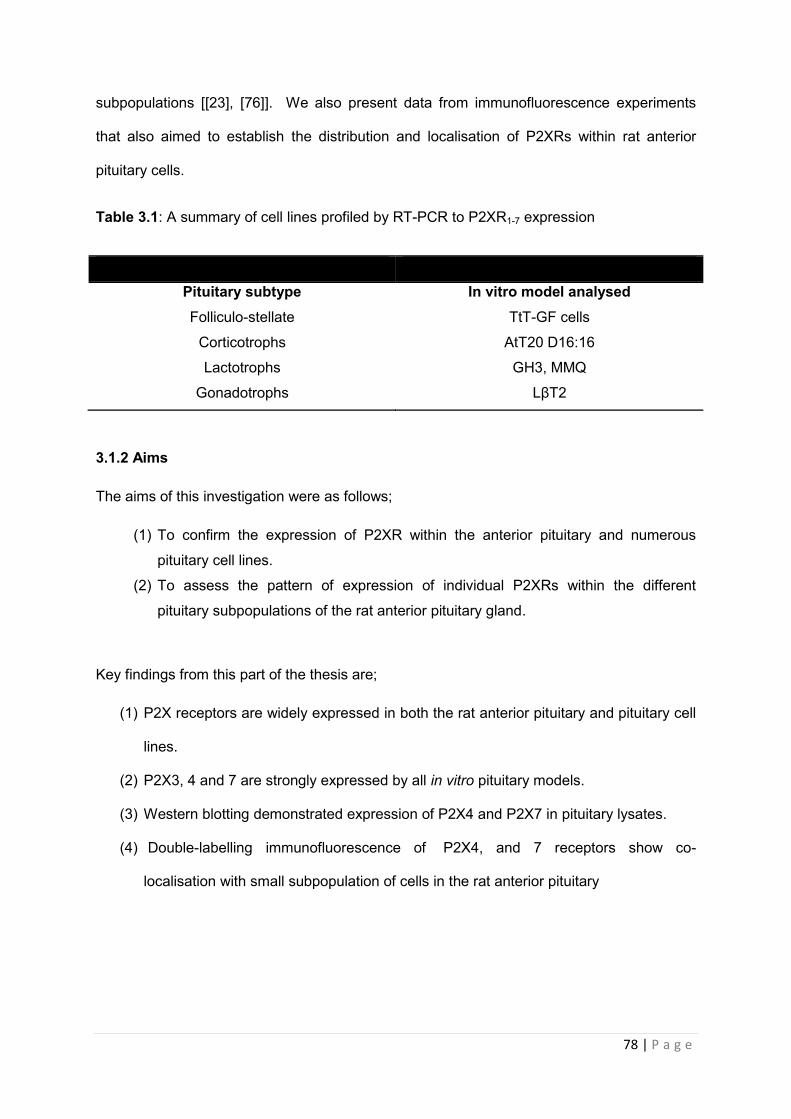
78 | P a g e
subpopulations [[23], [76]]. We also present data from immunofluorescence experiments
that also aimed to establish the distribution and localisation of P2XRs within rat anterior
pituitary cells.
Table 3.1: A summary of cell lines profiled by RT-PCR to P2XR1-7 expression
Pituitary subtype In vitro model analysed Folliculo-stellate TtT-GF cells
Corticotrophs AtT20 D16:16
Lactotrophs GH3, MMQ
Gonadotrophs LβT2
3.1.2 Aims
The aims of this investigation were as follows;
(1) To confirm the expression of P2XR within the anterior pituitary and numerous
pituitary cell lines.
(2) To assess the pattern of expression of individual P2XRs within the different
pituitary subpopulations of the rat anterior pituitary gland.
Key findings from this part of the thesis are;
(1) P2X receptors are widely expressed in both the rat anterior pituitary and pituitary cell
lines.
(2) P2X3, 4 and 7 are strongly expressed by all in vitro pituitary models.
(3) Western blotting demonstrated expression of P2X4 and P2X7 in pituitary lysates.
(4) Double-labelling immunofluorescence of P2X4, and 7 receptors show co-
localisation with small subpopulation of cells in the rat anterior pituitary
79 | P a g e
3.3 RESULTS
3.3.1 Expression of P2X receptor transcripts in rat pituitary by RT PCR
We first studied the expression of the various purinergic receptor mRNAs within rat pituitary
tissue by RT-PCR. As shown in Fig 3.1, a strong signal for mRNA transcripts of P2X2 and
P2X3, P2X4, P2X6 and P2X7 (Figure 3.1A.). A weak signal for P2X1 and P2X5 was also
detected (Figure 3.1A.). For a positive control we utilised rat brain tissue which contain P2X
receptor subtypes 2-7 (Figure 3.1B) and rat kidney tissue was used as a positive control
tissue for P2X1 (Figure 3.1A). The rat brain control tissues expressed the expected P2X
receptor transcripts, in the pituitary however, mRNA transcripts for variants P2X2a and P2X2b
were also detected.
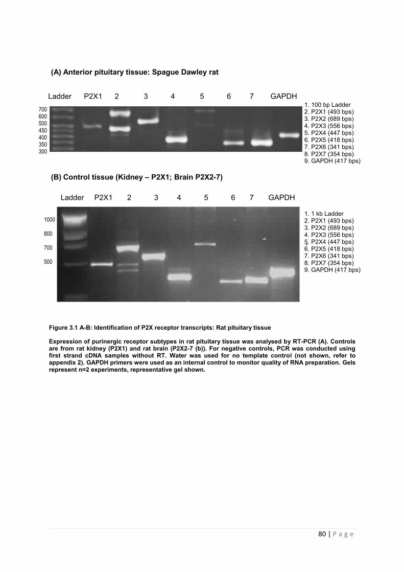
80 | P a g e
(A) Anterior pituitary tissue: Spague Dawley rat
(B) Control tissue (Kidney – P2X1; Brain P2X2-7)
.
Figure 3.1 A-B: Identification of P2X receptor transcripts: Rat pituitary tissue
Expression of purinergic receptor subtypes in rat pituitary tissue was analysed by RT-PCR (A). Controls are from rat kidney (P2X1) and rat brain (P2X2-7 (b)). For negative controls, PCR was conducted using first strand cDNA samples without RT. Water was used for no template control (not shown, refer to appendix 2). GAPDH primers were used as an internal control to monitor quality of RNA preparation. Gels represent n=2 experiments, representative gel shown.
Ladder P2X1 2 3 4 5 6 7 GAPDH
1. 100 bp Ladder 2. P2X1 (493 bps) 3. P2X2 (689 bps) 4. P2X3 (556 bps) 5. P2X4 (447 bps) 6. P2X5 (418 bps) 7. P2X6 (341 bps) 8. P2X7 (354 bps) 9. GAPDH (417 bps)
700 600 500 450 400 350 300
Ladder P2X1 2 3 4 5 6 7 GAPDH
1000 800 700 500
1. 1 kb Ladder 2. P2X1 (493 bps) 3. P2X2 (689 bps) 4. P2X3 (556 bps) 5. P2X4 (447 bps) 6. P2X5 (418 bps) 7. P2X6 (341 bps) 8. P2X7 (354 bps) 9. GAPDH (417 bps)
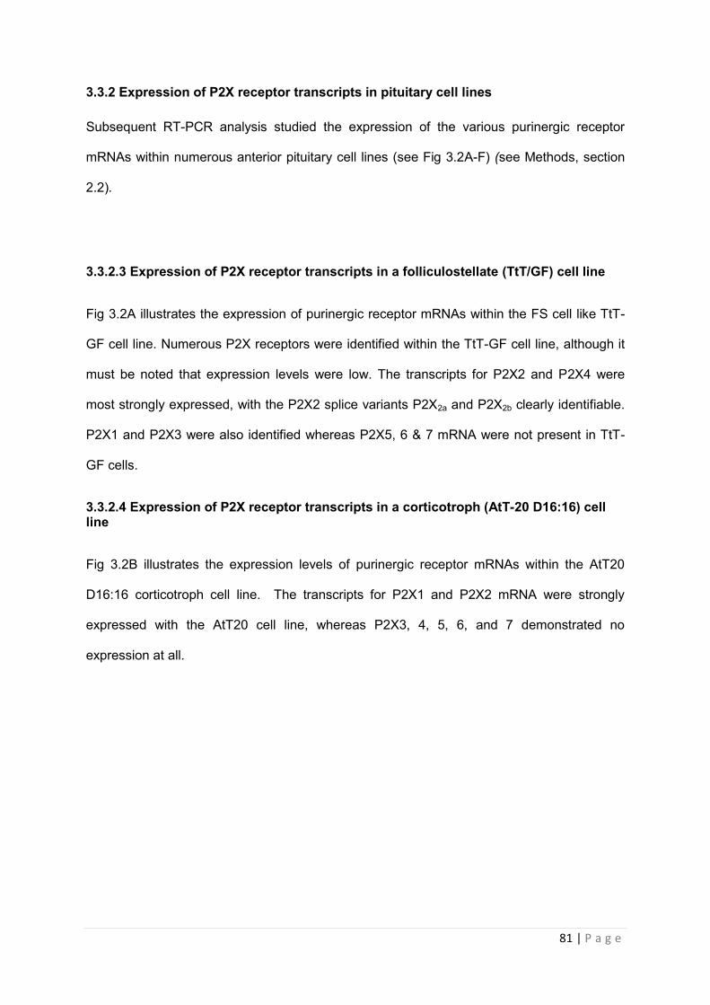
81 | P a g e
3.3.2 Expression of P2X receptor transcripts in pituitary cell lines
Subsequent RT-PCR analysis studied the expression of the various purinergic receptor
mRNAs within numerous anterior pituitary cell lines (see Fig 3.2A-F) (see Methods, section
2.2).
3.3.2.3 Expression of P2X receptor transcripts in a folliculostellate (TtT/GF) cell line
Fig 3.2A illustrates the expression of purinergic receptor mRNAs within the FS cell like TtT-
GF cell line. Numerous P2X receptors were identified within the TtT-GF cell line, although it
must be noted that expression levels were low. The transcripts for P2X2 and P2X4 were
most strongly expressed, with the P2X2 splice variants P2X2a and P2X2b clearly identifiable.
P2X1 and P2X3 were also identified whereas P2X5, 6 & 7 mRNA were not present in TtT-
GF cells.
3.3.2.4 Expression of P2X receptor transcripts in a corticotroph (AtT-20 D16:16) cell line
Fig 3.2B illustrates the expression levels of purinergic receptor mRNAs within the AtT20
D16:16 corticotroph cell line. The transcripts for P2X1 and P2X2 mRNA were strongly
expressed with the AtT20 cell line, whereas P2X3, 4, 5, 6, and 7 demonstrated no
expression at all.
82 | P a g e
(A) TtT-GF cells: Mouse
P2X1 2 3 4 5 6 7 GAPDH
(B) AtT20 D16:16 cells: Mouse
P2X1 2 3 4 5 6 7 GAPDH
Figure 3. 2 (A-B): Identification of P2X receptor transcripts in pituitary cell lines: TtTGF and AtT20 D16:16
Expression of purinergic receptor subtypes in immortalised pituitary cell lines was analysed by RT-PCR. Controls are from rat kidney (P2X1) and rat brain (P2X2-7). For negative controls, PCR was conducted using first strand cDNA samples without RT. Water was used for no template control. GAPDH primers were used as an internal control to monitor quality of RNA preparation. Expression of P2X2 and P2X4 was found in the TtT-GF cells (A). P2X1 and P2X2 were detected in AtT20 D16:16 (B) Gels represent n=2 experiments, representative gel shown.
700 600 500 400
700 600 500 400
83 | P a g e
3.3.2.5 Expression of P2X receptor transcripts in lactotroph (GH3 and MMQ) cell lines
Figure 3.2C illustrates the expression levels of purinergic receptor mRNAs within the GH3
‘somatolactotroph’ cells. Probing the GH3 cells revealed strong expression for P2X3, P2X4
and P2X7 mRNAs (Fig 3.2C) whereas P2X1, 2, 5 and 6 were not detected. Fig 3.2D
illustrates the expression levels of purinergic receptor mRNAs within the MMQ lactotroph-like
cell line. In MMQ cells, P2X3, P2X4 and P2X7 mRNA were strongly expressed which
correlates with expression in the GH3 ‘somatolactotroph’ cells. In contrast to GH3 cells, clear
(albeit weak) expression of P2X2 and P2X5 mRNA was also identified in the MMQ cells (Fig
3.2 D). P2X1 and P2X6 were not detected.
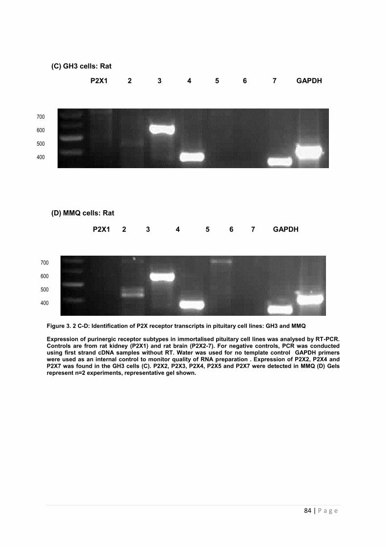
84 | P a g e
(C) GH3 cells: Rat
P2X1 2 3 4 5 6 7 GAPDH
(D) MMQ cells: Rat P2X1 2 3 4 5 6 7 GAPDH
Figure 3. 2 C-D: Identification of P2X receptor transcripts in pituitary cell lines: GH3 and MMQ
Expression of purinergic receptor subtypes in immortalised pituitary cell lines was analysed by RT-PCR. Controls are from rat kidney (P2X1) and rat brain (P2X2-7). For negative controls, PCR was conducted using first strand cDNA samples without RT. Water was used for no template control GAPDH primers were used as an internal control to monitor quality of RNA preparation . Expression of P2X2, P2X4 and P2X7 was found in the GH3 cells (C). P2X2, P2X3, P2X4, P2X5 and P2X7 were detected in MMQ (D) Gels represent n=2 experiments, representative gel shown.
700 600 500 400
700 600 500 400
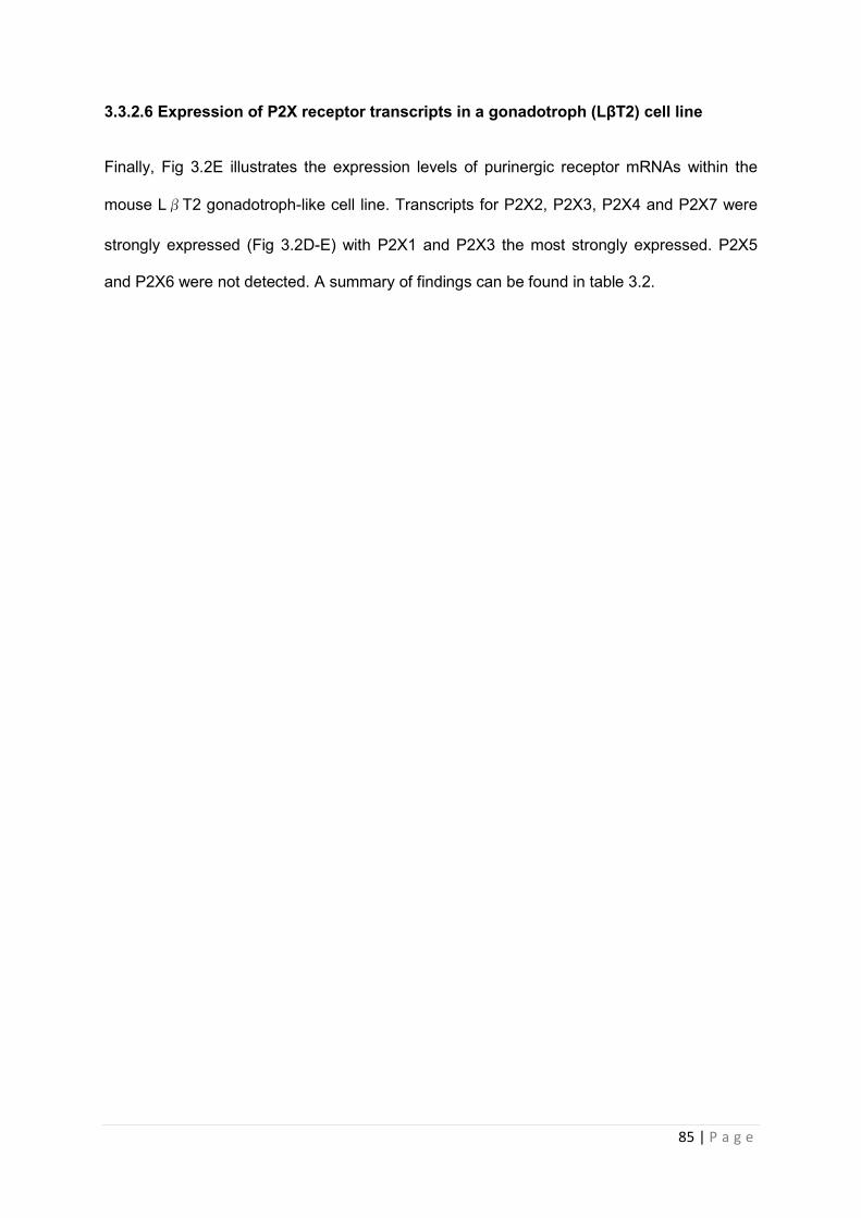
85 | P a g e
3.3.2.6 Expression of P2X receptor transcripts in a gonadotroph (LβT2) cell line
Finally, Fig 3.2E illustrates the expression levels of purinergic receptor mRNAs within the
mouse LβT2 gonadotroph-like cell line. Transcripts for P2X2, P2X3, P2X4 and P2X7 were
strongly expressed (Fig 3.2D-E) with P2X1 and P2X3 the most strongly expressed. P2X5
and P2X6 were not detected. A summary of findings can be found in table 3.2.
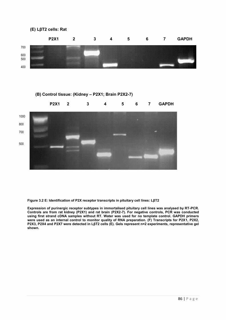
86 | P a g e
(E) LβT2 cells: Rat P2X1 2 3 4 5 6 7 GAPDH
(B) Control tissue: (Kidney – P2X1; Brain P2X2-7) P2X1 2 3 4 5 6 7 GAPDH
Figure 3.2 E: Identification of P2X receptor transcripts in pituitary cell lines: LβT2
Expression of purinergic receptor subtypes in immortalised pituitary cell lines was analysed by RT-PCR. Controls are from rat kidney (P2X1) and rat brain (P2X2-7). For negative controls, PCR was conducted using first strand cDNA samples without RT. Water was used for no template control. GAPDH primers were used as an internal control to monitor quality of RNA preparation. (F) Transcripts for P2X1, P2X2, P2X3, P2X4 and P2X7 were detected in LβT2 cells (E). Gels represent n=2 experiments, representative gel shown.
700 600 500 400
1000 800 700 500

87 | P a g e
Table3.2. Summary of P2X transcripts detected in rat pituitary tissue and pituitary cell lines
P2X Rat
Pituitary FS AtT20:D16 GH3 MMQ LβT2
P2X1 + - + - - -
P2X2 + W + - W +
P2X3 + W - + + +
P2X4 + - - + + +
P2X5 W - - - - -
P2X6 + - - - - -
P2X7 + - - + + +
Key
Expression (+) No expression (-) Weak expression (W)
88 | P a g e
3.3.2 Characterisation of purinergic receptor expression in whole pituitary tissue using Western blot
mRNA for P2X receptors 1 to 7 were identified in whole rat pituitary tissue using RT-PCR. In
order to verify expression of P2X receptors at the protein level, Western blot analysis (and
immunoflouresces see section 3.3.3) of rat pituitary protein extracts was conducted. This
was also to be used as a control experiment to validate the specificities of anti-P2X receptor
antibodies to be used in later studies. We initially chose to validate the antibodies for P2X3,
4, & 7 since these receptors were the most consistently expressed across the various
pituitary cell lines investigated.
We utilised protein extracts from rat brain as a positive control for the expression of P2X
receptor protein for P2X3 (approximately 65 kDa), P2X4 (approximately 65 KDa) and P2X7
(approximately 68KDa). Rat pituitary lysate that was also subjected to Western blot analysis
showed the presence of P2X4 (figure 3.3B) and P2X7 (figure 3.3C) receptor protein within
rat pituitary. How P2X 3 (approximately 65 kDa) was identified in brain tissue but not in the
pituitary gland (figure 3.3A). In all cases differences in glycosylation may explain differences
in protein size between brain and pituitary lysates . Summary of results can be found in table
3.3.
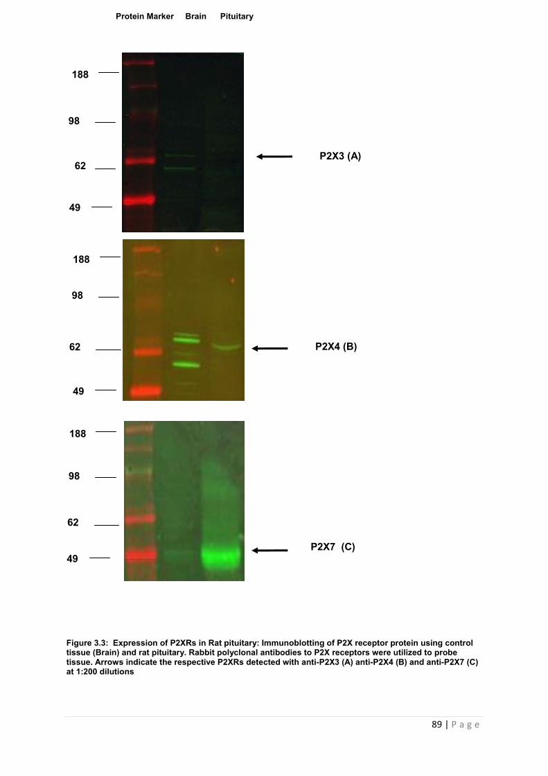
89 | P a g e
Figure 3.3: Expression of P2XRs in Rat pituitary: Immunoblotting of P2X receptor protein using control tissue (Brain) and rat pituitary. Rabbit polyclonal antibodies to P2X receptors were utilized to probe tissue. Arrows indicate the respective P2XRs detected with anti-P2X3 (A) anti-P2X4 (B) and anti-P2X7 (C) at 1:200 dilutions
Protein Marker Brain Pituitary
188
98
62
188
98
62
49
98
62
49
188
P2X3 (A)
P2X4 (B)
P2X7 (C)
49
90 | P a g e
Table 3.3: Summary of P2XR immunoreactivity detected in rat pituitary tissue and brain
tissue
Key
Expression (+); No expression (-)
3.3.3 Optimisation of immunoflourescent techniques for the detection and characterisation of purinergic receptors in whole pituitary tissue: Co-expression of P2X receptors with pituitary hormones.
The only study currently in the literature characterising P2 receptor localisation in rat
pituitary subtypes utilizes P2Y targeted antibodies. The following section includes our results
from attempts to characterise pituitary P2X receptor expression in a similar fashion. P2X3
antibody (alomone) showed no expression and was omitted from our study. Sprague Dawley
rat pituitary tissue was incubated with antibodies for P2X4 or 7 and pituitary hormones
ACTH, FSH, GH, PRL and TSH to determine colocalisation in the different pituitary
subtypes.
3.3.3.1 Coexpression of P2X4 receptor with ACTH, FSH, GH, PRL and TSH
Single staining showed P2X4 to have weak to moderate staining in pituitary tissue (Figure
3.4 A). Double-labelling immunofluorescence showed no true immunoreactive for between
ACTH (3.4A-B), GH (3.5E-F), PRL (3.4G-H) and TSH (3.4I-J), FSH (3.5 C-D) and P2X4.
P2X Rat Brain Rat Pituitary
P2X3 + -
P2X4 + +
P2X7 + +
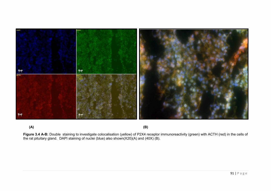
91 | P a g e
(A) (B)
Figure 3.4 A-B: Double staining to investigate colocalisation (yellow) of P2X4 receptor immunoreactivity (green) with ACTH (red) in the cells of the rat pituitary gland. DAPI staining of nuclei (blue) also shown(X20)(A) and (40X) (B).
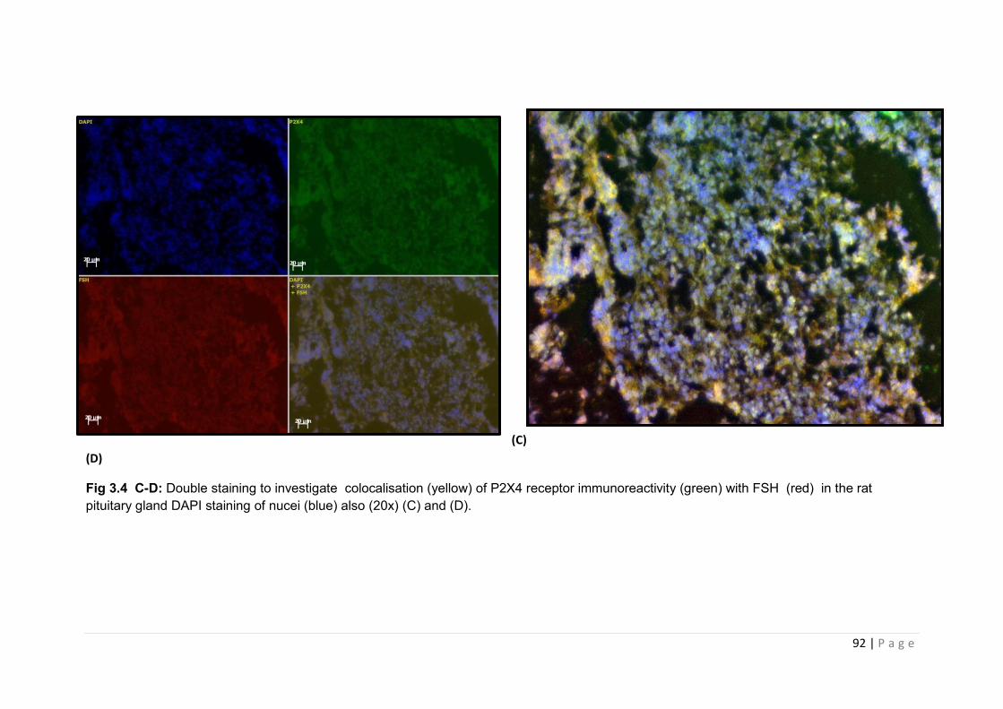
92 | P a g e
(C)
(D)
Fig 3.4 C-D: Double staining to investigate colocalisation (yellow) of P2X4 receptor immunoreactivity (green) with FSH (red) in the rat pituitary gland DAPI staining of nucei (blue) also (20x) (C) and (D).
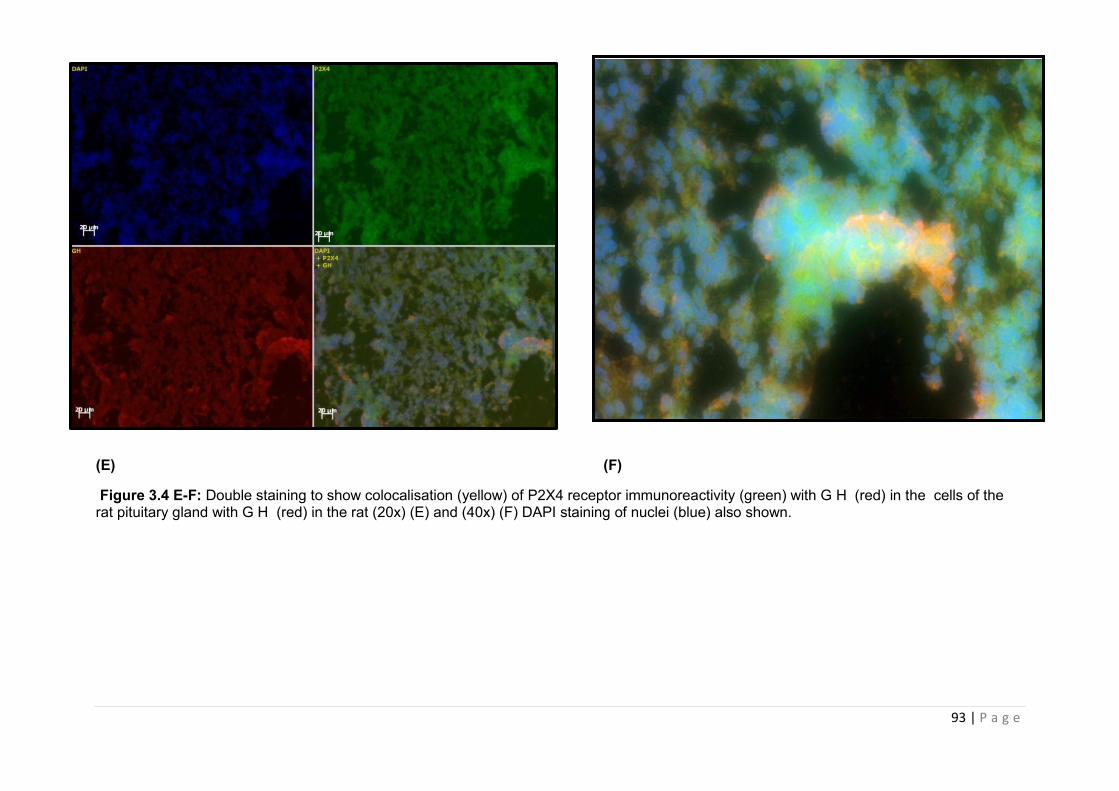
93 | P a g e
(E) (F)
Figure 3.4 E-F: Double staining to show colocalisation (yellow) of P2X4 receptor immunoreactivity (green) with G H (red) in the cells of the rat pituitary gland with G H (red) in the rat (20x) (E) and (40x) (F) DAPI staining of nuclei (blue) also shown.
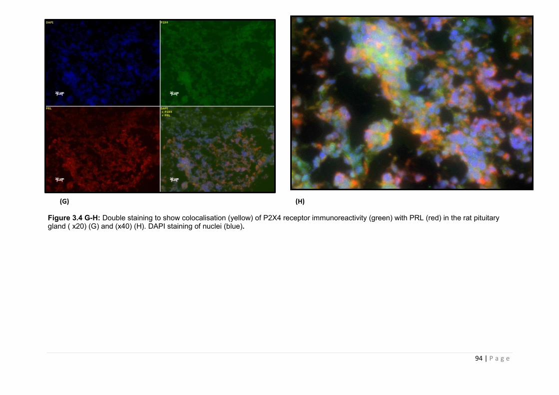
94 | P a g e
(G) (H)
Figure 3.4 G-H: Double staining to show colocalisation (yellow) of P2X4 receptor immunoreactivity (green) with PRL (red) in the rat pituitary gland ( x20) (G) and (x40) (H). DAPI staining of nuclei (blue).
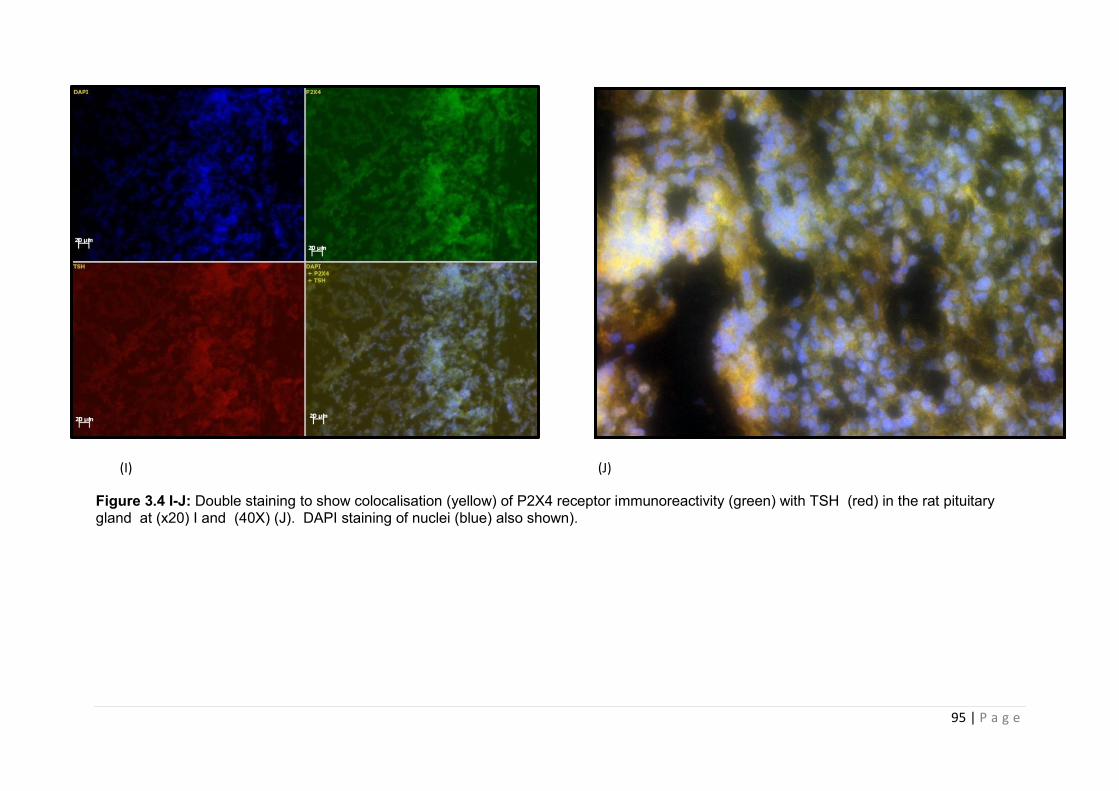
95 | P a g e
(I) (J)
Figure 3.4 I-J: Double staining to show colocalisation (yellow) of P2X4 receptor immunoreactivity (green) with TSH (red) in the rat pituitary gland at (x20) I and (40X) (J). DAPI staining of nuclei (blue) also shown).
96 | P a g e
3.3.3.2 Coexpression of P2X7 receptor with ACTH, FSH, GH, PRL and TSH
P2X7 showed very low levels of staining in the anterior pituitary tissue (figure 3.5 A-F).
Double staining with the nuclear stain DAPI showed P2X7 to be weakly immunoreactive in
the pituitary tissue (Figure 3.5 A). Double-labelling immunofluorescence showed no
colocalisation was seen with ACTH (3.5A), FSH (3.5 B), PRL (3.5 E), TSH (3.5 F), GH
(3.5C-D).
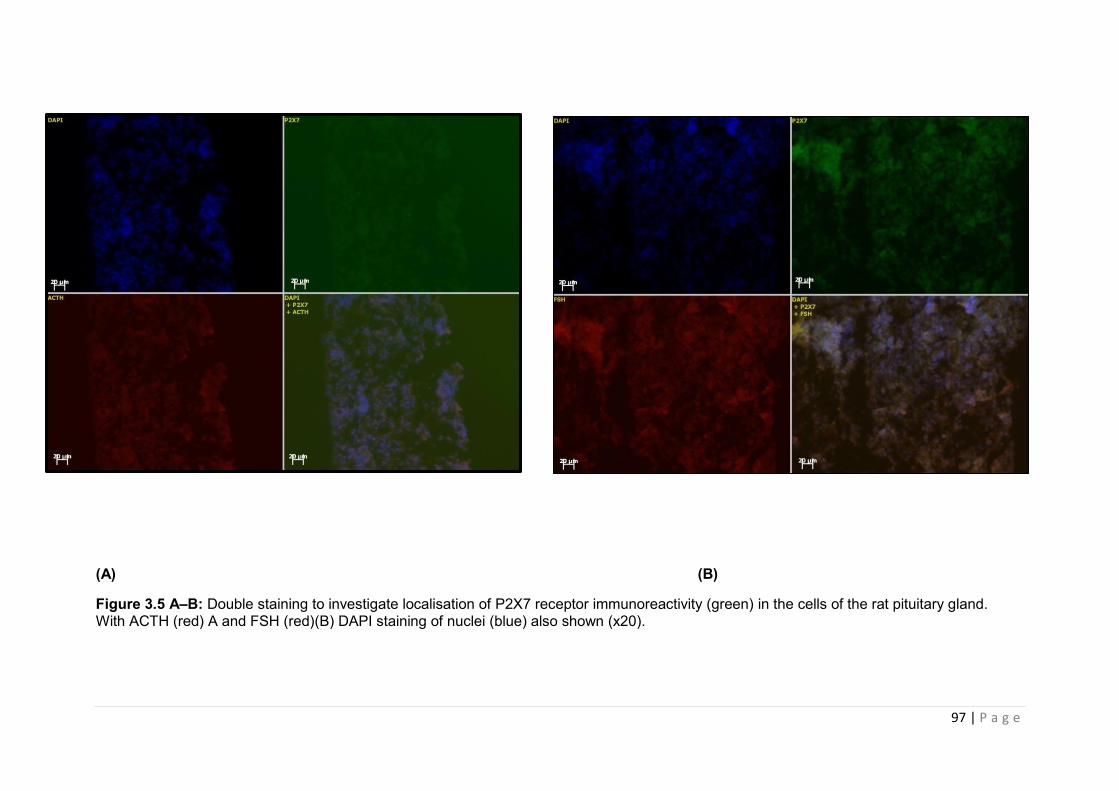
97 | P a g e
(A) (B)
Figure 3.5 A–B: Double staining to investigate localisation of P2X7 receptor immunoreactivity (green) in the cells of the rat pituitary gland. With ACTH (red) A and FSH (red)(B) DAPI staining of nuclei (blue) also shown (x20).
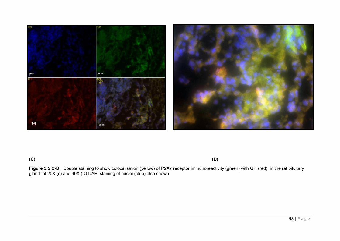
98 | P a g e
(C) (D)
Figure 3.5 C-D: Double staining to show colocalisation (yellow) of P2X7 receptor immunoreactivity (green) with GH (red) in the rat pituitary gland at 20X (c) and 40X (D) DAPI staining of nuclei (blue) also shown
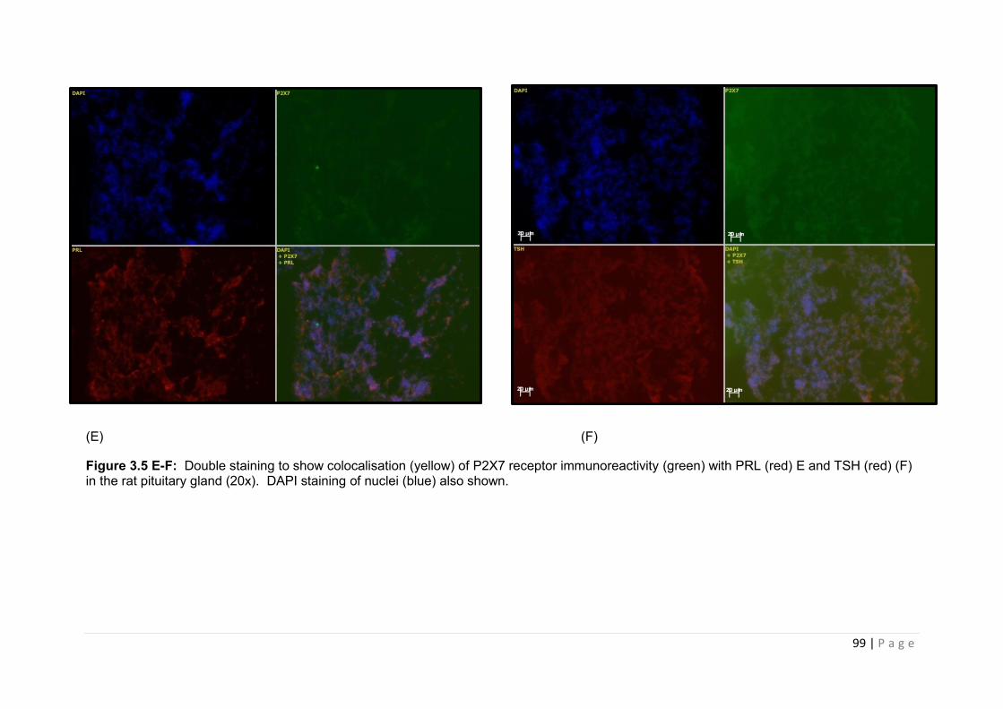
99 | P a g e
(E) (F)
Figure 3.5 E-F: Double staining to show colocalisation (yellow) of P2X7 receptor immunoreactivity (green) with PRL (red) E and TSH (red) (F) in the rat pituitary gland (20x). DAPI staining of nuclei (blue) also shown.
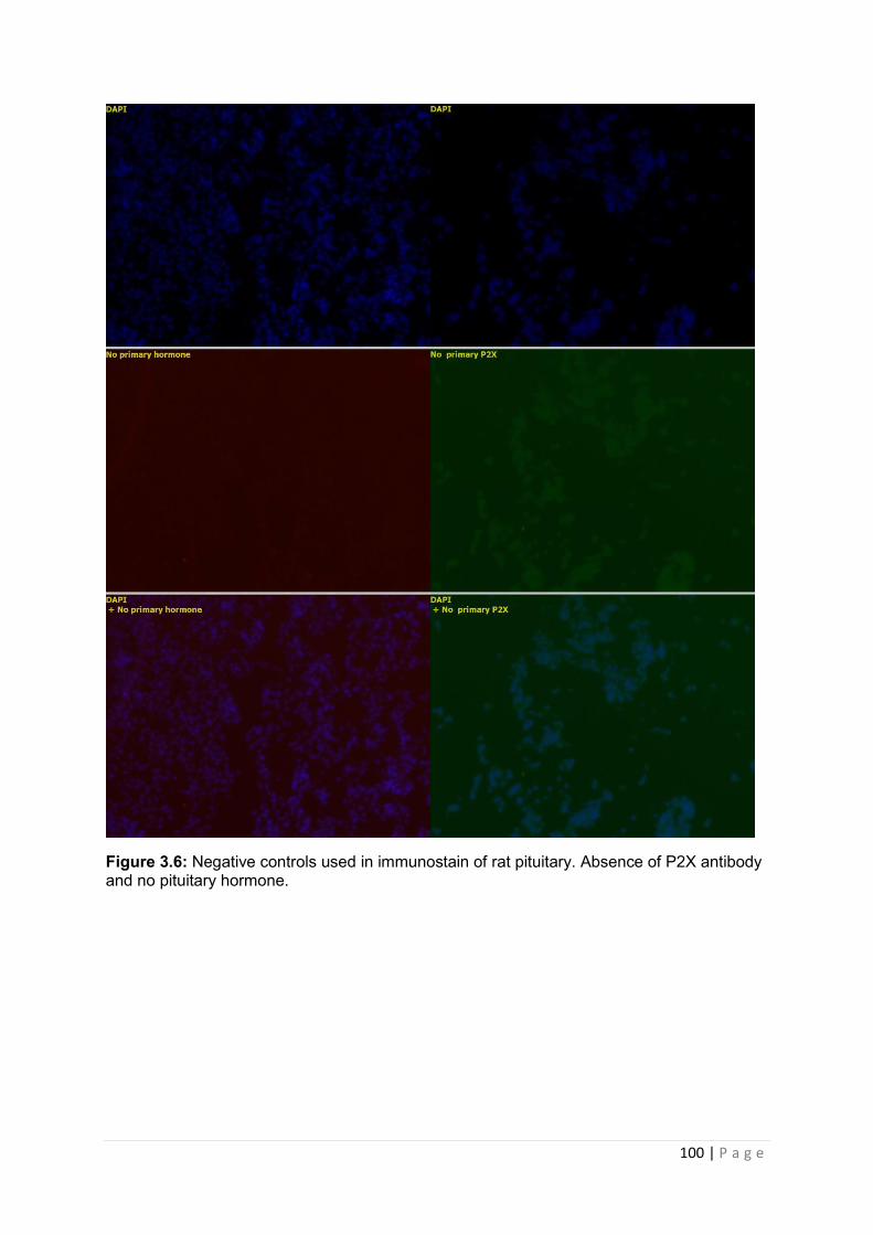
100 | P a g e
Figure 3.6: Negative controls used in immunostain of rat pituitary. Absence of P2X antibody and no pituitary hormone.
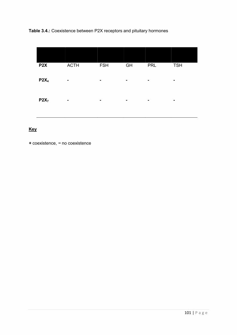
101 | P a g e
Table 3.4.: Coexistence between P2X receptors and pituitary hormones
Key
+ coexistence, − no coexistence
P2X ACTH FSH GH PRL TSH
P2X4 - - - - -
P2X7 - - - - -
102 | P a g e
3.4 Discussion
The results of the experiments presented in this chapter were designed to investigate the
pattern of P2X receptor expression within the pituitary. As demonstrated by RT- PCR
analysis, transcripts for P2X1-7 were expressed in the rat pituitary gland. This confirms
previous data concerning P2X receptor expression within the pituitary gland using Sprague
Dawley rats [[98]], with the exception of P2X5 which we demonstrated was highly expressed
in the pituitary, whereas the Koshimizu study failed to show P2X5 expression. We were also
able to detect P2X7 within rat pituitary tissue using qualitative RT-PCR [[98] [1]]. This is
significant, given a recent study using the more sensitive Q-PCR technique demonstrated
that amongst the various P2XR subtypes expressed in the rat pituitary, P2X7 is one of the
receptors with the lowest levels of expression [64]. Zemkova and colleagues used anterior
pituitary tissue only to determine levels of P2X receptor expression. P2X5 has been identified
in the posterior pituitary where it has been demonstrated to modulate oxytocyin release[[73]]
In our study we used whole pituitary to subject to RT-PCR, therefore our detection of P2X5
may represent expression of in the posterior pituitary. In accordance with previous studies,
our RT-PCR analysis identified two bands for P2X2 within the rat pituitary. The expression of
the wild type channel, P2X2a, and the spliced variant, P2X2b, have been identified in pituitary
tissue, as well as brain, kidney, spleen and intestine. Identification of P2X2 variants within
pituitary tissue is important, particularly with regard to subsequent functional analysis.
Evidence suggests that the two variants show different rates of desensitisation to ATP [100],
[92]], which would impact on the development of in vitro assays for pituitary function in
relation to P2XR. We utilised whole pituitary tissue for our expression studies, which is
consistent with previous investigations of purinergic receptors currently in the literature [[96]].
However, the failure to remove the posterior pituitary and to ensure the removal of
contaminating blood cells could lead to the reporting of false positives. Upon reviewing our
method for the preparation of pituitary tissue, although care was taken to remove blood prior
to RNA extraction, traces of contaminates in samples could potentially remain and also be a

103 | P a g e
source of false positive due to expression of P2X receptors in the blood and immune cells
contained within [1]. Perfusion of tissue with saline prior to extraction of pituitary would limit
this and has been used to great effect [102].
Our data demonstrate the expression of P2X1 and P2X2 mRNA within our corticotroph cell
line. A previous study investigated the expression of P2XR within the AtT-20 corticotroph cell
line, albeit using a different subclone (AtT-20 PL). RT-PCR analysis within this study
identified the presence of P2X3, P2X4, P2X6 and P2X7 transcripts in the AtT-20 cells [61].
Whilst there are significant differences between the two subclones, specifically a much
slower growth rate but a much quicker responsiveness of POMC promoter activity [103] this
does not explain the contrasting expression data between the two cell lines. In the literature,
the D16 subclone has been more extensively used , particularly for in vitro investigations of
stimulus induced ACTH release from corticotrophs [[104], [105]]. One could therefore
speculate that the corticotroph model used in our study is a more representative model of
this pituitary subtype. Unfortunately, available functional data does not indicate the
importance of individual P2X subtypes within corticotroph cells, since stimulation of P2XRs
within these cell types with available agonists does not influence the endocrine secretion of
ACTH [61].
As demonstrated by PCR analysis, transcripts for P2X2 and P2X4 were found to be
expressed in the TtT/GF folliculostellate cell line. To our knowledge this is the first time that
folliculstellate cells have been probed for P2XRs. It is interesting to note that P2X4 is
associated with immunological cells such as macrophages and stem cells [3], since
folliculostellate cells are often regarded as pituitary tissue specific macrophage like cells
derived from the lymphoid dendritic cell lineage. However, the fact that P2X2 and P2X4 are
only weakly expressed in TtT/GF cells in our studies is suggestive of a minor role in
regulating folliculostellate cell function. Current data concerning functional purinergic
signalling within folliculostellate cells indicates that responses are primarily driven by

104 | P a g e
adenosine [2], with adenosine receptor activation linked with IL-6 secretion. In addition P2Y2
receptors may also have a functional role in folliculostellate cells.
Molecular identification of P2XRs expressed within the lactotroph cell lines, GH3 and MMQs,
was also obtained by RT-PCR analysis. Transcripts encoding P2X3, P2X4 and P2X7 were
detected in both cell lines. With regard to GH3 cells, our data is in agreement with previous
studies using this cell line as well as data from molecular profiling of enriched lactotroph cell
populations [73]. RT-PCR analysis of the MMQ cell line also demonstrated weak expression
for P2x2 and P2x5 mRNA. To our knowledge this is first time this specific lactotroph cell line
has been probed for P2XR expression. MMQs are similar to primary tissue with regards to
the expression of a functional dopaminergic system and receptors for oxytocin [81]. As in
native pituitary tissue, secretions from MMQ cells are under the control of calcium and other
secondary messengers such as cAMP [81]. GH3 cells are established in vitro models of
lactotrophs, although its use as a direct model of the normal pituitary function is limited by
the absence of dopamine receptors and observations suggesting an abnormal
storage/release mechanism for PRL [4]. The difference in physiology may in part account for
the differences in P2X expression [75]. P2X5 has been associated with a range of cancer
states including prostate and skin cancer [108]. It is possible that expression of this receptor
within the MMQ cell line is related to the fact that this cell line is derived from a pituitary
adenoma [81]. This is an issue which of course is true for all immortalized cells lines used in
this investigation . It is important that in vitro systems used to assess purinergic function
within the pituitary are representative models of the native pituitary system we are trying to
investigate. As the result for MMQ illustrates, molecular profiling of cancerous tissue is vital
to ensure this is the case. Functional assays to assess purinergic function in the human
pituitary system may utilise a mixture of normal and cancerous tissue and profiling will be
conducted to ensure expression of receptors within dysregulated tissue is comparable to
normal tissue.
105 | P a g e
RT-PCR analysis revealed that P2X3, P2X4, P2X7 and P2X2 (weakly) are expressed in the
gonadotroph cell line, LT2. This is the first time that P2XR expression has been analysed
within a specific population of gonadotroph cells. Although there is no direct PCR data on
this cell line, our findings are in accordance with data from the characterisation of primary
gonadotrophs by Zemkova and co-workers who demonstrated P2X1, P2X2R, P2X3R, P2X4R
and P2X7R expression in both neonatal and 7 day old rats [34]. Our western blotting data
for P2X4 and P2X7 ( figure 3.3) was in accordance with previous studies [[64], [109]].
However in this study we were able to demonstrate expression in native pituitary tissue as
opposed to recombinant expression in an artificial system [109]. We detected double band
for expression of P2X3 and P2X4 in our control tissue, rat brain lysates. P2X4 bands
corresponding to ~ 50 and 65 kDa were detected in control tissue. These bands represent
expression of the unglcosylated and glycosylated forms of the receptor in both tissues. This
was also seen with P2X3 expression in the brain where two bands corresponding to ~60 and
70 kDa represent the unglycosylated and glcosylated variants respectively. Expression of
multiple bands for individual receptors has been observed in a range of tissues and is a
consequence of the variations in glycosylation and oligomerisation that occurs between
P2XR subtypes. Predicted sizes for receptors range from 45- 64kDa for unglycosylated
forms and 50-70kDa for the glycosylated forms [110]. Western blot analysis, along with
immunoprecipitation has suggested that a functional interaction between the two receptors
may occur [111]. Whilst our study cannot support this finding, this does suggest that
investigation of the level of colocalisation between different P2X receptors within the pituitary
tissue is an important area to explore. This could provide vital evidence as to the extent with
which different P2XRs exert regulatory effects on one another – something that has been
identified in other tissues, such as the smooth muscle and salivary glands [[112], [113],
[114]] Results from double immunoflourescence staining of P2XRs and the relevant
pituitary hormone demonstrated that corticotrophs, gonadotrophs, lactotrophs and
thyrotrophs express at least one member of the P2X family, but this is the first time that
P2XRs have been investigated for co-localisation with specific pituitary hormones within
106 | P a g e
specific pituitary cell types and this has provided new insight into the pattern of P2X
distribution within the pituitary. P2X4 was co-localised within the greatest number of pituitary
cell types (figure 3.5) This is not surprising given that P2X4 demonstrates the highest level of
expression in the rat pituitary gland within other studies[64]. Histological staining has also
identified expression within the adrenal gland [115] and studies using immunoflouresence
have indicated that P2X4 plays a fundamental role in the regulation of exocytosis of cellular
contents [116]. Given that we also found P2X4 widely distributed amongst pituitary cells, it is
possible that P2X4 possesses a similar role in the pituitary, although further
electrophysiological studies would be required to confirm this. P2X7 showed the least
immunoreactivity in the pituitary tissue (3.5). Expression of P2X7 within other tissues is
generally associated with apoptotic mechanisms, and the release of cytokines from immune
derived cells [[117], [118], [119], [120], [121], [122]] .Previous studies to identify a functional
role for P2X7 in the pituitary have shown that loss of P2X7 expression, using P2X7
knockout mice, has been associated with no alteration in the release of the pituitary
hormones, particularly the posterior pituitary hormones oxytocin and vasopressin [123]. The
fact that we have only shown low levels of pituitary P2X7 expression in our studies coupled
with low P2X7 expression demonstrated within the pituitary in previous studies [[64]]
suggests that P2X7 may not play as significant a role within the anterior pituitary compared
with the other P2X receptors expressed. Chapter 4 includes functional data that focuses on
the influence of individual P2XRs on anterior pituitary function.
We were unable to detected P2X3 after conducting western blot of rat pituitary lysates
(3.3A). This was surprising given that P2X3 had demonstrated to be expressed in the
cellular subpopulations of other endocrine organs. Western blotting had identified P2X3
expression in human islets and immunofluorescence techniques confirmed co-localisation of
P2X3 within insulin secreting -cells in human tissue in vitro [[124]]. There is evidence to
show that expression of P2X3 receptors changes with age within the rat brain and the
adrenal gland [[125], [115]]. Post-natally, expression of P2X3 is restricted to the bladder,
107 | P a g e
spinal cord and cerebellum of the rat [[126], [125]], This pattern of post-natal down regulation
of P2X3 receptor within the CNS and other organs of the rat may explain our inability to
detect P2X3 expression in our pituitary lysates . However, as we were able to detect P2X3 in
our control lysate (brain) could suggest that protein expression of this receptor in the pituitary
tissue used may be very low and beyond the detect limits of assay. This issue has been
overcome by other groups that have used separate expression systems such as GT1 cells
or xenapus oocytes, to artificially express receptor, thus ensuring a high levels expression
[64]. However, as we were interested in expression of the receptor in the native system, the
antibody may require further optimisation and/or increased amount of protein for use in
western blot.
3.5 Conclusions
Our investigations of P2XR expression in vitro support the published evidence and show that
P2XRs is a highly expressed protein component of mammalian pituitary tissue which we has
demonstrated to show species dependent differences (Rat, mouse). Characterisation of P2X
receptor expression within the whole pituitary and numerous pituitary cell lines by RT-PCR,
demonstrated that all P2XRs were expressed in pituitary tissue with P2X3, 4 and 7 most
commonly expressed within the pituitary cell lines. In the future, expression profile studies of
P2X receptors within the pituitary should also include investigations of human tissue. P2X
receptors are proving to be viable site of intervention in human health, and further
knowledge with regards to localisation and expression in the pituitary would be
advantageous. These expression profile studies provide the basis for future assessments of
P2X receptor targeted agents, which should take into account these species differences in
expression when assessing specificity and potency.

108 | P a g e
Chapter FOUR
Investigation of P2X receptor antagonists: Effects on pituitary function in vitro
109 | P a g e
4.1 Introduction
Whilst it is the case that a range of ATP analogues have been developed which specifically
target individual P2X receptors, most of these agents also act as partial agonists for other
members of the P2X family. However, a number of specific P2X receptor antagonists have
now been generated which might provide more conclusive evidence with regard to the
importance of individual P2X receptors in regulating pituitary function. I have demonstrated
that the rat pituitary gland expresses all P2X family members, and that these receptors
demonstrate co-localisation with hormones associated with the different pituitary subtypes to
varying degrees (see chapter 3). In this chapter I investigated the functional significance of
P2X receptors expressed in the pituitary gland using a range of ATP analogues and a
number of specific P2X receptor antagonists in order to assess the contribution made by
individual P2X receptors to pituitary hormone release in vitro.
4.1.1 Investigation of P2X receptor pharmacology in vitro
The native agonist of P2X receptors is ATP, however the development of ATP analogues
have proved to be useful tools in the study of the functional significance of individual P2X
channels. The majority of the data that has enhanced our understanding of P2X receptor
physiology has come from studies that have compared the interaction of ATP to that
observed with chemical ATP analogues such as αβ-methylene-ATP (αβmeATP) , 2',3'-O-(4-
benzoylbenzoyl)-ATP (BzATP) (See table 4.1 for list of ATP analogues ). ATP is susceptible
to enzyme degradation, and the use of non-hydrolysable isoforms has also allowed for
observations to be made over extended time points [[65], [127]]. The relative sensitivities of
the P2XR subtypes for ATP and the various ATP analogues are summarised in Table 4.1.
Table 4.2 Summarises IC50 data for ATP antagonists acting at the various P2XR subtypes.
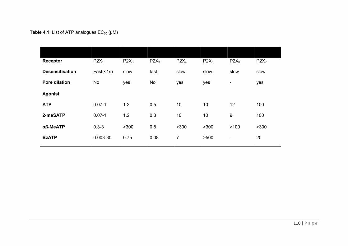
110 | P a g e
Table 4.1: List of ATP analogues EC50 (µM)
Receptor P2X1 P2X 2 P2X3 P2X4 P2X5 P2X6 P2X7
Desensitisation Fast(<1s) slow fast slow slow slow slow
Pore dilation No yes No yes yes - yes
Agonist
ATP 0.07-1 1.2 0.5 10 10 12 100
2-meSATP 0.07-1 1.2 0.3 10 10 9 100
αβ-MeATP 0.3-3 >300 0.8 >300 >300 >100 >300
BzATP 0.003-30 0.75 0.08 7 >500 - 20
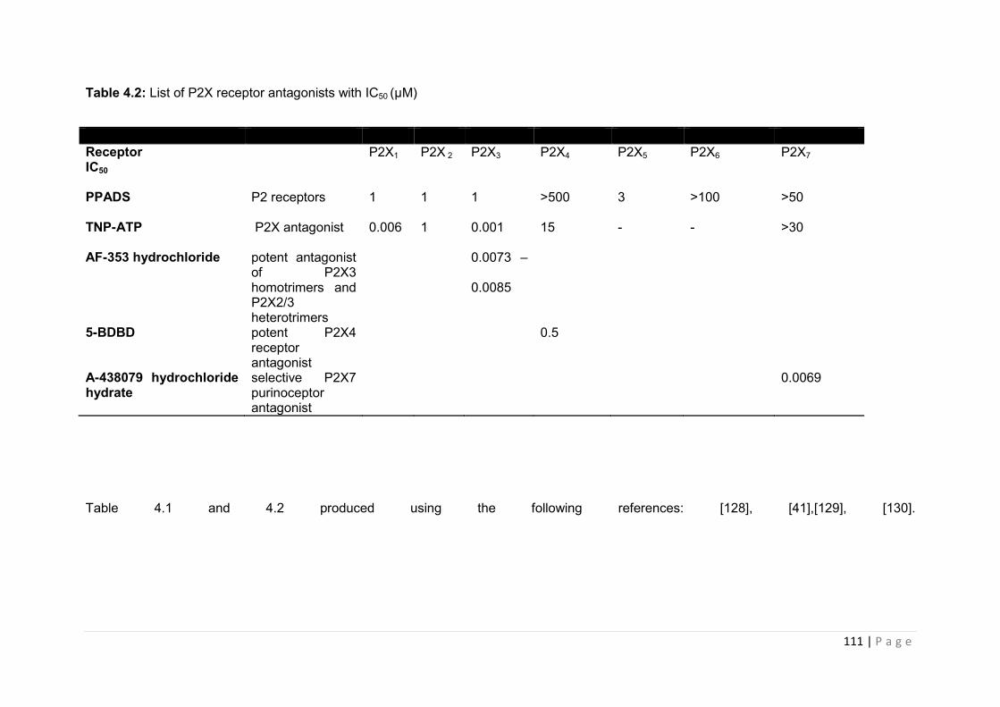
111 | P a g e
Table 4.2: List of P2X receptor antagonists with IC50 (µM)
Receptor P2X1 P2X 2 P2X3 P2X4 P2X5 P2X6 P2X7 IC50
PPADS P2 receptors 1 1 1 >500 3 >100 >50
TNP-ATP P2X antagonist 0.006 1 0.001 15 - - >30
AF-353 hydrochloride potent antagonist of P2X3 homotrimers and P2X2/3 heterotrimers
0.0073 –
0.0085
5-BDBD potent P2X4 receptor antagonist
0.5
A-438079 hydrochloride hydrate
selective P2X7 purinoceptor antagonist
0.0069
Table 4.1 and 4.2 produced using the following references: [128], [41],[129], [130].
112 | P a g e
As can be seen in Table 4.1 each of the various ATP analogues is capable of activating all of
the P2XR subtypes. These compounds have been useful tools in electrophysiology studies,
highlighting the role of P2X receptor mediated (and P2Y) calcium release, and the
associated depolarisation of the pituitary cell membrane prior to hormone
release[[74][131];[132][96]]. However this lack of true specificity makes it difficult to delineate
the role of individual receptors in hormone release within the pituitary subpopulation.
In contrast, selective P2XR antagonists have been generated which provide a more useful
tool for identifying the cell-specific physiological function of individual P2XRs.
Pyridoxalphosphate-6-azophenyl-2',4'-disulfonic acid (PPADS) has been described in the
literature as a broad spectrum P2 antagonist. It has the ability to block all members of the
P2XR family, however it has also been reported to show activity at both P2Y4 and P2Y6
[[133],[134, 135];[136]]. 2',3'-O-(2,4,6-Trinitrophenyl)adenosine-5'-triphosphate
tetra(triethylammonium) (TNP-ATP) is the latest in the line of broad spectrum P2XR
inhibitors, showing high affinity for all P2X receptors, albeit with some evidence for greater
selectivity for P2X1, P2X3 and heterometric P2X2/3 [[137], [138], [139]] There is mounting
clinical evidence supporting the importance of P2X receptors in a range human of
pathologies. The pharmaceutical industry has therefore responded with the development of
highly selective and highly potent P2X receptor antagonists [ [140],[141] [142]].
Recent studies using Q-PCR and electrophysiology have determined receptor subtypes
P2X2, P2X3 and heteromeric P2X2/3 play a key role in the relaying of painful stimuli to the
brain [ [143, 144], [145]], and thus the development of specific P2X3 inhibitors, e.g. AF-353
generated by Roche [146] are specifically targeted to treat neuropathic pain. Considerable
effort has been directed towards the development of selective P2X7 receptor antagonists
due to the fact that ATP-induced activation of P2X7R has been associated with numerous
disease pathophysiologies, for example P2X7R is believed to mediate some of the
113 | P a g e
neuroinflammation that contributes to the development of Alzheimer’s disease (AD) [ [147],
[148]].
Clearly the evidence supports the use of drugs that modulate P2XR activity as a therapeutic
intervention in a range of situations. As we have shown in the previous chapter, the pituitary
gland demonstrates the highest level of P2XR expression and therefore it is extremely
important that we understand how interference with purinoceptor function may potentially
influence homeostasis via actions within the hypothalamo-pituitary system.
The data presented in this chapter focuses on the effects of P2XR antagonists on pituitary
function, using primary pituitary cells and an established pituitary cell line model of
corticotrophs, AtT20:D16s and in vitro model of lactotrophs, GH3, which have both been
used by investigators to assess the contribution of ATP on pituitary hormone function
[[131],[61]].
4.2 Aims
The studies in this chapter aim to investigate the following:-
1. To determine the effects of P2XR stimulation on hormone release in vitro using
primary pituitary cultures and cell lines (AT20:D16 and GH3).
2. To evaluate the in vitro the effects specific P2XR antagonists on pituitary cell
function.
Key findings from this study are;
1. ATP stimulated the release of pituitary hormone from primary rat pituitary cultures
and pituitary cell lines in vitro in a dose dependant manner.
2. Effects of pituitary secretagogues, CRH and TRH were constantly potentiated when
administered with 100µM ATP.
3. Effects of secretagogue and/or ATP on hormone release from GH3 cells were
significantly decreased when pretreated with P2X antagonists PPADS, TNP-ATP,
AF-353 and A-438079.
114 | P a g e
4.3 Results
4.3.1 Validation of rat primary pituitary culture by RIA
We generated a rat primary pituitary culture and carried out preliminary studies to validate
this system with an assessment of the functional response to a well-established pituitary
stimulant. In this regard, the pituitary cultures were treated with CRF in the presence and
absence of the CRF antagonists R121919 and DMP904.
4.3.1.1 Effects of the CRF inhibitor R121919 on ACTH release from primary pituitary culture
ACTH secretion from primary rat pituitary cultures was significantly increased following 4hr
incubation with CRF (100nM CRF, p<0.001 vs control figure 4.1). Pre-incubation with the
CRF receptor antagonist R121919 or DMSO alone did not significantly influence basal
ACTH release. The CRF receptor antagonist, at all concentrations tested (1,5,10, 50 and
100nM), significantly decreased CRF induced ACTH release from primary pituitary cultures
(P<0.05 vs CRF and P<0.001 vs CRF ;figure 4.1A), with a maximal seven-fold decrease in
ACTH release (50nM R121919).
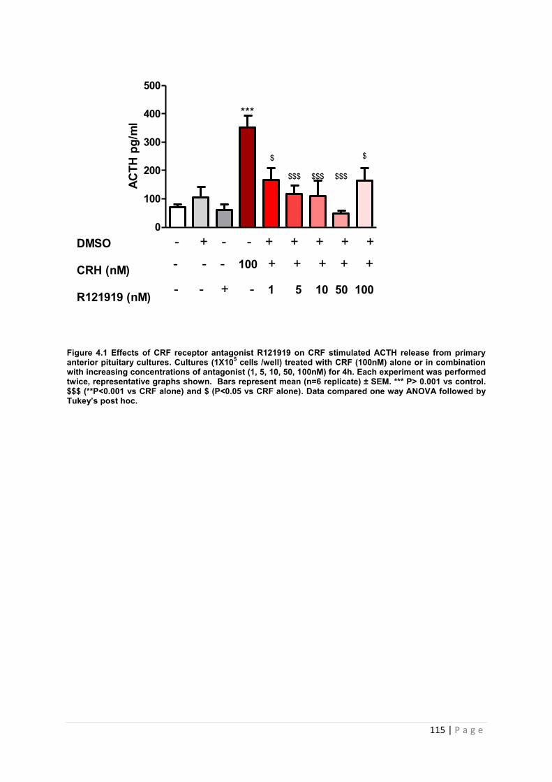
115 | P a g e
0
100
200
300
400
500
***
$ $
$$$ $$$ $$$
- + - - + + + + +
- - + - 1 5 10 50 100
DMSO
CRH (nM)
R121919 (nM)
ACTH
pg/
ml
- - - 100 + + + + +
Figure 4.1 Effects of CRF receptor antagonist R121919 on CRF stimulated ACTH release from primary anterior pituitary cultures. Cultures (1X10⁵ cells /well) treated with CRF (100nM) alone or in combination with increasing concentrations of antagonist (1, 5, 10, 50, 100nM) for 4h. Each experiment was performed twice, representative graphs shown. Bars represent mean (n=6 replicate) ± SEM. *** P> 0.001 vs control. $$$ (**P<0.001 vs CRF alone) and $ (P<0.05 vs CRF alone). Data compared one way ANOVA followed by Tukey’s post hoc.
116 | P a g e
4.3.1.2 Effects of CRF inhibitor DMP904 on ACTH release from primary pituitary
culture.
ACTH secretion from primary rat pituitary cultures was significantly increased following 4hr
incubation with CRF (100nM CRF, P<0.001 vs control figure 4.2). Pre-incubation with the
CRF receptor antagonist DMP90 or DMSO alone did not significantly influence basal ACTH
release. The CRF receptor antagonist, at all concentrations tested, significantly decreased
CRF induced ACTH release from primary pituitary cultures (P<0.001 vs CRF; figure 4.2),
with a maximal five-fold decrease in ACTH release (100nM DMP90).

117 | P a g e
0
100
200
300
400
500
***
$$$ $$$ $$$ $$$ACTH
pg/
ml
DMSO
CRH (nM)
DMP904 (nM)
- - - 100 + + + + +
- + - - + + + + +
- - + - 1 5 10 50 100
Figure 4.2: Effects of CRF receptor antagonist DMP904 on CRF stimulated ACTH release from primary anterior pituitary cultures. Cultures (1X10⁵ cells /well) treated with CRF (100nM) alone or in combination with increasing concentrations of antagonist (1, 5, 10, 50, 100nM) for 4h. Each experiment was repeated twice, representative graphs shown. Bars represent mean (n=6 replicates) ± SEM. *** P> 0.001 vs control. $$$ (**P<0.001 vs CRF alone). Data compared one way ANOVA followed by Tukey’s post hoc test.
118 | P a g e
4.3.2 Investigation into the effects of purinergic receptor stimulation on hormone release from rat primary pituitary cells
Following validation of our primary cell cultures, we then went on to identify which pituitary
hormones would be responsive ATP. Cells were then treated with a range of ATP doses
(0.001µM to 1000µM) which had been shown to be stimulatory in other endocrine organs
[149] and a range of time points (0 to 60 minutes) to assess the effects on ATP secretion.
4.3.2 .1 The effects of ATP on ACTH secretion from primary pituitary cells
Figure 4.3 illustrates the effects of increasing concentrations of ATP on the release of ACTH
from primary pituitary cells in vitro. For each dose of ATP investigated, an increase in ACTH
secretion was observed over time. The maximal ACTH secretion was between 120-149,000
pg/ml for all doses tested. With regard to timeframe, ATP consistently increased ACTH
secretion from cells at the 7 minute time point onwards.
0.001µM ATP did not induce a significant increase in ACTH secretion although it did cause
a 2 fold increase at 7 and 30 minutes (4.3.A). 0.01µM ATP caused a significant increase at
the 7 minute time point (P<0.05 vs. control, figure 4.3.B). 0.1µM ATP did not cause a
significant increase in ACTH, although a 2-fold increase in ACTH release was observed at
20 minutes (figure 4.3.C). 1µM ATP stimulated a 2-fold rise in ACTH levels at 10 minutes
(figure 4.3.D). 10µM ATP at the 30 minute time point has a significant effect on ACTH
secretion (P<0.05 vs. control, figure 4.1.E). 100µM ATP caused a significant release at the
time points of 7 (P<0.05 vs. contro1, 4.3F) and 60 (P<0.001 vs. control, figure 4.3F) minutes.
1000µM of ATP caused a significant increase at 20 (P<0.001 vs. control, 4.3G), 30 (P<0.05,
vs. control, figure 4.3G) and 60 (P<0.001 vs. control, figure 4.3G) minutes. Results are
summarised in table 4.3.
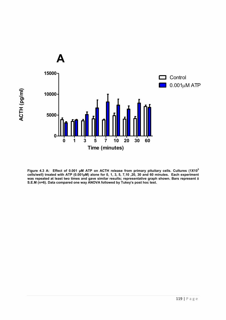
119 | P a g e
Figure 4.3 A: Effect of 0.001 µM ATP on ACTH release from primary pituitary cells. Cultures (1X10⁵ cells/well) treated with ATP (0.001µM) alone for 0, 1, 3, 5, 7,10 ,20, 30 and 60 minutes. Each experiment was repeated at least two times and gave similar results; representative graph shown. Bars represent ± S.E.M (n=6). Data compared one way ANOVA followed by Tukey’s post hoc test.
Time (minutes)
ACTH
(pg/
ml)
0 1 3 5 7 10 20 30 600
5000
10000
15000Control0.001M ATP
A

120 | P a g e
Figure 4.3 B: Effect of 0.01µM ATP on ACTH release from primary pituitary cells. Cultures (1X10⁵ cells/well) treated with ATP (0.01µM) alone for 0,1, 3, 5, 7, 10, 20, 30 and 60 minutes. Each experiment was repeated at least two times and gave similar results, representative graph shown. Bars represent ± S.E.M (n=6 replicates). * P<0.05 vs. control. Data compared one way ANOVA followed by Tukey’s post hoc test
B
Time (minutes)
AC
TH (
pg/m
l)l)
0 1 3 5 7 10 20 30 600
5000
10000
15000Control0.01M ATP
*
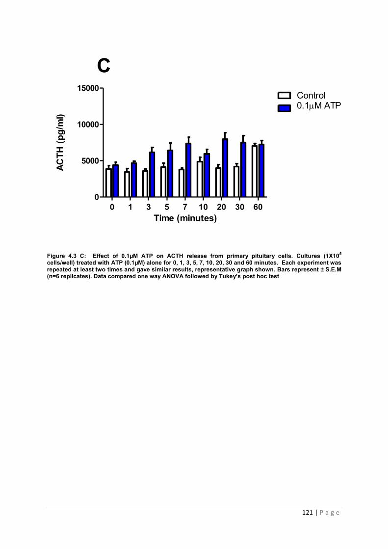
121 | P a g e
Figure 4.3 C: Effect of 0.1µM ATP on ACTH release from primary pituitary cells. Cultures (1X10⁵ cells/well) treated with ATP (0.1µM) alone for 0, 1, 3, 5, 7, 10, 20, 30 and 60 minutes. Each experiment was repeated at least two times and gave similar results, representative graph shown. Bars represent ± S.E.M (n=6 replicates). Data compared one way ANOVA followed by Tukey’s post hoc test
Time (minutes)
ACTH
(pg/
ml)
l)
0 1 3 5 7 10 20 30 600
5000
10000
15000Control0.1M ATP
C
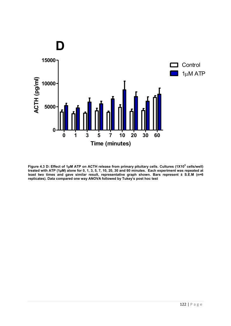
122 | P a g e
Figure 4.3 D: Effect of 1µM ATP on ACTH release from primary pituitary cells. Cultures (1X10⁵ cells/well) treated with ATP (1µM) alone for 0, 1, 3, 5, 7, 10, 20, 30 and 60 minutes. Each experiment was repeated at least two times and gave similar result, representative graph shown. Bars represent ± S.E.M (n=6 replicates). Data compared one way ANOVA followed by Tukey’s post hoc test
Time (minutes)
ACTH
(pg/
ml)
l)
0 1 3 5 7 10 20 30 600
5000
10000
15000Control1M ATP
D
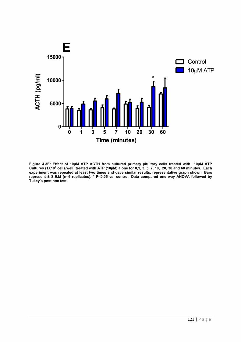
123 | P a g e
Figure 4.3E: Effect of 10µM ATP ACTH from cultured primary pituitary cells treated with 10µM ATP Cultures (1X10⁵ cells/well) treated with ATP (10µM) alone for 0,1, 3, 5, 7, 10, 20, 30 and 60 minutes. Each experiment was repeated at least two times and gave similar results, representative graph shown. Bars represent ± S.E.M (n=6 replicates). * P<0.05 vs. control. Data compared one way ANOVA followed by Tukey’s post hoc test.
Time (minutes)
ACTH
(pg/
ml)
l)
0 1 3 5 7 10 20 30 600
5000
10000
15000Control10M ATP
E
*

124 | P a g e
Figure 4.3F: Effect of 100µM ATP on ACTH release from primary pituitary cells. Cultures (1X10⁵ cells/well) treated with ATP (100µM) alone for 0, 1, 3, 5, 7, 10, 20, 30 and 60 minutes. Each experiment was repeated at least two times and gave similar results, representative graph shown. Bars represent ± S.E.M (n=6 replicates). *** P<0.001 vs. control (media only) and (* P<0.05 vs. control). Data compared one way ANOVA followed by Tukey’s post hoc test.
Time (minutes)
ACTH
(pg/
ml)
l)
0 1 3 5 7 10 20 30 600
5000
10000
15000Control100M ATP
F
*
***
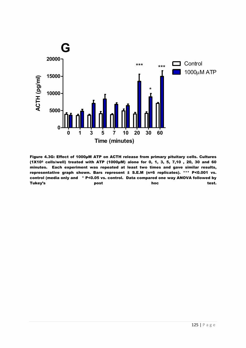
125 | P a g e
Figure 4.3G: Effect of 1000µM ATP on ACTH release from primary pituitary cells. Cultures
(1X10⁵ cells/well) treated with ATP (1000µM) alone for 0, 1, 3, 5, 7,10 , 20, 30 and 60
minutes. Each experiment was repeated at least two times and gave similar results,
representative graph shown. Bars represent ± S.E.M (n=6 replicates). *** P<0.001 vs.
control (media only and * P<0.05 vs. control. Data compared one way ANOVA followed by
Tukey’s post hoc test.
Time (minutes)
ACTH
(pg/
ml)
l)
0 1 3 5 7 10 20 30 600
5000
10000
15000
20000Control1000M ATP
G*** ***
*
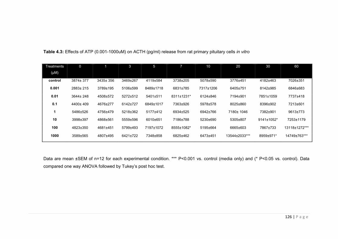
126 | P a g e
Table 4.3: Effects of ATP (0.001-1000uM) on ACTH (pg/ml) release from rat primary pituitary cells in vitro
Data are mean ±SEM of n=12 for each experimental condition. *** P<0.001 vs. control (media only) and (* P<0.05 vs. control). Data
compared one way ANOVA followed by Tukey’s post hoc test.
Treatments
(µM)
0 1 3 5 7 10 20 30 60
control 3874± 377 3435± 356 3469±267 4119±584 3738±205 5078±590 3776±451 4182±463 7026±351
0.001 2883± 215 3789±195 5106±599 8489±1718 6831±785 7317±1206 6405±751 8142±985 6846±683
0.01 3644± 248 4508±572 5272±512 5401±511 8311±1231* 6124±846 7194±901 7851±1059 7737±418
0.1 4400± 409 4676±277 6142±727 6849±1017 7363±926 5978±578 8025±860 8396±902 7213±601
1 5486±526 4756±479 5218±362 5177±412 6934±525 6942±766 7180± 1046 7382±901 9613±773
10 3998±397 4868±561 5559±596 6010±651 7186±788 5230±690 5305±807 9141±1052* 7253±1179
100 4823±350 4681±451 5799±493 7197±1072 8555±1082* 5195±664 6665±603 7867±733 13118±1272***
1000 3589±565 4807±495 6421±722 7348±858 6825±462 6473±451 13544±2033*** 8959±971* 14749±763***

127 | P a g e
4.3.2.2 The effects of ATP on GH from primary pituitary cells
Figure 4.3 illustrates the effects of graded concentrations of ATP on the release of GH from
primary pituitary cells in vitro. For each dose of ATP investigated, although none of the
doses caused a significant increase in GH release, a moderate increase in GH secretion in
response to ATP was observed over time, with all doses. The maximal GH secretion
observed was at 7 minutes and was between 892-922 pg/ml for all doses tested. 0.001µM
ATP did not cause a significant release although maximal release was observed at 7
minutes with a 24% increase in hormone (4.4.A). 0.01µM ATP caused a 30% increase at 7
minutes, however it also caused a 69% increase at the 20 minute time point (figure 4.4.B).
0.1µM ATP caused a maximal increase of GH at 7 minutes, with a 27% increase over basal,
(figure 4.4.C). 1µM ATP stimulated a peak rise in GH levels at 7 minutes with a 22%
increase above basal (figure 4.4D). 10µM ATP (figure 4.4.E) and 100µM ATP (figure 4.4F)
both caused a peak release at the time points of 7, with an average 26 % increase
observed. 1000µM of ATP caused only a marginal increase, peaking at 5 minutes with a 17%
increase in hormone (figure 4.4.G). Results summarised in table 4.4.
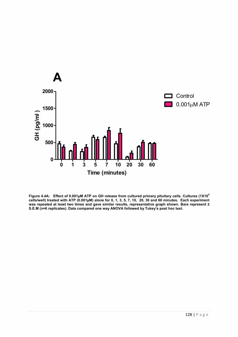
128 | P a g e
Figure 4.4A: Effect of 0.001µM ATP on GH release from cultured primary pituitary cells. Cultures (1X10⁵ cells/well) treated with ATP (0.001µM) alone for 0, 1, 3, 5, 7, 10, 20, 30 and 60 minutes. Each experiment was repeated at least two times and gave similar results, representative graph shown. Bars represent ± S.E.M (n=6 replicates). Data compared one way ANOVA followed by Tukey’s post hoc test.
Time (minutes)
GH
(pg/
ml )
0 1 3 5 7 10 20 30 600
500
1000
1500
2000Control0.001M ATP
A
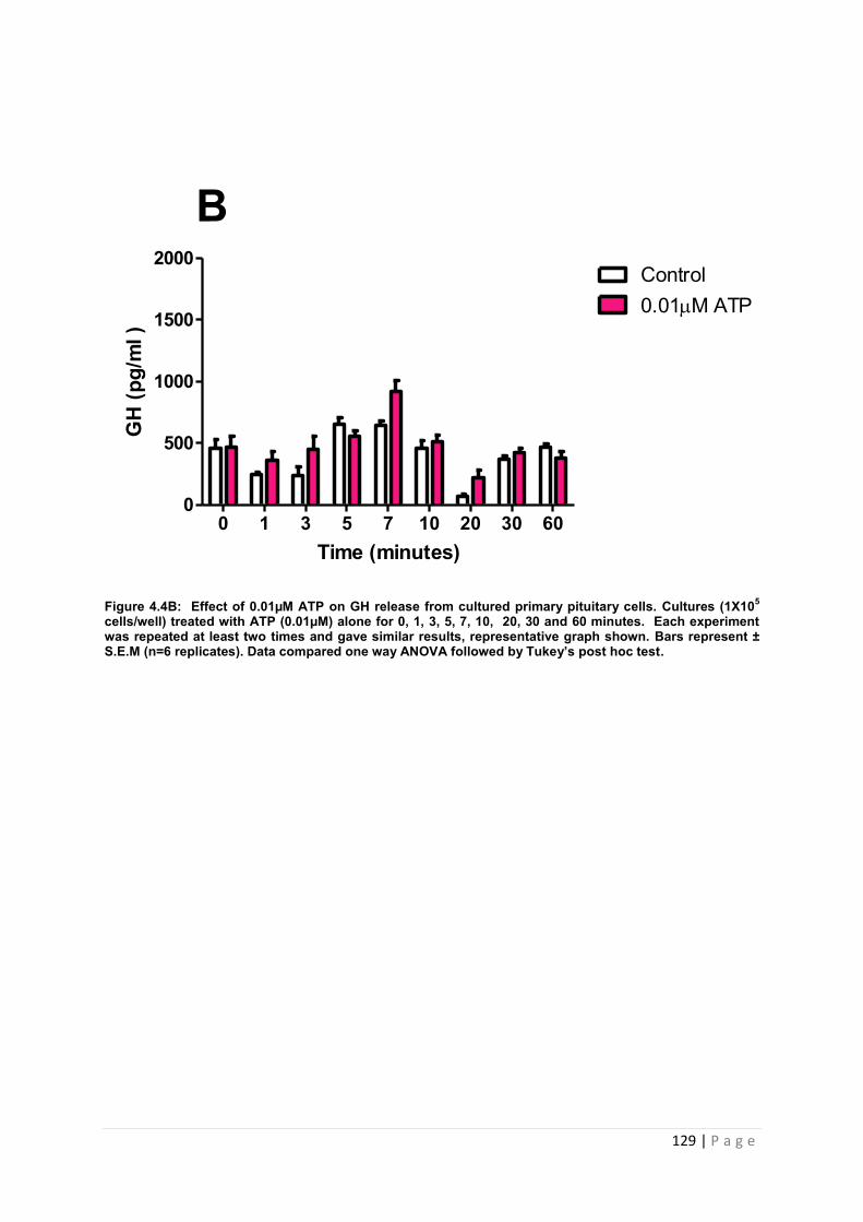
129 | P a g e
Figure 4.4B: Effect of 0.01µM ATP on GH release from cultured primary pituitary cells. Cultures (1X10⁵ cells/well) treated with ATP (0.01µM) alone for 0, 1, 3, 5, 7, 10, 20, 30 and 60 minutes. Each experiment was repeated at least two times and gave similar results, representative graph shown. Bars represent ± S.E.M (n=6 replicates). Data compared one way ANOVA followed by Tukey’s post hoc test.
Time (minutes)
GH
(pg/
ml )
0 1 3 5 7 10 20 30 600
500
1000
1500
2000Control0.01M ATP
B
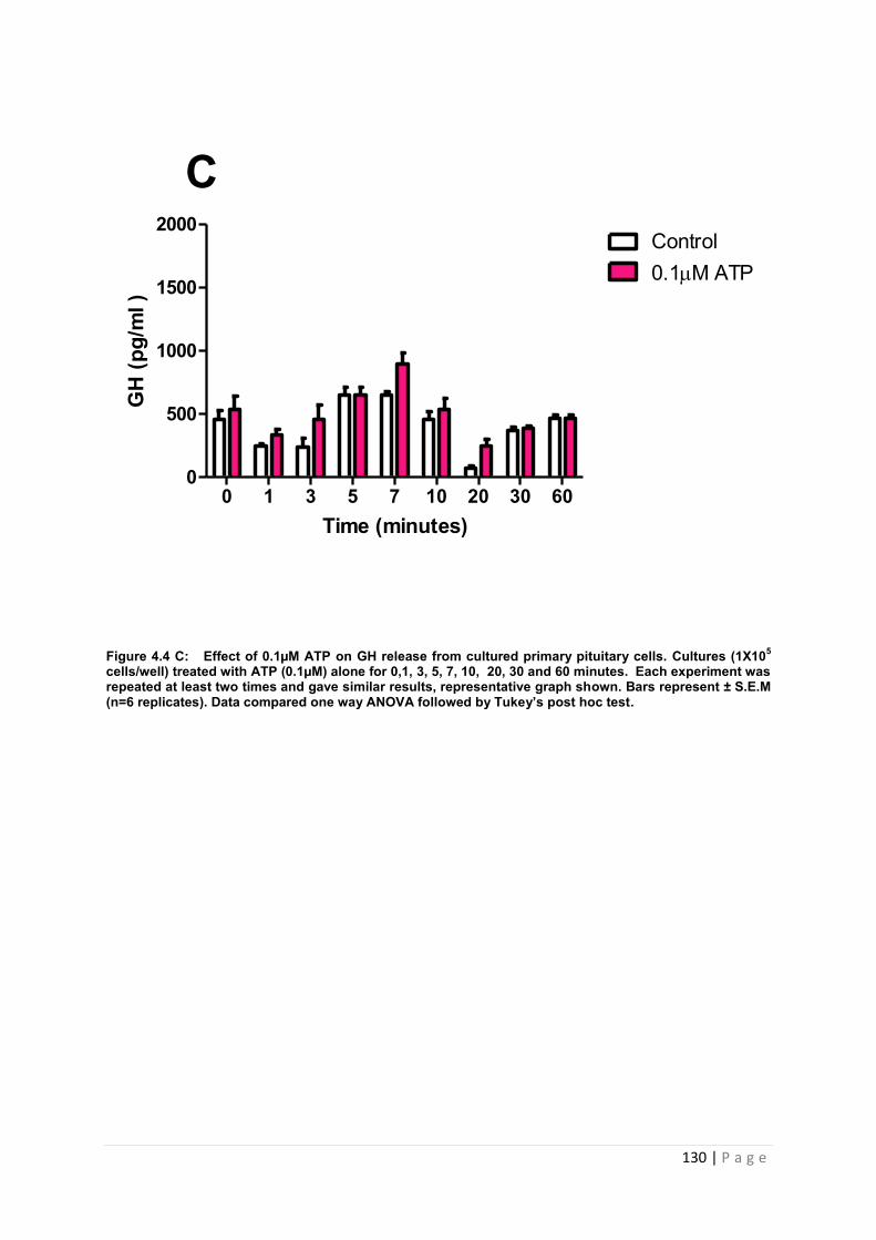
130 | P a g e
Figure 4.4 C: Effect of 0.1µM ATP on GH release from cultured primary pituitary cells. Cultures (1X10⁵ cells/well) treated with ATP (0.1µM) alone for 0,1, 3, 5, 7, 10, 20, 30 and 60 minutes. Each experiment was repeated at least two times and gave similar results, representative graph shown. Bars represent ± S.E.M (n=6 replicates). Data compared one way ANOVA followed by Tukey’s post hoc test.
Time (minutes)
GH
(pg/
ml )
0 1 3 5 7 10 20 30 600
500
1000
1500
2000Control0.1M ATP
C
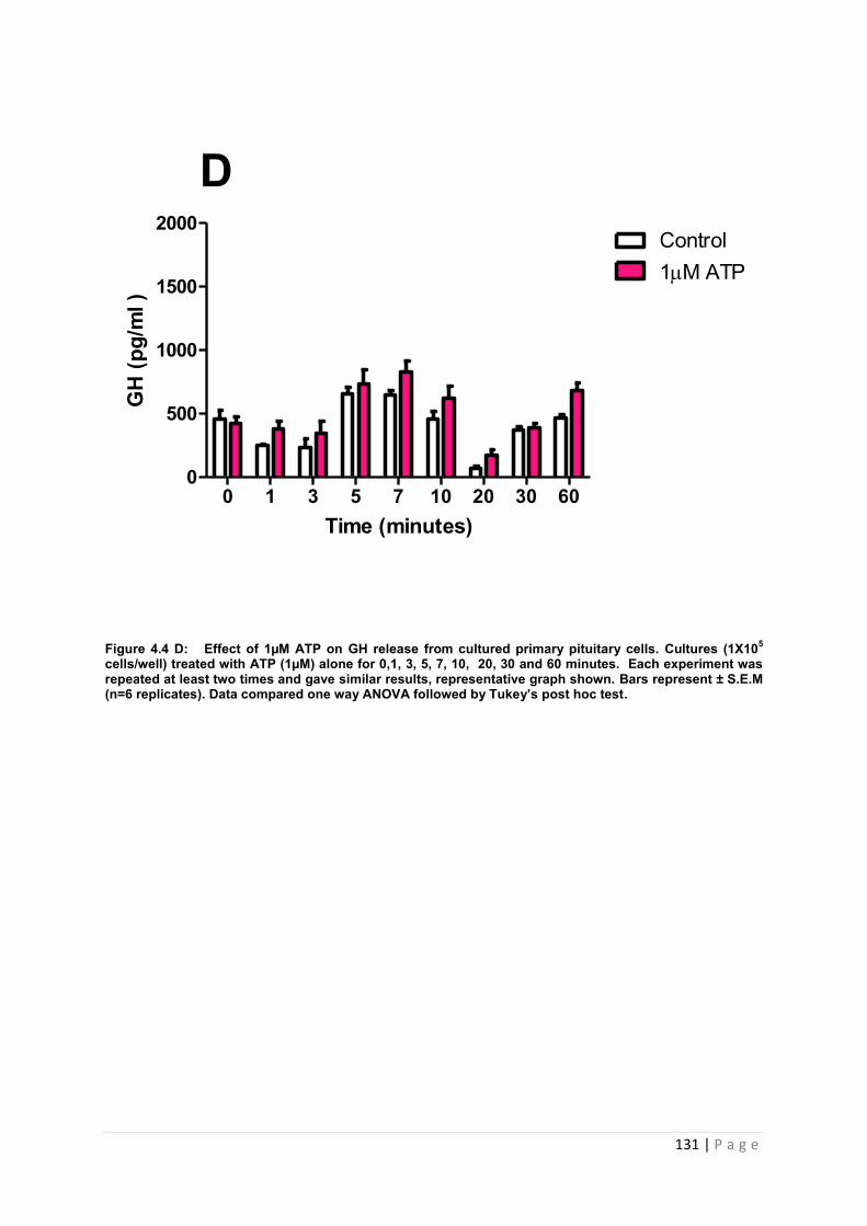
131 | P a g e
Figure 4.4 D: Effect of 1µM ATP on GH release from cultured primary pituitary cells. Cultures (1X10⁵ cells/well) treated with ATP (1µM) alone for 0,1, 3, 5, 7, 10, 20, 30 and 60 minutes. Each experiment was repeated at least two times and gave similar results, representative graph shown. Bars represent ± S.E.M (n=6 replicates). Data compared one way ANOVA followed by Tukey’s post hoc test.
Time (minutes)
GH
(pg/
ml )
0 1 3 5 7 10 20 30 600
500
1000
1500
2000Control1M ATP
D
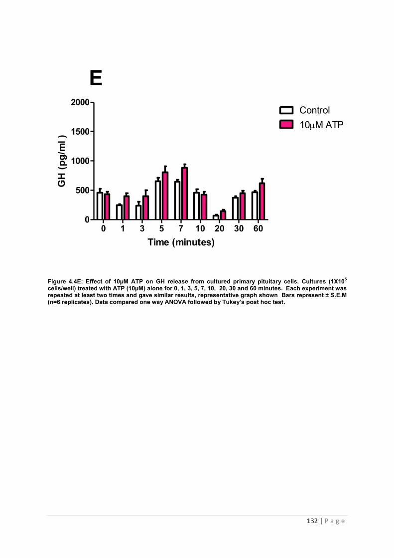
132 | P a g e
Figure 4.4E: Effect of 10µM ATP on GH release from cultured primary pituitary cells. Cultures (1X10⁵ cells/well) treated with ATP (10µM) alone for 0, 1, 3, 5, 7, 10, 20, 30 and 60 minutes. Each experiment was repeated at least two times and gave similar results, representative graph shown Bars represent ± S.E.M (n=6 replicates). Data compared one way ANOVA followed by Tukey’s post hoc test.
Time (minutes)
GH
(pg/
ml )
0 1 3 5 7 10 20 30 600
500
1000
1500
2000Control10M ATP
E
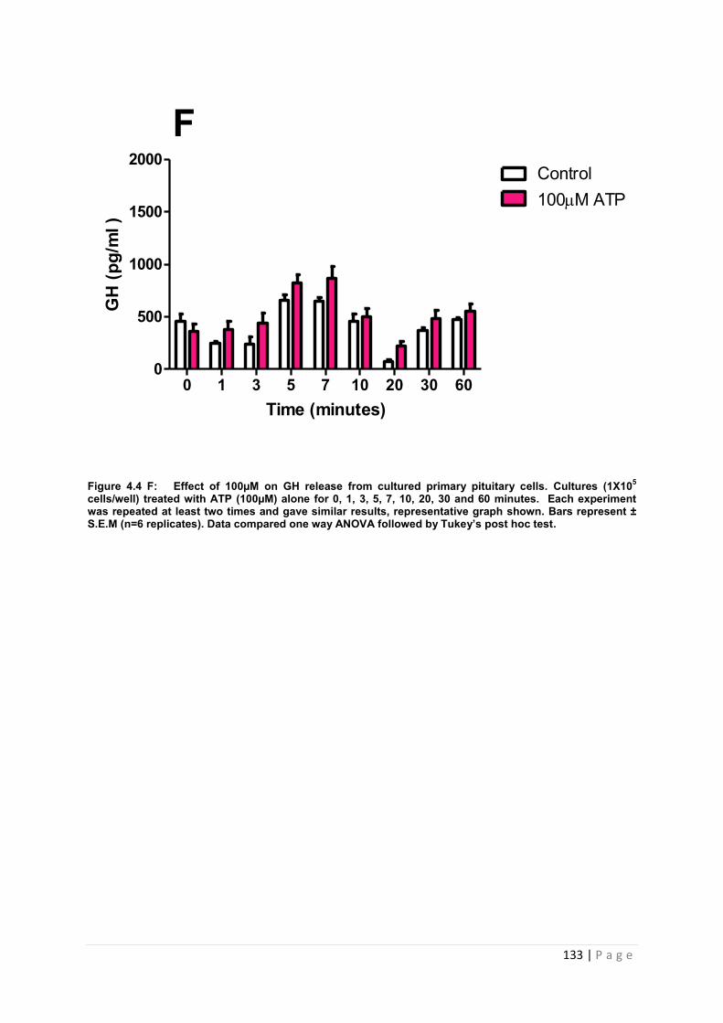
133 | P a g e
Figure 4.4 F: Effect of 100µM on GH release from cultured primary pituitary cells. Cultures (1X10⁵ cells/well) treated with ATP (100µM) alone for 0, 1, 3, 5, 7, 10, 20, 30 and 60 minutes. Each experiment was repeated at least two times and gave similar results, representative graph shown. Bars represent ± S.E.M (n=6 replicates). Data compared one way ANOVA followed by Tukey’s post hoc test.
Time (minutes)
GH
(pg/
ml )
0 1 3 5 7 10 20 30 600
500
1000
1500
2000Control100M ATP
F
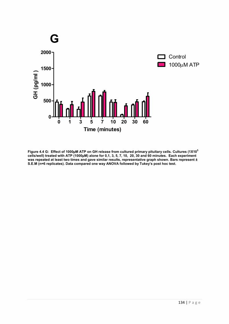
134 | P a g e
Figure 4.4 G: Effect of 1000µM ATP on GH release from cultured primary pituitary cells. Cultures (1X10⁵ cells/well) treated with ATP (1000µM) alone for 0,1, 3, 5, 7, 10, 20, 30 and 60 minutes. Each experiment was repeated at least two times and gave similar results, representative graph shown. Bars represent ± S.E.M (n=6 replicates). Data compared one way ANOVA followed by Tukey’s post hoc test.
Time (minutes)
GH
(pg/
ml )
0 1 3 5 7 10 20 30 600
500
1000
1500
2000Control1000M ATP
G
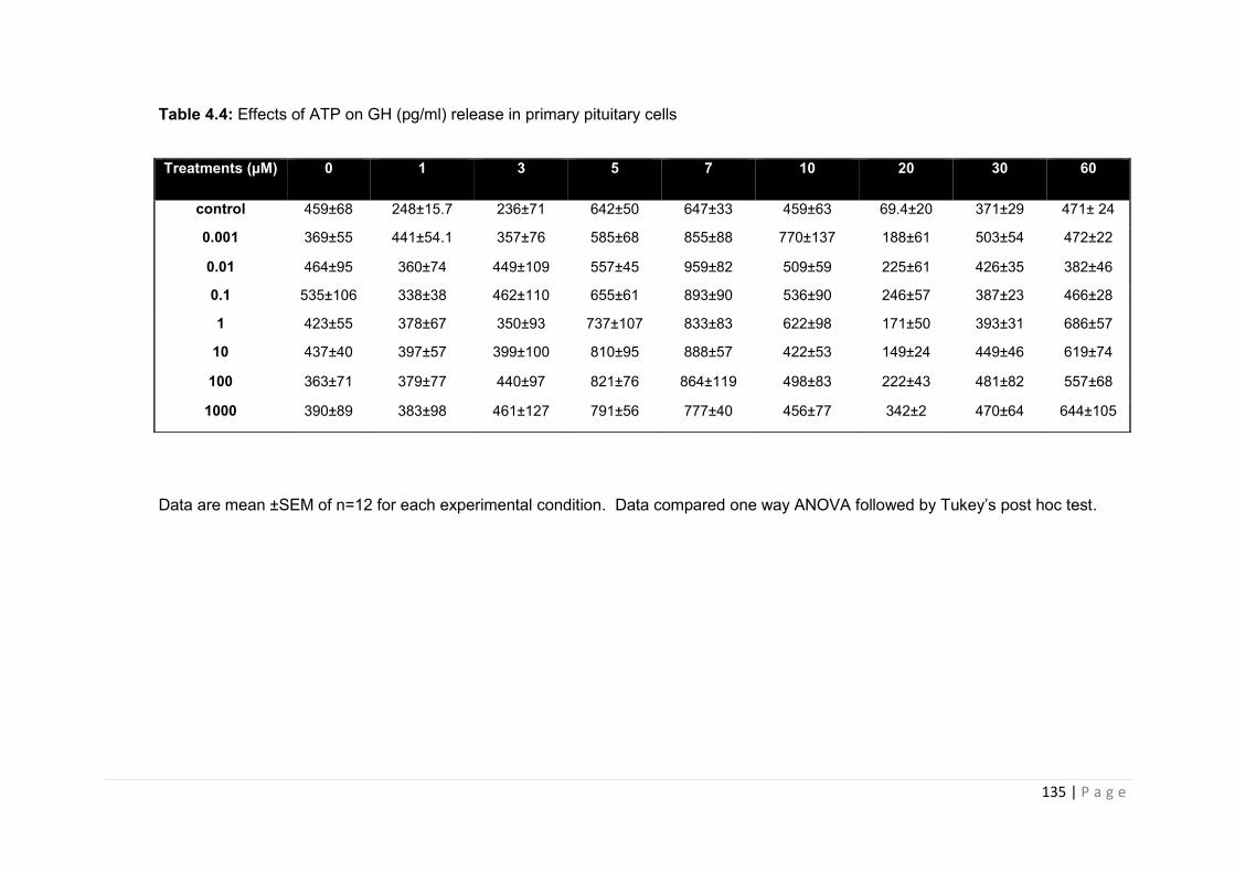
135 | P a g e
Table 4.4: Effects of ATP on GH (pg/ml) release in primary pituitary cells
Data are mean ±SEM of n=12 for each experimental condition. Data compared one way ANOVA followed by Tukey’s post hoc test.
Treatments (µM) 0 1 3 5 7 10 20 30 60
control 459±68 248±15.7 236±71 642±50 647±33 459±63 69.4±20 371±29 471± 24
0.001 369±55 441±54.1 357±76 585±68 855±88 770±137 188±61 503±54 472±22
0.01 464±95 360±74 449±109 557±45 959±82 509±59 225±61 426±35 382±46
0.1 535±106 338±38 462±110 655±61 893±90 536±90 246±57 387±23 466±28
1 423±55 378±67 350±93 737±107 833±83 622±98 171±50 393±31 686±57
10 437±40 397±57 399±100 810±95 888±57 422±53 149±24 449±46 619±74
100 363±71 379±77 440±97 821±76 864±119 498±83 222±43 481±82 557±68
1000 390±89 383±98 461±127 791±56 777±40 456±77 342±2 470±64 644±105
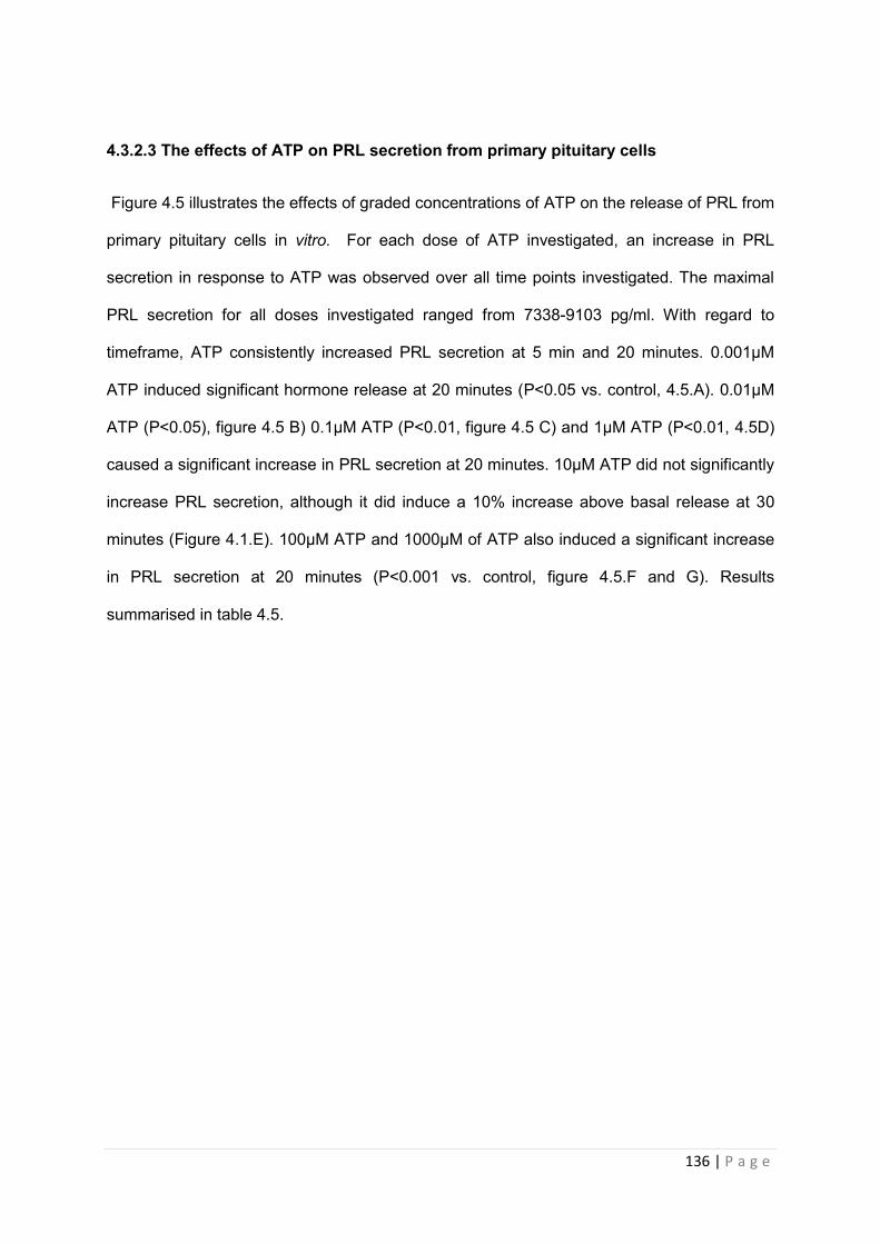
136 | P a g e
4.3.2.3 The effects of ATP on PRL secretion from primary pituitary cells
Figure 4.5 illustrates the effects of graded concentrations of ATP on the release of PRL from
primary pituitary cells in vitro. For each dose of ATP investigated, an increase in PRL
secretion in response to ATP was observed over all time points investigated. The maximal
PRL secretion for all doses investigated ranged from 7338-9103 pg/ml. With regard to
timeframe, ATP consistently increased PRL secretion at 5 min and 20 minutes. 0.001µM
ATP induced significant hormone release at 20 minutes (P<0.05 vs. control, 4.5.A). 0.01µM
ATP (P<0.05), figure 4.5 B) 0.1µM ATP (P<0.01, figure 4.5 C) and 1µM ATP (P<0.01, 4.5D)
caused a significant increase in PRL secretion at 20 minutes. 10µM ATP did not significantly
increase PRL secretion, although it did induce a 10% increase above basal release at 30
minutes (Figure 4.1.E). 100µM ATP and 1000µM of ATP also induced a significant increase
in PRL secretion at 20 minutes (P<0.001 vs. control, figure 4.5.F and G). Results
summarised in table 4.5.
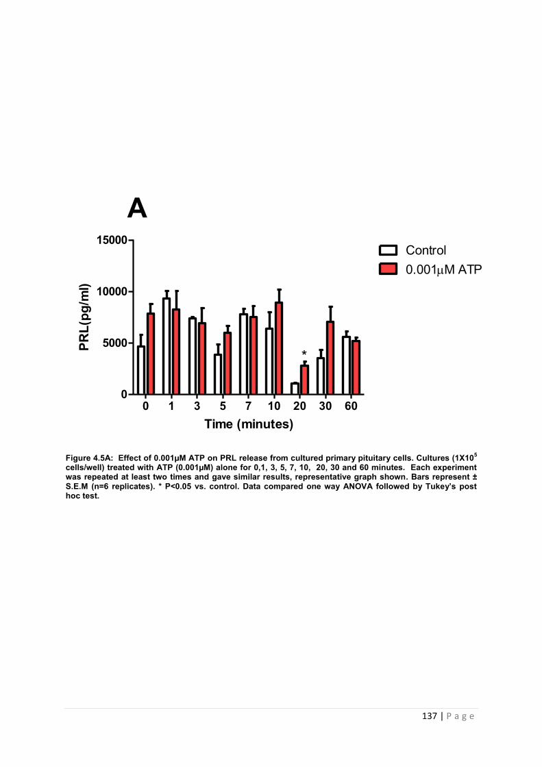
137 | P a g e
Figure 4.5A: Effect of 0.001µM ATP on PRL release from cultured primary pituitary cells. Cultures (1X10⁵ cells/well) treated with ATP (0.001µM) alone for 0,1, 3, 5, 7, 10, 20, 30 and 60 minutes. Each experiment was repeated at least two times and gave similar results, representative graph shown. Bars represent ± S.E.M (n=6 replicates). * P<0.05 vs. control. Data compared one way ANOVA followed by Tukey’s post hoc test.
Time (minutes)
PRL(
pg/m
l)l)
0 1 3 5 7 10 20 30 600
5000
10000
15000Control0.001M ATP
A
*
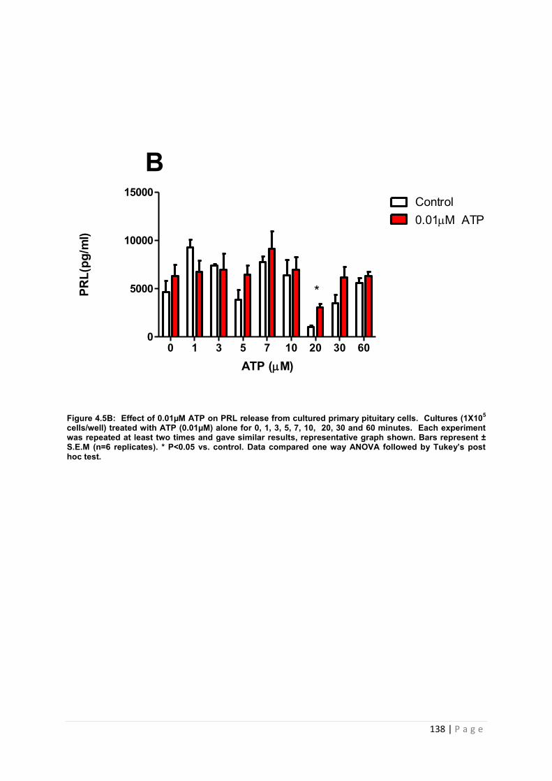
138 | P a g e
Figure 4.5B: Effect of 0.01µM ATP on PRL release from cultured primary pituitary cells. Cultures (1X10⁵ cells/well) treated with ATP (0.01µM) alone for 0, 1, 3, 5, 7, 10, 20, 30 and 60 minutes. Each experiment was repeated at least two times and gave similar results, representative graph shown. Bars represent ± S.E.M (n=6 replicates). * P<0.05 vs. control. Data compared one way ANOVA followed by Tukey’s post hoc test.
ATP (M)
PRL(
pg/m
l)l)
0 1 3 5 7 10 20 30 600
5000
10000
15000Control0.01M ATP
B
*
139 | P a g e
Figure 4.5C: Effect of 0.1µM ATP on PRL release from cultured primary pituitary cells. Cultures (1X10⁵ cells/well) treated with ATP (0.1µM) alone for 0, 1, 3, 5, 7, 10, 20, 30 and 60 minutes. Each experiment was repeated at least two times and gave similar results, representative graph shown. Bars represent ± S.E.M (n=6 replicates). ** P<0.01 vs. control. Data compared one way ANOVA followed by Tukey’s post hoc test.
Time (minutes)
PRL(
pg/m
l)l)
0 1 3 5 7 10 20 30 600
5000
10000
15000Control0.1m ATP
C
**
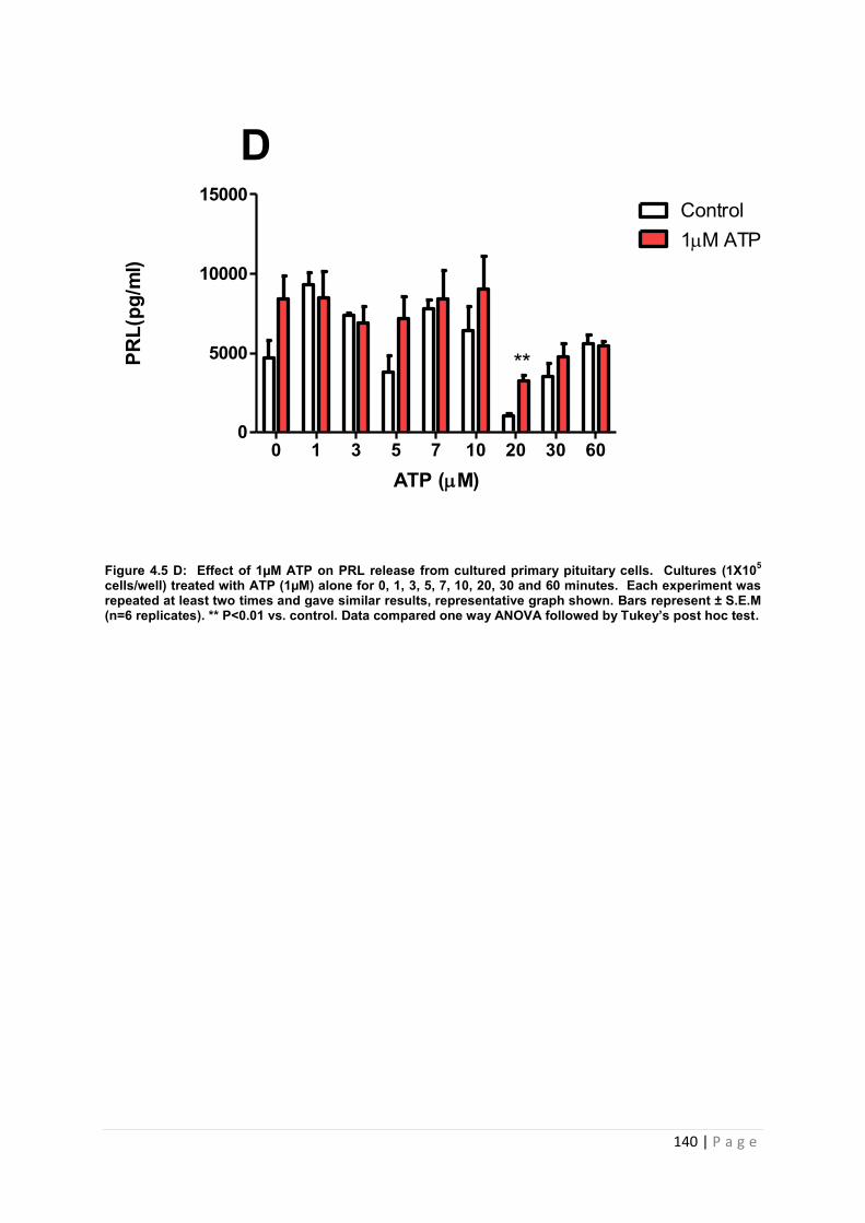
140 | P a g e
Figure 4.5 D: Effect of 1µM ATP on PRL release from cultured primary pituitary cells. Cultures (1X10⁵ cells/well) treated with ATP (1µM) alone for 0, 1, 3, 5, 7, 10, 20, 30 and 60 minutes. Each experiment was repeated at least two times and gave similar results, representative graph shown. Bars represent ± S.E.M (n=6 replicates). ** P<0.01 vs. control. Data compared one way ANOVA followed by Tukey’s post hoc test.
ATP (M)
PRL(
pg/m
l)l)
0 1 3 5 7 10 20 30 600
5000
10000
15000Control1M ATP
D
**
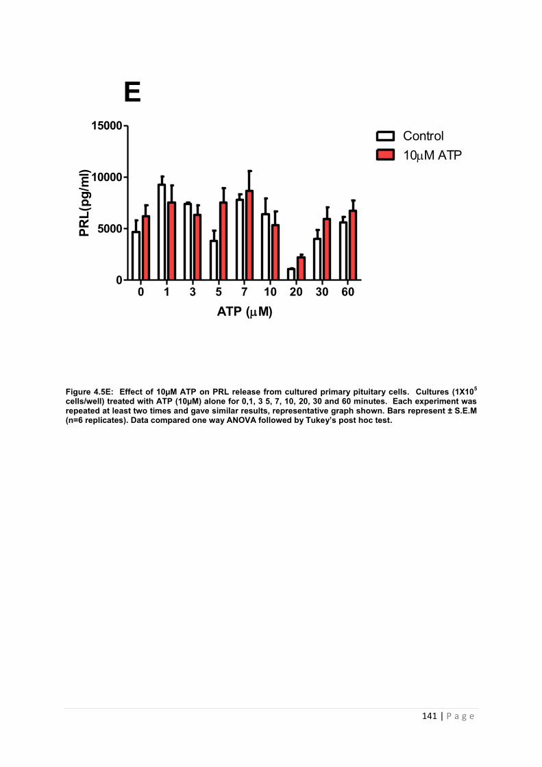
141 | P a g e
Figure 4.5E: Effect of 10µM ATP on PRL release from cultured primary pituitary cells. Cultures (1X10⁵ cells/well) treated with ATP (10µM) alone for 0,1, 3 5, 7, 10, 20, 30 and 60 minutes. Each experiment was repeated at least two times and gave similar results, representative graph shown. Bars represent ± S.E.M (n=6 replicates). Data compared one way ANOVA followed by Tukey’s post hoc test.
ATP (M)
PRL(
pg/m
l)l)
0 1 3 5 7 10 20 30 600
5000
10000
15000Control10M ATP
E
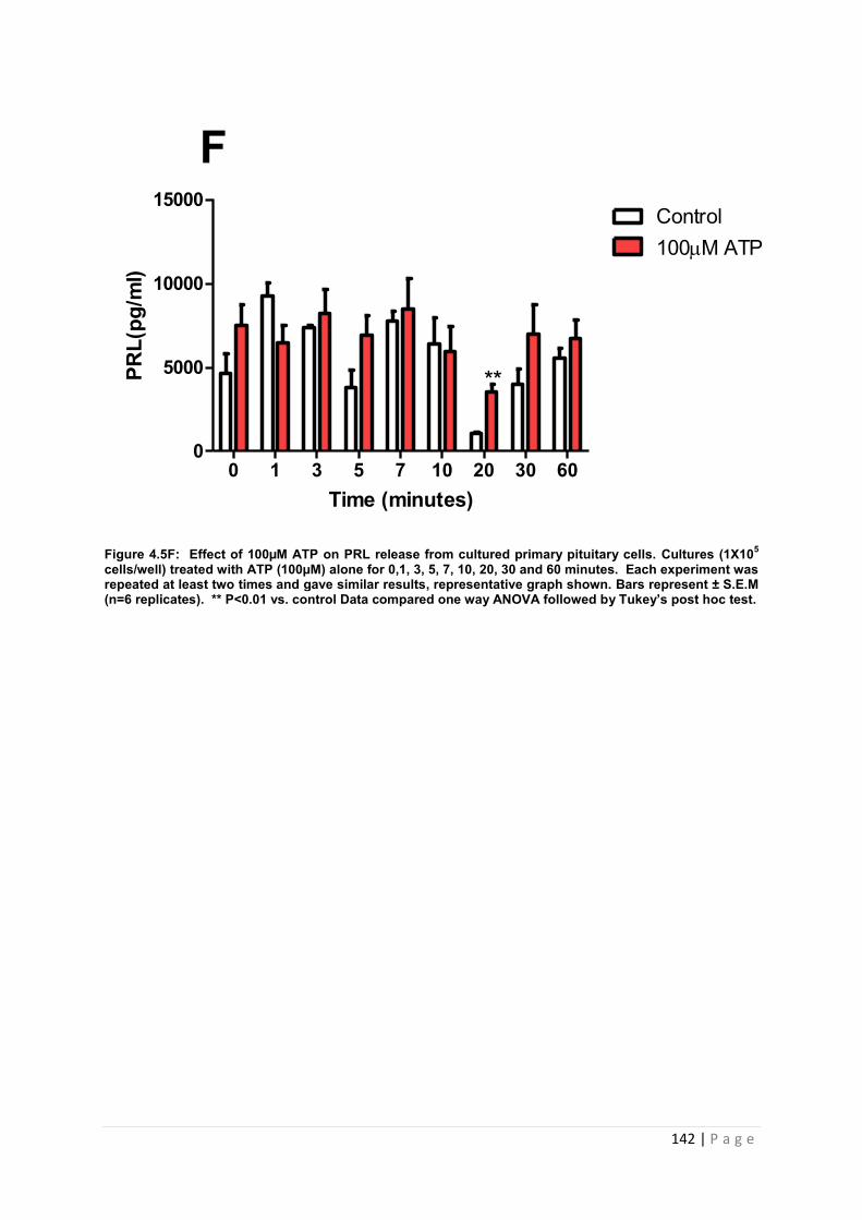
142 | P a g e
Figure 4.5F: Effect of 100µM ATP on PRL release from cultured primary pituitary cells. Cultures (1X10⁵ cells/well) treated with ATP (100µM) alone for 0,1, 3, 5, 7, 10, 20, 30 and 60 minutes. Each experiment was repeated at least two times and gave similar results, representative graph shown. Bars represent ± S.E.M (n=6 replicates). ** P<0.01 vs. control Data compared one way ANOVA followed by Tukey’s post hoc test.
Time (minutes)
PRL(
pg/m
l)l)
0 1 3 5 7 10 20 30 600
5000
10000
15000Control100M ATP
F
**
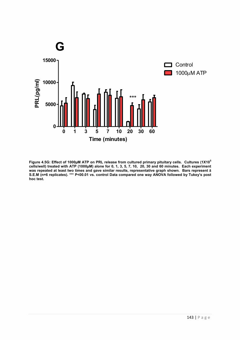
143 | P a g e
Figure 4.5G: Effect of 1000µM ATP on PRL release from cultured primary pituitary cells. Cultures (1X10⁵ cells/well) treated with ATP (1000µM) alone for 0, 1, 3, 5, 7, 10, 20, 30 and 60 minutes. Each experiment was repeated at least two times and gave similar results, representative graph shown. Bars represent ± S.E.M (n=6 replicates). *** P<00.01 vs. control Data compared one way ANOVA followed by Tukey’s post hoc test.
Time (minutes)
PRL(
pg/m
l)l)
0 1 3 5 7 10 20 30 600
5000
10000
15000Control1000M ATP
G
***
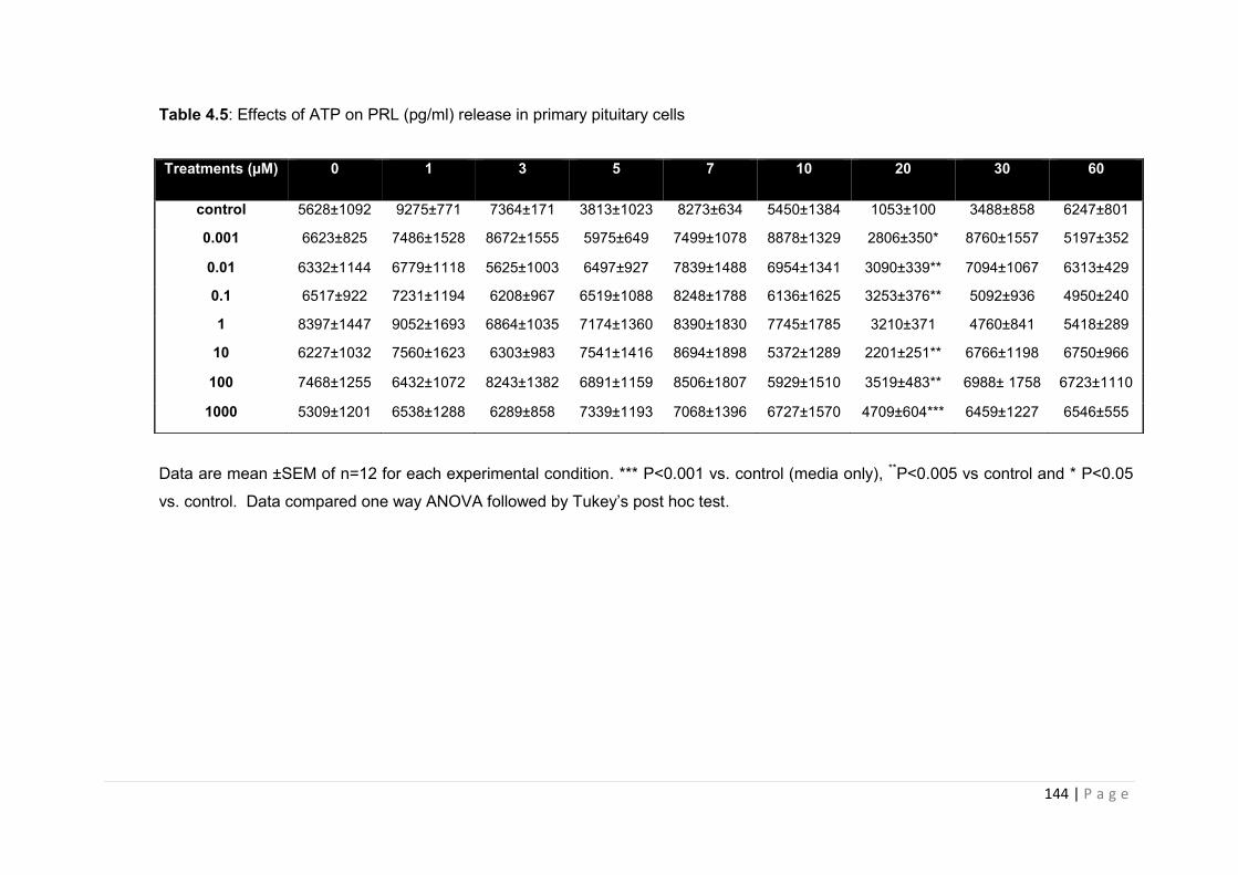
144 | P a g e
Table 4.5: Effects of ATP on PRL (pg/ml) release in primary pituitary cells
Data are mean ±SEM of n=12 for each experimental condition. *** P<0.001 vs. control (media only), **P<0.005 vs control and * P<0.05
vs. control. Data compared one way ANOVA followed by Tukey’s post hoc test.
Treatments (µM) 0 1 3 5 7 10 20 30 60
control 5628±1092 9275±771 7364±171 3813±1023 8273±634 5450±1384 1053±100 3488±858 6247±801
0.001 6623±825 7486±1528 8672±1555 5975±649 7499±1078 8878±1329 2806±350* 8760±1557 5197±352
0.01 6332±1144 6779±1118 5625±1003 6497±927 7839±1488 6954±1341 3090±339** 7094±1067 6313±429
0.1 6517±922 7231±1194 6208±967 6519±1088 8248±1788 6136±1625 3253±376** 5092±936 4950±240
1 8397±1447 9052±1693 6864±1035 7174±1360 8390±1830 7745±1785 3210±371 4760±841 5418±289
10 6227±1032 7560±1623 6303±983 7541±1416 8694±1898 5372±1289 2201±251** 6766±1198 6750±966
100 7468±1255 6432±1072 8243±1382 6891±1159 8506±1807 5929±1510 3519±483** 6988± 1758 6723±1110
1000 5309±1201 6538±1288 6289±858 7339±1193 7068±1396 6727±1570 4709±604*** 6459±1227 6546±555
145 | P a g e
4.3.2 .4 The effects of ATP on TSH from primary pituitary cells
Figure 4.6 illustrates the effects of graded concentrations of ATP on the release of TSH
from primary pituitary cells in vitro. For each dose of ATP investigated, an increase in TSH
secretion in response to ATP was observed at each time point investigated. The maximal
TSH secretion for all doses investigated ranged from 1902-2679 pg/ml. With regard to
timeframe, ATP consistently increased PRL secretion at 7 min and 10 minutes. 0.001µM
ATP induced a significant increase in TSH secretion at 10 minutes (P<0.05 vs. control, figure
4.6.A). 0.01µM ATP induced maximal TSH secretion of 25% above basal at 7 minutes
(figure 4.6.B). 0.1µM ATP induced maximal TSH secretion of 39% above basal at 10
minutes (figure 4.1.C). 1µM and 10 µM ATP stimulated a maximal rise in TSH levels at 10
and 7 minutes, respectively with a 28% increase observed (figure 4.6.D and E). 100µM ATP
induced a 14% increase in TSH secretion above basal at 7 minutes (figure 4.6F) whereas
1000µM of ATP induced a significant increase in TSH secretion at 20 (P<0.001 vs. control,
figure 4.6 G) and 60 minutes (P<0.001 vs. control, 4.6 G). Results summarised in table 4.6.
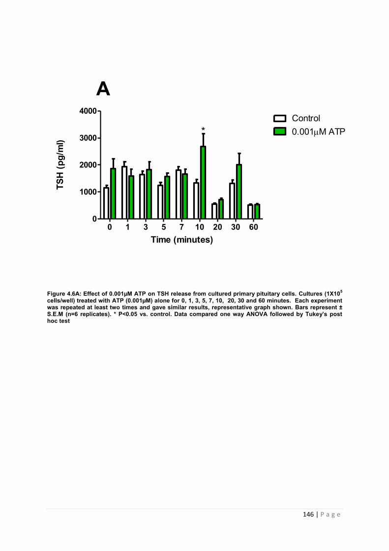
146 | P a g e
Figure 4.6A: Effect of 0.001µM ATP on TSH release from cultured primary pituitary cells. Cultures (1X10⁵ cells/well) treated with ATP (0.001µM) alone for 0, 1, 3, 5, 7, 10, 20, 30 and 60 minutes. Each experiment was repeated at least two times and gave similar results, representative graph shown. Bars represent ± S.E.M (n=6 replicates). * P<0.05 vs. control. Data compared one way ANOVA followed by Tukey’s post hoc test
Time (minutes)
TSH
(pg/
ml)
0 1 3 5 7 10 20 30 600
1000
2000
3000
4000Control0.001M ATP*
A
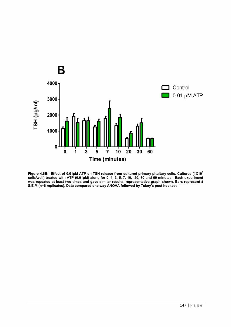
147 | P a g e
Figure 4.6B: Effect of 0.01µM ATP on TSH release from cultured primary pituitary cells. Cultures (1X10⁵ cells/well) treated with ATP (0.01µM) alone for 0, 1, 3, 5, 7, 10, 20, 30 and 60 minutes. Each experiment was repeated at least two times and gave similar results, representative graph shown. Bars represent ± S.E.M (n=6 replicates). Data compared one way ANOVA followed by Tukey’s post hoc test
Time (minutes)
TSH
(pg/
ml)
0 1 3 5 7 10 20 30 600
1000
2000
3000
4000Control0.01 M ATP
B
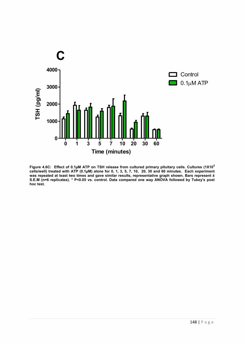
148 | P a g e
Figure 4.6C: Effect of 0.1µM ATP on TSH release from cultured primary pituitary cells. Cultures (1X10⁵ cells/well) treated with ATP (0.1µM) alone for 0, 1, 3, 5, 7, 10, 20, 30 and 60 minutes. Each experiment was repeated at least two times and gave similar results, representative graph shown. Bars represent ± S.E.M (n=6 replicates). * P<0.05 vs. control. Data compared one way ANOVA followed by Tukey’s post hoc test.
Time (minutes)
TSH
(pg/
ml)
0 1 3 5 7 10 20 30 600
1000
2000
3000
4000Control0.1M ATP
C
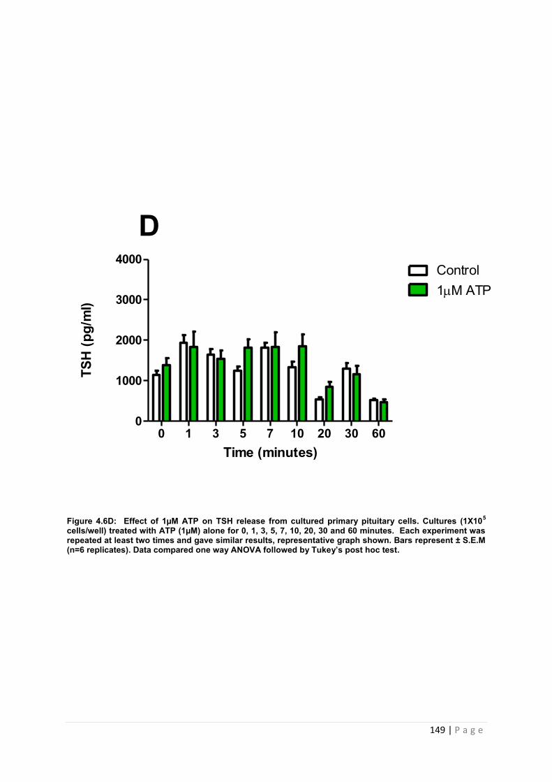
149 | P a g e
Figure 4.6D: Effect of 1µM ATP on TSH release from cultured primary pituitary cells. Cultures (1X10⁵ cells/well) treated with ATP (1µM) alone for 0, 1, 3, 5, 7, 10, 20, 30 and 60 minutes. Each experiment was repeated at least two times and gave similar results, representative graph shown. Bars represent ± S.E.M (n=6 replicates). Data compared one way ANOVA followed by Tukey’s post hoc test.
Time (minutes)
TSH
(pg/
ml)
0 1 3 5 7 10 20 30 600
1000
2000
3000
4000Control1M ATP
D
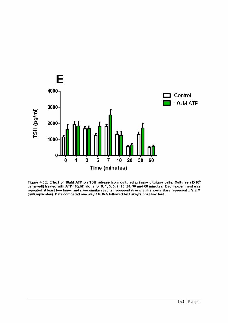
150 | P a g e
Figure 4.6E: Effect of 10µM ATP on TSH release from cultured primary pituitary cells. Cultures (1X10⁵ cells/well) treated with ATP (10µM) alone for 0, 1, 3, 5, 7, 10, 20, 30 and 60 minutes. Each experiment was repeated at least two times and gave similar results, representative graph shown. Bars represent ± S.E.M (n=6 replicates). Data compared one way ANOVA followed by Tukey’s post hoc test.
Time (minutes)
TSH
(pg/
ml)
l)
0 1 3 5 7 10 20 30 600
1000
2000
3000
4000Control10M ATP
E
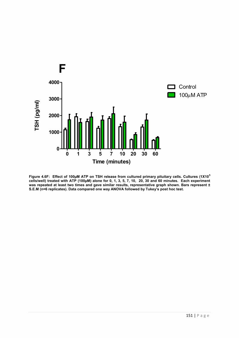
151 | P a g e
Figure 4.6F: Effect of 100µM ATP on TSH release from cultured primary pituitary cells. Cultures (1X10⁵ cells/well) treated with ATP (100µM) alone for 0, 1, 3, 5, 7, 10, 20, 30 and 60 minutes. Each experiment was repeated at least two times and gave similar results, representative graph shown. Bars represent ± S.E.M (n=6 replicates). Data compared one way ANOVA followed by Tukey’s post hoc test.
Time (minutes)
TSH
(pg/
ml)
0 1 3 5 7 10 20 30 600
1000
2000
3000
4000Control100M ATP
F
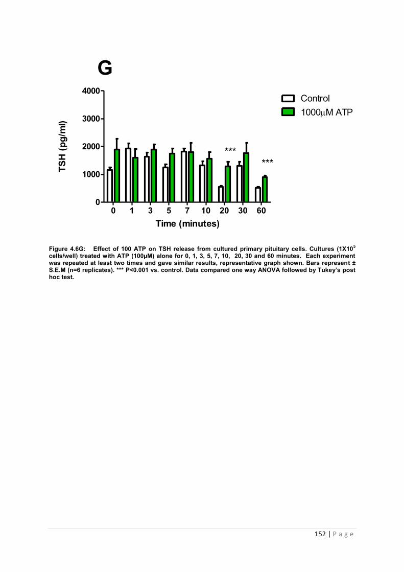
152 | P a g e
Figure 4.6G: Effect of 100 ATP on TSH release from cultured primary pituitary cells. Cultures (1X10⁵ cells/well) treated with ATP (100µM) alone for 0, 1, 3, 5, 7, 10, 20, 30 and 60 minutes. Each experiment was repeated at least two times and gave similar results, representative graph shown. Bars represent ± S.E.M (n=6 replicates). *** P<0.001 vs. control. Data compared one way ANOVA followed by Tukey’s post hoc test.
Time (minutes)
TSH
(pg/
ml)
0 1 3 5 7 10 20 30 600
1000
2000
3000
4000Control1000M ATP
G
******
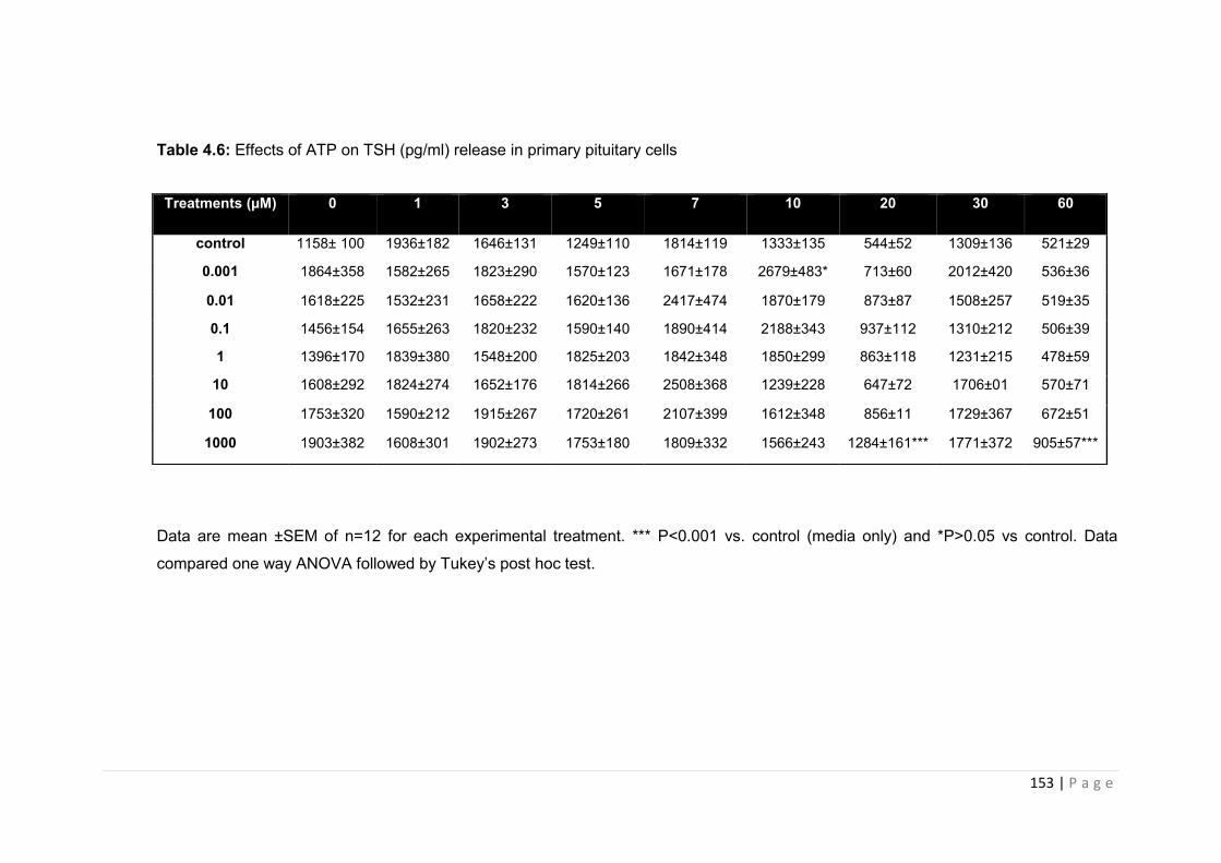
153 | P a g e
Table 4.6: Effects of ATP on TSH (pg/ml) release in primary pituitary cells
Data are mean ±SEM of n=12 for each experimental treatment. *** P<0.001 vs. control (media only) and *P>0.05 vs control. Data
compared one way ANOVA followed by Tukey’s post hoc test.
Treatments (µM) 0 1 3 5 7 10 20 30 60
control 1158± 100 1936±182 1646±131 1249±110 1814±119 1333±135 544±52 1309±136 521±29
0.001 1864±358 1582±265 1823±290 1570±123 1671±178 2679±483* 713±60 2012±420 536±36
0.01 1618±225 1532±231 1658±222 1620±136 2417±474 1870±179 873±87 1508±257 519±35
0.1 1456±154 1655±263 1820±232 1590±140 1890±414 2188±343 937±112 1310±212 506±39
1 1396±170 1839±380 1548±200 1825±203 1842±348 1850±299 863±118 1231±215 478±59
10 1608±292 1824±274 1652±176 1814±266 2508±368 1239±228 647±72 1706±01 570±71
100 1753±320 1590±212 1915±267 1720±261 2107±399 1612±348 856±11 1729±367 672±51
1000 1903±382 1608±301 1902±273 1753±180 1809±332 1566±243 1284±161*** 1771±372 905±57***
154 | P a g e
4.3.3 Investigating the effect of purinergic receptor activation on secretagogue-
induced hormone release from pituitary cell lines
Data from the previous experiments indicated that primary pituitary cells responsible for the
production of ACTH and PRL hormone showed a positive response to ATP treatment (See
figures 4.3, Table 4.3 and figure 4.5, table 4.5).
We therefore wanted to assess the effects of ATP on these two pituitary hormones in
isolation, and utilised two specific in vitro pituitary cell lines to do so. The mouse
corticotroph cell line AtT20 D16:16, was used to study the effects ACTH release. This cell
line had been used extensively by our group [[77]; [150]], primarily whilst investigating the
influence of annexin -1 on corticotrophin (ACTH) secretion. The rat cell, GH3 were utilised to
look at the effects of ATP on PRL secretion from lactotrophs.
Our aim was to identify which of the pituitary cell lines were capable of producing significant
increases in hormone secretion in response to both the relevant secretagogue and ATP/ATP
analogues. Cell lines that were responsive to both stimulants were then investigated further
with the use of P2X antagonists to attempt to block the secretagogue induced increases in
hormone secretion.
4.3.3.1 AtT20 D16:16 corticotroph cells – response to secretagogue (corticotrophin
releasing factor).
Previous studies in our group had established that 4 hours was the minimum incubation time
with which significant increases in secretagogue induced ACTH secretion from ATt20:D16
cells could be observed [150]. Corticotrophin releasing factor (CRF) is a well-established
secretagogue for ACTH and our CRF dose response was selected using doses either side of
concentrations that had previously been shown to be effective (Stefana, Ray et al. 1996).
Figure 4.7 illustrates the effects of graded concentrations of CRF on the release of ACTH
from AtT-20 cells in vitro. CRF (0.1-100nM) induced a concentration-dependent increase in
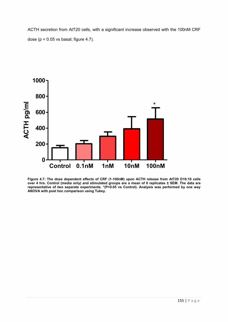
155 | P a g e
ACTH secretion from AtT20 cells, with a significant increase observed with the 100nM CRF
dose (p < 0.05 vs basal; figure 4.7).
Figure 4.7: The dose dependent effects of CRF (1-100nM) upon ACTH release from AtT20 D16:16 cells over 4 hrs. Control (media only) and stimulated groups are a mean of 6 replicates ± SEM. The data are representative of two separate experiments. *(P>0.05 vs Control). Analysis was performed by one way ANOVA with post hoc comparison using Tukey.
Control 0.1nM 1nM 10nM 100nM0
200
400
600
800
1000
*
ACTH
pg/
ml
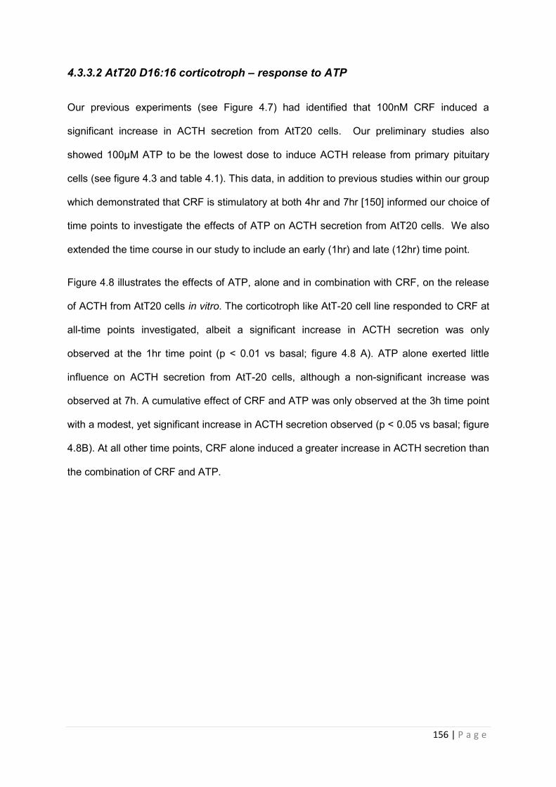
156 | P a g e
4.3.3.2 AtT20 D16:16 corticotroph – response to ATP
Our previous experiments (see Figure 4.7) had identified that 100nM CRF induced a
significant increase in ACTH secretion from AtT20 cells. Our preliminary studies also
showed 100µM ATP to be the lowest dose to induce ACTH release from primary pituitary
cells (see figure 4.3 and table 4.1). This data, in addition to previous studies within our group
which demonstrated that CRF is stimulatory at both 4hr and 7hr [150] informed our choice of
time points to investigate the effects of ATP on ACTH secretion from AtT20 cells. We also
extended the time course in our study to include an early (1hr) and late (12hr) time point.
Figure 4.8 illustrates the effects of ATP, alone and in combination with CRF, on the release
of ACTH from AtT20 cells in vitro. The corticotroph like AtT-20 cell line responded to CRF at
all-time points investigated, albeit a significant increase in ACTH secretion was only
observed at the 1hr time point (p < 0.01 vs basal; figure 4.8 A). ATP alone exerted little
influence on ACTH secretion from AtT-20 cells, although a non-significant increase was
observed at 7h. A cumulative effect of CRF and ATP was only observed at the 3h time point
with a modest, yet significant increase in ACTH secretion observed (p < 0.05 vs basal; figure
4.8B). At all other time points, CRF alone induced a greater increase in ACTH secretion than
the combination of CRF and ATP.
157 | P a g e
Figure 4.8: Effects of CRF and ATP on ACTH secretion from AtT20 D16:D16. AtT20 cells
were treated with ATP (100uM) alone or in combination with CRF (100nM) for 1(A), 4(B),
7(C) and 12(D) hrs. The medium from cells (2x 10⁵/well) was collected and ACTH detected
by RIA. Bars represent means (n=6)± SEM. *p<0.05 vs control **p<0.01 vs control. Data
represents average of one experiment. Data compared by one way ANOVA followed by
Tukey’s post hoc test.
4.3.3.4 GH3 lactotroph cell line - response to secretagogue (thyrotrophin releasing
hormone)
Given that AtT20:D16 were not responsive to ATP at the early time points (see figure 4.8 a
and b), we proceeded to look at the effects of ATP on PRL release using the in vitro model
of lactorophs, GH3 cells. Our previous expression profiling studies had shown the cell line
expresses P2X3, 4 and 7 (see figure 3.2 c and table 3.1). As our group had little experience
with these cells, we began our investigations by culturing the cells as described previously
[131] and treated with graded concentrations of thyrotrophin releasing hormone (TRH), a
ACTH
pg/
ml
Control CRH ATP CRH+ATP0
200
400
600
800
1000A
**
ACTH
pg/
ml
control CRH ATP ATP+CRH0
200
400
600
800
1000
*
BAC
TH p
g/m
l
control CRH ATP ATP+CRH0
200
400
600
800
1000C
******
**AC
TH p
g/m
l
control CRH ATP ATP+CRH0
200
400
600
800
1000D
******
**
158 | P a g e
well-established secretagogue for prolactin [[151];[2]]. A TRH dose response was conducted
utilising doses and time points (0, 10, 30, 60 and 90 minutes) that previously been shown to
be effective [[14];[2]].
Figure 4.9 illustrates the effects of graded concentrations of TRH on the release of PRL from
GH3 cells in vitro. All doses of TRH induced a significant increase in PRL secretion from
GH3 cells at 60 and 90 minutes. In addition, 100nM and 1000nM TRH induced a significant
increase in PRL secretion compared to basal levels at 10 minutes (P<0.05 vs. control).
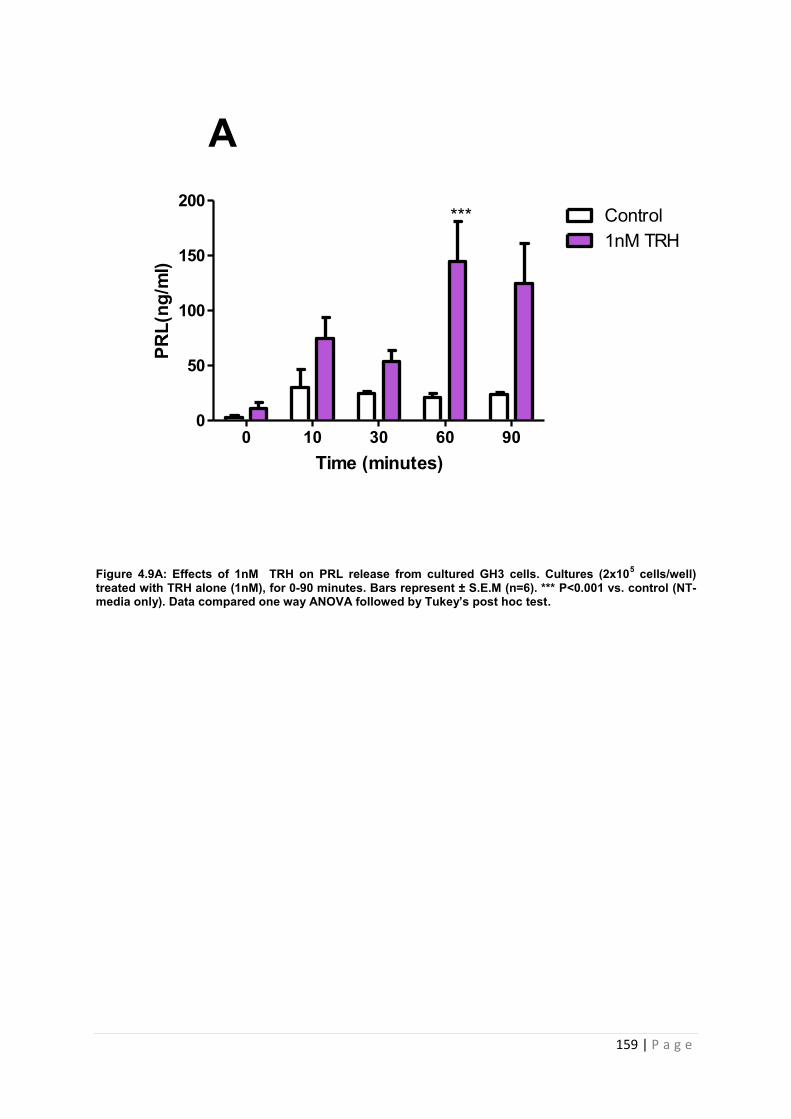
159 | P a g e
Figure 4.9A: Effects of 1nM TRH on PRL release from cultured GH3 cells. Cultures (2x10⁵ cells/well) treated with TRH alone (1nM), for 0-90 minutes. Bars represent ± S.E.M (n=6). *** P<0.001 vs. control (NT-media only). Data compared one way ANOVA followed by Tukey’s post hoc test.
0 10 30 60 900
50
100
150
200Control1nM TRH
Time (minutes)
PRL(
ng/m
l)
***
A
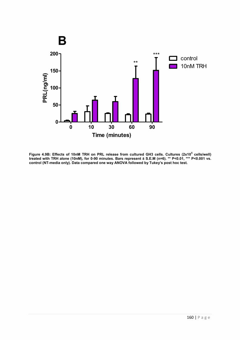
160 | P a g e
Figure 4.9B: Effects of 10nM TRH on PRL release from cultured GH3 cells. Cultures (2x10⁵ cells/well) treated with TRH alone (10nM), for 0-90 minutes. Bars represent ± S.E.M (n=6). ** P<0.01, *** P<0.001 vs. control (NT-media only). Data compared one way ANOVA followed by Tukey’s post hoc test.
0 10 30 60 900
50
100
150
200control10nM TRH
Time (minutes)
PRL(
ng/m
l)
*****
B
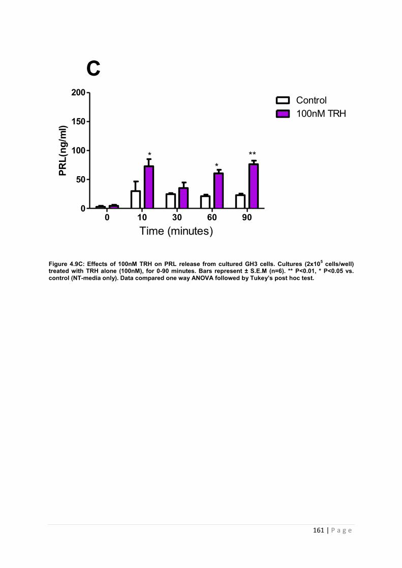
161 | P a g e
Figure 4.9C: Effects of 100nM TRH on PRL release from cultured GH3 cells. Cultures (2x10⁵ cells/well) treated with TRH alone (100nM), for 0-90 minutes. Bars represent ± S.E.M (n=6). ** P<0.01, * P<0.05 vs. control (NT-media only). Data compared one way ANOVA followed by Tukey’s post hoc test.
0 10 30 60 900
50
100
150
200Control100nM TRH
PRL(
ng/m
l)
**
**
C
Time (minutes)
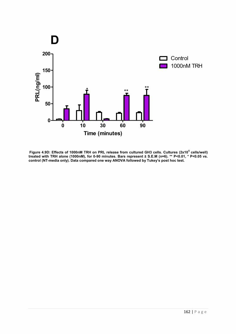
162 | P a g e
Figure 4.9D: Effects of 1000nM TRH on PRL release from cultured GH3 cells. Cultures (2x10⁵ cells/well) treated with TRH alone (1000nM), for 0-90 minutes. Bars represent ± S.E.M (n=6). ** P<0.01, * P<0.05 vs. control (NT-media only). Data compared one way ANOVA followed by Tukey’s post hoc test.
0 10 30 60 900
50
100
150
200Control1000nM TRH
Time (minutes)
PRL(
ng/m
l)
* ** **
D

163 | P a g e
4.3.3.5 GH3 lactotroph cell line - response to ATP
Our previous experiments (Figure 4.9) had identified that TRH induced a significant increase
in PRL secretion from GH3 cells at 60-90min at all doses tested. We therefore decided to
investigate the effects of ATP on PRL secretion from GH3 cells over the same timeframe
Table 4.7 shows the list of ATP analogues, doses and time points investigated
Table 4.7: Summary of ATP analogues and time points assessed in vitro
Cell line
ATP
analogue
Dose (µM) Time pints (minutes)
GH3 ATP,
BzATP,
ATPγS
0.001,0.01,0.1,1,10,100,1000 0,5,10,15,20,30,60
Figure 4.10 illustrates the effects of graded concentrations of ATP on the release of PRL
from GH3 cells in vitro. ATP induced a significant increase in PRL release from GH3 cells at
all concentrations investigated. A dose dependent increase in PRL secretion in response to
ATP was not observed, with maximal PRL secretion between 150-250ng/ml for all doses
tested. With regard to timeframe, ATP consistently increased PRL secretion at 5 min – only
the 1000uM dose failed to increase PRL release at 5 min. The timeframe for the ATP
response at each dose is detailed in the table below. 0.001µM ATP induced a significant
increase in PRL secretion at 5 (P<0.001 vs. control), 10 (P<0.01vs control), 15 (P<0.01vs
control), 30 (P<0.001 vs. control, figure 4.10A) and 60minutes (P<0.001 vs. control, figure
4.10.A). 0.01µM ATP induced a significant increase in PRL secretion at 10 (P<0.05 vs.
control,), 15 (P<0.05 vs. control) and 20 minutes (P<0.001 vs. control)(figure 4.10.B). 0.1µM
ATP induced a significant increase in PRL secretion at 5 (P<0.05 vs. control), 15 (P<0.05 vs.
control), 30 (P<0.001 vs. control) and 60 minutes (P<0.001 vs. control) (figure 4.10C). 1µM
164 | P a g e
ATP induced a significant increase in PRL secretion at 5 (P<0.01 vs control), 10 (P<0.05 vs.
control), 15 (P<0.001 vs. control), 20 (P<0.01vs control ) and 30 minutes (P<0.05 vs. control)
(figure 4.10D). 10µM ATP induced a significant increase in PRL secretion at 5 (P<0.001 vs.
control), 15 (P<0.001 vs. control), 30 (P<0.01vs control) and 60 minutes (P<0.001 vs.
control) (figur4.10.E). 100µM ATP induced a significant increase in PRL secretion at all-time
points investigated (P<0.001 vs. control)(figure 2.F), whereas 1000µM of ATP only induced a
significant increase at the 60 minute time point (P<0.001 vs. control)(figure 4.10.G.). Results
summarised in table 4.8.
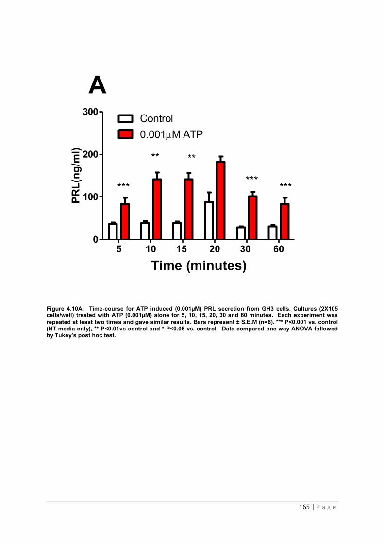
165 | P a g e
Figure 4.10A: Time-course for ATP induced (0.001µM) PRL secretion from GH3 cells. Cultures (2X10⁵ cells/well) treated with ATP (0.001µM) alone for 5, 10, 15, 20, 30 and 60 minutes. Each experiment was repeated at least two times and gave similar results. Bars represent ± S.E.M (n=6). *** P<0.001 vs. control (NT-media only), ** P<0.01vs control and * P<0.05 vs. control. Data compared one way ANOVA followed by Tukey’s post hoc test.
5 10 15 20 30 600
100
200
300 Control0.001M ATP
Time (minutes)
PRL(
ng/m
l)A
***
** **
*** ***
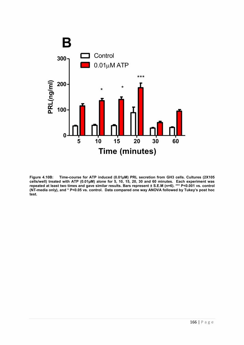
166 | P a g e
Figure 4.10B: Time-course for ATP induced (0.01µM) PRL secretion from GH3 cells. Cultures (2X10⁵ cells/well) treated with ATP (0.01µM) alone for 5, 10, 15, 20, 30 and 60 minutes. Each experiment was repeated at least two times and gave similar results. Bars represent ± S.E.M (n=6). *** P<0.001 vs. control (NT-media only), and * P<0.05 vs. control. Data compared one way ANOVA followed by Tukey’s post hoc test.
5 10 15 20 30 600
100
200
300 Control0.01M ATP
Time (minutes)
PRL(
ng/m
l)B
* ****
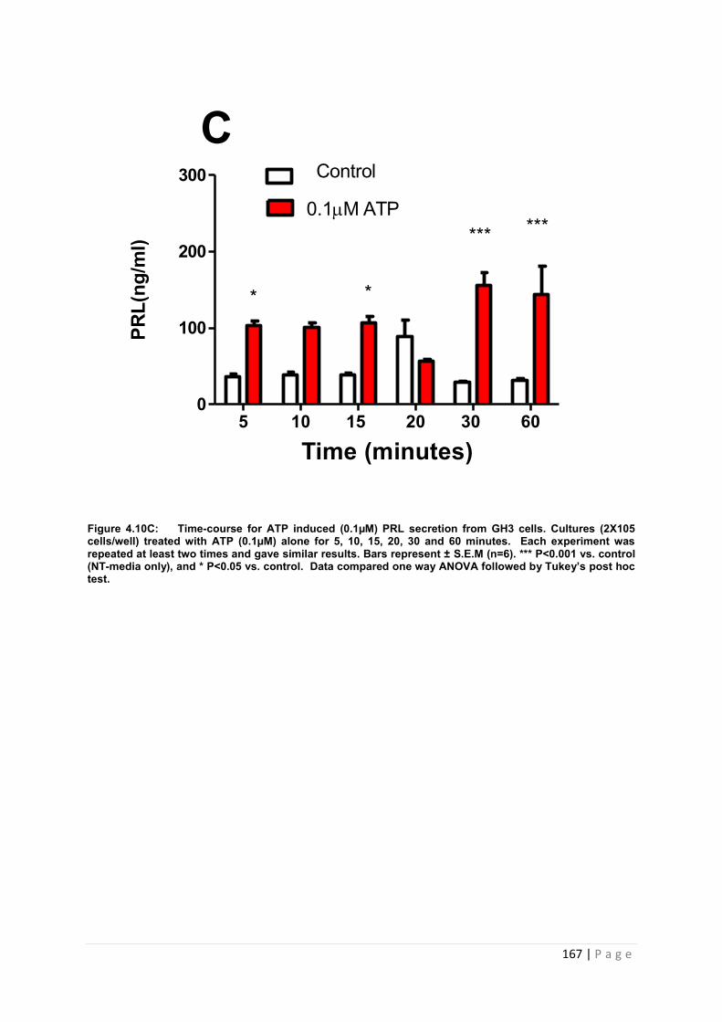
167 | P a g e
Figure 4.10C: Time-course for ATP induced (0.1µM) PRL secretion from GH3 cells. Cultures (2X10⁵ cells/well) treated with ATP (0.1µM) alone for 5, 10, 15, 20, 30 and 60 minutes. Each experiment was repeated at least two times and gave similar results. Bars represent ± S.E.M (n=6). *** P<0.001 vs. control (NT-media only), and * P<0.05 vs. control. Data compared one way ANOVA followed by Tukey’s post hoc test.
5 10 15 20 30 600
100
200
300
0.1M ATP
Time (minutes)
PRL(
ng/m
l)C
* *
*** ***
Control
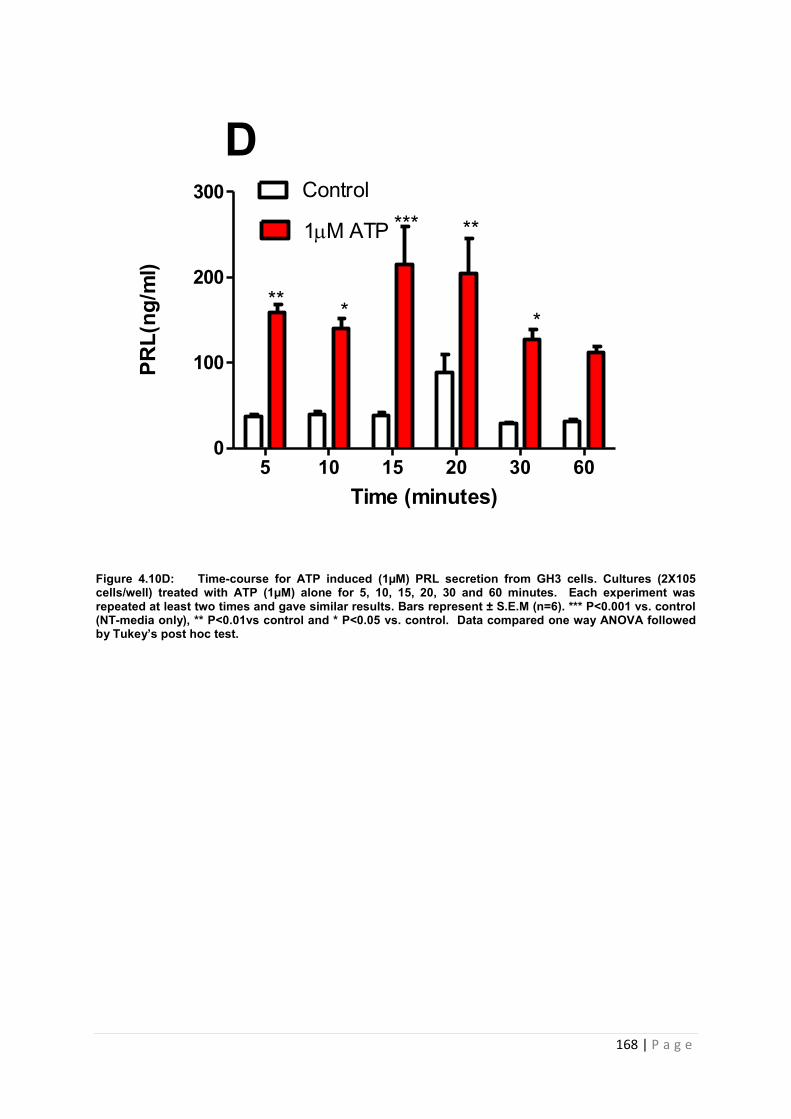
168 | P a g e
Figure 4.10D: Time-course for ATP induced (1µM) PRL secretion from GH3 cells. Cultures (2X10⁵ cells/well) treated with ATP (1µM) alone for 5, 10, 15, 20, 30 and 60 minutes. Each experiment was repeated at least two times and gave similar results. Bars represent ± S.E.M (n=6). *** P<0.001 vs. control (NT-media only), ** P<0.01vs control and * P<0.05 vs. control. Data compared one way ANOVA followed by Tukey’s post hoc test.
Time (minutes)
PRL(
ng/m
l)
5 10 15 20 30 600
100
200
300 Control
1M ATP
D
*
*** **
***
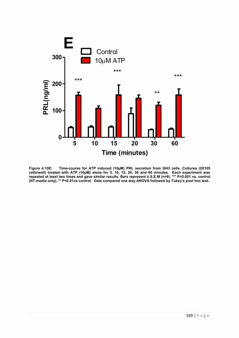
169 | P a g e
Figure 4.10E: Time-course for ATP induced (10µM) PRL secretion from GH3 cells. Cultures (2X10⁵ cells/well) treated with ATP (10µM) alone for 5, 10, 15, 20, 30 and 60 minutes. Each experiment was repeated at least two times and gave similar results. Bars represent ± S.E.M (n=6). *** P<0.001 vs. control (NT-media only), ** P<0.01vs control. Data compared one way ANOVA followed by Tukey’s post hoc test.
5 10 15 20 30 600
100
200
300 10M ATP
Time (minutes)
PRL(
ng/m
l)E
***
**
******
Control
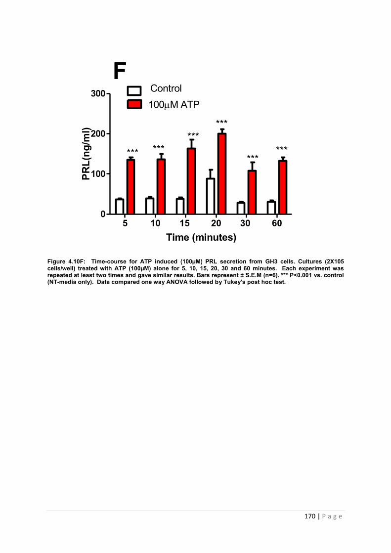
170 | P a g e
Figure 4.10F: Time-course for ATP induced (100µM) PRL secretion from GH3 cells. Cultures (2X10⁵ cells/well) treated with ATP (100µM) alone for 5, 10, 15, 20, 30 and 60 minutes. Each experiment was repeated at least two times and gave similar results. Bars represent ± S.E.M (n=6). *** P<0.001 vs. control (NT-media only). Data compared one way ANOVA followed by Tukey’s post hoc test.
5 10 15 20 30 600
100
200
300
Time (minutes)
PRL(
ng/m
l)F
100M ATPControl
******
***
*********
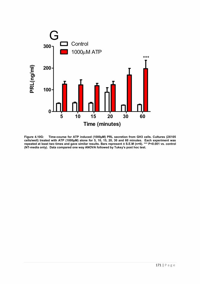
171 | P a g e
Figure 4.10G: Time-course for ATP induced (1000µM) PRL secretion from GH3 cells. Cultures (2X10⁵ cells/well) treated with ATP (1000µM) alone for 5, 10, 15, 20, 30 and 60 minutes. Each experiment was repeated at least two times and gave similar results. Bars represent ± S.E.M (n=6). *** P<0.001 vs. control (NT-media only). Data compared one way ANOVA followed by Tukey’s post hoc test.
5 10 15 20 30 600
100
200
300 Control1000M ATP
Time (minutes)
PRL(
ng/m
l)G
***
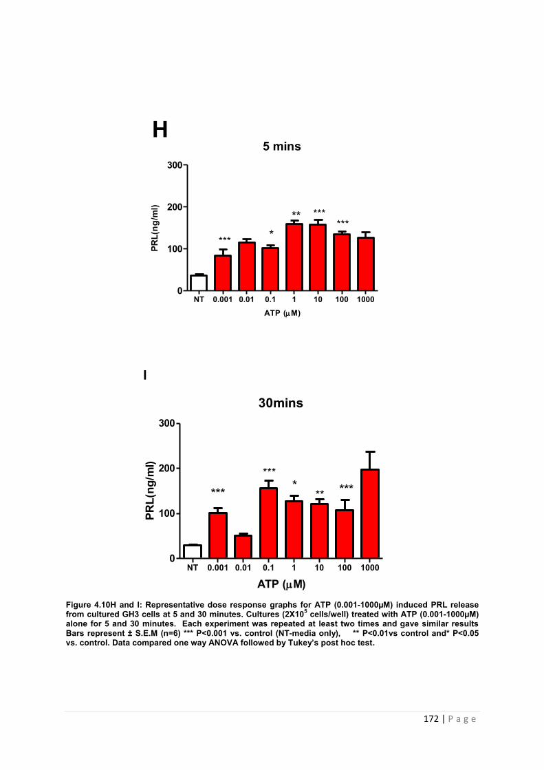
172 | P a g e
5 mins
ATP (M)
PRL(
ng/m
l)
NT 0.001 0.01 0.1 1 10 100 10000
100
200
300
***
******
***
H
30mins
ATP (M)
PRL(
ng/m
l)
NT 0.001 0.01 0.1 1 10 100 10000
100
200
300
***
** *******
I
Figure 4.10H and I: Representative dose response graphs for ATP (0.001-1000µM) induced PRL release from cultured GH3 cells at 5 and 30 minutes. Cultures (2X10⁵ cells/well) treated with ATP (0.001-1000µM) alone for 5 and 30 minutes. Each experiment was repeated at least two times and gave similar results Bars represent ± S.E.M (n=6) *** P<0.001 vs. control (NT-media only), ** P<0.01vs control and* P<0.05 vs. control. Data compared one way ANOVA followed by Tukey’s post hoc test.
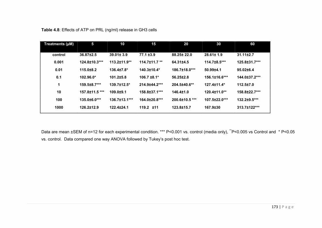
173 | P a g e
Table 4.8: Effects of ATP on PRL (ng/ml) release in GH3 cells
Data are mean ±SEM of n=12 for each experimental condition. *** P<0.001 vs. control (media only), **P<0.005 vs Control and * P<0.05
vs. control. Data compared one way ANOVA followed by Tukey’s post hoc test.
Treatments (µM) 5 10 15 20 30 60
control 36.87±2.5 39.01± 3.9 77.1 ±3.9 88.25± 22.0 28.61± 1.9 31.11±2.7
0.001 124.8±10.3*** 113.2±11.9** 114.7±11.7 ** 64.31±4.5 114.7±8.5*** 125.8±31.7***
0.01 115.0±8.2 136.4±7.8* 140.3±10.4* 186.7±18.0*** 50.99±4.1 95.02±6.4
0.1 102.96.0* 101.2±5.8 106.7 ±8.1* 56.25±2.8 156.1±16.6*** 144.0±37.2***
1 159.5±8.7*** 139.7±12.5* 214.9±44.2*** 204.5±40.6** 127.4±11.4* 112.5±7.0
10 157.8±11.5 *** 109.0±9.1 158.8±37.1*** 146.4±1.0 120.4±11.0** 158.8±22.7***
100 135.0±6.0*** 136.7±13.1*** 164.0±20.8*** 200.6±10.5 *** 107.5±22.0*** 132.2±9.5***
1000 126.2±12.9 122.4±24.1 119.2 ±11 123.8±15.7 167.9±30 313.7±122***
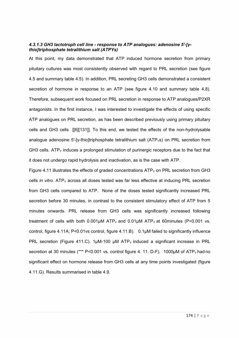
174 | P a g e
4.3.1.3 GH3 lactotroph cell line - response to ATP analogues: adenosine 5′-[γ-thio]triphosphate tetralithium salt (ATPΥs)
At this point, my data demonstrated that ATP induced hormone secretion from primary
pituitary cultures was most consistently observed with regard to PRL secretion (see figure
4.5 and summary table 4.5). In addition, PRL secreting GH3 cells demonstrated a consistent
secretion of hormone in response to an ATP (see figure 4.10 and summary table 4.8).
Therefore, subsequent work focused on PRL secretion in response to ATP analogues/P2XR
antagonists. In the first instance, I was interested to investigate the effects of using specific
ATP analogues on PRL secretion, as has been described previously using primary pituitary
cells and GH3 cells [[6][131]]. To this end, we tested the effects of the non-hydrolysable
analogue adenosine 5′-[γ-thio]triphosphate tetralithium salt (ATPΥs) on PRL secretion from
GH3 cells. ATPΥ induces a prolonged stimulation of purinergic receptors due to the fact that
it does not undergo rapid hydrolysis and inactivation, as is the case with ATP.
Figure 4.11 illustrates the effects of graded concentrations ATPΥ on PRL secretion from GH3
cells in vitro. ATPΥ across all doses tested was far less effective at inducing PRL secretion
from GH3 cells compared to ATP. None of the doses tested significantly increased PRL
secretion before 30 minutes, in contrast to the consistent stimulatory effect of ATP from 5
minutes onwards. PRL release from GH3 cells was significantly increased following
treatment of cells with both 0.001µM ATPΥ and 0.01µM ATPΥ at 60minutes (P<0.001 vs.
control, figure 4.11A; P<0.01vs control, figure 4.11.B). 0.1µM failed to significantly influence
PRL secretion (Figure 411.C). 1µM-100 µM ATPΥ induced a significant increase in PRL
secretion at 30 minutes (*** P<0.001 vs. control figure 4. 11. D-F). 1000µM of ATPΥ had no
significant effect on hormone release from GH3 cells at any time points investigated (figure
4.11.G). Results summarised in table 4.9.
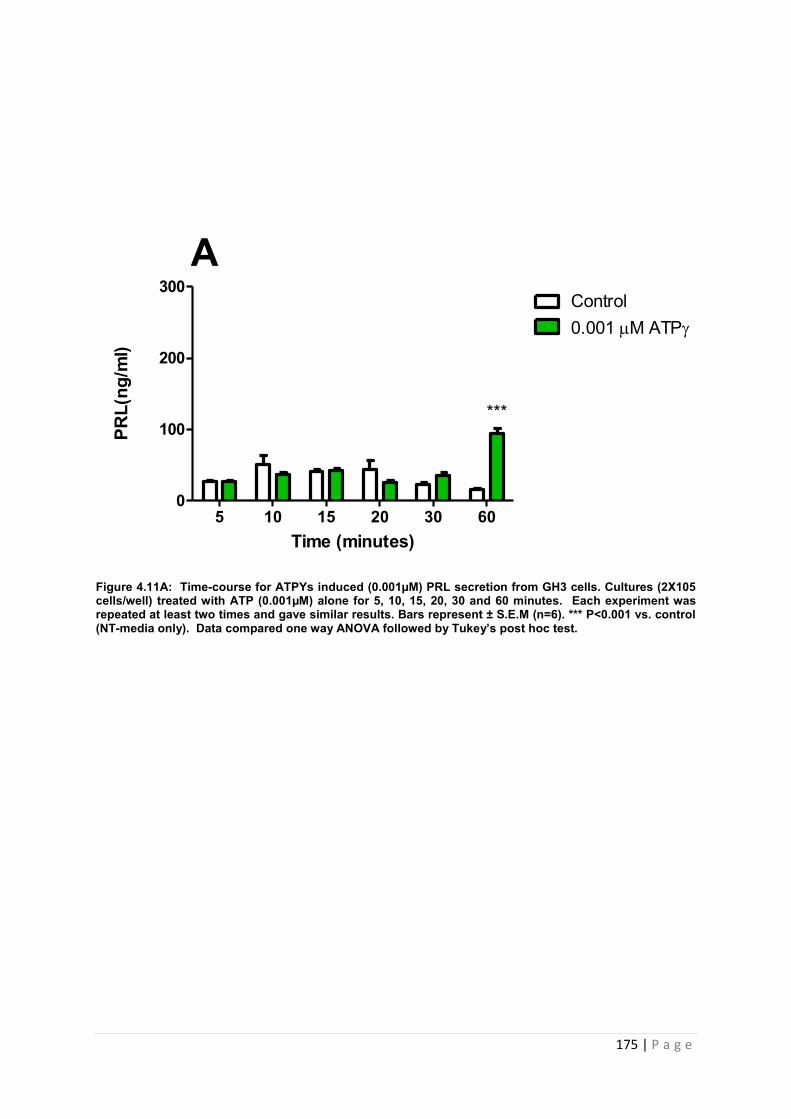
175 | P a g e
Figure 4.11A: Time-course for ATPYs induced (0.001µM) PRL secretion from GH3 cells. Cultures (2X10⁵ cells/well) treated with ATP (0.001µM) alone for 5, 10, 15, 20, 30 and 60 minutes. Each experiment was repeated at least two times and gave similar results. Bars represent ± S.E.M (n=6). *** P<0.001 vs. control (NT-media only). Data compared one way ANOVA followed by Tukey’s post hoc test.
Time (minutes)
PRL(
ng/m
l)
5 10 15 20 30 600
100
200
300Control0.001 M ATP
***
A
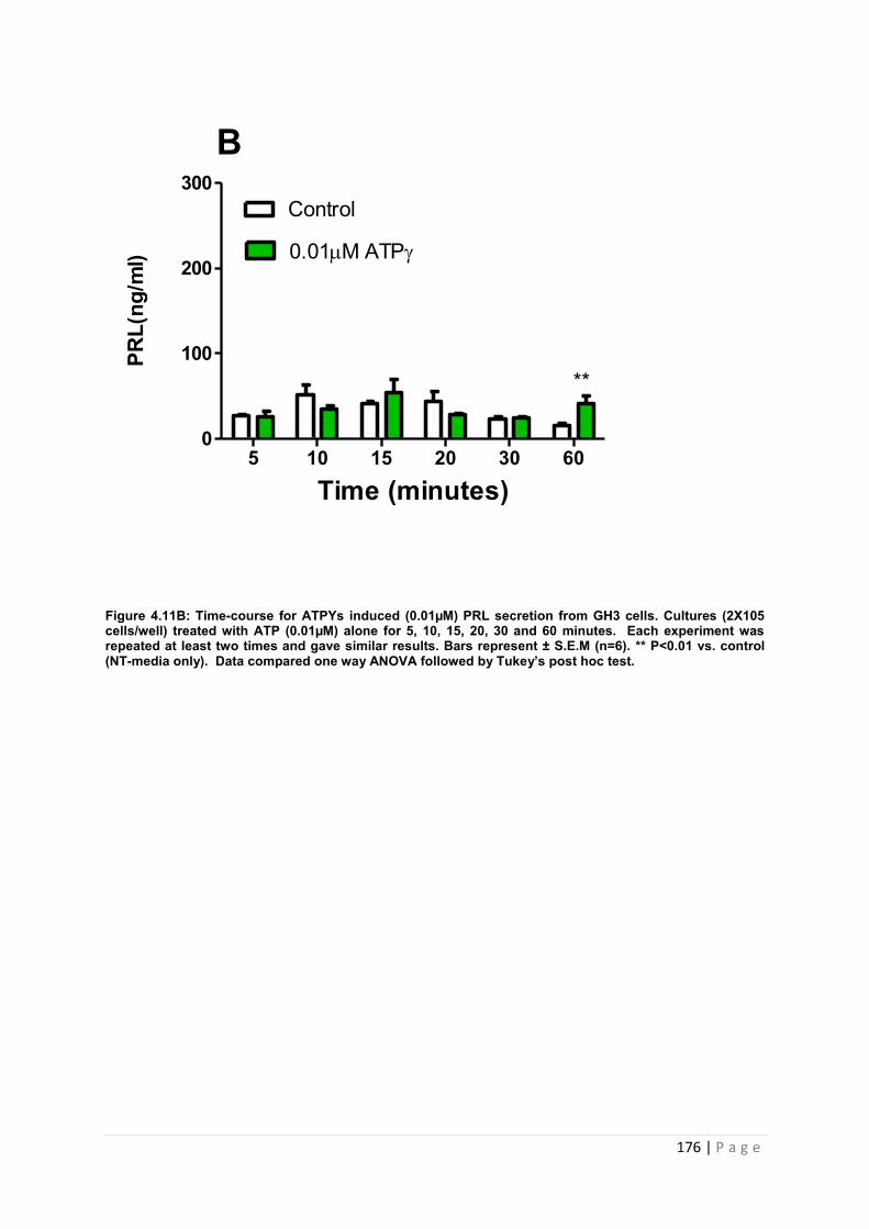
176 | P a g e
Figure 4.11B: Time-course for ATPYs induced (0.01µM) PRL secretion from GH3 cells. Cultures (2X10⁵ cells/well) treated with ATP (0.01µM) alone for 5, 10, 15, 20, 30 and 60 minutes. Each experiment was repeated at least two times and gave similar results. Bars represent ± S.E.M (n=6). ** P<0.01 vs. control (NT-media only). Data compared one way ANOVA followed by Tukey’s post hoc test.
Time (minutes)
PRL(
ng/m
l)
5 10 15 20 30 600
100
200
300Control
0.01M ATP
**
B
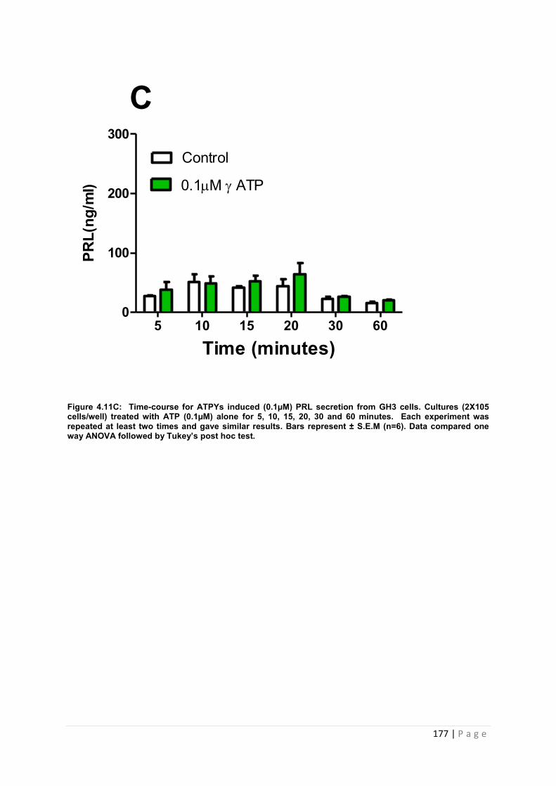
177 | P a g e
Figure 4.11C: Time-course for ATPYs induced (0.1µM) PRL secretion from GH3 cells. Cultures (2X10⁵ cells/well) treated with ATP (0.1µM) alone for 5, 10, 15, 20, 30 and 60 minutes. Each experiment was repeated at least two times and gave similar results. Bars represent ± S.E.M (n=6). Data compared one way ANOVA followed by Tukey’s post hoc test.
Time (minutes)
PRL(
ng/m
l)
5 10 15 20 30 600
100
200
300
Control
0.1M ATP
C
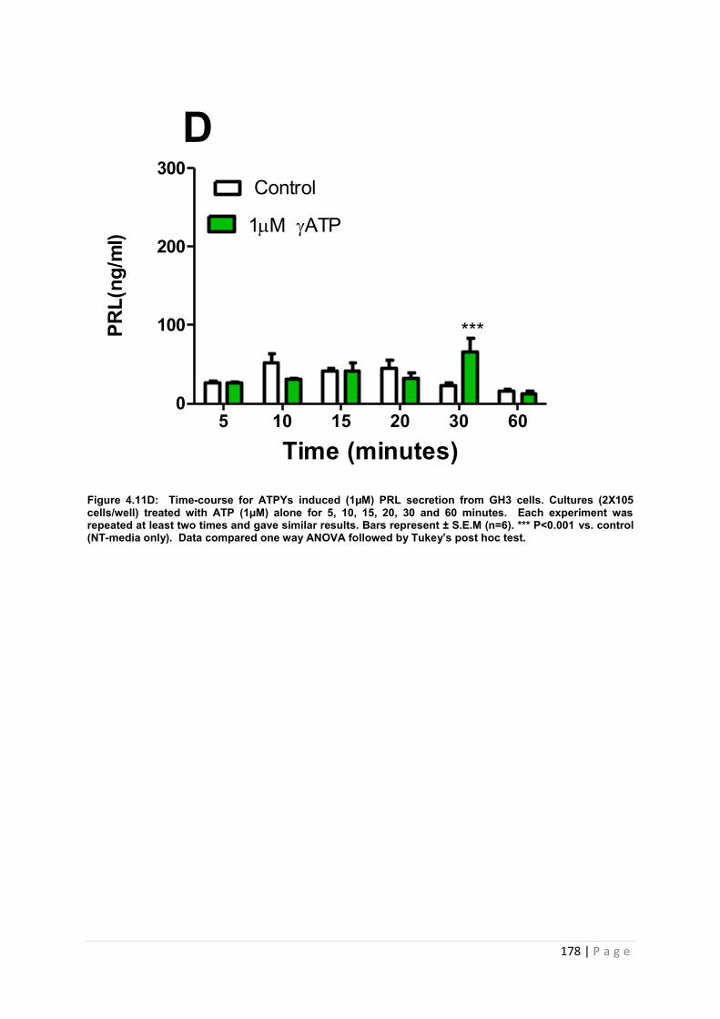
178 | P a g e
Figure 4.11D: Time-course for ATPYs induced (1µM) PRL secretion from GH3 cells. Cultures (2X10⁵ cells/well) treated with ATP (1µM) alone for 5, 10, 15, 20, 30 and 60 minutes. Each experiment was repeated at least two times and gave similar results. Bars represent ± S.E.M (n=6). *** P<0.001 vs. control (NT-media only). Data compared one way ANOVA followed by Tukey’s post hoc test.
Time (minutes)
PRL(
ng/m
l)
5 10 15 20 30 600
100
200
300Control
1M ATP
D
***
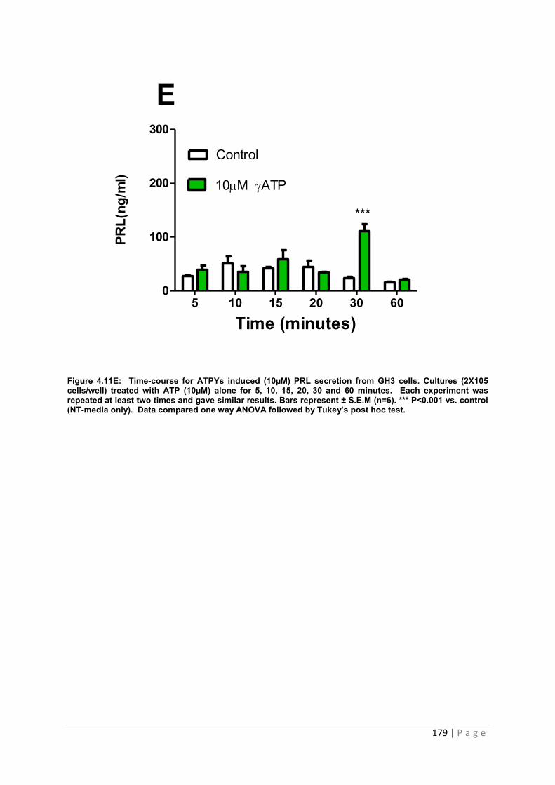
179 | P a g e
Figure 4.11E: Time-course for ATPYs induced (10µM) PRL secretion from GH3 cells. Cultures (2X10⁵ cells/well) treated with ATP (10µM) alone for 5, 10, 15, 20, 30 and 60 minutes. Each experiment was repeated at least two times and gave similar results. Bars represent ± S.E.M (n=6). *** P<0.001 vs. control (NT-media only). Data compared one way ANOVA followed by Tukey’s post hoc test.
Time (minutes)
PRL(
ng/m
l)
5 10 15 20 30 600
100
200
300
Control
10M ATP
***
E
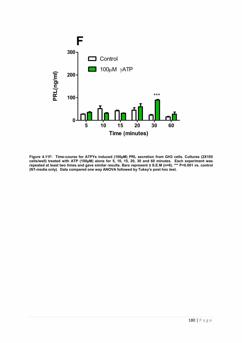
180 | P a g e
Figure 4.11F: Time-course for ATPYs induced (100µM) PRL secretion from GH3 cells. Cultures (2X10⁵ cells/well) treated with ATP (100µM) alone for 5, 10, 15, 20, 30 and 60 minutes. Each experiment was repeated at least two times and gave similar results. Bars represent ± S.E.M (n=6). *** P<0.001 vs. control (NT-media only). Data compared one way ANOVA followed by Tukey’s post hoc test.
Time (minutes)
PRL(
ng/m
l)
5 10 15 20 30 600
100
200
300Control
100M ATP
***
F
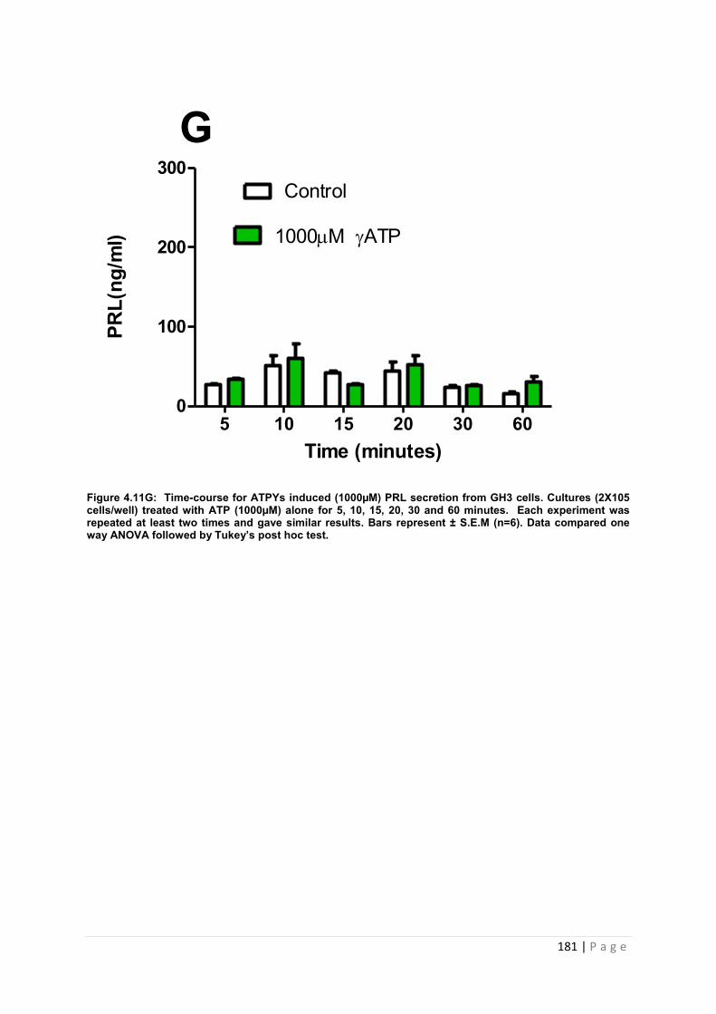
181 | P a g e
Figure 4.11G: Time-course for ATPYs induced (1000µM) PRL secretion from GH3 cells. Cultures (2X10⁵ cells/well) treated with ATP (1000µM) alone for 5, 10, 15, 20, 30 and 60 minutes. Each experiment was repeated at least two times and gave similar results. Bars represent ± S.E.M (n=6). Data compared one way ANOVA followed by Tukey’s post hoc test.
Time (minutes)
PRL(
ng/m
l)
5 10 15 20 30 600
100
200
300Control
1000M ATP
G
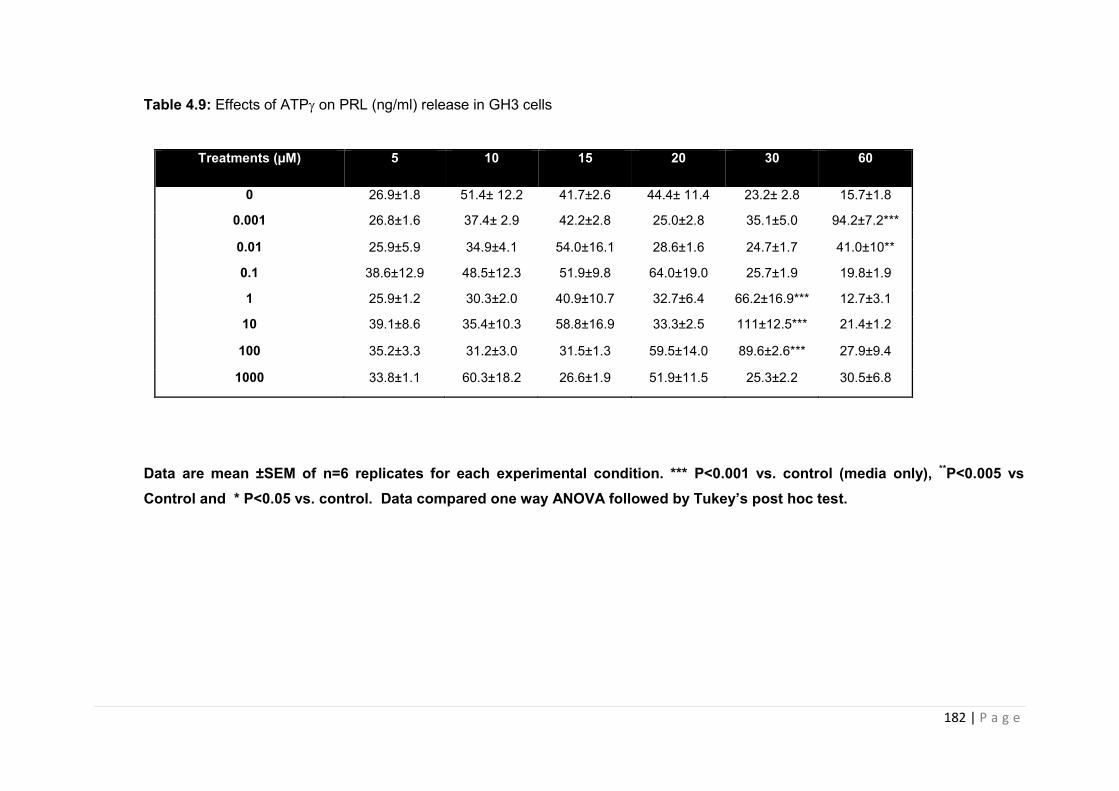
182 | P a g e
Table 4.9: Effects of ATP on PRL (ng/ml) release in GH3 cells
Data are mean ±SEM of n=6 replicates for each experimental condition. *** P<0.001 vs. control (media only), **P<0.005 vs Control and * P<0.05 vs. control. Data compared one way ANOVA followed by Tukey’s post hoc test.
Treatments (µM) 5 10 15 20 30 60
0 26.9±1.8 51.4± 12.2 41.7±2.6 44.4± 11.4 23.2± 2.8 15.7±1.8
0.001 26.8±1.6 37.4± 2.9 42.2±2.8 25.0±2.8 35.1±5.0 94.2±7.2***
0.01 25.9±5.9 34.9±4.1 54.0±16.1 28.6±1.6 24.7±1.7 41.0±10**
0.1 38.6±12.9 48.5±12.3 51.9±9.8 64.0±19.0 25.7±1.9 19.8±1.9
1 25.9±1.2 30.3±2.0 40.9±10.7 32.7±6.4 66.2±16.9*** 12.7±3.1
10 39.1±8.6 35.4±10.3 58.8±16.9 33.3±2.5 111±12.5*** 21.4±1.2
100 35.2±3.3 31.2±3.0 31.5±1.3 59.5±14.0 89.6±2.6*** 27.9±9.4
1000 33.8±1.1 60.3±18.2 26.6±1.9 51.9±11.5 25.3±2.2 30.5±6.8

183 | P a g e
4.3.1.4 GH3 lactotroph cell line - response to 3'-O-[4-benzoyl]benzoyl adenosine 5'-
triphosphate (BzATP), a selective P2X receptor agonist.
In addition to testing non-selective purinergic receptor agonists, we also tested the effects of
a selective purinergic P2X receptor agonist, 3'-O-[4-benzoyl]benzoyl adenosine 5'-
triphosphate (BzATP) on PRL secretion in vitro. BzATP has been reported in the literature
as showing selectivity for P2X receptors and has been shown to augment hormone secretion
in GH3 cells in vitro [[152], [153], [131]].
Figure 4.12 illustrates the effects of graded concentrations of BzATP on the secretion of PRL
from GH3 cells in vitro. In general, BzATP appears to be less potent than ATP, with maximal
PRL secretion in the range of 400-600ng/ml versus 54-78ng/ml compared to ATP alone.
Maximal secretion of PRL with BzATP usually occurs within the 20-30min time frame with a
reduction observed at 60 minutes, PRL secretion was not influenced by treatment with
0.001µM and 0.01µM of BzATP over the 5-60 minute time points (Figures 4.12A and 412.B
respectively) .0.1-10 µM BzATP induced a significant increase in PRL release at 20 and 30
minutes with maximal secretion observed at 20 minutes (P<0.001 vs. control, figure 4.12C-
E). This significant increase in PRL secretion was no longer observed at the 60 minute time
point for each of these doses tested. 100 µM BzATP significantly increases the release of
PRL when incubated for 30 minutes (P<0.05 vs. control figure4.12 F), although the maximal
PRL secretion was greatly reduced compared to other doses. A 1000 µM dose significantly
increases the release of PRL when incubated for 10 (* P<0.05 vs. control, figure 4.G)
Results summarised in table 4.10.
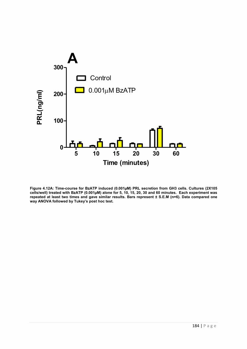
184 | P a g e
Figure 4.12A: Time-course for BzATP induced (0.001µM) PRL secretion from GH3 cells. Cultures (2X10⁵ cells/well) treated with BzATP (0.001µM) alone for 5, 10, 15, 20, 30 and 60 minutes. Each experiment was repeated at least two times and gave similar results. Bars represent ± S.E.M (n=6). Data compared one way ANOVA followed by Tukey’s post hoc test.
Time (minutes)
PRL(
ng/m
l)
5 10 15 20 30 600
100
200
300
Control
0.001M BzATP
A
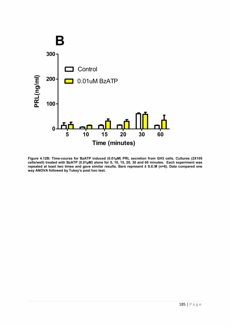
185 | P a g e
Figure 4.12B: Time-course for BzATP induced (0.01µM) PRL secretion from GH3 cells. Cultures (2X10⁵ cells/well) treated with BzATP (0.01µM) alone for 5, 10, 15, 20, 30 and 60 minutes. Each experiment was repeated at least two times and gave similar results. Bars represent ± S.E.M (n=6). Data compared one way ANOVA followed by Tukey’s post hoc test.
Time (minutes)
PRL(
ng/m
l)
5 10 15 20 30 600
100
200
300
Control
B
0.01uM BzATP
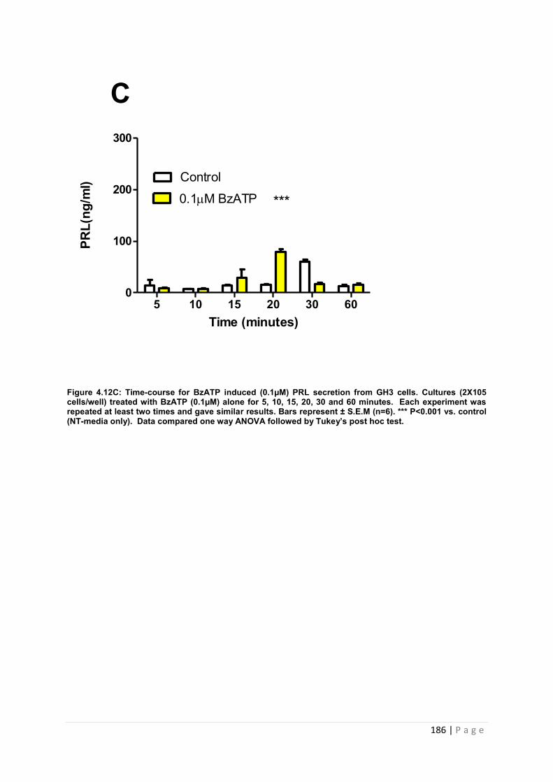
186 | P a g e
Figure 4.12C: Time-course for BzATP induced (0.1µM) PRL secretion from GH3 cells. Cultures (2X10⁵ cells/well) treated with BzATP (0.1µM) alone for 5, 10, 15, 20, 30 and 60 minutes. Each experiment was repeated at least two times and gave similar results. Bars represent ± S.E.M (n=6). *** P<0.001 vs. control (NT-media only). Data compared one way ANOVA followed by Tukey’s post hoc test.
Time (minutes)
PRL(
ng/m
l)
5 10 15 20 30 600
100
200
300
Control
0.1M BzATP
C
***
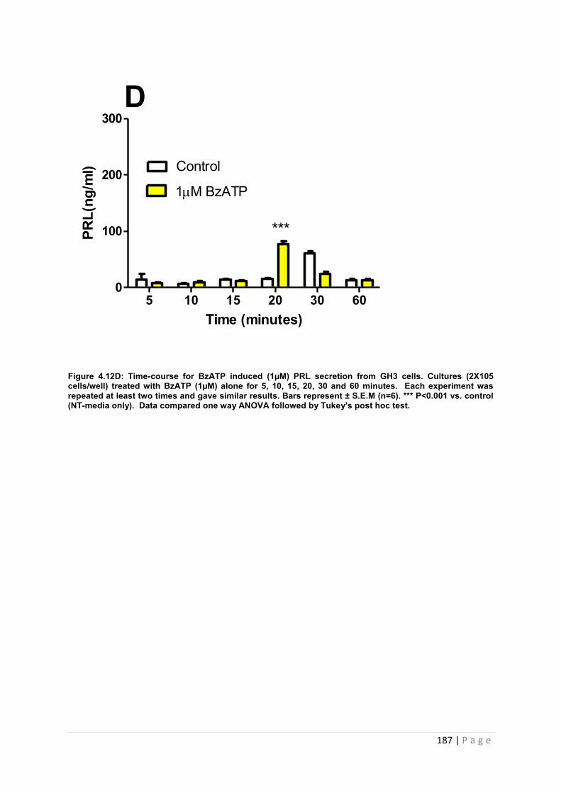
187 | P a g e
Figure 4.12D: Time-course for BzATP induced (1µM) PRL secretion from GH3 cells. Cultures (2X10⁵ cells/well) treated with BzATP (1µM) alone for 5, 10, 15, 20, 30 and 60 minutes. Each experiment was repeated at least two times and gave similar results. Bars represent ± S.E.M (n=6). *** P<0.001 vs. control (NT-media only). Data compared one way ANOVA followed by Tukey’s post hoc test.
Time (minutes)
PRL(
ng/m
l)
5 10 15 20 30 600
100
200
300
Control
D
1M BzATP
***

188 | P a g e
Figure 4.12E: Time-course for BzATP induced (10µM) PRL secretion from GH3 cells. Cultures (2X10⁵ cells/well) treated with BzATP (10µM) alone for 5, 10, 15, 20, 30 and 60 minutes. Each experiment was repeated at least two times and gave similar results. Bars represent ± S.E.M (n=6). *** P<0.001 vs. control (NT-media only). Data compared one way ANOVA followed by Tukey’s post hoc test.
Time (minutes)
PRL(
ng/m
l)
5 10 15 20 30 600
100
200
300
Control
10M BzATP
E
***
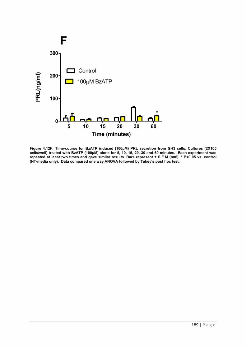
189 | P a g e
Figure 4.12F: Time-course for BzATP induced (100µM) PRL secretion from GH3 cells. Cultures (2X10⁵ cells/well) treated with BzATP (100µM) alone for 5, 10, 15, 20, 30 and 60 minutes. Each experiment was repeated at least two times and gave similar results. Bars represent ± S.E.M (n=6). * P<0.05 vs. control (NT-media only). Data compared one way ANOVA followed by Tukey’s post hoc test.
Time (minutes)
PRL(
ng/m
l)
5 10 15 20 30 600
100
200
300
Control
100M BzATP
F
*
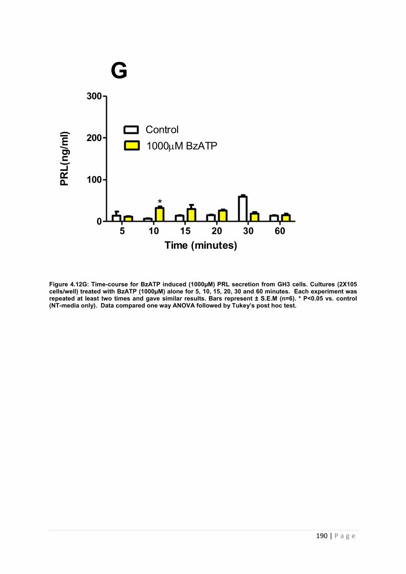
190 | P a g e
Figure 4.12G: Time-course for BzATP induced (1000µM) PRL secretion from GH3 cells. Cultures (2X10⁵ cells/well) treated with BzATP (1000µM) alone for 5, 10, 15, 20, 30 and 60 minutes. Each experiment was repeated at least two times and gave similar results. Bars represent ± S.E.M (n=6). * P<0.05 vs. control (NT-media only). Data compared one way ANOVA followed by Tukey’s post hoc test.
Time (minutes)
PRL(
ng/m
l)
5 10 15 20 30 600
100
200
300
Control1000M BzATP
G
*
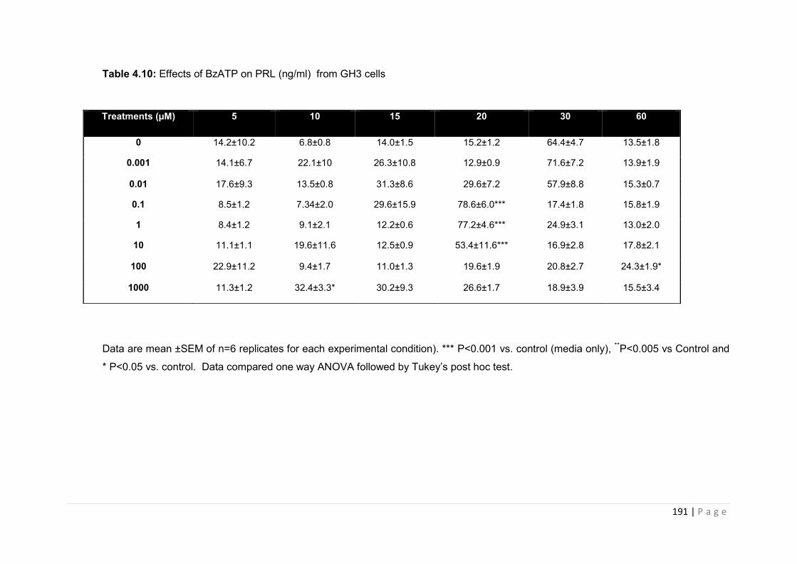
191 | P a g e
Table 4.10: Effects of BzATP on PRL (ng/ml) from GH3 cells
Data are mean ±SEM of n=6 replicates for each experimental condition). *** P<0.001 vs. control (media only), **P<0.005 vs Control and
* P<0.05 vs. control. Data compared one way ANOVA followed by Tukey’s post hoc test.
Treatments (µM) 5 10 15 20 30 60
0 14.2±10.2 6.8±0.8 14.0±1.5 15.2±1.2 64.4±4.7 13.5±1.8
0.001 14.1±6.7 22.1±10 26.3±10.8 12.9±0.9 71.6±7.2 13.9±1.9
0.01 17.6±9.3 13.5±0.8 31.3±8.6 29.6±7.2 57.9±8.8 15.3±0.7
0.1 8.5±1.2 7.34±2.0 29.6±15.9 78.6±6.0*** 17.4±1.8 15.8±1.9
1 8.4±1.2 9.1±2.1 12.2±0.6 77.2±4.6*** 24.9±3.1 13.0±2.0
10 11.1±1.1 19.6±11.6 12.5±0.9 53.4±11.6*** 16.9±2.8 17.8±2.1
100 22.9±11.2 9.4±1.7 11.0±1.3 19.6±1.9 20.8±2.7 24.3±1.9*
1000 11.3±1.2 32.4±3.3* 30.2±9.3 26.6±1.7 18.9±3.9 15.5±3.4
192 | P a g e
4.3.4 An investigation into the effects of purinergic receptor antagonism on hormone secretion from GH3 cells
The previous section demonstrated the effects of various purinergic receptor agonists on
hormone secretion within an in vitro model of pituitary lactotrophs, GH3 cells. ATP
analogues stimulated PRL hormone release from cells in the following order of
potency:ATP>ATPγ>BzATP. However, investigation into P2X function is limited by the range
of purinergic agonists currently available. Whilst the various agonists utilised induced
activation of various members of the P2 family (i.e ATP activates both P2Y and P2X; BzATP
is a P2XR agonist), they are not sufficiently selective to study the significance of individual
P2XR activation within pituitary cells and the subsequent effect on hormone secretion.
The aim of the experiments described in this section was to utilise purinergic receptor
antagonists who were more selective for individual P2XRs in order to assess the functional
significance of individual P2XRs within the pituitary (see table 4.11).
We explored the effect of P2X antagonists on secretagogue induced PRL secretion and ATP
stimulated hormone release. We also examined the ability of ATP to amplify TRH induced
PRL release from GH3 cells. This effect had been previously described in primary rat
gonadotrophs, using GnRH and ATP [34]. In the following section we demonstrated a similar
interaction between ATP and TRH and determined the effects of antagonists on PRL
release.
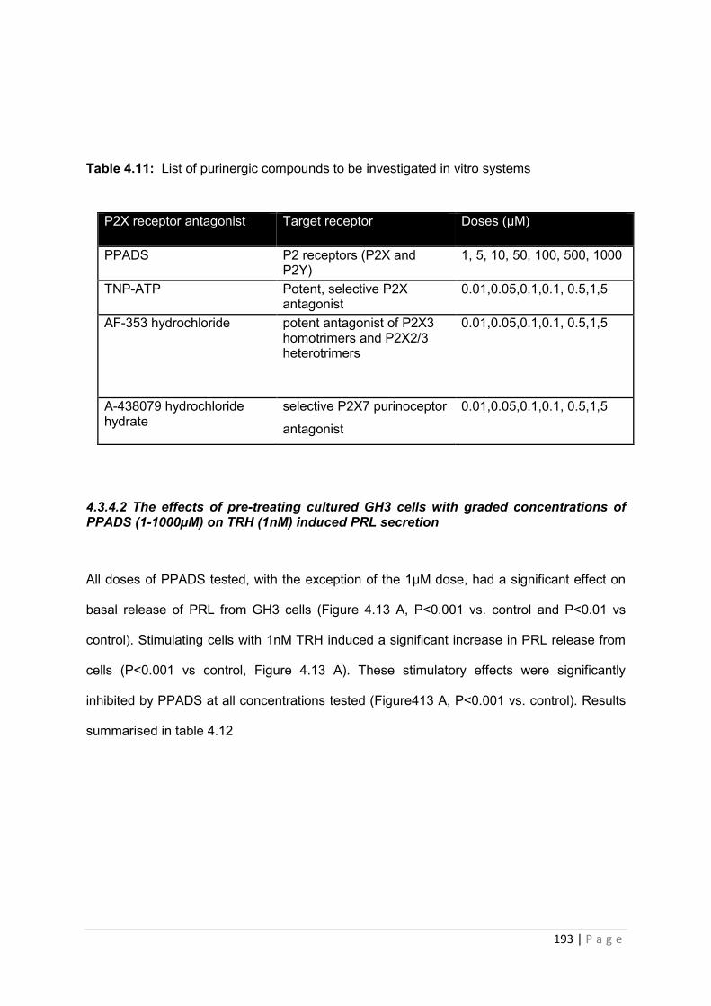
193 | P a g e
Table 4.11: List of purinergic compounds to be investigated in vitro systems
4.3.4.2 The effects of pre-treating cultured GH3 cells with graded concentrations of PPADS (1-1000µM) on TRH (1nM) induced PRL secretion
All doses of PPADS tested, with the exception of the 1µM dose, had a significant effect on
basal release of PRL from GH3 cells (Figure 4.13 A, P<0.001 vs. control and P<0.01 vs
control). Stimulating cells with 1nM TRH induced a significant increase in PRL release from
cells (P<0.001 vs control, Figure 4.13 A). These stimulatory effects were significantly
inhibited by PPADS at all concentrations tested (Figure413 A, P<0.001 vs. control). Results
summarised in table 4.12
P2X receptor antagonist Target receptor Doses (µM)
PPADS P2 receptors (P2X and P2Y)
1, 5, 10, 50, 100, 500, 1000
TNP-ATP Potent, selective P2X antagonist
0.01,0.05,0.1,0.1, 0.5,1,5
AF-353 hydrochloride potent antagonist of P2X3 homotrimers and P2X2/3 heterotrimers
0.01,0.05,0.1,0.1, 0.5,1,5
A-438079 hydrochloride hydrate
selective P2X7 purinoceptor
antagonist 0.01,0.05,0.1,0.1, 0.5,1,5
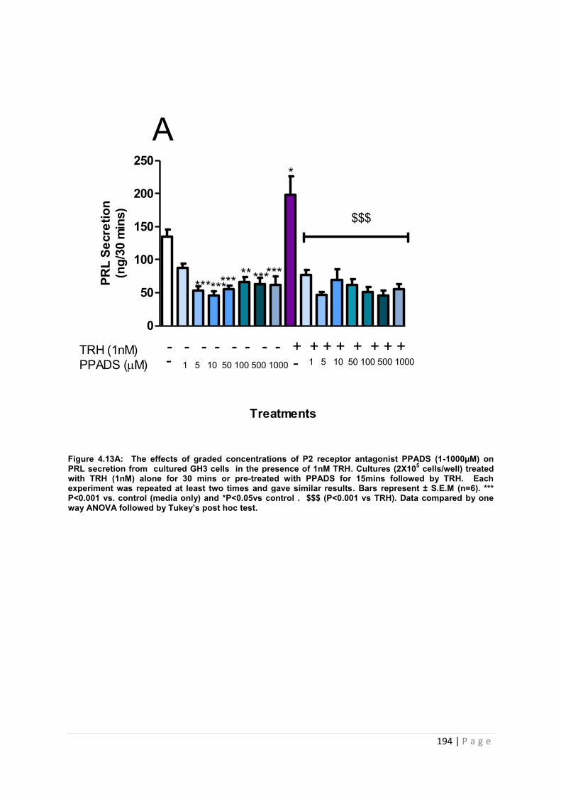
194 | P a g e
Figure 4.13A: The effects of graded concentrations of P2 receptor antagonist PPADS (1-1000µM) on PRL secretion from cultured GH3 cells in the presence of 1nM TRH. Cultures (2X10⁵ cells/well) treated with TRH (1nM) alone for 30 mins or pre-treated with PPADS for 15mins followed by TRH. Each experiment was repeated at least two times and gave similar results. Bars represent ± S.E.M (n=6). *** P<0.001 vs. control (media only) and *P<0.05vs control . $$$ (P<0.001 vs TRH). Data compared by one way ANOVA followed by Tukey’s post hoc test.
0
50
100
150
200
250
Treatments
PRL
Secr
etio
n(n
g/30
min
s)
*****************
*
TRH (1nM)PPADS (M)
- - - - - - - - + + + + + + + + - 1 5 10 50 100 500 1000 - 1 5 10 50 100 500 1000
A
$$$
195 | P a g e
4.3.4.3 The effects of pre-treating cultured GH3 cells with graded concentrations of PPADS (1-1000µM) on ATP (1µM and 100µM) induced PRL secretion
As before, all doses of PPADS tested, with the exception of the 1µM dose, had a significant
effect on basal release of PRL from GH3 cells (Figure 4.13 B, P<0.001 vs. control and
P<0.005 vs control). Stimulating cells with 1µM ATP induced a 25% increase in PRL release
compared to basal secretion, however this was not significant (Figure4.13 B). The
stimulatory effects of ATP on PRL release was significantly inhibited by all doses of PPADS
used (figure 4.15,B, P<0.001 vs ATP and P<0.01 vs ATP). 100uM ATP had no significant
effect on PRL release, but PRL secretion was significantly reduced by PPADS at 5, 10, 100,
500 (figure4.13 C, P<0.001 vs ATP) and 1000µM (figure4.13 c, P<0.05 vs ATP).
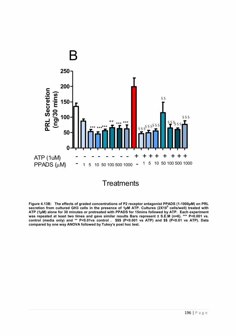
196 | P a g e
Figure 4.13B: The effects of graded concentrations of P2 receptor antagonist PPADS (1-1000µM) on PRL secretion from cultured GH3 cells in the presence of 1µM ATP. Cultures (2X10⁵ cells/well) treated with ATP (1µM) alone for 30 minutes or pretreated with PPADS for 15mins followed by ATP. Each experiment was repeated at least two times and gave similar results Bars represent ± S.E.M (n=6). *** P<0.001 vs. control (media only) and ** P<0.01vs control . $$$ (P<0.001 vs ATP) and $$ (P<0.01 vs ATP). Data compared by one way ANOVA followed by Tukey’s post hoc test.
0
50
100
150
200
250
PRL
Secr
etio
n(n
g/30
min
s)
**
$$$$$$$$$
$$$$$$
$$$
$$
B
ATP (1uM)PPADS (M)
- - - - - - - - + + + + + + + +1 5 10 50 100 500 1000 - 1 5 10 50 100 500 1000 -
*** *********
***
Treatments

197 | P a g e
Figure 4.13C: The effects of graded concentrations of P2 receptor antagonist PPADS (1-1000µM) on PRL secretion from cultured GH3 cells in the presence of 100µM ATP. Cultures (2X10⁵ cells/well) treated with ATP (100µM) alone for 30 minutes or pretreated with PPADS for 15mins followed by ATP. Each experiment was repeated at least two times and gave similar results Bars represent ± S.E.M (n=6). *** P<0.001 vs. control (media only) and ** P<0.01vs control . $$$ (P<0.001 vs ATP) and $$ (P<0.01 vs ATP). Data compared by one way ANOVA followed by Tukey’s post hoc test.
0
50
100
150
200
250PR
L Se
cret
ion
(ng/
30 m
ins)
ATP (100uM)PPADS (M)
- - - - - - - - + + + + + + + +1 5 10 50 100 500 1000 - 1 5 10 50 100 500 1000 -
****** ***********
$$$$$$ $$$
$$$
$
C
198 | P a g e
4.3.4.4 The effects of pre-treating cultured GH3 cells with graded concentrations of PPADS (1-1000µM) on ATP (1µM) and TRH (1nM) induced PRL secretion
As before, 1nM TRH induced a significant increase in PRL release from GH3 cells (P<0.05
vs control, figure 4.13 D), whereas 1µM ATP induced a non-significant rise (25%) in PRL
release.(figure 4.13 D). TRH and ATP in combination, did not induce a significant increase in
PRL release. PRL secretion was decreased by 1, 5, (figure 4.13D, P<0.05 vs ATP) and 100,
500 and 1000µM PPADS (figure4.13D, P<0.001 vs ATP).
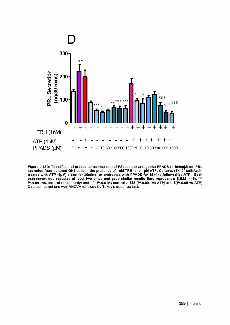
199 | P a g e
Figure 4.13D: The effects of graded concentrations of P2 receptor antagonist PPADS (1-1000µM) on PRL secretion from cultured GH3 cells in the presence of 1nM TRH and 1µM ATP. Cultures (2X10⁵ cells/well) treated with ATP (1µM) alone for 30mins or pretreated with PPADS for 15mins followed by ATP. Each experiment was repeated at least two times and gave similar results Bars represent ± S.E.M (n=6). *** P<0.001 vs. control (media only) and ** P<0.01vs control . $$$ (P<0.001 vs ATP) and $(P<0.05 vs ATP) Data compared one way ANOVA followed by Tukey’s post hoc test.
0
100
200
300
ATP (1uM)PPADS (M)
- - + - - - - - - - + + + + + + + - - 1 5 10 50 100 500 1000 1 5 10 50 100 500 1000 -
TRH (1nM)- + - - - - - - - - + + + + + + + +
D
$$$$
$$$$$$
**
$
******
***
*****PR
L Se
cret
ion
(ng/
30 m
ins)
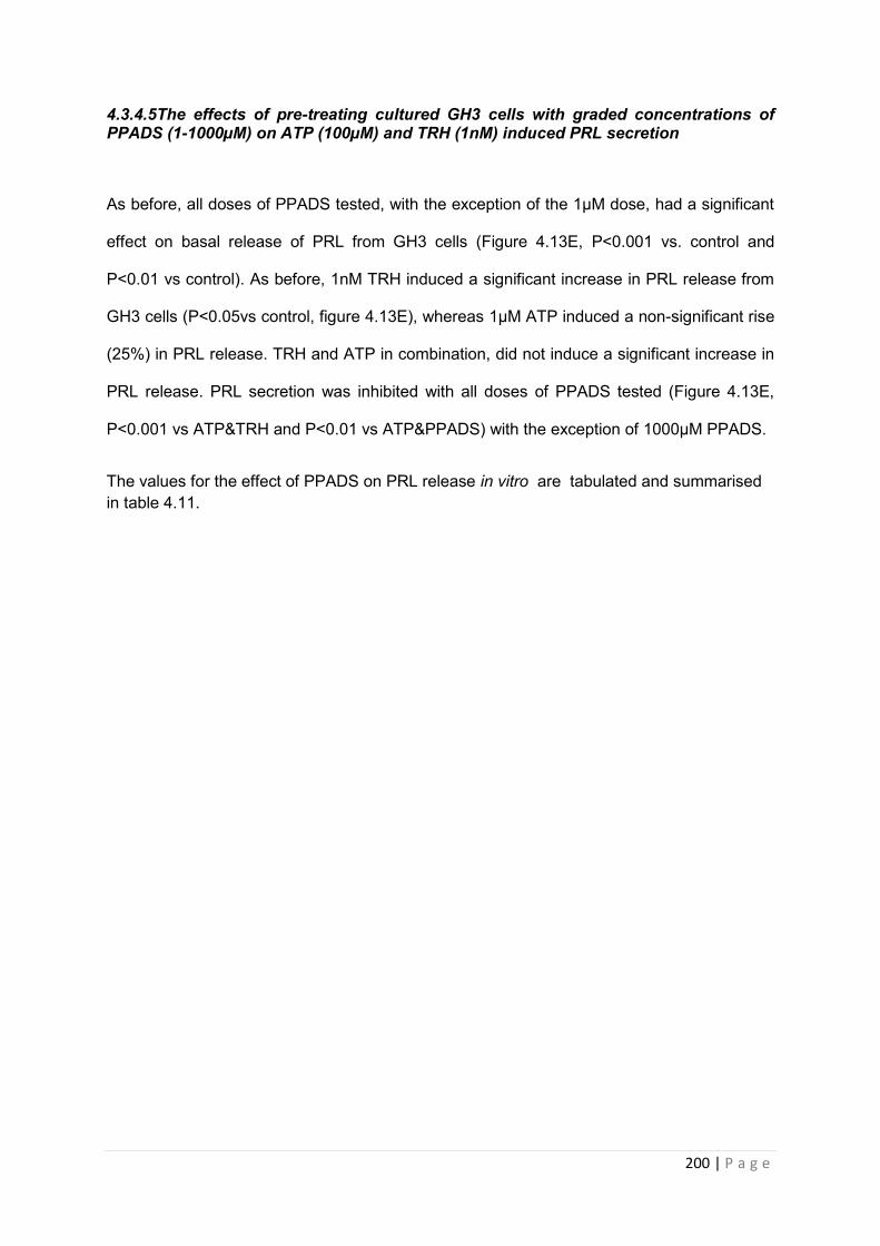
200 | P a g e
4.3.4.5The effects of pre-treating cultured GH3 cells with graded concentrations of PPADS (1-1000µM) on ATP (100µM) and TRH (1nM) induced PRL secretion
As before, all doses of PPADS tested, with the exception of the 1µM dose, had a significant
effect on basal release of PRL from GH3 cells (Figure 4.13E, P<0.001 vs. control and
P<0.01 vs control). As before, 1nM TRH induced a significant increase in PRL release from
GH3 cells (P<0.05vs control, figure 4.13E), whereas 1µM ATP induced a non-significant rise
(25%) in PRL release. TRH and ATP in combination, did not induce a significant increase in
PRL release. PRL secretion was inhibited with all doses of PPADS tested (Figure 4.13E,
P<0.001 vs ATP&TRH and P<0.01 vs ATP&PPADS) with the exception of 1000µM PPADS.
The values for the effect of PPADS on PRL release in vitro are tabulated and summarised in table 4.11.
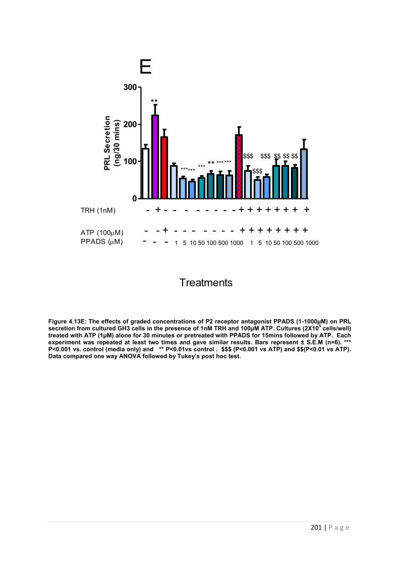
201 | P a g e
Figure 4.13E: The effects of graded concentrations of P2 receptor antagonist PPADS (1-1000µM) on PRL secretion from cultured GH3 cells in the presence of 1nM TRH and 100µM ATP. Cultures (2X10⁵ cells/well) treated with ATP (1µM) alone for 30 minutes or pretreated with PPADS for 15mins followed by ATP. Each experiment was repeated at least two times and gave similar results. Bars represent ± S.E.M (n=6). *** P<0.001 vs. control (media only) and ** P<0.01vs control . $$$ (P<0.001 vs ATP) and $$(P<0.01 vs ATP). Data compared one way ANOVA followed by Tukey’s post hoc test.
0
100
200
300
ATP (100M)PPADS (M)
- - + - - - - - - - + + + + + + + + - - 1 5 10 50 100 500 1000 1 5 10 50 100 500 1000 -
TRH (1nM) - + - - - - - - - - + + + + + + + +
****** ***********
**
$$$
$$$
$$$ $$ $$ $$
E
PRL
Secr
etio
n(n
g/30
min
s)
Treatments
202 | P a g e
Table 4.11: A summary of the effects of PPADS on PRL (ng/ml) release from GH3 cells in vitro.
Data
are mean ±SEM of n=6 replicates for each treatment group. *** P<0.001 vs. control (media only) and ** P<0.01vs control. $$$ (P<0.001 vs ATP) and $$(P<0.01 vs
ATP). Data compared one way ANOVA followed by Tukey’s post hoc test.
PPADS (0µM)
PPADS (1µM)
PPADS (5µM)
PPADS (10µM)
PPADS (50µM)
PPADS (100µM)
PPADS (500µM)
PPADS (1000µM)
Control 135.1±10.37 87.6±6.9 53.5±6.3*** 45.3±6.8*** 56±4.8 *** 66.3±7.8** 63.2±9.3*** 62.09±13.0*** TRH (1nM) 223±28.9 * 76.8±7.7$$$ 46.9±4.5$$$ 69.7±15.7$$$ 61.8±8.5$$$ 51.3±6.9$$$ 46.2±6.7$$$ 55.3±7.6$$$
ATP (1µM) 199.4±28.4 46.4±5.8$$$ 49.1±8.1$$$ 55.3±7.1$$$ 114.5±34.$$ 65.5±11.7$$$ 60.5±4.2$$$ 76.4±11.7$$$
ATP (100µM) 166.2±19.6
121.7±17.4
66.5±13.2$$$ 78.5±15.0$$$ 104.2±9.8 71.0±13.8$$$ 52.3±15.4$$$ 98.1±22.6$
TRH +ATP (1µM)
169.5±22.4 95.2±11.6$ 85.9±21.2$ 109.3±10.1 124.6±12.6 75.19±12.9$$$ 45.03±8.3$$$ 41.9±7.2$$$
TRH +ATP (100µM)
170.8±12.9 75.2±9.0$$$ 49.9±9.0$$$ 57.7±7.3$$$ 88.0±17.7$$ 88.9±11.6$$ 83.1±8.4$$ 132.6±26.0
203 | P a g e
4.3.5.1 An investigation into the effects of TNP-ATP on prolactin secretion from cultured GH3 cells
Having demonstrated that inhibition of P2 receptors with PPADS will decrease hormone
release from GH3 cells, we proceeded to investigate the influence of P2XR inhibition utilising
the P2X receptor antagonist TNP-ATP. TNP-ATP has been reported to be a potent P2X
inhibitor in nanomolar concentrations [ [154], [155]]. GH3 cells were incubated with TNP-
ATP for 15 minutes prior to treatment with TRH (1nM), ATP (1µM and 100µM) or TRH and
ATP combined (1nM TRH +1/100µM ATP). The effects of TNP-ATP on PRL secretion from
GH3 were determined by RIA.
4.3.5.2 The effects of pre-treating cultured GH3 cells with graded concentrations of TNP-ATP (1-5000nM) on TRH (1nM) induced PRL secretion.
TNP-ATP significantly reduced basal PRL secretion from GH3 cells at 1, 50 (figure,4.14A
P<0.05 vs. control), 10 and 100nM (figure, 4.14A P<0.01 vs. control ). Stimulating cells with
1nM TRH induced a significant increase in PRL secretion (P<0.01 vs control, figure 4.6 A).
These stimulatory effects were significantly inhibited by TNP-ATP at 10, 500, 1000 and 5000
nM (Figure 4.14A, P<0.01 vs TRH) and 5 and 100nM (Figure 4.14A, P<0.001 vs. TRH).
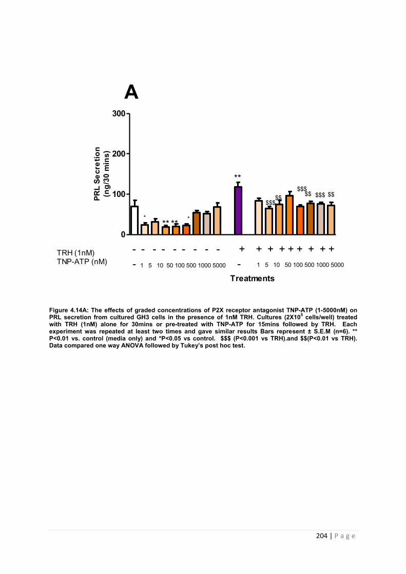
204 | P a g e
Figure 4.14A: The effects of graded concentrations of P2X receptor antagonist TNP-ATP (1-5000nM) on PRL secretion from cultured GH3 cells in the presence of 1nM TRH. Cultures (2X10⁵ cells/well) treated with TRH (1nM) alone for 30mins or pre-treated with TNP-ATP for 15mins followed by TRH. Each experiment was repeated at least two times and gave similar results Bars represent ± S.E.M (n=6). ** P<0.01 vs. control (media only) and *P<0.05 vs control. $$$ (P<0.001 vs TRH).and $$(P<0.01 vs TRH). Data compared one way ANOVA followed by Tukey’s post hoc test.
0
100
200
300
Treatments
TRH (1nM)TNP-ATP (nM)
- - - - - - - - - + + + + + + + + +- 1 5 10 50 100 500 1000 5000 - 1 5 10 50 100 500 1000 5000
*** **
**
*
$$$$$
$$$$$
$$$ $$
APR
L Se
cret
ion
(ng/
30 m
ins)
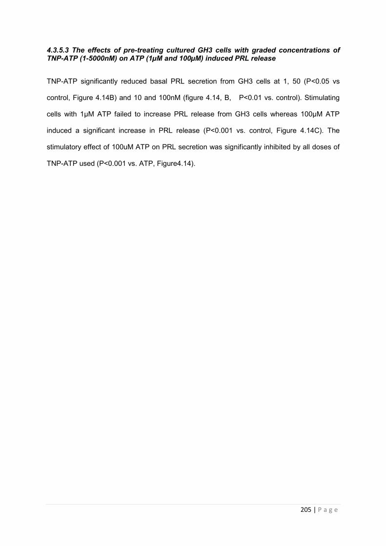
205 | P a g e
4.3.5.3 The effects of pre-treating cultured GH3 cells with graded concentrations of TNP-ATP (1-5000nM) on ATP (1µM and 100µM) induced PRL release
TNP-ATP significantly reduced basal PRL secretion from GH3 cells at 1, 50 (P<0.05 vs
control, Figure 4.14B) and 10 and 100nM (figure 4.14, B, P<0.01 vs. control). Stimulating
cells with 1µM ATP failed to increase PRL release from GH3 cells whereas 100µM ATP
induced a significant increase in PRL release (P<0.001 vs. control, Figure 4.14C). The
stimulatory effect of 100uM ATP on PRL secretion was significantly inhibited by all doses of
TNP-ATP used (P<0.001 vs. ATP, Figure4.14).
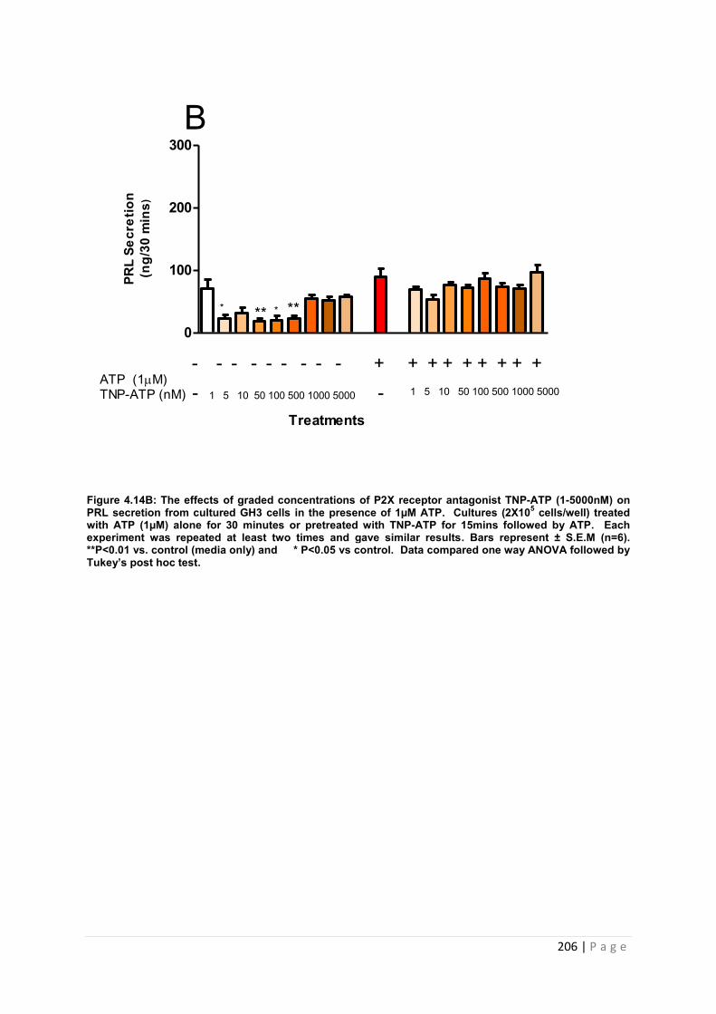
206 | P a g e
Figure 4.14B: The effects of graded concentrations of P2X receptor antagonist TNP-ATP (1-5000nM) on PRL secretion from cultured GH3 cells in the presence of 1µM ATP. Cultures (2X10⁵ cells/well) treated with ATP (1µM) alone for 30 minutes or pretreated with TNP-ATP for 15mins followed by ATP. Each experiment was repeated at least two times and gave similar results. Bars represent ± S.E.M (n=6). **P<0.01 vs. control (media only) and * P<0.05 vs control. Data compared one way ANOVA followed by Tukey’s post hoc test.
0
100
200
300
Treatments
ATP (1M)TNP-ATP (nM) - 1 5 10 50 100 500 1000 5000 - 1 5 10 50 100 500 1000 5000
- - - - - - - - - + + + + + + + + +
* *****
BPR
L Se
cret
ion
(ng/
30 m
ins)
207 | P a g e
Figure 4.14C: The effects of graded concentrations of P2X receptor antagonist TNP-ATP (1-5000nM) on PRL secretion from cultured GH3 cells in the presence of 100µM ATP. Cultures (2X10⁵ cells/well) treated with ATP (100µM) alone for 30 minutes or pretreated with TNP-ATP for 15mins followed by ATP. Each experiment was repeated at least two times and gave similar results. Bars represent ± S.E.M (n=6). **P<0.01 vs. control (media only) * P<0.05 vs control . Data compared one way ANOVA followed by Tukey’s post hoc test.
0
100
200
300
Treatments
ATP (100M)TNP-ATP (nM) - 1 5 10 50 100 500 1000 5000 - 1 5 10 50 100 500 1000 5000
- - - - - - - - - + + + + + + + + +
$$$
***
* *** ** **
CP
RL
Sec
retio
n(n
g/30
min
s)
208 | P a g e
4.3.5.4 The effects of pre-treating cultured GH3 cells with graded concentrations of
TNP-ATP (1-5000nM) on ATP (1µM) and TRH (1nM) induced PRL secretion
TNP-ATP significantly reduced basal PRL secretion from GH3 cells at 1, 50, 100 nM
(P<0.05 vs control, Figure 4.14D) and 10nM (P<0.01 vs. control, Figure 4.14D). 1nM TRH
induced a significant increase in PRL release from GH3 cells (P<0.01 vs control, figure 4.14
D). 1µM ATP failed to increase PRL release from cells. However co-incubation with 1µM
ATP and 1nM TRH induced a significant increase in PRL release (P<0.001 vs. control,
Figure4.14 D). The stimulatory effect of TRH both with and without ATP was significantly
inhibited at all doses of TNP-ATP tested (P<0.001 vs. TRH and ATP, Figure 4.14D).
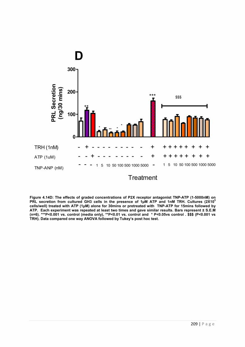
209 | P a g e
Figure 4.14D: The effects of graded concentrations of P2X receptor antagonist TNP-ATP (1-5000nM) on PRL secretion from cultured GH3 cells in the presence of 1µM ATP and 1nM TRH. Cultures (2X10⁵ cells/well) treated with ATP (1µM) alone for 30mins or pretreated with TNP-ATP for 15mins followed by ATP. Each experiment was repeated at least two times and gave similar results. Bars represent ± S.E.M (n=6). ***P<0.001 vs. control (media only), **P<0.01 vs. control and * P<0.05vs control . $$$ (P<0.001 vs TRH). Data compared one way ANOVA followed by Tukey’s post hoc test.
0
100
200
300
ATP (1uM)
TNP-ANP (nM)
- - + - - - - - - - - + + + + + + + + +TRH (1nM) - + - - - - - - - - - + + + + + + + + +
- 1 5 10 50 100 500 1000 5000 -- - 1 5 10 50 100 500 1000 5000
**
***
* *** *
$$$
DPR
L Se
cret
ion
(ng/
30 m
ins)
Treatment
210 | P a g e
4.3.5.5 The effects of pre-treating cultured GH3 cells with graded concentrations of
TNP-ATP (1-5000nM) on ATP (100µM) and TRH (1nM) induced PRL release.
TNP-ATP had a significant influence on basal PRL secretion from GH3 cells at 1nM (P<0.05
vs control, Figure 4.6 E) and 10 and 100nM (figure, P<0.01 vs. control, Figure 4.14E). 1nM
TRH induced a significant increase in PRL release from GH3 cells (P<0.01 vs control, figure
4.14E). 100µM ATP induced a significant increase in PRL release (P<0.001 vs. control,
Figure 4.14E). The stimulatory effect of 100uM ATP was significantly inhibited at all doses of
TNP-ATP tested (P<0.001 vs. ATP, Figure 4.6E). However co-incubation with 100µM ATP
and 1nM TRH induced a significant increase in PRL release (P<0.001 vs. control, Figure
4.14E). The stimulatory effect of TRH and ATP was significantly inhibited at all doses of
TNP-ATP tested (P<0.001 vs. TRH and ATP, Figure 4.14E). The values for the effect of
TNP-ATP on PRL release in vitro are tabulated and summarised in table 4.12.
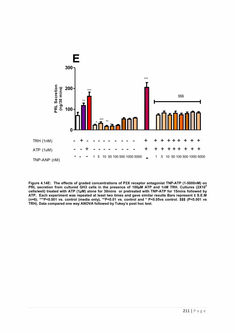
211 | P a g e
Figure 4.14E: The effects of graded concentrations of P2X receptor antagonist TNP-ATP (1-5000nM) on PRL secretion from cultured GH3 cells in the presence of 100µM ATP and 1nM TRH. Cultures (2X10⁵ cells/well) treated with ATP (1µM) alone for 30mins or pretreated with TNP-ATP for 15mins followed by ATP. Each experiment was repeated at least two times and gave similar results Bars represent ± S.E.M (n=6). ***P<0.001 vs. control (media only), **P<0.01 vs. control and * P<0.05vs control. $$$ (P<0.001 vs TRH). Data compared one way ANOVA followed by Tukey’s post hoc test.
0
100
200
300
**
***
***
E
**** **
**
$$$
ATP (1uM)
TNP-ANP (nM)
- - + - - - - - - - - + + + + + + + + +TRH (1nM) - + - - - - - - - - - + + + + + + + + +
- 1 5 10 50 100 500 1000 5000 1 5 10 50 100 500 1000 5000---
PR
L S
ecre
tion
(ng/
30 m
ins)
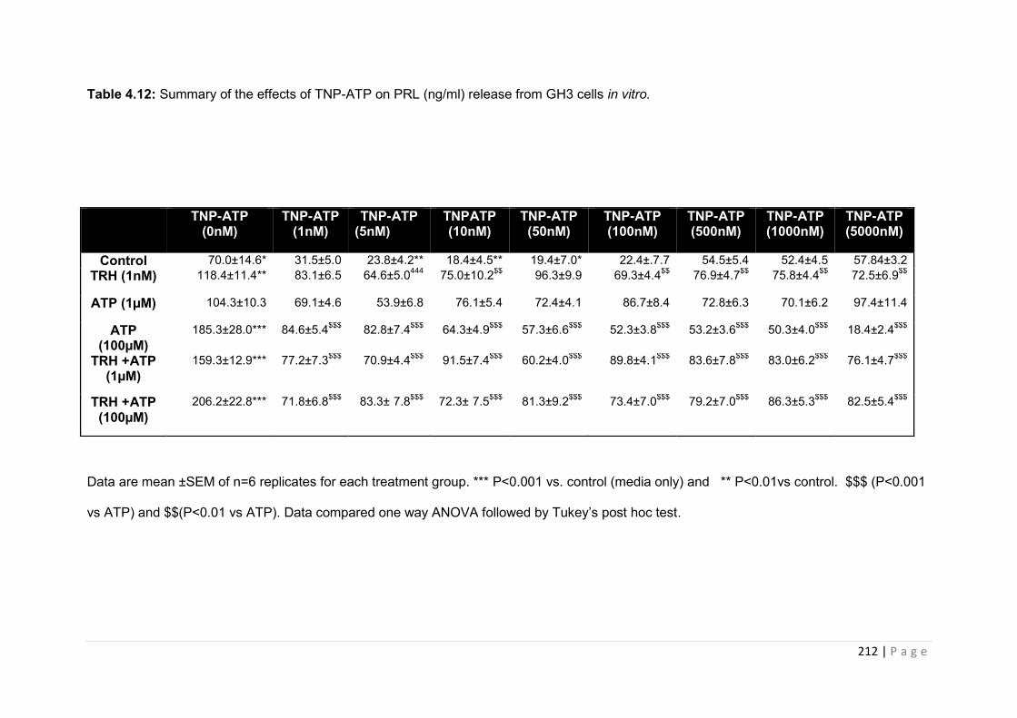
212 | P a g e
Table 4.12: Summary of the effects of TNP-ATP on PRL (ng/ml) release from GH3 cells in vitro.
Data are mean ±SEM of n=6 replicates for each treatment group. *** P<0.001 vs. control (media only) and ** P<0.01vs control. $$$ (P<0.001
vs ATP) and $$(P<0.01 vs ATP). Data compared one way ANOVA followed by Tukey’s post hoc test.
TNP-ATP (0nM)
TNP-ATP (1nM)
TNP-ATP (5nM)
TNPATP (10nM)
TNP-ATP (50nM)
TNP-ATP (100nM)
TNP-ATP (500nM)
TNP-ATP (1000nM)
TNP-ATP (5000nM)
Control 70.0±14.6* 31.5±5.0 23.8±4.2** 18.4±4.5** 19.4±7.0* 22.4±.7.7 54.5±5.4 52.4±4.5 57.84±3.2 TRH (1nM) 118.4±11.4** 83.1±6.5 64.6±5.0444 75.0±10.2$$ 96.3±9.9 69.3±4.4$$ 76.9±4.7$$ 75.8±4.4$$ 72.5±6.9$$
ATP (1µM) 104.3±10.3 69.1±4.6 53.9±6.8 76.1±5.4 72.4±4.1 86.7±8.4 72.8±6.3 70.1±6.2 97.4±11.4
ATP (100µM)
185.3±28.0*** 84.6±5.4$$$ 82.8±7.4$$$ 64.3±4.9$$$ 57.3±6.6$$$ 52.3±3.8$$$ 53.2±3.6$$$ 50.3±4.0$$$ 18.4±2.4$$$
TRH +ATP (1µM)
159.3±12.9*** 77.2±7.3$$$ 70.9±4.4$$$ 91.5±7.4$$$ 60.2±4.0$$$ 89.8±4.1$$$ 83.6±7.8$$$ 83.0±6.2$$$ 76.1±4.7$$$
TRH +ATP (100µM)
206.2±22.8*** 71.8±6.8$$$ 83.3± 7.8$$$ 72.3± 7.5$$$ 81.3±9.2$$$ 73.4±7.0$$$ 79.2±7.0$$$ 86.3±5.3$$$ 82.5±5.4$$$

213 | P a g e
4.3.5.4 An investigation into the effects of P2X3 inhibitor AF-353 on prolactin release from cultured GH3 cells
We subsequently investigated the effect of blocking individual P2X receptors in order to
determine the effects of specific receptor antagonism on PRL release from pituitary cells in
vitro. The P2X3 receptor antagonist, AF-353 has been characterised both in vivo and in
vitro as a potent P2X3R inhibitor but it effects on pituitary function are yet to be determined
[[144],[146] ]. We pretreated GH3 cells for 15 minutes with the P2X3 receptor antagonist AF-
353 and subsequently treated with TRH (1nM), ATP (1µM and 100µM) and TRH and ATP
combined (1nM TRH +1/100µM ATP). The effects of the compound on PRL secretion from
GH3 were determined by RIA.
4.3.5.4.1 The effects of pre-treating cultured GH3 cells with graded concentrations of AF-353 (1-5000nM) on TRH (1nM) induced PRL secretion.
All doses of A-353 tested failed to influence basal PRL release from GH3 cells (Figure
4.15A). Stimulating cells with 1nM TRH induced a significant increase in PRL release from
GH3 cells (P<0.001 vs control, Figure 4.15A). The stimulatory effects of TRH on PRL
secretion were significantly inhibited by AF-353 at all concentrations tested ( P<0.001 vs.
TRH, and P<0.01 vs TRH, Figure 4.15A).
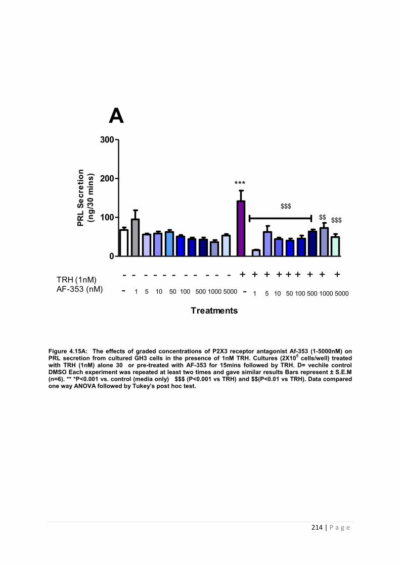
214 | P a g e
Figure 4.15A: The effects of graded concentrations of P2X3 receptor antagonist Af-353 (1-5000nM) on PRL secretion from cultured GH3 cells in the presence of 1nM TRH. Cultures (2X10⁵ cells/well) treated with TRH (1nM) alone 30 or pre-treated with AF-353 for 15mins followed by TRH. D= vechile control DMSO Each experiment was repeated at least two times and gave similar results Bars represent ± S.E.M (n=6). ** *P<0.001 vs. control (media only) $$$ (P<0.001 vs TRH) and $$(P<0.01 vs TRH). Data compared one way ANOVA followed by Tukey’s post hoc test.
0
100
200
300
***
$$
A
Treatments
TRH (1nM)AF-353 (nM) - 1 5 10 50 100 500 1000 5000 1 5 10 50 100 500 1000 5000
- - - - - - - - - - - + + + + + + + + +
PRL
Secr
etio
n(n
g/30
min
s)
$$$
$$$
-
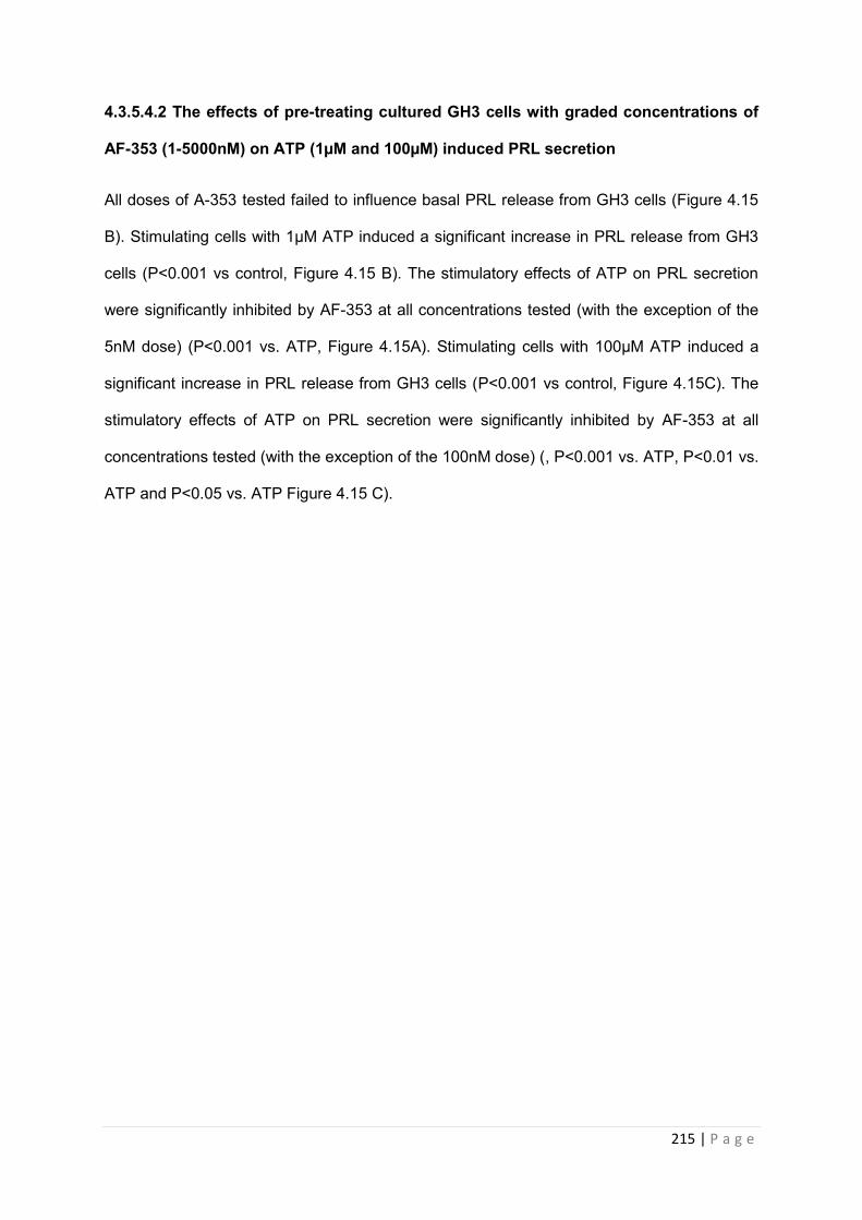
215 | P a g e
4.3.5.4.2 The effects of pre-treating cultured GH3 cells with graded concentrations of
AF-353 (1-5000nM) on ATP (1µM and 100µM) induced PRL secretion
All doses of A-353 tested failed to influence basal PRL release from GH3 cells (Figure 4.15
B). Stimulating cells with 1µM ATP induced a significant increase in PRL release from GH3
cells (P<0.001 vs control, Figure 4.15 B). The stimulatory effects of ATP on PRL secretion
were significantly inhibited by AF-353 at all concentrations tested (with the exception of the
5nM dose) (P<0.001 vs. ATP, Figure 4.15A). Stimulating cells with 100µM ATP induced a
significant increase in PRL release from GH3 cells (P<0.001 vs control, Figure 4.15C). The
stimulatory effects of ATP on PRL secretion were significantly inhibited by AF-353 at all
concentrations tested (with the exception of the 100nM dose) (, P<0.001 vs. ATP, P<0.01 vs.
ATP and P<0.05 vs. ATP Figure 4.15 C).
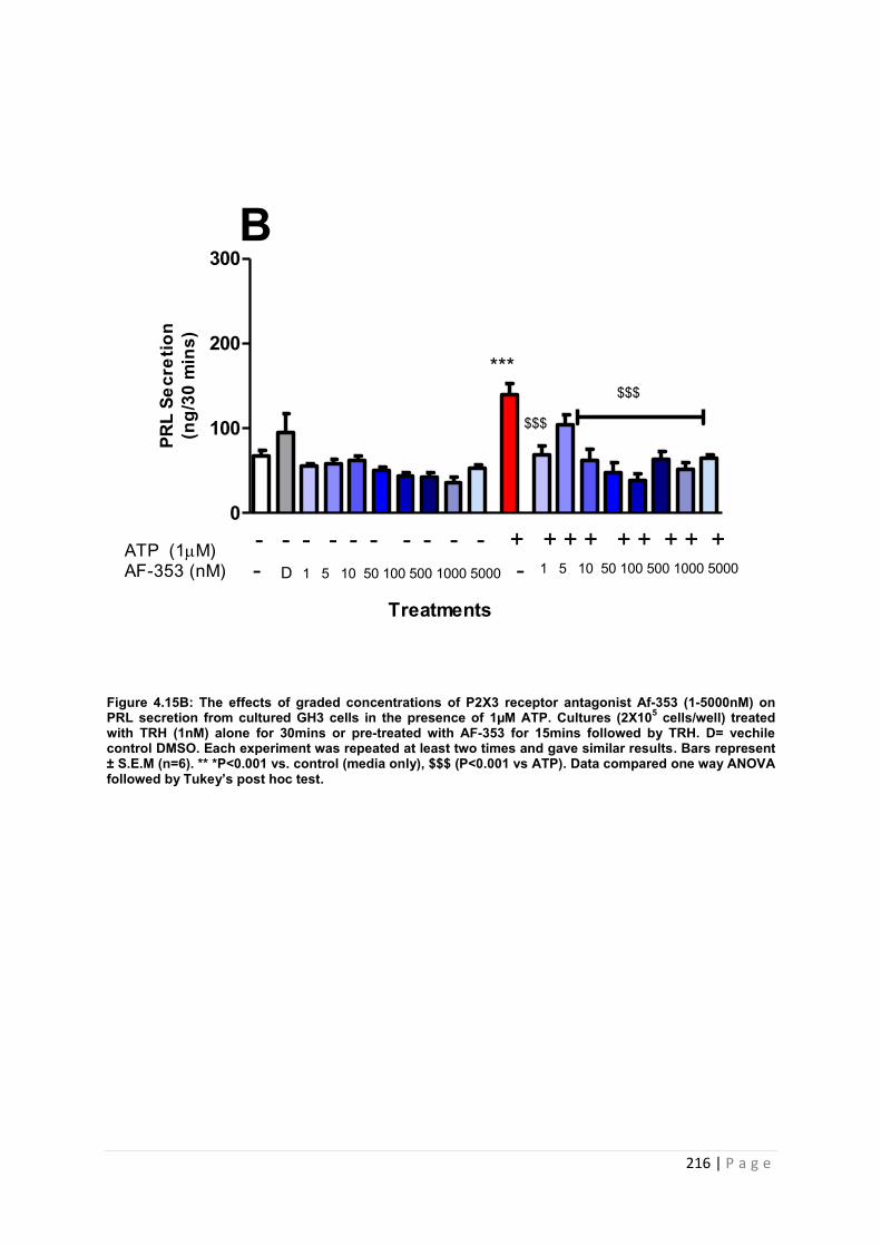
216 | P a g e
Figure 4.15B: The effects of graded concentrations of P2X3 receptor antagonist Af-353 (1-5000nM) on PRL secretion from cultured GH3 cells in the presence of 1µM ATP. Cultures (2X10⁵ cells/well) treated with TRH (1nM) alone for 30mins or pre-treated with AF-353 for 15mins followed by TRH. D= vechile control DMSO. Each experiment was repeated at least two times and gave similar results. Bars represent ± S.E.M (n=6). ** *P<0.001 vs. control (media only), $$$ (P<0.001 vs ATP). Data compared one way ANOVA followed by Tukey’s post hoc test.
0
100
200
300
***
$$$
B
Treatments
ATP (1M)AF-353 (nM) - D 1 5 10 50 100 500 1000 5000 - 1 5 10 50 100 500 1000 5000
- - - - - - - - - - + + + + + + + + +
PRL
Secr
etio
n(n
g/30
min
s)
$$$
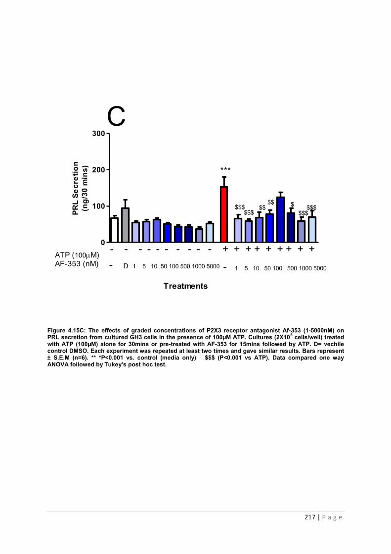
217 | P a g e
Figure 4.15C: The effects of graded concentrations of P2X3 receptor antagonist Af-353 (1-5000nM) on PRL secretion from cultured GH3 cells in the presence of 100µM ATP. Cultures (2X10⁵ cells/well) treated with ATP (100µM) alone for 30mins or pre-treated with AF-353 for 15mins followed by ATP. D= vechile control DMSO. Each experiment was repeated at least two times and gave similar results. Bars represent ± S.E.M (n=6). ** *P<0.001 vs. control (media only) $$$ (P<0.001 vs ATP). Data compared one way ANOVA followed by Tukey’s post hoc test.
0
100
200
300
***
$$$$$$ $$$
$$$$$ $$$
C
PRL
Secr
etio
n(n
g/30
min
s)
Treatments
ATP (100M)AF-353 (nM) - D 1 5 10 50 100 500 1000 5000 - 1 5 10 50 100 500 1000 5000
- - - - - - - - - - + + + + + + + + +
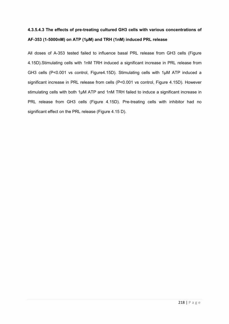
218 | P a g e
4.3.5.4.3 The effects of pre-treating cultured GH3 cells with various concentrations of
AF-353 (1-5000nM) on ATP (1µM) and TRH (1nM) induced PRL release
All doses of A-353 tested failed to influence basal PRL release from GH3 cells (Figure
4.15D).Stimulating cells with 1nM TRH induced a significant increase in PRL release from
GH3 cells (P<0.001 vs control, Figure4.15D). Stimulating cells with 1µM ATP induced a
significant increase in PRL release from cells (P<0.001 vs control, Figure 4.15D). However
stimulating cells with both 1µM ATP and 1nM TRH failed to induce a significant increase in
PRL release from GH3 cells (Figure 4.15D). Pre-treating cells with inhibitor had no
significant effect on the PRL release (Figure 4.15 D).
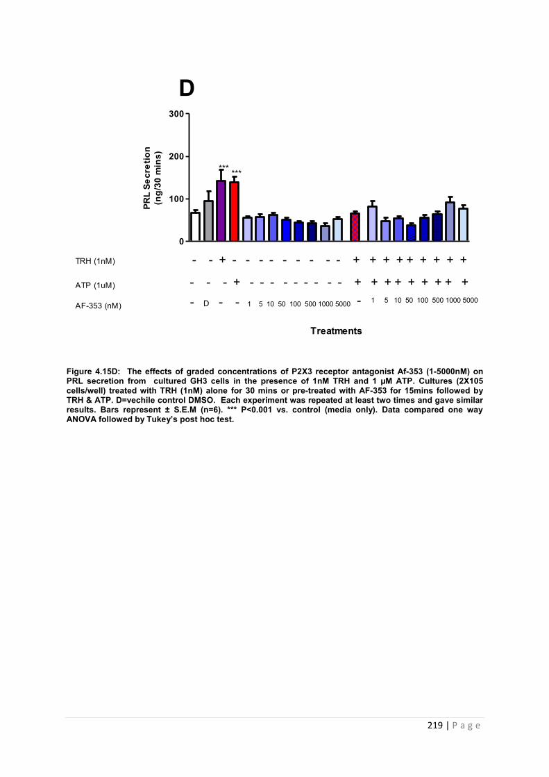
219 | P a g e
Figure 4.15D: The effects of graded concentrations of P2X3 receptor antagonist Af-353 (1-5000nM) on PRL secretion from cultured GH3 cells in the presence of 1nM TRH and 1 µM ATP. Cultures (2X10⁵ cells/well) treated with TRH (1nM) alone for 30 mins or pre-treated with AF-353 for 15mins followed by TRH & ATP. D=vechile control DMSO. Each experiment was repeated at least two times and gave similar results. Bars represent ± S.E.M (n=6). *** P<0.001 vs. control (media only). Data compared one way ANOVA followed by Tukey’s post hoc test.
0
100
200
300
******
D
ATP (1uM)
AF-353 (nM)
- - - + - - - - - - - - - + + + + + + + + +
TRH (1nM) - - + - - - - - - - - - + + + + + + + + +
Treatments
PRL
Secr
etio
n(n
g/30
min
s)
1 5 10 50 100 500 1000 50001 5 10 50 100 500 1000 5000 -- D - -
220 | P a g e
4.3.5.4.4 The effects of pre-treating cultured GH3 cells with graded concentrations of
AF-353 (1-5000nM) on ATP (100µM) and TRH (1nM) induced PRL release
All doses of A-353 tested failed to influence basal PRL release from GH3 cells (Figure
4.15E).
Stimulating cells with 1nM TRH induced a significant increase in PRL release from GH3 cells
(P<0.001 vs control, Figure 4.15E). Stimulating cells with 100µM ATP also induced a
significant increase in PRL release (P<0.001 vs control, Figure 4.15 E). However stimulating
cells with both 100µM ATP and 1nM TRH caused a marginal but non- significant increase in
PRL release from cells (Figure 4.15E). The stimulatory effects of TRH and ATP were
significantly inhibited by AF-353 at 1,10 ( P<0.05 vs. TRH&ATP, Figure 4.15E), 500 and
1000nM (P<0.01 vs. TRH&ATP, Figure 4.15 E). 5, 50,100 and 5000nM of AF-353 had no
effect. The values for the effect of AF-353 on PRL release in vitro are tabulated and
summarised in table 4.13.
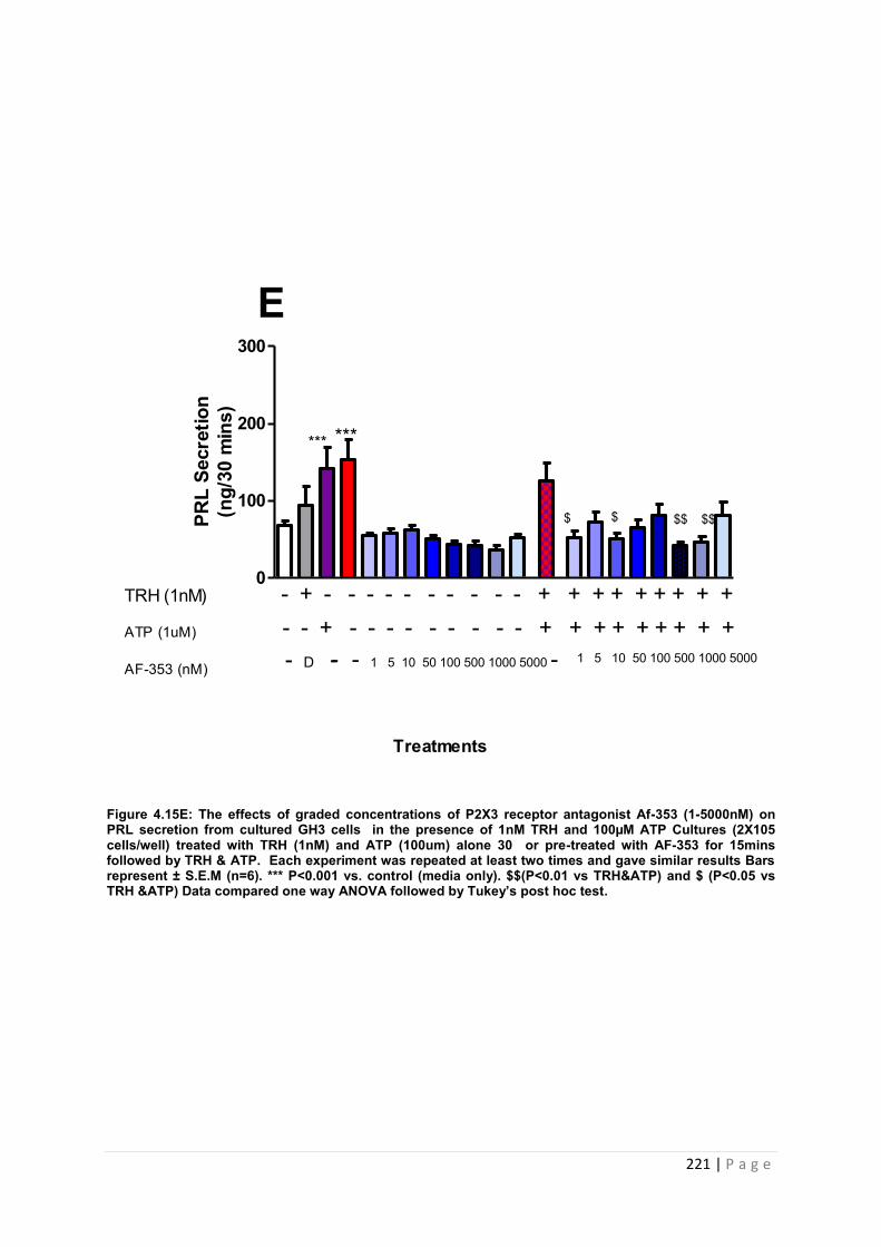
221 | P a g e
Figure 4.15E: The effects of graded concentrations of P2X3 receptor antagonist Af-353 (1-5000nM) on PRL secretion from cultured GH3 cells in the presence of 1nM TRH and 100µM ATP Cultures (2X10⁵ cells/well) treated with TRH (1nM) and ATP (100um) alone 30 or pre-treated with AF-353 for 15mins followed by TRH & ATP. Each experiment was repeated at least two times and gave similar results Bars represent ± S.E.M (n=6). *** P<0.001 vs. control (media only). $$(P<0.01 vs TRH&ATP) and $ (P<0.05 vs TRH &ATP) Data compared one way ANOVA followed by Tukey’s post hoc test.
0
100
200
300
E
$$ $$ $$
*** ***
ATP (1uM)
AF-353 (nM)
- - + - - - - - - - - - + + + + + + + + +TRH (1nM) - + - - - - - - - - - - + + + + + + + + +
- - 1 5 10 50 100 500 1000 5000 - 1 5 10 50 100 500 1000 5000
Treatments
PRL
Secr
etio
n(n
g/30
min
s)
- D -
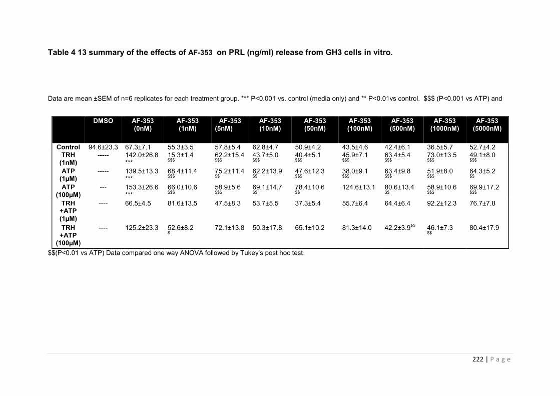
222 | P a g e
Table 4 13 summary of the effects of AF-353 on PRL (ng/ml) release from GH3 cells in vitro.
Data are mean ±SEM of n=6 replicates for each treatment group. *** P<0.001 vs. control (media only) and ** P<0.01vs control. $$$ (P<0.001 vs ATP) and
$$(P<0.01 vs ATP) Data compared one way ANOVA followed by Tukey’s post hoc test.
DMSO AF-353 (0nM)
AF-353 (1nM)
AF-353 (5nM)
AF-353 (10nM)
AF-353 (50nM)
AF-353 (100nM)
AF-353 (500nM)
AF-353 (1000nM)
AF-353 (5000nM)
Control 94.6±23.3 67.3±7.1 55.3±3.5 57.8±5.4 62.8±4.7 50.9±4.2 43.5±4.6 42.4±6.1 36.5±5.7 52.7±4.2 TRH
(1nM) ----- 142.0±26.8
*** 15.3±1.4 $$$
62.2±15.4 $$$
43.7±5.0 $$$
40.4±5.1 $$$
45.9±7.1 $$$
63.4±5.4 $$$
73.0±13.5 $$$
49.1±8.0 $$$
ATP (1µM)
----- 139.5±13.3 ***
68.4±11.4 $$$
75.2±11.4 $$
62.2±13.9 $$
47.6±12.3 $$$
38.0±9.1 $$$
63.4±9.8 $$$
51.9±8.0 $$$
64.3±5.2 $$
ATP (100µM)
--- 153.3±26.6 ***
66.0±10.6 $$$
58.9±5.6 $$$
69.1±14.7 $$
78.4±10.6 $$
124.6±13.1 80.6±13.4 $$
58.9±10.6 $$$
69.9±17.2 $$$
TRH +ATP (1µM)
---- 66.5±4.5 81.6±13.5 47.5±8.3 53.7±5.5 37.3±5.4 55.7±6.4 64.4±6.4 92.2±12.3 76.7±7.8
TRH +ATP
(100µM)
---- 125.2±23.3 52.6±8.2 $
72.1±13.8 50.3±17.8 65.1±10.2 81.3±14.0 42.2±3.9$$ 46.1±7.3 $$
80.4±17.9
223 | P a g e
4.3.5.5 An investigation into the effects of the P2X7 inhibitor A-438079 on prolactin
release from cultured GH3 cells
We then proceeded to investigate the influence of specific antagonism of the P2X7 receptor
on PRL release from GH3 cells in vitro [[156],[157]]. We utilised the P2X7 receptor
antagonist A-438079 to determine the effect of P2X7 blockade on pituitary function. GH3
cells were preincubated with A-438079 for 15 minutes prior to treatment with TRH (1nM),
ATP (1µM and 100µM) or TRH and ATP combined (1nM TRH +1/100µM ATP). The effects
of the compound on PRL secretion from GH3 were determined by RIA.
4.3.5.5 .1The effects of pre-treating cultured GH3 cells with various concentrations of
A-438079 (1-5000nM) on TRH (1nM) induced PRL release.
All doses of A-438079 tested significantly reduced the basal release of PRL from GH3 cells
(P<0.001 vs. control and P<0.01 vs control, Figure 4.16A). Stimulating cells with 1nM TRH
induced a significant increase in PRL release from cells (P<0.01 vs control, Figure 4.16A).
The stimulatory effects of TRH were significantly inhibited by A-438079 at all concentrations
(P<0.001 vs. TRH, Figure4.16 A).
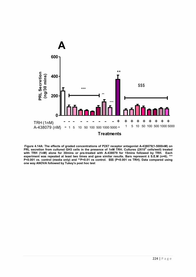
224 | P a g e
Figure 4.14A: The effects of graded concentrations of P2X7 receptor antagonist A-438079(1-5000nM) on PRL secretion from cultured GH3 cells in the presence of 1nM TRH. Cultures (2X10⁵ cells/well) treated with TRH (1nM) alone for 30mins or pre-treated with A-438079 for 15mins followed by TRH. Each experiment was repeated at least two times and gave similar results. Bars represent ± S.E.M (n=6). *** P<0.001 vs. control (media only) and **P<0.01 vs control. $$$ (P<0.001 vs TRH). Data compared using one way ANOVA followed by Tukey’s post hoc test
0
200
400
600
A
Treatments
TRH (1nM)A-438079 (nM) - 1 5 10 50 100 500 1000 5000 - 1 5 10 50 100 500 1000 5000
- - - - - - - - - + + + + + + + + +
**
***
***
**
PRL
Secr
etio
n(n
g/30
min
s)
$$$
225 | P a g e
4.3.3.52 The effects of pre-treating cultured GH3 cells with graded concentrations of
A-438079 (1-5000nM) on ATP (1µM and 100µM) induced PRL release
All doses of A-438079 significantly reduced the basal release of PRL from GH3 cells
(P<0.001 vs. control and P<0.01 vs control, Figure4.16B). Stimulating cells with 1µM ATP
induced a significant increase in PRL release from cells (P<0.001 vs. control, Figure 4.16 B).
This effect was also repeated with 100µM ATP (P<0.001 vs. control, Figure 4.16C). The
stimulatory effects of both does of ATP were significantly inhibited by all doses of A-438079
tested (P<0.001 vs. ATP, Figure 4.16B and 4.16C).
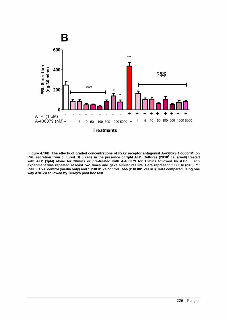
226 | P a g e
Figure 4.16B: The effects of graded concentrations of P2X7 receptor antagonist A-438079(1-5000nM) on PRL secretion from cultured GH3 cells in the presence of 1µM ATP. Cultures (2X10⁵ cells/well) treated with ATP (1µM) alone for 30mins or pre-treated with A-438079 for 15mins followed by ATP. Each experiment was repeated at least two times and gave similar results. Bars represent ± S.E.M (n=6). *** P<0.001 vs. control (media only) and **P<0.01 vs control. $$$ (P<0.001 vsTRH). Data compared using one way ANOVA followed by Tukey’s post hoc test
0
200
400
600
******
***
$$$
**
Treatments
ATP (1 M)A-438079 (nM)- 1 5 10 50 100 500 1000 5000 - 1 5 10 50 100 500 1000 5000
- - - - - - - - - + + + + + + + + +
BPR
L Se
cret
ion
(ng/
30 m
ins)
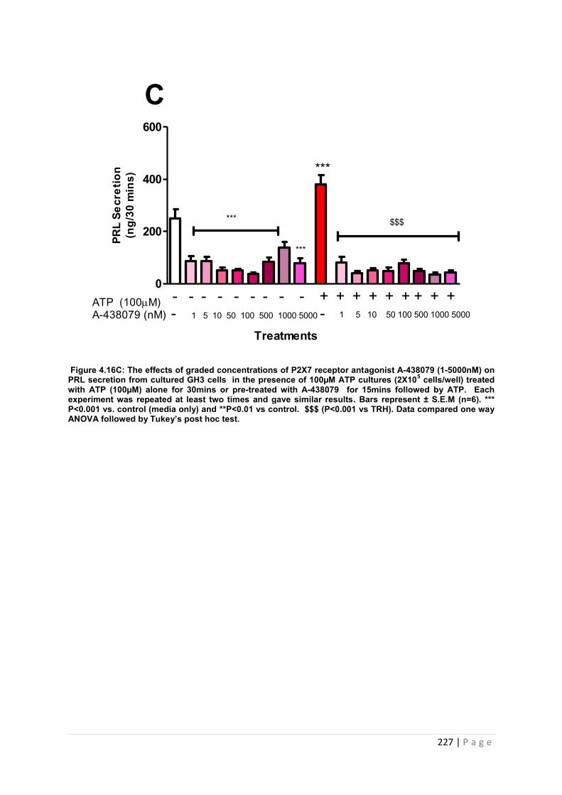
227 | P a g e
Figure 4.16C: The effects of graded concentrations of P2X7 receptor antagonist A-438079 (1-5000nM) on PRL secretion from cultured GH3 cells in the presence of 100µM ATP cultures (2X10⁵ cells/well) treated with ATP (100µM) alone for 30mins or pre-treated with A-438079 for 15mins followed by ATP. Each experiment was repeated at least two times and gave similar results. Bars represent ± S.E.M (n=6). *** P<0.001 vs. control (media only) and **P<0.01 vs control. $$$ (P<0.001 vs TRH). Data compared one way ANOVA followed by Tukey’s post hoc test.
0
200
400
600
Treatments
ATP (100M)A-438079 (nM) - 1 5 10 50 100 500 1000 5000 - 1 5 10 50 100 500 1000 5000
- - - - - - - - - + + + + + + + + +
***
***
*** $$$
CPR
L Se
cret
ion
(ng/
30 m
ins)
228 | P a g e
4.3.3.5.3 The effects of pre-treating cultured GH3 cells with graded concentrations of
A-438079 (1-5000nM) on ATP (1µM) and TRH (1nM) induced PRL release
All doses of A-438079 tested significantly reduced the basal release of PRL from GH3 cells
(P<0.001 vs. control and P<0.01 vs control, Figure 4.16D). Stimulating cells with 1nM TRH
induced a significant increase in PRL release from cells (P<0.01 vs control, Figure 4.16D).
Stimulating cells with 1µM ATP induced a significant increase in PRL release from cells
(P<0.001 vs. control, Figure 4.16D). However combining both secretagogue and ATP
together failed to significantly influence PRL release (Figure 4.16D). Pre-treating cells with
A-438079 did significantly decrease the release of PRL from TRH/ATP treated cells
(P<0.001 vs. TRH/ATP, Figure 4.16D).
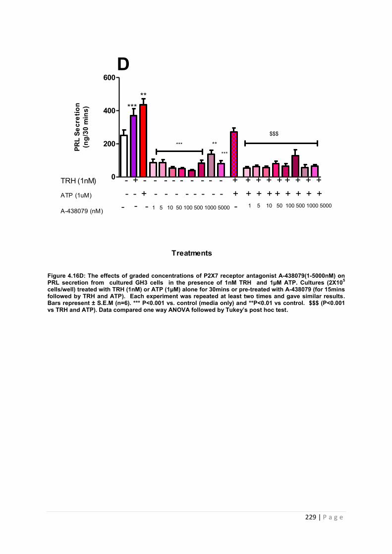
229 | P a g e
Figure 4.16D: The effects of graded concentrations of P2X7 receptor antagonist A-438079(1-5000nM) on PRL secretion from cultured GH3 cells in the presence of 1nM TRH and 1µM ATP. Cultures (2X10⁵ cells/well) treated with TRH (1nM) or ATP (1µM) alone for 30mins or pre-treated with A-438079 (for 15mins followed by TRH and ATP). Each experiment was repeated at least two times and gave similar results. Bars represent ± S.E.M (n=6). *** P<0.001 vs. control (media only) and **P<0.01 vs control. $$$ (P<0.001 vs TRH and ATP). Data compared one way ANOVA followed by Tukey’s post hoc test.
0
200
400
600
ATP (1uM)
A-438079 (nM)
- - + - - - - - - - - + + + + + + + + +TRH (1nM) - + - - - - - - - - - + + + + + + + + +
- 1 5 10 50 100 500 1000 5000 - 1 5 10 50 100 500 1000 5000--
*****
***
**
$$$
D
Treatments
PRL
Secr
etio
n(n
g/30
min
s)
***
230 | P a g e
4.3.3.5.4The effects of pre-treating cultured GH3 cells with various concentrations of
A-438079 (1-5000nM) on ATP (100µM) and TRH (1nM) induced PRL release
All doses A-438079 had a significant effect on basal release of PRL from GH3 cells
(P<0.001 vs. control and P<0.01 vs control, Figure 4.16E). Stimulating cells with 1nM TRH
caused a significant increase in PRL release from cells (P<0.01 vs control, Figure 4.16E).
Stimulating cells with 100µM ATP caused a significant increase in PRL release from cells
(P<0.001 vs. control, Figure4.16 E). Combining both secretagogue and ATP together did
have a significant effect on PRL release (P<0.01 vs control, Figure 4.16E). This stimulatory
effects was significantly inhibited by pre-treating cells with A-438079 at all doses
investigated (P<0.001 vs. TRH and ATP, Figure 4.16E). The values for the effect of A-
438079 on PRL release in vitro are tabulated and summarised in table 4.14.
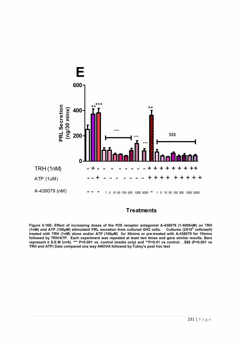
231 | P a g e
0
200
400
600
*******
$$$***
******
ATP (1uM)
A-438079 (nM)
- - + - - - - - - - + + + + + + + + +TRH (1nM) - + - - - - - - - - + + + + + + + ++
Treatments
EPR
L Se
cret
ion
(ng/
30 m
ins)
- - - 1 5 10 50 100 500 1000 5000 - 1 5 10 50 100 500 1000 5000
Figure 4.16E: Effect of increasing doses of the P2X receptor antagonist A-438079 (1-5000nM) on TRH (1nM) and ATP (100µM) stimulated PRL secretion from cultured GH3 cells. Cultures (2X10⁵ cells/well) treated with TRH (1nM) alone and/or ATP (100µM) for 30mins or pre-treated with A-438079 for 15mins followed by TRH/ATP. Each experiment was repeated at least two times and gave similar results. Bars represent ± S.E.M (n=6). *** P<0.001 vs. control (media only) and **P<0.01 vs control. . $$$ (P<0.001 vs TRH and ATP) Data compared one way ANOVA followed by Tukey’s post hoc test
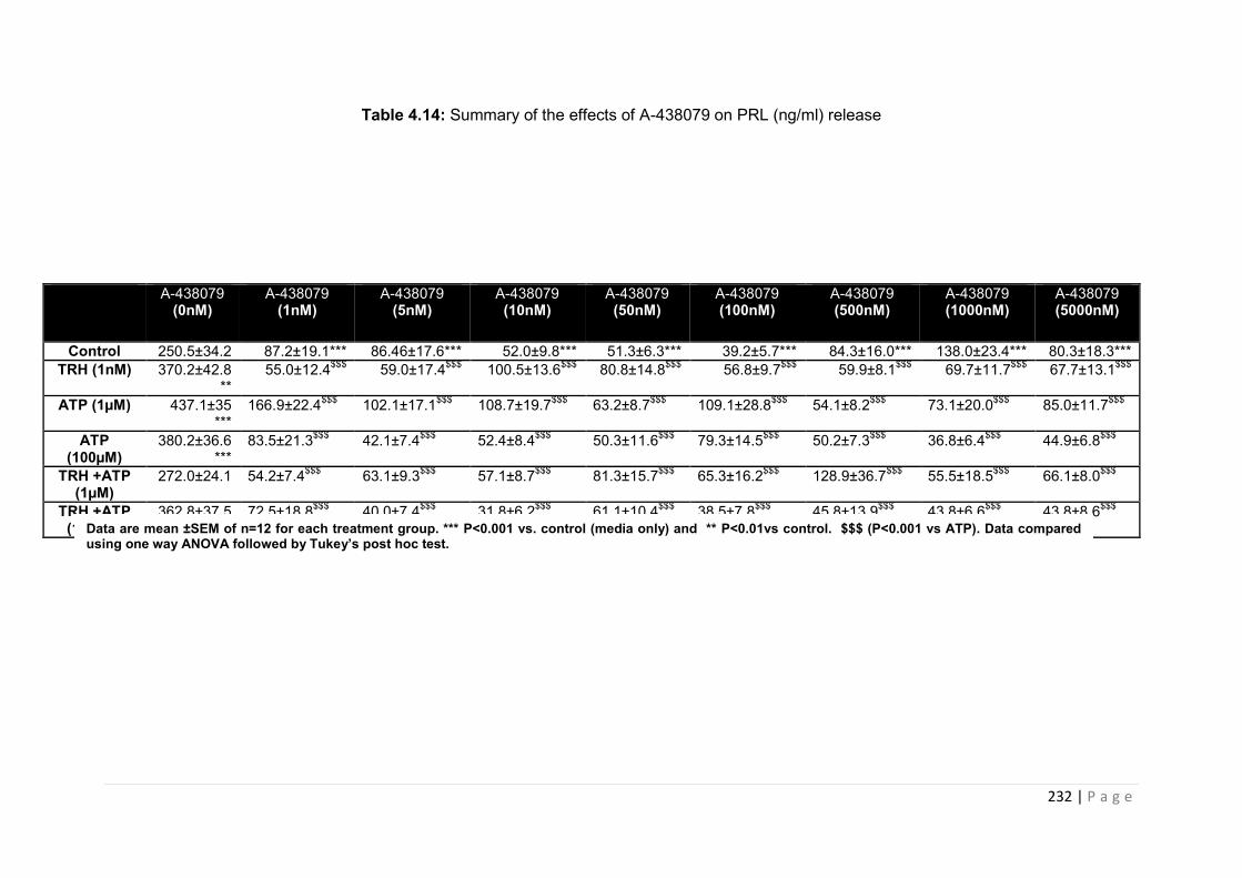
232 | P a g e
Table 4.14: Summary of the effects of A-438079 on PRL (ng/ml) release
A-438079 (0nM)
A-438079 (1nM)
A-438079 (5nM)
A-438079 (10nM)
A-438079 (50nM)
A-438079 (100nM)
A-438079 (500nM)
A-438079 (1000nM)
A-438079 (5000nM)
Control 250.5±34.2 87.2±19.1*** 86.46±17.6*** 52.0±9.8*** 51.3±6.3*** 39.2±5.7*** 84.3±16.0*** 138.0±23.4*** 80.3±18.3*** TRH (1nM) 370.2±42.8
** 55.0±12.4$$$ 59.0±17.4$$$ 100.5±13.6$$$ 80.8±14.8$$$ 56.8±9.7$$$ 59.9±8.1$$$ 69.7±11.7$$$ 67.7±13.1$$$
ATP (1µM) 437.1±35 ***
166.9±22.4$$$ 102.1±17.1$$$ 108.7±19.7$$$ 63.2±8.7$$$ 109.1±28.8$$$ 54.1±8.2$$$ 73.1±20.0$$$ 85.0±11.7$$$
ATP (100µM)
380.2±36.6 ***
83.5±21.3$$$ 42.1±7.4$$$ 52.4±8.4$$$ 50.3±11.6$$$ 79.3±14.5$$$ 50.2±7.3$$$ 36.8±6.4$$$ 44.9±6.8$$$
TRH +ATP (1µM)
272.0±24.1 54.2±7.4$$$ 63.1±9.3$$$ 57.1±8.7$$$ 81.3±15.7$$$ 65.3±16.2$$$ 128.9±36.7$$$ 55.5±18.5$$$ 66.1±8.0$$$
TRH +ATP (100µM)
362.8±37.5 72.5±18.8$$$ 40.0±7.4$$$ 31.8±6.2$$$ 61.1±10.4$$$ 38.5±7.8$$$ 45.8±13.9$$$ 43.8±6.6$$$ 43.8±8.6$$$ Data are mean ±SEM of n=12 for each treatment group. *** P<0.001 vs. control (media only) and ** P<0.01vs control. $$$ (P<0.001 vs ATP). Data compared using one way ANOVA followed by Tukey’s post hoc test.
233 | P a g e
4.4 Discussion
The studies described in this chapter investigated the hypothesis that P2X receptors play a
key role in the release of pituitary hormone and that treatment with P2XR antagonists would
significantly influence pituitary function. Our previous data demonstrated that all 7 P2XR
family members are expressed in the rat pituitary gland (see Chapter 3) and therefore the
possibility remained that any or all P2XRs may have a role in the regulation of the
mammalian pituitary gland. This chapter includes data that furthers our understanding of the
mechanism by which ATP and P2X interact to modulate pituitary function.
P2X receptors (P2XRs) are a novel class of inotropic receptors which can be found located
in a range of different tissues of both mammalian and non-mammalian origin. These ligand
gated ion channels, of which there are seven identified to date (P2X1-7), are activated by
extra cellular adenosine 5’-triphosphate (ATP), which, when bound to P2XRs affect the
homeostasis of host cells in the following ways : 1) increase the passage of cations (Na, K,)
out of the host cell which initials depolarisation, 2) facilitates the passage of calcium ions into
the cell which further activates internal cellular signalling and 3) upon prolonged activation,
dilation of the channel allows unrestricted passage of ions into out the cell [[158],[19]]. In the
excitable cells of the anterior pituitary, controlled secretion of hormones is driven by
extracellular calcium/calcium influx [Ca2+]i and occurs in a pituitary subtype specific manner
[3].The increase in [Ca2+]i stimulates electrical activity within the cell and facilitates larger
influxes via the activation of voltage-gated calcium channels (VGCCs) which penetrate all
areas of the cell [[53, 159]]. Rising levels of Ca2+ within the cell stimulate the production of
inositol triphosphate (IP3) (1, 4, 5) by PLC. IP3 binding to its receptor on the endoplasmic
reticulum allows the release of calcium from internal stores and aids in elevating cytosolic
levels of calcium which culminates in pituitary hormone release [[26] [48]]. It has been
demonstrated that intracellularly located ATP can achieve local concentrations in the
millimolar range, although there is currently no data concerning ATP concentrations within
pituitary secretory granules. Given that the process of secretion is a dynamic process, one
234 | P a g e
could speculate that ATP concentrations would fluctuate dependent on patterns/periods of
hormone secretion [[160],[161]]. Due to this fact, we tested a wide ATP dose range (0.001-
1000µM) in order to compare hormone secretion across this dose range. In order to validate
our primary pituitary culture model we first tested responses to a well know pituitary
secretagogues in the absence and presence of the relevant receptor antagonist. We utilised
CRH as our secretagogue and were provided with two potent CRF1 receptor antagonists
which were developed at Pfizer as anti-depressant agents. CRH induced a significant rise in
ACTH release from our primary pituitary cultures and both inhibitors caused a significant
decrease in stimulated ACTH release as expected.
Several studies have utilised primary cultures to characterise purinergic function in the rat
pituitary [[75],[96],[64],[34]]. In 2006, Zemkova and colleagues employed primary pituitary
cultures to demonstrate that ATP amplifies the effect of GnRH on LH secretion via activation
of P2X2Rs [34] and He and co-workers have demonstrated that ATP stimulation of P2X4
directly influences PRL release from lactotrophs [75].
In this investigation a mixed population of primary rat pituitary cells were used to assess the
effects of ATP on the release of a panel of pituitary hormones (ACTH, GH, PRL and TSH). In
accordance with previous reports, ATP was able to stimulate hormone release from static
primary cultures [[14], [65]]. We found the order of responsiveness of hormones to all doses
of ATP used in this study to show the following profile: ACTH>PRL>TSH>GH. Previous
studies of primary cells responsiveness to ATP had found a similar profile. 81% of
corticotrophs identified by immunocytochemistry were responsive to ATP, followed by 74 %
lactotrophs, and sommatotrophs, and 60% of thyrotrophs. [29]. However, this study utilised
live calcium imaging of Ca2+ ions following a single treatment with 50µM ATP to primary
cells. Pituitary subtypes were then identified by using antibodies raised against the different
pituitary hormones. Our study cannot be directly compared with the Villaobos study due to
differences in rat species used in generating the primary cultures and the functional measure
of pituitary cell responsiveness to ATP. However, our study did demonstrate that ATP exerts
235 | P a g e
control over pituitary hormone release from cells in a subtype specific manner. Our study
utilised the high throughput Multiplex system. This novel technique enabled us to directly
characterize the effects of ATP on the secretion of all pituitary hormones from rat primary
pituitary cultures in vitro. Multiplexing analysis has been used in clinical studies to identify
serum levels of a range of neuroendocrine hormones including ACTH, PRL, GH,
progesterone and cortisol [[162],[163]]. In vivo, multiplexing assays have been used to
detect changes in alpha melanocyte stimulating hormone (alpha-MSH) levels in mice. In fact
the multiplexing immunoassay is so sensitive that it has been used to identify pituitary
peptide hormones in paraffin embedded sections of tumours, for the purpose of staging
pituitary adenomas [164]. To our knowledge, this is the first time that a panel of pituitary cells
have been profiled by this method to measure functional responses to purinergic agonists.
This allowed us to measure all hormone profiles at once. Therefore our results may reflect
interactions between pituitary hormones, such as has been documented in rats in vivo )[165].
A significant finding from this project was that ATP stimulates the release of ACTH from
primary pituitary cells in vitro (figure 4.3). High dose ATP (100-1000µM) induced a significant
increase in ACTH secretion compared to basal ACTH release (see figure 4.3, table 4.3). To
our knowledge, we have shown for the first time that ATP can induce ACTH release from rat
primary pituitary cells. There is little evidence to support a role for P2XRs in modulating
pituitary function, although there is evidence of an effect in the adrenal gland. In cultured
bovine adrenocortical fasciculata cells, ATP has been demonstrated to potentiate ACTH
induced glucocorticoid synthesis [166]. A single low dose of ATP (5µM) was able to induce
cortisol secretion from cells when combined with ACTH, angiotensin II, sodium fluoride and
forskolin over a one hour period [166]. A recent study reported similar observations using the
human adrenal cortex cell line NCI-H295R (H295R)[167]. Only P2X5 and P2X7 were
identified by RT-PCR within this cell line, compared to a wider range of P2Y receptors (P2Y1,
P2Y2, P2Y6, P2Y12, P2Y13, and P2Y14). It was demonstrated through using the P2Y selective
agonist 2MeS-ATP, that stimulation of these subsets of the P2 family caused an increase in
236 | P a g e
both calcium mobilisation and glucocorticoid production [167]. Thus, previous studies
indicating an effect of P2XRs on adrenal function coupled with our data suggest that
purinergic signalling may have a role in the control of HPA function.
Interestingly, ATP has been shown to stabilise growth hormone granules in vitro. Interactions
between ATP in the pituitary cell golgi complex stabilises isolated growth hormone granules
and facilitates hormone secretion [168]. Previous studies have also shown that the addition
of ATP can stimulate the release of GH (and PRL) from isolated secretory granules [169].
However, using a mixed population of primary pituitary cells, we were unable to reproduce
these reported effects. GH showed little response to the addition of exogenous ATP to
primary pituitary cultures. Both humans and rats release GH hormone from the pituitary in
episodic bursts, which occur in a pulsatile manner, which is largely GHRH dependent [170].
Therefore accurately measuring GH in vitro becomes difficult, as we can only take a “snap
shot”, which will only depict the phase of release at that time. Also, we also need to take into
the account the gender differences in the amount of GH released, with females releasing
more than males [171]. As we used only male rats in our study, combined with the episodic
pattern of GH secretion may explain the varied basal levels recorded in our investigation for
GH.
To our knowledge, this is the first time that TSH secretion in response to ATP has been
determined in vitro. With regard to the effect of ATP on pituitary TSH release, 1000µM ATP
significantly increased TSH secretion at 20 and 60 minutes time point (figure 4.6, table 4.6).
There is little evidence concerning the effects of ATP on TSH release from the pituitary.
Studies have demonstrated a regulatory role for ATP in the production of thyroid hormone [1].
Using human thyrocytes in primary culture, ATP was observed to cause an increase in
intracellular calcium which is commonly used as a measure of cellular responses to
purinergic agonists. [173]. Kumari and co-workers have suggested that adeonsine induced
P1 receptor activation may contribute to pituitary TSH release [174]. Given the time-point at
which ATP stimulated TSH release, our data could potentially reflect an influence of
237 | P a g e
adenosine, since P1 receptors and the ectonucleases required for the breakdown of ATP are
present in the pituitary [127].
In contrast to the effects on GH and TSH, ATP induced significant increases in PRL release
from primary pituitary cells in vitro (figure 4.5, table 4.5). ATP, at all doses tested (with the
exception of 1µM ATP) stimulated a significant release of PRL from primary pituitary cells at
the 20 minute time point (figure, 4.5, table 4.5). This finding is in accordance with previous
investigations using primary pituitary cells which have demonstrated that ATP induced
activation of P2 receptors is required for the regulated release of PRL [[6] [175]]. Nunez and
colleagues demonstrated that stimulating rat primary pituitary cultures with 10µM ATP
stimulated a significant increase in PRL. This was observed in both male and lactating rats
[175]. They also demonstrated that enzymatic removal of ATP caused a decrease in both
basal and TRH induced PRL release [173]. This study employed the reverse haemolytic
plaque assay to determine and measure responses of single cells to ATP and/or TRH. He
and co-workers have used isolated enriched primary rat lactotrophs and measured hormone
release using cell column perfusion and RIA to investigate the role of P2 receptors in PRL
release [65]. This group found ATP to cause both an increase in PRL release and
intracellular calcium, when treated with 10µM ATP. The column perfusion allowed the group
to collect samples every minute and immediately assay for PRL responses. Potentially this
subtle difference in experiment design may account for our differences in response to ATP at
the earlier time points. However, our study using static mixed population cells and
multiplexing immunoassay can confirm both these findings and highlights that investigations
based primarily on the responses to P2 agonists may not be the most efficient measure of
P2X receptor function in the pituitary gland. In general, the relatively high basal levels of the
pituitary hormones released from the primary cultures, made it difficult to identify the
stimulatory effects of exogenously added ATP. This could potentially be due to the loss of
the hypothalamic regulators such as somatostatin and dopamine which control the levels GH
and PRL respectively. Dopamine has been shown to inhibit both the release of PRL and GH
238 | P a g e
from normal rat primary cells [176]. Binding of dopamine to the dopamine receptor 2 (D2R) is
associated with a decrease in the production of cAMP and a decrease in the release of PRL
from lactotrophs [177]. Potentially, the addition of dopamine would reduce basal levels of
PRL which might unmask the effects of ATP on hormone secretion.
With regard to our experiments with the rat primary pituitary cultures, PRL and ACTH were
the two hormones that were consistently stimulated in response to ATP treatment. In order
to further investigate the influence of P2XRs on pituitary function, we utilised two pituitary
cell lines – AtT20:D16s (a corticotroph cell line) and GH3s (a sommatolactotroph cell line).
Our initial experiments focused on the AtT20 cell line, and we studied the effects of ATP,
alone and in combination with the relevant secretagogue CRH, on ACTH release. It is
interesting to note that responsiveness of AtT20 cells to CRF was lower than observed in the
primary pituitary cultures (figure 4.1 and 4.3). Potentially, intact FS cells, which have been
demonstrated to facilitate the release of ACTH from corticotrophs in addition to other
pituitary derived mediators present in the primary culture milieu may have contributed to the
response to CRF [[78],[178]]. Thus the use of individual pituitary cell cultures is valid in order
to identify the influence of P2X receptors on hormone secretion in a pituitary cell subtype
specific manner. A significant observation that came from the investigations of ATt20:D16s
was that ATP and CRH co- administration caused a significant increase in ACTH release at
the 4, 7 and 12hr time point (figure 4.8). Our findings are in accordance with previous
investigations of P2XR function using this cell line which have also demonstrated that a
combination of ATP and CRH caused a significant increase in ACTH release from cells
following 3 hr incubation [61]. Zhao and co-workers used a different subclone of AtT20s
(AtT20PL cell line) to determine the effects of different purinergic agonists on the expression
of the pro-opiomelanocortin (POMC) gene and ACTH secretion from cells. RT-PCR
demonstrated that all P2X receptors, with the exception ofP2X2, and a wide range of P2Y
(1,2, 4 and 6) and P1 receptors ( A1 A2B A2a) were expressed in these cells. Treatment of
cells with adenosine (10 and 20µM) or ATP (100µM), alone or in combination with CRH
239 | P a g e
(100nM) caused a significant increase in POMC expression and ACTH release from cells [61]
. Previous investigations conducted by our group had demonstrated that the maximal effect
of CRH within AtT20 cells occurred at extended time points (7hr and 12hr) [[150], [150]]. By
looking at the effects of ATP at these extended time points we were able to show for the first
time that ATP alone can stimulate ACTH release.
We also studied the effects of ATP on PRL release from GH3 cells. ATP, at all doses tested,
significantly increased PRL release from GH3 cells from 5-60 minutes (figure 4.10). This
evidence is supported by recent studies using electrophysiological techniques to
demonstrate the responsiveness of GH3 cells to ATP and ATP analogues [[179],[131]].
Interestingly, the non-hydrolysable ATP analogue, ATPγS (figure 4.11), is less potent that
ATP at stimulating PRL release from GH3 cells. The major difference between ATP and
ATPγS, is that the former can be enzymatically cleaved by endogenous endonucleases (see
figure 1.4), with the breakdown products ADP, AMP or adenosine capable of activating both
P2 and P1 receptors, whereas the latter is resistant to breakdown and is therefore only
capable of activating P2 receptors. This suggests that the PRL response to ATP may be, in
part, mediated by both P2 and P1 receptors. It is important to note the previous studies have
demonstrated that both P2 and P1 receptors are expressed in the GH3 cell line [[180],
[66],[127]]. Therefore our results may reflect responses to agonist binding to both subtypes
of receptors. Treatment of GH3 cells with BzATP induced a minimal effect on PRL release
from cells in vitro. This is unexpected given the fact that BzATP is selective for P2XR
whereas ATP acts on all purinergic receptors [[181],[152]]. Previous characterisation studies
of P2X receptors using electrophysiology had demonstrated BzATP to induce Ca2+ influx
from cells [131]. Chung and Colleagues identified expression of P2X7 in the cell line GH3 by
RT-PCR and suggested that the P2X7 receptor facilitated the changes in calcium influx
[131]. Our RT-PCR analysis of GH3 cells is in accordance with this study although our data
also identified P2X3 and PX4Rs within this cell line (chapter 3). To our knowledge our study
is the first time that BzATP treatment of GH3 cells has been used to directly measure the

240 | P a g e
effects of P2X receptor stimulation on PRL release. Our results suggest that subsequent
investigation regarding in vitro characterisation of P2X receptors and hormone release in
GH3 cells should take a more integrative approach, utilising calcium release and hormone
secretion when assessing P2X function. In addition, a previous study into the effects of ATP
on insulin secretion in vitro observed decreases in insulin secretion following BzATP
treatment [182].This study utilised the hamster derived pancreatic cell line HIT-T15 to assess
the effect of ATP on insulin release. There is some controversy in the literature regarding the
effects of purinergic agents on insulin release, whereby ATP has been observed to both
increase insulin at low doses of ATP and inhibit at higher doses [[149],[182], [124]]. Lee and
colleagues found 1 hour stimulation of HIT-T15 grown in static culture with 100µM BzATP
caused a significant decrease in insulin release [182]. Jacques-Silva and co-workers also
observed a decreased insulin secretion from human islets treated with 50µM BzATP in
comparison to ATP [124]. In our study, a wide range of doses of ATP (0.001-1000µM) were
administered to both primary pituitary cells and to our in vitro models, AtT20:D16 and GH3
cells. In our study, ATP only displayed a positive effect on hormone release in vitro, albeit
demonstrating a similar profile when compared to the effects of BzATP on GH3 cells.
Our data generated utilising P2XR agonists indicated that P2XRs influence PRL secretion
from GH3 cells, but failed to indicate which specific P2XRs were mediating these effects. In
order to attempt to identify the importance of specific P2XRs, we studied the effects of
selective P2XR antagonists on PRL secretion from GH3 cells. Initially, GH3 cells were
treated with the secretagogue TRH and the native P2XR agonist, ATP, either alone or in
combination. In our study GH3 cells responded to both TRH and ATP in a reproducible
manner and in accordance with previous studies [ [79], [183],[131], [175]]. In addition,
coadministration of 100µM ATP with TRH resulted in a consistent potentiation of the effects
of TRH alone on PRL release from GH3 cells [figures 4.13 to 4.16]. The ability of ATP to
potentiate the effects of TRH on PRL release has been previously demonstrated in primary
pituitary cells [175]. In addition, the capability of ATP to potentiate pituitary hormone release
241 | P a g e
has also been demonstrated in the neurohypohysis [175]. Knotts and colleagues cultured
primary rat neurohypophysial cells and demonstrated that the electrically stimulated release
of vasopressin (AVP) and oxytocin (OT) was potentiated by ATP, most likely via P2X2 and
P2X3R activation [[123],[184],[1]]. In our study, the effects of ATP and TRH, alone and in
combination was significantly reduced by treatment with the non-selective P2R antagonist,
PPADS (figures 4.13). The capability of PPADS to reduce pituitary hormone secretion has
also been demonstrated in the neurohypophysis, where AVP release was significantly
decreased in the presence of the antagonist [[185], [123]]. PPADS has been demonstrated
to inhibit P2X mediated electrical currents in primary gonadotrophs [34], and decrease
insulin release following pre-treatment of cultured primary islet cells from rat and humans
[[124],[186] ]. The results from our studies are in accordance with this observation, whereby
PPADS inhibited both TRH and ATP stimulated PRL release form GH3 cells in vitro. Our data
contributes to the story that functional P2 receptors are expressed in the pituitary and
regulate hormone release from cells. The selective P2XR inhibitor, TNP-ATP also
significantly reduced both basal and stimulated PRL release from GH3 cells (figure 4.14). In
contrast, the effects of TNP-ATP on PRL release were exerted with nanomolar
concentrations, compared to PPADS which was only effective at micromolar concentrations
(figure 4.13-4.14). The fact that our data demonstrates that blockade of P2X receptors
significantly reduces both basal and stimulated PRL secretion from GH3 cells indicates that
members of the P2X family play a significant role in hormone release from pituitary cells.
TNP-ATP mediated blockade of P2X induced hormone release has been demonstrated in
human keratinocytes [155]. The release of ATPγs stimulated oxytocin from cultured skin
and epidermis derived keratinocytes was blocked following 5 minute pretreatment with
100nM of TNP-ATP [155]. Our study has demonstrated through the use of a selective P2XR
inhibitor that P2X receptors play a significant role in secretagogue and ATP stimulated
hormone release from GH3 cells in vitro.
242 | P a g e
The data obtained in this present study demonstrates for the first time that inhibition of
P2X3Rs significantly decreases PRL release (figure 4.15). Investigations of P2X3 R function
have predominantly focused on the role of this receptor in nociception, and a number of
other pathophysiological processes [144]. In this setting, AF-353 has been demonstrated,
using in vivo electrophysiology, to significantly alleviate pain in rat models of bone cancer
[[146], [144]]. Since its characterisation in 2010, AF-353 has yet to be exploited as an in vitro
tool for looking at P2XR function, and to our knowledge, this is the first time it has been used
in vitro to characterise P2X3R function. AF-353 had no significant effect on basal release of
PRL from GH3 cells (figure 4.15). This may suggests that basal release of PRL may not
involve P2X3 channels. Given the limited amount of data on this compound in vitro, one
cannot draw comparisons with previous investigations. However, in this study we have
demonstrated that GH3 express mRNA transcript for P2X3 receptors, which are functionally
expressed in cells and are significantly involved in PRL release.
All doses of the P2X7R antagonist, A-438079, consistently decreased basal and stimulated
release of PRL (4.16). In agreement with our study, previous investigations using GH3 cells
have shown P2X7R to be strongly expressed (chapter 3), and as we have described, P2X7R
is believed to be the major receptor via which the ATP mediated effect occurs in this cell line
[131]. Compared to AF-353, A-438079 has been studied more extensively in vitro. A-438079
treatment of primary rat synaptosomes from the cerebral cortex, significantly inhibited
glutamate release from tissues [187]. A-438079 was concluded to be a useful tool in
determining P2X7 versus non-P2X7 mediated exocytosis of the neurotransmitter glutamate
[187]. It has also been used to functionally demonstrate P2X7 expression in the rat lacrimal
gland [156]. Molecular and histological identification of P2X7R in primary lacrimal tissue was
confirmed by functional studies to determine the effects of P2X7 inhibition on gland
physiology. Treatment of primary lacrimal gland tissue with A-438079 decreased Bz-ATP
stimulated calcium influx and peroxidase secretion [156].
243 | P a g e
4.5 Conclusion
In conclusion, the data presented in this section of the thesis confirms that extra cellular ATP
present within the pituitary cultures is a major driver of basal hormone release. This was
demonstrated by the marked effects of P2X inhibitors on basal PRL release from GH3 cells.
Although the timing is not as yet fully clear, our data suggests that this occurs prior to
secretagogue induced release and is enough to maintain basal levels of hormone. In the
future, new selective P2X antagonists will become available for the treatment of a range of
disorders, making it critical that they are investigated in a similar manner, for the possible off
target effects on normal pituitary function.

244 | P a g e
5. General discussion
245 | P a g e
5.1 Conclusions
To test the hypothesis that P2X channels mediate the ability of extracellular ATP to modulate
hormone secretagogue function, we used molecular, biochemical and immunological studies
on both primary pituitary cells and pituitary cell lines.
In the proceeding chapters I have confirmed a relationship between P2X receptors and
normal pituitary function and that P2X receptors may play a role in modulating pituitary
function. From this work I can conclude the following:
1. A wide range of P2XRs are present on primary pituitary tissue, and specific pituitary
cell lines.
2. Although the timing is not fully clear, our data suggests ATP is secreted prior to
secretagogue-induced exocytosis, and is present in sufficient concentration to drive
basal hormone release.
3. Innate pituitary P2X receptors respond to ATP and may be involved in secretagogue
induced hormone release.
4. Therefore, ATP acts in an autocrine manner to facilitate pituitary hormone exocytosis
initiated by hypothalamic peptide.
5.2 Future work
5.2.1 What is significance of other P2X receptors expressed in the pituitary? : P2X4
Several studies, utilising both neuronal and non-neuronal cells, have demonstrated that
activation of P2X4R can induce changes in intracellular calcium concentration and ultimately
promote the exocytosis of cellular contents. A study by Miklavc and colleagues stated that
P2X4 activation mediated fusion-activated Ca2+ entry (FACE), a stage in calcium signalling
that proceeds release of cellular contents using primary rat alveolar type II (ATII) epithelial
cells [116]. Real time PCR demonstrated P2X4 receptors showed the highest level of
246 | P a g e
expression in ATII cells and western blot confirmed expression of protein. They observed the
receptor localised in the lamellar bodies (LBs), which are the secretory granules of the cells.
Calcium induced FACE of LBs to the plasma membrane of ATII cells, a step which proceeds
exocytosis of contents, was measured using electrophysiology, and was increased following
application of 100µM ATP. Removal of ATP from ATII cells significantly reduced FACE
following application of apyrase (300 U/mL, an ATP hydrolytic enzyme). Use of ivermectin
(IVM), a selective positive modulator of P2X4 receptor caused a significant increase in the
amplitude of FACE and the study concluded that functional P2X4 receptors regulated cell
exocytosis [116]. Trang and colleagues, used primary rat cortical microglia, to demonstrate
P2X4 regulated the release of brain-derived neurotrophic factor (BDNF) from cells [188].
Electrophysiology determined a mechanism whereby activation of P2X4 by extracellular ATP
preceded increases in intracellular calcium which activated Soluble NSF Attachment Protein-
receptor (SNARE) proteins and the release of BDNF. SNARE are membrane bound protein
complexes required for vesicle fusion and exocytosis of cellular contents [[57],[158]], and
P2X4 mediated activation of these proteins was found to be essential to the release of BDNF
from primary rat microglia [188]. Trang and co-workers used inhibition of P2X4 receptors,
using siRNA to suppress expression of P2X4 in microglia and PPADS and TNP-ATP to
prevent activation of membrane bound receptors, to investigate the role of the receptor in
cells. In both cases, they reported a reduction in FACE and subsequent decreased secretion
of BDNF from microglia [188]. There are similarities in observations reported regarding P2X4
and calcium controlled release of vesicles containing extracellular material from the cells.
Our understanding of P2X4 function in the pituitary at present is limited. Zemkova and
colleges, using q-PCR, found P2X4 to be highly expressed in the rat anterior pituitary. Using
electrophysiology, this group demonstrated that primary rat pituitary cells responded to IVM
with an increase in intracellular calcium, and a subsequent dose dependent increase in PRL
release from pituitary cells [64]. Thus the data presented above linking P2X4 with cell
exocytosis, coupled with our data and the Zemkova study, suggests that P2X4 may play a
significant role in the regulation of hormone secretion from pituitary cells. Therefore an

247 | P a g e
assessment of the functional significance of P2X4 expression in the pituitary is a worthy line
for follow up investigations.
At present only one P2X4 specific inhibitor is available, the compound 5-BDBD [189]. A
recent in vitro study using primary guinea pigs cochleas has utilised 5-BDBD to determine
the extent of P2X4 control of cochlear blood flow - an essential component in the process of
hearing [190]. Using electrophysiology, applications of 30µM 5-BDBD were observed to
produce a significant decrease in ATP induced electrical currents in cells. Fluorescent
calcium imaging was used to document both decreases in free calcium and the dilation of
capillary vessels supplying blood to the cochlear following treatment of cells with 5-BDBD
[[189] [190]]. The use of 5-BDBD as an investigative tool to assess P2X4 function in the
pituitary in vitro has yet to be explored. Therefore in vitro studies testing the effects of 5-
BDBD using the cell models presented in this thesis i.e. pituitary cultures, would extend our
knowledge of the functional role of P2X4 in the pituitary gland. I would also assess the
effects of the inhibitor on calcium release, using a similar method applied by Zemkova and
co-workers [64], whereby the effects of P2X4 activation were assessed in primary rat
pituitary cells using single cell calcium measurements [64]. Studying the effects of P2X4
inhibition on calcium release, which has been demonstrated to be essential to hormone
release, could then be examined. It would also be useful to examine the P2X inhibitors
utilised in this thesis with regard to the inhibitory effects on calcium release. This data could
be integrated with information gained from an assessment of the influence of the inhibitors
on hormone secretion from primary rat pituitary cells to generate a more complete picture of
the effect of individual P2XRs on the process of pituitary hormone secretion.
5.2.2 What is the significance of P2X receptors expressed in human pituitary tissue?
The above methodology could then be applied to determine the role of P2X receptors in
human pituitary tissue. Jacques-Silva and colleagues have indicated that ATP signalling
248 | P a g e
through P2X3 serves as a positive autocrine signal for insulin release from isolated primary
human islets cells [124]. Primary cultures of human pituitary adenomas have been used in
vitro to extend our understanding of the mechanisms underlying pituitary function, and to
help develop treatments for major pituitary disorders. Gentillin and co-workers have
examined the ability of mitotane, an adrenolytic drug used in the treatment of Cushing’s
disease, to influence pituitary function [191]. Using AtT20/D16v-F2, a mouse corticotroph cell
line and ACTH-secreting human pituitary adenoma cells, this study identified that mitotane
acted directly on corticotrophs to influence both ACTH release and cell function [191].
Similar studies could focus on the assessment of P2X function in human pituitary cultures
and the development of primary human cell cultures would also provide a useful in vitro tool
in the assessment of therapeutic P2X inhibitors prior to clinical trials.
5.2.3 What are the signalling pathways involved in P2X receptor function and
activation?
Our study determined that TRH/ATP evoked secretion of PRL from GH3 cells involved a
pathway that was inhibited by P2/P2X receptor inhibitors, although hormone release was not
absolutely dependent on P2XRs. It has been demonstrated that ATP modulation of P2X
receptors and subsequent calcium influx are responsible for physiological changes across a
range of cell types. Nishi and colleagues demonstrated that cortisol secretion from the
human adrenal cortex cell line, NCI-H295R, was increased following ATP treatment and
directly linked to calcium influx [167]. Treatment of NCI-H295R cells with ATP induce
transient increases in intracellular calcium influx (as measured by fluorescent calcium
imaging), which was blocked by the addition of nifedipine, a potent calcium channel blocker.
A similar effect has been observed in primary rat pituitary cells. As described in Chapter 4,
Stojilkovic and co-workers have linked ATP activation of P2X receptors with calcium
mobilisation and hormone release from pituitary cells. In brief, calcium influx through
activated P2X channels causes depolarisation of the cellular membrane and the generation
of action potentials which subsequently activate voltage-gated calcium channels which
249 | P a g e
facilitate an increase in intracellular calcium, both via the channels themselves and the
stimulation of calcium release from the endoplasmic reticulum. This involves secondary
intracellular messengers such as DAG and cAMP/PKA. Increased intracellular calcium
stimulates intracellular mechanisms which ultimately cause hormone exocytosis [[96], [53],
[6], [96], [62], [192]]. However, the signalling pathway via which ATP and P2X induce
secretion is still not completely understood. Recent studies have indicated that
phosphoinositides, which are known to interact with numerous ion channels (e.g TRP,
NDMA) may also interact with P2XRs. Phospholipids are vital components of the cell
membrane that allow the cell to adapt to changes in the environment [173]. In a recent
student by Mo and co-workers, it was found that inhibition of phospholipid signalling in
primary rat dorsal root ganglion (DRG) neurons significant reduced P2X3 mediated
responses to ATP[173]. Addition of wortmannin, which inhibits kinases involved in the
production of phosphoinosides, led to a significant decrease in the ATP stimulated currents.
A previous study by Bernier and co-workers found that P2X4 receptor function in mouse BV-
2 microglial cells required both PIP2 and PIP3 (intermediates in phospholipid production)
[2]. Treatment of cells with wortmannin led to ATP only acting as a partial agonist in PI
depleted conditions[2]. This data suggests that P2X channels operate as part of a larger
signalling complex [ [194], [195]]. Previous studies have shown that ATP is co-packaged in
the secretory vesicles of pituitary cells, and ATP is released from both primary and
immortalised cell lines in vitro [127][124]. In the context of the pituitary, within which we know
numerous P2X channels are expressed, one could speculate that phospholipid signalling
and potentially other as yet uncharacterised proteins may aid in fine tuning P2X channel
function via local regulation of the receptor. This requires further investigation.
The pituitary gland is composed of a range of cellular subtypes, the hormone products of
which interact to maintain homeostasis. Our general overview of how this occurs has been
described over the last 100 years, but more recent studies, such as the work presented in

250 | P a g e
this thesis, suggest more subtle molecular interactions may also play a crucial role in
regulating pituitary function.

251 | P a g e
Appendices 1
The general set of the RIAs are shown in table
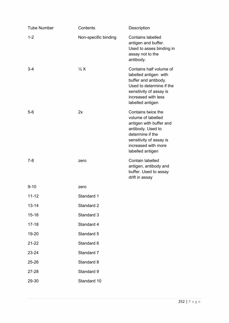
252 | P a g e
Tube Number Contents Description
1-2 Non-specific binding Contains labelled antigen and buffer. Used to asses binding in assay not to the antibody.
3-4 ½ X Contains half volume of labelled antigen with buffer and antibody. Used to determine if the sensitivity of assay is increased with less labelled antigen
5-6 2x Contains twice the volume of labelled antigen with buffer and antibody. Used to determine if the sensitivity of assay is increased with more labelled antigen
7-8 zero Contain labelled antigen, antibody and buffer. Used to assay drift in assay
9-10 zero
11-12 Standard 1
13-14 Standard 2
15-16 Standard 3
17-18 Standard 4
19-20 Standard 5
21-22 Standard 6
23-24 Standard 7
25-26 Standard 8
27-28 Standard 9
29-30 Standard 10

253 | P a g e
31-32 Standard 11
33-34 Zero
35…….. Samples
Every 30-50 tubes had a zero tube
---
Final two tubes Excess antibody Measure of lable quality over time
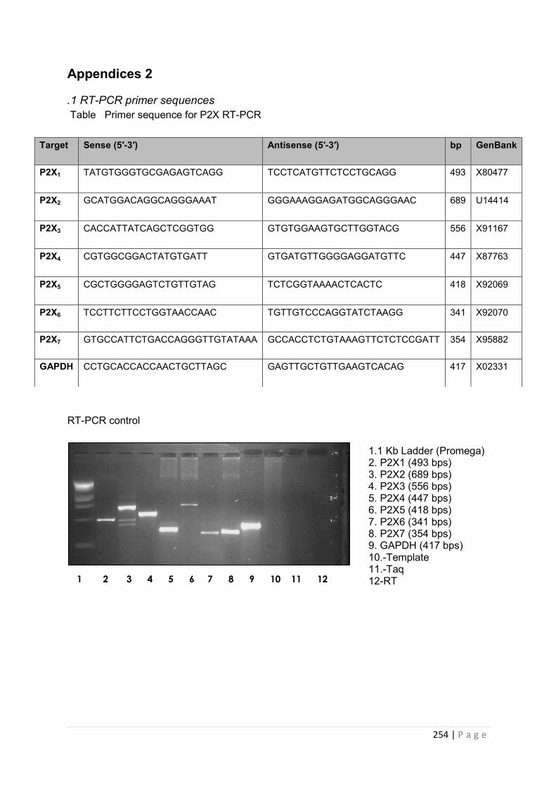
254 | P a g e
Appendices 2
.1 RT-PCR primer sequences Table Primer sequence for P2X RT-PCR
RT-PCR control
Target Sense (5′-3′) Antisense (5′-3′) bp GenBank
P2X1 TATGTGGGTGCGAGAGTCAGG TCCTCATGTTCTCCTGCAGG 493 X80477
P2X2 GCATGGACAGGCAGGGAAAT GGGAAAGGAGATGGCAGGGAAC 689 U14414
P2X3 CACCATTATCAGCTCGGTGG GTGTGGAAGTGCTTGGTACG 556 X91167
P2X4 CGTGGCGGACTATGTGATT GTGATGTTGGGGAGGATGTTC 447 X87763
P2X5 CGCTGGGGAGTCTGTTGTAG TCTCGGTAAAACTCACTC 418 X92069
P2X6 TCCTTCTTCCTGGTAACCAAC TGTTGTCCCAGGTATCTAAGG 341 X92070
P2X7 GTGCCATTCTGACCAGGGTTGTATAAA GCCACCTCTGTAAAGTTCTCTCCGATT 354 X95882
GAPDH CCTGCACCACCAACTGCTTAGC GAGTTGCTGTTGAAGTCACAG 417 X02331
1 2 3 4 5 6 7 8 9 10 11 12
1.1 Kb Ladder (Promega) 2. P2X1 (493 bps) 3. P2X2 (689 bps) 4. P2X3 (556 bps) 5. P2X4 (447 bps) 6. P2X5 (418 bps) 7. P2X6 (341 bps) 8. P2X7 (354 bps) 9. GAPDH (417 bps) 10.-Template 11.-Taq 12-RT
255 | P a g e
References 1. Heiss, H.W., Human Physiology. Edited by Robert F. Schmidt and Gerhard Thews. Springer-
Verlag, Berlin-Heidelberg-New York (1983) 725 pages, 569 figures, approx. $39.20 ISBN: 3-540-11669-9. Clinical Cardiology, 1983. 6(9): p. A43-A44.
2. Nussey, S. and S. Whitehead, in Endocrinology: An Integrated Approach2001, BIOS Scientific Publishers Limited: Oxford.
3. Beishuizen, A. and L.G. Thijs, Review: Endotoxin and the hypothalamo-pituitary-adrenal (HPA) axis. Journal of Endotoxin Research, 2003. 9(1): p. 3-24.
4. Ganong, W.F., Review of Medical Physiology. 22 ed2003, United States: Lange medical books/McGraw-Hill, .
5. Maxime, V.M.D., S.M.D. Siami, and D.M.D.P. Annane, Metabolism modulators in sepsis: The abnormal pituitary response. Critical Care Medicine Metabolic Support in Sepsis and Multiple Organ Failure: Proceedings of a Roundtable Conference in Brussels, Belgium, March 2007, 2007. 35(9): p. S596-S601.
6. Asa, S.L. and S. Ezzat, The pathogenesis of pituitary tumours. Nat Rev Cancer, 2002. 2(11): p. 836-849.
7. Chesnokova, V. and S. Melmed, Minireview: Neuro-Immuno-Endocrine Modulation of the Hypothalamic-Pituitary-Adrenal (HPA) Axis by gp130 Signaling Molecules. Endocrinology, 2002. 143(5): p. 1571-1574.
8. Beato, M., M. Truss, and S. Chavez, Control of transcription by steroid hormones. Ann N Y Acad Sci, 1996. 784: p. 93 - 123.
9. O'CONNOR, T.M., D.J. O'HALLORAN, and F. SHANAHAN, The stress response and the hypothalamic-pituitary-adrenal axis: from molecule to melancholia. QJM, 2000. 93(6): p. 323-333.
10. John, C.D. and J.C. Buckingham, Cytokines: regulation of the hypothalamo-pituitary-adrenocortical axis. Current Opinion in Pharmacology, 2003. 3(1): p. 78-84.
11. Szabo, N., et al., Role of neuroepithelial Sonic hedgehog in hypothalamic patterning. J Neurosci, 2009. 29: p. 6989 - 7002.
12. UNSWORTH, C.D. and R.G.J. JR., ATP Compartmentation in Neuroendocrine Secretory Vesicles. Annals of the New York Academy of Sciences, 1990. 603(Biological Actions of Extracellular ATP): p. 353-363.
13. Carew, M.A., et al., Extracellular ATP activates calcium entry and mobilization via P2U-purinoceptors in rat lactotrophs. Cell Calcium, 1994. 16(3): p. 227-235.
14. Chen, Z., et al., Effects of extracellular nucleotides in the pituitary: adenosine triphosphate receptor-mediated intracellular responses in gonadotrope- derived alpha T3-1 cells. Endocrinology, 1996. 137(1): p. 248-256.
15. Burnstock, G., Historical review: ATP as a neurotransmitter. Trends in Pharmacological Sciences, 2006. 27(3): p. 166-176.
16. Holton, F.A. and P. Holton, The capillary dilator substances in dry powders of spinal roots; a possible role of adenosine triphosphate in chemical transmission from nerve endings. The Journal of Physiology, 1954. 126(1): p. 124-140.
17. Holton, P., The liberation of adenosine triphosphate on antidromic stimulation of sensory nerves. The Journal of Physiology, 1959. 145(3): p. 494-504.
18. Burnstock, G., et al., INHIBITION OF THE SMOOTH MUSCLE ON THE TAENIA COLI. Nature, 1963. 200: p. 581-582.
19. Corriden, R. and P.A. Insel, Basal Release of ATP: An Autocrine-Paracrine Mechanism for Cell Regulation. Sci. Signal., 2010. 3(104): p. re1-.
20. Benarroch, E.E., Adenosine triphosphate: A multifaceted chemical signal in the nervous system. Neurology, 2010. 74(7): p. 601-607.
21. Burnstock, G., Physiology and Pathophysiology of Purinergic Neurotransmission. Physiol. Rev., 2007. 87(2): p. 659-797.

256 | P a g e
22. Tittle, R.K. and R.I. Hume, Opposite Effects of Zinc on Human and Rat P2X2 Receptors. J. Neurosci., 2008. 28(44): p. 11131-11140.
23. Li, S., et al., Expression and roles of pannexins in ATP release in the pituitary gland. Endocrinology, 2011. 152(6): p. 2342-52.
24. Chivasa, S., et al., Extracellular ATP Functions as an Endogenous External Metabolite Regulating Plant Cell Viability. Plant Cell, 2005. 17(11): p. 3019-3034.
25. Geoffrey Burnstock, Purinergic signalling. British Journal of Pharmacology, 2006. 147(S1): p. S172-S181.
26. North, R., Molecular Physiology of P2X Receptors. Physiol Rev, 2002. 82: p. 1013 - 1067. 27. Burnstock, G., Purinergic signalling: past, present and future. Braz J Med Biol Res, 2009.
42(1): p. 3-8. 28. Rees, D.A., et al., Adenosine-induced IL-6 expression in pituitary folliculostellate cells is
mediated via A2b adenosine receptors coupled to PKC and p38 MAPK. Br J Pharmacol, 2003. 140(4): p. 764-772.
29. JOUR, et al. 30. Tuduri, E., et al., Inhibition of Ca2+ signaling and glucagon secretion in mouse pancreatic
{alpha}-cells by extracellular ATP and purinergic receptors. Am J Physiol Endocrinol Metab, 2008. 294(5): p. E952-960.
31. Abbracchio, M.P., et al., Purinergic signalling in the nervous system: an overview. Trends in Neurosciences, 2009. 32(1): p. 19-29.
32. Khakh, B.S. and R. Alan North, P2X receptors as cell-surface ATP sensors in health and disease. Nature, 2006. 442(7102): p. 527-532.
33. Zemkova, H., et al., Molecular structure of purinergic P2X receptors and their expression in the hypothalamus and pituitary. Physiol Res, 2008. 57 Suppl 3: p. S23-38.
34. Zemkova, H., et al., Roles of Purinergic P2X Receptors as Pacemaking Channels and Modulators of Calcium-Mobilizing Pathway in Pituitary Gonadotrophs. Mol Endocrinol, 2006. 20(6): p. 1423-1436.
35. Colquhoun, D., Binding, gating, affinity and efficacy: the interpretation of structure-activity relationships for agonists and of the effects of mutating receptors. British Journal of Pharmacology, 1998. 125(5): p. 924-947.
36. Gever, J., et al., Pharmacology of P2X channels. Pflügers Archiv European Journal of Physiology, 2006. 452(5): p. 513-537.
37. Roberts, J., et al., Molecular properties of P2X receptors. Pflügers Archiv European Journal of Physiology, 2006. 452(5): p. 486-500.
38. Kawate, T., et al., Crystal structure of the ATP-gated P2X4 ion channel in the closed state. Nature. 460( 7255): p. 6.
39. Young, M.T., P2X receptors: dawn of the post-structure era. Trends in Biochemical Sciences, 2010. 35(2): p. 83-90.
40. Bianchi, B.R., et al., Pharmacological characterization of recombinant human and rat P2X receptor subtypes. European Journal of Pharmacology, 1999. 376(1-2): p. 127-138.
41. North, R.A. and A. Surprenant, Pharmacology of Cloned P2X Receptors. Annual Review of Pharmacology and Toxicology, 2000. 40(1): p. 563-580.
42. Olteanu, D., M.B. Hovater, and E.M. Schwiebert, Intraluminal autocrine purinergic signaling within cysts: implications for the progression of diseases that involve encapsulated cyst formation. Am J Physiol Renal Physiol, 2007. 292(1): p. F11-14.
43. Murrell-Lagnado, R.D. and O.S. Qureshi, Assembly and trafficking of P2X purinergic receptors (Review). Molecular Membrane Biology, 2008. 25(4): p. 321-331.
44. Bobanovic, L.K., S.J. Royle, and R.D. Murrell-Lagnado, P2X Receptor Trafficking in Neurons Is Subunit Specific. J. Neurosci., 2002. 22(12): p. 4814-4824.
257 | P a g e
45. Sharir, H. and M. Hershfinkel, The extracellular zinc-sensing receptor mediates intercellular communication by inducing ATP release. Biochemical and Biophysical Research Communications, 2005. 332(3): p. 845-852.
46. Schwiebert, E., et al., Extracellular zinc and ATP-gated P2X receptor calcium entry channels: New zinc receptors as physiological sensors and therapeutic targets. Purinergic Signalling, 2005. 1(4): p. 299-310.
47. Fu, J., et al., P2X receptors are expressed on neurons containing luteinizing hormone-releasing hormone in the mouse hypothalamus. Neuroscience Letters, 2009. 458(1): p. 32-36.
48. Rees, D., M. Scanlon, and J. Ham, Adenosine signalling pathways in the pituitary gland: one ligand, multiple receptors. J Endocrinol, 2003. 177(3): p. 357-364.
49. Gillespie, J.H., The biological significance of the linkages in adenosine triphosphoric acid. The Journal of Physiology, 1934. 80(4): p. 345-359.
50. Stojilkovic, S.S., Purinergic regulation of hypothalamopituitary functions. Trends in Endocrinology & Metabolism, 2009. 20(9): p. 460-468.
51. Terasawa, E., et al., Possible Role of 5'-Adenosine Triphosphate in Synchronization of Ca2+ Oscillations in Primate Luteinizing Hormone-Releasing Hormone Neurons. Mol Endocrinol, 2005. 19(11): p. 2736-2747.
52. Fischer, W., et al., Increase of intracellular Ca2+ by P2X and P2Y receptor-subtypes in cultured cortical astroglia of the rat. Neuroscience, 2009. 160(4): p. 767-783.
53. STOJILKOVIC, S.S., Pituitary cell type-specific electrical activity, calcium signaling and secretion. Biological Research, 2006. 39: p. 403-423.
54. Pellegatti, P., et al., A novel recombinant plasma membrane-targeted luciferase reveals a new pathway for ATP secretion. Mol Biol Cell, 2005. 16(8): p. 3659-65.
55. Jarvis, M.F. and B.S. Khakh, ATP-gated P2X cation-channels. Neuropharmacology, 2009. 56(1): p. 208-215.
56. Stojilkovic, S.S., H. Zemkova, and F. Van Goor, Biophysical basis of pituitary cell type-specific Ca2+ signaling-secretion coupling. Trends in Endocrinology and Metabolism. 16(4): p. 152-159.
57. Burgoyne, R.D. and A. Morgan, Secretory Granule Exocytosis. Physiol. Rev., 2003. 83(2): p. 581-632.
58. Balla, T., Regulation of Ca2+ entry by inositol lipids in mammalian cells by multiple mechanisms. Cell Calcium, 2009. 45(6): p. 527-534.
59. Anand-Srivastava, M.B., M. Cantin, and J. Gutkowska, Adenosine regulates the release of adrenocorticotropic hormone (ACTH) from cultured anterior pituitary cells. Molecular and Cellular Biochemistry, 1989. 89(1): p. 21-28.
60. Scaccianoce, S., et al., Adenosine and Pituitary-Adrenocortical Axis Activity in the Rat. Neuroendocrinology, 1989. 50(4): p. 464-468.
61. L.-F. Zhao, et al., Purinergic Receptor Ligands Stimulate Pro-Opiomelanocortin Gene Expression in AtT-20 Pituitary Corticotroph Cells. Journal of Neuroendocrinology, 2006. 18(4): p. 273-278.
62. Tomić, M., et al., Expression of Purinergic Receptor Channels and Their Role in Calcium Signaling and Hormone Release in Pituitary Gonadotrophs. Journal of Biological Chemistry, 1996. 271(35): p. 21200-21208.
63. Gonzalez-Iglesias, A.E., et al., Dependence of Electrical Activity and Calcium Influx-Controlled Prolactin Release on Adenylyl Cyclase Signaling Pathway in Pituitary Lactotrophs. Mol Endocrinol, 2006. 20(9): p. 2231-2246.
64. Zemkova, H., et al., Characterization of Purinergic P2X4 Receptor Channels Expressed in Anterior Pituitary Cells. Am J Physiol Endocrinol Metab, 2009: p. ajpendo.00558.2009.
258 | P a g e
65. He, M.-L., A.E. Gonzalez-Iglesias, and S.S. Stojilkovic, Role of Nucleotide P2 Receptors in Calcium Signaling and Prolactin Release in Pituitary Lactotrophs. J. Biol. Chem., 2003. 278(47): p. 46270-46277.
66. Lewis, B.M., et al., Adenosine stimulates connexin 43 expression and gap junctional communication in pituitary folliculostellate cells. FASEB J., 2006. 20(14): p. 2585-2587.
67. Nakajima, Y., et al., Acetylcholine increases intracellular Ca2+ in the rat pituitary folliculostellate cells in primary culture. Am J Physiol Endocrinol Metab, 2001. 280(4): p. E608-615.
68. Chen, L., et al., Cytological Characterization of a Pituitary Folliculo-Stellate-Like Cell Line, Tpit/F1, with Special Reference to Adenosine Triphosphate-Mediated Neuronal Nitric Oxide Synthase Expression and Nitric Oxide Secretion. Endocrinology, 2000. 141(10): p. 3603-3610.
69. Stojilkovic, S.S., et al., Multiple Roles of G<sub>i/o</sub> Protein-coupled Receptors in Control of Action Potential Secretion Coupling in Pituitary Lactotrophs. Annals of the New York Academy of Sciences, 2009. 1152(Mechanisms of Exocytosis): p. 174-186.
70. Chung, H.S., et al., ATP-induced [Ca<sup>2+</sup>]<sub>i</sub> changes and depolarization in GH3 cells. British Journal of Pharmacology, 2000. 130(8): p. 1843-1852.
71. Egan, T.M. and B.S. Khakh, Contribution of Calcium Ions to P2X Channel Responses. J. Neurosci., 2004. 24(13): p. 3413-3420.
72. Coddou, C., et al., Activation and Regulation of Purinergic P2X Receptor Channels. Pharmacological Reviews, 2011.
73. Stojilkovic, S.S., et al., Signaling by purinergic receptors and channels in the pituitary gland. Molecular and Cellular Endocrinology, 2010. 314(2): p. 184-191.
74. Villalobos, C., et al., Functional ATP receptors in rat anterior pituitary cells. Am J Physiol Cell Physiol, 1997. 273(6): p. C1963-1971.
75. Stojilkovic, S.S. and T.-a. Koshimizu, Signaling by extracellular nucleotides in anterior pituitary cells. Trends in Endocrinology and Metabolism, 2001. 12(5): p. 218-225.
76. Yu, Q., et al., Expression of P2Y receptors in the rat anterior pituitary. Purinergic Signal, 2011. 7(2): p. 207-19.
77. Tierney, T., et al., Evidence From Studies on Co-Cultures of TtT/GF and AtT20 Cells That Annexin 1 Acts as a Paracrine or Juxtacrine Mediator of the Early Inhibitory Effects of Glucocorticoids on ACTH Release. Journal of Neuroendocrinology, 2003. 15(12): p. 1134-1143.
78. Tierney, T., et al., Macrophage Migration Inhibitory Factor Is Released from Pituitary Folliculo-Stellate-Like Cells by Endotoxin and Dexamethasone and Attenuates the Steroid-Induced Inhibition of Interleukin 6 Release. Endocrinology, 2005. 146(1): p. 35-43.
79. Kanasaki, H., et al., Differential Regulation of Pituitary Hormone Secretion and Gene Expression by Thyrotropin-Releasing Hormone. A Role for Mitogen-Activated Protein Kinase Signaling Cascade in Rat Pituitary GH3 Cells1. Biology of Reproduction, 2002. 67(1): p. 107-113.
80. Leclerc, G.M. and F.R. Boockfor, Pulses of Prolactin Promoter Activity Depend on a Noncanonical E-Box that Can Bind the Circadian Proteins CLOCK and BMAL1. Endocrinology, 2005. 146(6): p. 2782-2790.
81. JUDD, A.M., et al., Characterization of the MMQ Cell, A Prolactin-Secreting Clonal Cell Line That Is Responsive to Dopamine. Endocrinology, 1988. 123(5): p. 2341-2350.
82. Baricordi, O., et al., An ATP-activated channel is involved in mitogenic stimulation of human T lymphocytes. Blood, 1996. 87: p. 682 - 690.
83. Ciccone, N.A. and U.B. Kaiser, The biology of gonadotroph regulation. Current Opinion in Endocrinology, Diabetes & Obesity, 2009. 16(4): p. 321-327.
84. Henderson, H.L., J. Townsend, and D.J. Tortonese, Direct effects of prolactin and dopamine on the gonadotroph response to GnRH. J Endocrinol, 2008. 197(2): p. 343-350.

259 | P a g e
85. Alarid, E.T., et al., Immortalization of pituitary cells at discrete stages of development by directed oncogenesis in transgenic mice. Development, 1996. 122(10): p. 3319-3329.
86. Ooi, G.T., N. Tawadros, and R.M. Escalona, Pituitary cell lines and their endocrine applications. Molecular and Cellular Endocrinology, 2004. 228(1-2): p. 1-21.
87. Yonehara, T., et al., Characterization of αT3-1 Cells Stably Transfected with Luteininzing Hormone β-Subunit Complementary Deoxyribonucleic Acid. Endocrine Journal, 2003. 50(3): p. 341-354.
88. Fraker, P.J. and J.C. Speck, Jr., Protein and cell membrane iodinations with a sparingly soluble chloroamide, 1,3,4,6-tetrachloro-3a,6a-diphenylglycoluril. 1978. Biochem Biophys Res Commun, 2012. 425(3): p. 510-8.
89. Brouns, I., et al., Triple Immunofluorescence Staining with Antibodies Raised in the Same Species to Study the Complex Innervation Pattern of Intrapulmonary Chemoreceptors. Journal of Histochemistry & Cytochemistry, 2002. 50(4): p. 575-582.
90. Balen, D., et al., Revised immunolocalization of the Na+-D-glucose cotransporter SGLT1 in rat organs with an improved antibody. Am J Physiol Cell Physiol, 2008. 295(2): p. C475-89.
91. Ruan, H.Z., et al., Expression of P2X and P2Y receptors in the intramural parasympathetic ganglia of the cat urinary bladder. Am J Physiol Renal Physiol, 2006. 290(5): p. F1143-52.
92. Greenwood, D., et al., P2X receptor signaling inhibits BDNF-mediated spiral ganglion neuron development in the neonatal rat cochlea. Development, 2007. 134(7): p. 1407-1417.
93. Doctor, R.B., et al., Purinergic regulation of cholangiocyte secretion: identification of a novel role for P2X receptors. Am J Physiol Gastrointest Liver Physiol, 2005. 288(4): p. G779-786.
94. Inoue, K., et al., Establishment of a folliculo-stellate-like cell line from a murine thyrotropic pituitary tumor. Endocrinology, 1992. 131(6): p. 3110-3116.
95. Herkenham, M., Folliculo-Stellate (FS) Cells of the Anterior Pituitary Mediate Interactions between the Endocrine and Immune Systems. Endocrinology, 2005. 146(1): p. 33-34.
96. Koshimizu, T.-a., et al., Characterization of Purinergic Receptors and Receptor-Channels Expressed in Anterior Pituitary Cells. Endocrinology, 2000. 141(11): p. 4091-4099.
97. Li, S., M. Tomić, and S.S. Stojilkovic, Characterization of novel Pannexin 1 isoforms from rat pituitary cells and their association with ATP-gated P2X channels. General and Comparative Endocrinology, (0).
98. Koshimizu, T.-a., et al., Characterization of Calcium Signaling by Purinergic Receptor-Channels Expressed in Excitable Cells. Mol Pharmacol, 2000. 58(5): p. 936-945.
99. Stojilkovic, S.S., et al., Signaling by purinergic receptors and channels in the pituitary gland. Molecular and Cellular Endocrinology. In Press, Corrected Proof.
100. Koshimizu, T.-a., et al., Functional Role of Alternative Splicing in Pituitary P2X2 Receptor-Channel Activation and Desensitization. Mol Endocrinol, 1998. 12(7): p. 901-913.
101. Di Virgilio, F., et al., Nucleotide receptors: an emerging family of regulatory molecules in blood cells. Blood, 2001. 97(3): p. 587-600.
102. Smalheiser, N.R., et al., Expression of reelin in adult mammalian blood, liver, pituitary pars intermedia, and adrenal chromaffin cells. Proc Natl Acad Sci U S A, 2000. 97(3): p. 1281-6.
103. Aoki, Y., et al., Regulation of the Rat Proopiomelanocortin Gene Expression in AtT-20 cells. II: Effects of the Pituitary Adenylate Cyclase-Activating Polypeptide and Vasoactive Intestinal Polypeptide. Endocrinology, 1997. 138(5): p. 1930-1934.
104. Wilson, M.L. and S.B. Guild, Effects of wortmannin upon the late stages of the secretory pathway of AtT-20 cells. European Journal of Pharmacology, 2001. 413(1): p. 55-62.
105. McFerran, B.W. and S.B. Guild, Effects of mastoparan upon the late stages of the ACTH secretory pathway of AtT-20 cells. Br J Pharmacol, 1995. 115(4): p. 696-702.
106. Volonté, C., et al., P2 receptor web: Complexity and fine-tuning. Pharmacology & Therapeutics, 2006. 112(1): p. 264-280.
107. BOOCKFOR, F.R. and L.K. SCHWARZ, CULTURES OF GH3 CELLS CONTAIN BOTH SINGLE AND DUAL HORMONE SECRETORS. Endocrinology, 1988. 122(2): p. 762-764.
260 | P a g e
108. Deli, T. and L. Csernoch, Extracellular ATP and Cancer—An Overview with Special Reference to P2 Purinergic Receptors. Pathology & Oncology Research, 2008. 14(3): p. 219-231.
109. Ohtani, M., K. Ohura, and T. Oka, Involvement of P2X Receptors in the Regulation of Insulin Secretion, Proliferation and Survival in Mouse Pancreatic β-Cells. Cell Physiol Biochem, 2011. 28(2): p. 355-366.
110. Suzuki-Kerr, H., et al., Molecular identification and localization of P2X receptors in the rat lens. Experimental Eye Research, 2008. 86(5): p. 844-855.
111. Guo, C., et al., Evidence for functional P2X4/P2X7 heteromeric receptors. Mol Pharmacol, 2007. 72(6): p. 1447-56.
112. Dutton, J.L., et al., Development of P2X receptor clusters on smooth muscle cells in relation to nerve varicosities in the rat urinary bladder. J Neurocytol, 1999. 28(1): p. 4-16.
113. Brandon, N.J. and S.J. Moss, Receptor Cross Talk: Ligand-Gated Ion Channels Start to Communicate. Sci. STKE, 2000. 2000(55): p. pe1-.
114. Casas-Pruneda, G., et al., Functional interactions between P2X4 and P2X7 receptors from mouse salivary epithelia. The Journal of Physiology, 2009. 587(12): p. 2887-2901.
115. Afework, M. and G. Burnstock, Distribution of P2X receptors in the rat adrenal gland. Cell and Tissue Research, 1999. 298(3): p. 449-456.
116. Miklavc, P., et al., Fusion-activated Ca2+ entry via vesicular P2X4 receptors promotes fusion pore opening and exocytotic content release in pneumocytes. Proc Natl Acad Sci U S A, 2011. 108(35): p. 14503-8.
117. Bulanova, E., et al., Extracellular ATP induces cytokine expression and apoptosis through P2X7 receptor in murine mast cells. J Immunol, 2005. 174: p. 3880 - 3890.
118. Lämmer, A., et al., Neuroprotective effects of the P2 receptor antagonist PPADS on focal cerebral ischaemia-induced injury in rats. European Journal of Neuroscience, 2006. 23(10): p. 2824-2828.
119. Clark, A.K., et al., P2X7-Dependent Release of Interleukin-1{beta} and Nociception in the Spinal Cord following Lipopolysaccharide. J. Neurosci., 2010. 30(2): p. 573-582.
120. Franke, H., et al., Involvement of P2X and P2Y receptors in microglial activation in vivo. Purinergic Signal, 2007. 3(4): p. 435-45.
121. Chessell, I., et al., Disruption of the P2X7 purinoceptor gene abolishes chronic inflammatory and neuropathic pain. Pain, 2005. 114: p. 386 - 396.
122. Solle, M., et al., Altered cytokine production in mice lacking P2X{sub7} receptors. J. Biol. Chem., 2000: p. M006781200.
123. Custer, E.E., et al., P2X purinergic receptor knockout mice reveal endogenous ATP modulation of both vasopressin and oxytocin release from the intact neurohypophysis. J Neuroendocrinol, 2012. 24(4): p. 674-80.
124. Jacques-Silva, M.C., et al., ATP-gated P2X3 receptors constitute a positive autocrine signal for insulin release in the human pancreatic β cell. Proceedings of the National Academy of Sciences, 2010. 107(14): p. 6465-6470.
125. Xiang, Z. and G. Burnstock, Changes in expression of P2X purinoceptors in rat cerebellum during postnatal development. Brain Res Dev Brain Res, 2005. 156(2): p. 147-57.
126. Studeny, S., A. Torabi, and M.A. Vizzard, P2X2 and P2X3 receptor expression in postnatal and adult rat urinary bladder and lumbosacral spinal cord. Am J Physiol Regul Integr Comp Physiol, 2005. 289(4): p. R1155-68.
127. He, M.-L., et al., Release and extracellular metabolism of ATP by ecto-nucleotidase eNTPDase 1–2 in hypothalamic and pituitary cells. Purinergic Signalling, 2005. 1(2): p. 135-144.
128. Bianchi, B., et al., Pharmacological characterization of recombinant human and rat P2X receptor subtypes. European Journal of Pharmacology, 1999. 376: p. 127 - 138.
129. North, R.A., Molecular Physiology of P2X Receptors. Physiol. Rev., 2002. 82(4): p. 1013-1067. 130. Surprenant, A. and R.A. North, Signaling at Purinergic P2X Receptors. Annual Review of
Physiology, 2009. 71(1): p. 333-359.
261 | P a g e
131. Chung, H.S., et al., ATP-induced [Ca(2+)](i) changes and depolarization in GH3 cells. Br J Pharmacol, 2000. 130(8): p. 1843-52.
132. Kretschmannova, K., et al., The Expression and Role of Hyperpolarization-Activated and Cyclic Nucleotide-Gated Channels in Endocrine Anterior Pituitary Cells. Molecular Endocrinology, 2012. 26(1): p. 153-164.
133. Alexander V Gourine, D.M.P., Nikolai Zhernosek, Ekaterina V Melenchuk, Rüdiger Gerstberger, K Michael Spyer, Valery N Gourine,, P2 receptor blockade attenuates fever and cytokine responses induced by lipopolysaccharide in rats. British Journal of Pharmacology, 2005. 146(1): p. 139-145.
134. Spelta, V., et al., Kinetics of antagonist actions at rat P2X2/3 heteromeric receptors. British Journal of Pharmacology, 2002. 135(6): p. 1524-1530.
135. Charlton, S.J., et al., PPADS and suramin as antagonists at cloned P2Y- and P2U-purinoceptors. Br J Pharmacol, 1996. 118(3): p. 704-10.
136. Dong, X., et al., P2Y receptors mediate Ca2+ signaling in duodenocytes and contribute to duodenal mucosal bicarbonate secretion. Am J Physiol Gastrointest Liver Physiol, 2009. 296(2): p. G424-32.
137. Xu, G.-Y., et al., P2X receptor-mediated visceral hyperalgesia in a rat model of chronic visceral hypersensitivity. Gut, 2008. 57(9): p. 1230-1237.
138. Thomas, S., et al., The antagonist trinitrophenyl-ATP reveals co-existence of distinct P2X receptor channels in rat nodose neurones. The Journal of Physiology, 1998. 509(2): p. 411-417.
139. Hodges, R.R., et al., Identification of P2X3 and P2X7 Purinergic Receptors Activated by ATP in Rat Lacrimal Gland. Investigative Ophthalmology & Visual Science, 2011.
140. Alcaraz, L., et al., Novel P2X7 receptor antagonists. Bioorg Med Chem Lett, 2003. 13: p. 4043 - 4046.
141. D L Donnelly-Roberts, M.F.J., Discovery of P2X<sub>7</sub> receptor-selective antagonists offers new insights into P2X<sub>7</sub> receptor function and indicates a role in chronic pain states. British Journal of Pharmacology, 2007. 151(5): p. 571-579.
142. Felix, R.A., et al., Development of a comprehensive set of P2 receptor pharmacological research compounds. Purinergic Signal, 2012. 8(Suppl 1): p. 101-12.
143. Ueno, S., et al., Involvement of P2X<SUB><FONT SIZE='-1'>2</FONT></SUB> and P2X<SUB><FONT SIZE='-1'>3</FONT></SUB> receptors in neuropathic pain in a mouse model of chronic constriction injury. Drug Development Research, 2003. 59(1): p. 104-111.
144. Kaan, T.K.Y., et al., Systemic blockade of P2X3 and P2X2/3 receptors attenuates bone cancer pain behaviour in rats. Brain, 2010. 133(9): p. 2549-2564.
145. Wirkner, K., B. Sperlagh, and P. Illes, P2X3 Receptor Involvement in Pain States. Molecular Neurobiology, 2007. 36(2): p. 165-183.
146. Gever, J.R., et al., AF-353, a novel, potent and orally bioavailable P2X3/P2X2/3 receptor antagonist. Br J Pharmacol, 2010. 160(6): p. 1387-98.
147. Sanz, J.M., et al., Activation of Microglia by Amyloid {beta} Requires P2X7 Receptor Expression. J Immunol, 2009. 182(7): p. 4378-4385.
148. Apolloni, S., et al., Membrane compartments and purinergic signalling: P2X receptors in neurodegenerative and neuroinflammatory events. FEBS Journal, 2009. 276: p. 354-364.
149. Verspohl, E.J., et al., Effect of purinergic agonists and antagonists on insulin secretion from INS-1 cells (insulinoma cell line) and rat pancreatic islets. Canadian Journal of Physiology and Pharmacology, 2002. 80(6): p. 562-568.
150. Mehet, D.K., et al., Evidence from in vitro and in vivo studies that NFκB within the pituitary folliculostellate cells and corticotrophs regulates ACTH secretion in experimental endotoxaemia. Journal of Neuroendocrinology, 2012: p. no-no.
262 | P a g e
151. Kanasaki, H., et al., Mitogen-Activated Protein Kinase Activation by Stimulation with Thyrotropin‐Releasing Hormone in Rat Pituitary GH3 Cells. Biology of Reproduction, 1999. 61(1): p. 319-325.
152. Jarvis, M.F., et al., Modulation of BzATP and formalin induced nociception: attenuation by the P2X receptor antagonist, TNP-ATP and enhancement by the P2X(3) allosteric modulator, cibacron blue. Br J Pharmacol, 2001. 132(1): p. 259-69.
153. Solini, A., et al., Multiple P2X receptors are involved in the modulation of apoptosis in human mesangial cells: evidence for a role of P2X4. Am J Physiol Renal Physiol, 2007. 292(5): p. F1537-47.
154. Ma, B., et al., Pharmacological properties of P2 receptors on rat otic parasympathetic ganglion neurons. Life Sciences, 2008. 83(5-6): p. 185-191.
155. Denda, S., et al., Oxytocin is expressed in epidermal keratinocytes and released upon stimulation with adenosine 5'-[gamma-thio]triphosphate in vitro. Exp Dermatol, 2012. 21(7): p. 535-7.
156. Hodges, R.R., et al., Characterization of P2X7 purinergic receptors and their function in rat lacrimal gland. Invest Ophthalmol Vis Sci, 2009. 50(12): p. 5681-9.
157. Marcellino, D., et al., On the role of P2X(7) receptors in dopamine nerve cell degeneration in a rat model of Parkinson's disease: studies with the P2X(7) receptor antagonist A-438079. J Neural Transm, 2010. 117(6): p. 681-7.
158. Hille, B., et al., Stimulation of exocytosis without a calcium signal. The Journal of Physiology, 1999. 520(1): p. 23-31.
159. Van Goor, F., et al., Dependence of Pituitary Hormone Secretion on the Pattern of Spontaneus Voltage-gated Calcium Influx. Journal of Biological Chemistry, 2001. 276(36): p. 33840-33846.
160. Chen, Z.-P., et al., Evidence for a Role of Pituitary ATP Receptors in the Regulation of Pituitary Function. Proceedings of the National Academy of Sciences of the United States of America, 1995. 92(11): p. 5219-5223.
161. Ataullakhanov, F.I. and V.M. Vitvitsky, What determines the intracellular ATP concentration. Biosci Rep, 2002. 22(5-6): p. 501-11.
162. Guest, P.C., et al., Altered levels of circulating insulin and other neuroendocrine hormones associated with the onset of schizophrenia. Psychoneuroendocrinology, 2011. 36(7): p. 1092-6.
163. Takagi, M., et al., Gradual loss of ACTH due to a novel mutation in LHX4: comprehensive mutation screening in Japanese patients with congenital hypopituitarism. PLoS ONE, 2012. 7(9): p. e46008.
164. Strathmann, F.G., et al., Multiplex immunoassays of peptide hormones extracted from formalin-fixed, paraffin-embedded tissue accurately subclassify pituitary adenomas. Clin Chem, 2012. 58(2): p. 366-74.
165. Besser, G.M., et al., Interaction between thyrotrophin, corticotrophin and growth hormone secretion in man. J Endocrinol, 1971. 51(4): p. 699-706.
166. Kawamura, M., et al., Extracellular ATP potentiates steroidogenic effect of adrenocorticotropic hormone in bovine adrenocortical fasciculata cells. Jpn J Pharmacol, 2001. 85(4): p. 376-81.
167. Nishi, H., H. Arai, and T. Momiyama, NCI-H295R, a Human Adrenal Cortex-Derived Cell Line, Expresses Purinergic Receptors Linked to Ca<sup>2+</sup>-Mobilization/Influx and Cortisol Secretion. PLoS ONE, 2013. 8(8): p. e71022.
168. HOWELL, S.L. and R.B.L. EWART, Synthesis and Secretion of Growth Hormone in the Rat Anterior Pituitary: II. Properties of the Isolated Growth Hormone Storage Granules. Journal of Cell Science, 1973. 12(1): p. 23-35.
169. LORENSON, M.Y. and L.S. JACOBS, Secretory Granule Growth Hormone and Prolactin Release: Independence from Granule Membrane ATPase. Endocrinology, 1984. 114(3): p. 717-724.
263 | P a g e
170. Luque, R.M., et al., Homologous and Heterologous Regulation of Pituitary Receptors for Ghrelin and Growth Hormone-Releasing Hormone. Endocrinology, 2004. 145(7): p. 3182-3189.
171. Veldhuis, J.D., Neuroendocrine control of pulsatile growth hormone release in the human: relationship with gender. Growth Horm IGF Res, 1998. 8 Suppl B: p. 49-59.
172. Raspe, E., et al., ATP, bradykinin, TRH and TSH activate the Ca(2+)-phosphatidylinositol cascade of human thyrocytes in primary culture. Mol Cell Endocrinol, 1991. 81(1-3): p. 175-83.
173. JOUR, et al. 174. Kumari, M., et al., Roles for adenosine A1- and A2-receptors in the control of thyrotrophin
and prolactin release from the anterior pituitary gland. Regulatory Peptides, 1999. 79(1): p. 41-46.
175. Nunez, L., C. Villalobos, and L.S. Frawley, Extracellular ATP as an autocrine/paracrine regulator of prolactin release. Am J Physiol, 1997. 272(6 Pt 1): p. E1117-23.
176. Johnston, J.M., et al., The dopamine D2 receptor is expressed in GH3 cells. Journal of Molecular Endocrinology, 1991. 7(2): p. 131-136.
177. Li, Q., et al., Dopamine receptor D2S gene transfer improves the sensitivity of GH3 rat pituitary adenoma cells to bromocriptine. Molecular and Cellular Endocrinology, 2014. 382(1): p. 377-384.
178. Gloddek, J., et al., The intrapituitary stimulatory effect of lipopolysaccharide on ACTH secretion is mediated by paracrine-acting IL-6. Exp Clin Endocrinol Diabetes, 2001. 109(8): p. 410-5.
179. Clark, A.R., J.R.S. Martins, and C.R. Tchen, Role of Dual Specificity Phosphatases in Biological Responses to Glucocorticoids. J. Biol. Chem., 2008. 283(38): p. 25765-25769.
180. Delahunty, T.M., M.J. Cronin, and J. Linden, Regulation of GH3-cell function via adenosine A1 receptors. Inhibition of prolactin release, cyclic AMP production and inositol phosphate generation. Biochem J, 1988. 255(1): p. 69-77.
181. Parvathenani, L., et al., P2X7 Mediates Superoxide Production in Primary Microglia and Is Up-regulated in a Transgenic Mouse Model of Alzheimer's Disease. J Biol Chem, 2003. 278: p. 13309 - 13317.
182. Lee, D.H., et al., Dual effect of ATP on glucose-induced insulin secretion in HIT-T15 cells. Pancreas, 2008. 37(3): p. 302-8.
183. Huang, C.-Y., et al., Prolactin secretion and intracellular Ca2+ change in rat lactotroph subpopulations stimulated by thyrotropin-releasing hormone. Journal of Cellular Biochemistry, 2002. 87(2): p. 126-132.
184. Knott, T.K., et al., Adenosine trisphosphate appears to act via different receptors in terminals versus somata of the hypothalamic neurohypophysial system. J Neuroendocrinol, 2012. 24(4): p. 681-9.
185. Knott, T.K., et al., Endogenous ATP potentiates only vasopressin secretion from neurohypophysial terminals. J Cell Physiol, 2008. 217(1): p. 155-61.
186. Richards-Williams, C., et al., Extracellular ATP and zinc are co-secreted with insulin and activate multiple P2X purinergic receptor channels expressed by islet beta-cells to potentiate insulin secretion. Purinergic Signalling, 2008. 4(4): p. 393-405.
187. Cervetto, C., et al., Calmidazolium selectively inhibits exocytotic glutamate release evoked by P2X7 receptor activation. Neurochem Int, 2012. 60(8): p. 768-72.
188. Trang, T., et al., P2X4-Receptor-Mediated Synthesis and Release of Brain-Derived Neurotrophic Factor in Microglia Is Dependent on Calcium and p38-Mitogen-Activated Protein Kinase Activation. J. Neurosci., 2009. 29(11): p. 3518-3528.
189. Balázs, B., et al., Investigation of the Inhibitory Effects of the Benzodiazepine Derivative, 5-BDBD on P2X<sub>4</sub> Purinergic Receptors by two Complementary Methods. Cellular Physiology and Biochemistry, 2013. 32(1): p. 11-24.
264 | P a g e
190. Wu, T., et al., Functional expression of P2X4 receptor in capillary endothelial cells of the cochlear spiral ligament and its role in regulating the capillary diameter. Am J Physiol Heart Circ Physiol, 2011. 301(1): p. H69-78.
191. Gentilin, E., et al., Mitotane reduces human and mouse ACTH-secreting pituitary cell viability and function. Journal of Endocrinology, 2013. 218(3): p. 275-285.
192. Tomic, M., et al., ROLE OF NON-SELECTIVE CATION CHANNELS IN SPONTANEOUS AND PROTEIN KINASE A STIMULATED CALCIUM SIGNALING IN PITUITARY CELLS. American Journal of Physiology - Endocrinology And Metabolism, 2011.
193. Bernier, L.-P., et al., Phosphoinositides Regulate P2X4 ATP-Gated Channels through Direct Interactions. J. Neurosci., 2008. 28(48): p. 12938-12945.
194. Chaumont, S., et al., Regulation of P2X2 Receptors by the Neuronal Calcium Sensor VILIP1. Sci. Signal., 2008. 1(41): p. ra8-.
195. Garcia-Marcos, M., J.-P. Dehaye, and A. Marino, Membrane compartments and purinergic signalling: the role of plasma membrane microdomains in the modulation of P2XR-mediated signalling. FEBS Journal, 2009. 276(2): p. 330-340.