characterisation of endophytic bacteria from a desert ... · pdf file1 plant protect. sci....
TRANSCRIPT

1
Plant Protect. Sci. Vol. 52, 2016, No. 4: 00–
doi: 10.17221/14/2016-PPS
Characterisation of Endophytic Bacteria from a Desert Plant Lepidium perfoliatum L.
Yuanting LI, Cong CHENG and Dengdi AN
Xinjiang Key Laboratory of Special Species Conservation and Regulatory Biology, College of Life Science, Xinjiang Normal University, Xinjiang, P.R. China
Abstract
Li Y., Cheng C., An D. (2017): Characterisation of endophytic bacteria from a desert plant Lepidium per-foliatum L. Plant Protect. Sci.
Sixty-two endophytic bacteria from the leaves, roots, and stems of a healthy Lepidium perfoliatum L. were isolated and characterised. From the results, 89, 87, 90, and 97% isolates could tolerate 12% NaCl, 30% PEG 6000, 50°C and pH 10, respectively. 74% isolates could form biofilm. Besides, 28 isolates could improve germination rate of host seeds under different degree of drought stress. These data suggest that the endophyte isolates show considerable resistance to abiotic stress and assist their plant hosts germination under drought stress.
Keywords: Lepidium perfoliatum L.; endophytic bacterial diversity; biofilm; salt tolerance; drought tolerance
Abiotic stresses such as drought, salinity, hot and soil pH are major limiting factors that affect crop productivity worldwide as a result of their debilitat-ing effects on all plant functions. Endophytic bac-teria colonise inner host tissues without damaging the host (Reinhold-Hurek & Hurek 2011). They have several beneficial effects on host plants such as plant growth promotion and increased resistance against plant pathogens and parasites (Hallmann et al. 2011). Furthermore, endophytic bacteria have recently been discovered to ameliorate the adverse effects of abiotic stress to host plant. Date palm root-associated bacteria promote plant growth under drought conditions (Cherif et al. 2015). Plant growth promoting endophytic bacteria, for example, Burk-holderia phytofirmans PsJN improve the growth of wheat and maize under drought conditions (Naveed et al. 2014a, b). Endophytic bacteria strains Achro-mobacter xylosoxidans and Bacillus pumilus enhance the growth of sunflower seedlings under water stress
(Forchetti et al. 2010). The use of bacterial endo-phytes (Pseudomonas fluorescens YsS6 and P. migulae 8R6) has the potential to facilitate tomato plant growth under high salt contents (Ali et al. 2014). Isolates from halophyte Limonium sinense (Girard) Kuntze, which possessed ACC deaminase activ-ity could enhance plant growth under saline stress conditions (Qin et al. 2014). Endophytic bacterium A. xylosoxidans that was isolated from the root tissue of Catharanthus roseus was able to ameliorate salt stress in C. roseus (Karthikeyan et al. 2012). Inocu-lation of rice (Oryza sativa L.) with an endophytic bacterium P. pseudoalcaligenes that was isolated from the root tissue could increase shoot biomass at lower saline levels ( Jha et al. 2011).
Lepidium perfoliatum L. (Brassicaceae) is an an-nual or biannual herb plant living in wilderness or desert (Yuan et al. 2013). It is the sand-fixing pioneer plant because of its fast growth and the resistance to drought and salinity (Huang et al. 2011). The
Supported by Chinese National Natural Science Fund, Grants Nos 31300238, 31360149, and 31370159, and Xinjiang Key Laboratory of Special Species Conservation and Regulatory Biology, Grants Nos XJDX1414-2015-02, XJDX1414-2016-05, and XJDX1414-2016-06.
All authors declare that they have no any conflict of interests.
2
Vol. 52, 2016, No. 4: 00– Plant Protect. Sci.
doi: 10.17221/14/2016-PPS
previous study of the L. perfoliatum was focussed on its seed coat mucilage, which is important for germination in a harsh environment (Li-Li et al. 2008). To the best of our knowledge, there are cur-rently no data on the culture-dependent bacterial endophyte diversity in L. perfoliatum. Thus, a better understanding of the presence of the endophytic bacteria in L. perfoliatum is important. These bac-teria may have been adapted to the salt, drought, hot, and alkali stress conditions and could provide significant benefits to the host plants. Studying the bacterial endophytes could offer a few important clues on how these endophytes mutual interaction with their host helps them to withstand the desert environment successfully.
This study aims to isolate the endophytic bacteria associated with the desert plant Lepidium perfolia-tum L., and to identify them using 16S rRNA gene sequence analysis. Also included in the objective is the characterisation of enzyme production, tests for salt, drought, hot and alkaline tolerance, and bio-film formation in non-stressed and stressed condi-tions. Finally, whether these isolates could improve germination rate of the host seeds under different degree of drought stress was tested.
MATERIAL AND METHODS
Sampling of plant materials. L. perfoliatum was collected in its native habitat from a hill near Urumqi City in Xinjiang Province, China (43°50'N, 87°35'E) in May 2013. Annual precipitation in this area is less than 200 mm, however, the evaporation capacity is as high as 2600 mm, and the relative humidity is only 30% which is the typical ecoclimatic condition in area of drought and desert. After digging 25–30 cm depth around the main root of plants, all whole plants were then immediately put in sterile polystyrene bags and brought to the laboratory on ice.
Isolation of endophytic bacteria. Fresh healthy L. per- foliatum plants were washed thoroughly with tap water to remove adhering soil and debris. One gram of leaves, stems and roots was weighed individually and immersed in 75% ethanol for 3 min three times, and then treated with 0.1% mercuric chloride for 3 min and finally rinsed 6 times in sterile distilled water (5 min each rinse). In order to examine the effect of the surface sterilization, aliquots of the final rinsing water were spread on beef extract peptone medium plates containing (in g/l) beef extract 3,
peptone 10, NaCl 5, pH 7.2, and cultured at 37°C. The absence of bacteria after 6 days of incubation was taken to confirm that surface disinfection process was successful. Ten grams of each surface-sterilised tissue was ground by sterilised mortar and pestle with 10 ml sterilised potassium phosphate buffer (pH 7.0). The suspension was used for serial dilution plating on beef extract peptone medium plates that were then incubated at 37°C. Bacterial colonies from each tissue were passed through four rounds of single colony isolation by streaking them on beef extract peptone medium to ensure purity of the organism. Pure cultures were stored in 20% glycerol at –80°C.
16S rRNA gene amplification, bacterial identifi-cation and phylogenetic analysis. Three milliliters of fresh cultures of the isolates in beef extract peptone medium was used for total DNA extraction (Sam-brook 1989). Genomic DNA was used as a template for 16S rRNA gene amplification with universal primer set 27F and 1492R (Chelius & Triplett 2001) in a polymerase chain reaction (PCR). Purified PCR products were sequenced with an ABI 3730 automated sequencer (Invitrogen, Shanghai, China). All the 16S rRNA gene sequences of the isolates were identified using the Eztaxon algorithm (http://www.ezbiocloud.net/eztaxon/identify). Multiple sequence alignment was performed with Clustal X software (Thompson et al. 1997). A phylogenetic tree was constructed by the neighbor-joining method (Saitou & Nei 1987) using MEGA 3 (Kumar et al. 2004).
Physiological and biochemical characterisation. All isolates were initially tested for their phenotypic features. Gram’s reaction was performed, and cell shape was observed under a light microscope by tak-ing a drop of bacterial culture suspension in sterile physiological solution (9 g/l NaCl). The growth of the isolates at different temperatures was determined in beef extract peptone liquid medium at the following temperatures: 4, 20, 37, 45, 50, 55, and 60°C. The pH range was tested on beef extract peptone liquid medium set at different pH values, using the buffers homopiperazine-N,N'-bis-2-(ethanesulfonic acid) (Homopipes; 25 mmol/l) for pHs ranging between 4 and 5, 2-(N-morpholino)ethanesulfonic acid (MES; 20 mmol/l) for pHs ranging between 5 and 7, 2-(N-mor- pholino)ethanesulfonic acid (MOPS; 20 mmol/l) for pHs ranging between 7 and 8, and adjusted with NaOH for pHs up to 11 (Fterich et al. 2012).
Catalase activity was determined by observation of O2 evolution from cell suspensions following the addition of 0.5 mM H2O2 (Rorth & Jensen 1967).
3
Plant Protect. Sci. Vol. 52, 2016, No. 4: 00–
doi: 10.17221/14/2016-PPS
To determine amylase activity, the isolates were in-oculated on starch agar. And after bacterial growth, 5 ml of a 1% iodine–potassium iodide solution was added to each plate, allowing the visualisation of clear halos around the colonies (Krishnan et al. 2012). To determine esterase activity, previously sterilised Tween 80 was added to the sterilised culture medium containing (in g/l) peptone 10, NaCl 5, CaCl·H2O 0.1, pH 7.4 to a final concentration of 1% (v/v) to evaluate the bacterial esterase activity. The presence of halos was considered to indicate enzymatic activity (Cas-tro et al. 2014). To determine protease activity, we
used a culture medium containing skim milk; and the formation of a halo around the colony was considered indicative of proteolytic activity (Castro et al. 2014).
Abiotic stress resistance. Resistance to salt was assessed by adding sodium chloride (NaCl) 2, 5, 7, 10, 12, and 15% (m/v) to beef extract peptone liquid medium and incubating at 37°C overnight. Tolerance to drought stress was evaluated by adding 10, 20, and 30% (m/v) of polyethylene glycol (PEG 6000) to beef extract peptone medium and the growth was checked after 16 h incubation at 37°C. Alkali resistance was as-sessed by adjusting medium pH with saturated NaOH
Table 1. Endophytic bacteria isolated from surface-sterilised Lepidium perfoliatum L.
Isolates GenBank Accession No.a Closest phylogenetic relative (GenBank Accession No.)
Percentage identity (%)
No. of isolates
Isolates from leavesNB3-1, NB3-2, NB3-4, NB3-5, NB3-7, NB4-2, NB4-4, NB4-5, NB4-6, NB4-7, NB4-8, NZ2-4-1, NZ2-4-2, NZ2-6, NL1-3-1, NL1-3-2
KR999901, KR999902, KR999904, KR999905, KR999907, KR999910, KR999912, KR999913, KR999914, KR999915, KR999916, KR999921, KR999922, KR999924, KR999932,
KR999933
Bacillus subtilis subsp. inaquosorum KCTC 13429T
(EU138467)
99.79, 99.79, 99.65, 99.65, 99.93, 99.58, 99.93, 99.72, 99.93, 99.93, 99.58, 99.31, 99.58, 99.57, 99.72,
99.93
16
NB3-3, NB3-6, NB3-8, NB4-1, NB4-3, NB4-9, NZ2-3, NZ4-2, NZ4-3
KR999903, KR999906, KR999908, KR999909, KR999911, KR999917, KR999920, KR999930, KR999931
Bacillus flexus IFO 15715T
(AB021185)
98.29, 99.72, 99.58, 99.93, 99.65, 99.86,
100, 99.58, 99.939
NZ2-1 KR999918 Bacillus cereus ATCC 14579T
(NR_074540) 99.93 1
NZ2-2, NZ3-2, NL3-1
KR999919, KR999927, KR999934
Bacillus licheniformis ATCC 14580T (X68416) 99.58, 99.37, 99.58 3
NZ2-5 KR999923 Bacillus safensis FO-36bT
(AF234854) 99.51 1
NZ2-7, NZ4-1-1, NZ4-1-2
KR999925, KR999928, KR999929
Bacillus mojavensis RO-H-1T
(JH600280) 99.79, 99.86, 99.65 3
NZ3-1 KR999926 Bacillus sonorensis NBRC 101234T (AF302118) 99.16 1
Isolates from rootsNG3-1, NG3-2, NG3-3, NG3-4, NG3-5, NG3-6, NG3-7, NG3-8, NG3-9, NG3-10, NG4-1, NG4-3, NG4-4, NG4-5, NG4-6, NG4-7, NG4-8, NG4-9, NG4-10, NG4-11, NG4-12, NG4-14, NG4-15, NG4-16, NG4-17
KR999935, KR999936, KR999937, KR999938, KR999939, KR999940, KR999941, KR999942, KR999943, KR999944, KR999945, KR999947, KR999948, KR999949, KR999950, KR999951, KR999952, KR999953, KR999954, KR999955, KR999956, KR999958, KR999959, KR999960,
KR999961
Bacillus subtilis subsp. inaquosorum KCTC 13429T
(EU138467)
99.44, 99.65, 99.72, 99.79, 99.57, 99.51, 99.86, 99.65, 99.86, 99.79, 99.44, 99.79, 99.93, 99.86, 99.72, 99.37, 99.93, 99.93, 99.65, 99.72, 99.58, 99.86, 99.36, 99.57,
99.86
25
NG4-2 KR999946 Bacillus sonorensis NBRC 101234T (AF302118) 98.82 1
NG4-13 KR999957 Bacillus licheniformis ATCC 14580T (X68416) 97.91 1
Isolates from stems
NJ4-1 KR999962 Bacillus sonorensis NBRC 101234T (AF302118) 99.03 1
aaccession numbers correspond with the isolate order

4
Vol. 52, 2016, No. 4: 00– Plant Protect. Sci.
doi: 10.17221/14/2016-PPS
from 7 to 10 and the isolates were incubated at 37°C overnight. Hot resistance was assessed by incubating overnight at 37, 45, 50, and 55°C, respectively.
Biofilm formation. Biofilm formation was quan-tified according to Tariq et al. (2014) with some modifications. Briefly, the strains were incubated at
Figure 1. Neighbor-joining tree of partial 16S rDNA sequences of 62 endophytic isolates from Lepi- didium perfoliatum and their close relatives. Sequences obtained in this work are in bold. Numbers at nodes are bootstrap scores (above 50%) obtained from 1000 repli- cationsBar = 1 nt substitution per 200 nt
5
Plant Protect. Sci. Vol. 52, 2016, No. 4: 00–
doi: 10.17221/14/2016-PPS
37°C for 24 h in LB liquid medium. The cells were harvested and resuspended to an OD600 of 0.2. An aliquot (1 ml) of bacterial cell suspension was added to test tubes and incubated at 37°C for 20 hours. Each isolate repeated 3 times. Finally, liquid of the cultures was discarded carefully and the biofilm attached to the wall of the tube. Then the biofilms were stained with 1 ml 0.01% crystal violet for 40 minutes. The excess dye was washed away with three changes of sterile distilled water. The dye that stained the bio-film was then solubilized with 3 ml 95% ethanol, and the amount of dye was quantified by measuring the absorbance at 570 nm. Means and standard errors of the mean were calculated.
All isolates were individually checked for biofilm formation by visually assessing its formation on the top of the medium in different salt, drought, hot, and alkali stress conditions. For salt stress, the medium was supplemented with various concentra-tions of NaCl (0, 5, 10, and 15%). For drought stress, the medium was supplemented with PEG 6000 (0, 10, 20, and 30%). For alkali stress, the medium was supplemented with NaOH to adjust pH from 7 to 10. For hot stress, the test tubes were incubated at 37, 45, 50, and 55°C, respectively, for 24 hours. The experiment was carried out in four replicates.
Seed germination experiment. Seeds of L. perfo-liatum were harvest in its native habitat. The seeds’ mucilage was removed by soaking in 18 M H2SO4 for 5 min and then the seeds were rinsed in sterile distilled water for three times. The treated seeds were placed on two layers of filter-paper which were moistened with 1.4 ml serials concentration of PEG 6000 (0, 5, 10, 15, 20, and 25%, m/v) and 0.7 ml beef extract peptone medium or 0.7 ml of the bacteria cultures (OD600 = 1.0) in 6 cm Petri dishes (Li-Li et al. 2008). Each treatment was performed in quadruplicate with 25 seeds per rep-licate. Germination rate was scored 8 days later after
placing the Petri dishes in an illumination cultivation chamber at 20°C, 60 %, 70 % humidity, 2500–3000 lux illumination.
Statistics analysis. Analysis of variance (ANOVA) test using SPSS 13.0 software was used for statistical analysis of biofilm formation and germination rate. Means among the treatments were compared with the least significance difference (LSD) test. Differences were considered to be significant at P < 0.05.
RESULTS
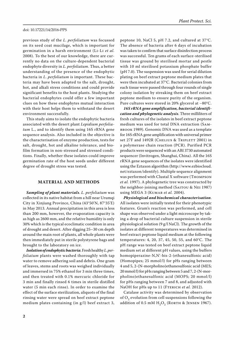
Endophytic bacteria isolation and phylogenet-ics. A total of 62 endophytic bacterial isolates were obtained from leaves, roots and stems of L. perfo-liatum. 16S rRNA gene sequences analysis showed that all of them were belonged to genus Bacillus and their GenBank accession numbers was KR999901 to KR999962 (Table 1). Phylogenetic tree constructed using the partial 16S rDNA sequences of the bacterial endophytes and taxa are shown in Figure 1.
Distribution and diversity of the bacterial endo-phytes are shown in Figure 2. Of the 62 isolates, 34 were from leaves, 27 were from roots, and 1 was from stems (Figure 2A). The relative amounts of the species isolated are as follows: species similar to B. subtilis (66%), B. flexus (14%), B. licheniformis (6%), B. mojavensis (5%), B. sonorensis (5%), B. cereus (2%) and B. safensis (2%) (Figure 2B).
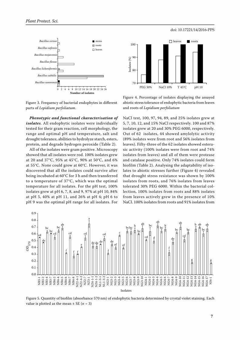
As shown in Figure 3, the isolates were shared at least 7 “species”. Similar to B. sonorensis were distributed in leaves, roots and stems; but similar to B. subtilis and B. licheniformis were obtained only from leaves and roots; and similar to B. flexus, B. mojavensis, B. cereus, and B. safensis were located leaves only, suggesting that more diversity of bacteria living in leaves than roots.
Figure 2. Number of obtained strains from different tissues (A) and endophytic bacterial diversity (B) of Lepidium perfoliatum
Bacillus subtilis, 41, 66%flexus, 9, 14%
Bacillus licheniformis,
4, 6%
Bacillus mojavensis,
Bacillus sonorensis,
Bacillus cereus, 1, 2% safensis,
1, 2%
Bacillus
3, 5%
3, 5%
Bacillus
leaves, 34, 55%
roots,27, 43%
stems, 1, 2%
(A) (B)
6
Vol. 52, 2016, No. 4: 00– Plant Protect. Sci.
doi: 10.17221/14/2016-PPS
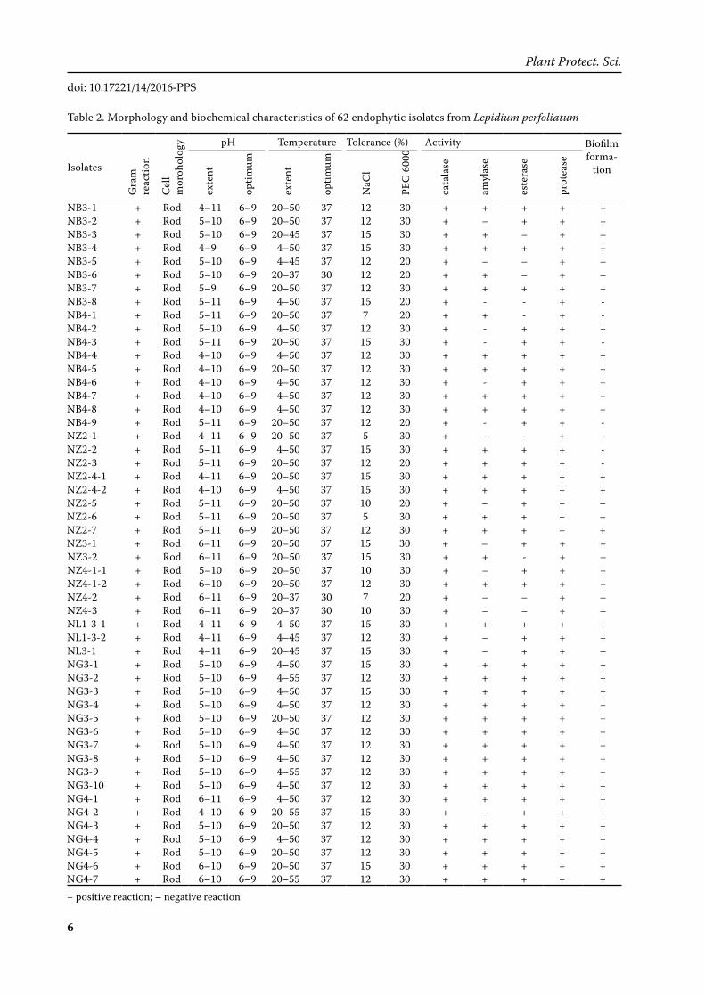
Table 2. Morphology and biochemical characteristics of 62 endophytic isolates from Lepidium perfoliatum
Isolates
Gra
m
reac
tion
Cel
l m
oroh
olog
y pH Temperature Tolerance (%) Activity Biofilm forma-
tion
exte
nt
optim
um
exte
nt
optim
um
NaC
l
PEG
600
0
cata
lase
amyl
ase
este
rase
prot
ease
NB3-1 + Rod 4–11 6–9 20–50 37 12 30 + + + + +NB3-2 + Rod 5–10 6–9 20–50 37 12 30 + – + + +NB3-3 + Rod 5–10 6–9 20–45 37 15 30 + + – + –NB3-4 + Rod 4–9 6–9 4–50 37 15 30 + + + + +NB3-5 + Rod 5–10 6–9 4–45 37 12 20 + – – + –NB3-6 + Rod 5–10 6–9 20–37 30 12 20 + + – + –NB3-7 + Rod 5–9 6–9 20–50 37 12 30 + + + + +NB3-8 + Rod 5–11 6–9 4–50 37 15 20 + - - + -NB4-1 + Rod 5–11 6–9 20–50 37 7 20 + + - + -NB4-2 + Rod 5–10 6–9 4–50 37 12 30 + - + + +NB4-3 + Rod 5–11 6–9 20–50 37 15 30 + - + + -NB4-4 + Rod 4–10 6–9 4–50 37 12 30 + + + + +NB4-5 + Rod 4–10 6–9 20–50 37 12 30 + + + + +NB4-6 + Rod 4–10 6–9 4–50 37 12 30 + - + + +NB4-7 + Rod 4–10 6–9 4–50 37 12 30 + + + + +NB4-8 + Rod 4–10 6–9 4–50 37 12 30 + + + + +NB4-9 + Rod 5–11 6–9 20–50 37 12 20 + - + + -NZ2-1 + Rod 4–11 6–9 20–50 37 5 30 + - - + -NZ2-2 + Rod 5–11 6–9 4–50 37 15 30 + + + + -NZ2-3 + Rod 5–11 6–9 20–50 37 12 20 + + + + -NZ2-4-1 + Rod 4–11 6–9 20–50 37 15 30 + + + + +NZ2-4-2 + Rod 4–10 6–9 4–50 37 15 30 + + + + +NZ2-5 + Rod 5–11 6–9 20–50 37 10 20 + – + + –NZ2-6 + Rod 5–11 6–9 20–50 37 5 30 + + + + –NZ2-7 + Rod 5–11 6–9 20–50 37 12 30 + + + + +NZ3-1 + Rod 6–11 6–9 20–50 37 15 30 + – + + +NZ3-2 + Rod 6–11 6–9 20–50 37 15 30 + + - + –NZ4-1-1 + Rod 5–10 6–9 20–50 37 10 30 + – + + +NZ4-1-2 + Rod 6–10 6–9 20–50 37 12 30 + + + + +NZ4-2 + Rod 6–11 6–9 20–37 30 7 20 + – – + –NZ4-3 + Rod 6–11 6–9 20–37 30 10 30 + – – + –NL1-3-1 + Rod 4–11 6–9 4–50 37 15 30 + + + + +NL1-3-2 + Rod 4–11 6–9 4–45 37 12 30 + – + + +NL3-1 + Rod 4–11 6–9 20–45 37 15 30 + – + + –NG3-1 + Rod 5–10 6–9 4–50 37 15 30 + + + + +NG3-2 + Rod 5–10 6–9 4–55 37 12 30 + + + + +NG3-3 + Rod 5–10 6–9 4–50 37 15 30 + + + + +NG3-4 + Rod 5–10 6–9 4–50 37 12 30 + + + + +NG3-5 + Rod 5–10 6–9 20–50 37 12 30 + + + + +NG3-6 + Rod 5–10 6–9 4–50 37 12 30 + + + + +NG3-7 + Rod 5–10 6–9 4–50 37 12 30 + + + + +NG3-8 + Rod 5–10 6–9 4–50 37 12 30 + + + + +NG3-9 + Rod 5–10 6–9 4–55 37 12 30 + + + + +NG3-10 + Rod 5–10 6–9 4–50 37 12 30 + + + + +NG4-1 + Rod 6–11 6–9 4–50 37 12 30 + + + + +NG4-2 + Rod 4–10 6–9 20–55 37 15 30 + – + + +NG4-3 + Rod 5–10 6–9 20–50 37 12 30 + + + + +NG4-4 + Rod 5–10 6–9 4–50 37 12 30 + + + + +NG4-5 + Rod 5–10 6–9 20–50 37 12 30 + + + + +NG4-6 + Rod 6–10 6–9 20–50 37 15 30 + + + + +NG4-7 + Rod 6–10 6–9 20–55 37 12 30 + + + + +
+ positive reaction; – negative reaction
7
Plant Protect. Sci. Vol. 52, 2016, No. 4: 00–
doi: 10.17221/14/2016-PPS
Phenotypic and functional characterisation of isolates. All endophytic isolates were individually tested for their gram reaction, cell morphology, the range and optimal pH and temperature, salt and drought tolerance, abilities to hydrolyze starch, esters, protein, and degrade hydrogen peroxide (Table 2).
All of the isolates were gram positive. Microscopy showed that all isolates were rod. 100% isolates grew at 20 and 37°C, 95% at 45°C, 90% at 50°C, and 6% at 55°C. None could grow at 60°C. However, it was discovered that all the isolates could survive after being incubated at 60°C for 3 h and then transferred to a temperature of 37°C, which was the optimal temperature for all isolates. For the pH test, 100% isolates grew at pH 6, 7, 8, and 9, 97% at pH 10, 84% at pH 5, 40% at pH 11, and 26% at pH 4; pH 6 to pH 9 was the optimal pH range for all isolates. For
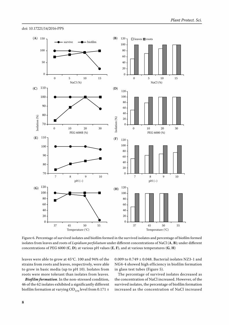
NaCl test, 100, 97, 94, 89, and 25% isolates grew at 5, 7, 10, 12, and 15% NaCl respectively. 100 and 87% isolates grew at 20 and 30% PEG 6000, respectively. Out of 62 isolates, 44 showed amylolytic activity (89% isolates were from root and 56% isolates from leaves). Fifty-three of the 62 isolates showed estera-sic activity (100% isolates were from root and 74% isolates from leaves) and all of them were protease and catalase positive. Only 74% isolates could form biofilm (Table 2). Analysing the adaptability of iso-lates to abiotic stresses further (Figure 4) revealed that drought stress resistance was shown by 100% isolates from roots, and 76% isolates from leaves tolerated 30% PEG 6000. Within the bacterial col-lection, 100% isolates from roots and 88% isolates from leaves actively grew in the presence of 10% NaCl. 100% isolates from roots and 91% isolates from
Figure 4. Percentage of isolates displaying the assayed abiotic stress tolerance of endophytic bacteria from leaves and roots of Lepidium perfoliatum
Figure 5. Quantity of biofilm (absorbance 570 nm) of endophytic bacteria determined by crystal violet staining. Each value is plotted as the mean ± SE (n = 3)
70
80
90
100
PEG 30% NaCl 10% T 45°C pH 10
Isol
ates
(%)
leaves roots
0.0
0.1
0.2
0.3
0.4
0.5
0.6
0.7
0.8
0.9
NB3
-1NB3
-2NB3
-4NB3
-7NB4
-2NB4
-4NB4
-5NB4
-6NB4
-7NB4
-8NZ2
-4-1
NZ2
-4-2
NZ2
-7NZ3
-1NZ4
-1-1
NZ4
-1-2
NL1
-3-1
NL1
-3-2
NG3-1
NG3-2
NG3-3
NG3-4
NG3-5
NG3-6
NG3-7
NG3-8
NG3-9
NG3-10
NG4-1
NG4-2
NG4-3
NG4-4
NG4-5
NG4-6
NG4-7
NG4-8
NG4-9
NG4-10
NG4-11
NG4-12
NG4-13
NG4-14
NG4-15
NG4-16
NG4-17
NJ4-1
OD57
0
Isolates
cd cd cd
de
bcbc
bcab
f f f
e
cd
de de de
f
a
cd
deef ef
cbcbc b
bc
ab
cd
bc
d
ab
bcb
f
de
bc
ab ab abbc
cd
bbc
g g
Figure 3. Frequency of bacterial endophytes in different parts of Lepidium perfoliatum.
0 2 4 6 8 10 12 14 16 18 20 22 24 26
Bacillus sonorensis
Bacillus subtilis
Bacillus licheniformis
Bacillus flexus
Bacillus mojavensis
Bacillus safensis
Bacillus cereus
Number of isolates
stemsrootsleaves
8
Vol. 52, 2016, No. 4: 00– Plant Protect. Sci.
doi: 10.17221/14/2016-PPS
leaves were able to grow at 45°C. 100 and 94% of the strains from roots and leaves, respectively, were able to grow in basic media (up to pH 10). Isolates from roots were more tolerant than isolates from leaves.
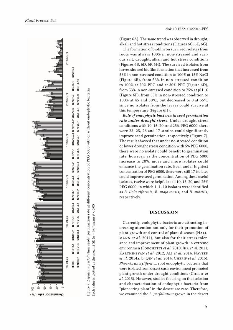
Biofilm formation. In the non-stressed condition, 46 of the 62 isolates exhibited a significantly different biofilm formation at varying OD570 level from 0.171 ±
0.009 to 0.749 ± 0.048. Bacterial isolates NZ3-1 and NG4-4 showed high efficiency in biofilm formation in glass test tubes (Figure 5).
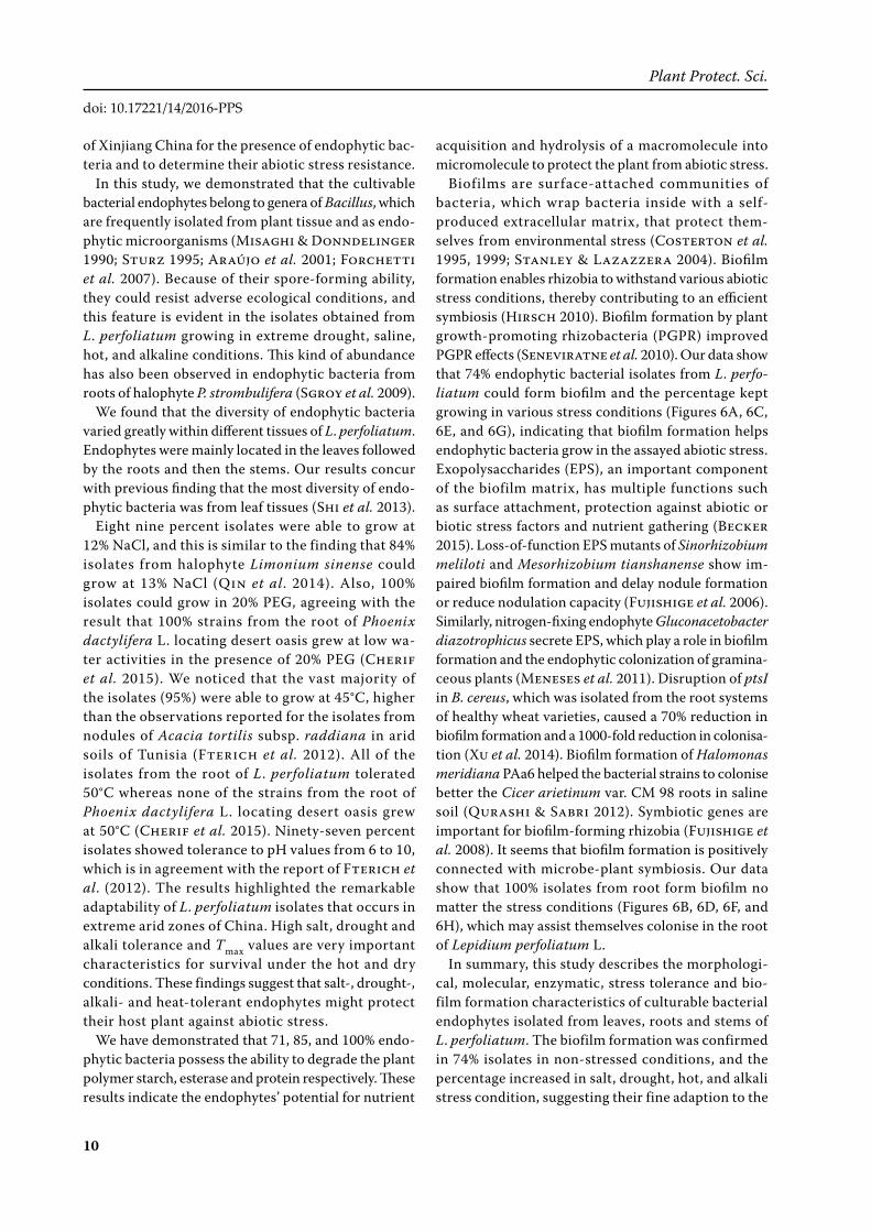
The percentage of survived isolates decreased as the concentration of NaCl increased. However, of the survived isolates, the percentage of biofilm formation increased as the concentration of NaCl increased
Figure 6. Percentage of survived isolates and biofilm formed in the survived isolates and percentage of biofilm formed isolates from leaves and roots of Lepidium perfoliatum under different concentrations of NaCl (A, B); under different concentrations of PEG 6000 (C, D); at various pH values (E, F), and at various temperatures (G, H)
0
20
40
60
80
100
120
0 5 10 15
Isol
ates
(%)
NaCl (%)
leaves roots
0
50
100
150
0 5 10 15
Isol
ates
(%)
NaCl (%)
survive biofilm
70
80
90
100
110
0 10 20 30
Isol
ates
(%)
PEG 6000 (%)
70
80
90
100
110
7 8 9 10
Isol
ates
(%)
pH
0
20
40
60
80
100
120
7 8 9 10
Isol
ates
(%)
pH
0
20
40
60
80
100
120
37 45 50 55
Isol
ates
(%)
Temperature (°C)
0
20
40
60
80
100
120
0 10 20 30
Isol
ates
(%)
PEG 6000 (%)
0
20
40
60
80
100
120
37 45 50 55Temperature (°C)
Isol
atio
n (%
)
Isol
atio
n (%
)
0 5 10 15 0 5 10 15 NaCl (%) NaCl (%)
0 10 20 30 0 10 20 30 PEG 6000l (%) PEG 6000 (%)
37 45 50 55 37 45 50 55 Temperature (°C) Temperature (°C)
7 8 9 10 7 8 9 10 pH (–) pH (–)
(A)
(C)
(B)
(D)
(E)
(G)
(F)
(H)
9
Plant Protect. Sci. Vol. 52, 2016, No. 4: 00–
doi: 10.17221/14/2016-PPS
Figu
re 7
. Lep
idiu
m p
erfo
liatu
m se
eds’
germ
inat
ion
rate
at d
iffer
ent c
once
ntra
tion
of P
EG 6
000
with
or w
ithou
t end
ophy
tic b
acte
ria
Each
val
ue is
plo
tted
as th
e m
ean
± SE
(n =
4);
*mea
ns P
< 0
.05
(Figure 6A). The same trend was observed in drought, alkali and hot stress conditions (Figures 6C, 6E, 6G).
The formation of biofilm on survived isolates from roots was always 100% in non-stressed and vari-ous salt, drought, alkali and hot stress conditions (Figures 6B, 6D, 6F, 6H). The survived isolates from leaves showed biofilm formation that increased from 53% in non-stressed condition to 100% at 15% NaCl (Figure 6B), from 53% in non-stressed condition to 100% at 20% PEG and at 30% PEG (Figure 6D), from 53% in non-stressed condition to 75% at pH 10 (Figure 6F), from 53% in non-stressed condition to 100% at 45 and 50°C, but decreased to 0 at 55°C since no isolates from the leaves could survive at this temperature (Figure 6H).
Role of endophytic bacteria in seed germination rate under drought stress. Under drought stress conditions with 10, 15, 20, and 25% PEG 6000, there were 23, 25, 26 and 17 strains could significantly improve seed germination, respectively (Figure 7). The result showed that under no stressed condition or lower drought stress condition with 5% PEG 6000, there were no isolate could benefit to germination rate, however, as the concentration of PEG 6000 increase to 20%, more and more isolates could enhance the germination rate. Even under hightest concentration of PEG 6000, there were still 17 isolates could improve seed germination. Among these useful isolates, twelve were helpful at all 10, 15, 20, and 25% PEG 6000, in which 1, 1, 10 isolates were identified as B. licheniformis, B. mojavensis, and B. subtilis, respectively.
DISCUSSION
Currently, endophytic bacteria are attracting in-creasing attention not only for their promotion of plant growth and control of plant diseases (Hall-mann et al. 2011), but also for their stress toler-ance and improvement of plant growth in extreme environmen (Forchetti et al. 2010; Jha et al. 2011; Karthikeyan et al. 2012; Ali et al. 2014; Naveed et al. 2014a, b; Qin et al. 2014; Cherif et al. 2015). Phoenix dactylifera L. root endophytic bacteria that were isolated from desert oasis environment promoted plant growth under drought conditions (Cherif et al. 2015). However, studies focusing on the isolation and characterisation of endophytic bacteria from “pioneering plant” in the desert are rare. Therefore, we examined the L. perfoliatum grown in the desert
10
Vol. 52, 2016, No. 4: 00– Plant Protect. Sci.
doi: 10.17221/14/2016-PPS
of Xinjiang China for the presence of endophytic bac-teria and to determine their abiotic stress resistance.
In this study, we demonstrated that the cultivable bacterial endophytes belong to genera of Bacillus, which are frequently isolated from plant tissue and as endo-phytic microorganisms (Misaghi & Donndelinger 1990; Sturz 1995; Araújo et al. 2001; Forchetti et al. 2007). Because of their spore-forming ability, they could resist adverse ecological conditions, and this feature is evident in the isolates obtained from L. perfoliatum growing in extreme drought, saline, hot, and alkaline conditions. This kind of abundance has also been observed in endophytic bacteria from roots of halophyte P. strombulifera (Sgroy et al. 2009).
We found that the diversity of endophytic bacteria varied greatly within different tissues of L. perfoliatum. Endophytes were mainly located in the leaves followed by the roots and then the stems. Our results concur with previous finding that the most diversity of endo-phytic bacteria was from leaf tissues (Shi et al. 2013).
Eight nine percent isolates were able to grow at 12% NaCl, and this is similar to the finding that 84% isolates from halophyte Limonium sinense could grow at 13% NaCl (Qin et al. 2014). Also, 100% isolates could grow in 20% PEG, agreeing with the result that 100% strains from the root of Phoenix dactylifera L. locating desert oasis grew at low wa-ter activities in the presence of 20% PEG (Cherif et al. 2015). We noticed that the vast majority of the isolates (95%) were able to grow at 45°C, higher than the observations reported for the isolates from nodules of Acacia tortilis subsp. raddiana in arid soils of Tunisia (Fterich et al. 2012). All of the isolates from the root of L. perfoliatum tolerated 50°C whereas none of the strains from the root of Phoenix dactylifera L. locating desert oasis grew at 50°C (Cherif et al. 2015). Ninety-seven percent isolates showed tolerance to pH values from 6 to 10, which is in agreement with the report of Fterich et al. (2012). The results highlighted the remarkable adaptability of L. perfoliatum isolates that occurs in extreme arid zones of China. High salt, drought and alkali tolerance and Tmax values are very important characteristics for survival under the hot and dry conditions. These findings suggest that salt-, drought-, alkali- and heat-tolerant endophytes might protect their host plant against abiotic stress.
We have demonstrated that 71, 85, and 100% endo-phytic bacteria possess the ability to degrade the plant polymer starch, esterase and protein respectively. These results indicate the endophytes’ potential for nutrient
acquisition and hydrolysis of a macromolecule into micromolecule to protect the plant from abiotic stress.
Biofilms are surface-attached communities of bacteria, which wrap bacteria inside with a self-produced extracellular matrix, that protect them-selves from environmental stress (Costerton et al. 1995, 1999; Stanley & Lazazzera 2004). Biofilm formation enables rhizobia to withstand various abiotic stress conditions, thereby contributing to an efficient symbiosis (Hirsch 2010). Biofilm formation by plant growth-promoting rhizobacteria (PGPR) improved PGPR effects (Seneviratne et al. 2010). Our data show that 74% endophytic bacterial isolates from L. perfo-liatum could form biofilm and the percentage kept growing in various stress conditions (Figures 6A, 6C, 6E, and 6G), indicating that biofilm formation helps endophytic bacteria grow in the assayed abiotic stress. Exopolysaccharides (EPS), an important component of the biofilm matrix, has multiple functions such as surface attachment, protection against abiotic or biotic stress factors and nutrient gathering (Becker 2015). Loss-of-function EPS mutants of Sinorhizobium meliloti and Mesorhizobium tianshanense show im-paired biofilm formation and delay nodule formation or reduce nodulation capacity (Fujishige et al. 2006). Similarly, nitrogen-fixing endophyte Gluconacetobacter diazotrophicus secrete EPS, which play a role in biofilm formation and the endophytic colonization of gramina-ceous plants (Meneses et al. 2011). Disruption of ptsI in B. cereus, which was isolated from the root systems of healthy wheat varieties, caused a 70% reduction in biofilm formation and a 1000-fold reduction in colonisa-tion (Xu et al. 2014). Biofilm formation of Halomonas meridiana PAa6 helped the bacterial strains to colonise better the Cicer arietinum var. CM 98 roots in saline soil (Qurashi & Sabri 2012). Symbiotic genes are important for biofilm-forming rhizobia (Fujishige et al. 2008). It seems that biofilm formation is positively connected with microbe-plant symbiosis. Our data show that 100% isolates from root form biofilm no matter the stress conditions (Figures 6B, 6D, 6F, and 6H), which may assist themselves colonise in the root of Lepidium perfoliatum L.
In summary, this study describes the morphologi-cal, molecular, enzymatic, stress tolerance and bio-film formation characteristics of culturable bacterial endophytes isolated from leaves, roots and stems of L. perfoliatum. The biofilm formation was confirmed in 74% isolates in non-stressed conditions, and the percentage increased in salt, drought, hot, and alkali stress condition, suggesting their fine adaption to the
11
Plant Protect. Sci. Vol. 52, 2016, No. 4: 00–
doi: 10.17221/14/2016-PPS
harsh environment. Besides, 28 isolates could improve seed germination rate under different drought stress. These data are valuable and essentially show that the endophytic bacteria from L. perfoliatum could assist their host to resist harsh environment.
R e f e r e n c e s
Ali S., Charles T.C., Glick B.R. (2014): Amelioration of high salinity stress damage by plant growth-promoting bacterial endophytes that contain ACC deaminase. Plant Physiology and Biochemistry, 80: 160–167.
Altschul S.F., Gish W., Miller W., Myers E.W., Lipman D.J. (1990): Basic local alignment search tool. Journal of Mo-lecular Biology, 215: 403–410.
Araújo W.L., Maccheroni Jr W., Aguilar-Vildoso C.I., Bar-roso P.A., Saridakis H.O., Azevedo J.L. (2001): Variability and interactions between endophytic bacteria and fungi isolated from leaf tissues of citrus rootstocks. Canadian Journal of Microbiology, 47: 229–236.
Becker A. (2015): Challenges and perspectives in combina-torial assembly of novel exopolysaccharide biosynthesis pathways. Frontiers in Microbiology, 6; 687. doi: 10.3389/fmicb.2015.00687
Castro R.A., Quecine M.C., Lacava P.T., Batista B.D., Luvi-zotto D.M., Marcon J., Ferreira A., Melo I.S., Azevedo J.L. (2014): Isolation and enzyme bioprospection of endophytic bacteria associated with plants of Brazilian mangrove eco-system. SpringerPlus, 3.382. doi: 10.1186/2193-1801-3-382
Chelius M.K., Triplett E.W. (2001): The diversity of archaea and bacteria in association with the roots of Zea mays L. Microbial Ecology, 41: 252–263.
Cherif H., Marasco R., Rolli E., Ferjani R., Fusi M., Soussi A., Mapelli F., Blilou I., Borin S., Boudabous A., Cherif A., Daf-fonchio D., Ouzari H. (2015): Oasis desert farming selects environment-specific date palm root endophytic commu-nities and cultivable bacteria that promote resistance to drought. Environmental Microbiology Reports, 7: 668–678.
Costerton J.W., Lewandowski Z., Caldwell D.E., Korber D.R., Lappin-Scott H.M. (1995): Microbial biofilms. An-nual Review of Microbiology, 49: 711–745.
Costerton J.W., Stewart P.S., Greenberg E.P. (1999): Bacte-rial biofilms: a common cause of persistent infections. Science, 284: 1318–1322.
Forchetti G., Masciarelli O., Alemano S., Alvarez D., Abdala G. (2007): Endophytic bacteria in sunflower (Helianthus annuus L.): isolation, characterization, and production of jasmonates and abscisic acid in culture medium. Ap-plied Microbiology and Biotechnology, 76: 1145–1152.
Forchetti G., Masciarelli O., Izaguirre M.J., Alemano S., Alvarez D., Abdala G. (2010): Endophytic bacteria im-
prove seedling growth of sunflower under water stress, produce salicylic acid, and inhibit growth of pathogenic fungi. Current Microbiology, 61: 485–493.
Fterich A., Mahdhi M., Lafuente A., Pajuelo E., Caviedes M.A., Rodriguez-Llorente I.D., Mars M. (2012): Taxo-nomic and symbiotic diversity of bacteria isolated from nodules of Acacia tortilis subsp. raddiana in arid soils of Tunisia. Canadian Journal of Microbiology, 58: 738–751.
Fujishige N.A., Kapadia N.N., De Hoff P.L., Hirsch A.M. (2006): Investigations of Rhizobium biofilm formation. FEMS Microbiology Ecology, 56: 195–206.
Fujishige N.A., Lum M.R., De Hoff P.L., Whitelegge J.P., Faull K.F., Hirsch A.M. (2008): Rhizobium common nod genes are required for biofilm formation. Molecular Mi-crobiology, 67: 504–515.
Hallmann J., Quadt-Hallmann A., Mahaffee W.F., Kloepper J.W. (2011): Bacterial endophytes in agricultural crops. Canadian Journal of Microbiology, 43: 895–914.
Hirsch A.M. (2010): How rhizobia survive in the absence of a legume host, a stressful world indeed. In: Seckbach J., Grube M. (eds): Symbioses and Stress. Joint Ventures in Biology. Dordrecht, Springer: 375–391.
Huang D.H., Zhao J., Yuan J.W., Dong-Sheng X.U., Chen S., Lan H.Y. (2011): A Preliminary Study on Methods for Dormancy Breaking of Seeds of Lepidium perfoliatum L. Cultivated Outdoor. Xinjiang Agricultural Sciences.
Jha Y., Subramanian R.B., Patel S. (2011): Combination of endophytic and rhizospheric plant growth promoting rhizobacteria in Oryza sativa shows higher accumulation of osmoprotectant against saline stress. Acta Physiologiae Plantarum, 33: 797–802.
Karthikeyan B., Joe M.M., Islam M.R., Sa T.M. (2012): ACC deaminase containing diazotrophic endophytic bacteria ameliorate salt stress in Catharanthus roseus through reduced ethylene levels and induction of antioxidative defense systems. Symbiosis, 56: 77–86.
Krishnan P., Bhat R., Kush A., Ravikumar P. (2012): Isolation and functional characterization of bacterial endophytes from Carica papaya fruits. Journal of Applied Microbiol-ogy, 113: 308–317.
Kumar S., Tamura K., Nei M. (2004): MEGA3: Integrated soft-ware for Molecular Evolutionary Genetics Analysis and se-quence alignment. Briefings in Bioinformatics, 5: 150–163.
Li-Li G.U., Liu L.H., You T.Y., Lan H.Y., Zhang F.C. (2008): Characterization of the seed coat mucilage properties of ephemeral plant Lepidium perfoliatum L. in Xinjiang. Acta Botanica Boreali-Occidentalia Sinica, 28: 2451–2460.
Meneses C.H., Rouws L.F., Simoes-Araujo J.L., Vidal M.S., Baldani J.I. (2011): Exopolysaccharide production is re-quired for biofilm formation and plant colonization by the nitrogen-fixing endophyte Gluconacetobacter di-
12
Vol. 52, 2016, No. 4: 00– Plant Protect. Sci.
doi: 10.17221/14/2016-PPS
azotrophicus. Molecular Plant-Microbe Interactions, 24: 1448–1458.
Misaghi I.J., Donndelinger C.R. (1990): Endophytic bacte-ria in symptom-free cotton plants. Phytopathology, 80: 808–811.
Naveed M., Hussain M.B., Zahir Z.A., Mitter B., Sessitsch A. (2014a): Drought stress amelioration in wheat through inoculation with Burkholderia phytofirmans strain PsJN. Plant Growth Regulation, 73: 121–131.
Naveed M., Mitter B., Reichenauer T.G., Wieczorek K., Ses-sitsch A. (2014b): Increased drought stress resilience of maize through endophytic colonization by Burkholderia phytofirmans PsJN and Enterobacter sp. FD17. Environ-mental & Experimental Botany, 97: 30–39.
Qin S., Zhang Y.J., Yuan B., Xu P.Y., Xing K., Wang J., Jiang J.H. (2014): Isolation of ACC deaminase-producing habi-tat-adapted symbiotic bacteria associated with halophyte Limonium sinense (Girard) Kuntze and evaluating their plant growth-promoting activity under salt stress. Plant and Soil, 374: 753–766.
Qurashi A.W., Sabri A.N. (2012): Biofilm formation in mod-erately halophilic bacteria is influenced by varying salinity levels. Journal of Basic Microbiology, 52: 566–572.
Reinhold-Hurek B., Hurek T. (2011): Living inside plants: bacterial endophytes. Current Opinion in Plant Biology, 14: 435–443.
Rorth M., Jensen P.K. (1967): Determination of catalase activ-ity by means of the Clark oxygen electrode. Biochimica et Biophysica Acta, 139: 171–173.
Saitou N., Nei M. (1987): The neighbor-joining method: a new method for reconstructing phylogenetic trees. Mo-lecular Biology and Evolution, 4: 406–425.
Sambrook J., Fritsch E.F., Maniatis T. (1989): Molecular Cloning: A Laboratory Manual. 2nd Ed. New York, CSH Laboratory Press.
Seneviratne G., Weerasekara M.L.M.A.W., Seneviratne K.A.C.N., Zavahir J.S., Kecskés M.L., Kennedy I.R. (2010): Importance of biofilm formation in plant growth promot-ing rhizobacterial action. In: Maheshwari D.K. (ed.): Plant Growth and Health Promoting Bacteria. Vol. 18. Berlin, Springer: 81–95.
Sgroy V., Cassan F., Masciarelli O., Del Papa M.F., Lagares A., Luna V. (2009): Isolation and characterization of en-dophytic plant growth-promoting (PGPB) or stress ho-meostasis-regulating (PSHB) bacteria associated to the halophyte Prosopis strombulifera. Applied Microbiology and Biotechnology, 85: 371–381.
Shi Y., Zhang X., Lou K. (2013): Isolation, characterization, and insecticidal activity of an endophyte of drunken horse grass, Achnatherum inebrians. Journal of Insect Science, 13: 151.
Stanley N.R., Lazazzera B.A. (2004): Environmental signals and regulatory pathways that influence biofilm formation. Molecular Microbiology, 52: 917–924.
Sturz A.V. (1995): The role of endophytic bacteria during seed piece decay and potato tuberization. Plant & Soil, 175: 257–263.
Tariq M., Hameed S., Yasmeen T., Zahid M., Zafar M. (2014): Molecular characterization and identification of plant growth promoting endophytic bacteria isolated from the root nodules of pea (Pisum sativum L.). World Journal of Microbiology & Biotechnology, 30: 719–725.
Thompson J.D., Gibson T.J., Plewniak F., Jeanmougin F., Higgins D.G. (1997): The CLUSTAL_X windows inter-face: flexible strategies for multiple sequence alignment aided by quality analysis tools. Nucleic Acids Research, 25: 4876–4882.
Xu Y.B., Chen M., Zhang Y., Wang M., Wang Y., Huang Q.B., Wang X., Wang G. (2014): The phosphotransferase system gene ptsI in the endophytic bacterium Bacillus cereus is required for biofilm formation, colonization, and biocontrol against wheat sharp eyespot. FEMS Mi-crobiology Letters, 354: 142–152.
Yuan J., Huang D., Dongsheng X.U., Zhao J., Ling L.I., Lan H., Zhang F. (2013): Cloning and analysis of seed coat mucilage-related gene MUM4 from Lepidium perfo-liatum. Acta Botanica Boreali-Occidentalia Sinica, 33: 1940–1952.
Received: 2016–01–25Accepted after corrections: 2016–03–08
Published online: 2016–08–20
Corresponding Author:
Dr Dengdi An, Xinjiang Normal University, College of Life Science, Xinjiang Key Laboratory of Special Species Conservation and Regulatory Biology, Urumqi 830017, Xinjiang, P.R. China; E-mail: [email protected]