characterisation and expression of the fasciola gigantica cathepsin l gene
TRANSCRIPT
Characterisation and expression of the Fasciola giganticacathepsin L geneq
Hiroshi Yamasakia,*, Reiko Minekib, Kimie Murayamab, Akira Itoc, Takashi Aokia
aDepartment of Parasitology, Central Laboratory of Medical Sciences, Juntendo University School of Medicine, Tokyo 113-0033, JapanbDivision of Biochemical Analysis, Central Laboratory of Medical Sciences, Juntendo University School of Medicine, Tokyo 113-0033, Japan
cDepartment of Parasitology, Asahikawa Medical College, Asahikawa 078-8510, Japan.
Received 21 December 2001; received in revised form 26 February 2002; accepted 28 February 2002
Abstract
The gene structure of a cathepsin L from Fasciola gigantica was characterised. The gene spans approximately 2.0 kb and comprises four
exons and three introns and is a compact gene as in the cases of crustaceous and platyhelminth cathepsins L. Southern blot analysis suggested
that a few copies of the genes are sparsely organised in the genome. Of the three intron insertion positions, two of which are in the same
position as in the mammalian cathepsin L gene. Phylogenetic analysis revealed that F. gigantica cathepsin L forms a clade with those from
Fasciola hepatica, but not with those from Spirometra erinacei and schistosomes. Putative TATA-boxes were found upstream of a
transcription initiation site. The sequence analysis of the 5 0-upstream of the transcript revealed that the cathepsin L gene is transcribed
by cis-splicing fashion. Furthermore, the experiments using recombinant F. gigantica procathepsin L showed that it was processed to an
enzymatically active cathepsin L by pH-dependent autocatalysis. However, the pro-peptide deleted cathepsin L showed no enzyme activity,
indicating that the pro-region of F. gigantica procathepsin L is essential for the folding and/or refolding of functional cathepsin L. These
results are consistent with the observations in mammalian cathepsin L and papain. q 2002 Australian Society for Parasitology Inc. Published
by Elsevier Science Ltd. All rights reserved.
Keywords: Fasciola gigantica; Cathepsin L; Gene structure; Gene family; Expression; In vitro processing
1. Introduction
The liver flukes Fasciola hepatica and Fasciola gigantica
are causative agents of fascioliasis in humans and rumi-
nants, especially cattle, goat and sheep. The disease has
been traditionally considered to be an important veterinary
disease because of the substantial production and large
economic losses in livestock production. In contrast,
human fascioliasis has been linked to cases among livestock
in the area concerned. However, it has been considered an
increasingly important chronic disease since 1980 (Chen
and Mott, 1990; Esteban et al., 1998; Mas-Coma et al.,
1999).
It has been reported that F. hepatica thiol-activated
proteolytic enzymes secreted by migrating parasites cleave
host immunoglobulins (Chapman and Mitchell, 1982).
Since then, the proteinases secreted by adult liver flukes
have been characterised, but most studies agree that the
major enzymes are cathepsin L-like cysteine proteases
that are homologues of the mammalian lysosomal cathepsin
L (Yamasaki et al., 1989; Rege et al., 1989; Dalton and
Hefferman, 1989; Fagbemi and Hillyer, 1992; Yamasaki
and Aoki, 1993; McGinty et al., 1993; Smith et al., 1993;
Wijffels et al., 1994a; Dowd et al., 1994; Heussler and
Dobbelaere, 1994; Hawthorne et al., 2000). It is considered
that Fasciola cathepsin L may be involved in crucial biolo-
gical functions such as host protein degradation, tissue
penetration and immune evasion. For these reasons, the
cathepsin L-like cysteine proteases of liver flukes have
been potential targets as immunodiagnostic antigens for
fascioliasis (Yamasaki et al., 1989; Fagbemi and Guobadia,
1995; O’Neill et al., 1999) or as vaccine candidates (Wijf-
fels et al., 1994a; Dalton et al., 1996b).
On the other hand, concerning genomic organisation of
cathepsin L genes in invertebrates, studies on a kinetoplastid
protozoon (Eakin et al., 1992), a malaria parasite (Rosenthal
and Nelson, 1992), a fruit fly (Matsumoto et al., 1995), a
International Journal for Parasitology 32 (2002) 1031–1042
0020-7519/02/$20.00 q 2002 Australian Society for Parasitology Inc. Published by Elsevier Science Ltd. All rights reserved.
PII: S0020-7519(02)00057-7
www.parasitology-online.com
q The nucleotide sequence data reported in the present paper have been
submitted to DDBJ, GenBank and EMBL databases with accession
numbers AB010923 and AB010924.
* Corresponding author. Present address: Department of Parasitology,
Asahikawa Medical College, Midorigaoka Higashi 2-1-1-1, Asahikawa
078-8510, Japan. Tel.: 181-166-68-2421; Fax: 181-166-68-2429.
E-mail address: [email protected] (H. Yamasaki).

larval cestode (Liu et al., 1996) and a shrimp (Boulay et al.,
1998) have been reported. In the present paper, we describe
the gene structure of a cathepsin L from F. gigantica and
compare it with those of known cathepsins L from other
organisms with a phylogenetic analysis. In addition, the
function of the pro-region is also discussed using recombi-
nant F. gigantica procathepsin L.
2. Materials and methods
2.1. Parasite
Adult Fasciola worms were collected from a cattle natu-
rally infected with the parasites at a local slaughterhouse in
Tokyo. The parasites were washed several times in sterile
PBS, pH 7.2, and immediately frozen in liquid nitrogen for
RNA preparation or stored at 280 8C for DNA samples. The
parasite used, F. gigantica, was identified based on the
nucleotide sequences of cytochrome c oxidase subunit I
and ribosomal RNA genes (Hashimoto et al., 1997).
2.2. Construction of the Fasciola genomic DNA library
Genomic DNA was prepared from 1 g of a frozen worm,
partially digested with Sau3AI, and fractionated into 5–20
kb fragments by sucrose density gradient method (Maniatis
et al., 1982). The fragments were ligated to BamHI/EcoRI
double-digested EMBL 3 lambda DNA, and allowed to
form phage particles using an in vitro packaging kit (Stra-
tagene). Approximately 6 £ 104 recombinant phages were
screened using a digoxigenin-labelled Fasciola cathepsin L
cDNA probe (Yamasaki and Aoki, 1993).
2.3. Southern blot analysis
Genomic DNA was digested with different restriction
enzymes whose cleavage sites are absent from the cathepsin
L gene, separated on a 0.7% agarose gel, transferred to a
positively charged nylon membrane (Boehringer Mannheim)
and prehybridised at 68 8C for 2 h in hybridisation buffer
containing 7% SDS. The filter was hybridised at 68 8C over-
night with a digoxigenin-labelled probe. The 1.9-kb probe
was prepared by PCR using primers based on the nucleotide
sequences of F. gigantica cathepsin L gene (B22-2) shown in
Fig. 2. Finally, the filter was washed at high stringency and
exposed to X-ray film for 5–10 min. CSPD (Boehringer
Mannheim) was used as a substrate for chemiluminescent
detection.
2.4. RNA preparation, 5 0-RACE and Northern blot analysis
Total RNA was extracted from a frozen worm (0.8 g
weight) by the acid guanidinium thiocyanate phenol-chloro-
form method (Chomczynski and Sacchi, 1987). Poly(A)1
RNA was purified on an oligo(dT) cellulose gel column
(Pharmacia Amersham Biotech, USA). 5 0-rapid amplifica-
tion of cDNA ends (5 0-RACE) was performed according to
the manufacturer’s instructions (Life Technologies Inc.) to
determine the sequence of the 5 0-untranslated region of the F.
gigantica cathepsin L gene (Fg-CATL). Gene-specific anti-
sense primers (GSP 1, 5 0-TTGATGCCACAAATCAT-
CATTCGAGCCAAG-3 0 and GSP 2, 5 0-ATCTTCCCAAA-
TATTTCGTCTGTG-3 0) were used for nested PCR. PCR
was performed for 35 cycles of 1 min at 94 8C, 1 min at 55
8C, 2 min at 72 8C, and 4 min at 72 8C for extension. In order to
clarify whether a spliced leader sequence is added at the 5 0-
end of the transcript of Fg-CATL, mRNA was reversibly
transcribed with M-MLV reverse transcriptase (Gibco
BRL) and oligo(dT) primer (Boehringer Mannheim),
followed by PCR performed at 94 8C for 1 min, 55 8C for 1
min, 72 8C for 2 min, for 35 cycles using a Fasciola spliced
leader primer (5 0-AACCTTAACGGTTCTCTG-3 0, Davis et
al., 1994) and GSP 1 primer.
mRNA was electrophoresed in a 1% agarose gel, blotted
onto a positively charged nylon membrane, and then hybri-
dised with a digoxigenin-labelled cathepsin L (B22-2)
cDNA probe overnight at 42 8C. The probe was prepared
by PCR using primers based on the nucleotide sequences of
F. gigantica cathepsin L gene (B22-2) and cDNA prepared
using cDNA synthesis kit (Pharmacia). The hybridisation
buffer used was 50 mM phosphate buffer, pH 7.0, containing
5£ standard saline citrate (SSC), 7% SDS, 0.1% sarcosine,
50% formamide and yeast total RNA (50 mg/ml). The filter
was washed extensively with 2£ SSC/0.1% SDS, 0:1£ SSC/
0.1% SDS at 42 8C, and chemiluminescent detection was
carried out using CSPD as a substrate.
2.5. DNA sequencing
A DNA fragment derived from a positive clone (B22-2
clone) was digested with restriction enzymes as indicated in
Fig. 1, and the resulting fragments were subcloned into
pUC18. The nucleotide sequences of the genomic DNA
were determined by the dideoxynucleotide chain termina-
tion method using Dye Primer and Dye Terminator Cycle
Sequencing kits (Applied Biosystems Inc.) and an ABI
DNA sequencer 373A. The nucleotide sequence of the
PCR-amplified cathepsin L gene (B7-3) was also deter-
mined by same protocol. Sequence data were analysed
using Genetix-Mac, and EMBL/GenBank database searches
were performed with the FASTA program.
2.6. Sequence alignment and phylogenetic analysis
The alignment of sequences was carried out using CLUS-
TAL W program available over the World Wide Web
(http://www.ddbj.nig.ac.jp/E-mail/homology.html). Phylo-
genetic tree was inferred with the neighbour-joining method
(Saitou and Nei, 1987) using TreeView PPC software
(version 1.5.3). Confidence values for each branch were
determined by 1000 bootstrap replications.
H. Yamasaki et al. / International Journal for Parasitology 32 (2002) 1031–10421032
2.7. Expression and refolding of the recombinant
procathepsin L and pro-peptide deleted cathepsin L
Two cDNAs encoding F. gigantica procathepsin L and
pro-peptide deleted cathepsin L (B22-2) were amplified by
PCR using cDNA as described above. Primer sets used were
pro 5 0-primer/3 0-primer, and mCL 5 0-primer/3 0-primer
based on the nucleotide sequence of Fg CATL (B22-2,
AB010923). PCR was performed using AmpliTaq DNA
polymerase (Perkin Elmer) as follows: 95 8C, 1 min; 55
8C, 1 min; 72 8C, 2 min; 35 cycles. The PCR product was
subcloned into pT7Blue T-vector (Novagene, USA) and
sequenced. cDNA corresponding to B22-2 clone was
inserted into the pGEX-4T-1 expression vector (Pharmacia
Amersham Biotech), and designated pGEX/proCATL for
procathepsin L or pGEX/mCATL for pro-peptide deleted
cathepsin L. Each construct was then introduced into
BL21 Escherichia coli strain (Novagene). The expression
of glutathione S-transferase (GST) fused with either
procathepsin L or pro-peptide deleted cathepsin L was
induced by adding a final concentration of 1 mM isopro-
pyl-b-d(2)thiogalactopyranoside (IPTG) for 2–3 h at 37 8C.
Cells from 1 l culture were harvested, and the cell pellet
was resuspended in chilled PBS containing 0.1% Triton X-
100 and 1% sarcosine, and then sonicated using a sonifier
(Branson). The resulting suspensions were centrifuged at
15,000 rpm for 10 min, and the pellets were rinsed in several
times in PBS and resuspended in 20 ml of 50 mM Tris–HCl,
pH 8.0, containing 50 mM NaCl, 5 mM EDTA and 10 mM
dithiothreitol (DTT). Urea was added to a final concentra-
tion of 8 M to solubilise the suspension completely. Refold-
ing was performed according to a previously described
procedure (Smith and Gottesman, 1989). Briefly, the urea-
solubilised GST-fusion proteins were slowly added drop-
wise (2 ml/h) into 400 volumes of 50 mM potassium phos-
phate buffer, pH 10.7, containing 5 mM EDTA, 0.1 mM
oxidised glutathione and 1 mM reduced glutathione to a
final protein concentration of approximately 10 mg/ml,
and stirred at 4 8C overnight. The solutions of solubilised
proteins were then adjusted to pH 8.0 and concentrated to 1/
300 of the original volume using an Amicon cell ultrafiltra-
tion unit. After concentration, the solutions were centri-
fuged briefly for further experiments.
2.8. Purification of recombinant and native cathepsins L
The refolded GST-fusion proteins were cleaved with
thrombin to remove GST according to the manufacturer’s
manual and the recombinant procathepsin L was dialysed
against 10 mM Na2HPO4/1.8 mM KH2PO4, pH 8.0, contain-
ing 140 mM NaCl, 2.7 mM KCl and 4 M urea, and the
resulting solution were applied to a Sephacryl S-200 HR
gel chromatography column (1:5 £ 120 cm) equilibrated
with same buffer. The eluates were analysed by SDS–
PAGE (Laemmli, 1970). In order to compare the activities
of recombinant and native cathepsin L, native cathepsin L
was partially purified from F. gigantica adult worms.
Briefly, 1.5 g of lyophilised parasites were homogenised
H. Yamasaki et al. / International Journal for Parasitology 32 (2002) 1031–1042 1033
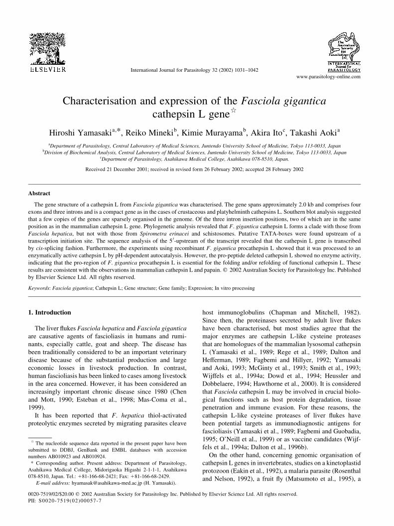
Fig. 1. Genomic organisation of Fasciola gigantica cathepsin L gene and sequencing strategy. In the cDNA, dotted, open and striped boxes indicate regions
encoding the signal sequence, pro-peptide and mature enzyme domains, respectively. In the genomic clone, closed boxes indicate exons and solid bars denote
introns. Bm, BamHI; M, MvaI; C, ClaI; St, StyI; Sp, SspI; A, AgeI; E, EcoO109I; B, BsmI. Arrows indicate the direction of sequencing.

in 40 mM sodium acetate buffer, pH 3.9, containing 150
mM NaCl, 1 mM EDTA, and 1 mM DTT. The resulting
supernatant was dialysed against the same buffer and
applied to TSK-Gel 3000SW column (21:5 £ 600 mm) in
a high-performance liquid chromatograph (Shimadzu,
Model LC-6A). Fractions showing activities toward carbo-
benzoxyl-l-phenylalanyl-l-arginine 4-methyl-coumaryl
amide (Z-Phe-Arg-MCA, Peptide Institute) were pooled
for further experiments.
2.9. Enzyme assay
Cathepsin L activity was assayed according to the method
of Barrett and Kirschke, (1981). Briefly, 10 ml of recombi-
nant or native cathepsin L in 80 ml of 80 mM sodium acetate
buffer, pH 5.5, containing 8 mM l-cysteine and 4 mM
EDTA were preincubated at 37 8C for 5 min, and the
enzyme reaction was initiated by adding 10 ml of 1 mM
Z-Phe-Arg-MCA at 37 8C for 10 min, and terminated by
adding 100 ml of 5% SDS. The 7-amino-4-methyl-coumarin
released was measured using a fluorescence spectrophoto-
metry (Hitachi, Model 850) at an excitation wavelength of
370 nm and emission wavelength of 460 nm.
2.10. NH2-terminal sequencing of recombinant
procathepsin L, pro-peptide deleted- and processed
cathepsins L
Both GST-fused procathepsin L and cathepsin L were
cleaved with thrombin, electrophoresed, and then were
blotted onto a PVDF membrane for NH2-terminal sequen-
cing according to the method of Matsudaira (1987). The
NH2-terminal amino acid sequence of recombinant cathe-
psin L processed in vitro was also electrophoresed. The
target bands were cut and then applied to a Protein Sequen-
cer (Applied Biosystems, Model 477A).
2.11. Protein assay
The protein content was determined by the Bradford
method using a protein assay kit (BioRad).
3. Results
3.1. Structure of the F. gigantica cathepsin L gene
Recombinant clones (6 £ 104) were screened with a
digoxigenin-labelled Fasciola cathepsin L cDNA probe
(Yamasaki and Aoki, 1993), and a positive clone (B22-2)
with a 16 kb DNA fragment was isolated. The DNA frag-
ment was digested with BamHI, a 4.3 kb fragment hybri-
dised with the probe was obtained, and 3,766 bp including a
5 0-flanking region of the fragment were determined. Fig. 1
illustrates the schematic structure of the F. gigantica cathe-
psin L gene (Fg-CATL) and sequence strategy. The nucleo-
tide and deduced amino acid sequences of the Fg-CATL
(B22-2, AB010923) are shown in Fig. 2. The gene consists
of four exons and three introns spanning approximately 2.0
kb in the genome. The length of each exon varies from 378,
222, 158, and 217 bp in exons 1, 2, 3 and 4, respectively.
Exon 1 encodes 15 amino acid residues of the signal
sequence, 90 amino acid residues of the pro-region, and
the first 21 residues of the mature enzyme. The remainder
of the mature enzyme is encoded by exons 2–4. Cys27, His164
and Asn184, which form the catalytic triad in the active sites,
are encoded in exons 2 and 4, respectively. The intron
breakpoints are not found at the junction of the pre-, pro-
peptide and mature enzyme domains, indicating that the
gene structure does not correspond to the functional units
of the protease as well other cathepsin L (Ishidoh et al.,
1989). The length of each intron varies from 53, 163 and
694 bp in introns 1, 2 and 3, respectively. Furthermore,
introns 1 and 2 interrupt the open reading frame between
two codons (type 0), whereas intron 3 interrupts the coding
sequence after the second nucleotide of a codon (type 2)
(Patthy, 1987). The exon–intron boundary sites, determined
by comparison with the cDNA sequence, are all consistent
with the GT-AG rule (Breathnach and Chambon, 1981). In
the 5 0-flanking region of Fg-CATL, two putative TATA
boxes were found 58 and 68 nucleotides upstream of a tran-
scription initiation site (Fig. 2), one of which might be
involved in the transcription activity of the gene. However,
no CCAAT (CAT box) that enhances the transcription activ-
ity was found in this region, as in the case of Spirometra
erinacei cathepsin L gene (Liu et al., 1996).
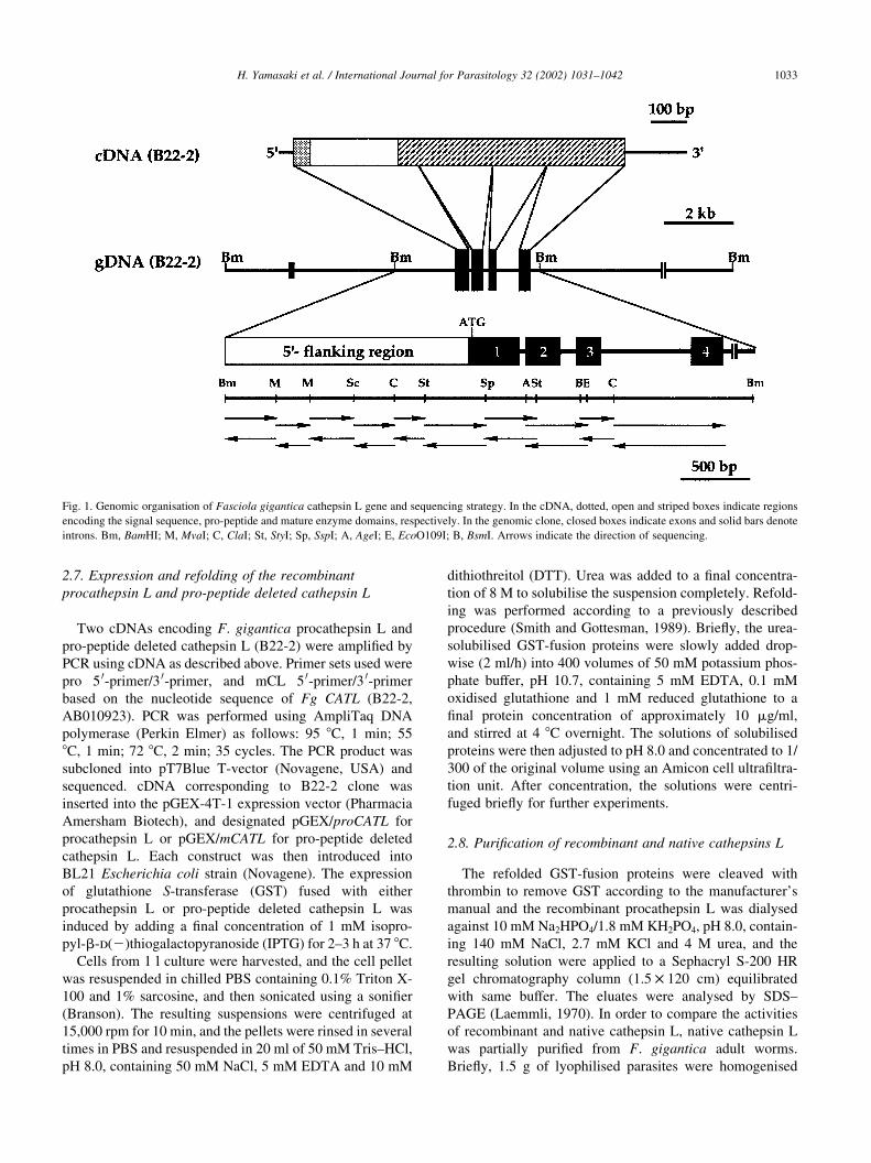
Southern blot analysis of F. gigantica genomic DNA
revealed two or three signals in each of the enzymes used
(Fig. 3). Apart from the genomic clone (B22-2), another1.8-
kb Fg-CATL was co-amplified (data not shown) when Fg-
CATL (B22-2) was amplified by PCR using genomic DNA
for probe preparation. Sequence analysis revealed the gene,
termed Fg-CATL (B7-3, accession number AB010924), is
distinct from Fg-CATL (B22-2) at the nucleotide and amino
acid levels. The remarked difference was observed in length
and sequence in the third intron, the sizes of 582 and 694 bp
were in the B22-2 and B7-3 Fg-CATLs, respectively. In
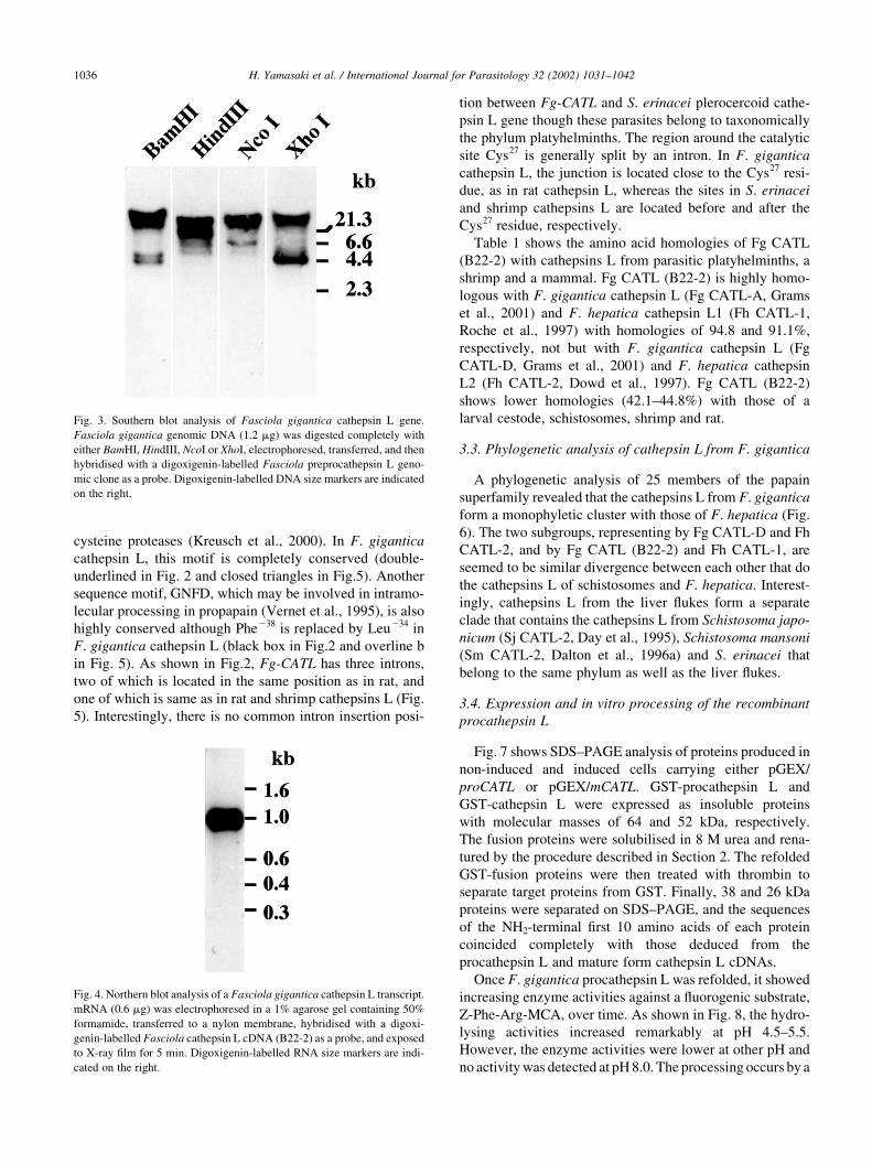
Northern blot analysis, an approximately 1.0 kb transcript
was detected when a B22-2 cathepsin L cDNA was used as a
probe (Fig. 4). Sequence analysis of the 5 0-untranslated
region of the Fg-CATL mRNAs revealed a predicted tran-
scription initiation site at an adenine 14 nucleotides
upstream of the translation initiation site.
3.2. Amino acid sequence characterisation of F. gigantica
cathepsin L and homology with other known cathepsin L
Fig. 5 shows the aligned sequences and intron insertion
positions of cathepsins L from F. gigantica, S. erinacei
plerocercoid (Liu et al., 1996), shrimp (Boulay et al.,
1998) and rat (Ishidoh et al., 1989). The amino acid
sequences of F. gigantica cathepsins L (Fg CATL B22-2
and B7-3) are homologous to those of cathepsins L from
other eukaryotes. The regions containing the Cys27, His164
H. Yamasaki et al. / International Journal for Parasitology 32 (2002) 1031–10421034

and Asn184 residues at the active sites are highly conserved.
Since it is confirmed that a cathepsin L (B22-2) is expressed
in the F. gigantica adult worms, the cathepsin L described
here is Fg CATL (B22-2). The ERFNIN motif (Karrer et al.,
1993) was found, but the first amino acid of the motif, Glu,
was replaced by Val (grey box in Fig. 2 and overlined a in
Fig. 5). Most recently, an evolutionarily conserved tripartite
tryptophan motif has been identified in cathepsin L-like
H. Yamasaki et al. / International Journal for Parasitology 32 (2002) 1031–1042 1035
Fig. 2. Nucleotide and deduced amino acid sequences of Fasciola gigantica cathepsin L (B22-2). Nucleotides are numbered from the transcription initiation
site (11). The amino acid sequence deduced from the nucleotide sequence is shown below the nucleotide sequences and is numbered from the first amino acid
(Arg11) of the mature enzyme. Intron sequences are shown in lowercase letters. Putative TATA boxes and a polyadenylation addition signal are in grey boxes.
The asterisk indicates a translation termination codon. Arrows a and b indicate putative cleavage sites for signal sequence and the pro-peptide. Active site
residues (Cys27, His164 and Asn184) are in black boxes. The ERFNIN motif (Val270 to Asn251) and the putative intramolecular processing motif (Gly238 to
Asp232) are in grey and black boxes, respectively. The three aromatic residues (Trp285, Trp282 and Trp262) in a tripartite tryptophan motif are underlined. The
NH2-terminal sequence of the recombinant cathepsin L processed in vitro is underlined (Glu24 to Gln21). GSP 1 and 2 primers were used for 5 0-rapid
amplification of cDNA ends. The underlined region (nucleotides 60–83), mCL 5 0- and 3 0-primers were used to amplify the procathepsin L and the pro-peptide
deleted cathepsin L cDNAs, respectively.
cysteine proteases (Kreusch et al., 2000). In F. gigantica
cathepsin L, this motif is completely conserved (double-
underlined in Fig. 2 and closed triangles in Fig.5). Another
sequence motif, GNFD, which may be involved in intramo-
lecular processing in propapain (Vernet et al., 1995), is also
highly conserved although Phe238 is replaced by Leu234 in
F. gigantica cathepsin L (black box in Fig.2 and overline b
in Fig. 5). As shown in Fig.2, Fg-CATL has three introns,
two of which is located in the same position as in rat, and
one of which is same as in rat and shrimp cathepsins L (Fig.
5). Interestingly, there is no common intron insertion posi-
tion between Fg-CATL and S. erinacei plerocercoid cathe-
psin L gene though these parasites belong to taxonomically
the phylum platyhelminths. The region around the catalytic
site Cys27 is generally split by an intron. In F. gigantica
cathepsin L, the junction is located close to the Cys27 resi-
due, as in rat cathepsin L, whereas the sites in S. erinacei
and shrimp cathepsins L are located before and after the
Cys27 residue, respectively.
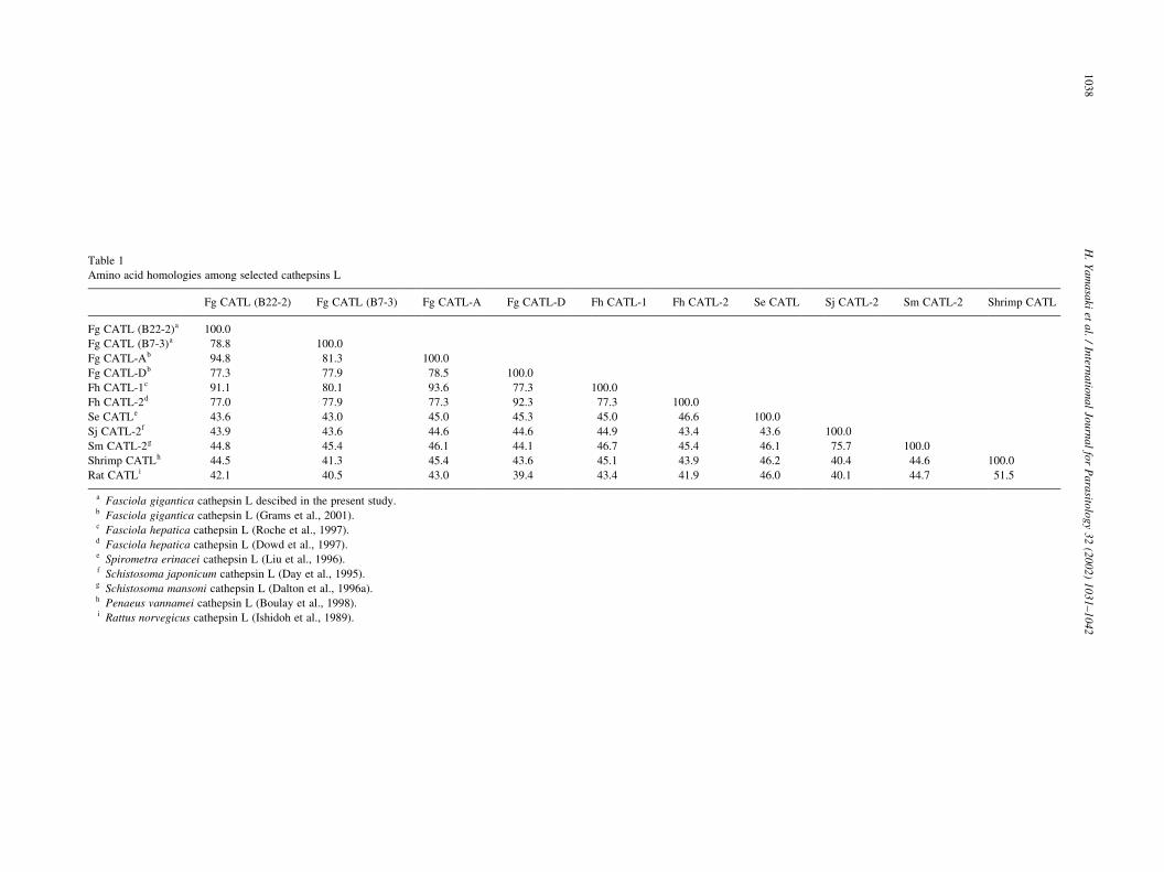
Table 1 shows the amino acid homologies of Fg CATL
(B22-2) with cathepsins L from parasitic platyhelminths, a
shrimp and a mammal. Fg CATL (B22-2) is highly homo-
logous with F. gigantica cathepsin L (Fg CATL-A, Grams
et al., 2001) and F. hepatica cathepsin L1 (Fh CATL-1,
Roche et al., 1997) with homologies of 94.8 and 91.1%,
respectively, not but with F. gigantica cathepsin L (Fg
CATL-D, Grams et al., 2001) and F. hepatica cathepsin
L2 (Fh CATL-2, Dowd et al., 1997). Fg CATL (B22-2)
shows lower homologies (42.1–44.8%) with those of a
larval cestode, schistosomes, shrimp and rat.
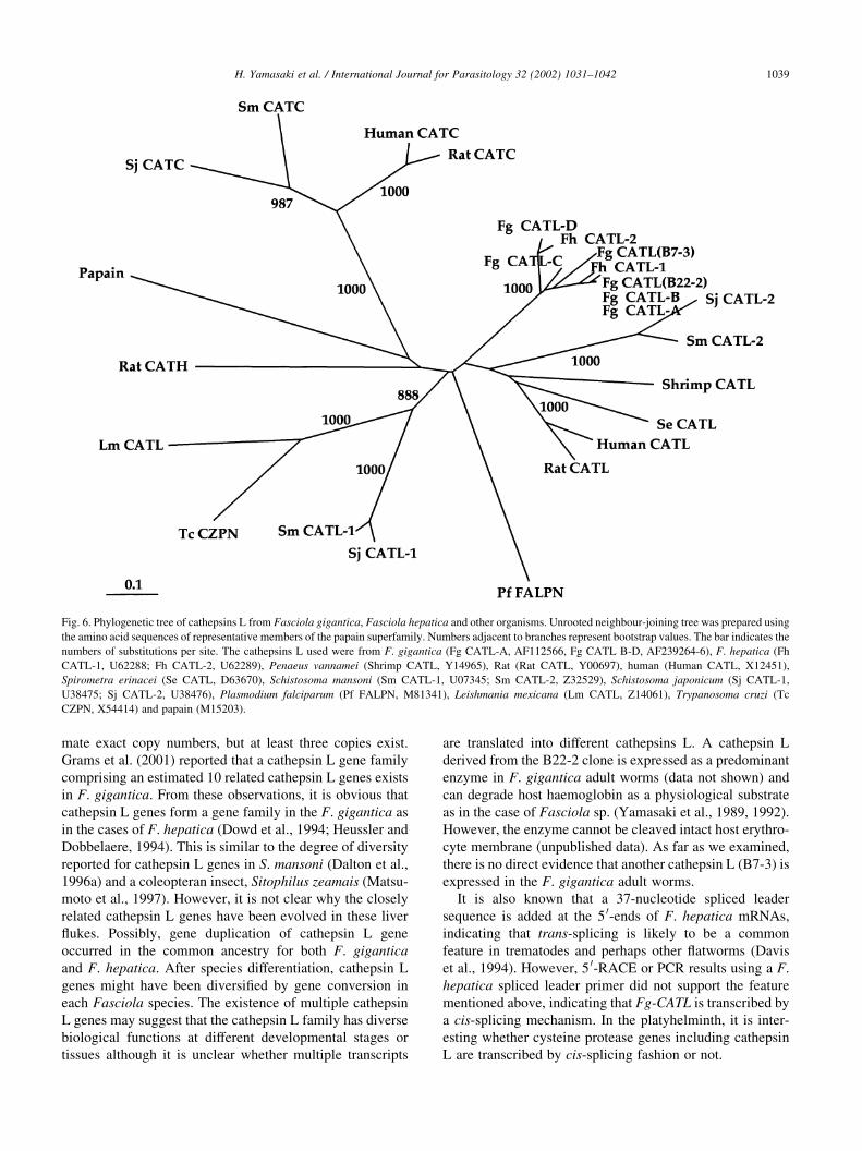
3.3. Phylogenetic analysis of cathepsin L from F. gigantica
A phylogenetic analysis of 25 members of the papain
superfamily revealed that the cathepsins L from F. gigantica
form a monophyletic cluster with those of F. hepatica (Fig.
6). The two subgroups, representing by Fg CATL-D and Fh
CATL-2, and by Fg CATL (B22-2) and Fh CATL-1, are
seemed to be similar divergence between each other that do
the cathepsins L of schistosomes and F. hepatica. Interest-
ingly, cathepsins L from the liver flukes form a separate
clade that contains the cathepsins L from Schistosoma japo-
nicum (Sj CATL-2, Day et al., 1995), Schistosoma mansoni
(Sm CATL-2, Dalton et al., 1996a) and S. erinacei that
belong to the same phylum as well as the liver flukes.
3.4. Expression and in vitro processing of the recombinant
procathepsin L
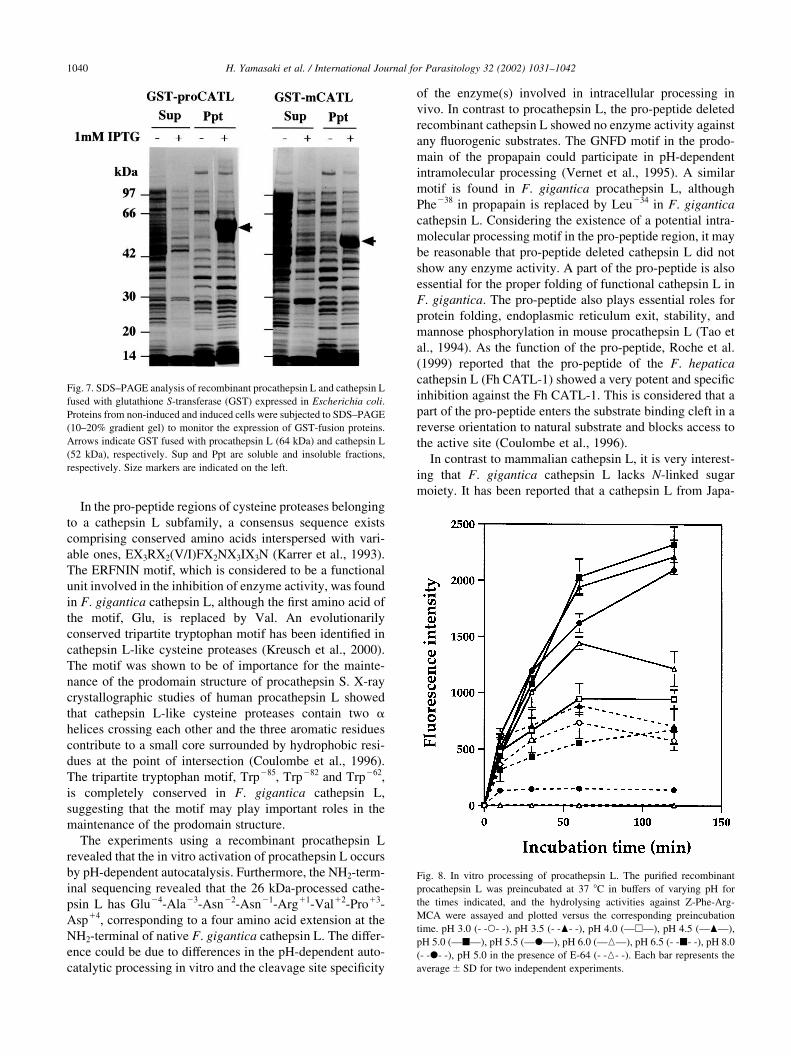
Fig. 7 shows SDS–PAGE analysis of proteins produced in
non-induced and induced cells carrying either pGEX/
proCATL or pGEX/mCATL. GST-procathepsin L and
GST-cathepsin L were expressed as insoluble proteins
with molecular masses of 64 and 52 kDa, respectively.
The fusion proteins were solubilised in 8 M urea and rena-
tured by the procedure described in Section 2. The refolded
GST-fusion proteins were then treated with thrombin to
separate target proteins from GST. Finally, 38 and 26 kDa
proteins were separated on SDS–PAGE, and the sequences
of the NH2-terminal first 10 amino acids of each protein
coincided completely with those deduced from the
procathepsin L and mature form cathepsin L cDNAs.
Once F. gigantica procathepsin L was refolded, it showed
increasing enzyme activities against a fluorogenic substrate,
Z-Phe-Arg-MCA, over time. As shown in Fig. 8, the hydro-
lysing activities increased remarkably at pH 4.5–5.5.
However, the enzyme activities were lower at other pH and
no activity was detected at pH 8.0. The processing occurs by a
H. Yamasaki et al. / International Journal for Parasitology 32 (2002) 1031–10421036
Fig. 4. Northern blot analysis of a Fasciola gigantica cathepsin L transcript.
mRNA (0.6 mg) was electrophoresed in a 1% agarose gel containing 50%
formamide, transferred to a nylon membrane, hybridised with a digoxi-
genin-labelled Fasciola cathepsin L cDNA (B22-2) as a probe, and exposed
to X-ray film for 5 min. Digoxigenin-labelled RNA size markers are indi-
cated on the right.
Fig. 3. Southern blot analysis of Fasciola gigantica cathepsin L gene.
Fasciola gigantica genomic DNA (1.2 mg) was digested completely with
either BamHI, HindIII, NcoI or XhoI, electrophoresed, transferred, and then
hybridised with a digoxigenin-labelled Fasciola preprocathepsin L geno-
mic clone as a probe. Digoxigenin-labelled DNA size markers are indicated
on the right.

pH-dependent autocatalytic mechanism in vitro and it seems
to occur in multiple steps based on the results by Western
blotting (data not shown). Interestingly, the processed active
cathepsin L degraded GST in the reaction mixture after over-
night incubation at pH 5.5. In addition, the processing reac-
tion was completely inhibited in the presence of an
irreversible inhibitor, E-64 (Fig. 8), indicating that the inter-
action with the prodomain of procathepsin L with the inhi-
bitor results in the inhibition of autocatalysis of the enzyme.
The specific activity of the processed cathepsin L was
4,282 nmol/mg protein per minute, corresponding to 80%
of that of native cathepsin L (5,308 nmol/mg protein per
minute). Km values for recombinant and native cathepsin L
toward Z-Phe-Arg-MCA were 83.3 and 23.3 mM, respec-
tively. The processed cathepsin L did not show any activities
against Z-Arg-Arg-MCA, a substrate for cathepsin B, and
Arg-MCA for cathepsin H as well as native F. gigantica
cathepsin L (data not shown). Furthermore, the sequencing
of the 26 kDa-processed cathepsin L revealed the NH2-term-
inal to be Glu24-Ala23-Asn22-Asn21-Arg11-Val12-Pro13-
Asp14 (underlined in Fig. 2), corresponding to a four-
amino-acid extension at the NH2-terminal of native F. gigan-
tica cathepsin L.
4. Discussion
We describe here the genomic structure and sequence
analysis of F. gigantica cathepsin L and the in vitro proces-
sing of the recombinant F. gigantica procathepsin L.
Sequence analysis revealed that Fg-CATL is as a basically
similar structure to the other cathepsin L genes. However,
Fg-CATL contains fewer introns compared with those of its
mammalian counterparts and is a compact gene as in the case
of cathepsin L from a platyhelminth S. erinacei (Liu et al.,
1996). Previously sequenced cathepsin L genes from proto-
zoan parasites are entirely devoid of any introns (Rosenthal
and Nelson, 1992; Eakin et al., 1992). Regarding intron inser-
tion positions, a junction close to the catalytic residue
(Asn185) found in rat cathepsin L is absent in F. gigantica
cathepsin L. The junction between Val200 and Glu201 is
conserved in shrimp and rat cathepsins L and in rat cathepsin
H. It is interesting to note that the intron insertion positions
are similar to those in mammalian and crustaceous cathepsins
L rather than to those in the taxonomically closely related S.
erinacei.
Southern blot analysis revealed that the Fg-CATL gene
family comprises only a few copies. It was difficult to esti-
H. Yamasaki et al. / International Journal for Parasitology 32 (2002) 1031–1042 1037
Fig. 5. Alignment of the predicted amino acid sequences and intron insertion positions of cathepsins L from Fasciola gigantica, Spirometra erinacei, shrimp
and rat. Cathepsin L genes whose intron insertion positions are determined are selected. The catalytic triad residues are marked with asterisks, and gaps (-) are
introduced to maximise alignment. Arrows a and b indicate the putative cleavage sites of the signal sequence and pro-peptide of F. gigantica cathepsin L,
respectively. Intron insertion positions are indicated by closed circles. The ERFNIN and the intramolecular processing motifs are shown by overlines a and b,
respectively. The tripartite tryptophan residues are denoted by closed triangles. Identical amino acid residues are shown in black boxes.
H.
Ya
ma
saki
eta
l./
Intern
atio
na
lJo
urn
al
for
Pa
rasito
log
y3
2(2
00
2)
10
31
–1
04
21
03
8
Table 1
Amino acid homologies among selected cathepsins L
Fg CATL (B22-2) Fg CATL (B7-3) Fg CATL-A Fg CATL-D Fh CATL-1 Fh CATL-2 Se CATL Sj CATL-2 Sm CATL-2 Shrimp CATL
Fg CATL (B22-2)a 100.0
Fg CATL (B7-3)a 78.8 100.0
Fg CATL-Ab 94.8 81.3 100.0
Fg CATL-Db 77.3 77.9 78.5 100.0
Fh CATL-1c 91.1 80.1 93.6 77.3 100.0
Fh CATL-2d 77.0 77.9 77.3 92.3 77.3 100.0
Se CATLe 43.6 43.0 45.0 45.3 45.0 46.6 100.0
Sj CATL-2f 43.9 43.6 44.6 44.6 44.9 43.4 43.6 100.0
Sm CATL-2g 44.8 45.4 46.1 44.1 46.7 45.4 46.1 75.7 100.0
Shrimp CATLh 44.5 41.3 45.4 43.6 45.1 43.9 46.2 40.4 44.6 100.0
Rat CATLi 42.1 40.5 43.0 39.4 43.4 41.9 46.0 40.1 44.7 51.5
a Fasciola gigantica cathepsin L descibed in the present study.b Fasciola gigantica cathepsin L (Grams et al., 2001).c Fasciola hepatica cathepsin L (Roche et al., 1997).d Fasciola hepatica cathepsin L (Dowd et al., 1997).e Spirometra erinacei cathepsin L (Liu et al., 1996).f Schistosoma japonicum cathepsin L (Day et al., 1995).g Schistosoma mansoni cathepsin L (Dalton et al., 1996a).h Penaeus vannamei cathepsin L (Boulay et al., 1998).i Rattus norvegicus cathepsin L (Ishidoh et al., 1989).
mate exact copy numbers, but at least three copies exist.
Grams et al. (2001) reported that a cathepsin L gene family
comprising an estimated 10 related cathepsin L genes exists
in F. gigantica. From these observations, it is obvious that
cathepsin L genes form a gene family in the F. gigantica as
in the cases of F. hepatica (Dowd et al., 1994; Heussler and
Dobbelaere, 1994). This is similar to the degree of diversity
reported for cathepsin L genes in S. mansoni (Dalton et al.,
1996a) and a coleopteran insect, Sitophilus zeamais (Matsu-
moto et al., 1997). However, it is not clear why the closely
related cathepsin L genes have been evolved in these liver
flukes. Possibly, gene duplication of cathepsin L gene
occurred in the common ancestry for both F. gigantica
and F. hepatica. After species differentiation, cathepsin L
genes might have been diversified by gene conversion in
each Fasciola species. The existence of multiple cathepsin
L genes may suggest that the cathepsin L family has diverse
biological functions at different developmental stages or
tissues although it is unclear whether multiple transcripts
are translated into different cathepsins L. A cathepsin L
derived from the B22-2 clone is expressed as a predominant
enzyme in F. gigantica adult worms (data not shown) and
can degrade host haemoglobin as a physiological substrate
as in the case of Fasciola sp. (Yamasaki et al., 1989, 1992).
However, the enzyme cannot be cleaved intact host erythro-
cyte membrane (unpublished data). As far as we examined,
there is no direct evidence that another cathepsin L (B7-3) is
expressed in the F. gigantica adult worms.
It is also known that a 37-nucleotide spliced leader
sequence is added at the 5 0-ends of F. hepatica mRNAs,
indicating that trans-splicing is likely to be a common
feature in trematodes and perhaps other flatworms (Davis
et al., 1994). However, 5 0-RACE or PCR results using a F.
hepatica spliced leader primer did not support the feature
mentioned above, indicating that Fg-CATL is transcribed by
a cis-splicing mechanism. In the platyhelminth, it is inter-
esting whether cysteine protease genes including cathepsin
L are transcribed by cis-splicing fashion or not.
H. Yamasaki et al. / International Journal for Parasitology 32 (2002) 1031–1042 1039
Fig. 6. Phylogenetic tree of cathepsins L from Fasciola gigantica, Fasciola hepatica and other organisms. Unrooted neighbour-joining tree was prepared using
the amino acid sequences of representative members of the papain superfamily. Numbers adjacent to branches represent bootstrap values. The bar indicates the
numbers of substitutions per site. The cathepsins L used were from F. gigantica (Fg CATL-A, AF112566, Fg CATL B-D, AF239264-6), F. hepatica (Fh
CATL-1, U62288; Fh CATL-2, U62289), Penaeus vannamei (Shrimp CATL, Y14965), Rat (Rat CATL, Y00697), human (Human CATL, X12451),
Spirometra erinacei (Se CATL, D63670), Schistosoma mansoni (Sm CATL-1, U07345; Sm CATL-2, Z32529), Schistosoma japonicum (Sj CATL-1,
U38475; Sj CATL-2, U38476), Plasmodium falciparum (Pf FALPN, M81341), Leishmania mexicana (Lm CATL, Z14061), Trypanosoma cruzi (Tc
CZPN, X54414) and papain (M15203).
In the pro-peptide regions of cysteine proteases belonging
to a cathepsin L subfamily, a consensus sequence exists
comprising conserved amino acids interspersed with vari-
able ones, EX3RX2(V/I)FX2NX3IX3N (Karrer et al., 1993).
The ERFNIN motif, which is considered to be a functional
unit involved in the inhibition of enzyme activity, was found
in F. gigantica cathepsin L, although the first amino acid of
the motif, Glu, is replaced by Val. An evolutionarily
conserved tripartite tryptophan motif has been identified in
cathepsin L-like cysteine proteases (Kreusch et al., 2000).
The motif was shown to be of importance for the mainte-
nance of the prodomain structure of procathepsin S. X-ray
crystallographic studies of human procathepsin L showed
that cathepsin L-like cysteine proteases contain two a
helices crossing each other and the three aromatic residues
contribute to a small core surrounded by hydrophobic resi-
dues at the point of intersection (Coulombe et al., 1996).
The tripartite tryptophan motif, Trp285, Trp282 and Trp262,
is completely conserved in F. gigantica cathepsin L,
suggesting that the motif may play important roles in the
maintenance of the prodomain structure.
The experiments using a recombinant procathepsin L
revealed that the in vitro activation of procathepsin L occurs
by pH-dependent autocatalysis. Furthermore, the NH2-term-
inal sequencing revealed that the 26 kDa-processed cathe-
psin L has Glu24-Ala23-Asn22-Asn21-Arg11-Val12-Pro13-
Asp14, corresponding to a four amino acid extension at the
NH2-terminal of native F. gigantica cathepsin L. The differ-
ence could be due to differences in the pH-dependent auto-
catalytic processing in vitro and the cleavage site specificity
of the enzyme(s) involved in intracellular processing in
vivo. In contrast to procathepsin L, the pro-peptide deleted
recombinant cathepsin L showed no enzyme activity against
any fluorogenic substrates. The GNFD motif in the prodo-
main of the propapain could participate in pH-dependent
intramolecular processing (Vernet et al., 1995). A similar
motif is found in F. gigantica procathepsin L, although
Phe238 in propapain is replaced by Leu234 in F. gigantica
cathepsin L. Considering the existence of a potential intra-
molecular processing motif in the pro-peptide region, it may
be reasonable that pro-peptide deleted cathepsin L did not
show any enzyme activity. A part of the pro-peptide is also
essential for the proper folding of functional cathepsin L in
F. gigantica. The pro-peptide also plays essential roles for
protein folding, endoplasmic reticulum exit, stability, and
mannose phosphorylation in mouse procathepsin L (Tao et
al., 1994). As the function of the pro-peptide, Roche et al.
(1999) reported that the pro-peptide of the F. hepatica
cathepsin L (Fh CATL-1) showed a very potent and specific
inhibition against the Fh CATL-1. This is considered that a
part of the pro-peptide enters the substrate binding cleft in a
reverse orientation to natural substrate and blocks access to
the active site (Coulombe et al., 1996).
In contrast to mammalian cathepsin L, it is very interest-
ing that F. gigantica cathepsin L lacks N-linked sugar
moiety. It has been reported that a cathepsin L from Japa-
H. Yamasaki et al. / International Journal for Parasitology 32 (2002) 1031–10421040
Fig. 7. SDS–PAGE analysis of recombinant procathepsin L and cathepsin L
fused with glutathione S-transferase (GST) expressed in Escherichia coli.
Proteins from non-induced and induced cells were subjected to SDS–PAGE
(10–20% gradient gel) to monitor the expression of GST-fusion proteins.
Arrows indicate GST fused with procathepsin L (64 kDa) and cathepsin L
(52 kDa), respectively. Sup and Ppt are soluble and insoluble fractions,
respectively. Size markers are indicated on the left.
Fig. 8. In vitro processing of procathepsin L. The purified recombinant
procathepsin L was preincubated at 37 8C in buffers of varying pH for
the times indicated, and the hydrolysing activities against Z-Phe-Arg-
MCA were assayed and plotted versus the corresponding preincubation
time. pH 3.0 (- -W- -), pH 3.5 (- -O- -), pH 4.0 (—A—), pH 4.5 (—O—),
pH 5.0 (—B—), pH 5.5 (—X—), pH 6.0 (—K—), pH 6.5 (- -B- -), pH 8.0
(- -X- -), pH 5.0 in the presence of E-64 (- -K- -). Each bar represents the
average ^ SD for two independent experiments.

nese Fasciola sp. localises in the secretory granules in
intestinal epithelial cells of the worms (Yamasaki et al.,
1992; Yamasaki and Aoki, 1993). This suggests that the
intracellular transport mechanism of F. gigantica cathepsin
L is different from those of mammalian cathepsins L. In
order to clarify the function of the pro-peptide in the intra-
cellular transport mechanism of F. gigantica cathepsin L,
experiments using deletion mutants of F. gigantica
procathepsin L fused with a green fluorescent protein are
currently in progress.
Acknowledgements
We thank Professor Dr Takeshi Agatsuma for the mole-
cular identification of the Fasciola parasite, and Naomi Mori,
Koichi Kawakami and Mami Kaneko for technical assis-
tance. This work was supported in part by grants-in-aid for
Scientific Research from the Ministry of Education, Science,
Culture and Sports, Japan (Nos. 05670241,07670291,
09670271).
References
Barrett, J.A., Kirschke, H., 1981. cathepsin B, cathepsin H and Cathepsin L.
Methods Enzymol. 80, 535–61.
Boulay, C.L., Sellos, D., Wormhoudt, A.V., 1998. Cathepsin L gene orga-
nization in crustaceans. Gene 218, 77–84.
Breathnach, R., Chambon, P., 1981. Organization and expression of eukar-
yotic split genes coding for proteins. Annu. Rev. Biochem. 50, 349–83.
Chapman, C.B., Mitchell, G.F., 1982. Proteolytic cleavage of immunoglo-
bulin by enzymes released by Fasciola hepatica. Vet. Parasitol. 11,
165–78.
Chen, M.G., Mott, K.E., 1990. Progress in morbidity due to Fasciola hepa-
tica infection. Trop. Dis. Bull. 87, 1–37.
Chomczynski, P., Sacchi, N., 1987. Single-step method of RNA isolation
by acid guanidinium thiocyanate-phenol-chloroform extraction. Anal.
Biochem. 162, 156–9.
Coulombe, R., Grochulski, R., Sivaraman, J., Menard, R., Mort, J.S.,
Cygler, M., 1996. Structure of human procathepsin L reveals the mole-
cular basis of inhibition by the prosegment. EMBO J. 15, 5492–503.
Dalton, J.P., Hefferman, M., 1989. Thiol proteases released in vitro by
Fasciola hepatica. Mol. Biochem. Parasitol. 35, 161–6.
Dalton, J.P., Clough, K.A., Jones, M.K., Brindley, P.J., 1996a. Character-
ization of the cathepsin-like cysteine proteinases of Schistosoma
mansoni. Infect. Immun. 64, 1328–34.
Dalton, J.P., McGonigle, S., Rolph, T.P., Andrews, S.J., 1996b. Induction
of protective immunity in cattle against infection with Fasciola hepa-
tica by vaccination with cathepsin L proteinase and hemoglobin. Infect.
Immun. 64, 5066–74.
Davis, E.R., Singh, H., Botka, C., Hardwick, C., el Meanawy, M.A., Villa-
nueva, J., 1994. RNA trans-splicing in Fasciola hepatica. Identification
of a spliced leader (SL) RNA and SL sequences on mRNAs. J. Biol.
Chem. 269, 20026–200230.
Day, S.R., Dalton, J.P., Clough, K.A., Leonardo, L., Tiu, W.U., Brindley,
P.J., 1995. Characterization and cloning of the cathepsin L proteinases
of Schistosoma japonicum. Biochem. Biophys. Res. Comm. 217, 1–9.
Dowd, A.J., Smith, M.A., McGonigle, S., Dalton, J.P., 1994. Purification
and characterization of a second cathepsin L proteinase secreted by the
trematode Fasciola hepatica. Eur. J. Biochem. 223, 91–98.
Dowd, A.J., Tort, J., Roche, L., Ryan, T., Dalton, J.P., 1997. Isolation of a
cDNA encoding Fasciola hepatica cathepsin L2 and functional expres-
sion in Saccharomyces cerevisiae. Mol. Biochem. Parasitol. 88, 163–
74.
Eakin, A.E., Mills, A.A., Harth, G., McKerrow, J.H., Craik, C.S., 1992. The
sequence, organization, and expression of the major cysteine protease
(cruzain) from Trypanosoma cruzi. J. Biol. Chem. 267, 7411–20.
Esteban, J.G., Bargues, M.D., Mas-Coma, S., 1998. Geographical distribu-
tion, diagnosis and treatment of human fascioliasis: a review. Res. Rev.
Parasitol. 58, 13–48.
Fagbemi, B.O., Guobadia, E.E., 1995. Immunodiagnosis of fascioliasis in
ruminants using a 28-kDa cysteine protease of Fasciola gigantica. Vet.
Parasitol. 57, 309–18.
Fagbemi, B.O., Hillyer, G.V., 1992. Partial purification and characteriza-
tion of the proteolytic enzymes of Fasciola gigantica adult worms. Vet.
Parasitol. 40, 217–26.
Grams, R., Vichasri-Grams, S., Sobhorn, P., Upatham, E.S., Viyanant, V.,
2001. Molecular cloning and characterization of cathepsin L encoding
genes from Fasciola gigantica. Parasitol. Int. 50, 105–14.
Hashimoto, K., Watanabe, T., Liu, C.X., Init, I., Blair, D., Ohnishi, S.,
Agatsuma, T., 1997. Mitochondrial DNA and nuclear DNA indicate
that the Japanese Fasciola species is F. gigantica. Parasitol. Res. 83,
220–5.
Hawthorne, S.J., Pagano, M., Halton, D.W., Walker, B., 2000. Partial char-
acterization of a novel cathepsin L-like protease from Fasciola hepa-
tica. Biochem. Biophys. Res. Commun. 277, 79–82.
Heussler, T.V., Dobbelaere, A.E.D., 1994. Cloning of a protease gene
family of Fasciola hepatica by the polymerase chain reaction. Mol.
Biochem. Parasitol. 64, 11–23.
Ishidoh, K., Kominami, E., Suzuki, K., Katsunuma, N., 1989. Gene struc-
ture and 5 0-upstream sequence of rat cathepsin L. FEBS Lett. 259, 71–
74.
Karrer, K.M., Peiffer, S.L., DiTomas, M.E., 1993. Two distinct gene subfa-
milies within the family of cysteine protease genes. Proc. Natl. Acad.
Sci. USA 90, 3063–7.
Kreusch, S., Fehn, M., Maubach, G., Nissler, K., Rommerskirch, W., Schil-
ling, K., Weber, E., Wenz, I., Wiederanders, B., 2000. An evolutiona-
rily conserved tripartite tryptophan motif stabilizes the prodomains of
cathepsin L-like cysteine proteases. Eur. J. Biochem. 267, 2965–72.
Laemmli, U.K., 1970. Cleavage of structural proteins during the assembly
of the head of bacteriophage T4. Nature 227, 680–5.
Liu, D.W., Kato, H., Nakamura, T., Sugane, K., 1996. Molecular cloning
and expression of the gene encoding a cysteine proteinase of Spirometra
erinacei. Mol. Biochem. Parasitol. 76, 11–21.
Maniatis, T., Fritsch, E.F., Sambrook, J., 1982. Molecular Cloning. A
Laboratory Manual, Cold Spring Harbor Laboratory, Cold Spring
Harbor, NY.
Mas-Coma, S., Esteban, J.G., Bargues, M.D., 1999. Epidemiology of
human fascioliasis: a review and proposed new classification. Bull.
WHO 77, 340–6.
Matsudaira, P., 1987. Sequence from picomole quantities of proteins elec-
troblotted onto polyvinylidene difluoride membrane. J. Biol. Chem.
262, 10035–8.
Matsumoto, I., Watanabe, H., Abe, K., Arai, S., Emori, Y., 1995. A putative
digestive cysteine proteinase from Drosophila melanogaster is predo-
minantly expressed in the embryonic and larval midgut. Eur. J.
Biochem. 227, 582–7.
Matsumoto, I., Emori, Y., Abe, K., Arai, S., 1997. Characterization of a
gene encoding cysteine proteinases of Sitophilus zeamais (Maize
Weevil), and analysis of the protein distribution in various tissues
including alimentary tract and germ cells. J. Biochem. 121, 464–76.
McGinty, A., Moore, M., Halton, D.W., Walker, B., 1993. Characterization
of the cysteine proteinase of the common liver fluke Fasciola hepatica
using novel, active-site directed affinity labels. Parasitology 106, 487–
93.
O’Neill, S.M., Parkinson, M., Dowd, A.J., Strauss, W., Angles, R., Dalton,
J.P., 1999. Immunodiagnosis of human fascioliasis using recombinant
Fasciola hepatica cathepsin L1 cysteine proteinase. Am. J. Trop. Med.
Hyg. 60, 749–51.
H. Yamasaki et al. / International Journal for Parasitology 32 (2002) 1031–1042 1041

Patthy, L., 1987. Intron-dependent evolution: preferred types of exons and
introns. FEBS Lett. 214, 1–7.
Rege, A.A., Herrera, P.R., Lopez, M., Dresden, M.H., 1989. Isolation and
characterization of a cysteine proteinase from Fasciola hepatica adult
worms. Mol. Biochem. Parasitol. 35, 89–95.
Roche, L., Dowd, A.J., Tort, J., McGonigle, S., McSweeney, A., Curley, P.,
Ryan, T., Dalton, J.P., 1997. Functional expression of Fasciola hepatica
cathepsin L1 in Saccharomyces cerevisiae. Eur. J. Biochem. 245, 373–
80.
Roche, L., Tort, J., Dalton, J.P., 1999. The pro-peptide of Fasciola hepatica
cathepsin L is a potent and selective inhibitor of the mature enzyme.
Mol. Biochem. Parasitol. 98, 271–7.
Rosenthal, P.J., Nelson, R.G., 1992. Isolation and characterization of a
cysteine proteinase gene of Plasmodium falciparum. Mol. Biochem.
Parasitol. 51, 143–52.
Saitou, N., Nei, M., 1987. The neighbour-joining method: a new method for
reconstructing phylogenetic trees. Mol. Biol. Evol. 4, 406–25.
Smith, A.M., Dowd, A.J., McGonigle, S., Keegan, P.S., Brennan, G., Trud-
gett, A., Dalton, J.P., 1993. Purification of a cathepsin L-like proteinase
secreted by adult Fasciola hepatica. Mol. Biochem. Parasitol. 62, 1–8.
Smith, S.M., Gottesman, M.M., 1989. Activity and deletion analysis of
recombinant human cathepsin L expressed in Escherichia coli. J.
Biol. Chem. 264, 20487–95.
Tao, K., Stearns, A.N., Dong, J., Wu, Q.-L., Sahagian, G.G., 1994. The
proregion of cathepsin L is required for proper folding, stability, and ER
exit. Arch. Biochem. Biophys. 311, 19–27.
Vernet, T., Berti, J.P., Montigny, C., Musil, R., Tessier, C.D., Menard, R.,
Magny, M.-C., Stoner, C.A., Thomas, Y.D., 1995. Processing of the
papain precursor. The ionization state of a conserved amino acid motif
within the pro region participates in the regulation of intramolecular
processing. J. Biol. Chem. 270, 10838–46.
Wijffels, G.L., Panaccio, M., Salvatore, L., Wilson, L., Walker, I.D., Spit-
hill, T.W., 1994a. The second cathepsin L-like proteinases of the trema-
tode, Fasciola hepatica, contain 3-hydroxyproline residues. Biochem.
J. 299, 781–90.
Wijffels, G.L., Salvatore, L., Dosen, M., Waddington, J., Thompson, C.,
Cambell, N., Sexton, J., Wicker, J., Bowen, F., Friedel, T., Spithill,
T.W., 1994b. Vaccination of sheep with purified cysteine proteinase
of Fasciola hepatica decreases worm fecundity. Exp. Parasitol. 78,
132–48.
Yamasaki, H., Aoki, T., Oya, H., 1989. A cysteine proteinase from the liver
fluke Fasciola spp.: purification, characterization, localization and
application to immunodiagnosis. Jpn. J. Parasitol. 38, 373–84.
Yamasaki, H., Kominami, E., Aoki, T., 1992. Immunocytochemical loca-
lization of a cysteine protease in adult worms of the liver fluke Fasciola
sp. Parasitol. Res. 78, 574–80.
Yamasaki, H., Aoki, T., 1993. Cloning and sequence analysis of the major
cysteine protease expressed the trematode parasite Fasciola sp.
Biochem. Mol. Biol. Int. 31, 537–42.
H. Yamasaki et al. / International Journal for Parasitology 32 (2002) 1031–10421042