chapter11 wound healing and the presence of biomaterials 11-1 introduction: formation of granulation...
TRANSCRIPT
CHAPTER
1111Wound Healingand the Presenceof Biomaterials
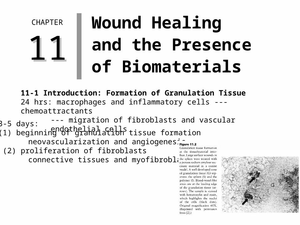
11-1 Introduction: Formation of Granulation Tissue 24 hrs: macrophages and inflammatory cells --- chemoattractants
--- migration of fibroblasts and vascular endothelial cells
3-5 days: (1) beginning of granulation tissue formation
neovascularization and angiogenesis (2) proliferation of fibroblasts
connective tissues and myofibroblasts
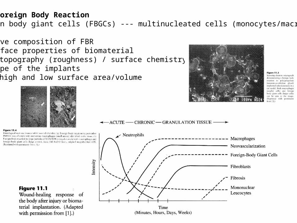
11-2 Foreign Body Reaction Foreign body giant cells (FBGCs) --- multinucleated cells (monocytes/macrophage)
Relative composition of FBR (1)Surface properties of biomaterial
topography (roughness) / surface chemistry (2)Shape of the implants
high and low surface area/volume

11-3 Fibrous Encapsulation Granulation tissue maturation (large blood vessels & collagen fiber alignment)
Long-term capsule formation (4 weeks post-implantation) (1) Degree of original injury during implantation (2) Amount of subsequent cell death (3) Location of implant site (4) Degradation time of implant
Thickness of the capsule (1)Amount & chemical composition of
small particulates produced (2) Mechanical factors at implant site (3) Shape of implant (4) Electrical currents
11-4 Chronic Inflammation Between acute and full development of granulation tissue
Granulomas Epitheloid cells

11-5 Four Types of Resolution Extrusion / Resolution / Integration / Encapsulation
- Failure or success
11-6 Repair vs Regeneration: Wound Healing in Skin - Tissue repair with scar tissue - Tissue regeneration with identical tissue
11.6.1 Skin Repair Skin = internal dermal layer + outer epidermal layer
Injury --- blood clotting --- fibrin network --- acute inflammation --- influx of fibroblasts into ECM --- beginning of granulation tissue --- Remodeling and scar formation
collagen III turnover (1) collagen I (2) GAG ratio 변화
collagen accumulation

11.6.2 Skin Regeneration Erosions: small skin wounds within the epidermal layer
Re-epithelialization Defect --- cell flattening --- cell migration --- cell proliferation migration edge in contact with other epithelial cells --- cuboid morphology and re-attach to ECM ---
proliferation and matrix production --- restoring the original thickness of tissue
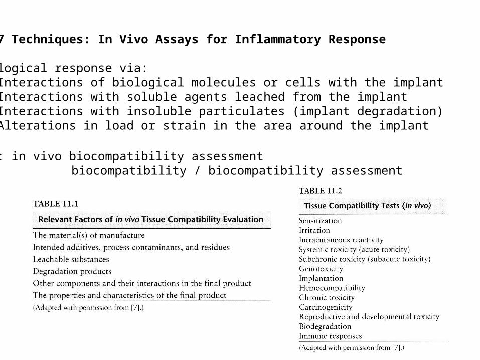
11-7 Techniques: In Vivo Assays for Inflammatory Response
Biological response via: (1)Interactions of biological molecules or cells with the implant (2)Interactions with soluble agents leached from the implant (3)Interactions with insoluble particulates (implant degradation) (4)Alterations in load or strain in the area around the implant
FDA: in vivo biocompatibility assessment biocompatibility / biocompatibility assessment
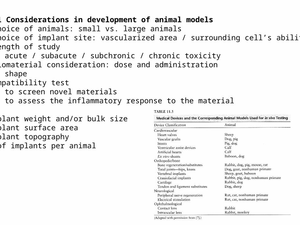
11.7.1 Considerations in development of animal models (1) Choice of animals: small vs. large animals(2) Choice of implant site: vascularized area / surrounding cell’s ability (3) Length of study
acute / subacute / subchronic / chronic toxicity (4) Biomaterial consideration: dose and administration
shape Biocompatibility test
to screen novel materials to assess the inflammatory response to the material
a. Implant weight and/or bulk sizeb. Implant surface areac. Implant topographyd. # of implants per animal
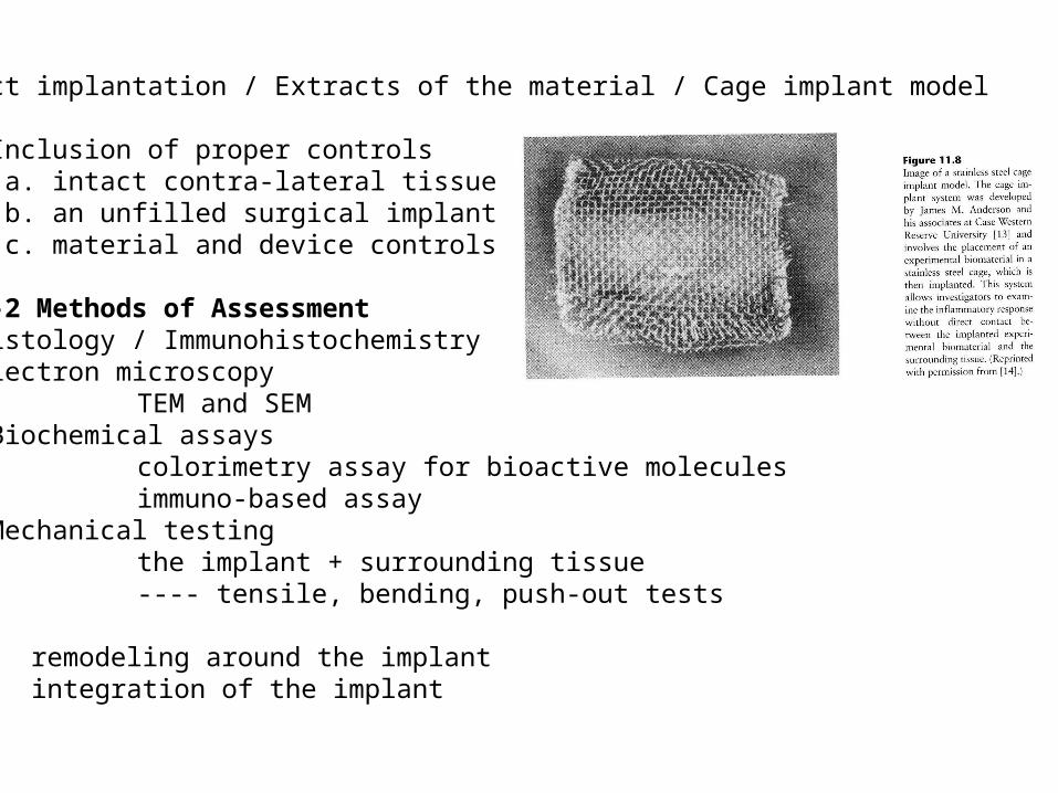
Direct implantation / Extracts of the material / Cage implant model
(5) Inclusion of proper controls a. intact contra-lateral tissue b. an unfilled surgical implant site c. material and device controls
11-7-2 Methods of Assessment (1)Histology / Immunohistochemistry (2)Electron microscopy TEM and SEM (3) Biochemical assays
colorimetry assay for bioactive molecules immuno-based assay
(4) Mechanical testing the implant + surrounding tissue ---- tensile, bending, push-out tests
remodeling around the implant integration of the implant