chapter 4shodhganga.inflibnet.ac.in/bitstream/10603/39779/9/09_chapter4.pdf · were performed using...
TRANSCRIPT

CHAPTER – 4
IN VITRO STUDY OF NOVEL SULFA
HYDRAZONE SUBSTITUTED 4-(3H)-
QUINAZOLINONE HETEROCYCLIC
DERIVATIVES

Chapter -4
Sardar Pate l Univers i ty 229
In this chapter the antibacterial activity of twenty
screened substituted quinazolinone derivetives against
bacterial strains Escherichia coli, Pseudomonas aeruginosa,
Bacillus megaterium and Proteus vulgaris are discussed and
were performed using Agar cup method.
4.1 INTRODUCTION :
The ever-growing resistance to antibiotics leads to
continuous screening for the new biologically effective
substances of natural or synthetic origin [1]. Quinazolinone
derivatives are a subject of increasing interest in bioorganic
and coordination chemistry. A sustained research activity
has been devoted to quinazolinone derivatives, due to their
successful multiple applications as diagnostic tools in
biomedical tribulations like infection and diseases.
Infectious diseases caused by bacteria affect millions of
people worldwide. Rigorous and systematic programs to
discover and develop new antibiotics have been driven to a
considerable extent by the development of resistance by
these organisms to the drugs commonly used against them.
The quinazoline skeleton is one of the ever used as
biologically active molecules since last several decades. In
particular, 4(3H)-quinazolinone derivatives display a broad
range of biological properties such as antihypertensive [2,
3], CNS depressant [4, 5], antitumor, analgesic and anti-
inflammatory [6]. Moreover, 4(3H)-quinazolinone derivatives
indicates the antibacterial and antifungal activities [7]. This
class of compounds also shows the growth inhibition
against Staphylococcus aureus bacteria [8]. Our main focus
is to evaluate the quinazolines derivative for bacteriostatic
effect on multi-drug resistance strains of Staphylococcus
aureus.

Chapter -4
Sardar Pate l Univers i ty 230
Looking to the vital role of this class of compounds in
many infections and diseases, it was planned to explore the
field of novel anti-infective agents. Thus, the present work
comprises in vitro study of twenty selected substituted
quinazolinone derivetives against Bacillus megaterium,
Escherichia coli, Pseudomonas aerugenosa and Proteus
vulgaris. Besides that the compounds were also analyzed for
their response against the Methicillin-Resistant
Staphylococcus Aureus (MRSA) strains. MRSA is a bacterium
responsible for infections in humans. It may also be
referred to as multiple-resistant Staphylococcus aureus.
4.2. BRIEF HISTORY OF ANTIMICROBIAL SCREENING :
Infection is a major cause of human disease and
skilled management of antimicrobial drugs is of the prime
importance. The term chemotherapy is used for the drug
treatment of parasitic infections in which the parasites
(viruses, bacteria, protozoa, fungi, and worms) are
destroyed or removed without injuring the host. Many
substances that we now know to possess therapeutic
efficacy were first used in the distant past. The Ancient
Greeks used male fern, and the Aztecs chenopodium, as
intestinal anthelmintics. The Ancient Hindus treated leprosy
with chaulmoogra. For hundreds of years moulds have been
applied to wounds. Despite the introduction of mercury as a
treatment for syphilis (16 th century), and the use of
cinchona bark against malaria (17 th century), the history of
modern rational chemotherapy did not begin until Paul
Ehrlich developed the idea from his observation that aniline
dyes selectively stained bacteria in tissue microscopic
preparations and could selectively kill them. He invented
the word ‘chemotherapy’ and in 1906 he wrote : “In order to
use chemotherapy successfully, we must search for

Chapter -4
Sardar Pate l Univers i ty 231
substances which have an affinity for the cells of the
parasites and a power of killing them greater than the
damage such substances cause to the organism itself… This
means… we must, learn to aim with chemical substances.”
The antimalerials pamaquin and mepacrine were
developed from dyes and in 1935 the first sulphonamides,
linked with a dye (Prontosil), was introduced as a result of
systematic studies by Domagk. The results obtained with
sulphonamides in puerperal sepsis, pneumonia and
meningitis were dramatic and caused a revolution in
scientific and medical thinking. In 1928, Fleming
accidentally discovered the long-known ability of Penicill ium
fungi to suppress the growth of bacterial cultures. In 1939,
principally as an academic exercise, Florey and Chain
undertook an investigation on antibiotics, i.e. substances
produced by microorganisms that are antagonistic to the
growth or life of other microorganisms. They prepared
penicillin and confirmed its remarkable non-toxicity. When
the preparation was administered to a policeman with
combined staphylococcal and streptococcal septicemia,
there was dramatic improvement; unfortunately the
manufacture of penicillin (in the local Pathology Laboratory)
could not keep pace with the requirements (it was also
extracted from the patient’s urine and re-injected); it ran
out and the patient later succumbed to infection.
Subsequent development thoroughly demonstrated the
remarkable therapeutic efficacy of penicillin.
4.3 CLASSIFICATION OF ANTIMICROBIAL DRUGS :
Antimicrobial agents may be classified according to the
type of organism against which they are active.
• Antibacterial drugs.
• Antiviral drugs.
Chapter -4
Sardar Pate l Univers i ty 232
• Antifungal drugs.
• Antiprotozoal drugs.
A few antimicrobials have useful activity across several
of these groups and they are known as broad spectrum
antibiotics. For example, metronidazole inhibits obligate
anaerobic bacteria (such as Clostridium perfringens) as well
as some protozoa that rely on anarobolic pathways (such as
Trichomonas vaginalis).
Antimicrobial drugs have also been classified broadly
into :
• Bacteriostatic : Those that act primarily by arresting
bacterial multiplication (groth), such as
sulphonamides, tetracyclines and chloramphenicol.
• Bactericidal : Those which act primarily by killing
bacteria, such as penicillins, cephalosporins,
aminoglycosides, isoniazide and rifampicin.
4.4 EVALUATION TECHNIQUES :
The following conditions must be met for the screening
of antimicrobial activity :
• Aseptic/sterile environment should be maintained.
• Mandatory conditions should be provided for the
growth of microorganisms.
• There should be an intimate contact between test
organisms and the substance to be evaluated for its
activity.
• Conditions should be maintained same throughout the
study.
Various methods have been used from time to time by
several workers to evaluate the antimicrobial activity [9-11].
• Turbidometric method.
• Agar streak dilution method.

Chapter -4
Sardar Pate l Univers i ty 233
• Serial dilution method.
• Agar diffusion method.
Agar diffusion method is again sub classified into three
types:
• Agar cup method.
• Agar ditch method.
• Paper disc Method.
However, in the present work Agar cup method was used.
4.4.1 Agar cup method :
When an antibiotic is added in agar cup (make in
medium previously inoculated with the test organism), the
radial diffusion of an antibiotic through the agar produces a
concentration gradient. Test organism is inhibited at the
minimum inhibitory concentration (MIC), giving rise to a
clear zone on inhibition. Greater the MIC, larger is the zone
of inhibition.
4.4.2 Advantages of agar cup method :
Top agar also known as soft agar or seed agar having
concentration of 0.5% is inoculated with the test organism
and then overlaid on the surface of sterilized nutrient agar
plate. The agar overlay method gives better results due to
the following reasons
• Since it has less concentration of agar (0.5%) it
permits faster diffusion of antibiotics.
• Organism can obtain nourishment from both the top
agar as well as the base agar.
• Since the base agar is free of any inoculum, the
zone of inhibition is sharply defined and clearly
measured.
• The zone of inhibition can be measured clearly as
compared to Paper disc and Agar ditch method.
• The growth of the microorganism in the agar cup
method is uniform in the cups of control, standard

Chapter -4
Sardar Pate l Univers i ty 234
drug and the antibiotic to be tested, while in the
case of ditch method the growth depends on the
placement of the dish in the upright position.
• Using this method In situ comparison can be done
with the standard drug easily. While it is not so in
the Agar ditch and the paper disc method.
4.5 FACTORS AFFECTING ZONE OF INHIBITION :
Following factors are important for the screening of the
chemical substances (looking promising as the potential
therapeutic agents). These variables are to be kept constant
for the authentic and reproducible results.
4.5.1 Ingredients of culture media :
Many substances are present in culture media, which
may affect the zone of inhibition. Common ingredients such
as peptone, agar, etc. may vary in their contents. Many of
these minerals may influence the activity of some
antimicrobials. It is well known that Ca, Mg, Fe, etc. ions
affect the sensitivity of zone produced by the tetracycline,
gentamycin. NaCl reduce the activity of amino glycosides
and enhances the effect of fungicides.
4.5.2 Choice of media :
Consistent and reproducible results are obtained in
media prepared especially for sensitivity testing. The plates
must be poured flat with an even depth.
4.5.3 Effect of pH :
The activity of amino glycosides is enhanced in
alkaline media and reduced in acidic media. Exactly the
reverse is found true for the drugs of tetracycline family.
4.5.4 Size of inoculums :
Although large number of organisms does not markedly
affect many antibiotics, all inhibition zones are diminished
by heavy inoculum. The ideal inoculum is one, which gives
Chapter -4
Sardar Pate l Univers i ty 235
an even dense growth without being confluent. Overnight
broth cultures of organisms and suitable suspensions from
solid media can be diluted accurately to give optimum
inoculum for sensitivity testing.
4.6 BACTERIA :
Bacteria are microscopic living creature classified in
the prokaryotic group of organisms. Many bacteria are
useful in our daily life from making the food like curd to the
production of pharmacological compound like antibiotics
and enzymes. On other side, many bacteria are found to be
pathogenic to the human and animal.
They cause the illness in human beings through the
infection. For treatment of the disease many natural and
synthetic compounds are currently used depending upon
the types of bacteria involved in the disease.
Classification of bacteria are based on several
characteristics exhibited by bacteria. However, the
frequently used is the Gram reaction. It was developed by
the Danish physician Christian Gram in 1884, discovered a
stain known as Gram stain, which can divide all bacteria
into two class “Gram positive” and “Gram negative”. The
Gram-positive bacteria resist discoloration with acetone,
alcohol and remain stained (methyl violet) as dark blue
colored, while Gram-negative bacteria are decolorized and
stains pink with the counter stain safranine. Bacteria are
also classified on the basis of their potency to cause the
disease. Those which are responsible for creating the
disease are known as a pathogenic and rests of other are
known as non-pathogenic
4.6.1 Pathogenic bacteria :
Those bacteria which cause the disease in the human
called the pathogenic bacteria. Many groups of bacteria are
Chapter -4
Sardar Pate l Univers i ty 236
responsible to cause the disease in human, some are
obligatory pathogenic, while some are opportunistic
pathogenic.
4.6.2 Gram Negative Bacteria :
Pseudomonas aeruginosa is Gram negative opportunistic
pathogenic which is able to cause infection when the
natural resistance of the body is low. They are mostly
associated with hospital infection (nosocomial) and post
burn infection. They also cause infection of middle ear, eye
and urinary tract and also reported to cause the diarrhea
and pneumonia.
Escherichia coli is one of the most studied and well
characterized bacteria and opportunistic pathogenic cause
the traveler diarrhea and urinary tract infection. E.coli is
Gram negative, short rod, non-capsulated and non-
sporulated bacteria.
Proteus vulgaris is associated with urinary tract infection
as well as infection of wounds and burns. It is also
responsible for summer diarrhea and infantile diarrhea.
They are short rod, highly motile, Gram negative bacteria.
Organisms are non-sporulated and non-capsulated.
4.6.3 Gram positive bacteria :
Bacillus megaterium : It is one of the representative
species of the wide studied genus Baccillus bacteria and are
rod shape with round shape ends. Cells are non capsulated
but are motile and produce the spore. They are non
pathogenic but some strains cause the infection.
Staphylococcus aureus are Gram positive coccoi having a
round or oval shape with size of 0.8 to 0.10 µm in diameter.
They are non motile, non-sporulated and most of the strains
are non-capsulated but few strains show the capsule. They
are responsible for the pyogenic infection of the skin and

Chapter -4
Sardar Pate l Univers i ty 237
also cause the infection like staphylococcal pneumonia,
wound infection, post burning and post surgical infections.
Colonies are smooth, raised, glistening, circular, entire
and translucent, and single colony may obtain a size of 6-8
mm in diameter on non selective media used for the
propagation of Staphylococci.
Facultative anaerobes : Growth is best under aerobic
condition. Growth in the anaerobic portion of a semisolid
thioglycolate medium is rapid and uniformly dense.
Terminal pH in glucose broth under anaerobic condition, is
4.3- 4.6. Catalase is produced by cell growing aerobically. It
is immunologically distinct from the catalase of other
species, as determined by double immuno diffusion and
micro complement fixation analyses. Catalase may be
absent in respiratory deficient mutants.
At least four different hemolysins (exotoxins) are
produced, including -, β-, - and δ- hemolysins. Nearly all
strains produce one or a combination of several hemolysins.
β-Hemolysin is produced more frequently by strains of
animal origin; it is a phospholipase C, specific for
sphingomyelin, and may be referred to as sphingomyelinase
C.
Most strains are susceptible to novobiocin (MIC < 0.4
μg/ml). Novobiocin resistance when it occurs is usually
chromosomally determined. Susceptibility to benzylpencillin
(penicillin G), erythromycin, tetracycline, chloremphenicol
and streptomycin is variable. Resistance to one or more of
these antibiotics and/or heavy metals (e.g. mercury, lead,
cadmium, arsenite, bismuth, antimony) is often plasmid
determined. Resistance to penicillin is due to production of
β-lactamases (penicillinase).
On the basis of phenotypic characterization,
Staphylococcus aureus strains living on certain different

Chapter -4
Sardar Pate l Univers i ty 238
host species (e.g. cattle, pigs, hares, poultry, humans) can
be distinguished from one another and may be regarded as
representing different ecovars (or biovars). The distinction
of ecovar in is made on the basis of susceptibility to phase,
antigenic components of the cell wall, nutritional
requirement, coagulation of different plasmas, hemolysins,
fibronolysin activity, serological differences of nucleases
and crystal violet type. Cross contamination may give rise to
the isolation of more then one ecovar from an individual
host species (e.g. different species of animals on the same
farm, humans and their pets or farm animals).
4.7 ANTIMICROBIAL ESSAY :
The antimicrobial assay [12] is essential to evaluate
the compound against the microorganisms, the entire
procedure furnished in brief as under.
1. Method employed : Agar Cup Technique.
2. Concentration of the compound : 100 ppm.
3. Culture used : Gram +Ve and Gram –Ve bacteria.
4. Control : DMSO.
6. Results of inhibitory activity : Diameter of the inhibition
of zone measured in mm.
4.8 ORIGIN OF METHICILLIN RESISTANT STAPHYLO
COCCUS AUREUS (MRSA) :
As the new antibiotics introduced Staphylococcus
aureus has evolved resistance against those antibiotics. In
the late 1930’s, sulphonamides offered the first challenge to
S. aureus, but they failed because of their poor clinical
performance in the presence of pus, and the acquisition of
resistance by Steph bacteria [13].
As the progressive development of antibiotic, the
introduction of benzylpenicillin (Penicillin G) in to clinical

Chapter -4
Sardar Pate l Univers i ty 239
practice in the early 1940s, dramatically improved this
prognosis. However, in 1942 resistance to this compound
was described, in a few S. aureus isolates. The mechanism
of resistance in Staphylococci was clarified shortly after
kirby recognized that resistant S. aureus produced a
penicillin inactivator. Bondi and Dietz identified the
inactivator to be penicillinase, an enzyme. 80% of S. aureus
showed resistance to benzyl penicillin by Melinda et al .
Along with it S. aureus had also acquired resistance to most
of the available antibiotics [14].
The isolation of the penicillin precursor 6-amino
penicillanic acid in 1959 made production of semisynthetic
penicillins possible. Modification of acyl side chain resulted
in steric protection of the β-lactam ring which prevented
hydrolysis of β-lactamase. Methicillin and oxacillin were
first to be introduced for clinical use in the early 1960’s.
These new compounds and related congeners like nafcillin,
cloxacillin, dicloxacillin provided a temporary solution to
the problem of penicillin resistance in Staphylococci.
Resistance to them was recognized almost immediately [14].
4.9 MATERIALS AND METHOD :
4.9.1 Selection of compounds for the antimicrobial
assay :
The various synthesized sulfa hydrazone substituted
4-(3H)-quinazolinone heterocyclic derivatives were screened
with the help of virtual screening as described in the
Chapter-3 in this thesis. Thus, the carcinogenic and
mutagenic compounds were removed. Then the screened
twenty compounds were used for the present study as
shown in Table 4.1.

Chapter -4
Sardar Pate l Univers i ty 240
Table 4.1 Screened compounds for antibacterial activity:
COMPOUND ID
IUPAC NAME OF THE COMPOUNDS
Q1 4-amino-N-(2-methyl-4-oxoquinazolin-3-(4H)-yl)benzenesulfonamide.
Q2 4-amino-N-(6-bromo-2-methyl-4-oxoquinazolin-3-(4H)-yl)benzenesulfonamide.
Q8 4-amino-N-[2-(4-chlorophenyl)-4-oxoquinazolin-3-(4H)-yl]benzenesulfonamide.
Q9 4-amino-N-[6-bromo-2-(4-chlorophenyl)-4-oxoquinazolin-3-(4H)-yl]benzenesulfonamide.
Q10 4-amino-N-[6,8-dibromo-2-(4-chlorophenyl)-4-oxoquinazolin-3-(4H)-yl]benzenesulfonamide.
Q11 Ethyl-2-[(4-{[(2-methyl-4-oxoquinazolin-3-(4H)-yl)amino]sulfonyl}phenyl)hydrazono]-3-oxobutanoate.
Q12 2-{[4-(6-Bromo-2-methyl-4-oxo-4H-quinazolin-3-yl-sulfamoyl)phenyl]hydrazono}-3-oxobutyricacid ethylester.
Q22 4-[N'-(1-Acetyl-2-oxopropylidene)hydrazino]-N-(6-bromo-2-methyl-4-oxo-4H-quinazolin-3-yl)benzenesulfonamide.
Q25 4-[N'-(1-Acetyl-2-oxopropylidene)hydrazino]-N-(4-oxo-2-phenyl-4H-quinazolin-3-yl)benzenesulfonamide.
Q28 4-[N'-(1-Acetyl-2-oxo-propylidene)hydrazino]-N-[6-bromo-2-(4-chlorophenyl)-4-oxo-4H-quinazolin-3-yl]benzenesulfonamide.
Q31 N-(2-methyl-4-oxoquinazolin-3-(4H)-yl)-4-[(2E)-2-(4-oxo-2-thioxo-1,3-thiazolidin-5-ylidene)hydrazino]benzenesulfonamide.
Q35 N-(4-Oxo-2-phenyl-4H-quinazolin-3-yl)-4-{N'-[4- oxo-2-thioxothiazolidin-(5E)-ylidene]hydrazino}benzenesulfonamide.
Q41 4-{N'-[3-Methyl-5-oxo-1,5-dihydropyrazol-(4Z)-ylidene]hydrazino}-N-(2-methyl-4-oxo-4H-quinazolin-3-yl)benzenesulfonamide.
Q42 N-(6-Bromo-2-methyl-4-oxo-4H-quinazolin-3-yl)-4-{N'-[3-methyl-5-oxo-1,5-dihydropyrazol-(4Z)-ylidene]hydrazino}benzenesulfonamide.
Q45 4-{N'-[3-Methyl-5-oxo-1,5-dihydropyrazol-(4Z)-ylidene]hydrazino}-N-(4-oxo-2-phenyl-4H-quinazolin-3-yl)benzenesulfonamide.
Q46 N-(6-Bromo-4-oxo-2-phenyl-4H-quinazolin-3-yl)-4-{N'-[3-methyl-5-oxo-1,5-dihydropyrazol-(4Z)-ylidene]hydrazino}benzenesulfonamide.
Q61 4-[N'-(4,6-Dimethyl-2-oxo-2H-pyrimidin-5-ylidene)hydrazino]-N-(2-methyl-4-oxo-4H-quinazolin-3-yl)benzenesulfonamide.

Chapter -4
Sardar Pate l Univers i ty 241
Q62 N-(6-Bromo-2-methyl-4-oxo-4H-quinazolin-3-yl)-4-[N'-(4,6-dimethyl-2-oxo-2H-pyrimidin-5-ylidene)hydrazino]benzenesulfonamide.
Q65 4-[N'-(4,6-Dimethyl-2-oxo-2H-pyrimidin-5-ylidene)hydrazino]-N-(4-oxo-2-phenyl-4H-quinazolin-3-yl)benzenesulfonamide.
Q68 N-[2-(4-Chlorophenyl)-4-oxo-4H-quinazolin-3-yl]-4-[N'-(4,6-dimethyl-2-oxo-2H-pyrimidin-5-ylidene)hydrazino]benzenesulfonamide.
4.9.2 Selection of bacteria :
The antibacterial activities of the synthesized
compounds were assayed against both the groups of
bacteria i.e. Grams positive and Grams negative to widen
the experiment approach.
4.9.3 Active culture preparation :
Nutrient broth was prepared by dissolving peptone
(0.5%), yeast extract (0.15%), beef extract (0.15%) and
sodium chloride (0.5%) in 100 ml distilled water. The pH of
the solution was adjusted to 7.2 by adding sodium
hydroxide solution (4%) and the resulting medium was
autoclaved for 15 min. at 15 psi. One day prior to the
experiment, all the cultures of bacteria were inoculated in
nutrient broth (inoculation medium) and incubated
overnight at 37 0C.
Similarly, nutrient agar medium was prepared with the
addition of 1.5% agar-agar powder as a solidifying agent in
to nutrient broth. Agar-agar was dissolved by heating the
medium followed by autoclave for 15 min. at 15 psi.
4.9.4 Antibacterial activity :
Each test compounds (1 mg) were dissolved in 1ml
DMSO, and 0.1ml from this prepared solution (100 μg) was
used for the testing of antibacterial activity. 24 hrs. old
culture was used as an inoculum and added aseptically to
the melted nutrient agar medium and mixed thoroughly to
get the uniform distribution of bacteria. Then medium was

Chapter -4
Sardar Pate l Univers i ty 242
poured (25 ml in each dish) into Petri dishes and then
allowed to solidify at room temperature.
4.9.5 Cup borer method (Drug diffusion method) :
Solidified Plates were punched with a sterile cork borer
and scooping out the punched medium part made the cups.
The diameter of each cup was 6 mm. 0.1 ml of test
compound was added in to cups and DMSO was used as the
solvent control. The test samples were tested at a
concentration of 100 μg. The plates were allowed to keep at
low temperature for an hour in order to facilitate the
diffusion of the loaded solution. Then the plates were
incubated at 37 0C for 48 hrs.
4.10 RESULTS AND DISCUSSION :
Bacillus megaterium and multi-drug resistance
Staphylococcus aureus (MRSA) strain were used as a Gram
positive whereas, Escherichia coli, Pseudomonas aerugenosa
and Proteus vulgaris were included as a Gram negative. In
the present study MRSA was previously isolated form the
medical waste of two different hospitals and tested against
commercially available 12 different antibiotics, result of the
test indicates resistance against all the tested antibiotics
[16].
Overnight growth of all the bacteria was found enough
to use 0.1 ml inoculum in the assay medium. All the
bacteria were indicated the more or less equal growth.
Nutrient agar plates were indicated the lawn grown.
4.10.1 Measurement of antibacterial activity :
The zone of growth inhibition was observed and
measured in millimeters (mm) using antibiotic zone reader.
Zone of DMSO was considered as a control and subtracts
that with the zone of compounds. Zone against different
bacteria were compared to get the better ideas regarding the
Chapter -4
Sardar Pate l Univers i ty 243
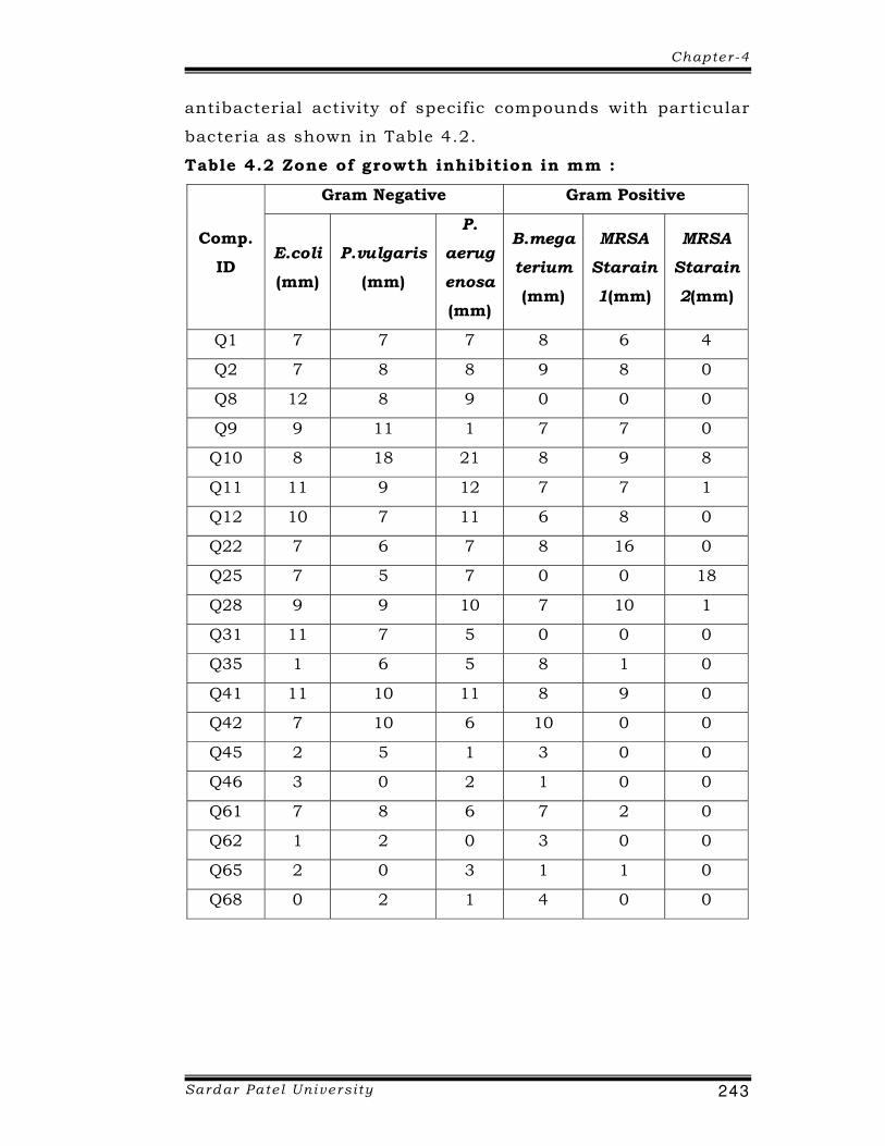
antibacterial activity of specific compounds with particular
bacteria as shown in Table 4.2.
Table 4.2 Zone of growth inhibition in mm :
Comp.
ID
Gram Negative Gram Positive
E.coli
(mm)
P.vulgaris
(mm)
P.
aerug
enosa
(mm)
B.mega
terium
(mm)
MRSA
Starain
1(mm)
MRSA
Starain
2(mm)
Q1 7 7 7 8 6 4
Q2 7 8 8 9 8 0
Q8 12 8 9 0 0 0
Q9 9 11 1 7 7 0
Q10 8 18 21 8 9 8
Q11 11 9 12 7 7 1
Q12 10 7 11 6 8 0
Q22 7 6 7 8 16 0
Q25 7 5 7 0 0 18
Q28 9 9 10 7 10 1
Q31 11 7 5 0 0 0
Q35 1 6 5 8 1 0
Q41 11 10 11 8 9 0
Q42 7 10 6 10 0 0
Q45 2 5 1 3 0 0
Q46 3 0 2 1 0 0
Q61 7 8 6 7 2 0
Q62 1 2 0 3 0 0
Q65 2 0 3 1 1 0
Q68 0 2 1 4 0 0

Chapter -4
Sardar Pate l Univers i ty 244
4.10.2 Comparative analysis :
The normal strain of E. coli was also similarly inhibited
by all the compounds included in study within the range of
0 to 12 mm. Compound Q8 was found to be best inhibitor
with zone diameter of 12 mm followed by compound Q11,
Q31 and Q41. The least inhibition was found for Q62 and
Q68 compounds as shown in Figure 4.1.
Figure 4.1 Zone of inhibition of E. Coli.
Antimicrobial inhibition study with P. Proteus
demonstrated the activity in the range of 0 to 18 mm. The
compound Q10 was found to have the maximum
antimicrobial potential with zone inhibition of 18 mm,
followed by Q41, Q42 and Q11. The lowest inhibition was
reported for the compound Q46 and Q65 as shown in Figure
4.2.

Chapter -4
Sardar Pate l Univers i ty 245
Figure 4.2 Zone of inhibition of P. vugaris
The entire compounds were found to inhibit growth of
Pseudomonas aerugenosa except compound Q8 in the range
of 0 to 21 mm. Maximum inhibition with zone diameter of
21 mm was recorded of compound Q10, followed by Q11,
Q12, Q28 and Q41 while minimum was for compounds Q45,
Q62 and Q68 as shown in Figure 4.3.
Figure 4.3 Zone of inhibition of P. aerugenosa
Escherichia coli, P. Proteus and Pseudomonas aerugenosa
was good inhibited by the compound Q8, indicate that

Chapter -4
Sardar Pate l Univers i ty 246
compound Q8 was active against Grams negative bacteria
but not active against Gram positive bacteria like Bacillus
megaterium and MRSA strains.
In Bacillus megaterium, highest growth was found in
compound Q42 (Figure 4.4).
Figure 4.4 Zone of inhibition of B.megaterium
Antimicrobial activities of all the synthetic compounds
were found different against tested organisms. The above
study was also supported by Habib N. S. et al. [15].
Compounds Q1, Q2, Q9, Q11, Q12, Q22, Q28, and Q41 were
active against only one MRSA strain 1. Where as compound
Q1 and Q10 inhibited all the bacteria including MRSA stains
2. Therefore, both the compounds were active against all the
tested organisms. Similarly, compound Q25 was also active
against most of the strains of bacteria, but it was not good
inhibitor for Bacillus megaterium and one MRSA strain 1 as
shown in Figures 4.5 & 4.6.
In contrast to wide range inhibition of all the
compounds against all normal isolates the wide variation
was recorded for two MRSA strain 1 and MRSA strain 2. The
above study was also supported by Malik N. et al. [16].

Chapter -4
Sardar Pate l Univers i ty 247
Figure 4.5 Zone of inhibition of MRSA stains 1
Figure 4.6 Zone of inhibition of MRSA stains 2
Thus, the present synthetic compounds have a potency
to use as a drug against MRSA strains and compounds may
have a future prospect to develop against MRSA strains,

Chapter -4
Sardar Pate l Univers i ty 248
which are often difficult to treat but these synthetic
compounds are active against the MRSA strains.
4.11 CONCLUSION :
Based on the above study, it is to be concluded that
the antibacterial activities of the compounds were
influenced by the halogen group i.e. microbial activity was
enhanced on substitution of the bromine group, whereas
declining in the activity was observed in case of chlorine
group substitution in the present study. Moreover, bromine
groups were active against most of the bacteria as well as
MRSA strains. So it proves that, complexity of the
compound does not increase the antibacterial activity but
the activeness depends on the types of the substituted
groups and their position in the structure.
Escherichia coli was maximum inhibited by compound
Q8. Proteus vulgaris and Pseudomonas aerugenosa was
maximum inhibited by the compound Q10, and Bacillus
megaterium, highest inhibited by compound Q42.
Escherichia coli, Proteus vulgaris and Pseudomonas
aerugenosa was good inhibited by the compound Q8,
indicate that compound Q8 was active against Grams
negative bacteria.
The different pattern of inhibition with two MRSA
strains isolates from the diverge location indicates the
biochemical heterogeneity and drug resistance development
among both the isolates. The result indicates the good
potentiality of the compound Q1 and Q10 as a potent
candidate for the development of novel antimicrobial against
multidrug resistant strains of MRSA. Even currently many
reports are available regarding the sensitivity of MRSA
against quinazolinone derivatives. Therefore, the work
incorporated in the present thesis also supports this

Chapter -4
Sardar Pate l Univers i ty 249
concept of inhibition of MRSA by the quinazolinone
derivatives.
MRSA strains are not easy to inhibit them. Although
three synthesized compounds inhibited the growth of MRSA
strain, these compounds are Q25 followed by Q10 and Q1
suggest that it may have a future potency to use against the
MRSA infection as a magic bullet to treat them in case of
hospital acquired infection of MRSA. However, further study
needs to evaluate the compounds in details.

Chapter -4
Sardar Pate l Univers i ty 250
4.12 REFERENCES :
[1] Aguilar L., Gimenez M. J., Garcia C., Martin J. E.
Antimicrob. Chemother. 50, 93 (2002).
[2] Hess H. J., Cronin T. H., Scriabine A. J. Med. Chem.
11, 130 (1968).
[3] Ishikawa M., Azuma H., Eguchi Y., Sugimoto A., Ito S.,
Takashima Y., Ebisawa H., Moriguchi S., Kotoku I.,
Suzuki H. Chem. Pharm. Bull. 30, 744 (1982).
[4] Gujral M. L., Saxona P. N., Tiwari R. S. Ind. J. Med.
Res. 43, 637 (1955).
[5] Chenard B. L., Reinhold A. R., Welch W. M. Eur. Pat.
EP 0884310 A1 (1998).
[6] Alagarsamy V., Dhanabal K., Parthiban P., Murugesan
B., Rajkumar S., Beevi A. J. J. Pharm. Pharmacol. 59,
669 (2007).
[7] Pendergast W., Johnson J., Dickerson S., Ferone R.,
Hall W., Duch D., Kelly J., Wilson D. J. Med. Chem.,
36, 2279 (1993).
[8] Jantova S., Greif G., Spirkova K., Stankovsky S.,
Oravcova M. Folia. Microbiol. 45(2), 133 (2000).
[9] Robert C. “Medical Microbiology”. ELBS, Livingston,
11 th Ed., 815 (1970).
[10] Sujatha G.D. Ind. J. Expt. Biol., 13, 286 (1975).
[11] Walksman S.A. “Microbial Antagonism and Antibiotic
Substances”, 2nd Ed., 72 (1947).
[12] Perry J.J.,Stalay J. J. Microbiology Dynemics and
Diversity, Fort Worth : Saunders College Pub., 7, 165
(1997).
[13] William B., Jeremy H. M. Journal of Medicine,
320(18), 1188 (1989).
[14] Maranan M.C., Moreira B., Daum R.S. Infectious
Disease Clinics of North America, 11(4), 813 (1997).

Chapter -4
Sardar Pate l Univers i ty 251
[15] Soliman R., Habib N. S., Ashour F. A. Boll Chim Farm.
140(3), 140 (2001).
[16] Malik N., Butt T., Arfan-U. B. J Coll Physicians Surg
Pak. 19(5), 287 (2009).