chapter vi. controlled degradation of a cell-adhesive ... degradation of a cell-adhesive,...
TRANSCRIPT
VI-1
Chapter VI. Controlled degradation of a cell-adhesive, elastomeric protein
through incorporation of a fluorinated amino acid
Abstract
The design of biomaterials with controlled mechanical, cell adhesive, and
degradative properties is a common goal in tissue engineering and drug delivery systems.
Towards this goal, a series of protein-based biomaterials were synthesized in an
engineered bacterial production system. These modular proteins include domains from
fibronectin that are known to adhere endothelial cells and elastin-derived repeating units
to provide mechanical integrity. Fluorination of the protein by in vivo replacement of the
amino acid isoleucine with the noncanonical amino acid 5,5,5-trifluoroisoleucine (5TFI)
resulted in a tenfold decrease in degradation by the enzyme human leukocyte elastase
compared to non-fluorinated protein. However, even after significant fluorination, the
materials retain their ability to adhere endothelial cells in a sequence-specific manner.
Incorporation of a noncanonical amino acid, without requiring a change in the encoding
genetic sequence, represents a novel strategy to tune the rate of degradation of protein-
based biomaterials without compromising cell adhesion.
Manuscript prepared for submission by Sarah C. Heilshorn*, Marissa L. Mock*, and
David A. Tirrell.
*These authors contributed equally to this work.
VI-2
1. Introduction
Genetic engineering techniques allow the templated production of protein
polymers with precisely controlled sequence, molecular weight, and functionality using
microbial biosynthesis. Such techniques have been employed in the de novo design and
synthesis of engineered proteins with a variety of novel physical and biological
activities1-8. These materials have demonstrated potential in tissue engineering and
reconstruction and drug delivery.
Elastin-like domains are of particular interest for engineered, protein-based
biomaterials due to their high expression levels, ease of purification, biocompatibility,
and tunable mechanical properties9-15. The extensive work of Urry and coworkers on the
family of elastin-like polypentapeptides (VPGZG)x, where Z is any amino acid, has
shown that the hydrophobicity of the biopolymer can be used to tune the lower critical
solution temperature (LCST)16,17. Polymers are soluble at temperature below the LCST
but phase separate into a polymer-rich coacervate as the temperature is increased. This
LCST phase transition allows straightforward purification of elastin-like polymers after
biosynthesis using a simple thermal cycling technique.
We have employed this method to purify a set of engineered proteins designed for
use as implantable biomaterials. The protein sequences (Figure 1) are a result of a
modular design incorporating domains from fibronectin to adhere endothelial cells10,11,
which are important for a healthy vasculature, and elastin-like domains to provide
mechanical integrity12,18. Sequence CS5 contains the authentic cell-binding domain,
which adheres human umbilical vein endothelial cells (HUVEC) in a sequence-specific

VI-3
manner11. As a negative control, sequence SC5 contains a scrambled cell-binding
domain incapable of promoting sequence-specific HUVEC adhesion19.
Figure 1. Amino acid sequences of the engineered proteins. Each protein has three cassettes of a cell-
binding domain interspersed with an elastin-like domain. Protein CS5 contains the authentic CS5 binding
domain while SC5 contains a negative control, scrambled domain. Proteins CS5-F and SC5-F are identical
to CS5 and SC5 except they are synthesized in the presence of 5TFI.
Depending on the specific medical application, e.g., drug delivery or tissue
regeneration, the success of implanted biomaterials will depend on optimization of the in
vivo degradation characteristics. Often, it is desirable to combine multiple degradation
rates in one material. For example, a single system could combine rapid delivery of a
pharmaceutical along with sustained release of growth factors for cell infiltration.
Protein-based materials are degraded by a class of enzymes called proteases; however,
native elastin is resistant to many of these proteases with the notable exception of
elastase20. Human leukocyte elastase (HLE) is the predominant form of this protease that
circulates the body in the blood stream21. HLE preferentially cuts after small,
hydrophobic amino acids, and previous work in our laboratory showed that HLE prefers
to cut after isoleucine in protein CS5. We hypothesized we could alter the degradation
Proteins CS5 (I=isolecuine) and CS5-F (I=5TFI):M-MASMTGGQQMG-HHHHHHH-DDDDK-{LD-GEEIQIGHIPREDVDYHLYP-G[(VPGIG)2VPGKG(VPGIG)2]4VP}3-LE
Proteins SC5 (I=isolecuine) and SC5-F (I=5TFI):M-MASMTGGQQMG-HHHHHHH-DDDDK-{LD-GEEIQIGHIPREVDDYHLYP-G[(VPGIG)2VPGKG(VPGIG)2]4VP}3-LE
T7 tag His tag Cleavagesite
CS5 binding domain Elastin-like domain
T7 tag His tag Cleavagesite
Scrambled CS5 binding domain Elastin-like domain
VI-4
properties of this engineered protein by replacing isoleucine with a noncanonical amino
acid.
The introduction of functional groups not contained within the 20 canonical
amino acids into proteins is a valuable tool for the protein engineer, providing access to
new chemical reactivity22-26 and physical properties27-31. In many cases it is desirable to
retain the biological activity of a protein upon introduction of these novel properties,
requiring minimal disruption of the active, folded structure and making fluorinated amino
acids of special interest. While fluorine is similar in size to hydrogen, the hydrophobicity
of the CF3 group is higher than the CH3 group due to the low polarizability of fluorine32,
giving fluorination of proteins the potential to dramatically change physical properties of
the protein without impairing its biological function31,33. The ability to tune the rate of
degradation of a biomaterial by varying the extent of incorporation of a noncanonical
amino acid, without requiring a change in the encoding genetic sequence, would provide
powerful control in target applications ranging from drug delivery to tissue engineering.
2. Results and discussion
2.1. Protein synthesis and characterization
Using an engineered bacterial strain, the genetic message encoding the CS5
protein can be alternatively read to produce protein CS5-F, a fluorinated version of CS5.
The high isoleucine content of the CS5 protein permits extensive fluorination through
incorporation of the noncanonical amino acid 5TFI, which has demonstrated levels of
isoleucine replacement from 85 – 93% in bacterial systems33. 5TFI was prepared as
previously reported33,34 and used to express proteins CS5-F and SC5-F with yields of

VI-5
0.83 mg/g and 0.71 mg/g wet cell mass, respectively. Nickel column purification yielded
5.4 mg pure CS5-F and 3.6 mg pure SC5-F from 1 L shake-flask fermentations. In
contrast, proteins CS5 and SC5 express well and are easily purified using the thermal
cycling technique to provide multi-gram quantities12,19. Typical yields are 2-5 g pure
protein from 10 L batch fermentations. Further optimization is required to synthesize the
fluorinated proteins above milligram yields.
To confirm the replacement of isoleucine with 5TFI, the engineered proteins were
digested with the protease trypsin to yield protein fragments of predicted sequence which
were then analyzed by MALDI-TOF mass spectroscopy (Figure 2). The peak at
approximately 2576 Da has been assigned to two proteolytic fragments consisting of
residues 136-161 and 262-286. These identical fragments contain five potential
isoleucine replacement sites. Accordingly, the higher mass peaks are assigned to
fragments with incorporation of one, two, three, four, and five 5TFI residues, each with a
shift of 53.88 Da corresponding to the mass difference between isoleucine and 5TFI.
Subsequent amino acid analysis reported 5TFI replacement of 82% of isoleucine residues
in CS5-F and 92% in SC5-F. This is consistent with results from previous studies
demonstrating high incorporation efficiency of 5TFI into recombinant proteins33.

VI-6
Figure 2. MALDI-TOF of tryptic digest fragments of a) CS5-F and b) SC5-F. The expected mass of the
unsubstituted peptide fragments comprising residues 136-161 and 261-286 = 2576. Peaks are apparent at
masses expected for replacement of 1 through 5 isoleucines with 5TFI.
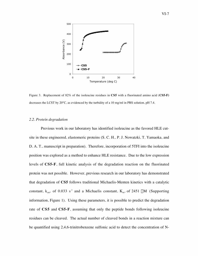
We were interested in the affect of fluorination on the thermodynamic phase
behavior of these proteins, as they are commonly purified through thermal cycling9,35. As
discussed above, proteins with elastin-like domains are known to exhibit an inverse
temperature transition that is affected by the identity of the amino acid in the Z
position16,17,30,36. Relative to the most common pentapeptide repeat in bovine and porcine
elastin, VPGVG37,38, the LCST is lowered when the amino acid occupying the Z position
is more hydrophobic than valine and raised when Z is more hydrophilic. As expected,
introducing the highly hydrophobic amino acid 5TFI into the Z position of the elastin-like
domain results in a decrease of the LCST by more than 20°C (Figure 3). Therefore, the
LCST of elastin-like proteins can be tuned by incorporation of fluorophilic 5TFI side
chains, which may aid in optimization of thermal cycling purification techniques.
2550 2650 2750 2850
+ 1 5TFI
+ 2 5TFI
+ 3 5TFI
+ 4 5TFI
Residues 136-161, 261-286GVPGIGVPGIGVPLDGEEIQIGHIPR
2576.12
a.+ 5 5TFI
2550 2650 2750 2850
Residues 136-161, 261-286GVPGIGVPGIGVPLDGEEIQIGHIPRb.
2576.80
+ 1 5TFI
+ 2 5TFI
+ 3 5TFI
+ 4 5TFI
+ 5 5TFI
VI-7
Figure 3. Replacement of 82% of the isoleucine residues in CS5 with a fluorinated amino acid (CS5-F)
decreases the LCST by 20°C, as evidenced by the turbidity of a 10 mg/ml in PBS solution, pH 7.4.
2.2. Protein degradation
Previous work in our laboratory has identified isoleucine as the favored HLE cut-
site in these engineered, elastomeric proteins (S. C. H., P. J. Nowatzki, T. Yamaoka, and
D. A. T., manuscript in preparation). Therefore, incorporation of 5TFI into the isoleucine
position was explored as a method to enhance HLE resistance. Due to the low expression
levels of CS5-F, full kinetic analysis of the degradation reaction on the fluorinated
protein was not possible. However, previous research in our laboratory has demonstrated
that degradation of CS5 follows traditional Michaelis-Menten kinetics with a catalytic
constant, kcat, of 0.033 s-1 and a Michaelis constant, Km, of 2451 mM (Supporting
information, Figure 1). Using these parameters, it is possible to predict the degradation
rate of CS5 and CS5-F, assuming that only the peptide bonds following isoleucine
residues can be cleaved. The actual number of cleaved bonds in a reaction mixture can
be quantified using 2,4,6-trinitrobenzene sulfonic acid to detect the concentration of N-
0
100
200
300
400
500
0 10 20 30 40
Temperature (deg C)
Abso
rban
ce (
V)
CS5CS5-F

VI-8
termini. After 4 h of HLE degradation, 90% fewer peptide cleavages were observed on
the fluorinated protein compared to the non-fluorinated protein (Figure 4). These
experimental results were in good agreement with the values predicted using the
Michaelis-Menten model, which supports the assumption that HLE can only cleave
peptide bonds following isoleucine in the fluorinated elastomer.
Figure 4. HLE degradation of CS5-F produces 90% fewer new N-termini per original molecule than CS5
after 4 h of reaction. Observed data represent two independent experiments, both testing three replicates of
each substrate; error bars represent one standard deviation. Predicted data are based on the assumption that
the reactions follow Michaelis-Menten kinetics and only the peptide bonds following isoleucine residues
can be cleaved.
To examine the time course of degradation of CS5 and CS5-F, Western analysis
was used to determine the amount of full-length, intact protein remaining after HLE
exposure for various times, and densitometry was employed to quantify the percent of
full-length protein remaining at each time point (Figure 5). Incorporation of 5TFI into
the elastin-like protein significantly inhibited elastase activity. At 6 h, 46% of CS5-F
0.0
0.2
0.4
0.6
0.8
1.0
1.2
1.4
1.6
CS5 CS5-F
Num
ber
of new
N-t
erm
ini per
mole
cule
Predicted
Observed

VI-9
remained intact compared to 0% of CS5. Furthermore, full-length CS5-F was still
detectable after 24 h exposure to HLE. Similar to the analysis performed above, the
degradation reaction rates can be predicted using the Michaelis-Menten parameters for
peptide cleavage after isoleucine residues. Using the simple assumption that each peptide
cleavage results in the loss of one full-length protein chain, the predicted degradation
rates for CS5 and CS5-F are in good agreement with the observed values.
Figure 5. HLE degradation of protein was a) monitored by Western blot and b) quantified using
densitometry analysis of CS5 (°) and protein CS5-F (s). No full-length CS5 was observed after 6 h,
while nearly half of CS5-F is still intact. The rate of chain degradation was predicted for CS5 (dashed line)
and CS5-F (solid line) using Michaelis-Menten parameters for the cleavage of peptide bonds after
isoleucine residues.
0
20
40
60
80
100
120
0 10 20 30 40 50
Time (hours)
Inta
ct P
rote
in R
emai
nin
g (
mM
)
Time (h): 0 1 3 6 12 24 48
CS5
CS5-F
a.
b.

VI-10
2.3. Endothelial cell adhesion
We also investigated the ability of the fluorinated proteins CS5-F and SC5-F to
promote cell adhesion. CS5 was previously reported to be adherent to human umbilical
vein endothelial cells (HUVEC) in a sequence-specific manner11. Such adhesion is
mediated through interactions with the REDV minimal binding sequence within the CS5
cell-binding domain. We wished to confirm that sequence-specific HUVEC adhesion
would not be compromised by significant fluorination of the elastin-like regions. The
ability of these engineered proteins to adhere HUVEC was examined using a buoyant
centrifugation assay (Figure 6). At a detachment force of 24 pN, protein CS5-F exhibited
HUVEC adhesion greater than that of protein CS5 and negative control proteins SC5-F
and SC5, which contain scrambled cell-binding domains. Adhesion to fibronectin, which
contains multiple cell-binding domains, was included as a positive control.
Figure 6. Percent of HUVEC remaining adherent to adsorbed fibronectin and engineered proteins after 10
min exposure to 24 pN detachment force (100g) relative to HUVEC remaining on adsorbed fibronectin
after 10 min exposure to 0.24 pN (1g). Data represent three independent experiments in which six
replicates of each substrate were tested; error bars represent one standard deviation.
0.0
20.0
40.0
60.0
80.0
100.0
120.0
Fibronectin CS5 SC5 CS5-F SC5-F
Cel
l Adhes
ion I
ndex
(%
)

VI-11
These results suggest that 5TFI incorporation into these artificial proteins does not
inhibit sequence-specific HUVEC binding. Therefore, fluorination of this engineered
protein can successfully alter the thermodynamic behavior and proteolytic susceptibility
without impairing the desired biological activity. While the observed increase in
HUVEC adhesion to the fluorinated protein CS5-F, relative to the non-fluorinated protein
CS5, is interesting, these results require further investigation. Ongoing research in our
laboratory has shown similar context-dependence of adhesion strength to engineered
proteins containing the CS5 domain.
We have demonstrated the ability to control the rate of proteolysis of elastin-like
biomaterials through the incorporation of a noncanonical amino acid. From a single
genetic message, two protein-based materials with varying degree of fluorination were
created. Fluorination of the engineered elastomer retards elastase degradation of the
protein while also altering the thermodynamic phase behavior. The ability of the material
to adhere endothelial cells through the sequence-specific interaction with the CS5 cell-
binding domain is unaffected. Residue-specific incorporation of noncanonical amino
acids into proteins is an additional tool for the biomedical engineer in the attempt to
precisely control the material properties and biological activity of protein-based
biomaterials.
VI-12
3. Methods
3.1. Protein expression and purification
Proteins CS5 and SC5 were expressed as previously described11. To express
proteins CS5-F and SC5-F, a competent isoleucine auxotrophic derivative of E. coli
strain BL21(DE3), designated AI (E. coli B F- ompT hsdS(rB- mB
-)gal dcm l(DE3)
ilvD691), constructed in our laboratory33 and harboring the plasmid pLysS (Qiagen), was
transformed with, respectively, the plasmids pET28-CS5 and pET28-SC512 to yield
strains AI-pET28-CS5 and AI-pET28-SC5. To express proteins from these strains, a
culture was grown overnight in 2xYT medium and used to inoculate 1 L of M9AA
medium supplemented with the antibiotics chloramphenicol and kanamycin. At an OD600
of 0.8-1.0, the M9AA cultures were induced by adding 1 mM IPTG. After 20 additional
minutes of growth, the cells were washed twice with 0.9% NaCl and resuspended in M9
medium containing 19 amino acids (excluding isoleucine) to a final volume of 1 L. The
cultures were supplemented with 100 mg/L of L-5,5,5-trifluoroisoleucine (5TFI) and
grown for 2 h. Fluorinated proteins were purified by Ni-affinity chromatography using
Qiagen Ni-NTA agarose resin. Purity was assessed by SDS-PAGE and Western blotting
with anti-T7 tag-horseradish peroxidase conjugate antibody (Amersham). Level of 5TFI
incorporation was assessed by amino acid analysis at the University of California, Davis
Molecular Structure Facility (Beckman 6300 amino acid analyzer).
3.2. Tryptic digest/MALDI
Purified proteins CS5-F and SC5-F were incubated with trypsin (50 mM
ammonium bicarbonate buffer, overnight, room temperature). The proteolysis products
were purified by eluting from a C18 ZipTip (Millipore) with 75:25 acetonitrile:0.1%
VI-13
trifluoroacetic acid, spotted on an analysis plate at 4°C, and analyzed by MALDI-TOF
mass spectrometry on an Applied Biosystems Voyager DE Pro instrument.
3.3. LCST measurement
The LCST of proteins CS5 and CS5-F was measured at 10 mg/ml in phosphate
buffered saline (PBS), pH 7.4, by increasing the temperature at a rate of 30°C/h and
measuring the percent transmission (measured in volts) at 300 nm on an Aviv model
62DS spectrophotometer (Lakewood, NJ).
3.4. Analysis of elastase degradation
For quantification of full-length chains, the degradation reaction was carried out
at 37°C for 3 days in sodium borate buffer, pH 8, with 0.22 mM human leukocyte elastase
(HLE, Elastin Products Company, Owensville, MO) and 100 mM protein. Samples were
taken at 0, 1, 3, 6, 12, 24, 48, and 72 h and diluted with an equal amount of 2X SDS-
sample buffer including b-mercaptoethanol and frozen at -20°C. Samples were boiled for
5 min, run on a 12% Tris-tricine gel at 150 V for 1 h, and transferred to nitrocellulose for
Western blot analysis using an anti-T7 tag-horseradish peroxidase conjugate antibody
(Amersham). Densitometry was performed on Western blots using Image J (NIH
freeware image analysis program) to quantify the amount of whole-length protein
remaining at each time point.
For quantification of the number of cleaved peptide bonds, the degradation
reaction was carried out at 37°C for 6 h in sodium borate buffer, pH 8, with 0.22 mM
HLE and 50 mM protein under constant mixing. The extent of reaction was characterized
using 2,4,6-trinitrobenzene sulfonic acid at 4°C to quantify the number of N-termini in
solution at 4 h.

VI-14
3.5. Cell adhesion
Human umbilical vein endothelial cells (HUVEC, Bio Whittaker) were
maintained in a 37°C, 5% CO2 humidified environmental chamber. The cells were grown
in Endothelial Growth Medium-2 (5% serum, Bio Whittaker), which was replaced every
two days. Near confluent HUVEC cultures were passaged non-enzymatically by
treatment with 0.61 mM EDTA (Gibco). Passages 2-5 were used.
Solutions of engineered proteins (1 mg/ml in PBS) and fibronectin (10 mg/ml in
PBS) were adsorbed onto tissue culture polystyrene overnight at 4°C. Surfaces were
rinsed with PBS, blocked with 0.2% heat-inactivated bovine serum albumin (BSA
fraction V, Sigma) for 30 minutes, and rinsed.
HUVEC in suspension were labeled with a 5 m M solution of calcein
acetoxymethyl ester (Molecular Probes) in serum-free Endothelial Basal Medium (EBM,
Cell Applications, San Diego, CA) at room temperature for 30 min. Cells were rinsed
twice and resuspended in EBM at 2.67x10⁵ cell/ml. Cells (150 ml/well) were added to
adsorbed protein substrates in 96-well plates and incubated for 30 min. A solution of
21% w/w PercollTM (Sigma) and PBS was added (200 ml/well) and plates were
centrifuged at 100g for 10 min. Non-adherent cells were removed using harvesting
frames (Molecular Devices) with the filters removed. PBS (100 ml/well) was added and
fluorescence was measured using a Perkin Elmer HTS 7000 Bio Assay Reader at an
excitation of 485 nm and emission of 538 nm. A cell adhesion index was calculated as
the fluorescence reading of a test well divided by the fluorescence reading of HUVEC
attached to fibronectin subjected to 1 g. The detachment force applied was estimated to
be 26 pN using Archimedes’ theorem19.

VI-15
4. Supporting information
Supporting Information Figure 1. Kinetic analysis of HLE degradation of CS5. Error bars represent a 90%
confidence interval. The dashed line represents a best fit of the observed data to the Michaelis-Menten
kinetic model.
5. References
(1) MacPherson, D. T.; Xu, J.; Urry, D. W. Prot. Express. Purif. 1996, 7, 51-57.
(2) Petka, W. A.; Harden, J. L.; McGrath, K. P.; Wirtz, D.; Tirrell, D. A. Science
1998, 281, 389-392.
(3) Krejchi, M. T.; Atkins, E. D. T.; Waddon, A. J.; Fournier, M. J.; Mason, T. L.;
Tirrell, D. A. Science 1994, 265, 1427-1432.
(4) Szela, S.; Avtges, P.; Valluzzi, R.; Winkler, S.; Wilson, D.; Kirschner, D.;
Kaplan, D. L. Biomacromolecules 2000, 1, 534-542.
(5) O'Brien, J. P.; Fahnestock, S. R.; Termonia, Y.; Gardner, K. H. Adv. Mater.
1998, 10, 1185-1195.
(6) Kaplan, D. Nat. Biotechnol. 2002, 20, 239-240.
0.000
0.001
0.002
0.003
0.004
0.005
0.006
0.007
0 20 40 60 80 100 120
[Protein] (mM)
Rea
ctio
n V
eloci
ty (
mM
/sec
)
VI-16
(7) Meyer, D.; Chilkoti, A. Nat. Biotechnol. 1999, 17, 1112-1115.
(8) Meyer, D.; Chilkoti, A. Biomacromolecules 2002, 3, 357-367.
(9) Welsh, E. R.; Tirrell, D. A. Biomacromolecules 2000, 1, 23-30.
(10) Panitch, A.; Yamaoka, T.; Fournier, M. J.; Mason, T. L.; Tirrell, D. A.
Macromolecules 1999, 32, 1701-1703.
(11) Heilshorn, S. C.; Di Zio, K. A.; Welsh, E. R.; Tirrell, D. A. Biomaterials 2003,
24, 4245-4252.
(12) Di Zio, K.; Tirrell, D. A. Macromolecules 2003, 36, 1553-1558.
(13) Hong, M.; Isailovic, D.; McMillan, R. A.; Conticello, V. P. Biopolymers 2003,
70, 158-168.
(14) Nagarsekar, A.; Crissman, J.; Crissman, M.; Ferrari, F.; Cappello, J.;
Ghandehari, H. Biomacromolecules 2003, 4, 602-607.
(15) Betre, H.; Setton, L. A.; Meyer, D. E.; Chilkoti, A. Biomacromolecules 2002, 3,
910-916.
(16) Urry, D. W.; Gowda, C.; Parker, T. M.; Luan, C.-H.; Reid, M. C.; Harris, C.
M.; Pattanaik, A.; Harris, R. D. Biopolymers 1992, 32, 1243-1250.
(17) Urry, D. W.; Luan, C.-H.; Harris, C. M.; Parker, T. M. In Protein-Based
Materials; McGrath; Kaplan, D., Eds.; Birkahuser: Boston, 1997.
(18) Nowatzki, P. J.; Tirrell, D. A. Biomaterials 2004, 25, 1261-1267.
(19) Liu, J. C.; Heilshorn, S. C.; Tirrell, D. A. Biomacromolecules 2004, 5, 497-504.
(20) Robert, L.; Robert, A. M.; Jacotot, B. Atherosclerosis 1998, 140, 281-295.
(21) Oudijk, E. J. D.; Nieuwenhuis, H. K.; Bos, R.; Fijnheer, R. Thromb. Haemost.
2000, 83, 906-908.
VI-17
(22) Zhang, Z. W.; Wang, L.; Brock, A.; Schultz, P. G. Angew. Chem.-Int. Edit.
2002, 41, 2840-2842.
(23) Liu, H. T.; Wang, L.; Brock, A.; Wong, C. H.; Schultz, P. G. J. Am. Chem. Soc.
2003, 125, 1702-1703.
(24) Link, A. J.; Tirrell, D. A. J. Am. Chem. Soc. 2003, 125, 11164-11165.
(25) Kiick, K. L.; Saxon, E.; Tirrell, D. A.; Bertozzi, C. R. Proc. Natl. Acad. Sci.
2002, 99, 19-24.
(26) Chin, J. W.; Schultz, P. G. ChemBioChem 2002, 3, 1135-1137.
(27) Bae, J. H.; Rubini, M.; Jung, G.; Wiegand, G.; Seifert, M. H. J.; Azim, M. K.;
Kim, J. S.; Zumbusch, A.; Holak, T. A.; Moroder, L.; Huber, R.; Budisa, N. J.
Mol. Biol. 2003, 328, 1071-1081.
(28) Bilgicer, B.; Fichera, A.; Kumar, K. J. Am. Chem. Soc. 2001, 123, 4393-4399.
(29) Budisa, N.; Rubini, M.; Bae, J. H.; Weyher, E.; Wenger, W.; Golbik, R.;
Huber, R.; Moroder, L. Angew. Chem.-Int. Edit. 2002, 41, 4066-4069.
(30) Tamura, T.; Yamaoka, T.; Kunugi, S.; Panitch, A.; Tirrell, D. A.
Biomacromolecules 2000, 1, 552-555.
(31) Tang, Y.; Ghirlanda, G.; Vaidehi, N.; Kua, J.; Mainz, D. T.; Goddard, W. A.;
DeGrado, W. F.; Tirrell, D. A. Biochemistry 2001, 40, 2790-2796.
(32) Bilgicer, B.; Kumar, K. Tetrahedron 2002, 58, 4105-4112.
(33) Wang, P.; Tang, Y.; Tirrell, D. A. J. Am. Chem. Soc. 2003, 125, 6900-6906.
(34) Muller, N. J. Fluor. Chem. 1987, 36, 163-170.
(35) Meyer, D. E.; Chilkoti, A. Nat. Biotechnol. 1999, 17, 1112-1115.
(36) Urry, D. W. Angew. Chem.-Int. Edit. 1993, 32, 819-841.
VI-18
(37) Yeh, H.; Ornstein-Goldstein, N.; Indik, Z.; Sheppard, P.; Anderson, N.;
Rosenbloom, J. C.; Cicila, G.; Yoon, K. G.; Rosenbloom, J. Coll. Relat. Res.
1987, 7, 235-247.
(38) Sandberg, L. B.; Leslie, J. G.; Leach, C. T.; Alvarez, V. L.; Torres, A. R.;
Smith, D. W. Pathol. Biol. 1985, 33, 266-274.