chapter v: diabetic foot - angiologia para clínicos€¦ · chapter v: diabetic foot s61 and...
TRANSCRIPT

European Journal of Vascular and Endovascular Surgery (2011) 42(S2), S60–S74
Chapter V: Diabetic Foot
M. Lepantaloa,b,*, J. Apelqvistc,d, C. Setaccie, J.-B. Riccof, G. de Donatoe,F. Beckerg, H. Robert-Ebadig, P. Caoh, H.H. Ecksteini, P. De Rangok,N. Diehml, J. Schmidlim, M. Teraan,o, F.L. Molln, F. Dickm, A.H. Daviesp
a Department of Vascular Surgery, Helsinki University Central Hospital, Helsinki, Finlandb Institute of Clinical Medicine, Faculty of Medicine, University of Helsinki, Helsinki, Finlandc The Diabetic Foot Center at the Department of Endocrinology, Skane University Hospital, Malmo, Swedend Division for Clinical Sciences, University of Lund, Lund, Swedene Department of Surgery, Unit of Vascular and Endovascular Surgery, University of Siena, Siena, Italyf Department of Vascular Surgery, University Hospital of Poitiers, Poitiers, Franceg Division of Angiology and Hemostasis, Geneva University Hospitals, Geneva, Switzerlandh Unit of Vascular Surgery, Department of Cardiosciences, Hospital S. Camillo-Forlanini, Rome, Italyi Clinic for Vascular Surgery, Klinikum rechts der Isar, Technische Universitat Munchen, Munich, Germanyk Unit of Vascular and Endovascular Surgery, Hospital S. M. Misericordia, Loc. S. Andrea delle Fratte, Perugia, Italyl Clinical and Interventional Angiology, Swiss Cardiovascular Centre, University Hospital Berne, Berne, Switzerlandm Department of Cardiovascular Surgery, Swiss Cardiovascular Centre, University Hospital Berne, Berne, Switzerlandn Department of Vascular Surgery, University Medical Center Utrecht, Utrecht,The Netherlandso Department of Nephrology & Hypertension, University Medical Center Utrecht, Utrecht, The Netherlandsp Academic Section of Vascular Surgery, Imperial College School of Medicine, Charing Cross Hospital, London, United
Kingdom
KEYWORDSDiabetic foot;Ischaemia;Neuroischaemia;Vascular impairment;Ulcer healing;Revascularisation
Abstract Ulcerated diabetic foot is a complex problem. Ischaemia, neuropathy andinfection are the three pathological components that lead to diabetic foot complications,and they frequently occur together as an aetiologic triad. Neuropathy and ischaemia arethe initiating factors, most often together as neuroischaemia, whereas infection is mostlya consequence. The role of peripheral arterial disease in diabetic foot has long beenunderestimated as typical ischaemic symptoms are less frequent in diabetics with ischaemiathan in non-diabetics. Furthermore, the healing of a neuroischaemic ulcer is hamperedby microvascular dysfunction. Therefore, the threshold for revascularising neuroischaemiculcers should be lower than that for purely ischaemic ulcers. Previous guidelines havelargely ignored these specific demands related to ulcerated neuroischaemic diabetic feet.Any diabetic foot ulcer should always be considered to have vascular impairment unlessotherwise proven. Early referral, non-invasive vascular testing, imaging and interventionare crucial to improve diabetic foot ulcer healing and to prevent amputation. Timing isessential, as the window of opportunity to heal the ulcer and save the leg is easily missed.
This chapter underlines the paucity of data on the best way to diagnose and treat thesediabetic patients. Most of the studies dealing with neuroischaemic diabetic feet are notcomparable in terms of patient populations, interventions or outcome. Therefore, thereis an urgent need for a paradigm shift in diabetic foot care; that is, a new approach
* Corresponding author. Mauri Lepantalo, Department of Vascular Surgery, Helsinki University Central Hospital, P.O. Box 440, 00029 HUS,Finland. Mobile: +358 50 4271282. E-mail address: [email protected] (M. Lepantalo)
1078-5884/$36 © 2011 European Society for Vascular Surgery. Published by Elsevier Ltd. All rights reserved.

Chapter V: Diabetic Foot S61
and classification of diabetics with vascular impairment in regard to clinical practice andresearch. A multidisciplinary approach needs to implemented systematically with a vascularsurgeon as an integrated member. New strategies must be developed and implementedfor diabetic foot patients with vascular impairment, to improve healing, to speed uphealing rate and to avoid amputation, irrespective of the intervention technology chosen.Focused studies on the value of predictive tests, new treatment modalities as well asselective and targeted strategies are needed. As specific data on ulcerated neuroischaemicdiabetic feet are scarce, recommendations are often of low grade.© 2011 European Society for Vascular Surgery. Published by Elsevier Ltd. All rights reserved.
1. Introduction
Diabetic foot ulcers are a major healthcare problem.In 2011, 350 million people worldwide (6.6% of thepopulation) and more than 55 million in Europe suffer fromdiabetes mellitus,1 and estimates for 2025 cite a totalof over 65 million patients.1 Complications of foot ulcersare the leading cause of hospitalisation and amputationin diabetic patients. Indeed, 20––40% of the healthcareresources spent on diabetes are related to diabetic feet.2,3
Individuals suffering from diabetes and neuropathy withno other confounders will develop an ulcer in 7––10% of thecases annually, whereas the rate for patients with additionalrisk factors –– such as peripheral arterial disease (PAD),foot deformity, previous ulcers or previous amputation –– is25––30%.2––4
Major amputation will be needed within 1 year in 5––8%of patients with diabetic ulcers.5––7 Of all amputationson diabetic patients, 85% are preceded by a foot ulcerwhich subsequently deteriorates to a severe infection organgrene.2––4 Diabetes increases the risk of amputation8-fold in patients aged >45 years,8 12-fold in patients aged>65 years and 23-fold in those aged 65––74 years.9
2. NeuropathyIschaemia, neuropathy and infection are the three patho-logical components that lead to diabetic foot complications,and they frequently occur together as an aetiologic triad.10
Neuropathy and ischaemia are the initiating factors, with adifferent weight in different patients (Fig. 1), and infectionis mostly a consequence.11
Fig. 1. Pathway to diabetic ulcer. Modified from the International Working Group on the Diabetic Foot, International Consensus on theDiabetic Foot, 1999, with permission.

S62 M. Lepantalo et al.
Due to the lack of protective sensation, the footis vulnerable to unattended minor injuries caused byexcess pressure, mechanical or thermal injury. Motorneuropathy alters the biomechanics and, gradually, thefoot anatomy. Foot deformities, limited joint mobility andaltered loading of the foot are obvious consequences fromthese disarrangements. The most important feature of thetreatment of any ulcer with neuropathy is to restrict weightbearing, irrespective of the presence of ischaemia.
The treatment of purely neuropathic ulcers is beyondthe scope of these guidelines, and neuropathy is furtherdealt with only in conjunction to ischaemia –– i.e. asneuroischaemic ulcers. For the purposes of this chapter, theterm diabetic foot refers to an ulcerated diabetic foot withvascular impairment.
3. Ischaemia and neuroischaemia of thediabetic foot
3.1. Underestimation of the role of ischaemia
Poor glucose control accelerates the manifestation of PAD.For every 1% increase in haemoglobin A1c (HbA1c), thereis a corresponding increase of 25––28% in the relative riskof PAD.12 Diabetes increases the prevalence of symptomaticPAD 3.5-fold in men and 8.6-fold in women.13 Recent largeEuropean cohort studies of individuals with diabetes andfoot ulcers confirm that at least half are of neuroischaemicor ischaemic origin.14––16 Yet the strategy of preventionand treatment of the diabetic foot has predominantlybeen focused on neuropathy and its consequences,2,17
although ischaemia is the most important factor preventinghealing.11 PAD in diabetics is often multisegmental, typicallyinfrapopliteal and poorly collateralised.18––20 Ischaemia hasbeen reported to be at least a contributing factor in 90% indiabetics undergoing major amputation.21
RecommendationIschaemia should not be excluded as a cause of a diabeticfoot ulcer unless proven absent.22,23 (Level 5; Grade D)
3.2. Inadequate understanding of neuroischaemia
Neuroischaemia is the combined effect of diabetic neuro-pathy and ischaemia, impairing the oxygen delivery to meetmetabolic tissue demands in a synergetic way. Macrovasculardisease and microvascular dysfunction both impair perfusionin a diabetic foot.24 Peripheral autonomic neuropathy, orauto-sympathectomy, causes deficient sweating and alteredblood flow regulation with an opening of arteriovenousshunts and precapillary sphincter malfunction, whichdecreases nutritive blood flow and manifests as warm, dryskin, increasing the likelihood of skin breakdown.25
The microvascular dysfunction is further characterised bythe subsequent capillary leakage and venous pooling as wellas hormonal activity in the vessel and inflammation in thewall, all indicating that decreased perfusion in the diabeticfoot is more complex and not only related to PAD.26––28 YetPAD is the most important cause of vascular impairment ofdiabetic foot.24
RecommendationIn neuroischaemic legs, healing is primarily affected bythe severity of ischaemia.11 Therefore, from a practicalpoint of view, neuroischaemic and ischaemic lesionsshould be considered together as both may requirerevascularisation. (Level 2b; Grade C)
3.3. Assessment of vascular impairment beyond ischaemia
The use of rigid non-invasive methods is mostly based onthe haemodynamic changes in the macrovascular arterialtree, and criteria applicable to non-diabetic legs are notgood enough to predict the healing of diabetic foot lesions.23
There is a clear need to recognise decreased perfusionor vascular impairment as an indicator for the need forrevascularisation in the diabetic foot in order to achieveand maintain healing and to avoid or delay a futureamputation.4,6,16,23,29––31
RecommendationThe International Working Group for the Diabetic Footrecommends further vascular studies in case the ulcerhas not healed with proper treatment in 6 weeks evenif initial diagnostics have suggested only questionable ormild disease.22 (Level 5; Grade D)
Critical issueCriteria for impaired perfusion should be established.
3.4. Delay in revascularisation
As less than 25% of diabetics with PAD report intermittentclaudication, and rest pain is far less common than in non-diabetics, the diagnosis of ischaemia is often delayed.2 Theobvious consequence has been that a vascular consultationis arranged too late for diabetics. Indeed, 30––50% of theirfoot ulcers are already gangrenous, and, therefore, vascularsurgeons are too often not consulted at all.2,4
RecommendationTo prevent a delay in vascular consultation and revascular-isation, early non-invasive vascular evaluation is importantin identifying patients with poor ulcer healing and a highrisk for amputation.2,4,6,17,29––31 (Level 2b; Grade B)
3.5. Ischaemia, infection and tissue damage
Neuroischaemic ulcers are susceptible to infection. Infectionis seldom the direct cause of an ulcer but strongly relatedto the probability of amputation, especially in combinationwith ischaemia (PAD).11 Deep infections are manifestedeither as osteomyelitis or a soft tissue infection spreadingalong the tendons in the compromised foot. A deep infectionis a limb-threatening condition and the immediate causeof amputation in 25––50% of diabetic patients.2,4,32––34 Inseveral studies, the outcome of deep foot infection has beenrelated to the extent of tissue involved, comorbidity andco-existing PAD.2,4,14,16

Chapter V: Diabetic Foot S63
RecommendationThe combination of ischaemia and infection alwaysnecessitates urgent treatment, as “time is tissue”.22
(Level 2c; Grade C)
4. Clinical examination from the vascularperspective
4.1. History
4.1.1. GeneralThe primary evaluation with regard to the diabetic footshould include information on the presence of concomitantdiseases and their medications; cardiovascular risk factors;occupation and hobbies; lifestyle; smoking as well as theuse of alcohol, drugs and other intoxicants; in additionto diabetes-related complications, especially nephropathy,retinopathy and neuropathy. Special attention should alsobe paid to impaired vision, renal replacement therapy,previous foot education, social isolation and poor access tohealthcare.2
4.1.2. Foot-specific historyThe main aim of the examination of a diabetic foot is toassess the risk factors for foot ulceration and, in case therealready is an ulcer, to evaluate its specific aetiology andduration to allow targeted treatment.17,25
4.2. Inspection
A clinical examination of the foot of a diabetic patientshould be performed at least once a year and morefrequently in the presence of risk factors. The role of aregular inspection of the diabetic foot cannot be emphasisedenough.3,11,35 As Andrew Boulton has put it, “For one mistakemade for not knowing, ten mistakes are made for notlooking.” A neuropathic foot frequently has a characteristicappearance upon inspection.10
4.3. Vascular clinical examination
Pulse palpation is the cornerstone of vascular examinationalthough it is not necessarily a method of good reproducibil-ity.36 Therefore, clinically significant arterial disease canmost often be ruled out only if both dorsalis pedis andposterior tibial pulses are palpable with certainty. Yet, indiabetics even this may not suffice to exclude impairedperfusion.37 Furthermore, the arteria dorsalis pedis pulse ismissing in 8% and tibialis posterior pulse in 3% of healthyindividuals.38
An ischaemic foot may appear pink and relatively warmeven with impaired perfusion due to arteriovenous shunting.Delayed discolouration (rubor) or venous refilling >5 s ondependency may indicate poor arterial perfusion.39 Slowcapillary refilling time has little diagnostic value.39
RecommendationEvery foot ulcer should be examined for the presence ofischaemia.2 (Level 5; Grade 4)
4.4. Neurological clinical examination
Sensory loss tested by pressure perception with a 10-gram (5.07) Semmes––Weinstein monofilament is the mostimportant single test.2,17 Vibration perception using a128 Hz-tuning fork, pinprick discrimination and tactilesensation testing with cotton wool on the dorsum of thefoot, as well as testing Achilles tendon reflexes, belong tothe neurological examination in addition to looking for footdeformities or bony prominences.2
RecommendationEvery foot ulcer should be examined for the presence ofneuropathy.2 (Level 5; Grade 4)
4.5. Ulcers
The diabetic foot ulcer is not a disease of the skinbut a sign of abnormal loading and impaired perfusion.A systematic classification of foot ulcers would be helpful forthe comparison of data, but only few scoring systems havebeen validated.40 The most frequently used systems includeperfusion, the extent and size of tissue involvement as wellas infection.29,41
Critical issueThe validity of scoring systems needs to be evaluated
specifically in ischaemic diabetic ulcers.
4.6. Infection
Ulcer infections are diagnosed clinically on the basis of localsigns and symptoms of inflammation. These include purulentsecretion in the ulcer or at least two of the followingsigns or symptoms: redness, warmth, swelling, pain, delayedimprovement or bad odour. The clinical signs of infection canbe reduced due to diminished leucocyte function, PAD, poormetabolic control and neuropathy.42 Occasional systemicsigns are fever and poor general condition.34,43 In almost50% of patients with diabetes and deep foot infections,signs such as increased white blood cell count, erythrocytesedimentation rate, C-reactive protein concentration andfever have been found absent, resulting in a delay indiagnosis.32––34 Some patients with a diabetic foot infectionalso have a worsening in their glycaemic control. A swollenfoot with a long-lasting ulceration or a red swollen digitshould always arouse suspicion of an infection extendingto deep tissue. The most common sign of a diabeticfoot infection with an ulcer is increased exudation rate.32,44
Unroofing a superficial eschar may reveal deeper abscesses.10
Indeed, the severity of infection should be assessed afterdebridement, based on its extent and depth as well as thepresence of any systemic findings.22 Tissue specimens shouldbe obtained by biopsy, curettage or aspiration, preferable towound swab specimens, prior to starting empirical antibiotictherapy.45––47
A continuous extension of a soft tissue infection to theunderlying bone poses both diagnostic and therapeutic chal-lenges.32––34,48 Imaging studies may help detect pathologicalfindings in the bone.49 Plain radiographs of the foot may beof value in revealing the presence of a foreign body, gas,osteolysis or joint effusion. Radiological diagnosis is often
S64 M. Lepantalo et al.
difficult because changes suggesting osteomyelitis usuallytake several weeks to become visible on X-ray.
Fever as well as an increased erythrocyte sedimentationrate (ESR), white cell count and C-reactive proteinconcentrations (CRP) are usually helpful in recognising softtissue infections or abscess. An MRI, bone scan or CT scancan be of value in evaluating the presence and extent of adeep foot infection.
RecommendationEvery diabetic foot ulcer should be examined for thepresence of infection.2 (Level 5; Grade D)
4.7. Non-invasive vascular studies –– special considerationsrelated to the diabetic foot
In the case of any uncertainty as to foot perfusion, themeasurement of ankle pressure, the ankle-brachial systolicpressure index (ABI) and toe pressures should be included.Normal ABI values range between 0.9 and 1.3, as high valuessuggest non-compressible arteries (pseudohypertension)characteristic of advanced mediasclerosis, which is typical indiabetes. Less severe calcification may result in a normal ABIdespite clinically significant PAD.50 In a series of 554diabetics with vascular impairment, ankle pressures couldnot be measured in 35% of the patients.51 An ABI <0.4––0.45,absolute systolic ankle pressure <55 mmHg and toe pressure<30 mmHg have most frequently been used to indicate theneed for revascularisation.10,52,53 Pseudohypertension maybe revealed by pulse volume recording (PVR),54 but there arenot enough data to support the use of methods such as thepole test.55 In hand-held Doppler examination, an absent ormonophasic flow velocity signal from a foot artery indicatesocclusion or collateral flow.
RecommendationTrust ABI when low but not when high. An ABI <0.6indicates significant ischaemia in respect to wound healingpotential, whereas an ABI >0.6 has little predictive valueand, therefore, at least the toe pressure should bemeasured.22 (Level 5; Grade D)
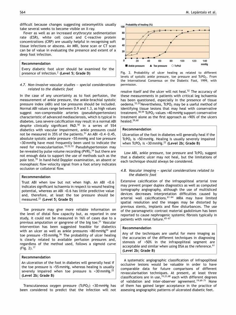
Toe pressure may give more reliable information onthe level of distal flow capacity but, as reported in onestudy, it could not be measured in 16% of cases due to aprevious amputation or gangrene of the big toe.51 Vascularintervention has been suggested feasible for diabeticswith an ulcer as well as ankle pressures <80 mmHg54 andtoe pressure <55 mmHg.56 The probability of ulcer healingis clearly related to available perfusion pressures and,regardless of the method used, follows a sigmoid curve(Fig. 2).17
RecommendationAn ulceration of the foot in diabetes will generally heal ifthe toe pressure is >55 mmHg, whereas healing is usuallyseverely impaired when toe pressure is <30 mmHg.22
(Level 2b; Grade B)
Transcutaneous oxygen pressure (TcPO2) <30 mmHg hasbeen considered to predict that the infection will not
Fig. 2. Probability of ulcer healing as related to differentlevels of systolic ankle pressure, toe pressure and TcPO2. Fromthe International Consensus on the Diabetic Foot, 1999, withpermission.
resolve itself and the ulcer will not heal.52 The accuracy ofthese measurements in patients with critical leg ischaemiahas been questioned, especially in the presence of tissueoedema.53,57Nevertheless, TcPO2 may be a useful method ofidentifying tissue lesions that may heal with conservativetreatment.58,59 TcPO2-values >40 mmHg support conservativetreatment alone as the first approach as >90% of the ulcershealed.59,60
RecommendationUlceration of the foot in diabetes will generally heal if theTcPO2 is >50 mmHg. Healing is usually severely impairedwhen TcPO2 is <30 mmHg.22 (Level 2b; Grade B)
Low ABI, ankle pressure, toe pressure and TcPO2 suggestthat a diabetic ulcer may not heal, but the limitations ofeach technique should always be considered.
4.8. Vascular imaging –– special considerations related tothe diabetic foot
Extensive calcification of the infrapopliteal arterial treemay prevent proper duplex diagnostics as well as computedtomography angiography, although the use of multisliceddevices decreases interpretation difficulties caused byarterial wall calcifications.61––64 MRA may have limitedspatial resolution and the images may be distorted byprevious stents, implants and flow disturbances. The useof the paramagnetic contrast material gadolinium has beenreported to cause nephrogenic systemic fibrosis typically inpatients with renal failure.65––67
RecommendationAny of the techniques are useful for mere imaging asthe accuracies of the different techniques in diagnosingstenosis of >50% in the infrapopliteal segment areacceptable and similar when using DSA as the reference.61
(Level 2b; Grade B)
A systematic angiographic classification of infrapoplitealocclusive lesions would be valuable in order to havecomparable data for future comparisons of differentrevascularisation techniques. At present, at least threeclassifications are in use,19,23,68 each with different degreesof validation and inter-observer agreement.19,69––71 Noneof them has gained larger acceptance in the practice ofassessing angiographic patterns of ulcerated diabetic feet.

Chapter V: Diabetic Foot S65
Chronic renal failure is increasingly common in diabeticswith a foot ulcer. Metformin treatment should be discon-tinued before angiography as it may cause lactic acidosis.72
Renal insufficiency influences the choice of imaging method,because contrast media are nephrotoxic agents. In the caseof mild chronic renal failure, regular DSA and CTA canbe performed, but intravenous hydration of the patient isrecommended before and after the examination.72,73 In moresevere cases, selective angiography with a minimal amountof contrast media, preferably diluted non-ionic iso-osmolar,can produce excellent imaging when focused on the targetlesion.73 Alternatively, duplex ultrasound can be used forimaging and sometimes also for guiding the endovascularprocedure.74
RecommendationDetailed visualisation of infrapopliteal arteries, includingthe arteries of the foot, is necessary for a completeevaluation of diabetic patients.22 (Level 5; Grade D)
Critical issueThe risks of gadolinium-enhanced MRA for imaging diabetic
patients with kidney failure should be considered and furtherevaluated.
5. Treatment of ulcerated neuroischaemicdiabetic feet
5.1. Multifactorial approach mandatory
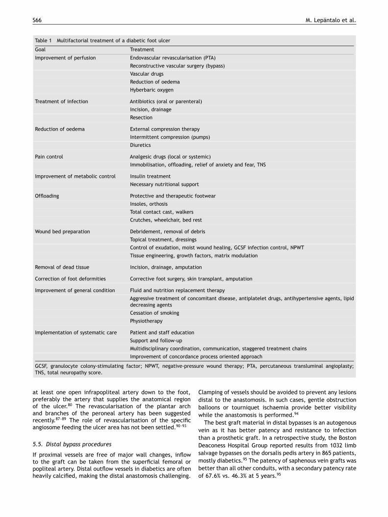
The complexity of diabetic foot ulcers necessitates in-depth knowledge of the underlying pathophysiology and amultifactorial approach in which aggressive management ofischaemia and infection is of major importance (Table 1).
RecommendationPatients in need of revascularisation to improve per-fusion and achieve healing should be identified by anextensive clinical examination and non-invasive, vasculartesting.22,23 (Level 5; Grade D)
Metabolic control also plays an important role incomprehensive treatment. Blood glucose control may bedifficult because of infection. If the patient is on oralantidiabetic drugs, a temporary switch to insulin may benecessary. On the other hand, high blood glucose worsensinfection and is associated with poorer operative results,morbidity and mortality. The recommended target level ofHbA1c should be <7.0––7.5% but higher if hypoglycaemicepisodes are a problem, and the LDL level should be<1.8 mmol/L and blood pressure <130/80 mmHg, whileless stringent goals should be accepted for elderly andmultimorbid patients.75
RecommendationIntensive management of diabetes, including glycaemicand platelet aggregation control, treatment of hyperten-sion and dyslipidaemia as well as non-pharmacologicalinterventions, decreases vascular complications in thelong run.76 (Level 1a; Grade A)
5.2. Management of infection
Antibiotic therapy is necessary for virtually all infectedwounds, but it is not beneficial for non-infected ulcersand is insufficient without appropriate wound care. Inlong-standing ulcers or ulcers with delayed healing andischaemia or necrotic tissue, polymicrobial flora with anunknown causative agent is frequently present. Broad-spectrum empirical therapy is not routinely required but isindicated for moderate to severe infections.34,77 Antibiotictherapy is continued until there is evidence that theinfection has been resolved but not necessarily until thewound has healed.49
Patients with uncontrolled or limb-threatening infectionsrequire immediate hospitalisation, immobilisation andintravenous antibiotics. Infections accompanied by a deepabscess, extensive bone or joint involvement, crepitus,substantial necrosis or gangrene, or necrotising fasciitis,need prompt surgical intervention. Infections can spreadextremely rapidly in a diabetic foot, which may lead toa life-threatening general septic infection if treatmentis delayed. Urgent evaluation of lower limb circulation,treatment of infections and surgical procedures, includingdebridement and revascularisations, are often needed asfirst-line leg salvage strategies.48,50,78
RecommendationSurgical intervention for moderate or severe infectionsis likely to decrease the risk of major amputation.22
(Level 2c; Grade B)
5.3. Infrainguinal revascularisation
The crucial issue is to decide whether revascularisationis needed for a certain lesion in a certain patient.10
Although non-invasive evaluation is helpful, the decision tointervene is made according to the symptoms and clinicalfindings.10 Anatomical imaging should be considered only asstrategic.73
If both an endovascular and a bypass procedureare possible with an equal outcome to be expected,endovascular treatments should be preferred.79 Especiallypatients with chronic neuroischaemic ulcers, borderline toepressures and short lesions are candidates for endovasculartreatment. However, continuous surveillance and a lowthreshold for secondary imaging, percutaneous transluminalangioplasty (PTA) or bypass are basic principles when treat-ing diabetic ulcers with an endovascular procedure.80 Bypassgrafting is to be used for long occlusions. Patency rates aftercrural and pedal bypasses are similar in diabetics and non-diabetics.81 (For femoropopliteal reconstructions, see Chap-ter IV, Treatment of Critical Limb Ischaemia, pp. S43––S59.)
5.4. Infrapopliteal endovascular procedures
Endovascular therapy for infrapopliteal arterial disease isgaining acceptance as a first-line revascularisation methodto improve ulcer healing and limb salvage.79 The angioplastyof isolated crural arterial lesions in diabetic patients withan unhealed ulcer is also considered an effective andsafe therapeutic modality to avoid limb loss.82 There areseveral studies showing good results and patency rates afterendovascular treatment of PAD with critical ischaemia.82––86
An important task for any revascularisation is to achieve
S66 M. Lepantalo et al.
Table 1 Multifactorial treatment of a diabetic foot ulcer
Goal Treatment
Improvement of perfusion Endovascular revascularisation (PTA)
Reconstructive vascular surgery (bypass)
Vascular drugs
Reduction of oedema
Hyberbaric oxygen
Treatment of infection Antibiotics (oral or parenteral)
Incision, drainage
Resection
Reduction of oedema External compression therapy
Intermittent compression (pumps)
Diuretics
Pain control Analgesic drugs (local or systemic)
Immobilisation, offloading, relief of anxiety and fear, TNS
Improvement of metabolic control Insulin treatment
Necessary nutritional support
Offloading Protective and therapeutic footwear
Insoles, orthosis
Total contact cast, walkers
Crutches, wheelchair, bed rest
Wound bed preparation Debridement, removal of debris
Topical treatment, dressings
Control of exudation, moist wound healing, GCSF infection control, NPWT
Tissue engineering, growth factors, matrix modulation
Removal of dead tissue Incision, drainage, amputation
Correction of foot deformities Corrective foot surgery, skin transplant, amputation
Improvement of general condition Fluid and nutrition replacement therapy
Aggressive treatment of concomitant disease, antiplatelet drugs, antihypertensive agents, lipiddecreasing agents
Cessation of smoking
Physiotherapy
Implementation of systematic care Patient and staff education
Support and follow-up
Multidisciplinary coordination, communication, staggered treatment chains
Improvement of concordance process oriented approach
GCSF, granulocyte colony-stimulating factor; NPWT, negative-pressure wound therapy; PTA, percutaneous transluminal angioplasty;TNS, total neuropathy score.
at least one open infrapopliteal artery down to the foot,preferably the artery that supplies the anatomical regionof the ulcer.80 The revascularisation of the plantar archand branches of the peroneal artery has been suggestedrecently.87––89 The role of revascularisation of the specificangiosome feeding the ulcer area has not been settled.90––93
5.5. Distal bypass procedures
If proximal vessels are free of major wall changes, inflowto the graft can be taken from the superficial femoral orpopliteal artery. Distal outflow vessels in diabetics are oftenheavily calcified, making the distal anastomosis challenging.
Clamping of vessels should be avoided to prevent any lesionsdistal to the anastomosis. In such cases, gentle obstructionballoons or tourniquet ischaemia provide better visibilitywhile the anastomosis is performed.94
The best graft material in distal bypasses is an autogenousvein as it has better patency and resistance to infectionthan a prosthetic graft. In a retrospective study, the BostonDeaconess Hospital Group reported results from 1032 limbsalvage bypasses on the dorsalis pedis artery in 865 patients,mostly diabetics.95 The patency of saphenous vein grafts wasbetter than all other conduits, with a secondary patency rateof 67.6% vs. 46.3% at 5 years.95

Chapter V: Diabetic Foot S67
5.6. Immediate outcome after revascularisation
Several conditions, such as chronic renal failure requiringlong-term dialysis, an arterial graft of poor quality orsevere foot infection may indicate problems in leg salvage.96
Diabetes along with coronary artery disease, foot gangreneand an urgent operation have been found to be independentpredictors of 30-day post-operative mortality and/or majorlower limb amputation after revascularisation for CLI.96
Systemic complications are encountered in approximately10% of patients.97
5.7. Endovascular intervention or surgical bypass
There is not a single randomised controlled trial availablecomparing endovascular and surgical revascularisation inthe treatment of impaired perfusion or critical ischaemiain diabetics.97,98 A literature search revealed only sevencase series on revascularisations exclusively for diabeticfeet, provided that all patients were diabetics, had an ulcerand were treated with an infrainguinal revascularisation(Table 2).
As the infrapopliteal region is strongly affected bydiabetic PAD, current interest is increasingly targetedon infrapopliteal revascularisations. New endovasculartechniques are rapidly evolving, despite the lack of RCTscomparing open and endovascular revascularisations belowthe knee. However, a recent meta-analysis is available forboth infrapopliteal surgery and infrapopliteal endovascularinterventions,106,107 with 29 and 30 studies included,respectively. As 88% of the patients were diabetics and88% had tissue loss in the bypass group with 2320 graftsstudied, the results may be accepted to be indicative of adiabetic population. Unfortunately, only 61% of the patientsin the endovascular group were diabetics and only 76% ofthem suffered from tissue loss, and the group thereforerather represented a mixed group. No distal pressure datawere available. Primary and secondary mid-term patencyrates were better after bypass, but there was no differencein limb salvage. The so-called patency/leg-salvage gapseemed wider in the endovascular than in the surgicalseries –– i.e. occlusion of the revascularised segment wasless likely to lead to amputation after an endovascularprocedure than surgical bypass. It is unclear whetherbypass patients had more severe ischaemia pre-operativelyor whether open surgery caused more leg morbidity. Asthe BASIL trial showed, only 29% of patients are suitablefor both treatment methods, and patient populations arethus bound to be different in endovascular and surgicalseries.108 Bypass surgery and endovascular interventions aretherefore complementary techniques for revascularisationin diabetic patients with non-healing ulcers.56 Indeed, ananalysis of infrapopliteal revascularisations in 611 diabeticswith 417 open and 194 endovascular revascularisationsshowed a comparable outcome in terms of amputation-freesurvival.79
RecommendationThe choice between different methods of revasculari-sation –– open, endovascular or hybrid –– depends oncomorbidity, severity and extension of the arterial lesionsas well as the expertise of the centre. (Level 2c;Grade B)
5.8. Microvascular flaps
Microvascular free flaps may be used to cover large tissuedefects and ulcers overtaking tendons and bones in diabeticfeet. In a recent review there were 17 case series, thelargest with 79 patients, 85% of whom were diabeticsand 66% of whom underwent a procedure combiningrevascularisation and free flap transfer.109 Revascularisationsof ischaemic diabetic feet combined with free flap transferrepresent only a small fraction –– 4% at most –– of allinterventions to improve diabetic foot perfusion.110
RecommendationWhen ischaemia coincides with a large diabetic footdefect, major amputation may be prevented in anambulatory patient by combining revascularisation withmicrovascular flap transfer.109 (Level 4; Grade C)
5.9. Timing of the treatment of infection vs.revascularisation
The most important step in controlling deep infection isurgent incision and drainage of an abscess as well asradical debridement of all infected, nonviable necrotictissue.32––34 The debridement should be done first andrevascularisation thereafter.32––34,48 Distal bypass, whenneeded, is usually delayed 2––5 days to control theinfection.94,111,112 Simultaneous revascularisation, preferablyendovascularly, in patients without systemic sepsis allowsmaximising blood flow at the initial debridement. Thosehaving a minor amputation before bypass have beenreported to fare worse than those who were revascularisedfirst.113 Common sense is essential in this setting sincepurulent lesions necessitate an amputation first whereasmummified gangrene allows revascularisation first.
In situations with no limb-threatening infection, theblood supply to the wound/extremity should be optimisedbefore surgical debridement to ensure that potentiallyviable tissue is not unnecessarily removed. This may takeweeks.
RecommendationThe severity of infection guides the decision whetherto debride, to revascularise or to use a simultaneousapproach first. (Level 2c; Grade C)
5.10. Debridement
Debridement after the damage control phase has beenstudied extensively. A large review comprising surgicaldebridement, surgical excision, the use of hyperbaricoxygen, negative-pressure wound therapy, skin grafting,bioactive local therapy products as well as electrical,magnetic, ultrasound and laser therapies114 showed noevidence to prove that one method was better than theothers with regard to the probability of healing accordingto Cochrane Database.114––116
RecommendationNo single method is outstanding in terms of enhancingdiabetic ulcer healing. (Level 1c; Grade A)

S68 M. Lepantalo et al.
Tabl
e2
Case
seri
esin
clud
ing
excl
usiv
ely
infr
aing
uina
lre
vasc
ular
isat
ions
for
isch
aem
icul
cera
ted
diab
etic
feet
Auth
orPa
tien
ts;
N/g
ende
r/ag
e(m
ean/
med
ian)
Com
orbi
dity
Inte
rven
tion
Infr
a-po
plit
eal
dist
ribu
tion
30-d
ayco
mpl
icat
ions
Follo
w-u
p(f
u)O
utco
me
Rose
nblu
m,
1994
9939
/M33
,F6
/62.
3yr
sN
AIn
frap
oplit
eal
bypa
ssgr
afts
79%
Maj
oram
puta
tion
3%,
mor
talit
yN
A21
.2m
onth
s(m
ean)
,ra
nge
2––6
4
83%
prim
ary
ulce
rhe
alin
gw
ith
graf
tpa
tenc
ydu
ring
fu
Wol
fle,
2000
100
125/
NA/
70yr
sCA
D57
%,ES
RD25
%In
frap
oplit
eal
bypa
ssgr
afts
100%
Maj
oram
puta
tion
NA,
mor
talit
y2%
24m
onth
s(m
ean)
Leg
salv
age
80%
and
pate
ncy
76%
at1
yr,
mor
talit
y51
%du
ring
fu
84/N
A/68
yrs
CAD
48%,
ESRD
42%
Infr
apop
litea
lPT
A(m
ostl
yst
enos
es)
100%
Maj
oram
puta
tion
NA,
mor
talit
y6%
24m
onth
s(m
ean)
Leg
salv
age
82%
at1
yr,
mor
talit
y35
%du
ring
fu
Schn
eide
r,20
0110
111
0/M
67,
F43/
69yr
sCA
D43
%,ES
RD69
%Re
vasc
ular
isat
ion
usin
gei
ther
fem
-dis
tal
bypa
ss,
com
bine
dSF
APT
Aan
ddi
stal
bypa
ssgr
afti
ngor
shor
tdi
stal
bypa
ssgr
aft
100%
NA
23m
onth
s(m
ean)
Leg
salv
age
89%,
pate
ncy
78%
at2
yrs,
mor
talit
yN
A
Fagl
ia,
2002
102
219/
NA/
NA
CAD
55%,
ESRD
4%Fe
mor
odis
tal
and
infr
apop
litea
lPT
A(o
fst
enos
es>5
0%)
94%
Maj
oram
puta
tion
5%,
mor
talit
y0%
12m
onth
s(m
edia
n),
rang
e5–
–30
Leg
salv
age
NA,
mor
talit
y5.
3%at
1yr
Dor
wei
ler,
2002
103
46/M
36,
F10/
69yr
sCA
D46
%,ES
RD13
%Pe
dal
bypa
ssgr
afts
100%
Maj
oram
puta
tion
7%,
mor
talit
y2%
28m
onth
s(m
edia
n),
rang
e1–
–70
Leg
salv
age
87%
at2
yrs
Barg
ellin
i,20
0810
460
/M41
,F1
9/69
.4yr
sCA
D42
%,CV
D25
%M
ulti
-lev
elsu
bint
imal
PTA
inpa
tien
tsun
fitfo
rsu
rger
y43
%M
ajor
ampu
tati
on5%
,m
orta
lity
5%23
mon
ths
(mea
n),
rang
e0–
–48
Leg
salv
age
93.3
%,m
orta
lity
10%
at1y
r
Ferr
ares
i,20
0910
510
1/M
85,
F16/
66yr
sCA
D28
%,ES
RD3%
Infr
apop
litea
lPT
A10
0%N
A35
mon
ths
(mea
n)Le
gsa
lvag
e93
%,m
orta
lity
9%du
ring
fu
Chapter V: Diabetic Foot S69
As to hyperbaric oxygen therapy, a recent double-blind RCT demonstrated a significantly improved outcomein the intervention group as the treated patients weremore likely to heal within 12 months: 25/48 (52%) vs.12/42 (27%); p = 0.03.117 Notably, a favourable outcomeseems to be connected to moderate rather than severeischaemia.117––119 A recent systematic review by the NICEGuidelines Development Group in the UK concluded thatthe available data were insufficient to demonstrate that theintervention was cost-effective.120
RecommendationHyperbaric oxygen therapy may be indicated for a selectedgroup of diabetic ulcers, but it is not clear which patientsare likely to benefit and what is the optimal duration.(Level 1b; Grade A)
Negative-pressure wound therapy (NPWT) is used toaccelerate healing and to ease local wound therapy. Theprerequisite for optimal effect is that there is sufficientblood supply for ulcer healing. Armstrong et al. used aTcPO2 �50 mmHg or toe pressure �30 mmHg as inclusioncriteria in their large multicentre trial.58 NPWT does notreplace surgical wound debridement and measures toimprove blood circulation. There must be no significantinfection or gangrene in the wound when NPWT is initiated.
RecommendationNegative-pressure wound therapy appears to be as ef-fective and, under certain circumstances, more effectivethan other available local wound treatments in patientswithout significant infection.121,122 (Level 1a; Grade A)
5.11. Foot surgery and correction of deformities
There are surgical techniques to offload non-infected ulcers,including surgical excision, arthroplasties, metatarsal headresections and Achilles tendon lengthening. These proce-dures seem to expedite healing and reduce ulcer recurrenceafter revascularisation or if tissue perfusion is adequate.123
Elective surgery should be considered to correct structuraldeformities that cannot be accommodated by therapeuticfootwear.
RecommendationFoot surgery to offload pressure areas may be beneficial toprevent ulcer recurrence after revascularisation for neuro-ischaemic diabetic foot ulcers. (Level 4; Grade 5)
5.12. Minor amputation and removal of necrotic tissue
Minor amputations can be performed under ankle blockade.Minor amputations should be left open whenever skinviability is compromised. Patients with restricted acralgangrene or dry lesions usually benefit from revascularisationfirst. Patients frequently need several debridements andcare lasting several months before ulcers have healedeven after successful bypass.111 Heel ulcers are especiallyvulnerable as poor perfusion in the heel fat pad and thedanger of debriding into the calcaneus may expose the areato deep infection. Once the ulcer bed infection has subsided,
healing per second intention, or covering of the woundshould be discussed.
RecommendationToe, ray and transmetatarsal amputations are preferredwhenever possible as they enable a broader distributionof weight during ambulation.10 (Level 4; Grade 5)
5.13. Amputations
Amputations are urgent or curative.124 Indications for anamputation include the removal of infected or gangrenoustissue, controlling infection and creating a functional foot orstump that can accommodate footwear or a prosthesis. Thepreservation of leg length aids ambulation and decreasesenergy expenditure. Yet the surgical site should healprimarily. A closed toe and metatarsal amputation typicallyleave the patient with a functional foot for walking.10,125––127
If the healing of a toe is in doubt, metatarsal amputationsshould be used liberally after revascularisation. Piecemealamputations should be avoided. In situations involvingextensive tissue loss and precluding a functional foot, aswell as when there are non-healing wounds despite patentrevascularisation and for controlling sepsis, amputationbelow the knee is necessary.128
RecommendationBedridden patients, poor ambulation that is not worsenedby amputation, life expectancy less than 1 year, and anon-revascularizable leg are indications for performing amajor amputation, even above the knee when necessary.(Level 4; Grade D)
6. Outcomes
6.1. Ulcer healing
As an example of the recent positive trend in healing rates,it has been observed that 50––60% of ulcers had healed at20 weeks of observation and more than 75% had healedat 1 year.11 Yet it is difficult to obtain reliable data onulcer healing rates in diabetic populations. Furthermore,the definition and observation time may cause problemsin the assessment of wound healing. Typically, heel ulcersheal slowly. The completeness of revascularisation seemsimportant as shown by the predictive value of post-procedural TcPO2 measurements by Faglia et al.59 Completetissue healing after infrainguinal bypass, including thehealing of ischaemic tissue lesions and surgical wounds,was 26% at 6 months and 63% at 1 year, which was slowerthan in non-diabetics.129 The median time to completetissue healing was 213 days in diabetics and 159 days innon-diabetics.129 In a large study by Apelqvist et al.,801 patients underwent angiography, and 297 were treatedmedically, 314 by an endovascular technique, and 190 byopen surgical revascularisation. Revascularisations improvedulcer healing, whereas the number of ulcers and severity ofPAD as well as congestive heart failure and renal functionimpairment were associated with poor ulcer healing.130
Renal failure has been reported to independently predictnon-healing of neuroischaemic foot lesions (OR 3.04).6

S70 M. Lepantalo et al.
6.2. Leg salvage
Leg salvage is a composite endpoint and only an indirectmeasure of successful revascularisation –– only half of thediabetic patients with CLI were observed to undergo majoramputation within 6 months if they were not candidates forrevascularisation.131 Occlusion of all three crural arteries,dialysis, wound infection, multiple ulcers, oedema andnon-compliance to treatment increase the risk of majoramputation.16,51 Leg salvage rates of approximately 80% at1 year and roughly 70% at 3 years have been reported afterrevascularisations.97 Diabetic patients with end-stage renaldisease (ESRD) and gangrene are at high risk of losing theirleg despite successful infrapopliteal revascularisation.132,133
Proper patency data on revascularisations for ulcerateddiabetic feet are not available as almost all series mixdiabetics and non-diabetics as well as different indicationsand levels of disease.97
6.3. Mortality
Diabetic patients with CLI have been observed tohave 53% mortality at 6 months if not suitable forrevascularisation.131 ESRD and coronary heart diseaseincrease mortality.51,130 Peri-operative mortality in reportedrevascularisation series tends to be mostly below 5%.97
Mortality is roughly 10––20% at 1 year and 40––50% at5 years after open surgery; long-term data are missing inendovascular series.98
6.4. Quality of life
Successful revascularisation for critical ischaemia improvesthe quality of life for diabetics.134,135 Concurrent diseaseslimit the chances of improving quality of life. Indeed,diabetics with impaired ambulatory status and gangreneat presentation had an 83% probability (OR 10.5) of notbenefiting from the intervention, and for those also withend-stage renal disease and prior vascular surgery, theprobability of failure was 93% (OR 23.7).136
RecommendationComorbidities, especially renal failure and impairedambulatory status, at presentation are major factors forpoor outcome in diabetics with ischaemic ulcers. Thesecomorbidities should be taken into consideration whendeciding whether or not to revascularise. (Level 2a;Grade B)
7. Multidisciplinary team approach
7.1. Multidisciplinary team
Diabetic foot ulcers should be managed by a multidisci-plinary team, comprising individuals who can deliver all thenecessary and wide-ranging skills: medical and surgical aswell as podiatric, nursing and orthotic experts.123 Using aprotocol-driven and multidisciplinary approach will lowerthe number of diabetics suffering from numerous footcomplications. Education, presented in a structured andorganised manner, also plays an important role in theprevention of foot problems when combined with podiatry
and the use of adequate preventive footwear and offloadingtechniques.
The associated systemic factors that impair woundhealing need to be treated; these include hyperglycaemia,cardiovascular disease, peripheral vascular disease, in-creased incidence of bacterial infections, and plantarpressure redistribution.137 The medical management ofulcers includes offloading, treatment of infection (local,cellulitis, osteomyelitis or sepsis), debridement, wound bedpreparation and dressings. Surgery is often needed torevascularise the limb, to treat the infected ulcers and toachieve offloading.
A multidisciplinary approach is supported by the complex-ity of the disease in patients with diabetic foot ulcers asmost of them present with multi-organ disease. Comparativecohort studies with regard to healing and amputations,epidemiological studies on incidence and diabetic-foot-related amputations as well as health economic studiesstrongly support this approach.
Vascular diagnostics and intervention are an integral partof the strategy but are implemented conservatively, themain reason being a poorly functioning treatment chainwith delayed referrals to vascular centres.2,17 To improveamputation prevention, this window of opportunity shouldnot be missed.4,6,16,29,30 According to the most optimisticview, up to 85% of amputations may be prevented by amultidisciplinary approach.17
RecommendationEarly referral and intervention are crucial for to improvediabetic foot ulcer healing and to prevent amputation:
• Do non-invasive vascular testing to all individuals withdiabetes and a foot ulcer.
• Image if non-invasive tests indicate ischaemia orwhen mild or questionable ischaemia is diagnosed andconservative treatment (Table 1) does not promoteulcer healing (in 4––8 weeks)
• Revascularise to repair distal perfusion to promote ulcerhealing whenever feasible.
(Level 2b; Grade B)
8. Summary
The incidence of diabetes is increasing, and diabetic footulcers continue to be a growing challenge for healthcare aswell as for vascular services. A neuroischaemic diabetic footis far more common than is usually thought. From a practicalpoint of view, diabetics with neuroischaemic feet and thesmall group of diabetics with purely ischaemic ulcerateddiabetic feet should be lumped together. A diabeticfoot ulcer should always be considered to have vascularimpairment unless otherwise proven.
There is a paucity of data on how to diagnose and treatthese diabetic patients in the best possible way. Most of thestudies dealing with neuroischaemic diabetic feet are notcomparable in terms of patient populations, interventionsor outcome. Therefore, there is an urgent need for aparadigm shift in diabetic foot care –– i.e. a new approachand classification of diabetics with impaired perfusion withregard to clinical practice and research. A multidisciplinaryapproach needs to be implemented systematically so as to

Chapter V: Diabetic Foot S71
intervene with a diabetic foot with impaired arterial supplyto improve healing and to avoid amputation irrespective ofthe technique chosen.
Conflict of Interest/Funding
None
References
1 www.diabetesatlas.org2 International Working Group on the Diabetic Foot. International
Consensus on the Diabetic Foot and Practical Guidelines onthe Management and the Prevention of the Diabetic Foot.Amsterdam, the Netherlands, 2011. Available on CD-ROM at:www.idf.org/bookshop or www.diabeticfoot.nl.
3 Boulton AJ, Vileikyte L, Ragnarson-Tennvall G, Apelqvist J. Theglobal burden of diabetic foot disease. Lancet 2005;366(9498):1719––24.
4 Apelqvist J, Larsson J. What is the most effective way to reduceincidence of amputation in the diabetic foot? Diabetes MetabRes Rev 2000;16(Suppl 1):S75––83.
5 Apelqvist J, Larsson J, Agardh CD. Long-term prognosis forpatients with diabetic ulcers. J Intern Med 1993;233:485––91.
6 Prompers L, Schaper N, Apelqvist J, Edmonds M, Jude E,Mauricio D, et al. Prediction of outcome in individualswith diabetic foot ulcers: focus on the differences betweenindividuals with and without peripheral arterial disease. TheEURODIALE Study. Diabetologia 2008;51(5):747––55.
7 Jonasson JM, Ye W, Sparen P, Apelqvist J, Nyren O, Brismar K.Risks of nontraumatic lower-extremity amputations in patientswith type 1 diabetes: a population-based cohort study inSweden. Diabetes Care 2008;31:1536––40.
8 Johannesson A, Larsson GU, Ramstrand N, Turkiewicz A,Wirehn AB, Atroshi I. Incidence of lower-limb amputationin the diabetic and nondiabetic general population: a 10-year population-based cohort study of initial unilateral andcontralateral amputations and reamputations. Diabetes Care2009;32(2):275––80.
9 Beckman JA, Creager MA, Libby P. Diabetes and atherosclerosis:epidemiology, pathophysiology, and management. JAMA 2002;287(19):2570––81.
10 Kalish J, Hamdan A. Management of diabetic foot problems.J Vasc Surg 2010;51(2):476––86.
11 Apelqvist J. The foot in perspective. Diabetes Metab Res Rev2008;24(Suppl 1):S110––5.
12 Selvin E, Marinopoulos S, Berkenblit G, Rami T, Brancati FL,Powe NR, et al. Meta-analysis: glycosylated hemoglobin and CVDdisease in diabetes mellitus. Ann Intern Med 2004;21:421––31.
13 Kannel WB, McGee DL. Update on some epidemiologic features ofintermittent claudication: the Framingham Study. J Am GeriatrSoc 1985;33(1):13––8.
14 Jeffcoate WJ, Chipchase SY, Ince P, Game FL. Assessing theoutcome of the management of diabetic foot ulcers using ulcer-related and person-related measures. Diabetes Care 2006;29:1784––7.
15 Prompers L, Huijberts M, Apelqvist J, Jude E, Piaggesi A,Bakker K, et al. High prevalence of ischaemia, infection andserious comorbidity in patients with diabetic foot disease inEurope. Baseline results from the Eurodiale study. Diabetologia2007;50(1):18––25.
16 Gershater MA, Londahl M, Nyberg P, Larsson J, Thorne J,Eneroth M, et al. Complexity of factors related to outcomeof neuropathic and neuroischaemic/ischaemic diabetic footulcers: a cohort study. Diabetologia 2009;52(3):398––407.
17 Apelqvist J, Bakker K, van Houtum WH, Schaper NC; InternationalWorking Group on the Diabetic Foot (IWGDF) Editorial Board.Practical guidelines on the management and prevention of thediabetic foot: based upon the International Consensus on theDiabetic Foot (2007). Prepared by the International WorkingGroup on the Diabetic Foot. Diabetes Metab Res Rev 2008;24(Suppl 1):S181––7.
18 Diehm N, Shang A, Silvestro A, Do DD, Dick F, Schmidli J, et al.Association of cardiovascular risk factors with pattern of lowerlimb atherosclerosis in 2659 patients undergoing angioplasty.Eur J Vasc Endovasc Surg 2006;31(1):59––63.
19 Graziani L, Silvestro A, Bertone V, Manara E, Andreini R, Sigala A,et al. Vascular involvement in diabetic subjects with ischemicfoot ulcer: a new morphologic categorization of disease severity.Eur J Vasc Endovasc Surg 2007;33(4):453––60.
20 De Vivo S, Palmer-Kazen U, Kalin B, Wahlberg E. Risk factorsfor poor collateral development in claudication. Vasc EndovascSurg 2005;39:519––24.
21 Eskelinen E, Lepantalo M, Hietala EM, Sell H, Kauppila L,Maenpaa I, et al. Lower limb amputations in Southern Finlandin 2000 and trends up to 2001. Eur J Vasc Endovasc Surg2004;27:193––200.
22 IWGDF-PAD Working Group 2011. Specific guidelines on diagnosisand treatment of PAD in the diabetic patient with a foot ulcer.www.idf.org; 2011.
23 Norgren L, Hiatt WR, Dormandy JA, et al.; on behalf of the TASC IIWorking Group. Inter-Society Consensus for the Management ofPeripheral Arterial Disease (TASC II). J Vasc Surg 2007;45(Suppl):S5––67.
24 LoGerfo FW, Coffman JD. Current concepts. Vascular andmicrovascular disease of the foot in diabetes. Implications forfoot care. N Engl J Med 1984;311:1615––9.
25 Steed D, Attinger C, Colaizzi T, Crossland M, Franz M, Harkless L,et al. Guidelines for the treatment of diabetic ulcers. WoundRep Reg 2006;14:680––92.
26 Schaper NC, Nabuurs-Franssen, MH, Huijberts SP. Peripheralvascular disease and type 2 diabetes mellitus. Diabetes MetabRes Rev 2000;16(Suppl 1): S11––5.
27 Schaper NC, Huijberts M, Pickwell K. Neurovascular control andneurogenic inflammation in diabetes. Diabetes Metab Res Rev2008;24(Suppl 1):S40––4.
28 Jorneskog G, Brismar K, Fagrell B. Skin capillary circulationis more impaired in the toes of diabetic than non-diabetic patients with peripheral vascular disease. Diabet Med1995;12(1):36––41.
29 Schaper NC. Diabetic foot ulcer classification system for researchpurposes: a progress report on criteria for including patients inresearch studies. Diabetes Metab Res Rev 2004;20(Suppl 1):90––5.
30 Armstrong DG, Lavery LA, Harkless LB. Validation of a diabeticwound classification system. The contribution of depth,infection, and ischemia to risk of amputation. Diabetes Care1998;21(5):855––9.
31 Beckert S, Witte M, Wicke C, et al. A new wound-based severity score for diabetic foot ulcers. Diabetes Care2006;29(5):988––92.
32 Eneroth M, Larsson J, Apelqvist J. Foot infections in diabetesmellitus –– entity with different characteristics, treatment andprognosis. J Diabet Complications 1999;13(5––6):254––63.
33 Lipsky BA, Berendt AR, Embil J, de Lalla F. Diagnosing andtreating diabetic foot infections. Diabetes Metab Res Rev2004;20(Suppl 1):S56––64.
34 Lipsky BA; International consensus group on diagnosing andtreating the infected diabetic foot. A report from theinternational consensus on diagnosing and treating the infecteddiabetic foot. Diabetes Metab Res Rev 2004;20(Suppl 1):S68––77.

S72 M. Lepantalo et al.
35 Boulton AJM. The diabetic foot: from art to science. Diabetologia2004;47(8):1343––53.
36 Lundin M, Wiksten JP, Perakyla T, Lindfors O, Savolainen H,Skytta J, et al. Distal pulse palpation: is it reliable? WorldJ Surg 1999;23:252––5.
37 Andros G, Harris RW, Dulawa LB, Oblath RW, Salles-Cunha SX.The need for arteriography in diabetic patients with gangreneand palpable foot pulses. Arch Surg 1984;119(11):1260––3.
38 McGee SR, Boyko EJ. Physical examination and chronic lower-extremity ischemia: a critical review. Arch Intern Med 1998;158(12):1357––64.
39 Boyko EJ, Ahroni JH, Davignon D, Stensel V, Prigeon RL, Smith DG.Diagnostic utility of the history and physical examinationfor peripheral vascular disease among patients with diabetesmellitus. J Clin Epidemiol 1997;50(6):659––68.
40 Karthikesalingam A, Holt PJ, Moxey P, Jones KG, Thompson MM,Hinchliffe RJ.A systematic review of scoring systems for diabeticfoot ulcers. Diabet Med 2010;27(5):544––9.
41 Lavery LA, Armstrong DG, Harkless LB. Classification of diabeticfoot wounds. J Foot Ankle Surg 1996;35(6):528––31.
42 International Working Group on the Diabetic Foot (IWGDF)Editorial Board. The Diabetic Foot. Diabetes Metab Res Rev2008;24(Suppl 1):S1––193.
43 Cavanagh PR, Lipsky BA, Bradbury AW, Botek G. Treatment fordiabetic foot ulcers. Lancet 2005;366:1725––35.
44 Eneroth M, Apelqvist J. Clinical characteristics and outcome indiabetic patients with deep foot infections. Foot and Ankle1997;18:716––22.
45 Sapico FL, Witte JL, Canawati HN, Montgomerie JZ, Bessman AN.The infected foot of the diabetic patient: quantitativemicrobiology and analysis of clinical features. Rev Infect Dis1984;6(Suppl 1):S171––6.
46 Pellizer G, Strazzabosco M, Presi S, Furlan F, Lora L, Benedetti P,et al. Deep tissue biopsy vs superficial swap culture monitoringin the microbiological assessment of limb-threatening diabeticfoot infection. Diabet Med 2001;18:822––7.
47 Lipsky BA, Pecoraro RE, Larson SA, Hanley ME, Ahroni JH.Outpatient management of uncomplicated lower-extremityinfections in diabetic patients. Arch Intern Med 1990;150:790––7.
48 Berendt AR, Peters EJ, Bakker K, Embil JM, Enenroth M,Hinchliffe RJ, et al. Diabetic foot osteomyelitis: a progressreport on diagnosis and a systematic review of treatment.Diabetes Metab Res Rev 2008;24(Suppl 1):S145––61.
49 Lipsky BA, Berendt AR, Deery HG, Embil JM, Warren SJ,Karchmer AW, et al. Diagnosis and treatment of diabetic footInfections. Clin Infect Dis 2004;39:885––910.
50 Schaper NC, Andros G, Apelqvist J, Bakker K, Lammer J,Lepantalo M, et al. Diagnosis and treatment of peripheralarterial disease in diabetic patients with a foot ulcer.A Progress Report. www.idf.org; 2011.
51 Faglia E, Clerici G, Clerissi J, Gabrielli L, Losa S, Mantero M,et al. Long-term prognosis of diabetic patients with criticallimb ischemia: a population-based cohort study. Diabetes Care2009;32(5):822––7.
52 Takolander R, Rauwerda JA. The use of noninvasive vascularassessment in diabetic patients wit foot lesions. Diab Med 1995;13(Suppl 1):S39––42.
53 Levin ME. The diabetic neuropathic foot ulcer: pathogenesis,management, and prevention. In: Yao JST, Pearce WH (editors),The Ischemic Extremity: Advances in Treatment. Norwalk,Conneticut: Appleton & Lange; 1995, pp. 269––82.
54 Raines JK, Darling RC, Buth J, Brewster DC, Austen WG. Vascularlaboratory criteria for the management of peripheral vasculardisease of the lower extremities. Surgery 1976;79:21––9.
55 Vuorisalo S, Venermo M, Lepantalo M. Treatment of diabetic footulcers. J Cardiovasc Surg 2009;50(3):275––91.
56 Mills Sr JL. Open bypass and endoluminal therapy:complementary techniques for revascularization in diabeticpatients with critical limb ischaemia. Diabetes Metab Res Rev2008;24(Suppl 1):S34––9.
57 Lukkari-Rautiainen E, Lepantalo M, Pietila J. Reproducibilityof skin blood flow, perfusion pressure and oxygen tensionmeasurements in advanced lower limb ischaemia. Eur J VascSurg 1989;3:345––50.
58 Armstrong DG, Lavery LA; Diabetic Foot Study Consortium.Negative pressure wound therapy after partial diabetic footamputation: a multicentre, randomised controlled trial. Lancet2005;366:1704––10.
59 Faglia E, Clerici G, Caminiti M, Quarantiello A, Curci V,Morabito A. Predictive values of transcutaneous oxygentension for above-the-ankle amputation in diabetic patientswith critical limb ischemia. Eur J Vasc Endovasc Surg2007;33:731––6.
60 Padberg FT, Back TL, Thompson PN, Hobson 2nd RW.Transcutaneous oxygen (TcPO2) estimates probability of healingin the ischemic extremity. J Surg Res 1996;60:365––9.
61 Collins R, Burch J, Cranny G, Aguiar-Ibanez R, Craig D, Wright K,et al. Duplex ultrasonography, magnetic resonance angiography,and computed tomography angiography for diagnosis andassessment of symptomatic, lower limb peripheral arterialdisease: systematic review. BMJ 2007;334(7606):1257.
62 Catalano C, Fraioli F, Laghi A, Napoli A, Bezzi M, Pediconi F,et al. Infrarenal aortic and lower-extremity arterial disease:diagnostic performance of multi-detector row CT angiography.Radiology 2004;231(2):555––63.
63 Romano M, Mainenti PP, Imbriaco M, Amato B, Markabaoui K,Tamburrini O, et al. Multidetector row CT angiography ofthe abdominal aorta and lower extremities in patients withperipheral arterial occlusive disease: diagnostic accuracy andinterobserver agreement. Eur J Radiol 2004;50(3):303––8.
64 Adriaensen ME, Kock MC, Stijnen T, van Sambeek MR, van Urk H,Pattynama PM, et al. Peripheral arterial disease: therapeuticconfidence of CT versus digital subtraction angiography andeffects on additional imaging recommendations. Radiology2004;233(2):385––91.
65 Prince MR, Zhang H, Morris M, MacGregor JL, Grossman ME,Silberzweig J, et al. Incidence of nephrogenic systemic fibrosisat two large medical centers. Radiology 2008;248(3):807––16.
66 Rydahl C, Thomsen HS, Marckmann P. High prevalence of nephro-genic systemic fibrosis in chronic renal failure patients exposedto gadodiamide, a gadolinium-containing magnetic resonancecontrast agent. Invest Radiol 2008;43(2):141––4.
67 Wertman R, Altun E, Martin DR, Mitchell DG, Leyendecker JR,O’Malley RB, et al. Risk of nephrogenic systemic fibrosis:evaluation of gadolinium chelate contrast agents at fourAmerican universities. Radiology 2008;248(3):799––806.
68 Bollinger A, Breddin K, Hess H, Heystraten FM, Kollath J,Konttila A, et al. Semiquantitative assessment of lowerlimb atherosclerosis from routine angiographic images.Atherosclerosis 1981;38(3––4):339––46.
69 Bradbury AW, Adam DJ, Bell J, Forbes JF, Fowkes FG, Gillespie I,et al.; BASIL trial Participants. Bypass versus Angioplasty inSevere Ischaemia of the Leg (BASIL) trial: A description of theseverity and extent of disease using the Bollinger angiogramscoring method and the TransAtlantic Inter-Society Consensus IIclassification. J Vasc Surg 2010;51(5 Suppl):32S––42S.
70 Kukkonen T, Korhonen M, Halmesmaki K, Lehti L, Tiitola M, Aho P,et al. Poor inter-observer agreement on the TASC II classificationof femoropopliteal lesions. Eur J Vasc Endovasc Surg 2010 Feb;39(2):220––4.
71 Zimmermann A, Wendorff H, Schuster T, Auer F, Berger H,Eckstein HH. Interobserver agreement of the TASC II

Chapter V: Diabetic Foot S73
classification for supra- and infrainguinal lesions. Eur J VascEndovasc Surg 2010 May;39(5):586––90.
72 Heikkinen M, Salmenpera M, Lepantalo A, Lepantalo M. Diabetescare for patients with peripheral arterial disease. Eur J VascEndovasc Surg 2007;33:583––91.
73 Pomposelli F. Arterial imaging in patients with lower extremityischemia and diabetes mellitus. J Vasc Surg 2010;52(3 Suppl):81S––91S.
74 Ascher E, Marks NA, Hingorani AP, Schutzer RW, Nahata S. Duplex-guided balloon angioplasty and subintimal dissection of infra-popliteal arteries: early results with a new approach to avoidradiation exposure and contrast material. J Vasc Surg 2005;42:1114––21.
75 American Diabetes Association (ADA). Standards of medical carein diabetes –– 2006. Diabetes Care 2006;29(Suppl 1):S4––42.
76 Muntner P, Wildman RP, Reynolds K, Desalvo KB, Chen J,Fonseca V. Relationship between HbA1c level and peripheralarterial disease. Diabetes Care 2005;28:1981––7.
77 Lipsky BA. Evidence-based antibiotic therapy of diabetic footinfections. FEMS Immunol Med Microbiol 1999;26:267––76.
78 Berendt AR, Peters EJ, Bakker K, Embil JM, Eneroth M,Hinchliffe RJ, et al. Specific guidelines for treatmentof diabetic foot osteomyelitis. Diabetes Metab Res Rev2008;24(Suppl 1):S190––1.
79 Soderstrom MI, Arvela EM, Korhonen M, Halmesmaki KH,Alback AN, Biancari F, et al. Infrapopliteal percutaneoustransluminal angioplasty versus bypass surgery as first-linestrategies in critical leg ischemia: a propensity score analysis.Ann Surg 2010;252(5):765––73.
80 Faglia E, Clerici G, Clerissi J, Mantero M, Caminiti M,Quarantiello A, et al. When is a technically successful peripheralangioplasty effective in preventing above-the-ankle amputationin diabetic patients with critical limb ischaemia? Diabet Med2007;24:823––9.
81 Fransson T, Thorne J. In situ saphenous vein bypass grafting ––still first line treatment? A prospective study comparing surgicalresults between diabetic and non-diabetic populations. Vasa2010;39:59––65.
82 Sigala F, Menenakos Ch, Sigalas P, Baunach Ch, Langer S,Papalambros E, et al. Transluminal angioplasty of isolated cruralarterial lesions in diabetics with critical limb ischemia. Vasa2005;34:186––91.
83 Met R, Van Lienden KP, Koelemay MJ, Bipat S, Legemate DA,Reekers JA. Subintimal angioplasty for peripheral arterialocclusive disease: a systematic review. Cardiovasc InterventRadiol 2008;31:687––97.
84 Dosluoglu HH, Cherr GS, Lall P, Harris LM, Dryjski ML. Peronealartery-only runoff following endovascular revascularizations iseffective for limb salvage in patients with tissue loss. J VascSurg 2008;48:137––43.
85 Faglia E, Dalla Paola L, Clerici G, Clerissi J, Graziani L, Fusaro M,et al. Peripheral angioplasty as the first-choice revascularizationprocedure in diabetic patients with critical limb ischemia:prospective study of 993 consecutive patients hospitalized andfollowed between 1999 and 2003. Eur J Vasc Endovasc Surg2005;29(6):620––7.
86 Graziani L, Piaggesi A. Indications and clinical outcomes forbelow knee endovascular therapy: review article. CatheterCardiovasc Interv 2010;75(3):433––43.
87 Manzi M, Fusaro M, Ceccacci T, Erente G, Dalla Paola L, Brocco E.Clinical results of below-the knee intervention using pedal-plantar loop technique for the revascularization of foot arteries.J Cardiovasc Surg 2009;50(3):331––7.
88 Fusaro M, Dalla Paola L, Biondi-Zoccai G. Pedal-plantar looptechnique for a challenging below-the-knee chronic totalocclusion: a novel approach to percutaneous revascularization
in critical lower limb ischemia. J Invasive Cardiol 2007;19(2):E34––7.
89 Graziani L, Silvestro A, Monge L, Boffano GM, Kokaly F,Casadidio I, et al. Transluminal angioplasty of peroneal arterybranches in diabetics: initial technical experience. CardiovascIntervent Radiol 2008;31(1):49––55.
90 Alexandrescu VA, Hubermont G, Philips Y, Guillaumie B,Ngongang C, Vandenbossche P, et al. Selective primaryangioplasty following an angiosome model of reperfusion inthe treatment of Wagner 1––4 diabetic foot lesions: practicein a multidisciplinary diabetic limb service. J Endovasc Ther2008;15(5):580––93.
91 Setacci C, de Donato G, Setacci F, Chisci E. Ischemic foot:definition, etiology and angiosome concept. J Cardiovasc Surg2010;51(2):223––31.
92 Neville RF, Attinger CE, Bulan EJ, Ducic I, Thomassen M,Sidawy AN. Revascularization of a specific angiosome forlimb salvage: does the target artery matter? Ann Vasc Surg2009;23(3):367––73.
93 Attinger CE, Evans KK, Bulan E, Blume P, Cooper P. Angiosomesof the foot and ankle and clinical implications for limb salvage:reconstruction, incisions, and revascularization. Plast ReconstrSurg 2006;117(7 Suppl):261S––293S.
94 Lepantalo M, Biancari F, Tukiainen E. Never amputate withoutconsultation of a vascular surgeon. Diabetes Metab Res Rev2000;16(Suppl 1):S27––32.
95 Pomposelli FB, Kansal N, Hamdan AD, Belfield A, Sheahan M,Campbell DR, et al. A decade of experience with dorsalis pedisartery bypass: analysis of outcome in more than 1000 cases.J Vasc Surg 2003;37:307––15.
96 Biancari F, Kantonen I,AlbackA, Matzke S, Luther M, Lepantalo M.Limits of infrapopliteal bypass surgery for critical leg ischemia:when not to reconstruct. World J Surg 2000;24:727––33.
97 Hinchliffe RJ, Andros G, Apelqvist J, Bakker K, Friedrichs S,Lammer J, et al. A systematic review of the effectivenessof revascularisation of the ulcerated foot in patients withdiabetes and peripheral arterial disease. www.idf.org; 2011.
98 Ihnat DM, Mills Sr JL. Current assessment of endovascular therapyfor infrainguinal arterial occlusive disease in patients withdiabetes. J Vasc Surg 2010;52(Suppl 3):92S––95S.
99 Rosenblum BI, Pomposelli FB, Giurini JM, Gibbons GW,Freeman D, Chrzan JS, et al. Maximizing foot salvage by acombined approach to foot ischemia and neuropathic ulcerationin patients with diabetes. Diabetes Care 1994;17:983––7.
100 Wolfle KD, Bruijnen H, Reeps C, Reutemann S, Wack C,Campbell P, et al. Tibioperoneal arterial lesions and criticalfoot ischaemia: successful management by the use of shortvein grafts and percutaneous transluminal angioplasty. Vasa2000;29:207––14.
101 Schneider PA, Caps MT, Ogawa DY, Hayman ES. Intraoperativesuperficial femoral artery balloon angioplasty and poplitealto distal bypass graft: An option for combined open andendovascular treatment of diabetic gangrene. J Vasc Surg2001;33:955––62.
102 Faglia E, Mantero M, Caminiti M, Caravaggi C, de Giglio R,Pritelli C, et al. Extensive use of peripheral angioplasty,particularly infrapopliteal, in the treatment of ischaemicdiabetic foot ulcers: clinical results of a multicentricstudy of 221 consecutive diabetic subjects. J Intern Med2002;252:225––32.
103 Dorweiler B, Neufang A, Schmiedt W, Oelert H. Pedal arterialbypass for limb salvage in patients with diabetes mellitus. EurJ Vasc Endovasc Surg 2002;24:309––13.
104 Bargellini I, Petruzzi P, Scatena A, Cioni R, Cicorelli A, Vignali C,et al. Primary infrainguinal subintimal angioplasty in diabeticpatients. Cardiovasc Intervent Radiol 2008;31:713––22.
105 Ferraresi R, Centola M, Ferlini M, Da Ros R, Caravaggi C,

S74 M. Lepantalo et al.
Assaloni R, et al. Long-term outcomes after angioplastyof isolated, below-the-knee arteries in diabetic patientswith critical limb ischaemia. Eur J Vasc Endovasc Surg2009;37:336––42.
106 Albers M, Romiti M, Brochado-Neto FC, De Luccia N, Pereira CA.Meta-analysis of popliteal-to-distal vein bypass grafts for criticalischemia. J Vasc Surg 2006;43:498––503.
107 Romiti M, Albers M, Brochado-Neto FC, Durazzo AE, Pereira CA,De Luccia N. Meta-analysis of infrapopliteal angioplasty forchronic critical limb ischemia. J Vasc Surg 2008;47:975––81.
108 Adam DJ, Beard JD, Cleveland T, Bell J, Bradbury AW, Forbes JF,et al.; on behalf of the BASIL trial participants. Bypass versusangioplasty in severe ischaemia of the leg (BASIL): multicentre,randomised controlled trial. Lancet 2005;366:1925––34.
109 Fitzgerald O’Connor EJ, Vesely M, Holt PJ, Jones KG,Thompson MM, Hinchliffe RJ. A systematic review of free tissuetransfer in the management of non-traumatic lower extremitywounds in patients with diabetes. Eur J Vasc Endovasc Surg2011;41(3):391––9.
110 Tukiainen E, Kallio M, Lepantalo M. Advanced leg salvage ofthe critically ischemic leg with major tissue loss by vascularand plastic surgeon teamwork: Long-term outcome. Ann Surg2006;244:949––57.
111 Tannenbaum GA, Pomposelli Jr FB, Marcaccio EJ, Gibbons GW,Campbell DR, Freeman DV, et al. Safety of vein bypass graftingto the dorsal pedal artery in diabetic patients with footinfections. J Vasc Surg 1992;15:982––8.
112 Chang BB, Darling RC, Paty PSK, Lloyd WE, Shah DM, Leather RP.Expeditious management of ischemic invasive foot infections.Cardiovasc Surg 1996;4:792––5.
113 Sheahan MG, Hamdan AD, Veraldi JR, McArthur CS, Skillman JJ,Campbell DR, et al. Lower extremity minor amputations: theroles of diabetes mellitus and timing of revascularization. J VascSurg 2005;42:476––80.
114 Hinchliffe RJ, Valk GD, Apelqvist J, Armstrong DG, Bakker K,Game FL, et al. A systematic review of the effectiveness ofinterventions to enchance the healing of chronic ulcers of thefoot in diabetes. Diabetes Metab Res Rev 2008;24(Suppl 1):S119––44.
115 Smith J. Debridement of diabetic foot ulcers. Cochrane DatabaseSyst Rev 2002;(4):CD003556.
116 Bergin SM, Wraight P. Silver based wound dressings and topicalagents for treating diabetic foot ulcers. Cochrane Database SystRev 2006;(1):CD005082.
117 Londahl M, Katzman P, Nilsson A, Hammarlund C. Hyperbaricoxygen therapy facilitates healing of chronic foot ulcers inpatients with diabetes. Diabetes Care 2010;33:998––1003.
118 Faglia E, Favales F, Aldeghi A, Calia P, Quarantiello A, Oriani G,et al. Adjunctive systemic hyperbaric oxygen therapy intreatment of severe prevalently ischemic diabetic foot ulcer.A randomized study. Diabetes Care 1996;19:1338––43.
119 Abidia A, Laden G, Kuhan G, Johnson BF, Wilkinson AR,Renwick PM, et al. The role of hyperbaric oxygen therapyin ischaemic diabetic lower extremity ulcers: a double-blind randomised-controlled trial. Eur J Vasc Endovasc Surg2003;25:513––8.
120 Tan T, Shaw EJ, Siddiqui F, Kandaswamy P, Barry PW, BakerM; Guideline Development Group. Inpatient management ofdiabetic foot problems: summary of NICE guidance. BMJ2011;342:702––3.
121 Vikatmaa P, Juutilainen V, Kuukasjarvi P, Malmivaara A. Negativepressure wound therapy: a systematic review on effectivenessand safety. Eur J Vasc Endovasc Surg 2008;36:438––48.
122 Ubbink DT, Westerbos SJ, Nelson EA, Vermeulen H. A systematicreview of topical negative pressure therapy for acute andchronic wounds. Br J Surg 2008;95:685––92.
123 Game F. The advantages and disadvantages of non-surgicalmanagement of the diabetic foot. Diabetes Metab Res Rev2008;24(Suppl 1):S72––5.
124 Frykberg RG, Armstrong DG, Giurini J, Edwards A, Kravette M,Kravitz S, et al.; American College of Foot and Ankle Surgeons.Diabetic foot disorders: a clinical practice guideline. AmericanCollege of Foot and Ankle Surgeons. J Foot Ankle Surg 2000;39(Suppl 5):S1––60.
125 Pollard J, Hamilton GA, Rush SM, Ford LA. Mortality and morbidityafter transmetatarsal amputation: retrospective review of101 cases. J Foot Ankle Surg 2006;45(2):91––7.
126 Stone PA, Back MR, Armstrong PA, Flaherty SK, Keeling WB,Johnson BL, et al. Midfoot amputations expand limbsalvage rates for diabetic foot infections. Ann Vasc Surg2005;19(6):805––11.
127 Dalla Paola L, Faglia E, Caminiti M, Clerici G, Ninkovic S,Deanesi V. Ulcer recurrence following first ray amputation indiabetic patients: a cohort prospective study. Diabetes Care2003;26(6):1874––8.
128 Abou-Zamzam Jr AM, Gomez NR, Molkara A, Banta JE, Teruya TH,Killeen JD, et al. A prospective analysis of critical limbischemia: factors leading to major primary amputation versusrevascularization. Ann Vasc Surg 2007;21(4):458––63.
129 Soderstrom M, Arvela E, Alback A, Aho PS, Lepantalo M. Healingof ischaemic tissue lesions after infrainguinal bypass surgery forcritical leg ischaemia. Eur J Vasc Endovasc Surg 2008;36:90––5.
130 Apelqvist J, Elgzyri T, Larsson J, Londahl M, Nyberg P, Thorne J.Factors related to outcome of neuroischemic/ischemic footulcer in diabetic patients. J Vasc Surg 2011;53(6):1582––8.
131 Lepantalo M, Matzke S. Outcome of unreconstructed chroniccritical leg ischaemia. Eur J Vasc Endovasc Surg 1996;11(2):153––7.
132 Venermo M, Biancari F, Arvela E, Korhonen M, Soderstrom M,Halmesmaki K, et al. The role of chronic kidney disease as apredictor of outcome after revascularisation of the ulcerateddiabetic foot. Diabetologia 2011 [Epub Aug 16, 2011].
133 Khan MU, Lall P, Harris LM, Dryjski ML, Dosluoglu HH. Predictorsof limb loss despite a patent endovascular-treated arterialsegment. J Vasc Surg 2009;49:1440––5.
134 Khalifa AA, Gueret G, Badra A, Gouny P. Diabetic critical ischemiaof lower limbs: distal arterial revascularisation. Acta Chir Belg2009;109(3):321––6.
135 Lumsden AB, Davies MG, Peden EK. Medical and endovascularmanagement of critical limb ischemia. J Endovasc Ther 2009;16(2 Suppl 2):II31––62.
136 Taylor SM, York JW, Cull DL, Kalbaugh CA, Cass AL, Langan 3rd EM.Clinical success using patient-oriented outcome measures afterlower extremity bypass and endovascular intervention forischemic tissue loss. J Vasc Surg 2009;50(3):534––41.
137 Sibbald R, Woo K. The biology of chronic foot ulcers in personswith diabetes. Diabetes Metab Res Rev 2008;24(Suppl 1):S25––30.