chapter mass spectrometry - buch.de electron beam and records the result as a spectrum of positive...
TRANSCRIPT

Silverstein c01.tex V3 - 07/10/2014 3:46 P.M. Page 1
CHAPTER 1MASS SPECTROMETRY
1.1 INTRODUCTION
The concept of mass spectrometry is relatively simple:
a compound is ionized (ionization method), the ions are
separated on the basis of their mass/charge ratio (ion sep-
aration method), and the number of ions representing each
mass/charge unit is recorded as a spectrum. There are many
ionization methods and many methods for separating the
resulting ions (see Section 1.2). For instance, in the com-
monly used electron impact (EI) mode, the mass spectrom-
eter bombards molecules in the vapor phase with a high-
energy electron beam and records the result as a spectrum
of positive ions, which have been separated on the basis of
mass/charge (m∕z).*To illustrate, the EI mass spectrum of benzamide is
given in Figure 1.1 showing a plot of abundance (% of the
base peak, the most intense peak in the spectrum) versus
m∕z. The positive ion peak at m∕z 121 represents the intact
molecule (M) less one electron, which was removed by the
impacting electron beam; it is designated as the molecular
ion, M•+. The energetic molecular ion produces a series of
fragment ions, some of which are rationalized in
Figure 1.1.
It is routine to couple a mass spectrometer to some form
of chromatographic instrument, such as a gas chromatograph
(GC-MS) or a liquid chromatograph (LC-MS). The mass
spectrometer finds widespread use in the analysis of com-
pounds whose mass spectrum is known and in the analysis
of completely unknown compounds. In the case of known
compounds, a computer search is conducted comparing the
mass spectrum of the compound in question with a library of
mass spectra. Electron impact mass spectrometry is particu-
larly useful in this regard since EI mass spectrometry leads
to considerable fragmentation. Congruence of mass spectra
is convincing evidence for identification and is often even
admissible in court. In the case of an unknown compound,
the molecular ion, the fragmentation pattern, and evidence
from other forms of spectrometry (e.g., IR and NMR) can
lead to the identification of a new compound. Our focus and
goal in this chapter is to develop skill in the latter use,
12011010090807060504030200
50
100
18 2844
51
77105
121
% o
f B
ase
Pea
k
m/z
C+
O
-CO
O NH2
-C6H
5
- NH 2
m/z 44
m/z 105 m/z 77
O NH2 O NH2
- 2e-
+ e-
M m/z 121
Benzamide
C7H7NO
Mol. Wt.: 121
M+
FIGURE 1.1 The EI mass spectrum of benzamide, above which is a fragmentation pathway to explain some of the
important ions.
*The unit of mass is the Dalton (Da), defined as1
12of the mass of an atom
of the isotope 12C, which is arbitrarily 12.0000… mass units.
1
COPYRIG
HTED M
ATERIAL

Silverstein c01.tex V3 - 07/10/2014 3:46 P.M. Page 2
2 CHAPTER 1 MASS SPECTROMETRY
especially using the EI method. For other applications or for
more detail, mass spectrometry texts and spectral compila-
tions are listed online at www.wiley.com/college/silverstein.
1.2 INSTRUMENTATION
As with all modern analytical instrumentation, there has
been recent, rapid growth and change in instrumentation for
mass spectrometry. Instead of discussing individual instru-
ments, the discussion will be broken down into (1) ionization
methods and (2) ion separation methods. In general, the
method of ionization is independent of the method of
ion separation and vice versa, although there are excep-
tions. Some of the ionization methods depend on a spe-
cific chromatographic front end (e.g., LC-MS), while still
others are precluded from using chromatography for intro-
duction of the sample (e.g., FAB and MALDI). Before
delving further into instrumentation, let us make a distinc-
tion between two types of mass spectrometers based on
resolution.
The minimum requirement for the organic chemist is
the ability to record the molecular weight of the compound
under examination to the nearest whole number. Thus, the
spectrum should show a peak at, say, m∕z 400, which is
distinguishable from a peak at m∕z 399 or at m∕z 401. In
order to select possible molecular formulas by measuring
isotope peak intensities (see Section 1.5.2.1), adjacent peaks
must be cleanly separated. Arbitrarily, the valley between
two such peaks should not be more than 10% of the height
of the larger peak. This degree of resolution is qualitatively
termed “unit” resolution and can be obtained up to a mass of
approximately 3000 Da on readily available “unit resolution”
instruments.
h
HhH 100 ≤ 10%
Mm Mn
( (
To determine the resolution* of an instrument, consider
two adjacent peaks of approximately equal intensity. These
peaks should be chosen so that the height of the valley
between the peaks is less than 10% of the intensity of the
peaks. The resolution (R) is R = Mn∕(Mn − Mm), where Mnis the higher mass number of the two adjacent peaks, and
Mm is the lower mass number.
There are two important categories of mass spec-
trometers: low (unit) resolution and high resolution. Low-
resolution instruments can be defined arbitrarily as the
instruments that separate unit masses up to m∕z 3000 [R =3000∕(3000 − 2999) = 3000]. A high-resolution instrument
(e.g., R = 20,000) can distinguish between C16H26O2 and
C15H24NO2 [R= 250.1933∕(250.1933− 250.1807)=19857].This important class of mass spectrometers, which can have
R as large as 100,000, can measure the mass of an ion
with sufficient accuracy to determine its atomic composi-
tion (molecular formula). As a practical matter, the term
high-resolution mass spectrometry will be used to designate
accurate mass measurement. The number of decimal places
needed for an unambiguous determination of elemental com-
position is related to the mass of the ion. For instance, an
accuracy of 0.0025 Da should be sufficient for ions with a
mass of less than 500 Da.
All mass spectrometers share common features (see
Figure 1.2). Introduction of the sample into the mass spec-
trometer is an important consideration, but it often depends
on the type of ionization method (see below). All mass spec-
trometers have methods for ionizing the sample and for sep-
arating the ions on the basis of m∕z. These methods are
discussed in detail below. Once separated, the ions must be
detected and quantified. A typical ion collector consists of
collimating slits that direct only one set of ions at a time
into the collector, where they are detected and amplified by
an electron multiplier. Ion detectors are designed to balance
sensitivity, accuracy, and response time. Generally speaking,
fast response times and high accuracy are mutually exclusive.
The method of ion detection is dependent to some extent on
the method of ion separation.
*This definition is the most common way to calculate resolution, but not the
only way.
Computer system
Ionization
method
Sample
introduction
Ion separation
method
Detector
FIGURE 1.2 Block diagram of features of a typical mass spectrometer.

Silverstein c01.tex V3 - 07/10/2014 3:46 P.M. Page 3
1.3 IONIZATION METHODS 3
Nearly all mass spectrometers today are interfaced with
a computer. Typically, the computer controls the operation of
the instrument, which includes any chromatography, collects
and stores the data, and provides either graphical output
(essentially a bar graph) or tabular lists of the data.
1.3 IONIZATION METHODS
The large number of ionization methods, some of which
are highly specialized, precludes complete coverage. The
most common ones in the three general areas of gas-phase,
desorption, and evaporative ionization are described below.
1.3.1 Gas-Phase Ionization MethodsGas-phase methods for generating ions for mass spectrom-
etry are the oldest and most popular methods for organic
chemists. These methods are applicable to compounds that
have a minimum vapor pressure of ca. 10−6 Torr at a tem-
perature at which the compound is stable; this criterion
applies to a large number of nonionic organic molecules with
MW<1000 Da.
1.3.1.1 Electron Impact Ionization. Electron impact (EI)
has historically been the most widely used method for gener-
ating ions for mass spectrometry. It is also the main focus in
this chapter for interpreting mass spectra for structure deter-
mination. Vapor-phase sample molecules are bombarded
with high-energy electrons (generally 70 eV), the purpose of
which is to eject an electron from a sample molecule to pro-
duce a radical cation, known as the molecular ion. Because
the ionization potential of typical organic compounds is
generally less than 15 eV, the bombarding electrons impart
50 eV (or more) of excess energy to the newly created molec-
ular ion, which is dissipated, in part, by the breaking of cova-
lent bonds, which have strengths between 3 and 10 eV.
Bond breaking is usually extensive and critically, highly
reproducible, and characteristic of the compound. Further-
more, this fragmentation process is also partly predictable
and is the source of the powerful structure elucidation poten-
tial of mass spectrometry. Often, the excess energy imparted
to the molecular ion is too great, which leads to a mass spec-
trum with no discernible molecular ion. Reduction of the
ionization voltage is a commonly used strategy to obtain a
molecular ion; the strategy is often successful because there
is greatly reduced fragmentation. The disadvantage of this
strategy is that the spectrum changes and cannot be compared
to standard literature spectra.
To many organic chemists, mass spectrometry is
synonymous with EI mass spectrometry. This view is
understandable for two reasons. First, historically, EI was
universally available before other ionization methods were
developed. Much of the early work was EI mass spectrom-
etry. Second, the major libraries and databases of mass
spectral data, which are relied upon so heavily and cited
so often, are of EI mass spectra. Some of the readily
accessible databases contain EI mass spectra of over 390000
compounds and they are easily searched by efficient com-
puter algorithms. The uniqueness of the EI mass spectrum
for a given organic compound, even for diastereomers, is
an almost certainty. This uniqueness, coupled with the great
sensitivity of the method, is what makes GC-MS such a pow-
erful and popular analytical tool. We will discuss EI mass
spectra beginning in Section 1.5.
1.3.1.2 Chemical Ionization. Electron impact ionization
often leads to such extensive fragmentation that no molec-
ular ion is observed. One way to avoid this problem is to
use an indirect ionization method; chemical ionization (CI)
is popular and readily available on many commercial instru-
ments. In CI, sample molecules (in the vapor phase) are
not subjected to bombardment by high-energy electrons.
Instead, a reagent gas (usually methane, isobutane, ammo-
nia, but others are also used) is introduced into the ion-
ization source and ionized. Sample molecules collide with
ionized reagent gas molecules (CH5+,C4H9
+, etc.) in the
relatively high-pressure CI source and undergo secondary
ionization (i.e., chemical ionization) by proton transfer pro-
ducing an [M + 1]+ ion, by electrophilic addition produc-
ing [M + 15]+, [M + 29]+, [M + 41]+, or [M + 18]+ (with
NH4+ ions), or by charge exchange (rare) producing a [M]+
ion. Chemical ionization spectra sometimes have prominent
[M − 1]+ ion peaks because of hydride abstraction. The ions
thus produced are even electron species. The excess energy
transfered to the sample molecules during the ionization
phase is small, generally less than 5 eV, so that much less
fragmentation takes place. There are several important con-
sequences, the most valuable of which are an abundance of
quasimolecular ions and greater sensitivity because the total
ion current is concentrated into a few ions. There is how-
ever, less information on structure. The quasimolecular ions
are usually quite stable and they are readily detected. Often-
times, there are only one or two fragment ions produced and
sometimes there are none.
For example, the EI mass spectrum of 3, 4-dimethoxy-
acetophenone (Figure 1.3) shows, in addition to the molec-
ular ion at m∕z 180, numerous fragment peaks in the range
of m∕z 15 to 167; these include the base peak at m∕z 165
and prominent peaks at m∕z 137 and m∕z 77. The CI mass
spectrum (methane, CH4, as reagent gas) shows the quasi-
molecular ion ([M + 1]+, m∕z 181) as the base peak (100%),
and no fragment ion peaks. The only other peaks, each of
just a few percent intensity, are the molecular ion peak,
m∕z 180,m∕z 209 ([M + 29]+ or M + C2H5+), and m∕z 221
([M + 41]+ or M + C3H5+). These last two peaks are a result
of electrophilic addition of carbocations and are very useful
in identifying the molecular ion. The excess methane carrier
gas is ionized by electron impact to the primary ions CH4+
and CH3+. These react with the excess methane to give sec-
ondary ions.
CH3+ + CH4 −→ C2H5
+ and H2
CH4 + C2H5+ −→ C3H5
+ and 2H2

Silverstein c01.tex V3 - 07/10/2014 3:46 P.M. Page 4
4 CHAPTER 1 MASS SPECTROMETRY
% o
f B
ase
Pea
k
m/z 200150100500
50
100
1543
5177
94 109137
165
180
% o
f B
ase
Pea
k
m/z 200150100500
50
100
181
209221
CI reagent gas methane
EI O
CH3
H3CO
H3CO
3,4-Dimethoxyacetophenone
C10H12O3
Mol. Wt.: 180
[M+1]+ = M+H
+
[M+29]+ = M+C2H5
+
[M+41]+ = M+C3H5
+
M+
FIGURE 1.3 The EI and CI mass spectra of 3,4-dimethoxyacetophenone.
The energy content of the various secondary ions (from,
respectively, methane, isobutane, and ammonia) decrease in
the order: C3H5+ > t-C4H9
+ > NH4+. Thus, by choice of
reagent gas, we can control the tendency of the CI produced
[M + 1]+ ion to fragment. For example, when methane is
the reagent gas, dioctyl phthalate shows its [M + 1]+ peak
(m∕z 391) as the base peak; more importantly, the fragment
peaks (e.g., m∕z 113 and 149) are 30% to 60% of the intensity
of the base beak. When isobutane is used, the [M + 1]+ peak
is still large, while the fragment peaks are only roughly 5%
as intense as the [M + 1]+ peak.
Chemical ionization mass spectrometry is neither useful
for peak matching (either manually or by computer) nor is
it particularly useful for structure elucidation; its main use
is for the detection of molecular ions and hence molecular
weights.
1.3.2 Desorption Ionization MethodsDesorption ionization methods are those techniques in which
sample molecules are emitted directly from a condensed
phase into the vapor phase as ions. The primary use of
these methods is for large, nonvolatile, or ionic compounds.
There can be significant disadvantages. Desorption methods
generally do not use available sample efficiently. Often times,
the information content is limited. For unknown compounds,
the methods are used primarily to provide molecular weight,
and in some cases to obtain an exact mass. However, even
for this purpose, it should be used with caution because the
molecular ion or the quasimolecular ion may not be evident.
The resulting spectra are often complicated by abundant
matrix ions.
1.3.2.1 Field Desorption Ionization. In the field desorp-
tion (FD) method, the sample is applied to a metal emitter
on the surface of which is found carbon microneedles. The
microneedles activate the surface, which is maintained at the
accelerating voltage and functions as the anode. Very high
voltage gradients at the tips of the needles remove an electron
from the sample, and the resulting cation is repelled away
from the emitter. The ions generated have little excess energy
so there is minimal fragmentation, that is, the molecular ion
is usually the only significant ion seen. For example, with
cholest-5-ene-3,16,22,26-tetrol, the EI and CI mass spectra
do not show a molecular ion peak. However, the FD mass
spectrum (Figure 1.4) shows predominately the molecular
ion with virtually no fragmentation.
1.3.2.2 Fast Atom Bombardment Ionization. Fast atom
bombardment (FAB) uses high-energy xenon or argon atoms
(6 keV to 10 keV) to bombard samples dissolved in a liquid
of low vapor pressure (e.g., glycerol). The matrix protects the
sample from excessive radiation damage. A related method,
liquid secondary ionization mass spectrometry, LSIMS, is
similar except that it uses somewhat more energetic cesium
ions (10 keV to 30 keV).
In both methods, positive ions (by cation attachment
([M + 1]+ or [M + 23,Na]+) and negative ions (by

Silverstein c01.tex V3 - 07/10/2014 3:46 P.M. Page 5
1.3 IONIZATION METHODS 5
% o
f B
ase
Pea
k
m/z
400350300250200150100500
50
100
417
399
381
283
271
255
99
% o
f B
ase
Pea
k
m/z
400350300250200150100500
50
100
434
CH3
CH3
CH3
CH2
CH3
HO
OH
OH
OH
Cholest-5-ene-3,16,22,26-tetrol
C27H46O4
Mol. Wt.: 434
% o
f B
ase
Pea
k
m/z
EI
CI reagent gas isobutane
FD (18 MA)
400350300250200150100500
50
100
44
5582
99
117
145 159 271 300318
416
M+
FIGURE 1.4 The electron impact (EI), chemical ionization (CI), and field desorption (FD) mass spectra of cholest-5-ene-3,
16, 22, 26-tetrol.
deprotonation [M − 1]−) are formed; both types of ions are
usually singly charged and, depending on the instrument,
FAB can be used in high-resolution mode. FAB is used
primarily with large nonvolatile molecules, particularly to
determine molecular weight. For most classes of compounds,
the rest of the spectrum is less useful, partially because the
lower mass ranges may be composed of ions produced by the
matrix itself. However, for certain classes of compounds that
are composed of “building blocks,” such as polysaccharides
and peptides, some structural information may be obtained
because fragmentation usually occurs at the glycosidic and
peptide bonds, respectively, thereby affording a method of
sequencing these classes of compounds.
The upper mass limit for FAB (and LSIMS) ionization
is between 10 kDa and 20 kDa, and FAB is really most
useful up to about 6 kDa. FAB is seen most often with
double focusing magnetic sector instruments where it has a
resolution of about 0.3 m∕z over the entire mass range; FAB
can, however, be used with most types of mass analyzers. The
biggest drawback to using FAB is that the spectrum always
shows a high level of matrix generated ions, which limit
sensitivity and which may obscure important fragment ions.
1.3.2.3 Plasma Desorption Ionization. Plasma desorp-
tion ionization is a highly specialized technique used almost
exclusively with a time-of-flight (TOF) mass analyzer
(Section 1.4.4). The fission products from californium-
252 (252Cf), with energies in the range of 80 MeV to
100 MeV, are used to bombard and ionize the sample. Each
time a 252Cf splits, two particles are produced moving in
opposite directions. One of the particles hits a triggering
detector and signals a start time. The other particle strikes
the sample matrix ejecting some sample ions into a time-
of-flight mass spectrometer (TOF-MS). The sample ions

Silverstein c01.tex V3 - 07/10/2014 3:46 P.M. Page 6
6 CHAPTER 1 MASS SPECTROMETRY
are most often released as singly, doubly, or triply proto-
nated moieties. These ions are of fairly low energy so that
structurally useful fragmentation is rarely observed and, for
polysaccharides and polypeptides, sequencing information is
not available. The mass accuracy of the method is limited
by the TOF mass spectrometer. The technique is useful on
compounds with molecular weights up to at least 45 kDa.
1.3.2.4 Laser Desorption Ionization. A pulsed laser
beam can be used to ionize samples for mass spectrometry.
Because this method of ionization is pulsed, it must be used
with either a TOF or a Fourier transform mass spectrometer
(Section 1.4.5). Two types of lasers have found widespread
use: a CO2 laser, which emits radiation in the far infrared
region, and a frequency-quadrupled neodymium/yttrium-
aluminum-garnet (Nd∕YAG) laser, which emits radiation
in the UV region at 266 nm. Without matrix assistance,
the method is limited to low molecular weight molecules
(<2 kDa).The power of the method is greatly enhanced by using
matrix assistance (matrix-assisted laser desorption ioniza-
tion, or MALDI). Two matrix materials, 2,5-dihydroxyben-
zoic acid and sinapinic acid, which have absorption bands
coinciding with the laser employed, have found widespread
use and sample molecular weights of up to two to three hun-
dred thousand Da have been successfully analyzed. A few
picomoles of sample are mixed with the matrix compound
followed by pulsed irradiation, which causes sample ions
(usually singly charged monomers but occasionally multiply
charged ions and dimers have been observed) to be ejected
from the matrix into the mass spectrometer.
The ions have little excess energy and show little propen-
sity to fragment. For this reason, the method is fairly useful
for mixtures. MALDI is used most often with a TOF-MS
or a Fourier transform mass spectrometer (FT-MS); both
mass analyzers are capable of accurate mass measurement.
As with other matrix-assisted methods, MALDI suffers from
background interference from the matrix material, which is
further exacerbated by matrix adduction. Thus, the assign-
ment of a molecular ion of an unknown compound can be
uncertain.
1.3.3 Evaporative Ionization MethodsThere are two important methods in which ions or, less
often, neutral compounds in solution (often containing
formic acid) have their solvent molecules stripped by evap-
oration, with simultaneous ionization leaving behind the
ions for mass analysis. Coupled with liquid chromatogra-
phy instrumentation, these methods have become immensely
popular.
1.3.3.1 Thermospray Mass Spectrometry. In the ther-
mospray method, a solution of the sample is introduced into
the mass spectrometer by means of a heated capillary tube.
The tube nebulizes and partially vaporizes the solvent, form-
ing a stream of fine droplets which enter the ion source. When
the solvent completely evaporates, the sample ions can be
mass analyzed. This method can handle high flow rates and
buffers; it was an early solution to interfacing mass spectrom-
eters with aqueous liquid chromatography. The method has
largely been supplanted by electrospray.
1.3.3.2 Electrospray Mass Spectrometry. The electro-
spray (ES) ion source (Figure 1.5) is operated at or near
atmospheric pressure and, thus is also called atmospheric
pressure ionization or API. The sample in solution (usually a
polar, volatile solvent) enters the ion source through a stain-
less steel capillary, which is surrounded by a co-axial flow of
nitrogen, called the nebulizing gas. The tip of the capillary
is maintained at a high potential with respect to a counter-
electrode. The potential difference produces a field gradient
of up to 5 kV/cm. As the solution exits the capillary, an
aerosol of charged droplets forms. The flow of nebulizing
gas directs the effluent toward the mass spectrometer.
Droplets in the aerosol shrink as the solvent evapo-
rates, thereby concentrating the charged sample ions. When
Mass spectrometer
Solvent/sample
Nebulizer gas
Nebulizer needle
Nebulizer gas
2 kV to 5 kVPower supply
ESI spray droplets withexcess charge on surface
Charged Plates
Capillary entrance
FIGURE 1.5 A diagram showing the evaporation of solvent leading to individual ions in an electrospray instrument.

Silverstein c01.tex V3 - 07/10/2014 3:46 P.M. Page 7
1.3 IONIZATION METHODS 7
the electrostatic repulsion among the charged sample ions
reaches a critical point, the droplet undergoes a so-called
Coulombic explosion, which releases the sample ions into
the vapor phase. The vapor phase ions are focused with a
number of sampling orifices into the mass analyzer.
Electrospray MS has undergone an explosion of activity
since about 1990, mainly for compounds that have multiple
charge-bearing sites. With proteins, for example, ions with
multiple charges are formed. Since the mass spectrometer
measures mass to charge ratio (m∕z) rather than mass
directly, these multiply charged ions are recorded at apparent
mass values of1
2,
1
3,… 1
n of their actual masses, where n is
the number of charges (z). Large proteins can have 40 or
more charges so that molecules of up to 100 kDa can be
detected in the range of conventional quadrupole, ion trap,
or magnetic sector mass spectrometers. The appearance of
the spectrum is a series of peaks increasing in mass, which
correspond to pseudomolecular ions possessing sequentially
one less proton and therefore one less charge.
Determination of the actual mass of the ion requires
that the charge of the ion be known. If two peaks, which
differ by a single charge, can be identified, the calculation
is reduced to simple algebra. Recall that each ion of the
sample molecule (Ms) has the general form (Ms + zH)z+where H is the mass of a proton (1.0079 Da). For two
ions differing by one charge, m1 = [Ms + (z + 1)H]∕(z +1) and m2 = [(Ms + zH)∕z]. Solving the two simultaneous
equations for the charge z, yields z = (m1 − H)∕(m2 − m1).
A simple computer program automates this calculation for
every peak in the spectrum and calculates the mass directly.
Many manufacturers have introduced inexpensive mass
spectrometers dedicated to electrospray for two reasons.
First, the method has been very successful while remaining a
fairly simple method to employ. Second, the analysis of pro-
teins and smaller peptides has grown in importance, and they
are probably analyzed best by the electrospray method.
Figure 1.6 compares the EI mass spectrum (lower por-
tion of the figure) of lactose to its ES mass spectrum (upper
portion of figure). Lactose is considered in more detail
in Chapter 5. The EI mass spectrum is completely useless
because lactose has low vapor pressure, it is thermally labile,
and the spectrum shows no characteristic peaks. The ES mass
spectrum shows a weak molecular ion peak at m∕z 342 and
a characteristic [M + 23]+ peak, the molecular ion peak plus
sodium. Because sodium ions are ubiquitous in aqueous solu-
tion, these sodium adducts are very common.
The ES mass spectrum of a tetra peptide comprised
of valine, glycine, serine, and glutamic acid (VGSE) is
given in Figure 1.7. VGSE is also an example compound in
Chapter 5. The base beak is the [M + 1]+ ion at m∕z 391
and the sodium adduct, [M + 23]+, is nearly 90% of the
base peak. In addition, there is some useful fragmentation
information characteristic of each of the amino acids. For
small peptides, it is not uncommon to find some helpful
fragmentation, but for proteins it is less likely.
Methods of ionization are summarized in Table 1.1.
350300250200150100500
50
100
200 251 342
365
% o
f B
ase
Pea
k%
of
Base
Pea
k
m/z
m/z
ES
EI
O
HO
H
H
HO
H
HOHH
O
OH
O
H
H
HOH
OH
OHH H
OH
Lactose (C12H22O11) Mol. Wt.: 342
350300250200150100500
50
10057
60
73
85
103
131163 191
M+
[M+23]+
(Na)
FIGURE 1.6 The EI and ES mass spectra of lactose.

Silverstein c01.tex V3 - 07/10/2014 3:46 P.M. Page 8
8 CHAPTER 1 MASS SPECTROMETRY
400350300250200150
50
100413
391
373
292
244
235
157
% o
f B
ase
Pea
k
m/z
M-17M-(E)M-(E,S)
M-(V)
M-(V,G)
H2N CH C
CH
NH
O
CH3
CH3
CH2C NH
O
CH C
CH2
NH
O
OH
CH C
CH2
OH
O
CH2
C
OH
O
Valine (V)
C5H10ON
mw=100
Glycine (G)
C2H3ON
mw=57
Serine (S)
C3H5O2N
mw=87
Glutamate (E)
C5H8O4N
mw=146
12
3
4
5
1
2
312
12
3
45
ES
[M + 1]+
[M + H]+ [M + 23]
+
[M + Na]+
FIGURE 1.7 The electrospray (ES) mass spectrum for the tetra peptide whose structure is given in the figure. See text for
explanation.
TABLE 1.1 Summary of Ionization Methods
Ionization Method Ions Formed Sensitivity Advantage Disadvantage
Electron impact M+ ng – pg Data base searchable
Structural information
M+ occasionally absent
Chemical ionization M+ 1,M+ 18, etc. ng – pg M+ usually present Little structural information
Field desorption M+ μg − ng Nonvolatile compounds Specialized equipment
Fast atom M+ 1,M+ cation μg − ng Nonvolatile compounds Matrix interference
bombardment M+matrix Sequencing information Difficult to interpret
Plasma desorption M+ μg − ng Nonvolatile compounds Matrix interference
Laser desorption M+ 1,M+matrix μg − ng Nonvolatile compounds Matrix interference
Burst of ions
Thermospray M+ μg − ng Nonvolatile compounds Outdated
Electrospray M+,M++,M+++,
etc.
ng – pg Nonvolatile compounds
interfaces w/LC
Limited classes of compounds
Forms multiply charged ions Little structural information
1.4 MASS ANALYZERS
The mass analyzer, which separates the mixture of ions that is
generated during the ionization step by m∕z in order to obtain
a spectrum, is the heart of each mass spectrometer, and there
are several different types with different characteristics. Each
of the major types of mass analyzers is described below. This
section concludes with a brief discussion of tandem MS and
related processes.
1.4.1 Magnetic Sector MassSpectrometersMass spectrometers were originally developed in the early
twentieth century; the 1922 Nobel Prize in chemistry was
awarded partly for the development of the mass spectrograph.
All of the early instruments were of the magnetic sector
type. The magnetic sector mass spectrometer uses a magnetic
field to deflect moving ions around a curved path (see
Figure 1.8). Even though magnetic sector mass spectrome-
ters were the first commercially available instruments, they
remain important today. Separation of ions occurs based on
the mass/charge ratio, with lighter ions deflected to a greater
extent than the heavier ions. Resolution depends on each ion
entering the magnetic field (from the source) with the same
kinetic energy, accomplished by accelerating the ions (which
have a charge z) with a voltage V . Each ion acquires kinetic
energy E = zV = mv2∕2. When an accelerated ion enters the
magnetic field (B), it experiences a deflecting force (Bzv),which bends the path of the ion orthogonal to its original

Silverstein c01.tex V3 - 07/10/2014 3:46 P.M. Page 9
1.4 MASS ANALYZERS 9
Ionization
source
Sample
Introduction
Lens stack
Mag
net secto
r (B)
Detector Computer
Collector slitsB0
N
FIGURE 1.8 Schematic diagram of a single focusing, 180∘ sector mass analyzer. The magnetic field
is perpendicular to the page. The radius of curvature varies from one instrument to another.
Ionization
source
Sample
Introduction
Lens stack Mag
net secto
r (B)
Detector Computer
Collector slit
Electric secto
r
Focusing slit
Focusing element m = 65
o
r = 35 cm
FIGURE 1.9 Schematic of double-focusing mass spectrometer.
direction. The ion is now traveling in a circular path of radius
r, given by Bzv = mv2∕r. The two equations can be com-
bined to give the familiar magnetic sector equation: m∕z =B2r2∕2V . Because the radius of the instrument is fixed, the
magnetic field is scanned to bring the ions of different m∕zsequentially into focus. As these equations show, a magnetic
sector instrument separates ions on the basis of momentum,
which is the product of mass and velocity, rather than mass
alone; therefore, ions of the same mass but different energies
will come into focus at different points.
An electrostatic analyzer (ESA) can greatly reduce the
energy distribution of an ion beam by forcing ions of the
same charge (z) and kinetic energy (regardless of mass) to
follow the same path. A slit at the exit of the ESA further
focuses the ion beam before it enters the detector. The
combination of an ESA and a magnetic sector is known
as double focusing because the two fields counteract the
dispersive effects each has on direction and velocity.
The resolution of a double-focusing magnetic sec-
tor instrument (Figure 1.9) can be as high as 100000
through the use of extremely small slit widths. This very
high resolution allows the measurement of “exact masses,”
which unequivocally provide molecular formulas and is
enormously useful. By comparison, slits allowing an energy
distribution for about 5000 resolution give at least 0.5 m∕zaccuracy across the entire mass range, that is, the “unit res-
olution” that is used in a standard mass spectrometer. The
upper mass limit for commercial magnetic sector instruments
is about m∕z 15000. Raising this upper limit is theoretically
possible but impractical.
1.4.2 Quadrupole Mass SpectrometersThe quadrupole mass analyzer (sometimes abbreviated QMF
for quadrupole mass filter), also known as the transmission
quadrupole, is much smaller and cheaper than a magnetic

Silverstein c01.tex V3 - 07/10/2014 3:46 P.M. Page 10
10 CHAPTER 1 MASS SPECTROMETRY
Ion volume
Source lenses
Detector
Ion beam Quadrupole Resonant ions
++
DC and RF voltages
FIGURE 1.10 Schematic representation of a quadrupole “mass filter” or ion separator.
sector instrument. A quadrupole setup (seen schematically
in Figure 1.10) consists of four cylindrical (or of hyperbolic
cross-section) rods (100 mm to 200 mm long) mounted
parallel to each other, at the corners of a square. A complete
mathematical analysis of the quadrupole mass analyzer is
complex, but we can discuss how it works in a simplified
form. This nonmagnetic mass analyzer uses a constant DC
voltage, which is modified by a radiofrequency voltage,
applied to the rods. Ions are introduced to the “tunnel”
formed by the four rods of the quadrupole in the center of
the square at one end to the rods and travel down the axis.
For any given combination of DC voltage and modified
voltage applied at the appropriate frequency (always at a
constant ratio), only ions with a certain m∕z value possess
a stable trajectory and therefore are able to pass all the
way to the end of the quadrupole to the detector. All ions
with different m∕z values travel unstable or erratic paths and
collide with one of the rods or pass outside the quadrupole.
An easy way to look at the quadrupole mass analyzer is as
a tunable mass filter. In other words, as the ions enter at
one end, only one m∕z ion will pass through. In practice,
the filtering can be carried out at a very fast rate so that the
entire mass range can be scanned in considerably less than
1 second.
The development of the QMF forever changed mass
spectrometry. Lower cost and ease-of-use led to “benchtop”
instruments, which in turn led to everyday use by chemists
and technicians. Also, the very fast scan times enabled the
coupling of the quadrupole mass spectrometer with the gas
chromatograph.
With respect to resolution and mass range, the quad-
rupole is generally inferior to the magnetic sector. For
instance, the current upper mass range is generally less than
5000 m∕z. On the other hand, sensitivity is generally high
because there is no need for resolving slits, which would
remove a portion of the ions. An important advantage of
quadrupoles is that they operate most efficiently on ions
of low velocity, which means that their ion sources can
operate close to ground potential (i.e., low voltage). Since
the entering ions generally have energies of less than 100 eV,
the quadrupole mass spectrometer is ideal for interfacing
to LC systems and for atmospheric pressure ionization
(API) techniques such as electrospray (see Section 1.3.3.2).
These techniques work best on ions of low energy so that
fewer high-energy collisions will occur before they enter the
quadrupole.
1.4.3 Ion Trap Mass SpectrometerThe ion trap, also known as the quadrupole ion trap, is
sometimes considered as a variant of the quadrupole, since
it resulted as a direct outgrowth of quadrupole research.
However, the ion trap is much more versatile and clearly
has greater potential for development. At one time the ion
trap had a bad reputation because the earliest versions gave
inferior results compared to quadrupoles. These problems
have been overcome and the EI spectra obtained with
an ion trap are now fully searchable with commercial
databases. Furthermore, the ion trap is more sensitive than
the quadrupole arrangement, and the ion trap is routinely
configured to carry out tandem experiments with no extra
hardware needed.
In one sense, an ion trap is aptly named because, unlike
the quadrupole, which merely acts as a mass filter, the
ion trap literally “traps” ions for relatively long periods of
time, with important consequences. The simplest use of
the trapped ions is to sequentially eject them to a detector,
producing a conventional mass spectrum. Before other uses
of trapped ions are briefly described, a closer look at the ion
trap itself will be helpful.
The ion trap generally consists of three electrodes
(hence, it is often called a 3D quadrupole ion trap or 3D QIT):
one ring electrode with a hyperbolic inner surface, and two
hyperbolic endcap electrodes at either end (a cross section
of an ion trap is found in Figure 1.11). The ring electrode
is operated with a sinusoidal radiofrequency field while the
endcap electrodes are operated in one of three modes. The
endcap may be operated at ground potential, or with either a
DC or an AC voltage.

Silverstein c01.tex V3 - 07/10/2014 3:46 P.M. Page 11
1.4 MASS ANALYZERS 11
Ion volume
Trap end caps
Einzel lens,
Central element Trap ring electrode
Source lenses Einzel lens,
First element
FIGURE 1.11 Cross-sectional view of an ion trap.
The mathematics that describes the motion of ions
within the ion trap are given by the Mathieu equation. Details
and discussions of three-dimensional ion stability diagrams
can be found in either March and Hughes (1989) or Nourse
and Cooks (1990). The beauty of the ion trap is that by
controlling the three parameters of RF voltage, AC voltage,
and DC voltage, a wide variety of experiments can be run
quite easily (for details see March and Hughes, 1989).
There are three basic modes in which the ion trap can be
operated. First, when the ion trap is operated with a fixed
RF voltage and no DC bias between the endcap and ring
electrodes, all ions above a certain cutoff m∕z ratio will
be trapped. As the RF voltage is raised, the cutoff m∕z is
increased in a controlled manner and the ions are sequen-
tially ejected and detected. The result is the standard mass
spectrum and this procedure is called the “mass-selective
instability” mode of operation. The maximum RF potential
that can be applied between the electrodes limits the upper
mass range in this mode. Ions of mass contained beyond the
upper limit are removed after the RF potential is brought back
to zero.
The second mode of operation uses a DC potential
across the endcaps; the general result is that there is now both
a low- and high-end cutoff (m∕z) of ions. The possibilities
of experiments in this mode of operation are tremendous,
and most operations with the ion trap use this mode. As few
as one ion mass can be selected. Selective ion monitoring
is an important use of this mode of operation. There is no
practical limit on the number of ionic masses that can be
selected.
The third mode of operation is similar to the second,
with the addition of an auxiliary oscillatory field between
the endcap electrodes, which results in adding kinetic energy
selectively to a particular ion. With a small amplitude
auxiliary field, selected ions gain kinetic energy slowly,
during which time they usually undergo a fragmenting
collision; the result can be a nearly 100% MS-MS efficiency.
If the inherent sensitivity of the ion trap is considered along
with the nearly 100% tandem efficiency, the use of the ion
trap for the tandem MS experiment greatly outshines the so-
called triple quad (see below).
Another way to use this kinetic energy addition mode is
to selectively reject unwanted ions from the ion trap. These
could be ions derived from solvent or from the matrix in FAB
or LSIMS experiments. A constant frequency field at high
voltage during the ionization period will selectively reject a
single ion. Multiple ions can also be selected in this mode.
1.4.4 Time-of-Flight MassSpectrometerThe concept of time-of-flight (TOF) mass spectrometers is
simple. Ions are accelerated through a potential (V) and
are then allowed to “drift” down a tube to a detector. If
the assumption is made that all of the ions arriving at the
beginning of the drift tube have the same energy given by
zeV = mv2∕2, then ions of different mass will have different
velocities: v = (2zeV∕m)1
2 . If a spectrometer possesses a
drift tube of length L, the time of flight for an ion is given
by: t = (L2m∕2zeV)1
2 , from which the mass for a given ion
can be easily calculated.
The critical aspect of this otherwise simple instrument
is the need to produce the ions at an accurately known start
time and position. These constraints generally limit TOF
spectrometers to use pulsed ionization techniques, which
include plasma and laser desorption (e.g., MALDI, matrix
assisted laser desorption ionization).
The resolution of TOF instruments is usually less than
20000 because some variation in ion energy is unavoidable.
Also, since the difference in arrival times at the detector can
be less than 10−7 seconds, fast electronics are necessary for
adequate resolution. On the positive side, the mass range of
these instruments is unlimited, and, like quadrupoles, they
have excellent sensitivity due to lack of resolving slits. Thus,
the technique is most useful for large biomolecules.

Silverstein c01.tex V3 - 07/10/2014 3:46 P.M. Page 12
12 CHAPTER 1 MASS SPECTROMETRY
1.4.5 Fourier Transform MassSpectrometerIn a Fourier transform mass spectrometer (formerly called an
ion cyclotron resonance mass spectrometer), ions are held
in a cell with an electric trapping potential within a strong
magnetic field. Within the cell, each ion orbits in a direction
perpendicular to the magnetic field, with a frequency propor-
tional to the ion’s m∕z value. A radiofrequency pulse applied
to the cell brings all of the cycloidal frequencies into reso-
nance simultaneously to yield an interferogram, conceptually
similar to the free induction decay (FID) signal in NMR or
the interferogram generated in FTIR experiments. The inter-
ferogram, which is a time domain spectrum, is Fourier trans-
formed into a frequency domain spectrum, which then yields
the conventional m∕z spectrum. Pulsed Fourier transform
methods applied to nuclear magnetic resonance spectroscopy
are discussed in Chapters 3, 4, and 5.
Because the instrument is operated at fixed mag-
netic field strength, extremely high field superconducting
magnets can be used. Also, because mass range is directly
proportional to magnetic field strength, very high mass
detection is possible. Finally, since all of the ions from a
single ionization event can be trapped and analyzed, the
method is very sensitive and works well with pulsed ion-
ization methods. The most compelling aspect of the method
is its high resolution, making FT mass spectrometers an
attractive alternative to other mass analyzers. The FT mass
spectrometer can be coupled to chromatographic instrumen-
tation and various ionization methods, which means that it
can be easily used with small molecules. Further informa-
tion on FT mass spectrometers can be found in the book by
Gross (1990).
1.4.6 Tandem Mass SpectrometryTandem mass spectrometry or MS-MS (“MS squared”) is
useful in studies with both known and unknown compounds;
with certain ion traps, MS to the nth (MS(n)) is possible
where n = 2 to 9. In practice, n rarely exceeds 2 or 3. With
MS-MS, a “parent” ion from the initial fragmentation (the
initial fragmentation gives rise to the conventional mass
spectrum) is selected and allowed or induced to fragment
further thus giving rise to “daughter” ions. In complex
mixtures, these daughter ions provide unequivocal evidence
for the presence of a known compound. For unknown or new
compounds, these daughter ions provide potential for further
structural information.
One popular use of MS-MS involves ionizing a crude
sample, selectively “fishing out” an ion characteristic for
the compound under study and obtaining the diagnostic
spectrum of the daughter ions produced from that ion. In this
way, a compound can be unequivocally detected in a crude
sample, with no prior chromatographic (or other separation
steps) being required. Thus, MS-MS can be a very powerful
screening tool. This type of analysis alleviates the need for
complex separations of mixtures for many routine analyses.
For instance, the analysis of urine samples from humans (or
from other animals such as race horses) for the presence of
drugs or drug metabolites can be carried out routinely on
whole urine (i.e., no purification or separation) by MS-MS.
For unknown compounds, these daughter ions can provide
structural information as well.
One way to carry out MS-MS is to link two or more
mass analyzers in series to produce an instrument capa-
ble of selecting a single ion, and examining how that ion
(either a parent or daughter ion) fragments. For instance,
three quadrupoles can be linked (a so-called triple quad) to
produce a tandem mass spectrometer. In this arrangement,
the first quadrupole selects a specific ion for further analysis,
the second quadrupole functions as a collision cell (collision
induced dissociation, CID) and is operated with radiofre-
quency only, and the third quadrupole separates the prod-
uct ions to produce a spectrum of daughter ions. The field
of tandem mass spectrometry is already rather mature with
good books available (Benninghoven et al., 1987; Wilson
et al., 1989).
In order for an instrument to carry out MS-MS, it must
be able to do the three operations outlined above. As we
have seen, however, ion-trap systems capable of MS-MS and
MS(n) do not use a tandem arrangement of mass analyzers at
all, but rather use a single ion trap for all three operations
simultaneously. As has already been stated, these ion-trap
tandem mass spectrometer experiments are very sensitive
and are now user friendly. The ion trap brings the capability
for carrying out MS-MS experiments to the benchtop at
relatively low cost.
A summary of mass analyzers and ionization methods
is displayed in Table 1.2.
1.5 INTERPRETATION OF EI MASSSPECTRA
Our discussion of interpreting mass spectra is limited to EI
mass spectrometry. Fragmentation in EI mass spectra is rich
with structural information; mastery of EI mass spectra is
especially useful for the organic chemist.
EI mass spectra are routinely obtained at an electron
beam energy of 70 eV. The desired and simplest event that
occurs is the removal of a single electron from the molecule
in the gas phase by an electron of the electron beam to form
the molecular ion, which is a radical cation. For example,
methanol forms a molecular ion in which the single dot
represents the remaining odd electron as seen in Scheme 1.1.
When the charge can be localized on one particular atom, the
charge is shown on that atom:
CH3
•+O• •H
CH3OH + e− → CH3OH•+(m∕z 32) + 2e−
(Sch 1.1)
Many of these molecular ions rapidly disintegrate in
10−10 seconds to 10−3 seconds to give, in the simplest

Silverstein c01.tex V3 - 07/10/2014 3:46 P.M. Page 13
1.5 INTERPRETATION OF EI MASS SPECTRA 13
TABLE 1.2 Summary of Mass Analyzers
Mass Analyzer Mass Range Resolution Sensitivity Advantage Disadvantage
Magnetic sector 1 – 15000 m∕z 0.0001 Low High resolution Low sensitivity
Very expensive
High technical expertise
Quadrupole 1 – 5000 m∕z Unit High Easy to use
Inexpensive
High sensitivity
Low resolution
Low mass range
Ion trap 1 – 5000 m∕z Unit High Easy to use
Inexpensive
High sensitivity
Tandem MS (MSn)
Low resolution
Low mass range
Time of flight Unlimited 0.0001 High High mass range
Simple design
Very high resolution
Fourier transform Up to 70 kDa 0.0001 High Very high resolution and
mass range
Very expensive
High technical expertise
case, a positively charged fragment ion and a radical. Many
fragment ions are thus formed, and each of these can cleave
to yield smaller fragments; examples of possible cleavages
for methanol are given in Scheme 1.2.
CH3OH•+ −→ CH2OH+(m∕z 31) + H•
CH3OH•+ −→ CH3
+(m∕z 15) + •OHCH2OH+ −→ CHO+(m∕z 29) + H2
(Sch 1.2)
If some of the molecular ions remain intact long enough
to reach the detector, we see a molecular ion peak. It is impor-
tant to recognize the molecular ion peak because this gives
the molecular weight of the compound. With unit resolu-
tion, this weight is the molecular weight to the nearest whole
number.
A mass spectrum is a presentation of the masses of the
positively charged fragments (including the molecular ion)
versus their relative abundances. The most intense peak in
the spectrum, called the base peak, is assigned a value of
100%, and the intensities (height × sensitivity factor) of the
other peaks, including the molecular ion peak, are reported
as percentages of the base peak. Of course, the molecular
ion peak may sometimes be the base peak. In Figure 1.1, the
molecular ion peak is m∕z 121, and the base peak is m∕z 77.
A tabular or graphic presentation of a spectrum may
be used. A graph has the advantage of presenting patterns
that, with experience, can be quickly recognized. However,
a graph must be drawn so that there is no difficulty in dis-
tinguishing mass units. Mistaking a peak at, say, m∕z 79 for
m∕z 80 can result in total confusion. The molecular ion peak
is usually the peak of highest mass number except for the
isotope peaks.
1.5.1 Recognition of the MolecularIon PeakQuite often, under electron impact (EI), recognition of the
molecular ion peak (M)+ poses a problem. The peak may be
very weak or it may not appear at all; how can we be sure
that it is the molecular ion peak and not a fragment peak or
an impurity? Often the best solution, if there is doubt, is to
obtain a chemical ionization spectrum (see Section 1.3.1.2).
The usual result is an intense peak at [M + 1]+ and little
fragmentation.
Many peaks can be ruled out as possible molecular
ions simply on grounds of reasonable structure requirements.
The nitrogen rule is often helpful. It states that a molecule
of even-numbered molecular weight must contain either no
nitrogen atoms or an even number of nitrogen atoms; an
odd-numbered molecular weight requires an odd number
of nitrogen atoms.* This rule holds for all compounds
containing carbon, hydrogen, oxygen, nitrogen, sulfur, and
the halogens, as well as many of the less usual atoms such as
phosphorus, boron, silicon, arsenic, and the alkaline earths.
A useful corollary of the nitrogen rule states that
fragmentation at a single bond gives an odd-numbered
ion fragment from an even-numbered molecular ion, and
an even-numbered ion fragment from an odd-numbered
molecular ion. For this corollary to hold, the ion fragment
must contain all of the nitrogen (if any) of the molecular ion.
Consideration of the breakdown pattern coupled with
other information will also assist in identifying molecular
ions. It should be kept in mind that Appendix A contains
fragment formulas as well as molecular formulas. Some of
the formulas may be discarded as trivial in attempts to solve
a particular problem.
The intensity of the molecular ion peak depends on the
stability of the molecular ion. The most stable molecular ions
are those of purely aromatic systems. If substituents that have
favorable modes of cleavage are present, the molecular ion
peak will be less intense, and the fragment peaks relatively
more intense. In general, the following group of compounds
will, in order of decreasing ability, give prominent molecu-
lar ion peaks: aromatic compounds > conjugated alkenes >
*For the nitrogen rule to hold, only unit atomic masses (i.e., integers) are
used in calculating the formula masses.

Silverstein c01.tex V3 - 07/10/2014 3:46 P.M. Page 14
14 CHAPTER 1 MASS SPECTROMETRY
cyclic compounds > organic sulfides > short, normal al-
kanes > mercaptans. Recognizable molecular ions are usu-
ally produced for these compounds in order of decreasing
ability: ketones > amines > esters > ethers > carboxylicacids ∼ aldehydes ∼ amides ∼ halides. The molecular ion
is frequently not detectable in aliphatic alcohols, nitrites,
nitrates, nitro compounds, nitriles, and in highly branched
compounds.
The presence of an M− 15 peak (loss of CH3), or
an M− 18 peak (loss of H2O), or an M− 31 peak (loss
of OCH3 from methyl esters), and so on, is taken as
confirmation of a molecular ion peak. An M− 1 peak is
common, and occasionally an M− 2 peak (loss of H2 by
either fragmentation or thermolysis), or even a rare M−3 peak (from alcohols) is reasonable. Peaks in the range
of M− 3 to M− 14, however, indicate that contaminants
may be present or that the presumed molecular ion peak
is actually a fragment ion peak. Losses of fragments of
masses of 19 to 25 are also unlikely (except for loss of
F = 19 or HF = 20 from fluorinated compounds). Loss
of 16 (O), 17 (OH), or18 (H2O) are likely only if an oxygen
atom is in the molecule.
1.5.2 Determination of a MolecularFormula1.5.2.1 Unit-Mass Molecular Ion and Isotope Peaks.So far, we have discussed the mass spectrum in terms of
unit resolutions: The unit mass of the molecular ion of
C7H7NO (Figure 1.1) is m∕z 121—that is, the sum of the unit
masses of the most abundant isotopes: (7 × 12 [for 12C]) +(7 × 1 [for 1H]) + (1 × 14 [for 14N] + (1 × 16 [for16O]) =121.
In addition, molecular species exist that contain the
less abundant isotopes, and these give rise to the “isotope
peaks” at M+ 1,M+ 2, etc. In Figure 1.1, the M+ 1 peak
is approximately 8% of the intensity of the molecular ion
peak, which for this purpose, is assigned an intensity of
100%. Contributing to the M+ 1 peak are the isotopes,13C, 2H, 15N, and 17O. Table 1.3 gives the abundances of
these isotopes relative to those of the most abundant isotopes.
The only contributor to the M+ 2 peak of C7H7NO is 18O,
whose relative abundance is very low (or a combination
of two of the isotopes that contribute to the M+ 1, for
example, one 13C and one 2H); thus the M+ 2 peak is
undetected. If only C, H, N, O, F, P, and I are present, the
approximate expected percentage (M + 1) and percentage
(M + 2) intensities can be calculated by use of the following
equations for a compound of formula CnHmNxOy (note: F,
P, and I are monoisotopic and do not contribute and can be
ignored for the calculation):
% (M + 1) ≈ (1.1 • n) + (0.36 • x) and % (M + 2) ≈(1.1 • n)2∕200 + (0.2 • y)
If these isotope peaks are intense enough to be measured
accurately, the above calculations may be useful in determin-
ing the molecular formula.*
If sulfur or silicon is present, the M+ 2 peak will
be more intense. In the case of a single sulfur atom, 34Scontributes approximately 4.40% to the M+ 2 peak; for
a single silicon in the molecule, 30Si contributes about
3.35% to the M+ 2 peak (see Section 1.6.15). A single
chlorine atom results in a contribution of 32.50% to the
M+ 2 peak, while a single bromine atom contributes 98.00%
to the M+ 2 isotope peak. The effect of several bromine
and chlorine atoms is described in Section 1.6.16. Note the
appearance of additional isotope peaks in the case of multiple
bromine and chlorine atoms. Obviously the mass spectrum
should be routinely scanned for the relative intensities of
the M+ 2,M+ 4, and higher isotope peaks, and the relative
intensities should be carefully measured. Since F, P, and I are
monoisotopic, they can be difficult to spot.
For most of the Problems in this text, the unit-resolution
molecular ion, used in conjunction with IR and NMR, will
suffice for determining the molecular formula by browsing
*There are limitations beyond the difficulty of measuring small peaks: The13C∕12C ratio differs with the source of the compound—synthetic compa-
red with a natural source. A natural product from different organisms or
regions may show differences. Furthermore, isotope peaks may be more
intense than the calculated value because of ion – molecule interactions that
vary with the sample concentration or with the class of compound involved.
TABLE 1.3 Relative Isotopic Abundances of Common Elements
Relative Relative RelativeElement Isotope Abundance Isotope Abundance Isotope Abundance
Carbon 12C 100 13C 1.11
Hydrogen 1H 100 2H 0.016
Nitrogen 14N 100 15N 0.38
Oxygen 16O 100 17O 0.04 18O 0.2
Fluorine 19F 100
Silicon 28Si 100 29Si 5.1 30Si 3.35
Phosphorus 31P 100
Sulfur 32S 100 33S 0.78 34S 4.4
Chlorine 35Cl 100 37Cl 32.5
Bromine 79Br 100 81Br 98
Iodine 127I 100

Silverstein c01.tex V3 - 07/10/2014 3:46 P.M. Page 15
1.5 INTERPRETATION OF EI MASS SPECTRA 15
Appendix A. For several more difficult Problems, the high-
resolution formula masses—for use with Appendix A (see
Section 1.5.2.2)—have been supplied.
Table 1.3 lists the principal stable isotopes of the com-
mon elements and their relative abundance calculated on the
basis of 100 molecules containing the most common iso-
tope. Note that this presentation differs from many isotopic
abundance tables, in which the sum of all the isotopes of an
element adds up to 100%.
1.5.2.2 High-Resolution Molecular Ion. A unique molec-
ular formula (or fragment formula) can often be derived
from a sufficiently accurate mass measurement alone (high-
resolution mass spectrometry). This is possible because
the nuclide masses are not integers (see Table 1.4). For
example, we can distinguish at a unit mass of 28 among
CO,N2,CH2N, and C2H4. The exact mass of CO is: 12.0000
(for 12C) + 15.9949 (for 16O) = 27.9949; the exact mass of
N2 is: 2 × 14.0031 (for 14N) = 28.0062. Similar calculations
give an exact mass of 28.0187 for CH2N and 28.0312 for
C2H4.
Thus, the mass observed for the molecular ion of CO, for
example, is the sum of the exact formula masses of the most
abundant isotope of carbon and of oxygen. This differs from
a molecular weight of CO based on atomic weights that are
the average of weights of all natural isotopes of an element
(e.g., C = 12.01,O = 15.999).Table 1.4 gives the masses to four or five decimal
places for the common naturally occurring isotopes; it also
gives the familiar atomic weights (average weights for the
elements).
TABLE 1.4 Exact Masses of Isotopes
AtomicElement Weight Nuclide Mass
Hydrogen 1.00794 1H 1.00783
D(2H) 2.01410
Carbon 12.01115 12C 12.00000 (std)13C 13.00336
Nitrogen 14.0067 14N 14.003115N 15.0001
Oxygen 15.9994 16O 15.994917O 16.999118O 17.9992
Fluorine 18.9984 19F 18.9984
Silicon 28.0855 28Si 27.976929Si 28.976530Si 29.9738
Phosphorus 30.9738 31P 30.9738
Sulfur 32.0660 32S 31.972133S 32.971534S 33.9679
Chlorine 35.4527 35Cl 34.968937Cl 36.9659
Bromine 79.9094 79Br 78.918381Br 80.9163
Iodine 126.9045 127I 126.9045
Appendix A lists molecular and fragment formulas in
order of the unit masses. Under each unit mass, the formulas
are listed in order of the standard Chemical Abstract system.
The calculated formula mass (FM) to four decimal places is
given for each formula. Appendix A is designed for brows-
ing, on the assumption that the student has a unit molecu-
lar mass from a unit-resolution mass spectrometer and clues
from other spectra. Note that the table includes only C, H, N,
and O.
1.5.3 Use of the Molecular Formula.Index of Hydrogen DeficiencyIf organic chemists had to choose a single item of information
above all others that are usually available from spectra or
from chemical manipulations, they would certainly choose
the molecular formula.
In addition to the kinds and numbers of atoms, the
molecular formula gives the index of hydrogen deficiency.
The index of hydrogen deficiency is the number of pairsof hydrogen atoms that must be removed from the corre-
sponding “saturated” formula to produce the molecular for-
mula of the compound of interest. The index of hydrogen
deficiency is also called the number of “sites (or degrees)
of unsaturation”; this description is incomplete since hydro-
gen deficiency can result from cyclic structures as well as
from multiple bonds. The index is thus the sum of the num-
ber of rings, the number of double bonds, and twice the
number of triple bonds.
The index of hydrogen deficiency can be calculated for
compounds containing carbon, hydrogen, nitrogen, halogen,
oxygen, and sulfur having the generalized molecular for-
mula, CnHmXxNyOz, from the equation
Index = (n) − (m∕2) − (x∕2) + (y∕2) + 1
Thus, the compound C7H7NO has an index of 7 − 3.5 +0.5 + 1 = 5. Note that divalent atoms (oxygen and sulfur) are
not counted in the formula.
For the generalized molecular formula 𝛼I𝛽II𝛾III𝛿IV, the
index is given by (IV) − (I∕2) + (III∕2) + 1, where 𝛼 is H,
D, or halogen (i.e., any monovalent atom), 𝛽 is O, S, or any
other bivalent atom, 𝛾 is N, P, or any other trivalent atom, and
𝛿 is C, Si, or any other tetravalent atom. The numerals I – IV
designate the numbers of the mono-, di-, tri-, and tetravalent
atoms, respectively.
For simple molecular formulas, we can arrive at the
index by comparison of the formula of interest with the
molecular formula of the corresponding saturated com-
pound. Compare C6H6 and C6H14; the index is 4 for the
former and 0 for the latter.
The index for C7H7NO is 5, and a possible structure is
benzamide (see Figure 1.1). Of course, other isomers (i.e.,
compounds with the same molecular formula) are possible,
such as
H
O
H2N

Silverstein c01.tex V3 - 07/10/2014 3:46 P.M. Page 16
16 CHAPTER 1 MASS SPECTROMETRY
Dimethyl
sulfoxide
H3C CH3
O
S
Nitromethane
H3C
Triphenylphosphine
oxide
(C6H5)3P O
O
O
N
FIGURE 1.12 “Polar” Lewis structures of dimethyl
sulfoxide, nitromethane, and triphenylphosphine
oxide that correctly account for the index of hydrogen
deficiency.
Note that the benzene ring itself accounts for four sites of
unsaturation: three for the double bonds and one for the ring.
“Polar” structures must be used for compounds con-
taining an atom in a higher valence state, such as sulfur or
phosphorus. Thus, if we treat sulfur in dimethyl sulfoxide
(DMSO) formally as a divalent atom, the calculated index,
0, is compatible with the structure in Figure 1.12. We must
use only formulas with filled valence shells; that is, the Lewis
octet rule must be obeyed.
Similarly, if we treat the nitrogen in nitromethane as
a trivalent atom, the index is 1, which is compatible with
Figure 1.12. If we treat phosphorus in triphenylphosphine
oxide as trivalent, the index is 12, which fits the Lewis
structure in Figure 1.12. As an example, let us consider the
molecular formula C13H9N2O4BrS. The index of hydrogen
deficiency would be 13 − 10
2+ 2
2+ 1 = 10 and a consistent
structure would be
H
H
NO2
O2N S C Br
(Index of hydrogen deficiency = 4 per benzene ring and 1
per NO2 group.)
The formula above for the index can be applied to
fragment ions as well as to the molecular ion. When it is
applied to even-electron (all electrons paired) ions, the result
is always an odd multiple of 0.5. As an example, consider
C7H5O+ with an index of 5.5. A reasonable structure is
C O
since 5.5 pairs of hydrogen atoms would be necessary
to obtain the corresponding saturated formula C7H16O(CnH2n+ 2O). Odd-electron fragment ions will always give
integer values of the index.
Such simple considerations give the chemist very ready
information about structure. As another example, a com-
pound containing a single oxygen atom might quickly be
determined to be an ether or a carbonyl compound simply
by the degree of hydrogen deficiency. Much of the potential
structural information is readily confirmed with information
from IR and NMR spectra (See Chapters 2, 3, and 4).
1.5.4 FragmentationAs a first impression, fragmenting a molecule with a huge
excess of energy would seem a brute-force approach to
molecular structure. The rationalizations used to corre-
late spectral patterns with structure, however, can only
be described as elegant, though sometimes arbitrary. The
insight of such pioneers as McLafferty, Beynon, Stenhagen,
Ryhage, and Meyerson led to a number of rational mech-
anisms for fragmentation. These were masterfully summa-
rized and elaborated by Biemann (1962), Budzikiewicz et al.
(1967), and others.
Generally, the tendency is to represent the molecular ion
with a localized charge. The approach of Budzikiewicz et al.
(1967) is to localize the positive charge on either a 𝜋 bond
(except in conjugated systems), or on a heteroatom. Whether
or not this concept is totally rigorous, it is, at the least, a
pedagogic tour de force. We shall use such locally charged
molecular ions in this book.
Structures A, B, and C in Figure 1.13, for example,
represent the molecular ion of cyclohexadiene. Compound Ais a delocalized structure with one less electron than the
original uncharged diene; both the electron and the positive
charge are delocalized over the 𝜋 system. Since the electron
removed to form the molecular ion is a 𝜋 electron, other
structures, such as B or C (resonance structures) can be
used. Structures such as B and C localize the electron
and the positive charge and thus are useful for describing
fragmentation processes.
Fragmentation is initiated by electron impact. Only a
small part of the driving force for fragmentation is energy
transferred as the result of the impact. The major driving
force is the cation-radical character that is imposed upon the
structure.
Fragmentation of the odd-electron molecular ion
(radical-cation, M•+) may occur by homolytic or heterolytic
cleavage of a single bond. In homolytic cleavage (Scheme 1.3,
I) each electron moves independently as shown by a (single-
barbed) fishhook: the fragments are an even-electron cation
and a free radical (odd electron). To prevent clutter, only
one of each pair of fishhooks need to be shown (Scheme 1.3,
II). In heterolytic cleavage, a pair of electrons move together
A B C
FIGURE 1.13 Different representations
of the radical cation of cyclohexadiene.

Silverstein c01.tex V3 - 07/10/2014 3:46 P.M. Page 17
1.5 INTERPRETATION OF EI MASS SPECTRA 17
toward the charged site as shown by the conventional curved
arrow; the fragments are again an even-electron cation and a
radical, but here the final charge site is on the alkyl product
(Scheme 1.3, III).
CH3 CH2 O RI
II
III
IV
H3 H2C O R
CH3 CH2 O R CH3 H2C O R
CH3 CH2 CH2 CH3 H2C CH
CH3 CH2 CH2 Br CH3 CH2 CH2 Br
C
(Sch 1.3)
In the absence of rings (whose fragmentation requires
cleavage of two or more bonds), most of the prominent
fragments in a mass spectrum are even-electron cations
formed as above by a single cleavage. Further fragmentation
of an even-electron cation usually results in another even-
electron cation and an even-electron neutral molecule or
fragment (Scheme 1.3, IV).
Simultaneous or consecutive cleavage of several bonds
may occur when energy benefits accrue from formation of a
highly stabilized cation and/or a stable radical, or a neutral
molecule, often through a well-defined low-energy pathway.
These are treated in Section 1.5.5 (rearrangements) and in
Section 1.6 under individual chemical classes.
The probability of cleavage of a particular bond is
related to the bond strength, to the possibility of low energy
transitions, and to the stability of the fragments, both charged
and uncharged, formed in the fragmentation process. Our
knowledge of pyrolytic cleavages can be used, to some
extent, to predict likely modes of cleavage of the molecu-
lar ion. Because of the extremely low pressure in the mass
spectrometer, there are very few fragment collisions; we
are dealing largely with unimolecular decompositions. This
assumption, backed by a large collection of reference spec-
tra, is the basis for the vast amount of information available
from the fragmentation pattern of a molecule. Whereas con-
ventional organic chemistry deals with reactions initiated by
chemical reagents, by thermal energy, or by light, mass spec-
trometry is concerned with the consequences suffered by an
organic molecule at a vapor pressure of about 10−6 mm Hg
struck by an ionizing electron beam.
A number of general guidelines for predicting prominent
peaks in EI spectra can be written and rationalized by using
standard concepts of physical organic chemistry:
1. The relative intensity of the molecular ion peak is
greatest for the straight-chain compound and decreases
as the degree of branching increases (see rule 3).
2. The relative intensity of the molecular ion peak usually
decreases with increasing molecular weight in a homol-
ogous series. Fatty esters appear to be an exception.
3. Cleavage is favored at alkyl-substituted carbon atoms:
the more substituted, the more likely is cleavage. This
is a consequence of the increased stability of a tertiary
carbocation over a secondary, which in turn is more
stable than a primary. Generally, the largest substituent
at a branch is eliminated most readily as a radical,
presumably because a long-chain radical can achieve
some stability by delocalization of the lone electron.
Cation stability order:
CH3+ < R2CH2
+ < R3CH+ < R3C+
4. Double bonds, cyclic structures, and especially aromatic
(or heteroaromatic) rings stabilize the molecular ion and
thus increase the probability of its appearance.
5. Double bonds favor allylic cleavage and give the reso-
nance-stabilized allylic carbocation. This rule does not
hold for simple alkenes because of the ready migration
of the double bond, but it does hold for cycloalkenes.
6. Saturated rings tend to lose alkyl side chains at the 𝛼
bond. This is merely a special case of branching (rule 3).
The positive charge tends to stay with the ring fragment.
See Scheme 1.4. Unsaturated rings can undergo a retro-
Diels-Alder reaction (see Scheme 1.5).
RR
(Sch 1.4)
CH2
CH2
(Sch 1.5)
7. In alkyl-substituted aromatic compounds, cleavage is
very probable at the bond 𝛽 to the ring, giving the
resonance-stabilized benzyl ion or, more likely, the
tropylium ion (see Scheme 1.6).
CH2
R
H shift
RCH2
H
H
H
HH
HH
H
H
H
HH
H
H
(Sch 1.6)

Silverstein c01.tex V3 - 07/10/2014 3:46 P.M. Page 18
18 CHAPTER 1 MASS SPECTROMETRY
8. The C⏤C bonds next to a heteroatom are frequently
cleaved, leaving the charge on the fragment containing
the heteroatom whose nonbonding electrons provide
resonance stabilization.
9. Cleavage is often associated with elimination of small,
stable, neutral molecules, such as carbon monoxide,
alkenes, water, ammonia, hydrogen sulfide, hydrogen
cyanide, mercaptans, ketenes, or alcohols, often with
rearrangement (Section 1.5.5).
It should be kept in mind that the fragmentation guide-
lines above apply to EI mass spectrometry. Since other ion-
izing techniques (CI, etc.) often produce molecular ions with
much lower energy or quasimolecular ions with very differ-
ent fragmentation patterns, different rules govern the frag-
mentation of these molecular ions.
1.5.5 RearrangementsRearrangement ions are fragments whose origin cannot be
described by simple cleavage of bonds in the molecular
ion but are a result of intramolecular atomic rearrangement
during fragmentation. Rearrangements involving migration
of hydrogen atoms in molecules that contain a heteroatom are
especially common. One important example is the so-called
McLafferty rearrangement; it is illustrated in Scheme 1.7 for
the general case.
Hwhere Y
H, R, OH,
OR, NH2
RO RHC CH2
Y
R
R R R
OH
CH
OH
OH
CHYY CHY
(Sch 1.7)
To undergo a McLafferty rearrangement, a molecule
must possess an appropriately located heteroatom (e.g., O),
a 𝜋 system (usually a double bond), and an abstractable
hydrogen atom 𝛾 to the C⏤⏤O system.
Such rearrangements often account for prominent char-
acteristic peaks and are consequently very useful for our
purpose. They can frequently be rationalized on the basis
of low-energy transitions and increased stability of the prod-
ucts. Rearrangements resulting in elimination of a stable neu-
tral molecule are common (e.g., the alkene product in the
McLafferty rearrangement) and will be encountered in the
discussion of mass spectra of chemical classes.
Rearrangement peaks, with loss of a neutral molecule,
can be recognized by considering the unit mass number
(m∕z) for fragment ions and for their corresponding mole-
cular ions. A simple (no rearrangement) cleavage of an even-
numbered molecular ion gives an odd-numbered fragment
ion and simple cleavage of an odd-numbered molecular
ion gives an even-numbered fragment. Observation of a
fragment ion mass different by 1 unit from that expected
for a fragment resulting from simple cleavage (e.g., an even-
numbered fragment mass from an even-numbered molecular
ion mass) indicates rearrangement of a hydrogen atom has
accompanied fragmentation. Rearrangement peaks may be
recognized by considering the corollary to the “nitrogen
rule” (Section 1.5.1). Thus, an even-numbered peak derived
from an even-numbered molecular ion is a result of two
cleavages, which may involve a rearrangement.
Seemingly random rearrangements of hydrocarbons
were noted by the early mass spectrometrists in the petroleum
industry. For example, the rearrangement of the neo-pentyl
radical-cation to the ethyl cation, shown in Scheme 1.8,
defies a straightforward explanation.
CH3
CH3
H3C C CH3 [C2H5]
(Sch 1.8)
1.6 MASS SPECTRA OF SOMECHEMICAL CLASSES
Mass spectra of a number of chemical classes are briefly
described in this section in terms of the most useful
generalizations for identification. For more details, the refer-
ences cited should be consulted (in particular, the thorough
treatment by Budzikiewicz et al., 1967). Databases are avail-
able both from publishers and as part of instrument capabili-
ties. The references are selective rather than comprehensive.
A table of frequently encountered fragment ions is given
in Appendix B. A table of fragments (uncharged) that are
commonly eliminated and some structural inferences are pre-
sented in Appendix C. More exhaustive listings of common
fragment ions have been compiled (see References).
1.6.1 Hydrocarbons1.6.1.1 Saturated Hydrocarbons. Most of the early work
in mass spectrometry was done on hydrocarbons of interest
to the petroleum industry. Guidelines 1 – 3, (Section 1.5.4)
apply quite generally; rearrangement peaks, though com-
mon, are not usually intense (random rearrangements), and
numerous reference spectra are available.
The molecular ion peak of a straight-chain, saturated
hydrocarbon is always present, though of low intensity for

Silverstein c01.tex V3 - 07/10/2014 3:46 P.M. Page 19
1.6 MASS SPECTRA OF SOME CHEMICAL CLASSES 19
% o
f B
ase
Pea
k
m/z
% o
f B
ase
Pea
k
m/z
200150100500
50
100
43 57
71
99 113 141 155 183 197 226
200150100500
50
100
43
71
85
99127 141
168211 226
5-Methylpentadecane
C16H34
Mol. Wt.: 226
57
169
85
141
n-Hexadecane
C16H34
Mol. Wt.: 226
57
M+
M+
FIGURE 1.14 EI mass spectra of isomeric C16 hydrocarbons.
long-chain compounds. The fragmentation pattern is char-
acterized by clusters of peaks, and the corresponding peaks
of each cluster are 14 mass units (⏤CH2⏤) apart. The
largest peak in each cluster represents a CnH2n+ 1 fragment
and thus occurs at m∕z = 14n + 1; this peak is accompanied
by CnH2n and CnH2n− 1 fragments. The most abundant frag-
ments are at C3 and C4, and the fragment abundances loga-
rithmically decrease down to [M − C2H5]+; the [M − CH3]+peak is characteristically very weak or missing. Compounds
containing more than eight carbon atoms show fairly simi-
lar spectra; identification then depends on the molecular ion
peak.
Spectra of branched saturated hydrocarbons are grossly
similar to those of straight-chain compounds, but the smooth
curve of decreasing intensities is broken by preferred frag-
mentation at each branch. The smooth curve for the
n-alkane in Figure 1.14 (top) is in contrast to the discon-
tinuity at C12 for the branched alkane (Figure 1.14, bot-
tom). This discontinuity indicates that the longest branch of
5-methylpentadecane has 10 carbon atoms.
In the bottom spectrum of Figure 1.14, the peaks at
m∕z 169 and 85 represent cleavage on either side of the
branch with charge retention on the substituted carbon
atom. Subtraction of the molecular weight from the sum
of these fragments accounts for the fragment ⏤CH⏤CH3.
Again, we appreciate the absence of a C11 unit, which
cannot form by a single cleavage. Finally, the presence
of a distinct M− 15 peak also indicates a methyl branch.
The fragment resulting from cleavage at a branch tends to
lose a single hydrogen atom so that the resulting CnH2n
peak is prominent and sometimes more intense than the
corresponding CnH2n+ 1 peak.
A saturated ring in a hydrocarbon increases the rela-
tive intensity of the molecular ion peak and favors cleavage
at the bond connecting the ring to the rest of the molecule
(guideline 6, Section 1.5.4). Fragmentation of the ring is usu-
ally characterized by the loss of two carbon atoms as C2H4
(28) and C2H5 (29). This tendency to lose even-numbered
fragments gives a spectrum that contains a greater propor-
tion of even-numbered mass ions than the spectrum of an
acyclic hydrocarbon. As in branched hydrocarbons, C⏤Ccleavage is accompanied by the loss of a hydrogen atom.
The characteristic peaks are therefore in the CnH2n−1 and
CnH2n−2 series.
The mass spectrum of cyclohexane (Figure 1.15) shows
a much more intense molecular ion than those of acyclic
compounds, since fragmentation requires the cleavage of two
carbon-carbon bonds. This spectrum has its base peak at m∕z56 (because of a loss of C2H4) and a large peak at m∕z 41,
which is a fragment in the CnH2n− 1 series with n = 3.
1.6.1.2 Alkenes (Olefins). The molecular ion peak of
alkenes, especially polyalkenes, is usually distinct. Loca-
tion of the double bond in acyclic alkenes is sometimes dif-
ficult because of its facile migration in the fragments. In
cyclic (especially polycyclic) alkenes, location of the double
bond is frequently evident as a result of a strong tendency
for allylic cleavage without much double-bond migration
(guideline 5, Section 1.5.4). Conjugation with a carbonyl
group also fixes the position of the double bond. As with
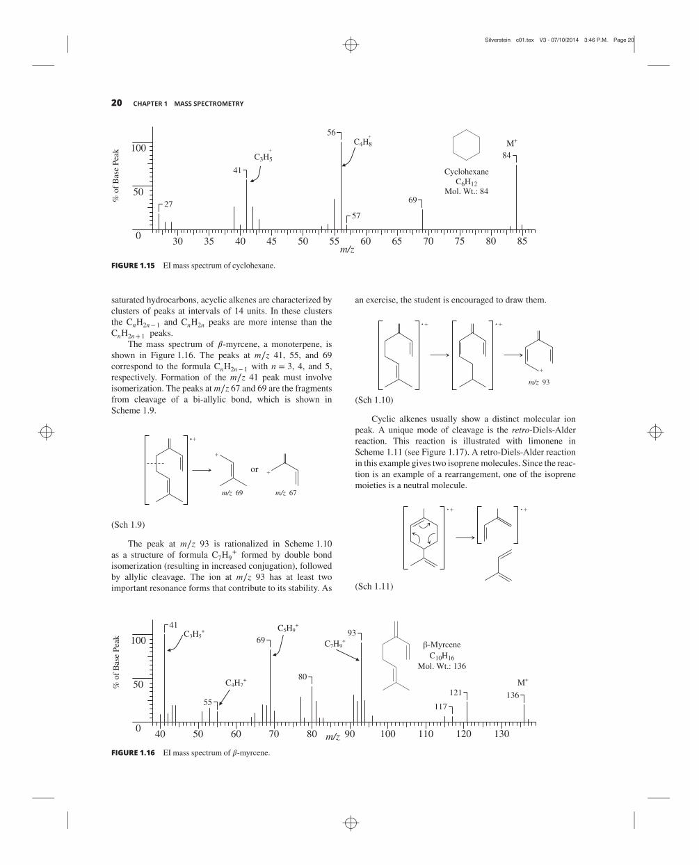
Silverstein c01.tex V3 - 07/10/2014 3:46 P.M. Page 20
20 CHAPTER 1 MASS SPECTROMETRY
% o
f B
ase
Pea
k
m/z858075706560555045403530
0
50
100
27
41
56
57
69
84
Cyclohexane
C6H12
Mol. Wt.: 84
C3H5
C4H8 M+
FIGURE 1.15 EI mass spectrum of cyclohexane.
saturated hydrocarbons, acyclic alkenes are characterized by
clusters of peaks at intervals of 14 units. In these clusters
the CnH2n− 1 and CnH2n peaks are more intense than the
CnH2n+ 1 peaks.
The mass spectrum of 𝛽-myrcene, a monoterpene, is
shown in Figure 1.16. The peaks at m∕z 41, 55, and 69
correspond to the formula CnH2n− 1 with n = 3, 4, and 5,
respectively. Formation of the m∕z 41 peak must involve
isomerization. The peaks at m∕z 67 and 69 are the fragments
from cleavage of a bi-allylic bond, which is shown in
Scheme 1.9.
m/z 69 m/z 67
or
(Sch 1.9)
The peak at m∕z 93 is rationalized in Scheme 1.10
as a structure of formula C7H9+ formed by double bond
isomerization (resulting in increased conjugation), followed
by allylic cleavage. The ion at m∕z 93 has at least two
important resonance forms that contribute to its stability. As
an exercise, the student is encouraged to draw them.
93m/z
(Sch 1.10)
Cyclic alkenes usually show a distinct molecular ion
peak. A unique mode of cleavage is the retro-Diels-Alder
reaction. This reaction is illustrated with limonene in
Scheme 1.11 (see Figure 1.17). A retro-Diels-Alder reaction
in this example gives two isoprene molecules. Since the reac-
tion is an example of a rearrangement, one of the isoprene
moieties is a neutral molecule.
(Sch 1.11)
kae
P esa
B fo
%
m/z
-Myrcene
C10H16
Mol. Wt.: 136
1301201101009080706050400
50
100
136121
117
93
80
69
55
41C3H5
+ C5H9+
C4H7+
C7H9+
M+
FIGURE 1.16 EI mass spectrum of 𝛽-myrcene.

Silverstein c01.tex V3 - 07/10/2014 3:46 P.M. Page 21
1.6 MASS SPECTRA OF SOME CHEMICAL CLASSES 21
130120110100908070605040
m/z
25
50
75
100
136121
108
107
93
79
77
68
5341
CH2
CH3
CH3
% o
f B
ase
Pea
k
FIGURE 1.17 EI mass spectrum of limonene.
% o
f B
ase
Pea
k
m/z130120110100908070605040
0
50
100
128
102756451
39
Naphthalene
C10H8
Mol. Wt.: 128
128 (M) 100.0%
129 (M+1) 11.0%
130 (M+2) 0.4%
M+
FIGURE 1.18 EI mass spectrum of naphthalene.
1.6.1.3 Aromatic and Aralkyl Hydrocarbons. An aro-
matic ring in a molecule stabilizes the molecular ion peak
(guideline 4, Section 1.5.4), which is usually sufficiently
large that accurate intensity measurements can be made on
the M+ 1 and M+ 2 peaks.
Shown in Figure 1.18 is the mass spectrum of naphtha-
lene. The molecular ion peak is also the base peak, and the
largest fragment peak, m∕z 51, is only 12.5% as intense as
the molecular ion peak.
An alkyl-substituted benzene ring frequently gives a
prominent peak (often the base peak) at m∕z 91 (C6H5CH2+).
Branching at the 𝛼-carbon leads to masses higher than 91,
by increments of 14, the largest substituent being eliminated
most readily (guideline 3, Section 1.5.4). The mere presence
of a peak at mass 91, however, does not preclude branching
at the 𝛼-carbon because this highly stabilized fragment may
result from rearrangements. A distinct and sometimes promi-
nent M− 1 peak results from similar benzylic cleavage of a
C⏤H bond.
It has been proposed that, in most cases, the ion of mass
91 is a tropylium rather than a benzylic cation. This explains
the ready loss of a methyl group, which seems to violate
guideline 7, from xylenes (Scheme 1.12, see Figure 1.19).
By comparison, toluene does not easily lose a methyl group.
The incipient molecular radical ion of xylene rearranges to
the methylcycloheptatriene radical ion, which then cleaves
to the tropylium ion (C7H+7). The frequently observed peak
at m∕z 65 results from the elimination of a neutral acetylene
molecule from the tropylium ion.
m/z 91
CH3
CH3 CH3
CH3
HH
(Sch 1.12)
1049688807264564840
m/z
25
50
75
100
106
91
776553
5139
CH3 CH3
% o
f B
ase
Pea
k
FIGURE 1.19 EI mass spectrum of p-xylene.

Silverstein c01.tex V3 - 07/10/2014 3:46 P.M. Page 22
22 CHAPTER 1 MASS SPECTROMETRY
Hydrogen migration with elimination of a neutral alkene
molecule accounts for the peak at m∕z 92 observed when the
alkyl group is longer than C2. Scheme 1.13 illustrates with
a general example. Note again that this is an example of a
rearrangement.
RHm/z 92
H
H
H2C CHR
(Sch 1.13)
A characteristic cluster of ions resulting from an 𝛼
cleavage and hydrogen migration in monoalkylbenzenes
appears at m∕z 77 (C6H5+), 78 (C6H6
+), and 79 (C6H7+).
Alkylated polyphenyls and alkylated polycyclic aro-
matic hydrocarbons exhibit the same 𝛽 cleavage as alkylben-
zene compounds.
1.6.2 Hydroxy Compounds1.6.2.1 Alcohols. The molecular ion peak of a primary
or secondary alcohol is usually quite small and for a
tertiary alcohol is often undetectable. The molecular ion
of 1-pentanol is extremely weak compared with its near
homologs. Expedients such as CI, or derivatization, may be
used to obtain the molecular weight.
Cleavage of the C⏤C bond next to the oxygen atom
is of general occurrence (guideline 8, Section 1.5.4). Thus,
primary alcohols show a prominent peak resulting from+CH2⏤OH (m∕z 31). Secondary and tertiary alcohols
cleave analogously to give a prominent peak resulting from+CHR⏤OH (m∕z 45, 59, 73, etc.) and +CRR′⏤OH (m∕z59, 73, 87, etc.), respectively. The largest substituent is
expelled most readily (guideline 3). It is not unusual that the
C⏤H bond next to the oxygen atom is cleaved; this less (or
least) favored pathway gives rise to an M− 1 peak.
Primary alcohols, in addition to the principal C⏤Ccleavage next to the oxygen atom, show a homologous series
of peaks of progressively decreasing intensity resulting from
cleavage at C⏤C bonds successively removed from the oxy-
gen atom. In long-chain (>C6) alcohols, the fragmentation
becomes dominated by the hydrocarbon pattern; in fact, the
spectrum resembles that of the corresponding alkene. The
spectrum in the vicinity of a very weak or missing molecular
ion peak of a primary alcohol is sometimes complicated by
weak M− 2 and M− 3 peaks.
A distinct and sometimes prominent peak can usually
be found at M− 18 from loss of water. This peak is most
noticeable in spectra of primary alcohols. This elimination
by electron impact has been rationalized and a mechanism
in which a 𝛿-hydrogen is lost is shown in Scheme 1.14, I. A
similar mechanism can be drawn in which a 𝛾-hydrogen is
lost. The M− 18 peak is frequently exaggerated by thermal
decomposition of higher alcohols on hot inlet surfaces.
Elimination of water, together with elimination of an alkene
from primary alcohols (see Scheme 1.14, II), accounts for
the presence of a peak at M− (alkene + H2O), that is, a peak
at M− 46,M− 74,M− 102,…
OHR
I
II
HH2C CH2 [H2C CHR ]
H2O
OHR H
OHR H R
H2O
(Sch 1.14)
Alcohols containing branched methyl groups (e.g., ter-
pene alcohols) frequently show a fairly strong peak atM− 33
resulting from loss of CH3 and H2O.
Cyclic alcohols undergo fragmentation by complicated
pathways; for example, cyclohexanol (Figure 1.20) (M =m∕z 100) forms C6H11O+ by simple loss of the 𝛼-hydrogen,
loses H2O to form C6H10+ (which appears to have more than
one possible bridged bicyclic structure), and forms C3H5O+
(m∕z 57) by a complex ring cleavage pathway.
A peak at m∕z 31 (see above) is quite diagnostic
for a primary alcohol provided it is more intense than
peaks at m∕z 45, 59, 73…. However, the first-formed ion
of a secondary alcohol can decompose further to give a
moderately intense m∕z 31 peak.
Figure 1.21 gives the characteristic spectra of isomeric
primary, secondary, and tertiary C5 alcohols.
Benzyl alcohols and their substituted homologs and
analogs constitute a distinct class. Generally, the parent peak
is strong. A moderate benzylic peak (M − OH) may be
present as expected from cleavage 𝛽 to the ring.
% o
f B
ase
Pea
k
9688807264564840
m/z
25
50
75
100
10083
82
71
67
58
57
51
44
OH
FIGURE 1.20 EI mass spectrum of cyclohexanol.

Silverstein c01.tex V3 - 07/10/2014 3:46 P.M. Page 23
1.6 MASS SPECTRA OF SOME CHEMICAL CLASSES 23
% o
f B
ase
Pea
k
m/z 90807060504030200
50
100
87
70
57
55
45
42
31
90807060504030200
50
100
8773
55
46
45
3927
90807060504030200
50
100
73
59
55
45
43
39
31
29
% o
f B
ase
Pea
k
m/z
% o
f B
ase
Pea
k
m/z
OH
1-Pentanol
C5H12O
Mol. Wt.: 88
31
57
OH2-Pentanol
C5H12O
Mol. Wt.: 88
45
43
OH
2-Methyl-2-butanol
C5H12O
Mol. Wt.: 88
59
29
M –1
M – H2O
M – (H2O + CH3)
M – (H2O + CH2=CH2)
CH2O+H
M –1 M+ (88)
M – (H2O + CH3)
M – H2O M – CH3
M – CH3
M – H2O
M – (H2O + CH3)
M – (H2O + CH2=CH2)
M – (H2O + CH2=CH2)
FIGURE 1.21 EI mass spectra of isomeric pentanols.
A complicated sequence leads to prominent M− 1,M− 2,
and M− 3 peaks. Benzyl alcohol itself (Figure 1.22) frag-
ments to give sequentially the M− 1 ion, the C6H7+ ion
by loss of CO, and the C6H5+ ion by loss of H2 (see
Scheme 1.15).
H
H
HH H
H
H
CH2OH OH
m/z 108 m/z 107
m/z 79
H
H2
CO
m/z 77
(Sch 1.15)
Loss of H2O to give a distinct M− 18 peak is a
common feature, especially pronounced and mechanistically
straightforward in some ortho-substituted benzyl alcohols
(see Figure 1.23). The loss of water shown in Scheme 1.16
works equally well with an oxygen atom at the ortho-
position (a phenol). The aromatic cluster at m∕z 77, 78,
and 79 resulting from complex degradation is prominent
here also.
H
OH H2O
(Sch 1.16)
1.6.2.2 Phenols. A conspicuous molecular ion peak facil-
itates identification of phenols. In phenol itself, the molecu-
lar ion peak is the base peak, and the M− 1 peak is small.
In cresols, the M− 1 peak is larger than that of the molec-
ular ion as a result of a facile benzylic C⏤H cleavage.

Silverstein c01.tex V3 - 07/10/2014 3:46 P.M. Page 24
24 CHAPTER 1 MASS SPECTROMETRY
1049688807264564840
m/z
25
50
75
100108
91
79
6553
5139
% o
f B
ase
Pea
k
OH
FIGURE 1.22 EI mass spectrum of benzyl alcohol.
% o
f B
ase
Pea
k
1201121049688807264564840
m/z
25
50
75
100
122107
104
9179
65
635139
CH3
OH
FIGURE 1.23 EI mass spectrum of o-methylbenzyl alcohol.
A rearrangement peak at m∕z 77 and peaks resulting from the
loss of CO (M − 28) and CHO (M − 29) are usually found in
the mass spectra of phenols.
The mass spectrum of o-ethylphenol, a typical phenol,
is shown in Figure 1.24. This spectrum shows that a methyl
group is lost much more readily than an 𝛼-hydrogen.
1.6.3 Ethers1.6.3.1 Aliphatic Ethers (and Acetals). The molecular
ion peak (two mass units larger than that of an analogous
hydrocarbon) is small, but larger sample size usually will
make the molecular ion peak or the M+ 1 peak obvious (H•
transfer during ion-molecule collision).
The presence of an oxygen atom can be deduced from
strong peaks at m∕z 31, 45, 59, 73,… These peaks repre-
sent the RO+ and ROCH2+ fragments. A typical aliphatic
ether is shown in Figure 1.25. Fragmentation occurs in two
principal ways:
1. Cleavage of the C⏤C bond next to the oxygen atom
(𝛼, 𝛽 bond, guideline 8, Section 1.5.4). One or the other
of these oxygen-containing ions may account for the
base peak. In the case shown in Figure 1.25, the first
cleavage (i.e., at the branch position leading to loss of the
larger fragment) is preferred. However, the first-formed
fragment decomposes further by loss of ethylene to give
the base peak; this decomposition is important when the
𝛼-carbon is substituted (see McLafferty rearrangement,
Section 1.5.5)
% o
f B
ase
Pea
k
HO
o-Ethylphenol
C8H10O
Mol. Wt.: 122
1201101009080706050400
50
100
122
107
10391
77
5139
m/z
[M – H2O]+
[M – CH3]+
C6H5+
M+
FIGURE 1.24 EI mass spectrum of o-ethylphenol.

Silverstein c01.tex V3 - 07/10/2014 3:46 P.M. Page 25
1.6 MASS SPECTRA OF SOME CHEMICAL CLASSES 25
100908070605040300
50
100
10287
73
59
56
45
41
29
% o
f B
ase
Pea
k
m/z
Ethyl sec-butyl ether
C6H14O Mol. Wt.: 102
O–CH3CH2
O–H2C C H2
O HO
HO
m/z 73 m/z 45
M – CH
H
3
CH3CH2+
M+
FIGURE 1.25 EI mass spectrum of ethyl sec-butyl ether.
2. C⏤O bond cleavage with the charge remaining on
the alkyl fragment. The spectrum of long-chain ethers
becomes dominated by the hydrocarbon pattern.
Acetals are a special class of ethers. Their mass spectra
are characterized by an extremely weak molecular ion peak,
by the prominent peaks at M− R and M−OR (and/or
M−OR′), and a weak peak at M−H. Each of these
cleavages is mediated by an oxygen atom and thus facile.
As usual, elimination of the largest group is preferred. As
with aliphatic ethers, the first-formed oxygen-containing
fragments can decompose further with hydrogen migration
and alkene elimination. Ketals behave similarly.
H
OR
R C OR
1.6.3.2 Aromatic Ethers. The molecular ion peak of aro-
matic ethers is prominent. Primary cleavage occurs at the
bond 𝛽 to the ring, and the first-formed ion can decompose
further. Thus, anisole (Figure 1.26, MW 108) gives ions of
m∕z 93 and 65. The characteristic aromatic peaks at m∕z 78
and 77 may arise from anisole.
% o
f B
ase
Pea
k
m/z 110100908070605040300
50
100
108
93
7865
5751
39
OCH2 –CH2O –H
m/z 77m/z 78
OCH3
H
O
m/z 93
–CH3 –CO
m/z 65
H
H
Anisole
C7H8O
Mol. Wt.: 108
M+
FIGURE 1.26 EI mass spectrum of anisole.

Silverstein c01.tex V3 - 07/10/2014 3:46 P.M. Page 26
26 CHAPTER 1 MASS SPECTROMETRY
O
CH3
% o
f B
ase
Pea
k
15014013012011010090807060504030
m/z
25
50
75
100
150
107
94
7766574129
FIGURE 1.27 EI mass spectrum of butyl phenyl ether.
When the alkyl portion of an aromatic alkyl ether is C2
or larger, cleavage 𝛽 to the ring is accompanied by hydrogen
migration (Scheme 1.17) as noted above for alkylbenzenes.
Clearly, cleavage is mediated by the ring rather than by the
oxygen atom; C⏤C cleavage next to the oxygen atom is
insignificant. An example of this type is illustrated in the
spectrum of butyl phenyl ether, Figure 1.27.
O
RH
O OH
m/z 94
H2C CHR
H
H
(Sch 1.17)
Diphenyl ethers show peaks at M−H, M− CO, and
M− CHO by complex rearrangements.
1.6.4 Ketones1.6.4.1 Aliphatic Ketones. The molecular ion peak of
ketones is usually quite pronounced. Major fragmenta-
tion peaks of aliphatic ketones result from cleavage at
one of the C⏤C bonds adjacent to the oxygen atom, the
charge remaining with the resonance-stabilized acylium ion
(Scheme 1.18). Thus, as with alcohols and ethers, cleavage
is mediated by the oxygen atom. This cleavage gives rise to
a peak at m∕z 43 or 57 or 71…. The base peak very often
results from loss of the larger alkyl group.
R C OC O
R
R
R C OR
(Sch 1.18)
When one of the alkyl chains attached to the C⏤⏤Ogroup is C3 or longer, cleavage of the C⏤C bond once
removed (𝛼, 𝛽-bond) from the C⏤⏤O group occurs with
hydrogen migration to give a major peak (McLafferty rear-
rangement, Scheme 1.19). Simple cleavage of the 𝛼, 𝛽-bond,
which does not occur to any extent, would give an ion of low
stability because it would have two adjacent positive centers.
HO RHC CH2
R
R
OH
CH2R
OH
OH
CH2R CH2R
(Sch 1.19)
Note that in long-chain ketones the hydrocarbon peaks
are indistinguishable (without the aid of high-resolution
techniques) from the acyl peaks, since the mass of the C⏤⏤Ounit (28) is the same as two methylene units. The multiple
cleavage modes in ketones sometimes make difficult the
determination of the carbon chain configuration. Reduction
of the carbonyl group to a methylene group yields the
corresponding hydrocarbon whose fragmentation pattern
leads to the carbon skeleton.
1.6.4.2 Cyclic Ketones. The molecular ion peak in cyclic
ketones is prominent. As with acyclic ketones, the primary
cleavage of cyclic ketones is adjacent to the C⏤⏤O group, but
the ion thus formed must undergo further cleavage in order
to produce a fragment. The base peak in the spectrum of
cyclopentanone and of cyclohexanone (Figure 1.28) is m∕z55. The mechanisms are similar in both cases: hydrogen
shift to convert a primary radical to a conjugated secondary
radical followed by formation of the resonance-stabilized
ion, m∕z 55. The other distinctive peaks at m∕z 83 and 42
in the spectrum of cyclohexanone have been rationalized as
depicted in Figure 1.28.
1.6.4.3 Aromatic Ketones. The molecular ion peak of an
aromatic ketone is prominent. Cleavage of aryl alkyl ketones
occurs at the bond 𝛽 to the ring, leaving a characteristic
ArC⏤⏤⏤O+ fragment (m∕z 105 when Ar = phenyl), which
usually accounts for the base peak. Loss of CO from this
fragment gives the “aryl” ion (m∕z 77 in the case of
acetophenone). Cleavage of the bond adjacent to the ring
to form a RC⏤⏤⏤O+ fragment (R = alkyl) is less important
though somewhat enhanced by electron-withdrawing groups

Silverstein c01.tex V3 - 07/10/2014 3:46 P.M. Page 27
1.6 MASS SPECTRA OF SOME CHEMICAL CLASSES 27
% o
f B
ase
Pea
k
m/z 100908070605040300
50
100
98
8380
55
42
H2CH
O
CO
CH3 CH
C
OO
C
CH
CH2
O
C
CH
CH2
-C3H7
m/z 55Cyclohexanone
C6H10O
Mol. Wt.: 98H2C
CO
- CH2=CH2
- COCH2
H2C
CH2
m/z 42
or
[M - H2O]+
[M – CH3]+
M+
FIGURE 1.28 EI mass spectrum of cyclohexanone.
(and diminished by electron-donating groups) in the para-
position of the Ar group.
When the alkyl chain is C3 or longer, cleavage of the
C⏤C bond once removed from the C⏤⏤O group occurs
with hydrogen migration. This is the same cleavage noted
for aliphatic ketones that proceeds through a cyclic transition
state and results in elimination of an alkene and formation of
a stable ion.
The mass spectrum of an unsymmetrical diaryl ketone,
p-chlorobenzophenone, is displayed in Figure 1.29. The
molecular ion peak (m∕z 216) is prominent and the intensity
of the M+ 2 peak (33.99%, relative to the molecular ion
peak) demonstrates that chlorine is in the structure (see the
discussion of Table 1.5 and Figure 1.35 in Section 1.6.16).
Since the intensity of the m∕z 141 peak is about1
3the intensity of the m∕z 139 peak, these peaks, which are
200150100500
50
100
216
181
139
105
94
77
51
% o
f B
ase
Pea
k
m/z
C
Cl
O
lCCC
Cl
OO
m/z 105
m/z 77
m/z 139
m/z 111
OC-OC-
216 M 100.00%217 M+1 19.28%218 M+2 33.99%219 M+3 6.21%220 M+4 0.98%
p-ChlorobenzophenoneC13H9ClO
Mol. Wt.: 216
+
FIGURE 1.29 EI mass spectrum of p-chlorobenzophenone.

Silverstein c01.tex V3 - 07/10/2014 3:46 P.M. Page 28
28 CHAPTER 1 MASS SPECTROMETRY
due to fragments which contain chlorine, correspond to the
same fragment. The same can be said about the fragments
producing the m∕z 111 and 113 peaks.
The fragmentation leading to the major peaks is sketched
in Figure 1.29. The Cl⏤ArC⏤⏤⏤O+ peak is larger than the
Cl⏤Ar+ peak, and theArC⏤⏤⏤O+ peak is larger than theAr+peak (𝛽 cleavage favored). If the [f ragment + 2] peaks for the
Cl-substituted moieties are taken into account however, there
is little difference in abundance between Cl⏤ArCO+ and
ArCO+, or between Cl⏤Ar+ and Ar+; the inductive (elec-
tron withdrawing) and resonance (electron releasing) effects
of the para-substituted Cl are roughly balanced out as they
are in electrophilic aromatic substitution reactions.
1.6.5 Aldehydes1.6.5.1 Aliphatic Aldehydes. The molecular ion peak of
aliphatic aldehydes is usually discernible. Cleavage of the
C⏤H and C⏤C bonds next to the oxygen atom results in
an M− 1 peak and in an M− R peak (m∕z 29,CHO+). The
M− 1 peak is a good diagnostic peak even for long-chain
aldehydes, but the m∕z 29 peak present in C4 and higher
aldehydes results from the hydrocarbon C2H5+ ion.
In the C4 and higher aldehydes, McLafferty cleavage of
the 𝛼, 𝛽 C⏤C bond occurs to give a major peak at m∕z 44,
58, or 72,…, depending on the 𝛼 substituents. This is the
resonance-stabilized ion (Scheme 1.20) formed through the
cyclic transition state as shown in Scheme 1.7, whereY⏤⏤H.
HH
R
OH
HH
R
OH
(Sch 1.20)
In straight-chain aldehydes, other unique, diagnostic
peaks are at M− 18 (loss of water), M− 28 (loss of ethy-
lene), M− 43 (loss of CH2⏤⏤CH⏤O•), and M− 44 (loss
of CH2⏤⏤CH⏤OH). The rearrangements leading to these
peaks have been rationalized (see Budzikiewicz et al., 1967).
As the chain lengthens, the hydrocarbon pattern (m∕z 29, 43,
57, 71, …) becomes dominant. These features are evident in
the spectrum of nonanal (Figure 1.30).
1.6.5.2 Aromatic Aldehydes. Aromatic aldehydes are
characterized by a large molecular ion peak and by an M− 1
peak (Ar⏤C⏤⏤O+) that is always large and may be larger
than the molecular ion peak. The M− 1 ion, C6H5⏤CO+
when Ar = phenyl, eliminates CO to give the phenyl ion
(m∕z 77), which in turn eliminates acetylene to give the
C4H3+ ion (m∕z 51).
1.6.6 Carboxylic Acids1.6.6.1 Aliphatic Acids. The molecular ion peak of a
straight-chain monocarboxylic acid is weak but usually
discernible. The most characteristic (sometimes the base)
peak is m∕z 60 resulting from the McLafferty rearrangement
(Scheme 1.21). Branching at the 𝛼-carbon enhances this
cleavage.
R
R
HO RHC CH2
O
HO
R
H
CHHO
R
OH
CHHO
(Sch 1.21)
In short-chain acids, peaks at M−OH and M− CO2Hare prominent: these represent cleavage of bonds next to
C⏤⏤O. In long-chain acids, the spectrum consists of two
series of peaks resulting from cleavage at each C⏤C bond
with retention of charge either on the oxygen-containing
fragment (m∕z 45, 59, 73, 87,…) or on the alkyl fragment
(m∕z 29, 43, 57, 71, 85, …). As previously discussed, the
hydrocarbon pattern also shows peaks at m∕z 27, 28; 41, 42;
140130120110100908070605040300
50
100
142124114
98
9582
69
5741
29
% o
f B
ase
Pea
k
O
Nonanal
C9H18O
Mol. Wt.: 142OH
m/z
M - H2O
M - CH2=CH2 M - 44
M-1M
+
FIGURE 1.30 EI mass spectrum of nonanal.

Silverstein c01.tex V3 - 07/10/2014 3:46 P.M. Page 29
1.6 MASS SPECTRA OF SOME CHEMICAL CLASSES 29
% o
f B
ase
Pea
k
m/z15010050
0
50
100
172143
129
112
73
60
43
CH3 CH2 CH2 CH2 CH2 CH2 CH2 CH2 CH2 C OH
O29
143
43
129
7157
115 101
85 99 113 128
87 73 59 45
Decanoic acidC10H20O2
Mol. Wt.: 172
O
CH2HO
H
M+
FIGURE 1.31 EI mass spectrum of decanoic acid.
55, 56; 69, 70;…. In summary, besides the McLafferty rear-
rangement peak, the spectrum of a long-chain acid resem-
bles the series of hydrocarbon clusters at intervals of 14
mass units. In each cluster, however, is a prominent peak at
CnH2n− 1O2. Decanoic acid, (Figure 1.31), nicely illustrates
many of the points discussed above.
Dibasic acids usually have low volatility and hence are
converted to esters to increase vapor pressure. Trimethylsilyl
esters are often successful.
1.6.6.2 Aromatic Acids. The molecular ion peak of aro-
matic acids is large. The other prominent peaks are formed
by loss of OH (M − 17) and of CO2H (M − 45). Loss of
H2O (M − 18) is prominent if a hydrogen-bearing orthogroup is available as outlined in Scheme 1.22. This is one
example of the general “ortho effect” noted when the sub-
stituents can be in a six-membered transition state to facili-
tate loss of a neutral molecule of H2O, ROH, or NH3.
HZZ
O
YH
CO
Y
Z = OH, OR, NH2; Y = CH2, O, NH
(Sch 1.22)
1.6.7 Carboxylic Esters1.6.7.1 Aliphatic Esters. The molecular ion peak of a
methyl ester of a straight-chain aliphatic acid is usually
distinct. Even waxes usually show a discernible molecular
ion peak. The molecular ion peak is weak in the range m∕z130 to ∼200, but becomes somewhat more intense beyond
this range. The most characteristic peak results from the
familiar McLafferty rearrangement (Scheme 1.23 gives the
rearrangement for an ester) and cleavage one bond removed
from the C⏤⏤O group. Thus, a methyl ester of an aliphatic
acid unbranched at the 𝛼-carbon gives a strong peak at m∕z
74, which in fact, is the base peak in straight-chain methyl
esters from C6 to C26. The alcohol moiety and/or the 𝛼
substituent can often be deduced by the location of the peak
resulting from this cleavage.
R
R
HO RHC CH2
O
RO
R
H
CH2RO
R
OH
CHRO
(Sch 1.23)
For the general ester, four ions can result from bond
cleavage next to C⏤⏤O.O
R C OR
The ion R+ is prominent for the short-chain esters but
diminishes rapidly with increasing chain length and is barely
perceptible in methyl hexanoate. The ion R⏤C⏤⏤⏤O+ gives
an easily recognizable peak for esters. In methyl esters, it
occurs at M− 31. It is the base peak in methyl acetate and
is still 4% of the base peak in the C26 methyl ester. The ions
[OR′]+ and [C(⏤⏤O)OR′]+ are usually of little importance.
The latter is discernible when R′⏤⏤CH3 (see m∕z 59 peak of
Figure 1.32).
First, consider esters in which the acid portion is the
predominant portion of the molecule. The fragmentation
pattern for methyl esters of straight-chain acids can be
described in the same terms used for the pattern of the
free acid. Cleavage at each C⏤C bond gives an alkyl
ion (m∕z 29, 43, 57,…) and an oxygen-containing ion,
CnH2n− 1O2+ (m∕z 59, 73, 87,…). Thus, there are hydrocar-
bon clusters at intervals of 14 mass units; in each cluster is a

Silverstein c01.tex V3 - 07/10/2014 3:46 P.M. Page 30
30 CHAPTER 1 MASS SPECTROMETRY
% o
f B
ase
Pea
k
m/z15010050
0
50
100
× 20
2943
55
74
87
101 127
158
O
OMethyl octanoate
C9H18O2
Mol. Wt.: 158CH2CH2COC H3
O
CH2 COCH3
OH
H3COC O
M – OCH3 H
M – 31
M+
FIGURE 1.32 EI mass spectrum of methyl octanoate.
prominent peak atCnH2n− 1O2+. The peak (m∕z 87) formally
represented by the ion [CH2CH2COOCH3]+ is always more
intense than its homologs, but the reason is not immediately
obvious. However, it seems clear that the CnH2n− 1O2+ ions
do not at all arise from simple cleavage.
The spectrum of methyl octanoate is presented as
Figure 1.32. This spectrum illustrates one difficulty in using
the M+ 1 peak to arrive at a molecular formula (previ-
ously mentioned, Section 1.5.2.1). The measured value for
the M+ 1 peak is 12%. The calculated value is 10.0%. The
measured value is high because of an ion-molecule reaction
induced by the relatively large sample that was used to see
the weak molecular ion peak.
Now let us consider esters in which the alcohol portion
is the predominant portion of the molecule. Esters of fatty
alcohols (except methyl esters) eliminate a molecule of acid
in the same manner that alcohols eliminate water. A scheme
similar to that described earlier for alcohols, involving a sin-
gle hydrogen transfer to the alcohol oxygen of the ester,
can be written. An alternative mechanism involves a hydride
transfer to the carbonyl oxygen (McLafferty rearrange-
ment).
The loss of acetic acid by the mechanism described
above is so facile in steroidal acetates that they frequently
show no detectable molecular ion peak. Steroidal systems
also seem unusual in that they often display significant
molecular ions as alcohols, even when the corresponding
acetates do not.
Esters of long-chain alcohols show a diagnostic peak at
m∕z 61, 75, or 89… from elimination of the alkyl moiety
as an alkene and transfer of two hydrogen atoms to the
fragment containing the oxygen atoms, which in essence is
the protonated carboxylic acid.
Esters of dibasic acids ROOC(CH2)nCOOR, in general,
give recognizable molecular ion peaks. Intense peaks are
found at [ROOC(CH2)nC⏤⏤O]+ and at [ROOC(CH2)n]+.
1.6.7.2 Benzyl and Phenyl Esters. Benzyl acetate (also
furfuryl acetate and other similar acetates) and phenyl
acetate eliminate the neutral molecule ketene (Scheme 1.24);
frequently this gives rise to the base peak.
O
m/z 108
H2C C O
O OH
(Sch 1.24)
Of course, the m∕z 43 peak (CH3C⏤⏤O)+ and m∕z91 (C7H7)+ peaks are prominent for benzyl acetate.
1.6.7.3 Esters of Aromatic Acids. The molecular ion
peak of methyl esters of aromatic acids is prominent
(ArCOOR,R⏤⏤CH3). As the size of the alcohol moiety
increases, the intensity of the molecular ion peak decreases
rapidly to practically zero at C5. The base peak results from
elimination of • OR, and elimination of • COOR accounts for
another prominent peak. In methyl esters, these peaks are at
M− 31 and M− 59, respectively.
As the alkyl moiety increases in length, three modes of
cleavage become important: (1) McLafferty rearrangement,
(2) rearrangement of two hydrogen atoms with elimination
of an allylic radical, and (3) retention of the positive charge
by the alkyl group.
The familiar McLafferty rearrangement pathway gives
rise to a peak for the aromatic acid, (ArCOOH)+. The
second, similar pathway gives the protonated aromatic acid,
(ArCOOH2)+. The third mode of cleavage gives the alkyl
cation, R+.
Appropriately, ortho-substituted benzoates eliminate
ROH through the general “ortho” effect described above
under aromatic acids. Thus, the base peak in the spectrum
of methyl salicylate is m∕z 120; this ion eliminates carbon
monoxide to give a strong peak at m∕z 92.
A strong characteristic peak at mass 149 is found in the
spectra of all esters of phthalic acid, starting with the diethyl

Silverstein c01.tex V3 - 07/10/2014 3:46 P.M. Page 31
1.6 MASS SPECTRA OF SOME CHEMICAL CLASSES 31
% o
f B
ase
Pea
k
m/z 100908070605040300
50
10028
2941
43
56
85
100
O
O
–CH3CH O
H2C
O-Valerolactone
C5H8O2
Mol. Wt.: 100
m/z 56
M+
M - 15
FIGURE 1.33 EI mass spectrum of 𝛾-valerolactone.
ester. This peak is not significant in the dimethyl or methyl
ethyl ester of phthalic acid, nor in esters of isophthalic or
terephthalic acids, all of which give the expected peaks at
M− R,M− 2R,M− CO2R, and M− 2CO2R. Since long-
chain phthalate esters are widely used as plasticizers, a strong
peak at m∕z 149 may indicate contamination. The m∕z 149
fragment (essentially a protonated phthalic anhydride) is
probably formed by two ester cleavages involving the shift
of two hydrogen atoms and then another hydrogen atom,
followed by elimination of H2O.
1.6.8 LactonesThe molecular ion peak of five-membered ring lactones is
distinct but is weaker when an alkyl substituent is present at
C4. Facile cleavage of the side chain at C4 (guidelines 3 and
8, Section 1.5.4) gives a strong peak at M − alkyl.
The base peak (m∕z 56) of 𝛾-valerolactone (Figure 1.33)
and the same strong peak of butyrolactone are rationalized,
which shows the elimination of acetaldehyde.
Labeling experiments indicate that some of the m∕z 56
peak in 𝛾-valerolactone arises from the C4H8+ ion. The other
intense peaks in 𝛾-valerolactone are at m∕z 27 (C2H3+),
28 (C2H4+), 29 (C2H5
+), 41 (C3H5+), and 43 (C3H7
+), and
85 (C4H5O2+, loss of the methyl group). In butyrolactone,
there are strong peaks at m∕z 27, 28, 29, 41, and 42 (C3H6+).
1.6.9 Amines1.6.9.1 Aliphatic Amines. The molecular ion peak of an
aliphatic monoamine is an odd number, but it is usu-
ally quite weak and, in long-chain or highly branched
amines, undetectable. The base peak frequently results from
C⏤C cleavage next to the nitrogen atom (𝛼, 𝛽 guideline
8, Section 1.5.4); for primary amines unbranched at the 𝛼-
carbon, this is m∕z 30 (CH2NH2+) shown in Scheme 1.25.
This cleavage accounts for the base peak in all primary
amines and secondary and tertiary amines that are not
branched at the 𝛼-carbon. Loss of the largest branch (• R″ in
Scheme 1.25) from the 𝛼-C atom is preferred.
H2N CR
H2N C R
R
R
H2N C
R
R
R
R
(Sch 1.25)
When branching at the 𝛼-carbon is absent, an M− 1
peak is usually visible. This is the same type of cleavage
noted above for alcohols. The effect is more pronounced in
amines because of the better resonance stabilization of the
ion fragment by the less electronegative N atom compared
with the O atom.
Primary straight-chain amines show a homologous
series of peaks of progressively decreasing intensity (the
cleavage at the 𝜖-bond is slightly more important than at the
neighboring bonds) at m∕z 30, 44, 58,…resulting from cleav-
age at C⏤C bonds successively removed from the nitro-
gen atom with retention of the charge on the N-containing
fragment. These peaks are accompanied by the hydrocar-
bon pattern ofCnH2n+ 1,CnH2n, andCnH2n− 1 ions. Thus, we
note characteristic clusters at intervals of 14 mass units, each
cluster containing a peak resulting from a CnH2n+ 2N ion.
Because of the very facile cleavage to form the base peak,
the fragmentation pattern in the high mass region becomes
extremely weak.
Cyclic fragments apparently occur during the frag-
mentation of longer chain amines. The fragment shown
in Scheme 1.26 gives a six-membered ring; five-membered
rings are also commonly formed.
NH2
R
NH2
m/z 86
R
(Sch 1.26)

Silverstein c01.tex V3 - 07/10/2014 3:46 P.M. Page 32
32 CHAPTER 1 MASS SPECTROMETRY
A peak at m∕z 30 is good though not conclusive evidence
for a straight-chain primary amine. Further decomposition of
the first-formed ion from a secondary or tertiary amine leads
to a peak at m∕z 30, 44, 58, 72, …. This is a process similar
to that described for aliphatic alcohols and ethers above and,
similarly, is enhanced by branching at one of the 𝛼-carbon
atoms.
Cleavage of amino acid esters occurs at both C⏤Cbonds (dashed lines below) next to the nitrogen atom; loss of
the carbalkoxy group (⏤COOR′) is preferred. The aliphatic
amine fragment (+NH2⏤⏤CHCH2CH2R) decomposes fur-
ther to give a peak at m∕z 30.
NH2
HR OOC C CH2CH2R
1.6.9.2 Cyclic Amines. In contrast to acyclic amines, the
molecular ion peaks of cyclic amines are usually intense
unless there is substitution at the 𝛼 position; for example, the
molecular ion peak of pyrrolidine is strong. Primary cleavage
at the bonds next to the N atom leads either to loss of an
𝛼-hydrogen atom to give a strong M− 1 peak or to opening
of the ring; the latter event is followed by elimination of
ethylene to give • CH2⏤+NH⏤⏤CH2 (m∕z 43, base peak),
hence by loss of a hydrogen atom to give CH2⏤⏤N+⏤⏤CH2
(m∕z 42). N-methyl pyrrolidine also gives a C2H4N+ (m∕z42) peak, apparently by more than one pathway.
Piperidine likewise shows a strong molecular ion and
M− 1 (base) peak. Ring opening followed by several avail-
able sequences leads to characteristic peaks at m∕z 70, 57,
56, 44, 43, 42, 30, 29, and 28. Substituents are cleaved from
the ring (guideline 6, Section 1.5.4).
1.6.9.3 Aromatic Amines (Anilines). The molecular ion
peak (odd number) of an aromatic monoamine is intense.
Loss of one of the amino H atoms of aniline gives a
moderately intense M− 1 peak; loss of a neutral molecule of
HCN followed by loss of a hydrogen atom gives prominent
peaks at m∕z 66 and 65, respectively.
It was noted above that cleavage of alkyl aryl ethers
occurs with rearrangement involving cleavage of theArO⏤Rbond: that is, cleavage was controlled by the ring rather than
by the oxygen atom. In the case of alkyl aryl amines, cleav-
age of the C⏤C bond next to the nitrogen atom is dominant
(Scheme 1.27): that is, the heteroatom controls cleavage.
CH2RCH2
m/z 106
CH2R
HN HN
(Sch 1.27)
1.6.10 Amides1.6.10.1 Aliphatic Amides. The molecular ion peak of
straight-chain monoamides is usually discernible. The dom-
inant modes of cleavage depend on the length of the acyl
moiety and on the lengths and number of the alkyl groups
attached to the nitrogen atom.
The base peak (m∕z 59,H2NC(⏤⏤OH+)CH2•) in all
straight-chain primary amides higher than propionamide
results from the familiar McLafferty rearrangement. Branch-
ing at the 𝛼-carbon (CH3, etc.) gives a homologous peak at
m∕z 73 or 87,….
Primary amides give a strong peak at m∕z 44 from cleav-
age of the R⏤CONH2 bond: (O⏤⏤C⏤⏤+NH2); this is the
base peak in C1⏤C3 primary amides and in isobutyramide.
A moderate peak at m∕z 86 results from 𝛾,𝛿 C⏤C cleavage,
possibly accompanied by cyclization (Scheme 1.28).
NRH2
m/z 86
RNH2
O O
(Sch 1.28)
Secondary and tertiary amides with an available hydro-
gen on the 𝛾-carbon of the acyl moiety and methyl groups
on the N atom show the dominant peak resulting from the
McLafferty rearrangement. When the N-alkyl groups are C2
or longer and the acyl moiety is shorter than C3, another
mode of cleavage predominates. This is cleavage of the
N-alkyl group 𝛽 to the nitrogen atom, and cleavage of the
carbonyl C⏤N bond with migration of an 𝛼-hydrogen atom
of the acyl moiety (expelling a neutral ketene molecule) and
leaving +NH2⏤⏤CH2 (m∕z 30).
1.6.10.2 Aromatic Amides. Benzamide (Figure 1.1) is a
typical example. Loss of NH2 from the molecular ion yields
a resonance-stabilized benzoyl cation that in turn undergoes
cleavage to a phenyl cation. A separate fragmentation path-
way gives rise to a modest m∕z 44 peak.
1.6.11 Aliphatic NitrilesThe molecular ion peaks of aliphatic nitriles (except for
acetonitrile and propionitrile) are weak or absent, but the
M+ 1 peak can usually be located by its behavior on
increasing the sample size (Section 1.5.2.1). A weak but
diagnostically useful M− 1 peak is formed by loss of an
𝛼-hydrogen to form the stable ion: RCH⏤⏤C⏤⏤N+.
The base peak of straight-chain nitriles between C4 and
C9 is m∕z 41. This peak corresponds to the ion resulting
from hydrogen rearrangement in a six-membered transition
state, similar to a McLafferty rearrangement giving a peak
at m∕z 41 CH2⏤⏤C⏤⏤N+⏤H. However, this peak lacks
diagnostic value because of the presence of C3H5+ (m∕z 41)
for all molecules containing a hydrocarbon chain.
A peak at m∕z 97 is characteristic and intense (some-
times the base peak) in straight-chain nitriles C8 and higher.

Silverstein c01.tex V3 - 07/10/2014 3:46 P.M. Page 33
1.6 MASS SPECTRA OF SOME CHEMICAL CLASSES 33
The mechanism depicted in Scheme 1.29 has been proposed
to account for this ion.
HN
R
N
m/z 97
H2C CHR
H
(Sch 1.29)
Simple cleavage at each C⏤C bond (except the one
next to the N atom) gives a characteristic series of homol-
ogous peaks of even mass number down the entire length
of the chain (m∕z 40, 54, 68, 82,…) resulting from the
(CH2)nC⏤⏤⏤N+ ions. Accompanying these peaks are the
usual peaks of the hydrocarbon pattern.
1.6.12 Nitro Compounds1.6.12.1 Aliphatic Nitro Compounds. The molecular ion
peak (odd number) of an aliphatic mononitro compound is
weak or absent (except in the lower homologs). The main
peaks are attributable to the hydrocarbon fragments up to
M−NO2. Presence of a nitro group is indicated by an appre-
ciable peak at m∕z 30 (NO+) and a smaller peak at mass
46 (NO2+).
1.6.12.2 Aromatic Nitro Compounds. The molecular ion
peak of aromatic nitro compounds (odd number for one N
atom) is strong. Prominent peaks result from elimination of
an NO2 radical (M− 46, the base peak in nitrobenzene), and
of a neutral NO molecule with rearrangement to form the
phenoxy cation (M − 30); both are good diagnostic peaks.
Loss of acetylene from the M− 46 ion accounts for a strong
peak at M− 72; loss of CO from the M− 30 ion gives a
peak at M− 58. A diagnostic peak at m∕z 30 results from
the NO+ ion.
The isomeric o-,m-, and p-nitroanilines each give a
strong molecular ion (even number). They all give prominent
peaks resulting from two sequences. The first pathway entails
a loss of an NO2 group (M − 46) to give an m∕z 92; this ion
loses HCN to give an m∕z 65. The second sequence records
a loss of NO (M − 30) to give m∕z 108, which loses CO to
give m∕z 80.
Aside from differences in intensities, the three isomers
give very similar spectra. The meta and para compounds give
a small peak at m∕z 122 from loss of an O atom, whereas the
ortho compound eliminates OH as depicted in Scheme 1.30
to give a small peak at m∕z 121.
ON
m/z 121
OH
O
NH
H
NO
NH
(Sch 1.30)
1.6.13 Aliphatic NitritesThe molecular ion peak (odd number) of aliphatic nitrites
(one N present) is weak or absent. The peak at m∕z 30 (NO+)is always large and is often the base peak. There is a large
peak at m∕z 60 (CH2⏤⏤+ONO) in all nitrites unbranched at
the 𝛼-carbon; this represents cleavage of the C⏤C bond next
to the ONO group. An 𝛼 branch can be identified by a peak
at m∕z 74, 88, or 102…. Absence of a large peak at m∕z 46
permits differentiation from nitro compounds. Hydrocarbon
peaks are prominent, and their distribution and intensities
describe the arrangement of the carbon chain.
1.6.14 Aliphatic NitratesThe molecular ion peak (odd number) of aliphatic nitrates
(one nitrogen present) is weak or absent. A prominent
(frequently the base) peak is formed by cleavage of theC⏤Cbond next to the ONO2 group with loss of the heaviest alkyl
group attached to the 𝛼-carbon. The NO2+ peak at m∕z 46
is also prominent. As in the case of aliphatic nitrites, the
hydrocarbon fragment ions are distinct.
1.6.15 Sulfur CompoundsThe contribution (4.4%, see Table 1.3 and Figure 1.34) of the34S isotope to the M+ 2 peak, and often to a (f ragment + 2)peak, affords ready recognition of sulfur-containing com-
pounds. A homologous series of sulfur containing frag-
ments is four mass units higher than the hydrocarbon
fragment series. The number of sulfur atoms can be deter-
mined from the size of the contribution of the 34S isotope
to the M+ 2 peak. The mass of the sulfur atom(s) present is
subtracted from the molecular weight. In diisopentyl disul-
fide, for example, the molecular weight is 206, and the
molecule contains two sulfur atoms. The formula for the rest
of the molecule is therefore found under mass 142, that is,
206 − (2 × 32).
1.6.15.1 Aliphatic Mercaptans (Thiols). The molecular
ion peak of aliphatic mercaptans, except for higher tertiary
mercaptans, is usually strong enough so that the M+ 2 peak
can be accurately measured. In general, the cleavage modes
resemble those of alcohols. Cleavage of the C⏤C bond
(𝛼,𝛽-bond) next to the SH group gives the characteristic ion
CH2⏤⏤SH+ (m∕z 47). Sulfur is poorer than nitrogen, but
better than oxygen, at stabilizing such a fragment. Cleavage
at the 𝛽,𝛾 bond gives a peak at m∕z 61 of about one-half the
intensity of the m∕z 47 peak. Cleavage at the 𝛾,𝛿-bond gives
a small peak at m∕z 75, and cleavage at the 𝛿,𝜖-bond gives a
peak at m∕z 89 that is more intense than the peak at m∕z 73;
presumably the m∕z 89 ion is stabilized by cyclization:
S H

Silverstein c01.tex V3 - 07/10/2014 3:46 P.M. Page 34
34 CHAPTER 1 MASS SPECTROMETRY
% o
f B
ase
Pea
k
m/z 150100500
50
10043
61
70
103
117
131
174
S
Di-n-pentyl sulfide
C10H22S
Mol. Wt.: 174
174 M 100.00%
175 M+1 11.33%
176 M+2 4.67%
C5H11S+
C5H11S+=CH2
C5H11S+H
C5H10
H2
C
H2C SH
CH2=S+H
+
FIGURE 1.34 EI mass spectrum of di-n-pentyl sulfide.
Again analogous to alcohols, primary mercaptans frag-
ment to give H2S and a strong M− 34 peak, the resulting
ion then eliminating ethylene: thus the homologous series
M−H2S − (CH2⏤⏤CH2)n arises.
Secondary and tertiary mercaptans cleave at the
𝛼-carbon atom with loss of the largest group to give a promi-
nent peak M− CH3,M− C2H5,M− C3H7,…. However, a
peak at m∕z 47 may also appear as a rearrangement peak of
secondary and tertiary mercaptans. A peak at M− 33 (loss
of HS) is usually present for secondary mercaptans.
In long-chain mercaptans, the hydrocarbon pattern is
superimposed on the mercaptan pattern. As for alcohols, the
alkenyl peaks (i.e., m∕z 41, 55, 69,…) are as intense or even
more so than the alkyl peaks (m∕z 43, 57, 71,…).
1.6.15.2 Aliphatic Sulfides. The molecular ion peak of
aliphatic sulfides is usually intense enough so that the
M+ 2 peak can be accurately measured. The cleavage modes
generally resemble those of ethers. Cleavage of one or
the other of the 𝛼, 𝛽 C⏤C bonds occurs, with loss of
the largest group being favored. These first-formed ions
decompose further with hydrogen transfer and elimination
of an alkene. The steps for aliphatic ethers also occur for
sulfides (Scheme 1.31); the end result is the ion RCH⏤⏤SH+
(see Figure 1.34 for an example.)
S
m/z 61
CH3 C2H4
S
H
SH
SH
(Sch 1.31)
For a sulfide unbranched at either 𝛿-carbon atom, this
ion is CH2⏤⏤SH+ (m∕z 47), and its intensity may lead to
confusion with the same ion derived from a mercaptan.
However, the absence of M−H2S or M− SH peaks in
sulfide spectra makes the distinction.
A moderate to strong peak at m∕z 61 is present (see alkyl
sulfide cleavage, Figure 1.34) in the spectrum of all except
tertiary sulfides. When an 𝛼-methyl substituent is present,
m∕z 61 is the ion CH3CH⏤⏤SH+, resulting from the double
cleavage. Methyl primary sulfides cleave at the 𝛼, 𝛽-bond to
give the m∕z 61 ion, CH3⏤S+⏤⏤CH2.
However, a strong m∕z 61 peak in the spectrum of
a straight-chain sulfide calls for a different explanation.
Scheme 1.32 offers a plausible explanation.
S
H
H
R R
R
CH2
m/z 61
H2C CHR RCH2
S
H
S
(Sch 1.32)
Sulfides give a characteristic ion by cleavage of the
C⏤S bond with retention of charge on sulfur. The resulting
RS+ ion gives a peak at m∕z 32 + CH3, 32 + C2H5, 32 +C3H7,… . The ion of m∕z 103 seems especially favored
possibly because of formation of a rearranged cyclic ion
(Scheme 1.33). These features are illustrated by the spectrum
of di-n-pentyl sulfide (Figure 1.34).
S
m/z 103
H
S
(Sch 1.33)
As with long-chain ethers, the hydrocarbon pattern may
dominate the spectrum of long-chain sulfides; the CnH2npeaks seem especially prominent. In branched chain sulfides,
cleavage at the branch may reduce the relative intensity of the
characteristic sulfide peaks.
1.6.15.3 Aliphatic Disulfides. The molecular ion peak for
at least up to C10 disulfides is strong. A major peak found
in these spectra results from cleavage of one of the C⏤Sbonds with retention of the charge on the alkyl fragment.
Another major peak results from the same cleavage along

Silverstein c01.tex V3 - 07/10/2014 3:46 P.M. Page 35
1.6 MASS SPECTRA OF SOME CHEMICAL CLASSES 35
with a shift of a hydrogen atom to form the RSSH fragment,
which retains the charge. Other peaks apparently result from
cleavage between the sulfur atoms without rearrangement,
and with migration of one or two hydrogen atoms to give,
respectively, RS+,RS+ − 1, and RS+ − 2.
1.6.16 Halogen CompoundsA compound that contains one chlorine atom will have an
M+ 2 peak approximately one-third the intensity of the
molecular ion peak because of the presence of a molecular
ion containing the 37Cl isotope (see Table 1.4). A compound
that contains one bromine atom will have an M+ 2 peak
almost equal in intensity to the molecular ion because of
the presence of a molecular ion containing the 81Br isotope.
A compound that contains two chlorines, or two bromines,
or one chlorine and one bromine will show a distinct M+ 4
peak, in addition to the M+ 2 peak, because of the presence
of a molecular ion containing two atoms of the heavy isotope.
In general, the number of chlorine and/or bromine atoms in a
molecule can be ascertained by the number of alternate peaks
beyond the molecular ion peak. Thus, three chlorine atoms
in a molecule will give peaks at M+ 2,M+ 4, and M+ 6; in
polychloro compounds, the peak of highest mass may be so
weak as to escape notice.
The relative abundances of the peaks (molecular ion,
M+ 2,M+ 4, and so on) have been calculated by Beynon
et al. (1968) for compounds containing chlorine and bromine
(atoms other than chlorine and bromine were ignored).
A portion of these results is presented here, somewhat
modified, as Table 1.5. We can now tell what combination
of chlorine and bromine atoms is present. It should be noted
that Table 1.5 presents the isotope contributions in terms of
percent of the molecular ion peak. Figure 1.35 provides the
corresponding bar graphs.
As required by Table 1.5, the M+ 2 peak in the spec-
trum of p-chlorobenzophenone (Figure 1.29) is about one-
third the intensity of the molecular ion peak (m∕z 218). As
mentioned earlier, the chlorine containing fragments (m∕z141 and 113) show [f ragment + 2] peaks of the proper inten-
sity.
Unfortunately, the application of isotope contributions,
though generally useful for aromatic halogen compounds, is
limited by the weak molecular ion peak of many aliphatic
halogen compounds of more than about six carbon atoms
for a straight chain, or fewer for a branched chain. How-
ever, the halogen-containing fragments are recognizable by
the ratio of the (f ragment + 2) peaks to fragment peaks
in monochlorides or monobromides. In polychloro or poly-
bromo compounds, these (f ragment + isotope) peaks form
a distinctive series of multiplets (Figure 1.36). Coincidence
of a fragment ion with one of the isotope fragments, with
another disruption of the characteristic ratios, must always
be kept in mind.
1.6.16.1 Aliphatic Chlorides. The molecular ion peak is
detectable only in the lower monochlorides. Fragmentation
of the molecular ion is mediated by the chlorine atom but to a
much lesser degree than is the case in oxygen-, nitrogen-, or
sulfur-containing compounds. Thus, cleavage of a straight-
chain monochloride at the C⏤C bond adjacent to the chlo-
rine atom accounts for a small peak at m∕z 49,CH2⏤⏤Cl+
(and, of course, the isotope peak at m∕z 51).
Cleavage of the C⏤Cl bond leads to a small Cl+ peak
and to a R+ peak, which is prominent in the lower chlorides
but quite small when the chain is longer than about C5.
Straight-chain chlorides longer than C6 give C3H6Cl+,C4H8Cl+, and C5H10Cl+ ions. Of these, the C4H8Cl+ ion
forms the most intense (sometimes the base) peak; a five-
membered cyclic structure (Scheme 1.34) may explain its
stability.
R
R
ClCl
(Sch 1.34)
Loss of HCl occurs, possibly by 1,3 elimination, to give
a peak (weak or moderate) at M− 36.
In general, the spectrum of an aliphatic monochloride
is dominated by the hydrocarbon pattern to a greater extent
than that of a corresponding alcohol, amine, or mercaptan.
1.6.16.2 Aliphatic Bromides. The remarks under aliphatic
chlorides apply quite generally to the corresponding bro-
mides.
1.6.16.3 Aliphatic Iodides. Aliphatic iodides give the
strongest molecular ion peak of the aliphatic halides. Since
iodine is monoisotopic, there is no distinctive isotope peak.
The presence of an iodine atom can sometimes be deduced
from isotope peaks that are suspiciously low in relation to the
molecular ion peaks, and from several distinctive peaks; in
polyiodo compounds, the large interval between major peaks
is characteristic.
TABLE 1.5 Intensities of Isotope Peaks (Relative to the Molecular
Ion) for Combinations of Chlorine and Bromine
Halogen % % % % % %Present M + 2 M + 4 M + 6 M + 8 M + 10 M + 12
Cl 32.6
Cl2 65.3 10.6
Cl3 97.8 31.9 3.5
Cl4 131.0 63.9 14.0 1.2
Cl5 163.0 106.0 34.7 5.7 0.4
Cl6 196.0 161.0 69.4 17.0 2.2 0.1
Br 97.9
Br2 195.0 95.5
Br3 293.0 286.0 93.4
BrCl 130.0 31.9
BrCl2 163.0 74.4 10.4
Br2Cl 228.0 159.0 31.2

Silverstein c01.tex V3 - 07/10/2014 3:46 P.M. Page 36
36 CHAPTER 1 MASS SPECTROMETRY
0
50
100
150
200
0
50
100
150
200
250
300
Cl Cl2 Cl3 Cl4 Cl5 Cl6
Br Br2 Br3 BrCl BrCl2 Br2Cl
M+2 M+12 M+10 M+8 M+6 M+4 Missing
M+2
M+6 M+4
M+6 M+4
M+4
% o
f M
ole
cula
r Io
n%
of
Mo
lecu
lar
Ion
FIGURE 1.35 Predicted patterns of M, M+ 2,M+ 4,… for compounds with various combinations of chlorine and bromine.
% o
f B
ase
Pea
k
m/z160150140130120110100908070605040
0
50
100
123
121
119117
86
8482
5949
47
37
35
CCl3
Cl
CCl2
CCl
FIGURE 1.36 EI mass spectrum of carbon tetrachloride (CCl4).
Iodides cleave much as do chlorides and bromides,
but the C4H8I+ ion is not as evident as the corresponding
chloride and bromide ions.
1.6.16.4 Aliphatic Fluorides. Aliphatic fluorides give the
weakest molecular ion peak of the aliphatic halides. Fluorine
is monoisotopic, and its detection in polyfluoro compounds
depends on suspiciously small isotopic peaks relative to
the molecular ion, on the intervals between peaks, and on
characteristic peaks. Of these, the most characteristic is m∕z69 resulting from the ion CF3
+, which is the base peak
in all perfluorocarbons. Prominent peaks are noted at m∕z119, 169, 219…; these are increments of CF2. The stable
ions C3F5+ and C4F7
+ give large peaks at m∕z 131 and
181. The M− F peak is frequently visible in perfluorinated
compounds. In monofluorides, cleavage of the 𝛼, 𝛽 C⏤Cbond is less important than in the other monohalides,
but cleavage of a C⏤H bond on the 𝛼-carbon atom is
more important. This reversal is a consequence of the high
electronegativity of the F atom and is rationalized by placing
the positive charge on the 𝛼-carbon atom. The secondary
carbonium ion thus depicted in Scheme 1.35 by a loss of a
hydrogen atom is more stable than the primary carbonium
ion resulting from loss of an alkyl radical.
HR CH2 FR CH F
RCH2 F
(Sch 1.35)

Silverstein c01.tex V3 - 07/10/2014 3:46 P.M. Page 37
STUDENT EXERCISES 37
1.6.16.5 Benzyl Halides. The molecular ion peak of ben-
zyl halides is usually detectable. The benzyl (or tropylium)
ion from loss of the halide (guideline 8, Section 1.5.4) is
favored even over 𝛽-bond cleavage of an alkyl substituent. A
substituted phenyl ion (𝛼-bond cleavage) is prominent when
the ring is polysubstituted.
1.6.16.6 Aromatic Halides. The molecular ion peak of an
aryl halide is readily apparent. The M−X peak is large for
all compounds in which X is attached directly to the ring.
1.6.17 Heteroaromatic CompoundsThe molecular ion peak of heteroaromatics and alkylated het-
eroaromatics is intense. Cleavage of the bond 𝛽 to the ring, as
in alkylbenzenes, is the general rule; in pyridine, the position
of substitution determines the ease of cleavage of the 𝛽-bond
(see below).
Localizing the charge of the molecular ion on the
heteroatom, rather than in the ring 𝜋 structure, provides
a satisfactory rationale for the observed mode of cleav-
age. The present treatment follows that used by Djerassi
(Budzikiewicz et al., 1967).
The five-membered ring heteroaromatics (furan, thio-
phene, and pyrrole) show very similar ring cleavage pat-
terns. The first step in each case is cleavage of the
carbon-heteroatom bond, followed by loss of either a neutral
acetylene molecule or by loss of radical fragments. Thus,
furan exhibits two principal peaks: C3H3+(m∕z 39) and
HC⏤⏤⏤O+ (m∕z 29). For thiophene, there are three peaks,
C3H3+(m∕z 39),HC⏤⏤⏤S+ (m∕z 45), and C2H2S+ (m∕z 58).
For pyrrole, there are three peaks: C3H3+(m∕z 39),
HC⏤⏤NH+ (m∕z 28) and C2H2 NH+ (m∕z 41). Pyrrole also
eliminates a neutral molecule of HCN to give an intense
peak at m∕z 40. The base peak of 2,5-dimethylfuran is
m∕z 43 (CH3C⏤⏤⏤O+).Cleavage of the 𝛽 C⏤C bond in alkylpyridines
(Scheme 1.36) depends on the position of the ring substitu-
tion, being more pronounced when the alkyl group is in the
3 position. An alkyl group of more than three carbon atoms
in the 2 position can undergo migration of a hydrogen atom
to the ring nitrogen.
N
H
R
CH2
m/z 93
RHC CH2
N
H
CH2N
H
(Sch 1.36)
A similar cleavage is found in pyrazines since all ring
substituents are necessarily ortho to one of the nitrogen
atoms.
REFERENCES
For a list of Chapter References, please visit: www.wiley.com/college/silverstein.
STUDENT EXERCISES
1.1 Using Table 1.4, calculate the exact mass for the compounds
below (a – o).
1.2 Determine the index of hydrogen deficiency for the compounds
below.
1.3 Write the structure for the molecular ion for each compound
(a – o) showing, when possible, the location of the radical
cation.
1.4 Predict three major fragmentation/rearrangement pathways for
the compounds below. For each pathway, cite the guideline
from Section 1.5.4 that supports your prediction.
1.5 For each fragmentation/rearrangement pathway from exercise
1.4, show a detailed mechanism using either single barbed or
double barbed arrows as appropriate.
1.6 Match each of the exact masses to the mass spectra shown on
the following pages (labelled A – W). Note that two compounds
have the same exact mass, and you will need to consider the
CI mass spectrum when given. The masses are: (a) 56.0264,
(b) 73.0896, (c) 74.0363, (d) 89.0479, (e) 94.0535,
(f) 96.0572, (g) 98.0736, (h) 100.0893, (i) 102.0678,
(j) 113.0845, (k) 114.1043, (l) 116.0841, (m) 116.1206,
(n) 122.0733, (o) 122.0733, (p) 126.1041, (q) 138.0687,
(r) 150.0041, (s) 152.0476, (t) 156.9934, (u) 161.9637,
(v) 169.9735, (w) 208.0094.
1.7 For each of the mass spectra (A–W), determine if there are any
of the following heteroatoms in the compound: S, Cl, Br.
1.8 For each exact mass corresponding to mass spectra A–W,
determine the molecular formula. Remember to look at the het-
eroatoms that were determined in exercise 1.7.
1.9 Determine the index of hydrogen deficiency for each of the for-
mulas in exercise 1.6.
1.10 List the base peak and molecular ion peak for each of the EI
mass spectra (A – W).
1.11 Choose three ions (besides the molecular ion) from each EI
mass spectrum (A – W, except for H), determine the molecular
formula for each fragment ion, and give the molecular formula
for the portion that is lost from the molecular ion. Indicate
which ions result from a rearrangement.

Silverstein c01.tex V3 - 07/10/2014 3:46 P.M. Page 38
38 CHAPTER 1 MASS SPECTROMETRY Exercise 1.1
O
H
O
Br
O
OH
OH
OH
S
O
NO2
OH
COOH
OAc
a b c
d e f
g ih
j lk
m on

Silverstein c01.tex V3 - 07/10/2014 3:46 P.M. Page 39
STUDENT EXERCISES 39Exercise 1.6 (A – C)
% o
f B
ase
Pea
k
m/z110100908070605040
50
10043
58
71
86 99
114
% o
f B
ase
Pea
k
m/z110100908070605040
50
100
41
43
45
5969
87
98115
% o
f B
ase
Pea
k
m/z
% o
f B
ase
Pea
k
m/z15010050
50
100
39 50
65
75
91
170
172
11511010510095908580757065600
50
100
99
117
A
B CI Reagent gas Methane
C
B

Silverstein c01.tex V3 - 07/10/2014 3:46 P.M. Page 40
40 CHAPTER 1 MASS SPECTROMETRY Exercise 1.6 (D – G)%
of
Base
Pea
k
m/z
% o
f B
ase
Pea
k
m/z
% o
f B
ase
Pea
k
m/z
% o
f B
ase
Pea
k
m/z
150140130120110100908070605040
50
100
× 10
43
55
71
107 109 121 123 150
152
100908070605040
50
100
43
58
71 85
100
7570656055504540
50
100
45
56
57
73
74
75
7570656055504540
50
100
4144
56
73
D
E
F
G

Silverstein c01.tex V3 - 07/10/2014 3:46 P.M. Page 41
STUDENT EXERCISES 41Exercise 1.6 (H – I)
% o
f B
ase
Pea
k
m/z
% o
f B
ase
Pea
k
m/z
% o
f B
ase
Pea
k
m/z
% o
f B
ase
Pea
k
m/z
55504540
50
100
5756
55
39
9085807570656055504540
50
100
9072
43
1351301251201151101051009590858075700
50
100
90
119
130
1051009590858075706560550
50
100
57
85 97
I CI Reagent gas Methane
H
I
H CI Reagent gas Methane

Silverstein c01.tex V3 - 07/10/2014 3:46 P.M. Page 42
42 CHAPTER 1 MASS SPECTROMETRY Exercise 1.6 (J – M)%
of
Base
Pea
k
m/z
% o
f B
ase
Pea
k
m/z
% o
f B
ase
Pea
k
m/z
% o
f B
ase
Pea
k
m/z
140130120110100908070605040
50
100
138
107
94
77
66514539
120110100908070605040
50
100
122
94
7766
5139
100908070605040
50
100
102
87
74
71
59
43
110100908070605040
50
100
113
85
84
67
56
55
42
L
K
J
M
Silverstein c01.tex V3 - 07/10/2014 3:46 P.M. Page 43
STUDENT EXERCISES 43Exercise 1.6 (N – Q)
% o
f B
ase
Pea
k
m/z
% o
f B
ase
Pea
k
m/z
% o
f B
ase
Pea
k
m/z
% o
f B
ase
Pea
k
m/z
959085807570656055504540
50
100
94
67
5342
40
150140130120110100908070605040
50
100
152
135
122
107
92
7763
5339
160150140130120110100908070605040
50
100
157
141
129
127113
111
101
99
75
50
200150100500
50
100
193191
181
179151149
129
111
83
69
60
5541
N
P
Q
O
159

Silverstein c01.tex V3 - 07/10/2014 3:46 P.M. Page 44
44 CHAPTER 1 MASS SPECTROMETRY Exercise 1.6 (Q – T)%
of
Base
Pea
k
m/z
% o
f B
ase
Pea
k
m/z
% o
f B
ase
Pea
k
m/z
% o
f B
ase
Pea
k
m/z
959085807570656055504540
50
100
98
9783
7970
5754
41
39
130120110100908070605040
50
100
126
111
108
9383
69
58
55
43
2001501000
50
100
111129
191 193
209 211
237
239
1101009080706050400
50
100
4145 56
60
73
87
116
T
S
R
Q CI Reagent gas Methane

Silverstein c01.tex V3 - 07/10/2014 3:46 P.M. Page 45
STUDENT EXERCISES 45Exercise 1.6 (T – W)
% o
f B
ase
Pea
k
m/z
% o
f B
ase
Pea
k
m/z
% o
f B
ase
Pea
k
m/z
15010050
50
100
162
128
12698
73
63
120110100908070605040
50
100
122107
917977
65
% o
f B
ase
Pea
k
m/z
959085807570656055504540
50
100
96
68
55
5342
40
1601501401301201101009080700
50
100
99
117
145157
T CI Reagent gas Methane
W
V
U

Silverstein c01.tex V3 - 07/10/2014 3:46 P.M. Page 46
46 CHAPTER 1 MASS SPECTROMETRY
APPENDIX AFORMULA MASSES (FM) FOR VARIOUS COMBINATIONS OFCARBON, HYDROGEN, NITROGEN, AND OXYGEN𝐚
FM FM FM FM
12 H4N2 32.0375 C2H6O 46.0419 CH3N2O 59.0246
C 12.0000 CH4O 32.0262 47 CH5N3 59.0484
13 33 HNO2 47.0007 C2H3O2 59.0133
CH 13.0078 HO2 32.9976 CH3O2 47.0133 C2H5NO 59.0371
14 H3NO 33.0215 CH5NO 47.0371 C2H7N2 59.0610
N 14.0031 34 48 C3H7O 59.0497
CH2 14.0157 H2O2 34.0054 O3 47.9847 C3H9N 59.0736
15 38 H2NO2 48.0085 60HN 15.0109 C3H2 38.0157 H4N2O 48.0324 CH2NO2 60.0085
CH3 15.0235 39 CH4O2 48.0211 CH4N2O 60.0324
16 C2HN 39.0109 49 CH6N3 60.0563
O 15.9949 C3H3 39.0235 H3NO2 49.0164 C2H4O2 60.0211
H2N 16.0187 40 52 C2H6NO 60.0450
CH4 16.0313 C2H2N 40.0187 C4H4 52.0313 C2H8N2 60.0688
17 C3H4 40.0313 53 C3H8O 60.0575
HO 17.0027 41 C3H3N 53.0266 C5 60.0000
H3N 17.0266 CHN2 41.0140 C4H5 53.0391 6118 C2H3N 41.0266 54 CH3NO2 61.0164
H2O 18.0106 C3H5 41.0391 C2H2N2 54.0218 CH5N2O 61.0402
24 42 C3H2O 54.0106 CH7N3 61.0641
C2 24.0000 N3 42.0093 C3H4N 54.0344 C2H5O2 61.0289
26 CNO 41.9980 C4H6 54.0470 C2H7NO 61.0528
CN 26.0031 CH2N2 42.0218 55 62C2H2 26.0157 C2H2O 42.0106 C2H3N2 55.0297 CH2O3 62.0003
27 C2H4N 42.0344 C3H3O 55.0184 CH4NO2 62.0242
CHN 27.0109 C3H6 42.0470 C3H5N 55.0422 CH6N2O 62.0480
C2H3 27.0235 43 C4H7 55.0548 C2H6O2 62.0368
28 HN3 43.0170 56 63N2 28.0062 CHNO 43.0058 C2O2 55.9898 HNO3 62.9956
CO 27.9949 CH3N2 43.0297 C2H2NO 56.0136 CH5NO2 63.0320
CH2N 28.0187 C2H3O 43.0184 C2H4N2 56.0375 64C2H4 28.0313 C2H5N 43.0422 C3H4O 56.0262 C5H4 64.0313
29 C3H7 43.0548 C3H6N 56.0501 65HN2 29.0140 44 C4H8 56.0626 C4H3N 65.0266
CHO 29.0027 N2O 44.0011 57 C5H5 65.0391
CH3N 29.0266 CO2 43.9898 C2H3NO 57.0215 66C2H5 29.0391 CH2NO 44.0136 C2H5N2 57.0453 C4H4N 66.0344
30 CH4N2 44.0375 C3H5O 57.0340 C5H6 66.0470
NO 29.9980 C2H4O 44.0262 C3H7N 57.0579 67H2N2 30.0218 C2H6N 44.0501 C4H9 57.0705 C3H3N2 67.0297
CH2O 30.0106 C3H8 44.0626 58 C4H3O 67.0184
CH4N 30.0344 45 CH2N2O 58.0167 C4H5N 67.0422
C2H6 30.0470 CH3NO 45.0215 CH4N3 58.0406 C5H7 67.0548
31 CH5N2 45.0453 C2H2O2 58.0054 68HNO 31.0058 C2H5O 45.0340 C2H4NO 58.0293 C3H4N2 68.0375
H3N2 31.0297 C2H7N 45.0579 C2H6N2 58.0532 C4H4O 68.0262
CH3O 31.0184 46 C3H6O 58.0419 C4H6N 68.0501
CH5N 31.0422 NO2 45.9929 C3H8N 58.0657 C5H8 68.0626
32 CH2O2 46.0054 C4H10 58.0783 69O2 31.9898 CH4NO 46.0293 59 C3H3NO 69.0215
H2NO 32.0136 CH6N2 46.0532 CHNO2 59.0007 C3H5N2 69.0453
aWith permission from J.H. Beynon, Mass Spectrometry and its Application to Organic Chemistry, Amsterdam, 1960. The columns headed FM contain the formula masses based on
the exact mass of the most abundant isotope of each element; these masses are based on the most abundant isotope of carbon having a mass of 12.0000. Note that the table includes
only C, H, N, and O.

Silverstein c01.tex V3 - 07/10/2014 3:46 P.M. Page 47
APPENDIX A 47
APPENDIX A (Continued)
FM FM FM FM
C4H5O 69.0340 C2H6NO2 76.0399 C4H9N2 85.0767 C3H8NO2 90.0555
C4H7N 69.0579 C2H8N2O 76.0637 C5H9O 85.0653 C3H10N2O 90.0794
C5H9 69.0705 C3H8O2 76.0524 C5H11N 85.0892 C4H10O2 90.0681
70 C5H2N 76.0187 C6H13 85.1018 C7H6 90.0470
C2H4N3 70.0406 C6H4 76.0313 86 91C3H2O2 70.0054 77 C2H2N2O2 86.0116 C2H3O4 91.0031
C3H4NO 70.0293 CH3NO3 77.0113 C2H4N3O 86.0355 C2H5NO3 91.0269
C3H6N2 70.0532 C2H5O3 77.0238 C2H6N4 86.0594 C2H7N2O2 91.0508
C4H6O 70.0419 C2H7NO2 77.0477 C3H4NO2 86.0242 C2H9N3O 91.0746
C4H8N 70.0657 C6H5 77.0391 C3H6N2O 86.0480 C3H7O3 91.0395
C5H10 70.0783 78 C3H8N3 86.0719 C3H9NO2 91.0634
71 C2H6O3 78.0317 C4H6O2 86.0368 C6H5N 91.0422
C2H3N2O 71.0246 C5H4N 78.0344 C4H8NO 86.0606 C7H7 91.0548
C2H5N3 71.0484 C6H6 78.0470 C4H10N2 86.0845 92C3H3O2 71.0133 79 C5H10O 86.0732 C2H4O4 92.0109
C3H5NO 71.0371 C5H5N 79.0422 C5H12N 86.0970 C2H6NO3 92.0348
C3H7N2 71.0610 C6H7 79.0548 C6H14 86.1096 C2H8N2O2 92.0586
C4H7O 71.0497 80 87 C3H8O3 92.0473
C4H9N 71.0736 C3H2N3 80.0249 C2H7N4 87.0672 C6H4O 92.0262
C5H11 71.0861 C4H4N2 80.0375 C3H3O3 87.0082 C6H6N 92.0501
72 C5H4O 80.0262 C3H5NO2 87.0320 C7H8 92.0626
C2H2NO2 72.0085 C5H6N 80.0501 C3H7N2O 87.0559 93C2H4N2O 72.0324 C6H8 80.0626 C3H9N3 87.0798 C2H5O4 93.0187
C2H6N3 72.0563 81 C4H7O2 87.0446 C2H7NO3 92.0426
C3H4O2 72.0211 C3H3N3 81.0328 C4H9NO 87.0684 C5H5N2 93.0453
C3H6NO 72.0449 C4H5N2 81.0453 C4H11N2 87.0923 C6H5O 93.0340
C3H8N2 72.0688 C5H5O 81.0340 C5H11O 87.0810 C6H7N 93.0579
C4H8O 72.0575 C5H7N 81.0579 C5H13N 87.1049 C7H9 93.0705
C4H10N 72.0814 C6H9 81.0705 88 94C5H12 72.0939 82 C2H4N2O2 88.0273 C2H6O4 94.0266
73 C3H4N3 82.0406 C2H6N3O 88.0511 C4H4N3 94.0406
C2H3NO2 73.0164 C4H4NO 82.0293 C2H8N4 88.0750 C5H4NO 94.0293
C2H5N2O 73.0402 C4H6N2 82.0532 C3H4O3 88.0160 C5H6N2 94.0532
C2H7N3 73.0641 C5H6O 82.0419 C3H6NO2 88.0399 C6H6O 94.0419
C3H5O2 73.0289 C5H8N 82.0657 C3H8N2O 88.0637 C6H8N 94.0657
C3H7NO 73.0528 C6H10 82.0783 C3H10N3 88.0876 C7H10 94.0783
C3H9N2 73.0767 83 C4H8O2 88.0524 95C4H9O 73.0653 C3H5N3 83.0484 C4H10NO 88.0763 C4H5N3 95.0484
C4H11N 73.0892 C4H3O2 83.0133 C4H12N2 88.1001 C5H5NO 95.0371
74 C4H5NO 83.0371 C5H12O 88.0888 C5H7N2 95.0610
C2H2O3 74.0003 C4H7N2 83.0610 89 C6H7O 95.0497
C2H4NO2 74.0242 C5H7O 83.0497 C2H5N2O2 89.0351 C6H9N 95.0736
C2H6N2O 74.0480 C5H9N 83.0736 C2H7N3O 89.0590 C7H11 95.0861
C2H8N3 74.0719 C6H11 83.0861 C2H9N4 89.0829 96C3H6O2 74.0368 84 C3H5O3 89.0238 C4H6N3 96.0563
C3H8NO 74.0606 C3H6N3 84.0563 C3H7NO2 89.0477 C5H4O2 96.0211
C3H10N2 74.0845 C4H4O2 84.0211 C3H9N2O 89.0715 C5H6NO 96.0449
C4H10O 74.0732 C4H6NO 84.0449 C3H11N3 89.0954 C5H8N2 96.0688
75 C4H8N2 84.0688 C4H9O2 89.0603 C6H8O 96.0575
C2H3O3 75.0082 C5H8O 84.0575 C4H11NO 89.0841 C6H10N 96.0814
C2H5NO2 75.0320 C5H10N 84.0814 C7H5 89.0391 C7H12 96.0939
C2H7N2O 75.0559 C6H12 84.0939 90 97C2H9N3 75.0798 85 C2H4NO3 90.0191 C3H5N4 97.0515
C3H7O2 75.0446 C3H5N2O 85.0402 C2H6N2O2 90.0429 C4H5N2O 97.0402
C3H9NO 75.0684 C3H7N3 85.0641 C2H8N3O 90.0668 C5H5O2 97.0289
76 C4H5O2 85.0289 C2H10N4 90.0907 C5H7NO 97.0528
C2H4O3 76.0160 C4H7NO 85.0528 C3H6O3 90.0317 C5H9N2 97.0767

Silverstein c01.tex V3 - 07/10/2014 3:46 P.M. Page 48
48 CHAPTER 1 MASS SPECTROMETRY
APPENDIX A (Continued)
FM FM FM FM
C6H9O 97.0653 102 C4H11NO2 105.0790 C4H6N4 110.0594
C6H11N 97.0892 C2H6N4O 102.0542 C6H5N2 105.0453 C5H6N2O 110.0480
C7H13 97.1018 C3H4NO3 102.0191 C7H5O 105.0340 C5H8N3 110.0719
98 C3H6N2O2 102.0429 C7H7N 105.0579 C6H6O2 110.0368
C3H4N3O 98.0355 C3H8N3O 102.0668 C8H9 105.0705 C6H8NO 110.0606
C3H6N4 98.0594 C3H10N4 102.0907 106 C6H10N2 110.0845
C4H4NO2 98.0242 C4H6O3 102.0317 C2H4NO4 106.0140 C7H10O 110.0732
C4H6N2O 98.0480 C4H8NO2 102.0555 C2H6N2O3 106.0379 C7H12N 110.0970
C4H8N3 98.0719 C4H10N2O 102.0794 C2H8N3O2 106.0617 C8H14 110.1096
C5H6O2 98.0368 C4H12N3 102.1032 C2H10N4O 106.0856 111C5H8NO 98.0606 C5H10O2 102.0681 C3H6O4 106.0266 C4H5N3O 111.0433
C5H10N2 98.0845 C5H12NO 102.0919 C3H8NO3 106.0504 C4H7N4 111.0672
C6H10O 98.0732 C5H14N2 102.1158 C3H10N2O2 106.0743 C5H5NO2 111.0320
C6H12N 98.0970 C6H14O 102.1045 C4H10O3 106.0630 C5H7N2O 111.0559
C7H14 98.1096 C8H6 102.0470 C6H4NO 106.0293 C5H9N3 111.0789
99 103 C6H6N2 106.0532 C6H7O2 111.0446
C3H5N3O 99.0433 C2H5N3O2 103.0382 C7H6O 106.0419 C6H9NO 111.0684
C3H7N4 99.0672 C2H7N4O 103.0621 C7H8N 106.0657 C6H11N2 111.0923
C4H3O3 99.0082 C3H3O4 103.0031 C8H10 106.0783 C7H11O 111.0810
C4H5NO2 99.0320 C3H5NO3 103.0269 107 C7H13N 111.1049
C4H7N2O 99.0559 C3H7N2O2 103.0508 C2H5NO4 107.0218 C8H15 111.1174
C4H9N3 99.0798 C3H9N3O 103.0746 C2H7N2O3 107.0457 112C5H7O2 99.0446 C3H11N4 103.0985 C2H9N3O2 107.0695 C3H4N4O 112.0386
C5H9NO 99.0685 C4H7O3 103.0395 C3H7O4 107.0344 C4H4N2O2 112.0273
C5H11N2 99.0923 C4H9NO2 103.0634 C3H9NO3 107.0583 C4H6N3O 112.0511
C6H11O 99.0810 C4H11N2O 103.0872 C5H5N3 107.0484 C4H8N4 112.0750
C6H13N 99.1049 C4H13N3 103.1111 C6H5NO 107.0371 C5H4O3 112.0160
C7H15 99.1174 C5H11O2 103.0759 C6H7N2 107.0610 C5H6NO2 112.0399
100 C5H13NO 103.0998 C7H7O 107.0497 C5H8N2O 112.0637
C2H4N4O 100.0386 C7H5N 103.0422 C7H9N 107.0736 C5H10N3 112.0876
C3H4N2O2 100.0273 C8H7 103.0548 C8H11 107.0861 C6H8O2 112.0524
C3H6N3O 100.0511 104 108 C6H10NO 112.0763
C3H8N4 100.0750 C2H4N2O3 104.0222 C2H6NO4 108.0297 C6H12N2 112.1001
C4H4O3 100.0160 C2H6N3O2 104.0460 C2H8N2O3 108.0535 C7H12O 112.0888
C4H6NO2 100.0399 C2H8N4O 104.0699 C3H8O4 108.0422 C7H14N 112.1127
C4H8N2O 100.0637 C3H4O4 104.0109 C4H4N4 108.0437 C8H16 112.1253
C4H10N3 100.0876 C3H6NO3 104.0348 C5H4N2O 108.0324 113C5H8O2 100.0524 C3H8N2O2 104.0586 C5H6N3 108.0563 C3H5N4O 113.0464
C5H10NO 100.0763 C3H10N3O 104.0825 C6H4O2 108.0211 C4H5N2O2 113.0351
C5H12N2 100.1001 C3H12N4 104.1063 C6H6NO 108.0449 C4H7N3O 113.0590
C6H12O 100.0888 C4H8O3 104.0473 C6H8N2 108.0688 C4H9N4 113.0829
C6H14N 100.1127 C4H10NO2 104.0712 C7H8O 108.0575 C5H5O3 113.0238
C7H16 100.1253 C4H12N2O 104.0950 C7H10N 108.0814 C5H7NO2 113.0477
101 C5H12O2 104.0837 C8H12 108.0939 C5H9N2O 113.0715
C3H3NO3 101.0113 C6H4N2 104.0375 109 C5H11N3 113.0954
C3H5N2O2 101.0351 C7H4O 104.0262 C2H7NO4 109.0375 C6H9O2 113.0603
C3H7N3O 101.0590 C7H6N 104.0501 C4H5N4 109.0515 C6H11NO 113.0841
C3H9N4 101.0829 C8H8 104.0626 C5H5N2O 109.0402 C6H13N2 113.1080
C4H5O3 101.0238 105 C5H7N3 109.0641 C7H13O 113.0967
C4H7NO2 101.0477 C2H5N2O3 105.0300 C6H5O2 109.0289 C7H15N 113.1205
C4H9N2O 101.0715 C2H7N3O2 105.0539 C6H7NO 109.0528 C8H17 113.1331
C4H11N3 101.0954 C2H9N4O 105.0777 C6H9N2 109.0767 114C5H9O2 101.0603 C3H5O4 105.0187 C7H9O 109.0653 C3H6N4O 114.0542
C5H11NO 101.0841 C3H7NO3 105.0426 C7H11N 109.0892 C4H4NO3 114.0191
C5H13N2 101.1080 C3H9N2O2 105.0664 C8H13 109.1018 C4H6N2O2 114.0429
C6H13O 101.0967 C3H11N3O 105.0903 110 C4H8N3O 114.0668
C6H15N 101.1205 C4H9O3 105.0552 C4H4N3O 110.0355 C4H10N4 114.0907

Silverstein c01.tex V3 - 07/10/2014 3:46 P.M. Page 49
APPENDIX A 49
APPENDIX A (Continued)
FM FM FM FM
C5H6O3 114.0317 C4H9N2O2 117.0664 C4H8O4 120.0422 C7H9NO 123.0684
C5H8NO2 114.0555 C4H11N3O 117.0903 C4H10NO3 120.0661 C7H11N2 123.0923
C5H10N2O 114.0794 C4H13N4 117.1142 C4H12N2O2 120.0899 C8H11O 123.0810
C5H12N3 114.1032 C5H9O3 117.0552 C5H4N4 120.0437 C8H13N 123.1049
C6H10O2 114.0681 C5H11NO2 117.0790 C5H12O3 120.0786 C9H15 123.1174
C6H12NO 114.0919 C5H13N2O 117.1029 C6H4N2O 120.0324 124C6H14N2 114.1158 C5H15N3 117.1267 C6H6N3 120.0563 C2H8N2O4 124.0484
C7H14O 114.1045 C6H13O2 117.0916 C7H6NO 120.0449 C4H4N4O 124.0386
C7H16N 114.1284 C6H15NO 117.1154 C7H8N2 120.0688 C5H4N2O2 124.0273
C8H18 114.1409 C8H7N 117.0579 C8H8O 120.0575 C5H6N3O 124.0511
C9H6 114.0470 C9H9 117.0705 C8H10N 120.0814 C5H8N4 124.0750
115 118 C9H12 120.0939 C6H4O3 124.0160
C3H5N3O2 115.0382 C2H4N3O3 118.0253 121 C6H6NO2 124.0399
C3H7N4O 115.0621 C2H6N4O2 118.0491 C2H5N2O4 121.0249 C6H8N2O 124.0637
C4H5NO3 115.0269 C3H4NO4 118.0140 C2H7N3O3 121.0488 C6H10N3 124.0876
C4H7N2O2 115.0508 C3H6N2O3 118.0379 C2H9N4O2 121.0726 C7H8O2 124.0524
C4H9N3O 115.0746 C3H8N3O2 118.0617 C3H7NO4 121.0375 C7H10NO 124.0763
C4H11N4 115.0985 C3H10N4O 118.0856 C3H9N2O3 121.0614 C7H12N2 124.1001
C5H7O3 115.0395 C4H6O4 118.0266 C3H11N3O2 121.0852 C8N2 124.0062
C5H9NO2 115.0634 C4H8NO3 118.0504 C4H9O4 121.0501 C8H12O 124.0888
C5H11N2O 115.0872 C4H10N2O2 118.0743 C4H11NO3 121.0739 C8H14N 124.1127
C5H13N3 115.1111 C4H12N3O 118.0981 C5H5N4 121.0515 C9H16 124.1253
C6H11O2 115.0759 C4H14N4 118.1220 C6H5N2O 121.0402 125C6H13NO 115.0998 C5H10O3 118.0630 C6H7N3 121.0641 C4H3N3O2 125.0226
C6H15N2 115.1236 C5H12NO2 118.0868 C7H5O2 121.0289 C4H5N4O 125.0464
C7H15O 115.1123 C5H14N2O 118.1107 C7H7NO 121.0528 C5H5N2O2 125.0351
C7H17N 115.1362 C6H14O2 118.0994 C7H9N2 121.0767 C5H7N3O 125.0590
C9H7 115.0548 C7H6N2 118.0532 C8H9O 121.0653 C5H9N4 125.0829
116 C8H6O 118.0419 C8H11N 121.0892 C6H5O3 125.0238
C2H4N4O2 116.0335 C8H8N 118.0657 C9H13 121.1018 C6H7NO2 125.0477
C3H4N2O3 116.0222 C9H10 118.0783 122 C6H9N2O 125.0715
C3H6N3O2 116.0460 119 C2H6N2O4 122.0328 C6H11N3 125.0954
C3H8N4O 116.0699 C2H5N3O3 119.0331 C2H8N3O3 122.0566 C7H9O2 125.0603
C4H4O4 116.0109 C2H7N4O2 119.0570 C2H10N4O2 122.0805 C7H11NO 125.0841
C4H6NO3 116.0348 C3H5NO4 119.0218 C3H8NO4 122.0453 C7H13N2 125.1080
C4H8N2O2 116.0586 C3H7N2O3 119.0457 C3H10N2O3 122.0692 C8H13O 125.0967
C4H10N3O 116.0825 C3H9N3O2 119.0695 C4H10O4 122.0579 C8H15N 125.1205
C4H12N4 116.1063 C3H11N4O 119.0934 C5H6N4 122.0594 C9H17 125.1331
C5H8O3 116.0473 C4H7O4 119.0344 C6H4NO2 122.0242 126C5H10NO2 116.0712 C4H9NO3 119.0583 C6H6N2O 122.0480 C3H2N4O2 126.0178
C5H12N2O 116.0950 C4H11N2O2 119.0821 C6H8N3 122.0719 C4H4N3O2 126.0304
C5H14N3 116.1189 C4H13N3O 119.1060 C7H6O2 122.0368 C4H6N4O 126.0542
C6H12O2 116.0837 C5H11O3 119.0708 C7H8NO 122.0606 C5H4NO3 126.0191
C6H14NO 116.1076 C5H13NO2 119.0947 C7H10N2 122.0845 C5H6N2O2 126.0429
C6H16N2 116.1315 C6H5N3 119.0484 C8H10O 122.0732 C5H8N3O 126.0668
C7H4N2 116.0375 C7H5NO 119.0371 C8H12N 122.0970 C5H10N4 126.0907
C7H16O 116.1202 C7H7N2 119.0610 C9H14 122.1096 C6H6O3 126.0317
C8H6N 116.0501 C8H7O 119.0497 123 C6H8NO2 126.0555
C9H8 116.0626 C8H9N 119.0736 C2H7N2O4 123.0406 C6H10N2O 126.0794
117 C9H11 119.0861 C2H9N3O3 123.0644 C6H12N3 126.1032
C2H5N4O2 117.0413 120 C3H9NO4 123.0532 C7H10O2 126.0681
C3H3NO4 117.0062 C2H6N3O3 120.0410 C5H5N3O 123.0433 C7H12NO 126.0919
C3H5N2O3 117.0300 C2H8N4O2 120.0648 C5H7N4 123.0672 C7H14N2 126.1158
C3H7N3O2 117.0539 C3H6NO4 120.0297 C6H5NO2 123.0320 C8H14O 126.1045
C3H9N4O 117.0777 C3H8N2O3 120.0535 C6H7N2O 123.0559 C8H16N 126.1284
C4H5O4 117.0187 C3H10N3O2 120.0774 C6H9N3 123.0798 C9H18 126.1409
C4H7NO3 117.0426 C3H12N4O 120.1012 C7H7O2 123.0446 127

Silverstein c01.tex V3 - 07/10/2014 3:46 P.M. Page 50
50 CHAPTER 1 MASS SPECTROMETRY
APPENDIX A (Continued)
FM FM FM FM
C3H3N4O2 127.0257 C8H19N 129.1519 C4H10N3O2 132.0774 C8H8NO 134.0606
C4H5N3O2 127.0382 C9H7N 129.0579 C4H12N4O 132.1012 C8H10N2 134.0845
C4H7N4O 127.0621 C10H9 129.0705 C5H8O4 132.0422 C9H10O 134.0732
C5H5NO3 127.0269 130 C5H10NO3 132.0661 C9H12N 134.0970
C5H7N2O2 127.0508 C3H4N3O3 130.0253 C5H12N2O2 132.0899 C10H14 134.1096
C5H9N3O 127.0746 C3H6N4O2 130.0491 C5H14N3O 132.1138 135C5H11N4 127.0985 C4H4NO4 130.0140 C5H16N4 132.1377 C3H7N2O4 135.0406
C6H7O3 127.0395 C4H6N2O3 130.0379 C6H4N4 132.0437 C3H9N3O3 135.0644
C6H9NO2 127.0634 C4H8N3O2 130.0617 C6H12O3 132.0786 C3H11N4O2 135.0883
C6H11N2O 127.0872 C4H10N4O 130.0856 C6H14NO2 132.1025 C4H9NO4 135.0532
C6H13N3 127.1111 C5H6O4 130.0266 C6H16N2O 132.1264 C4H11N2O3 135.0770
C7H11O2 127.0759 C5H8NO3 130.0504 C7H9N3 132.0563 C4H13N3O2 135.1009
C7H13NO 127.0998 C5H10N2O2 130.0743 C7H16O2 132.1151 C5H3N4O 135.0308
C7H15N2 127.1236 C5H12N3O 130.0981 C8H6NO 132.0449 C5H11O4 135.0657
C8H15O 127.1123 C5H14N4 130.1220 C8H8N2 132.0688 C5H13NO3 135.0896
C8H17N 127.1362 C6H10O3 130.0630 C9H8O 132.0575 C6H5N3O 135.0433
C9H19 127.1488 C6H12NO2 130.0868 C9H10N 132.0814 C6H7N4 135.0672
128 C6H14N2O 130.1107 C10H12 132.0939 C7H5NO2 135.0320
C3H4N4O2 128.0335 C6H16N3 130.1346 133 C7H7N2O 135.0559
C4H4N2O3 128.0222 C7H4N3 130.0406 C3H5N2O4 133.0249 C7H9N3 135.0798
C4H6N3O2 128.0460 C7H14O2 130.0994 C3H7N3O3 133.0488 C8H7O2 135.0446
C4H8N4O 128.0699 C7H16NO 130.1233 C3H9N4O2 133.0726 C8H9NO 135.0684
C5H4O4 128.0109 C7H18N2 130.1471 C4H7NO4 133.0375 C8H11N2 135.0923
C5H6NO3 128.0348 C8H6N2 130.0532 C4H9N2O3 133.0614 C9H11O 135.0810
C5H8N2O2 128.0586 C8H18O 130.1358 C4H11N3O2 133.0852 C9H13N 135.1049
C5H10N3O 128.0825 C9H8N 130.0657 C4H13N4O 133.1091 C10H15 135.1174
C5H12N4 128.1063 C10H10 130.0783 C5H9O4 133.0501 136C6H8O3 128.0473 131 C5H11NO3 133.0739 C3H8N2O4 136.0484
C6H10NO2 128.0712 C3H3N2O4 131.0093 C5H13N2O2 133.0978 C3H10N3O3 136.0723
C6H12N2O 128.0950 C3H5N3O3 131.0331 C5H15N3O 133.1216 C3H12N4O2 136.0961
C6H14N3 128.1189 C3H7N4O2 131.0570 C6H5N4 133.0515 C4H10NO4 136.0610
C7H12O2 128.0837 C4H5NO4 131.0218 C6H13O3 133.0865 C4H12N2O3 136.0848
C7H14NO 128.1076 C4H7N2O3 131.0457 C6H15NO2 133.1103 C5H2N3O2 136.0147
C7H16N2 128.1315 C4H9N3O2 131.0695 C7H5N2O 133.0402 C5H4N4O 136.0386
C8H16O 128.1202 C4H11N4O 131.0934 C7H7N3 133.0641 C5H12O4 136.0735
C8H18N 128.1440 C5H7O4 131.0344 C8H7NO 133.0528 C6H4N2O2 136.0273
C9H20 128.1566 C5H9NO3 131.0583 C8H9N2 133.0767 C6H6N3O 136.0511
C10H8 128.0626 C5H11N2O2 131.0821 C9H9O 133.0653 C6H8N4 136.0750
129 C5H13N3O 131.1060 C9H11N 133.0892 C7H4O3 136.0160
C3H3N3O3 129.0175 C5H15N4 131.1298 C10H13 133.1018 C7H6NO2 136.0399
C3H5N4O2 129.0413 C6H11O3 131.0708 134 C7H8N2O 136.0637
C4H5N2O3 129.0300 C6H13NO2 131.0947 C3H6N2O4 134.0328 C7H10N3 136.0876
C4H7N3O2 129.0539 C6H15N2O 131.1185 C3H8N3O3 134.0566 C8H8O2 136.0524
C4H9N4O 129.0777 C6H17N3 131.1424 C3H10N4O2 134.0805 C8H10NO 136.0763
C5H5O4 129.0187 C7H5N3 131.0484 C4H8NO4 134.0453 C8H12N2 136.1001
C5H7NO3 129.0426 C7H15O2 131.1072 C4H10N2O3 134.0692 C9H12O 136.0888
C5H9N2O2 129.0664 C7H17NO 131.1311 C4H12N3O2 134.0930 C9H14N 136.1127
C5H11N3O 129.0903 C8H7N2 131.0610 C4H14N4O 134.1169 C10H16 136.1253
C5H13N4 129.1142 C9H7O 131.0497 C5H10O4 134.0579 137C6H9O3 129.0552 C9H9N 131.0736 C5H12NO3 134.0817 C3H9N2O4 137.0563
C6H11NO2 129.0790 C10H11 131.0861 C5H14N2O2 134.1056 C3H11N3O3 137.0801
C6H13N2O 129.1029 132 C6H4N3O 134.0355 C4H11NO4 137.0688
C6H15N3 129.1267 C3H4N2O4 132.0171 C6H6N4 134.0594 C5H3N3O2 137.0226
C7H13O2 129.0916 C3H6N3O3 132.0410 C6H14O3 134.0943 C5H5N4O 137.0464
C7H15NO 129.1154 C3H8N4O2 132.0648 C7H6N2O 134.0480 C6H5N2O2 137.0351
C7H17N2 129.1393 C4H6NO4 132.0297 C7H8N3 134.0719 C6H7N3O 137.0590
C8H17O 129.1280 C4H8N2O3 132.0535 C8H6O2 134.0368 C6H9N4 137.0829

Silverstein c01.tex V3 - 07/10/2014 3:46 P.M. Page 51
APPENDIX A 51
APPENDIX A (Continued)
FM FM FM FM
C7H5O3 137.0238 C6H8N2O2 140.0586 C8H16NO 142.1233 C9H8N2 144.0688
C7H7NO2 137.0477 C6H10N3O 140.0825 C8H18N2 142.1471 C9H20O 144.1515
C7H9N2O 137.0715 C6H12N4 140.1063 C9H6N2 142.0532 C10H8O 144.0575
C7H11N3 137.0954 C7H8O3 140.0473 C9H18O 142.1358 C10H10N 144.0814
C8H9O2 137.0603 C7H10NO2 140.0712 C9H20N 142.1597 C11H12 144.0939
C8H11NO 137.0841 C7H12N2O 140.0950 C10H8N 142.0657 145C8H13N2 137.1080 C7H14N3 140.1189 C10H22 142.1722 C4H5N2O4 145.0249
C9H13O 137.0967 C8H12O2 140.0837 C11H10 142.0783 C4H7N3O3 145.0488
C9H15N 137.1205 C8H14NO 140.1076 143 C4H9N4O2 145.0726
C10H17 137.1331 C8H16N2 140.1315 C4H3N2O4 143.0093 C5H7NO4 145.0375
138 C9H16O 140.1202 C4H5N3O3 143.0331 C5H9N2O3 145.0614
C3H10N2O4 138.0641 C9H18N 140.1440 C4H7N4O2 143.0570 C5H11N3O2 145.0852
C5H4N3O2 138.0304 C10H6N 140.0501 C5H5NO4 143.0218 C5H13N4O 145.1091
C5H6N4O 138.0542 C10H20 140.1566 C5H7N2O3 143.0457 C6H5N4 145.0501
C6H4NO3 138.0191 C11H8 140.0626 C5H9N3O2 143.0695 C6H11NO3 145.0739
C6H6N2O2 138.0429 141 C5H11N4O 143.0934 C6H13N2O2 145.0978
C6H8N3O 138.0668 C4H3N3O3 141.0175 C6H7O4 143.0344 C6H15N3O 145.1216
C6H10N4 138.0907 C4H5N4O2 141.0413 C6H9NO3 143.0583 C6H17N4 145.1455
C7H6O3 138.0317 C5H3NO4 141.0062 C6H11N2O2 143.0821 C7H5N4 145.0515
C7H8NO2 138.0555 C5H5N2O3 141.0300 C6H13N3O 143.1060 C7H13O3 145.0865
C7H10N2O 138.0794 C5H7N3O2 141.0539 C6H15N4 143.1298 C7H15NO2 145.1103
C7H12N3 138.1032 C5H9N4O 141.0777 C7H11O3 143.0708 C7H17N2O 145.1342
C8H10O2 138.0681 C6H5O4 141.0187 C7H13NO2 143.0947 C7H19N3 145.1580
C8H12NO 138.0919 C6H7NO3 141.0426 C7H15N2O 143.1185 C8H5N2O 145.0402
C8H14N2 138.1158 C6H9N2O2 141.0664 C7H17N3 143.1424 C8H7N3 145.0641
C9H14O 138.1045 C6H11N3O 141.0903 C8H15O2 143.1072 C8H17O2 145.1229
C9H16N 138.1284 C6H13N4 141.1142 C8H17NO 143.1311 C8H19NO 145.1467
C10H18 138.1409 C7H9O3 141.0552 C8H19N2 143.1549 C9H7NO 145.0528
139 C7H11NO2 141.0790 C9H7N2 143.0610 C9H9N2 145.0767
C4H3N4O2 139.0257 C7H13N2O 141.1029 C9H19O 143.1436 C10H9O 145.0653
C5H3N2O3 139.0144 C7H15N3 141.1267 C9H21N 143.1675 C10H11N 145.0892
C5H5N3O2 139.0382 C8H13O2 141.0916 C10H7O 143.0497 C11H13 145.1018
C5H7N4O 139.0621 C8H15NO 141.1154 C10H9N 143.0736 146C6H5NO3 139.0269 C8H17N2 141.1393 C11H11 143.0861 C4H6N2O4 146.0328
C6H7N2O2 139.0508 C9H17O 141.1280 144 C4H8N3O3 146.0566
C6H9N3O 139.0747 C9H19N 141.1519 C4H4N2O4 144.0171 C4H10N4O2 146.0805
C6H11N4 139.0985 C10H7N 141.0579 C4H6N3O3 144.0410 C5H8NO4 146.0453
C7H7O3 139.0395 C10H21 141.1644 C4H8N4O2 144.0648 C5H10N2O3 146.0692
C7H9NO2 139.0634 C11H9 141.0705 C5H6NO4 144.0297 C5H12N3O2 146.0930
C7H11N2O 139.0872 142 C5H8N2O3 144.0535 C5H14N4O 146.1169
C7H13N3 139.1111 C4H4N3O3 142.0253 C5H10N3O2 144.0774 C6H10O4 146.0579
C8H11O2 139.0759 C4H6N4O2 142.0491 C5H12N4O 144.1012 C6H12NO3 146.0817
C8H13NO 139.0998 C5H4NO4 142.0140 C6H8O4 144.0422 C6H14N2O2 146.1056
C8H15N2 139.1236 C5H6N2O3 142.0379 C6H10NO3 144.0661 C6H16N3O 146.1295
C9H3N2 139.0297 C5H8N3O2 142.0617 C6H12N2O2 144.0899 C7H6N4 146.0594
C9H15O 139.1123 C5H10N4O 142.0856 C6H14N3O 144.1138 C7H14O3 146.0943
C9H17N 139.1362 C6H6O4 142.0266 C6H16N4 144.1377 C7H16NO2 146.1182
C10H19 139.1488 C6H8NO3 142.0504 C7H12O3 144.0786 C7H18N2O 146.1420
C11H7 139.0548 C6H10N2O2 142.0743 C7H14NO2 144.1025 C8H2O3 146.0003
140 C6H12N3O 142.0981 C7H16N2O 144.1264 C8H6N2O 146.0480
C4H4N4O2 140.0335 C6H14N4 142.1220 C7H18N3 144.1502 C8H8N3 146.0719
C5H4N2O3 140.0222 C7H10O3 142.0630 C8H6N3 144.0563 C8H18O2 146.1307
C5H6N3O2 140.0460 C7H12NO2 142.0868 C8H16O2 144.1151 C9H6O2 146.0368
C5H8N4O 140.0699 C7H14N2O 142.1107 C8H18NO 144.1389 C9H8NO 146.0606
C6H4O4 140.0109 C7H16N3 142.1346 C8H20N2 144.1628 C9H10N2 146.0845
C6H6NO3 140.0348 C8H14O2 142.0994 C9H6NO 144.0449 C10H10O 146.0732

Silverstein c01.tex V3 - 07/10/2014 3:46 P.M. Page 52
52 CHAPTER 1 MASS SPECTROMETRY
APPENDIX A (Continued)
FM FM FM FM
C10H12N 146.0970 C5H15N3O2 149.1165 C9H13NO 151.0998 C6H10N4O 154.0856
C11H14 146.1096 C6H5N4O 149.0464 C9H15N2 151.1236 C7H6O4 154.0266
147 C6H13O4 149.0814 C10H15O 151.1123 C7H8NO3 154.0504
C4H7N2O4 147.0406 C6H15NO3 149.1052 C10H17N 151.1362 C7H10N2O2 154.0743
C4H9N3O3 147.0644 C7H5N2O2 149.0351 C11H19 151.1488 C7H12N3O 154.0981
C4H11N4O2 147.0883 C7H7N3O 149.0590 152 C7H14N4 154.1220
C5H9NO4 147.0532 C7H9N4 149.0829 C4H12N2O4 152.0797 C8H10O3 154.0630
C5H11N2O3 147.0770 C8H5O3 149.0238 C5H4N4O2 152.0335 C8H12NO2 154.0868
C5H13N3O2 147.1009 C8H7NO2 149.0477 C6H4N2O3 152.0222 C8H14N2O 154.1107
C5H15N4O 147.1247 C8H9N2O 149.0715 C6H6N3O2 152.0460 C8H16N3 154.1346
C6H11O4 147.0657 C8H11N3 149.0954 C6H8N4O 152.0699 C9H14O2 154.0994
C6H13NO3 147.0896 C9H9O2 149.0603 C7H6NO3 152.0348 C9H16NO 154.1233
C6H15N2O2 147.1134 C9H11NO 149.0841 C7H8N2O2 152.0586 C9H18N2 154.1471
C6H17N3O 147.1373 C9H13N2 149.1080 C7H10N3O 152.0825 C10H18O 154.1358
C7H5N3O 147.0433 C10H13O 149.0967 C7H12N4 152.1063 C10H20N 154.1597
C7H7N4 147.0672 C10H15N 149.1205 C8H8O3 152.0473 C11H8N 154.0657
C7H15O3 147.1021 C11H17 149.1331 C8H10NO2 152.0712 C11H22 154.1722
C7H17NO2 147.1260 150 C8H12N2O 152.0950 C12H10 154.0783
C8H5NO2 147.0320 C4H10N2O4 150.0641 C8H14N3 152.1189 155C8H7N2O 147.0559 C4H12N3O3 150.0879 C9H12O2 152.0837 C5H3N2O4 155.0093
C8H9N3 147.0798 C4H14N4O2 150.1118 C9H14NO 152.1076 C5H5N3O3 155.0331
C9H7O2 147.0446 C5H12NO4 150.0766 C9H16N2 152.1315 C5H7N4O2 155.0570
C9H9NO 147.0684 C5H14N2O3 150.1005 C10H16O 152.1202 C6H5NO4 155.0218
C9H11N2 147.0923 C6H4N3O2 150.0304 C10H18N 152.1440 C6H7N2O3 155.0457
C10H11O 147.0810 C6H6N4O 150.0542 C11H6N 152.0501 C6H9N3O2 155.0695
C10H13N 147.1049 C6H14O4 150.0892 C11H20 152.1566 C6H11N4O 155.0934
C11H15 147.1174 C7H6N2O2 150.0429 C12H8 152.0626 C7H7O4 155.0344
148 C7H8N3O 150.0668 153 C7H9NO3 155.0583
C4H8N2O4 148.0484 C7H10N4 150.0907 C5H3N3O3 153.0175 C7H11N2O2 155.0821
C4H10N3O3 148.0723 C8H6O3 150.0317 C5H5N4O2 153.0413 C7H13N3O 155.1060
C4H12N4O2 148.0961 C8H8NO2 150.0555 C6H5N2O3 153.0300 C8H11O3 155.0708
C5H10NO4 148.0610 C8H10N2O 150.0794 C6H7N3O2 153.0539 C8H13NO2 155.0947
C5H12N2O3 148.0849 C8H12N3 150.1032 C6H9N4O 153.0777 C8H15N2O 155.1185
C5H16N4O 148.1325 C9H10O2 150.0681 C7H5O4 153.0187 C8H17N3 155.1424
C6H4N4O 148.0386 C9H12NO 150.0919 C7H7NO3 153.0426 C9H15O2 155.1072
C6H12O4 148.0735 C9H14N2 150.1158 C7H9N2O2 153.0664 C9H17NO 155.1311
C6H14NO3 148.0974 C10H14O 150.1045 C7H11N3O 153.0903 C9H19N2 155.1549
C6H16N2O2 148.1213 C10H16N 150.1284 C7H13N4 153.1142 C10H7N2 155.0610
C7H6N3O 148.0511 C11H18 150.1409 C8H9O3 153.0552 C10H19O 155.1436
C7H8N4 148.0750 151 C8H11NO2 153.0790 C10H21N 155.1675
C7H16O3 148.1100 C4H11N2O4 151.0719 C8H13N2O 153.1029 C11H7O 155.0497
C8H6NO2 148.0399 C4H13N3O3 151.0958 C8H15N3 153.1267 C11H9N 155.0736
C8H8N2O 148.0637 C5H3N4O2 151.0257 C9H13O2 153.0916 C11H23 155.1801
C8H10N3 148.0876 C5H13NO4 151.0845 C9H15NO 153.1154 C12H11 155.0861
C9H8O2 148.0524 C6H3N2O3 151.0144 C9H17N2 153.1393 156C9H10NO 148.0763 C6H5N3O2 151.0382 C10H17O 153.1280 C5H4N2O4 156.0171
C9H12N2 148.1001 C6H7N4O 151.0621 C10H19N 153.1519 C5H6N3O3 156.0410
C10H12O 148.0888 C7H5NO3 151.0269 C11H7N 153.0579 C5H8N4O2 156.0648
C10H14N 148.1127 C7H7N2O2 151.0508 C11H21 153.1644 C6H6NO4 156.0297
C11H16 148.1253 C7H9N3O 151.0746 C12H9 153.0705 C6H8N2O3 156.0535
149 C7H11N4 151.0985 154 C6H10N3O2 156.0774
C4H9N2O4 149.0563 C8H7O3 151.0395 C5H4N3O3 154.0253 C6H12N4O 156.1012
C4H11N3O3 149.0801 C8H9NO2 151.0634 C5H6N4O2 154.0491 C7H8O4 156.0422
C4H13N4O2 149.1040 C8H11N2O 151.0872 C6H4NO4 154.0140 C7H10NO3 156.0661
C5H11NO4 149.0688 C8H13N3 151.1111 C6H6N2O3 154.0379 C7H12N2O2 156.0899
C5H13N2O3 149.0927 C9H11O2 151.0759 C6H8N3O2 154.0617 C7H14N3O 156.1138

Silverstein c01.tex V3 - 07/10/2014 3:46 P.M. Page 53
APPENDIX A 53
APPENDIX A (Continued)
FM FM FM FM
C7H16N4 156.1377 C7H14N2O2 158.1056 C7H14NO3 160.0974 C8H10N4 162.0907
C8H12O3 156.0786 C7H16N3O 158.1295 C7H16N2O2 160.1213 C8H18O3 162.1256
C8H14NO2 156.1025 C7H18N4 158.1533 C7H18N3O 160.1451 C9H6O3 162.0317
C8H16N2O 156.1264 C8H6N4 158.0594 C7H20N4 160.1690 C9H8NO2 162.0555
C8H18N3 156.1502 C8H14O3 158.0943 C8H6N3O 160.0511 C9H10N2O 162.0794
C9H6N3 156.0563 C8H16NO2 158.1182 C8H8N4 160.0750 C9H12N3 162.1032
C9H16O2 156.1151 C8H18N2O 158.1420 C8H16O3 160.1100 C10H10O2 162.0681
C9H18NO 156.1389 C8H20N3 158.1659 C8H18NO2 160.1338 C10H12NO 162.0919
C9H20N2 156.1628 C9H6N2O 158.0480 C8H20N2O 160.1577 C10H14N2 162.1158
C10H6NO 156.0449 C9H8N3 158.0719 C9H6NO2 160.0399 C11H14O 162.1045
C10H8N2 156.0688 C9H18O2 158.1307 C9H8N2O 160.0637 C11H16N 162.1284
C10H20O 156.1515 C9H20NO 158.1546 C9H10N3 160.0876 C12H18 162.1409
C10H22N 156.1753 C10H6O2 158.0368 C9H20O2 160.1464 163C11H8O 156.0575 C10H8NO 158.0606 C10H8O2 160.0524 C5H11N2O4 163.0719
C11H10N 156.0814 C10H10N2 158.0845 C10H10NO 160.0763 C5H13N3O3 163.0958
C11H24 156.1879 C10H22O 158.1672 C10H12N2 160.1001 C5H15N4O2 163.1196
C12H12 156.0939 C11H10O 158.0732 C11H12O 160.0888 C6H13NO4 163.0845
157 C11H12N 158.0970 C11H14N 160.1127 C6H15N2O3 163.1083
C5H5N2O4 157.0249 C12H14 158.1096 C12H16 160.1253 C6H17N3O2 163.1322
C5H7N3O3 157.0488 159 161 C7H5N3O2 163.0382
C5H9N4O2 157.0726 C5H7N2O4 159.0406 C5H9N2O4 161.0563 C7H7N4O 163.0621
C6H7NO4 157.0375 C5H9N3O3 159.0644 C5H11N3O3 161.0801 C7H15O4 163.0970
C6H9N2O3 157.0614 C5H11N4O2 159.0883 C5H13N4O2 161.1040 C7H17NO3 163.1209
C6H11N3O2 157.0852 C6H9NO4 159.0532 C6H11NO4 161.0688 C8H5NO3 163.0269
C6H13N4O 157.1091 C6H11N2O3 159.0770 C6H13N2O3 161.0927 C8H7N2O2 163.0508
C7H9O4 157.0501 C6H13N3O2 159.1009 C6H15N3O2 161.1165 C8H9N3O 163.0746
C7H11NO3 157.0739 C6H15N4O 159.1247 C6H17N4O 161.1404 C8H11N4 163.0985
C7H13N2O2 157.0978 C7H11O4 159.0657 C7H5N4O 161.0464 C9H7O3 163.0395
C7H15N3O 157.1216 C7H13NO3 159.0896 C8H5N2O2 161.0351 C9H9NO2 163.0634
C7H17N4 157.1455 C7H15N2O2 159.1134 C8H7N3O 161.0590 C9H11N2O 163.0872
C8H5N4 157.0515 C7H17N3O 159.1373 C8H9N4 161.0829 C9H13N3 163.1111
C8H13O3 157.0865 C8H5N3O 159.0433 C8H17O3 161.1178 C10H11O2 163.0759
C8H15NO2 157.1103 C8H7N4 159.0672 C8H19NO2 161.1416 C10H13NO 163.0998
C8H17N2O 157.1342 C8H15O3 159.1021 C9H5O3 161.0238 C10H15N2 163.1236
C8H19N3 157.1580 C8H17NO2 159.1260 C9H7NO2 161.0477 C11H15O 163.1123
C9H5N2O 157.0402 C8H19N2O 159.1498 C9H9N2O 161.0715 C11H17N 163.1362
C9H7N3 157.0641 C8H21N3 159.1737 C9H11N3 161.0954 C12H19 163.1488
C9H17O2 157.1229 C9H5NO2 159.0320 C10H9O2 161.0603 164C9H19NO 157.1467 C9H7N2O 159.0559 C10H11NO 161.0841 C5H12N2O4 164.0797
C9H21N2 157.1706 C9H9N3 159.0798 C10H13N2 161.1080 C5H14N3O3 164.1036
C10H7NO 157.0528 C9H19O2 159.1385 C11H13O 161.0967 C5H16N4O2 164.1275
C10H9N2 157.0767 C9H21NO 159.1624 C11H15N 161.1205 C6H4N4O2 164.0335
C10H21O 157.1593 C10H7O2 159.0446 C12H17 161.1331 C6H14NO4 164.0923
C10H23N 157.1832 C10H9NO 159.0684 162 C6H16N2O3 164.1162
C11H9O 157.0653 C10H11N2 159.0923 C5H10N2O4 162.0641 C7H6N3O2 164.0460
C11H11N 157.0892 C11H11O 159.0810 C5H12N3O3 162.0879 C7H8N4O 164.0699
C12H13 157.1018 C11H13N 159.1049 C5H14N4O2 162.1118 C7H16O4 164.1049
158 C12H15 159.1174 C6H12NO4 162.0766 C8H6NO3 164.0348
C5H6N2O4 158.0328 160 C6H14N2O3 162.1005 C8H8N2O2 164.0586
C5H8N3O3 158.0566 C5H8N2O4 160.0484 C6H16N3O2 162.1244 C8H10N3O 164.0825
C5H10N4O2 158.0805 C5H10N3O3 160.0723 C6H18N4O 162.1482 C8H12N4 164.1063
C6H8NO4 158.0453 C5H12N4O2 160.0961 C7H6N4O 162.0542 C9H8O3 164.0473
C6H10N2O3 158.0692 C6H10NO4 160.0610 C7H14O4 162.0892 C9H10NO2 164.0712
C6H12N3O2 158.0930 C6H12N2O3 160.0848 C7H16NO3 162.1131 C9H12N2O 164.0950
C6H14N4O 158.1169 C6H14N3O2 160.1087 C7H18N2O2 162.1369 C9H14N3 164.1189
C7H10O4 158.0579 C6H16N4O 160.1325 C8H6N2O2 162.0429 C10H12O2 164.0837
C7H12NO3 158.0817 C7H12O4 160.0735 C8H8N3O 162.0668 C10H14NO 164.1076

Silverstein c01.tex V3 - 07/10/2014 3:46 P.M. Page 54
54 CHAPTER 1 MASS SPECTROMETRY
APPENDIX A (Continued)
FM FM FM FM
C10H16N2 164.1315 C7H7N2O3 167.0457 C8H11NO3 169.0739 C7H13N3O2 171.1009
C11H16O 164.1202 C7H9N3O2 167.0695 C8H13N2O2 169.0978 C7H15N4O 171.1247
C11H18N 164.1440 C7H11N4O 167.0934 C8H15N3O 169.1216 C8H11O4 171.0657
C12H20 164.1566 C8H7O4 167.0344 C8H17N4 169.1455 C8H13NO3 171.0896
165 C8H9NO3 167.0583 C9H13O3 169.0865 C8H15N2O2 171.1134
C5H13N2O4 165.0876 C8H11N2O2 167.0821 C9H15NO2 169.1103 C8H17N3O 171.1373
C5H15N3O3 165.1114 C8H13N3O 167.1060 C9H17N2O 169.1342 C8H19N4 171.1611
C6H5N4O2 165.0413 C8H15N4 167.1298 C9H19N3 169.1580 C9H5N3O 171.0433
C6H15NO4 165.1001 C9H11O3 167.0708 C10H7N3 169.0641 C9H7N4 171.0672
C7H5N2O3 165.0300 C9H13NO2 167.0947 C10H17O2 169.1229 C9H15O3 171.1021
C7H7N3O2 165.0539 C9H15N2O 167.1185 C10H19NO 169.1467 C9H17NO2 171.1260
C7H9N4O 165.0777 C9H17N3 167.1424 C10H21N2 169.1706 C9H19N2O 171.1498
C8H5O4 165.0187 C10H15O2 167.1072 C11H7NO 169.0528 C9H21N3 171.1737
C8H7NO3 165.0426 C10H17NO 167.1311 C11H9N2 169.0767 C10H7N2O 171.0559
C8H9N2O2 165.0664 C10H19N2 167.1549 C11H21O 169.1593 C10H9N3 171.0798
C8H11N3O 165.0903 C11H7N2 167.0610 C11H23N 169.1832 C10H19O2 171.1385
C8H13N4 165.1142 C11H19O 167.1436 C12H9O 169.0653 C10H21NO 171.1624
C9H9O3 165.0552 C11H21N 167.1675 C12H11N 169.0892 C10H23N2 171.1863
C9H11NO2 165.0790 C12H9N 167.0736 C12H25 169.1957 C11H7O2 171.0446
C9H13N2O 165.1029 C12H23 167.1801 C13H13 169.1018 C11H9NO 171.0684
C9H15N3 165.1267 C13H11 167.0861 170 C11H11N2 171.0923
C10H13O2 165.0916 168 C6H6N2O4 170.0328 C11H23O 171.1750
C10H15NO 165.1154 C6H4N2O4 168.0171 C6H8N3O3 170.0566 C11H25N 171.1988
C10H17N2 165.1393 C6H6N3O3 168.0410 C6H10N4O2 170.0805 C12H11O 171.0810
C11H17O 165.1280 C6H8N4O2 168.0648 C7H8NO4 170.0453 C12H13N 171.1049
C11H19N 165.1519 C7H6NO4 168.0297 C7H10N2O3 170.0692 C13H15 171.1174
C12H7N 165.0579 C7H8N2O3 168.0535 C7H12N3O2 170.0930 172C12H21 165.1644 C7H10N3O2 168.0774 C7H14N4O 170.1169 C6H8N2O4 172.0484
C13H9 165.0705 C7H12N4O 168.1012 C8H10O4 170.0579 C6H10N3O3 172.0723
166 C8H8O4 168.0422 C8H12NO3 170.0817 C6H12N4O2 172.0961
C5H14N2O4 166.0954 C8H10NO3 168.0661 C8H14N2O2 170.1056 C7H10NO4 172.0610
C6H4N3O3 166.0253 C8H12N2O2 168.0899 C8H16N3O 170.1295 C7H12H12N2O3 172.0848
C6H6N4O2 166.0491 C8H14N3O 168.1138 C8H18N4 170.1533 C7H14N3O2 172.1087
C7H6N2O3 166.0379 C8H16N4 168.1377 C9H6N4 170.0594 C7H16N4O 172.1325
C7H8N3O2 166.0617 C9H12O3 168.0786 C9H14O3 170.0943 C8H12O4 172.0735
C7H10N4O 166.0856 C9H14NO2 168.1025 C9H16NO2 170.1182 C8H14NO3 172.0974
C8H6O4 166.0266 C9H16N2O 168.1264 C9H18N2O 170.1420 C8H16N2O2 172.1213
C8H8NO3 166.0504 C9H18N3 168.1502 C9H20N3 170.1659 C8H18N3O 172.1451
C8H10N2O2 166.0743 C10H16O2 168.1151 C10H6N2O 170.0480 C8H20N4 172.1690
C8H12N3O 166.0981 C10H18NO 168.1389 C10H8N3 170.0719 C9H6N3O 172.0511
C8H14N4 166.1220 C10H20N2 168.1628 C10H18O2 170.1307 C9H8N4 172.0750
C9H10O3 166.0630 C11H8N2 168.0688 C10H20NO 170.1546 C9H16O3 172.1100
C9H12NO2 166.0868 C11H20O 168.1515 C10H22N2 170.1784 C9H18NO2 172.1338
C9H14N2O 166.1107 C11H22N 168.1753 C11H8NO 170.0606 C9H20N2O 172.1577
C9H16N3 166.1346 C12H8O 168.0575 C11H10N2 170.0845 C9H22N3 172.1815
C10H14O2 166.0994 C12H10N 168.0814 C11H22O 170.1671 C10H6NO2 172.0399
C10H16NO 166.1233 C12H24 168.1879 C11H24N 170.1910 C10H8N2O 172.0637
C10H18N2 166.1471 C13H12 168.0939 C12H10O 170.0732 C10H10N3 172.0876
C11H18O 166.1358 169 C12H12N 170.0970 C10H20O2 172.1464
C11H20N 166.1597 C6H5N2O4 169.0249 C12H26 170.2036 C10H22NO 172.1702
C12H8N 166.0657 C6H7N3O3 169.0488 C13H14 170.1096 C10H24N2 172.1941
C12H22 166.1722 C6H9N4O2 169.0726 171 C11H8O2 172.0524
C13H10 166.0783 C7H7NO4 169.0375 C6H7N2O4 171.0406 C11H10NO 172.0763
167 C7H9N2O3 169.0614 C6H9N3O3 171.0644 C11H12N2 172.1001
C6H5N3O3 167.0331 C7H11N3O2 169.0852 C6H11N4O2 171.0883 C11H24O 172.1828
C6H7N4O2 167.0570 C7H13N4O 169.1091 C7H9NO4 171.0532 C12H12O 172.0888
C7H5NO4 167.0218 C8H9O4 169.0501 C7H11N2O3 171.0770 C12H14N 172.1127

Silverstein c01.tex V3 - 07/10/2014 3:46 P.M. Page 55
APPENDIX A 55
APPENDIX A (Continued)
FM FM FM FM
C13H16 172.1253 C11H12NO 174.0919 C11H12O2 176.0837 C12H20N 178.1597
173 C11H14N2 174.1158 C11H14NO 176.1076 C13H8N 178.0657
C6H9N2O4 173.0563 C12H14O 174.1045 C11H16N2 176.1315 C13H22 178.1722
C6H11N3O3 173.0801 C12H16N 174.1284 C12H16O 176.1202 C14H10 178.0783
C6H13N4O2 173.1040 C13H18 174.1409 C12H18N 176.1440 179C7H11NO4 173.0688 175 C13H20 176.1566 C6H15N2O4 179.1032
C7H13N2O3 173.0927 C6H11N2O4 175.0719 177 C6H17N3O3 179.1271
C7H15N3O2 173.1165 C6H13N3O3 175.0958 C6H13N2O4 177.0876 C7H5N3O3 179.0331
C7H17N4O 173.1404 C6H15N4O2 175.1196 C6H15N3O3 177.1114 C7H7N4O2 179.0570
C8H13O4 173.0814 C7H13NO4 175.0845 C6H17N4O2 177.1353 C7H17NO4 179.1158
C8H15NO3 173.1052 C7H15N2O3 175.1083 C7H5N4O2 177.0413 C8H5NO4 179.0218
C8H17N2O2 173.1291 C7H17N3O2 175.1322 C7H15NO4 177.1001 C8H7N2O3 179.0457
C8H19N3O 173.1529 C7H19N4O 175.1560 C7H17N2O3 177.1240 C8H9N3O2 179.0695
C8H21N4 173.1768 C8H7N4O 175.0621 C7H19N3O2 177.1478 C8H11N4O 179.0934
C9H7N3O 173.0590 C8H15O4 175.0970 C8H5N2O3 177.0300 C9H7O4 179.0344
C9H9N4 173.0829 C8H17NO3 175.1209 C8H7N3O2 177.0539 C9H9NO3 179.0583
C9H17O3 173.1178 C8H19N2O2 175.1447 C8H9N4O 177.0777 C9H11N2O2 179.0821
C9H19NO2 173.1416 C8H21N3O 175.1686 C8H17O4 177.1127 C9H13N3O 179.1060
C9H21N2O 173.1655 C9H5NO3 175.0269 C8H19NO3 177.1365 C9H15N4 179.1298
C10H5O3 173.0238 C9H7N2O2 175.0508 C9H7NO3 177.0426 C10H11O3 179.0708
C10H7NO2 173.0477 C9H9N3O 175.0746 C9H9N2O2 177.0664 C10H13NO2 179.0947
C10H9N2O 173.0715 C9H11N4 175.0985 C9H11N3O 177.0903 C10H15N2O 179.1185
C10H11N3 173.0954 C9H19O3 175.1334 C9H13N4 177.1142 C10H17N3 179.1424
C10H21O2 173.1542 C9H21NO2 175.1573 C10H9O3 177.0552 C11H15O2 179.1072
C10H23NO 173.1781 C10H7O3 175.0395 C10H11NO2 177.0790 C11H17NO 179.1311
C11H9O2 173.0603 C10H9NO2 175.0634 C10H13N2O 177.1029 C11H19N2 179.1549
C11H11NO 173.0841 C10H11N2O 175.0872 C10H15N3 177.1267 C12H19O 179.1436
C11H13N2 173.1080 C10H13N3 175.1111 C11H13O2 177.0916 C12H21N 179.1675
C12H13O 173.0967 C11H11O2 175.0759 C11H15NO 177.1154 C13H9N 179.0736
C12H15N 173.1205 C11H13NO 175.0998 C11H17N2 177.1393 C13H23 179.1801
C13H17 173.1331 C11H15N2 175.1236 C12H17O 177.1280 C14H11 179.0861
174 C12H15O 175.1123 C12H19N 177.1519 180C6H10N2O4 174.0641 C12H17N 175.1362 C13H21 177.1644 C6H16N2O4 180.1111
C6H12N3O3 174.0879 C13H3O 175.0184 178 C7H6N3O3 180.0410
C6H14N4O2 174.1118 C13H19 175.1488 C6H14N2O4 178.0954 C7H8N4O2 180.0648
C7H12NO4 174.0766 176 C6H16N3O3 178.1193 C8H6NO4 180.0297
C7H14N2O3 174.1005 C6H12N2O4 176.0797 C6H18N4O2 178.1431 C8H8N2O3 180.0535
C7H16N3O2 174.1244 C6H14N3O3 176.1036 C7H6N4O2 178.0491 C8H10N3O2 180.0774
C7H18N4O 174.1482 C6H16N4O2 176.1275 C7H16NO4 178.1080 C8H12N4O 180.1012
C7H16N4O 174.1244 C7H14NO4 176.0923 C7H18N2O3 178.1318 C9H8O4 180.0422
C8H6N4O 174.0542 C7H16N2O3 176.1162 C8H6N2O3 178.0379 C9H10NO3 180.0661
C8H14O4 174.0892 C7H18N3O2 176.1400 C8H8N3O2 178.0617 C9H12N2O2 180.0899
C8H16NO3 174.1131 C7H20N4O 176.1639 C8H10N4O 178.0856 C9H14N3O 180.1138
C8H18N2O2 174.1369 C8H6N3O2 176.0460 C8H18O4 178.1205 C9H16N4 180.1377
C8H20N3O 174.1608 C8H8N4O 176.0699 C9H6O4 178.0266 C10H12O3 180.0786
C8H22N4 174.1846 C8H16O4 176.1049 C9H8NO3 178.0504 C10H14NO2 180.1025
C9H6N2O2 174.0429 C8H18NO3 176.1287 C9H10N2O2 178.0743 C10H16N2O 180.1264
C9H10N4 174.0907 C8H20N2O2 176.1526 C9H12N3O 178.0981 C10H18N3 180.1502
C9H18O3 174.1256 C9H6NO3 176.0348 C9H14N4 178.1220 C11H16O2 180.1151
C9H20NO2 174.1495 C9H8N2O2 176.0586 C10H10O3 178.0630 C11H18NO 180.1389
C9H22N2O 174.1733 C9H10N3O 176.0825 C10H12NO2 178.0868 C11H20N2 180.1628
C10H6O3 174.0317 C9H12N4 176.1063 C10H14N2O 178.1107 C12H8N2 180.0688
C10H8NO2 174.0555 C9H20O3 176.1413 C10H16N3 178.1346 C12H20O 180.1515
C10H10N2O 174.0794 C10H8O3 176.0473 C11H14O2 178.0994 C12H22N 180.1753
C10H12N3 174.1032 C10H10NO2 176.0712 C11H16NO 178.1233 C13H8O 180.0575
C10H22O2 174.1620 C10H12N2O 176.0950 C11H18N2 178.1471 C13H10N 180.0814
C11H10O2 174.0681 C10H14N3 176.1189 C12H18O 178.1358 C13H24 180.1879

Silverstein c01.tex V3 - 07/10/2014 3:46 P.M. Page 56
56 CHAPTER 1 MASS SPECTROMETRY
APPENDIX A (Continued)
FM FM FM FM
C14H12 180.0939 C13H12N 182.0970 C11H22NO 184.1702 C10H8N3O 186.0668
181 C13H26 182.2036 C11H24N2 184.1941 C10H10N4 186.0907
C7H5N2O4 181.0249 C14H14 182.1096 C12H8O2 184.0524 C10H18O3 186.1256
C7H7N3O3 181.0488 183 C12H10NO 184.0763 C10H20NO2 186.1495
C7H9N4O2 181.0726 C7H7N2O4 183.0406 C12H12N2 184.1001 C10H22N2O 186.1733
C8H7NO4 181.0375 C7H9N3O3 183.0644 C12H24O 184.1828 C10H24N3 186.1972
C8H9N2O3 181.0614 C7H11N4O2 183.0883 C12H26N 184.2067 C11H8NO2 186.0555
C8H11N3O2 181.0852 C8H9NO4 183.0532 C13H12O 184.0888 C11H10N2O 186.0794
C8H13N4O 181.1091 C8H11N2O3 183.0770 C13H14N 184.1127 C11H12N3 186.1032
C9H9O4 181.0501 C8H13N3O2 183.1009 C13H28 184.2192 C11H22O2 186.1620
C9H11NO3 181.0739 C8H15N4O 183.1247 C14H16 184.1253 C11H24NO 186.1859
C9H13N2O2 181.0978 C9H11O4 183.0657 185 C11H26N2 186.2098
C9H15N3O 181.1216 C9H13NO3 183.0896 C7H9N2O4 185.0563 C12H10O2 186.0681
C9H17N4 181.1455 C9H15N2O2 183.1134 C7H11N3O3 185.0801 C12H12NO 186.0919
C10H13O3 181.0865 C9H17N3O 183.1373 C7H13N4O2 185.1040 C12H14N2 186.1158
C10H15NO2 181.1103 C9H19N4 183.1611 C8H11NO4 185.0688 C12H26O 186.1985
C10H17N2O 181.1342 C10H7N4 183.0672 C8H13N2O3 185.0927 C13H14O 186.1045
C10H19N3 181.1580 C10H15O3 183.1021 C8H15N3O2 185.1165 C13H16N 186.1284
C11H7N3 181.0641 C10H17NO2 183.1260 C8H17N4O 185.1404 C14H18 186.1409
C11H17O2 181.1229 C10H19N2O 183.1498 C9H13O4 185.0814 187C11H19NO 181.1467 C10H21N3 183.1737 C9H15NO3 185.1052 C7H11N2O4 187.0719
C11H21N2 181.1706 C11H7N2O 183.0559 C9H17N2O2 185.1291 C7H13N3O3 187.0958
C12H7NO 181.0528 C11H9N3 183.0798 C9H19N3O 185.1529 C7H15N4O2 187.1196
C12H9N2 181.0767 C11H19O2 183.1385 C9H21N4 185.1768 C8H13NO4 187.0845
C12H21O 181.1593 C11H21NO 183.1624 C10H7N3O 185.0590 C8H15N2O3 187.1083
C12H23N 181.1832 C11H23N2 183.1863 C10H9N4 185.0829 C8H17N3O2 187.1322
C13H9O 181.0653 C12H7O2 183.0446 C10H17O3 185.1178 C8H19N4O 187.1560
C13H11N 181.0892 C12H9NO 183.0684 C10H19NO2 185.1416 C9H7N4O 187.0621
C13H25 181.1957 C12H11N2 183.0923 C10H21N2O 185.1655 C9H15O4 187.0970
C14H13 181.1018 C12H23O 183.1750 C10H23N3 185.1894 C9H17NO3 187.1209
182 C12H25N 183.1988 C11H9N2O 185.0715 C9H19N2O2 187.1447
C7H6N2O4 182.0328 C13H11O 183.0810 C11H11N3 185.0954 C9H21N3O 187.1686
C7H8N3O3 182.0566 C13H13N 183.1049 C11H21O2 185.1542 C9H23N4 187.1925
C7H10N4O2 182.0805 C13H27 183.2114 C11H23NO 185.1781 C10H7N2O2 187.0508
C8H8NO4 182.0453 C14H15 183.1174 C11H25N2 185.2019 C10H9N3O 187.0746
C8H10N2O3 182.0692 184 C12H9O2 185.0603 C10H11N4 187.0985
C8H12N3O2 182.0930 C7H8N2O4 184.0484 C12H11NO 185.0841 C10H19O3 187.1334
C8H14N4O 182.1169 C7H10N3O3 184.0723 C12H13N2 185.1080 C10H21NO2 187.1573
C9H10O4 182.0579 C7H12N4O2 184.0961 C12H25O 185.1906 C10H23N2O 187.1811
C9H12NO3 182.0817 C8H10NO4 184.0610 C12H27N 185.2145 C10H25N3 187.2050
C9H14N2O2 182.1056 C8H12N2O3 184.0848 C13H13O 185.0967 C11H7O3 187.0395
C9H16N3O 182.1295 C8H14N3O2 184.1087 C13H15N 185.1205 C11H9NO2 187.0634
C9H18N4 182.1533 C8H16N4O 184.1325 C14H17 185.1331 C11H11N2O 187.0872
C10H6N4 182.0594 C9H12O4 184.0735 186 C11H13N3 187.1111
C10H14O3 182.0943 C9H14NO3 184.0974 C7H10N2O4 186.0641 C11H23O2 187.1699
C10H16NO2 182.1182 C9H16N2O2 184.1213 C7H12N3O3 186.0879 C11H25NO 187.1937
C10H18N2O 182.1420 C9H18N3O 184.1451 C7H14N4O2 186.1118 C12H11O2 187.0759
C10H20N3 182.1659 C9H20N4 184.1690 C8H12NO4 186.0766 C12H13NO 187.0998
C11H8N3 182.0719 C10H6N3O 184.0511 C8H14N2O3 186.1005 C12H15N2 187.1236
C11H18O2 182.1307 C10H8N4 184.0750 C8H16N3O2 186.1244 C13H15O 187.1123
C11H20NO 182.1546 C10H16O3 184.1100 C8H18N4O 186.1482 C13H17N 187.1362
C11H22N2 182.1784 C10H18NO2 184.1338 C9H6N4O 186.0542 C14H19 187.1488
C12H8NO 182.0606 C10H20N2O 184.1577 C9H14O4 186.0892 188C12H10N2 182.0845 C10H22N3 184.1815 C9H16NO3 186.1131 C7H12N2O4 188.0797
C12H22O 182.1671 C11H8N2O 184.0637 C9H18N2O2 186.1369 C7H14N3O3 188.1036
C12H24N 182.1910 C11H10N3 184.0876 C9H20N3O 186.1608 C7H16N4O2 188.1275
C13H10O 182.0732 C11H20O2 184.1464 C10H6N2O2 186.0429 C8H14NO4 188.0923

Silverstein c01.tex V3 - 07/10/2014 3:46 P.M. Page 57
APPENDIX A 57
APPENDIX A (Continued)
FM FM FM FM
C8H16N2O3 188.1162 190 C14H9N 191.0736 C13H21O 193.1593
C8H18N3O2 188.1400 C7H14N2O4 190.0954 C14H23N 191.1801 C13H23N 193.1832
C8H20N4O 188.1639 C7H16N3O3 190.1193 C15H11 191.0861 C14H9O 193.0653
C9H6N3O2 188.0460 C7H18N4O2 190.1431 192 C14H11N 193.0892
C9H8N4O 188.0699 C8H6N4O2 190.0491 C7H16N2O4 192.1111 C14H25 193.1957
C9H16O4 188.1049 C8H16NO4 190.1080 C7H18N3O3 192.1349 C15H13 193.1018
C9H18NO3 188.1287 C8H18N2O3 190.1318 C7H20N4O2 192.1588 194C9H20N2O2 188.1526 C8H20N3O2 190.1557 C8H6N3O3 192.0410 C7H18N2O4 194.1267
C9H22N3O 188.1764 C8H22N4O 190.1795 C8H8N4O2 192.0648 C8H6N2O4 194.0328
C9H24N4 188.2003 C9H8N3O2 190.0617 C8H18NO4 192.1236 C8H8N3O3 194.0566
C10H8N2O2 188.0586 C9H10N4O 190.0856 C8H20N2O3 192.1475 C8H10N4O2 194.0805
C10H10N3O 188.0825 C9H18O4 190.1205 C9H6NO4 192.0297 C9H8NO4 194.0453
C10H12N4 188.1063 C9H20NO3 190.1444 C9H8N2O3 192.0535 C9H10N2O3 194.0692
C10H20O3 188.1413 C9H22N2O2 190.1682 C9H10N3O2 192.0774 C9H12N3O2 194.0930
C10H22NO2 188.1651 C10H8NO3 190.0504 C9H12N4O 192.1012 C9H14N4O 194.1169
C10H24N2O 188.1890 C10H10N2O2 190.0743 C9H20O4 192.1362 C10H10O4 194.0579
C11H8O3 188.0473 C10H12N3O 190.0981 C10H8O4 192.0422 C10H12NO3 194.0817
C11H10NO2 188.0712 C10H14N4 190.1220 C10H10NO3 192.0661 C10H14N2O2 194.1056
C11H12N2O 188.0950 C10H22O3 190.1569 C10H12N2O2 192.0899 C10H16N3O 194.1295
C11H14N3 188.1189 C11H10O3 190.0630 C10H14N3O 192.1138 C10H18N4 194.1533
C11H24O2 188.1777 C11H12NO2 190.0868 C10H16N4 192.1377 C11H14O3 194.0943
C12H12O2 188.0837 C11H14N2O 190.1107 C11H12O3 192.0786 C11H16NO2 194.1182
C12H14NO 188.1076 C11H16N3 190.1346 C11H14NO2 192.1025 C11H18N2O 194.1420
C12H16N2 188.1315 C12H14O2 190.0994 C11H16N2O 192.1264 C11H20N3 194.1659
C13H16O 188.1202 C12H16NO 190.1233 C11H18N3 192.1502 C12H8N3 194.0719
C13H18N 188.1440 C12H18N2 190.1471 C12H16O2 192.1151 C12H18O2 194.1307
C14H20 188.1566 C13H18O 190.1358 C12H18NO 192.1389 C12H20NO 194.1546
189 C13H20N 190.1597 C12H20N2 192.1628 C12H22N2 194.1784
C7H13N2O4 189.0876 C14H22 190.1722 C13H8N2 192.0688 C13H8NO 194.0606
C7H15N3O3 189.1114 C15H10 190.0783 C13H20O 192.1515 C13H10N2 194.0845
C7H17N4O2 189.1353 191 C13H22N 192.1753 C13H22O 194.1671
C8H15NO4 189.1001 C7H15N2O4 191.1032 C14H10N 192.0814 C13H24N 194.1910
C8H17N2O3 189.1240 C7H17N3O3 191.1271 C14H24 192.1879 C14H10O 194.0732
C8H19N3O2 189.1478 C7H19N4O2 191.1509 C15H12 192.0939 C14H12N 194.0970
C8H21N4O 189.1717 C8H7N4O2 191.0570 193 C14H26 194.2036
C9H7N3O2 189.0539 C8H17NO4 191.1158 C7H17N2O4 193.1189 C15H14 194.1096
C9H9N4O 189.0777 C8H19N2O3 191.1396 C7H19N3O3 193.1427 195C9H17O4 189.1127 C8H21N3O2 191.1635 C8H7N3O3 193.0488 C8H7N2O4 195.0406
C9H19NO3 189.1365 C9H7N2O3 191.0457 C8H9N4O2 193.0726 C8H9N3O3 195.0644
C9H21N2O2 189.1604 C9H9N3O2 191.0695 C8H19NO4 193.1315 C8H11N4O2 195.0883
C9H23N3O 189.1842 C9H11N4O 191.0934 C9H7NO4 193.0375 C9H9NO4 195.0532
C10H7NO3 189.0426 C9H19O4 191.1284 C9H9N2O3 193.0614 C9H11N2O3 195.0770
C10H9N2O2 189.0664 C9H21NO3 191.1522 C9H11N3O2 193.0852 C9H13N3O2 195.1009
C10H11N3O 189.0903 C10H7O4 191.0344 C9H13N4O 193.1091 C9H15N4O 195.1247
C10H13N4 189.1142 C10H9NO3 191.0583 C10H9O4 193.0501 C10H11O4 195.0657
C10H21O3 189.1491 C10H11N2O2 191.0821 C10H11NO3 193.0739 C10H13NO3 195.0896
C10H23NO2 189.1730 C10H13N3O 191.1060 C10H13N2O2 193.0978 C10H15N2O2 195.1134
C11H9O3 189.0552 C10H15N4 191.1298 C10H15N3O 193.1216 C10H17N3O 195.1373
C11H11NO2 189.0790 C11H11O3 191.0708 C10H17N4 193.1455 C10H19N4 195.1611
C11H13N2O 189.1029 C11H13NO2 191.0947 C11H13O3 193.0865 C11H7N4 195.0672
C11H15N3 189.1267 C11H15N2O 191.1185 C11H15NO2 193.1103 C11H15O3 195.1021
C12H13O2 189.0916 C11H17N3 191.1424 C11H17N2O 193.1342 C11H17NO2 195.1260
C12H15NO 189.1154 C12H15O2 191.1072 C11H19N3 193.1580 C11H19N2O 195.1498
C12H17N2 189.1393 C12H17NO 191.1311 C12H17O2 193.1229 C11H21N3 195.1737
C13H17O 189.1280 C12H19N2 191.1549 C12H19NO 193.1467 C12H7N2O 195.0559
C13H19N 189.1519 C13H19O 191.1436 C12H21N2 193.1706 C12H9N3 195.0798
C14H21 189.1644 C13H21N 191.1675 C13H9N2 193.0767 C12H19O2 195.1385

Silverstein c01.tex V3 - 07/10/2014 3:46 P.M. Page 58
58 CHAPTER 1 MASS SPECTROMETRY
APPENDIX A (Continued)
FM FM FM FM
C12H21NO 195.1624 C11H17O3 197.1178 C9H15N2O3 199.1083 C12H28N2 200.2254
C12H23N2 195.1863 C11H19NO2 197.1416 C9H17N3O2 199.1322 C13H12O2 200.0837
C13H9NO 195.0684 C11H21N2O 197.1655 C9H19N4O 199.1560 C13H14NO 200.1076
C13H11N2 195.0923 C11H23N3 197.1894 C10H7N4O 199.0621 C13H16N2 200.1315
C13H23O 195.1750 C12H9N2O 197.0715 C10H15O4 199.0970 C13H28O 200.2141
C13H25N 195.1988 C12H11N3 197.0954 C10H17NO3 199.1209 C14H16O 200.1202
C14H11O 195.0810 C12H21O2 197.1542 C10H19N2O2 199.1447 C14H18N 200.1440
C14H13N 195.1049 C12H23NO 197.1781 C10H21N3O 199.1686 C15H20 200.1566
C14H27 195.2114 C12H25N2 197.2019 C10H23N4 199.1925 201C15H15 195.1174 C13H9O2 197.0603 C11H7N2O2 199.0508 C8H13N2O4 201.0876
196 C13H11NO 197.0841 C11H9N3O 199.0746 C8H15N3O3 201.1114
C8H8N2O4 196.0484 C13H13N2 197.1080 C11H11N4 199.0985 C8H17N4O2 201.1353
C8H10N3O3 196.0723 C13H25O 197.1906 C11H19O3 199.1334 C9H15NO4 201.1001
C8H12N4O2 196.0961 C13H27N 197.2145 C11H21NO2 199.1573 C9H17N2O3 201.1240
C9H10NO4 196.0610 C14H13O 197.0967 C11H23N2O 199.1811 C9H19N3O2 201.1478
C9H12N2O3 196.0848 C14H15N 197.1205 C11H25N3 199.2050 C9H21N4O 201.1717
C9H14N3O2 196.1087 C14H29 197.2270 C12H9NO2 199.0634 C10H7N3O2 201.0539
C9H16N4O 196.1325 C15H17 197.1331 C12H11N2O 199.0872 C10H9N4O 201.0777
C10H12O4 196.0735 198 C12H13N3 199.1111 C10H17O4 201.1127
C10H14NO3 196.0974 C8H10N2O4 198.0641 C12H23O2 199.1699 C10H19NO3 201.1365
C10H16N2O2 196.1213 C8H12N3O3 198.0879 C12H25NO 199.1937 C10H21N2O2 201.1604
C10H18N3O 196.1451 C8H14N4O2 198.1118 C12H27N2 199.2176 C10H23N3O 201.1842
C10H20N4 196.1690 C9H12NO4 198.0766 C13H11O2 199.0759 C10H25N4 201.2081
C11H8N4 196.0750 C9H14N2O3 198.1005 C13H13NO 199.0998 C11H7NO3 201.0426
C11H16O3 196.1100 C9H16N3O2 198.1244 C13H15N2 199.1236 C11H9N2O2 201.0664
C11H18NO2 196.1338 C9H18N4O 198.1482 C13H27O 199.2063 C11H11N3O 201.0903
C11H20N2O 196.1577 C10H14O4 198.0892 C13H29N 199.2301 C11H13N4 201.1142
C11H22N3 196.1815 C10H16NO3 198.1131 C14H15O 199.1123 C11H21O3 201.1491
C12H8N2O 196.0637 C10H18N2O2 198.1369 C14H17N 199.1362 C11H23NO2 201.1730
C12H10N3 196.0876 C10H20N3O 198.1608 C15H19 199.1488 C11H25N2O 201.1968
C12H20O2 196.1464 C10H22N4 198.1846 200 C11H27N3 201.2207
C12H22NO 196.1702 C11H8N3O 198.0668 C8H12N2O4 200.0797 C12H9O3 201.0552
C12H24N2 196.1941 C11H10N4 198.0907 C8H14N3O3 200.1036 C12H11NO2 201.0790
C13H8O2 196.0524 C11H18O3 198.1256 C8H16N4O2 200.1275 C12H13N2O 201.1029
C13H10NO 196.0763 C11H20NO2 198.1495 C9H14NO4 200.0923 C12H15N3 201.1267
C13H12N2 196.1001 C11H22N2O 198.1733 C9H16N2O3 200.1162 C12H25O2 201.1855
C13H24O 196.1828 C11H24N3 198.1972 C9H18N3O2 200.1400 C12H27NO 201.2094
C13H26N 196.2067 C12H8NO2 198.0555 C9H20N4O 200.1639 C13H13O2 201.0916
C14H12O 196.0888 C12H10N2O 198.0794 C10H8N4O 200.0699 C13H15NO 201.1154
C14H14N 196.1127 C12H12N3 198.1032 C10H16O4 200.1049 C13H17N2 201.1393
C14H28 196.2192 C12H22O2 198.1620 C10H18NO3 200.1287 C14H17O 201.1280
C15H16 196.1253 C12H24NO 198.1859 C10H20N2O2 200.1526 C14H19N 201.1519
197 C12H26N2 198.2098 C10H22N3O 200.1764 C15H21 201.1644
C8H9N2O4 197.0563 C13H10O2 198.0681 C10H24N4 200.2003 202C8H11N3O3 197.0801 C13H12NO 198.0919 C11H8N2O2 200.0586 C8H14N2O4 202.0954
C8H13N4O2 197.1040 C13H14N2 198.1158 C11H10N3O 200.0825 C8H16N3O3 202.1193
C9H11NO4 197.0688 C13H26O 198.1985 C11H12N4 200.1063 C8H18N4O2 202.1431
C9H13N2O3 197.0927 C13H28N 198.2223 C11H20O3 200.1413 C9H6N4O2 202.0491
C9H15N3O2 197.1165 C14H14O 198.1045 C11H22NO2 200.1651 C9H16NO4 202.1080
C9H17N4O 197.1404 C14H16N 198.1284 C11H24N2O 200.1890 C9H18N2O3 202.1318
C10H13O4 197.0814 C14H30 198.2349 C11N26N3 200.2129 C9H20N3O2 202.1557
C10H15NO3 197.1052 C15H18 198.1409 C12H8O3 200.0473 C9H22N4O 202.1795
C10H17N2O2 197.1291 199 C12H10NO2 200.0712 C10H8N3O2 202.0617
C10H19N3O 197.1529 C8H11N2O4 199.0719 C12H12N2O 200.0950 C10H10N4O 202.0856
C10H21N4 197.1768 C8H13N3O3 199.0958 C12H14N3 200.1189 C10H18O4 202.1205
C11H7N3O 197.0590 C8H15N4O2 199.1196 C12H24O2 200.1777 C10H20NO3 202.1444
C11H9N4 197.0829 C9H13NO4 199.0845 C12H26NO 200.2015 C10H22N2O2 202.1682

Silverstein c01.tex V3 - 07/10/2014 3:46 P.M. Page 59
APPENDIX A 59
APPENDIX A (Continued)
FM FM FM FM
C10H24N3O 202.1921 C8H20N4O2 204.1588 C13H21N2 205.1706 C11H19N4 207.1611
C10H26N4 202.2160 C9H6N3O3 204.0410 C14H9N2 205.0767 C12H15O3 207.1021
C11H8NO3 202.0504 C9H8N4O2 204.0648 C14H21O 205.1593 C12H17NO2 207.1260
C11H10N2O2 202.0743 C9H18NO4 204.1236 C14H23N 205.1832 C12H19N2O 207.1498
C11H12N3O 202.0981 C9H20N2O3 204.1475 C15H9O 205.0653 C12H21N3 207.1737
C11H14N4 202.1220 C9H22N3O2 204.1713 C15H11N 205.0892 C13H9N3 207.0798
C11H22O3 202.1569 C9H24N4O 204.1952 C15H25 205.1957 C13H19O2 207.1385
C11H24NO2 202.1808 C10H8N2O3 204.0535 C16H13 205.1018 C13H21NO 207.1624
C11H26N2O 202.2046 C10H10N3O2 204.0774 206 C13H23N2 207.1863
C12H10O3 202.0630 C10H12N4O 204.1012 C8H18N2O4 206.1267 C14H9NO 207.0684
C12H12NO2 202.0868 C10H20O4 204.1362 C8H20N3O3 206.1506 C14H11N2 207.0923
C12H14N2O 202.1107 C10H22NO3 204.1600 C8H22N4O2 206.1744 C14H23O 207.1750
C12H16N3 202.1346 C10H24N2O2 204.1839 C9H6N2O4 206.0328 C14H25N 207.1988
C12H26O2 202.1934 C11H8O4 204.0422 C9H8N3O3 206.0566 C15H11O 207.0810
C13H14O2 202.0994 C11H10NO3 204.0661 C9H10N4O2 206.0805 C15H13N 207.1049
C13H16NO 202.1233 C11H12N2O2 204.0899 C9H20NO4 206.1393 C15H27 207.2114
C13H18N2 202.1471 C11H14N3O 204.1138 C9H22N2O3 206.1631 C16H15 207.1174
C14H18O 202.1358 C11H16N4 204.1377 C10H8NO4 206.0453 208C14H20N 202.1597 C11H24O3 204.1726 C10H10N2O3 206.0692 C8H20N2O4 208.1424
C15H22 202.1722 C12H12O3 204.0786 C10H12N3O2 206.0930 C9H8N2O4 208.0484
203 C12H14NO2 204.1025 C10H14N4O 206.1169 C9H10N3O3 208.0723
C8H15N2O4 203.1032 C12H16N2O 204.1264 C10H22O4 206.1518 C9H12N4O2 208.0961
C8H17N3O3 203.1271 C12H18N3 204.1502 C11H10O4 206.0579 C10H10NO4 208.0610
C8H19N4O2 203.1509 C13H16O2 204.1151 C11H12NO3 206.0817 C10H12N2O3 208.0848
C9H7N4O2 203.0570 C13H18NO 204.1389 C11H14N2O2 206.1056 C10H14N3O2 208.1087
C9H17NO4 203.1158 C13H20N2 204.1628 C11H16N3O 206.1295 C10H16N4O 208.1325
C9H19N2O3 203.1396 C14H20O 204.1515 C11H18N4 206.1533 C11H12O4 208.0735
C9H21N3O2 203.1635 C14H22N 204.1753 C12H14O3 206.0943 C11H14NO3 208.0974
C9H23N4O 203.1873 C15H10N 204.0814 C12H16NO2 206.1182 C11H16N2O2 208.1213
C10H7N2O3 203.0457 C15H24 204.1879 C12H18N2O 206.1420 C11H18N3O 208.1451
C10H9N3O2 203.0695 C16H12 204.0939 C12H20N3 206.1659 C11H20N4 208.1690
C10H11N4O 203.0934 205 C13H8N3 206.0719 C12H8N4 208.0750
C10H19O4 203.1284 C8H17N2O4 205.1189 C13H18O2 206.1307 C12H16O3 208.1100
C10H21NO3 203.1522 C8H19N3O3 205.1427 C13H20NO 206.1546 C12H18NO2 208.1338
C10H23N2O2 203.1761 C8H21N4O2 205.1666 C13H22N2 206.1784 C12H20N2O 208.1577
C10H25N3O 203.1999 C9H7N3O3 205.0488 C14H10N2 206.0845 C12H22N3 208.1815
C11H7O4 203.0344 C9H9N4O2 205.0726 C14H22O 206.1671 C13H8N2O 208.0637
C11H9NO3 203.0583 C9H19NO4 205.1315 C14H24N 206.1910 C13H10N3 208.0876
C11H11N2O2 203.0821 C9H21N2O3 205.1553 C15H10O 206.0732 C13H20O2 208.1464
C11H13N3O 203.1060 C9H23N3O2 205.1791 C15H12N 206.0970 C13H22NO 208.1702
C11H15N4 203.1298 C10H7NO4 205.0375 C15H26 206.2036 C13H24N2 208.1941
C11H23O3 203.1648 C10H9N2O3 205.0614 C16H14 206.1096 C14H10NO 208.0763
C11H25NO2 203.1886 C10H11N3O2 205.0852 207 C14H12N2 208.1001
C12H11O3 203.0708 C10H13N4O 205.1091 C8H19N2O4 207.1345 C14H24O 208.1828
C12H13NO2 203.0947 C10H21O4 205.1440 C8H21N3O3 207.1584 C14H26N 208.2067
C12H15N2O 203.1185 C10H23NO3 205.1679 C9H7N2O4 207.0406 C15H12O 208.0888
C12H17N3 203.1424 C11H9O4 205.0501 C9H9N3O3 207.0644 C15H14N 208.1127
C13H15O2 203.1072 C11H11NO3 205.0739 C9H11N4O2 207.0883 C15H28 208.2192
C13H17NO 203.1311 C11H13N2O2 205.0978 C9H21NO4 207.1471 C16H16 208.1253
C13H19N2 203.1549 C11H15N3O 205.1216 C10H9NO4 207.0532 209C14H19O 203.1436 C11H17N4 205.1455 C10H11N2O3 207.0770 C9H9N2O4 209.0563
C14H21N 203.1675 C12H13O3 205.0865 C10H13N3O2 207.1009 C9H11N3O3 209.0801
C15H9N 203.0736 C12H15N 205.1103 C10H15N4O 207.1247 C9H13N4O2 209.1040
C15H23 203.1801 C12H17N2O 205.1342 C11H11O4 207.0657 C10H11NO4 209.0688
204 C12H19N3 205.1580 C11H13NO3 207.0896 C10H13N2O3 209.0927
C8H16N2O4 204.1111 C13H17O2 205.1229 C11H15N2O2 207.1134 C10H15N3O2 209.1165
C8H18N3O3 204.1349 C13H19NO 205.1467 C11H17N3O 207.1373 C10H17N4O 209.1404

Silverstein c01.tex V3 - 07/10/2014 3:46 P.M. Page 60
60 CHAPTER 1 MASS SPECTROMETRY
APPENDIX A (Continued)
FM FM FM FM
C11H13O4 209.0814 C16H18 210.1409 C13H8O3 212.0473 C10H18N2O3 214.1318
C11H15NO3 209.1052 211 C13H10NO2 212.0712 C10H20N3O2 214.1557
C11H17N2O2 209.1291 C9H11N2O4 211.0719 C13H12N2O 212.0950 C10H22N4O 214.1795
C11H19N3O 209.1529 C9H13N3O3 211.0958 C13H14N3 212.1189 C11H8N3O2 214.0617
C11H21N4 209.1768 C9H15N4O2 211.1196 C13H24O2 212.1777 C11H10N4O 214.0856
C12H9N4 209.0829 C10H13NO4 211.0845 C13H26NO 212.2015 C11H18O4 214.1205
C12H17O3 209.1178 C10H15N2O3 211.1083 C13H28N2 212.2254 C11H20NO3 214.1444
C12H19NO2 209.1416 C10H17N3O2 211.1322 C14H12O2 212.0837 C11H22N2O2 214.1682
C12H21N2O 209.1655 C10H19N4O 211.1560 C14H14NO 212.1076 C11H24N3O 214.1921
C12H23N3 209.1894 C11H7N4O 211.0621 C14H16N2 212.1315 C11H26N4 214.2160
C13H9N2O 209.0715 C11H15O4 211.0970 C14H28O 212.2141 C12H8NO3 214.0504
C13H11N3 209.0954 C11H17NO3 211.1209 C14H30N 212.2380 C12H10N2O2 214.0743
C13H21O2 209.1542 C11H19N2O2 211.1447 C15H16O 212.1202 C12H12N3O 214.0981
C13H23NO 209.1781 C11H21N3O 211.1686 C15H18N 212.1440 C12H14N4 214.1220
C13H25N2 209.2019 C11H23N4 211.1925 C15H32 212.2505 C12H22O3 214.1569
C14H9O2 209.0603 C12H9N3O 211.0746 C16H20 212.1566 C12H24NO2 214.1808
C14H11NO 209.0841 C12H11N4 211.0985 213 C12H26N2O 214.2046
C14H13N2 209.1080 C12H19O3 211.1334 C9H13N2O4 213.0876 C12H28N3 214.2285
C14H25O 209.1906 C12H21NO2 211.1573 C9H15N3O3 213.1114 C13H10O3 214.0630
C14H27N 209.2145 C12H23N2O 211.1811 C9H17N4O2 213.1353 C13H12NO2 214.0869
C15H13O 209.0967 C12H25N3 211.2050 C10H15NO4 213.1001 C13H14N2O 214.1107
C15H15N 209.1205 C13H9NO2 211.0634 C10H17N2O3 213.1240 C13H16N3 214.1346
C15H29 209.2270 C13H11N2O 211.0872 C10H19N3O2 213.1478 C13H26O2 214.1934
C16H17 209.1331 C13H13N3 211.1111 C10H21N4O 213.1717 C13H28NO 214.2172
210 C13H23O2 211.1699 C11H7N3O2 213.0539 C13H30N2 214.2411
C9H10N2O4 210.0641 C13H25NO 211.1937 C11H9N4O 213.0777 C14H14O2 214.0994
C9H12N3O3 210.0879 C13H27N2 211.2176 C11H17O4 213.1127 C14H16NO 214.1233
C9H14N4O2 210.1118 C14H11O2 211.0759 C11H19NO3 213.1365 C14H18N2 214.1471
C10H12NO4 210.0766 C14H13NO 211.0998 C11H21N2O2 213.1604 C15H18O 214.1358
C10H14N2O3 210.1005 C14H15N2 211.1236 C11H23N3O 213.1842 C15H20N 214.1597
C10H16N3O2 210.1244 C14H27O 211.2063 C11H25N4 213.2081 C16H22 214.1722
C10H18N4O 210.1482 C14H29N 211.2301 C12H9N2O2 213.0664 215C11H14O4 210.0892 C15H15O 211.1123 C12H11N3O 213.0903 C9H15N2O4 215.1032
C11H16NO3 210.1131 C15H17N 211.1362 C12H13N4 213.1142 C9H17N3O3 215.1271
C11H18N2O2 210.1369 C15H31 211.2427 C12H21O3 213.1491 C9H19N4O2 215.1509
C11H20N3O 210.1608 C16H19 211.1488 C12H23NO2 213.1730 C10H7N4O2 215.0570
C11H22N4 210.1846 212 C12H25N2O 213.1968 C10H17NO4 215.1158
C12H8N3O 210.0668 C9H12N2O4 212.0797 C12H27N3 213.2207 C10H19N2O3 215.1396
C12H10N4 210.0907 C9H14N3O3 212.1036 C13H9O3 213.0552 C10H21N3O2 215.1635
C12H18O3 210.1256 C9H16N4O2 212.1275 C13H11NO2 213.0790 C10H23N4O 215.1873
C12H20NO2 210.1495 C10H14NO4 212.0923 C13H13N2O 213.1029 C11H7N2O3 215.0457
C12H22N2O 210.1733 C10H16N2O3 212.1162 C13H15N3 213.1267 C11H9N3O2 215.0695
C12H24N3 210.1972 C10H18N3O2 212.1400 C13H25O2 213.1855 C11H11N4O 215.0934
C13H8NO2 210.0555 C10H20N4O 212.1639 C13H27NO 213.2094 C11H19O4 215.1284
C13H10N2O 210.0794 C11H8N4O 212.0699 C13H29N2 213.2332 C11H21NO3 215.1522
C13H12N3 210.1032 C11H16O4 212.1049 C14H13O2 213.0916 C11H23N2O2 215.1761
C13H22O2 210.1620 C11H18NO3 212.1287 C14H15NO 213.1154 C11H25N3O 215.1999
C13H24NO 210.1859 C11H20N2O2 212.1526 C14H17N2 213.1393 C11H27N4 215.2238
C13H26N2 210.2098 C11H22N3O 212.1764 C14H29O 213.2219 C12H9NO3 215.0583
C14H10O2 210.0681 C11H24N4 212.2003 C15H17O 213.1280 C12H11N2O2 215.0821
C14H12NO 210.0919 C12H8N2O2 212.0586 C15H19N 213.1519 C12H13N3O 215.1060
C14H14N2 210.1158 C12H10N3O 212.0825 C16H21 213.1644 C12H15N4 215.1298
C14H26O 210.1985 C12H12N4 212.1063 214 C12H23O3 215.1648
C14H28N 210.2223 C12H20O3 212.1413 C9H14N2O4 214.0954 C12H25NO2 215.1886
C15H14O 210.1045 C12H22NO2 212.1651 C9H16N3O3 214.1193 C12H27N2O 215.2125
C15H16N 210.1284 C12H24N2O 212.1890 C9H18N4O2 214.1431 C12H29N3 215.2363
C15H30 210.2349 C12H26N3 212.2129 C10H16NO4 214.1080 C13H11O3 215.0708

Silverstein c01.tex V3 - 07/10/2014 3:46 P.M. Page 61
APPENDIX A 61
APPENDIX A (Continued)
FM FM FM FM
C13H13NO2 215.0947 C11H7NO4 217.0375 C14H22N2 218.1784 C11H16N4O 220.1325
C13H15N2O 215.1185 C11H9N2O3 217.0614 C15H10N2 218.0845 C12H12O4 220.0735
C13H17N3 215.1424 C11H11N3O2 217.0852 C15H22O 218.1671 C12H14NO3 220.0974
C14H15O2 215.1072 C11H13N4O 217.1091 C15H24N 218.1910 C12H16N2O2 220.1213
C14H17NO 215.1311 C11H21O4 217.1440 C16H10O 218.0732 C12H18N3O 220.1451
C14H19N2 215.1549 C11H23NO3 217.1679 C16H12N 218.0970 C12H20N4 220.1690
C15H19O 215.1436 C11H25N2O2 217.1917 C16H26 218.2036 C13H8N4 220.0750
C15H21N 215.1675 C11H27N3O 217.2156 C17H14 218.1096 C13H16O3 220.1100
C16H23 215.1801 C12H9O4 217.0501 219 C13H18NO2 220.1338
216 C12H11NO3 217.0739 C9H19N2O4 219.1345 C13H20N2O 220.1577
C9H16N2O4 216.1111 C12H13N2O2 217.0978 C9H21N3O3 219.1584 C13H22N3 220.1815
C9H18N3O3 216.1349 C12H15N3O 217.1216 C9H23N4O2 219.1822 C14H10N3 220.0876
C9H20N4O2 216.1588 C12H17N4 217.1455 C10H7N2O4 219.0406 C14H20O2 220.1464
C10H8N4O2 216.0648 C12H25O3 217.1804 C10H9N3O3 219.0644 C14H22NO 220.1702
C10H18NO4 216.1236 C12H27NO2 217.2043 C10H11N4O2 219.0883 C14H24N2 220.1941
C10H20N2O3 216.1475 C13H13O3 217.0865 C10H21NO4 219.1471 C15H10NO 220.0763
C10H22N3O2 216.1713 C13H15NO2 217.1103 C10H23N2O3 219.1710 C15H12N2 220.1001
C10H24N4O 216.1952 C13H17N2O 217.1342 C10H25N3O2 219.1948 C15H24O 220.1828
C11H8N2O3 216.0535 C13H19N3 217.1580 C11H9NO4 219.0532 C15H26N 220.2067
C11H10N3O2 216.0774 C14H17O2 217.1229 C11H11N2O3 219.0770 C16H12O 220.0888
C11H12N4O 216.1012 C14H19NO 217.1467 C11H13N3O2 219.1009 C16H14N 220.1127
C11H20O4 216.1362 C14H21N2 217.1706 C11H15N4O 219.1247 C16H28 220.2192
C11H22NO3 216.1600 C15H9N2 217.0767 C11H23O4 219.1597 C17H16 220.1253
C11H24N2O2 216.1839 C15H21O 217.1593 C11H25NO3 219.1835 221C11H26N3O 216.2077 C15H23N 217.1832 C12H11O4 219.0657 C9H21N2O4 221.1502
C11H28N4 216.2316 C16H11N 217.0892 C12H13NO3 219.0896 C9H23N3O3 221.1741
C12H8O4 216.0422 C16H25 217.1957 C12H15N2O2 219.1134 C10H9N2O4 221.0563
C12H10NO3 216.0661 C17H13 217.1018 C12H17N3O 219.1373 C10H11N3O3 221.0801
C12H12N2O2 216.0899 218 C12H19N4 219.1611 C10H13N4O2 221.1040
C12H14N3O 216.1138 C9H18N2O4 218.1267 C13H15O3 219.1021 C10H23NO4 221.1628
C12H16N4 216.1377 C9H20N3O3 218.1506 C13H17NO2 219.1260 C11H11NO4 221.0688
C12H24O3 216.1726 C9H22N4O2 218.1744 C13H19N2O 219.1498 C11H13N2O3 221.0927
C12H26NO2 216.1965 C10H8N3O3 218.0566 C13H21N3 219.1737 C11H15N3O2 221.1165
C12H28N2O 216.2203 C10H10N4O2 218.0805 C14H9N3 219.0798 C11H17N4O 221.1404
C13H12O3 216.0786 C10H20NO4 218.1393 C14H19O2 219.1385 C12H13O4 221.0814
C13H14NO2 216.1025 C10H22N2O3 218.1631 C14H21NO 219.1624 C12H15NO3 221.1052
C13H16N2O 216.1264 C10H24N3O2 218.1870 C14H23N2 219.1863 C12H17N2O2 221.1291
C13H18N3 216.1502 C10H26N4O 218.2108 C15H9NO 219.0684 C12H19N3O 221.1529
C13H28O2 216.2090 C11H8NO4 218.0453 C15H11N2 219.0923 C12H21N4 221.1768
C14H16O2 216.1151 C11H10N2O3 218.0692 C15H23O 219.1750 C13H9N4 221.0829
C14H18NO 216.1389 C11H12N3O2 218.0930 C15H25N 219.1988 C13H17O3 221.1178
C14H20N2 216.1628 C11H14N4O 218.1169 C16H11O 219.0810 C13H19NO2 221.1416
C15H20O 216.1515 C11H22O4 218.1518 C16H13N 219.1049 C13H21N2O 221.1655
C15H22N 216.1753 C11H24NO3 218.1757 C16H27 219.2114 C13H23N3 221.1894
C16H10N 216.0814 C11H26N2O2 218.1996 C17H15 219.1174 C14H9N2O 221.0715
C16H24 216.1879 C12H10O4 218.0579 220 C14H11N3 221.0954
C17H12 216.0939 C12H12NO3 218.0817 C9H20N2O4 220.1424 C14H21O2 221.1542
217 C12H14N2O2 218.1056 C9H22N3O3 220.1662 C14H23NO 221.1781
C9H17N2O4 217.1189 C12H16N3O 218.1295 C9H24N4O2 220.1901 C14H25N2 221.2019
C9H19N3O3 217.1427 C12H18N4 218.1533 C10H8N2O4 220.0484 C15H9O2 221.0603
C9H21N4O2 217.1666 C12H26O3 218.1883 C10H10N3O3 220.0723 C15H11NO 221.0841
C10H7N3O3 217.0488 C13H14O3 218.0943 C10H12N4O2 220.0961 C15H13N2 221.1080
C10H9N4O2 217.0726 C13H16NO2 218.1182 C10H22NO4 220.1549 C15H25O 221.1906
C10H19NO4 217.1315 C13H18N2O 218.1420 C10H24N2O3 220.1788 C15H27N 221.2145
C10H21N2O3 217.1553 C13H20N3 218.1659 C11H10NO4 220.0610 C16H13O 221.0967
C10H23N3O2 217.1791 C14H18O2 218.1307 C11H12N2O3 220.0848 C16H15N 221.1205
C10H25N4O 217.2030 C14H20NO 218.1546 C11H14N3O2 220.1087 C16H29 221.2270

Silverstein c01.tex V3 - 07/10/2014 3:46 P.M. Page 62
62 CHAPTER 1 MASS SPECTROMETRY
APPENDIX A (Continued)
FM FM FM FM
C17H17 221.1331 C14H9NO2 223.0634 C11H21N4O 225.1717 C14H28NO 226.2172
222 C14H11N2O 223.0872 C12H9N4O 225.0777 C14H30N2 226.2411
C9H22N2O4 222.1580 C14H13N3 223.1111 C12H17O4 225.1127 C15H14O2 226.0994
C10H10N2O4 222.0641 C14H23O2 223.1699 C12H19NO3 225.1365 C15H16NO 226.1233
C10H12N3O3 222.0879 C14H25NO 223.1937 C12H21N2O2 225.1604 C15H18N2 226.1471
C10H14N4O2 222.1118 C14H27N2 223.2176 C12H23N3O 225.1842 C15H30O 226.2298
C11H12NO4 222.0766 C15H11O2 223.0759 C12H25N4 225.2081 C15H32N 226.2536
C11H14N2O3 222.1005 C15H13NO 223.0998 C13H9N2O2 225.0664 C16H18O 226.1358
C11H16N3O2 222.1244 C15H27O 223.2063 C13H11N3O 225.0903 C16H20N 226.1597
C11H18N4O 222.1482 C15H29N 223.2301 C13H13N4 225.1142 C16H34 226.2662
C11N3O3 221.9940 C16H15O 223.1123 C13H21O3 225.1491 C17H22 226.1722
C12H14O4 222.0892 C16H17N 223.1362 C13H23NO2 225.1730 227C12H16NO3 222.1131 C16H31 223.2427 C13H25N2O 225.1968 C10H15N2O4 227.1032
C12H18N2O2 222.1369 C17H19 223.1488 C13H27N3 225.2207 C10H17N3O3 227.1271
C12H20N3O 222.1608 224 C14H9O3 225.0552 C10H19N4O2 227.1509
C12H22N4 222.1846 C10H12N2O4 224.0797 C14H11NO2 225.0790 C11H17NO4 227.1158
C13H8N3O 222.0668 C10H14N3O3 224.1036 C14H13N2O 225.1029 C11H21N3O2 227.1635
C13H10N4 222.0907 C10H16N4O2 224.1275 C14H15N3 225.1267 C11H23N4O 227.1873
C13H18O3 222.1256 C11H14NO4 224.0923 C14H25O2 225.1855 C12H7N2O3 227.0457
C13H20NO2 222.1495 C11H16N2O3 224.1162 C14H27NO 225.2094 C12H9N3O2 227.0695
C13H22N2O 222.1733 C11H18N3O2 224.1400 C14H29N2 225.2332 C12H11N4O 227.0934
C13H24N3 222.1972 C11H20N4O 224.1639 C15H13O2 225.0916 C12H19O4 227.1284
C14H10N2O 222.0794 C12H8N4O 224.0699 C15H15NO 225.1154 C12H21NO3 227.1522
C14H12N3 222.1032 C12H16O4 224.1049 C15H17N2 225.1393 C12H23N2O2 227.1761
C14H22O2 222.1620 C12H18NO3 224.1287 C15H29O 225.2219 C12H25N3O 227.1999
C14H24NO 222.1859 C12H20N2O2 224.1526 C15H31N 225.2458 C12H27N4 227.2238
C14H26N2 222.2098 C12H22N3O 224.1764 C16H17O 225.1280 C13H9NO3 227.0583
C15H10O2 222.0681 C12H24N4 224.2003 C16H19N 225.1519 C13H11N2O2 227.0821
C15H12NO 222.0919 C13H8N2O2 224.0586 C16H33 225.2584 C13H13N3O 227.1060
C15H14N2 222.1158 C13H10N3O 224.0825 C17H21 225.1644 C13H15N4 227.1298
C15H26O 222.1985 C13H12N4 224.1063 226 C13H25NO2 227.1886
C15H28N 222.2223 C13H20O3 224.1413 C10H14N2O4 226.0954 C13H27N2O 227.2125
C16H14O 222.1045 C13H22NO2 224.1651 C10H16N3O3 226.1193 C13H29N3 227.2363
C16H16N 222.1284 C13H24N2O 224.1890 C10H18N4O2 226.1431 C14H11O3 227.0708
C16H30 222.2349 C13H26N3 224.2129 C11H16NO4 226.1080 C14H13NO2 227.0947
C16NO 221.9980 C14H10NO2 224.0712 C11H18N2O3 226.1318 C14H15N2O 227.1185
C17H18 222.1409 C14H12N2O 224.0950 C11H20N3O2 226.1557 C14H17N3 227.1424
223 C14H14N3 224.1189 C11H22N4O 226.1795 C14H27O2 227.2012
C10H11N2O4 223.0719 C14H24O2 224.1777 C12H8N3O2 226.0617 C14H29NO 227.2250
C10H13N3O3 223.0958 C14H26NO 224.2015 C12H10N4O 226.0856 C15H15O2 227.1072
C10H15N4O2 223.1196 C14H28N2 224.2254 C12H18O4 226.1205 C15H17NO 227.1311
C11H13NO4 223.0845 C15H12O2 224.0837 C12H20NO3 226.1444 C15H19N2 227.1549
C11H15N2O3 223.1083 C15H14NO 224.1076 C12H22N2O2 226.1682 C15H31O 227.2376
C11H17N3O2 223.1322 C15H16N2 224.1315 C12H24N3O 226.1929 C15H33N 227.2615
C11H19N4O 223.1560 C15H28O 224.2141 C12H26N4 226.2160 C16H19O 227.1436
C12H7N4O 223.0621 C15H30N 224.2380 C13H10N2O2 226.0743 C16H21N 227.1675
C12H15O4 223.0970 C16H16O 224.1202 C13H12N3O 226.0981 C17H23 227.1801
C12H17NO3 223.1209 C16H18N 224.1440 C13H14N4 226.1220 228C12H19N2O2 223.1447 C16H32 224.2505 C13H22O3 226.1569 C10H16N2O2 228.1111
C12H21N3O 223.1686 C17H20 224.1566 C13H24NO2 226.1808 C10H18N3O3 228.1349
C12H23N4 223.1925 225 C13H26N2O 226.2046 C10H20N4O2 228.1588
C13H9N3O 223.0746 C10H13N2O4 225.0876 C13H28N3 226.2285 C11H8N4O2 228.0648
C13H11N4 223.0985 C10H15N3O3 225.1114 C14H10O3 226.0630 C11H18NO4 228.1236
C13H19O3 223.1334 C10H17N4O2 225.1353 C14H12NO2 226.0868 C11H20N2O3 228.1475
C13H21NO2 223.1573 C11H15NO4 225.1001 C14H14N2O 226.1107 C11H22N3O2 228.1713
C13H23N2O 223.1811 C11H17N2O3 225.1240 C14H16N3 226.1346 C11H24N4O 228.1952
C13H25N3 223.2050 C11H19N3O2 225.1478 C14H26O2 226.1934 C12H8N2O3 228.0535

Silverstein c01.tex V3 - 07/10/2014 3:46 P.M. Page 63
APPENDIX A 63
APPENDIX A (Continued)
FM FM FM FM
C12H12N4O 228.1012 C14H15NO2 229.1103 C10H23N4O2 231.1822 C13H20N4 232.1690
C12H20O4 228.1362 C14H17N2O 229.1342 C11H7N2O4 231.0406 C13H28O3 232.2039
C12H22NO3 228.1600 C14H19N3 229.1580 C11H9N3O3 231.0644 C14H16O3 232.1100
C12H24N2O2 228.1839 C14H29O2 229.2168 C11H11N4O2 231.0883 C14H18NO2 232.1338
C12H26N3O 228.2077 C14H31NO 229.2407 C11H21NO4 231.1471 C14H20N2O 232.1577
C12H28N4 228.2316 C15H17O2 229.1229 C11H23N2O3 231.1710 C14H22N3 232.1815
C13H8O4 228.0422 C15H19NO 229.1467 C11H25N3O2 231.1948 C15H10N3 232.0876
C13H10NO3 228.0661 C15H21N2 229.1706 C11H27N4O 231.2187 C15H20O2 232.1464
C13H12N2O2 228.0899 C16H21O 229.1593 C12H9NO4 231.0532 C15H22NO 232.1702
C13H14N3O 228.1138 C16H23N 229.1832 C12H11N2O3 231.0770 C15H24N2 232.1941
C13H24O3 228.1726 C17H9O 229.0653 C12H13N3O2 231.1009 C16H10NO 232.0768
C13H26NO2 228.1965 C17H11N 229.0892 C12H15N4O 231.1247 C16H12N2 232.1001
C13H28N2O 228.2203 C18H13 229.1018 C12H23O4 231.1597 C16H24O 232.1828
C13H30N3 228.2442 230 C12H25NO3 231.1835 C16H26N 232.2067
C14H12O3 228.0786 C10H18N2O4 230.1267 C12H27N2O2 231.2074 C17H12O 232.0888
C14H14NO2 228.1025 C10H20N3O3 230.1506 C12H29N3O 231.2312 C17H14N 232.1127
C14H16N2O 228.1264 C10H22N4O2 230.1744 C13H11O4 231.0657 C17H28 232.2192
C14H18N3 228.1502 C11H8N3O3 230.0566 C13H13NO3 231.0896 C18H16 232.1253
C14H28O2 228.2090 C11H10N4O2 230.0805 C13H15N2O2 231.1134 233C14H30NO 228.2329 C11H20NO4 230.1393 C13H17N3O 231.1373 C10H23N3O3 233.1741
C14H32N2 228.2567 C11H22N2O3 230.1631 C13H19N4 231.1611 C10H25N4O2 233.1979
C15H16O2 228.1151 C11H24N3O2 230.1870 C14H15O3 231.1021 C11H9N2O4 233.0563
C15H18NO 228.1389 C11H26N4O 230.2108 C14H17NO2 231.1260 C11H11N3O3 233.0801
C15H20N2 228.1628 C12H8NO4 230.0453 C14H19N2O 231.1498 C11H23NO4 233.1628
C15H32O 228.2454 C12H10N2O3 230.0692 C14H21N3 231.1737 C11H25N2O3 233.1866
C16H20O 228.1515 C12H12N3O2 230.0930 C15H9N3 231.0798 C11H27N3O2 233.2105
C16H22N 228.1753 C12H14N4O 230.1169 C15H19O2 231.1385 C12H11NO4 233.0688
C17H10N 228.0814 C12H22O4 230.1518 C15H21NO 231.1624 C12H13N2O3 233.0927
C17H24 228.1879 C12H24NO3 230.1757 C15H23N2 231.1863 C12H15N3O2 233.1165
C18H12 228.0939 C12H26N2O2 230.1996 C16H9NO 231.0684 C12H17N4O 233.1404
229 C12H28N3O 230.2234 C16H11N2 231.0923 C12H25O4 233.1753
C10H17N2O4 229.1189 C12H30N4 230.2473 C16H23O 231.1750 C12H27NO3 233.1992
C10H19N3O3 229.1427 C13H10O4 230.0579 C17H11O 231.0810 C13H13O4 233.0814
C10H21N4O2 229.1666 C13H12NO3 230.0817 C17H13N 231.1049 C13H15NO3 233.1052
C11H7N3O3 229.0488 C13H14N2O2 230.1056 C17H27 231.2114 C13H17N2O2 233.1291
C11H9N4O2 229.0726 C13H16N3O 230.1295 C18H15 231.1174 C13H19N3O 233.1529
C11H19NO4 229.1315 C13H18N4 230.1533 232 C13H21N4 233.1768
C11H21N2O3 229.1553 C13H26O3 230.1883 C10H20N2O4 232.1424 C14H9N4 233.0829
C11H23N3O2 229.1791 C13H28NO2 230.2121 C10H22N3O3 232.1662 C14H17O3 233.1178
C11H25N4O 229.2030 C13H30N2O 230.2360 C10H24N4O2 232.1901 C14H19NO2 233.1416
C12H9N2O3 229.0614 C14H14O3 230.0943 C11H8N2O4 232.0484 C14H21N2O 233.1655
C12H11N3O2 229.0852 C14H16NO2 230.1182 C11H10N3O3 232.0723 C15H9N2O 233.0715
C12H13N4O 229.1091 C14H18N2O 230.1420 C11H12N4O2 232.0961 C15H11N3 233.0954
C12H21O4 229.1440 C14H20N3 230.1659 C11H22NO4 232.1549 C15H21O2 233.1542
C12H23NO3 229.1679 C14H30O2 230.2247 C11H24N2O3 232.1788 C15H23NO 233.1781
C12H25N2O2 229.1917 C15H18O2 230.1307 C11H26N3O2 232.2026 C15H25N2 233.2019
C12H27N3O 229.2156 C15H20NO 230.1546 C11H28N4O 232.2265 C16H9O2 233.0603
C12H29N4 229.2394 C15H22N2 230.1784 C12H10NO4 232.0610 C16H11NO 233.0841
C13H9O4 229.0501 C16H10N2 230.0845 C12H12N2O3 232.0848 C16H13N2 233.1080
C13H11NO3 229.0739 C16H22O 230.1671 C12H14N3O2 232.1087 C16H25O 233.1906
C13H13N2O2 229.0978 C16H24N 230.1910 C12H16N4O 232.1325 C16H27N 233.2145
C13H15N3O 229.1216 C17H10O 230.0732 C12H24O4 232.1675 C17H13O 233.0967
C13H17N4 229.1455 C17H12N 230.0970 C12H26NO3 232.1914 C17H15N 233.1205
C13H25O3 229.1804 C17H26 230.2036 C12H28N2O2 232.2152 C17H29 233.2270
C13H27NO2 229.2043 C18H14 230.1096 C13H12O4 232.0735 C18H17 233.1331
C13H29N2O 229.2281 231 C13H14NO3 232.0974 234C13H31N3 229.2520 C10H19N2O4 231.1345 C13H16N2O2 232.1213 C10H22N2O4 234.1580
C14H13O3 229.0865 C10H21N3O3 231.1584 C13H18N3O 232.1451 C10H24N3O3 234.1819

Silverstein c01.tex V3 - 07/10/2014 3:46 P.M. Page 64
64 CHAPTER 1 MASS SPECTROMETRY
APPENDIX A (Continued)
FM FM FM FM
C10H26N4O2 234.2057 C15H13N3 235.1111 C12H21N4O 237.1717 C15H30N2 238.2411
C11H10N2O4 234.0641 C15H23O2 235.1699 C13H9N4O 237.0777 C16H14O2 238.0994
C11H12N3O3 234.0879 C15H25NO 235.1937 C13H17O4 237.1127 C16H16NO 238.1233
C11H14N4O2 234.1118 C15H27N2 235.2176 C13H19NO3 237.1365 C16H18N2 238.1471
C11H24NO4 234.1706 C16H11O2 235.0759 C13H21N2O2 237.1604 C16H30O 238.2298
C11H26N2O3 234.1945 C16H13NO 235.0998 C13H23N3O 237.1842 C16H32N 238.2536
C12H12NO4 234.0766 C16H15N2 235.1236 C13H25N 237.2081 C17H18O 238.1358
C12H14N2O3 234.1005 C16H27O 235.2063 C14H9N2O2 237.0664 C17H20N 238.1597
C12H16N3O2 234.1244 C16H29N 235.2301 C14H11N3O 237.0903 C17H34 238.2662
C12H18N4O 234.1482 C17H15O 235.1123 C14H13N4 237.1142 C18H22 238.1722
C12H26O4 234.1832 C17H17N 235.1362 C14H21O3 237.1491 239C13H14O4 234.0892 C17H31 235.2427 C14H23NO2 237.1730 C11H15N2O4 239.1032
C13H16NO3 234.1131 C18H19 235.1488 C14H25N2O 237.1968 C11H17N3O3 239.1271
C13H18N2O2 234.1369 236 C14H27N3 237.2207 C11H19N4O2 239.1509
C13H20N3O 234.1608 C10H24N2O4 236.1737 C15H9O3 237.0552 C12H17NO4 239.1158
C13H22N4 234.1846 C11H12N2O4 236.0797 C15H11NO2 237.0790 C12H19N2O3 239.1396
C14H10N4 234.0907 C11H14N3O3 236.1036 C15H13N2O 237.1029 C12H21N3O2 239.1635
C14H18O3 234.1256 C11H16N4O2 236.1275 C15H15N3 237.1267 C12H23N4O 239.1873
C14H20NO2 234.1495 C12H2N3O3 236.0096 C15H25O2 237.1855 C13H9N3O2 239.0695
C14H22N2O 234.1733 C12H4N4O2 236.0335 C15H27NO 237.2094 C13H11N4O 239.0934
C14H24N3 234.1972 C12H14NO4 236.0923 C15H29N2 237.2332 C13H19O4 239.1284
C15H10N2O 234.0794 C12H16N2O3 236.1162 C16H13O2 237.0916 C13H21NO3 239.1522
C15H12N3 234.1032 C12H18N3O2 236.1400 C16H15NO 237.1154 C13H23N2O2 239.1761
C15H22O2 234.1620 C12H20N4O 236.1639 C16H17N2 237.1393 C13H25N3O 239.1999
C15H24NO 234.1859 C13H8N4O 236.0699 C16H29O 237.2219 C13H27N4 239.2238
C15H26N2 234.2098 C13H16O4 236.1049 C16H31N 237.2458 C14H9NO3 239.0583
C16H10O2 234.0681 C13H18NO3 236.1287 C17H17O 237.1280 C14H11N2O2 239.0821
C16H12NO 234.0919 C13H20N2O2 236.1526 C17H19N 237.1519 C14H13N3O 239.1060
C16H14N2 234.1158 C13H22N3O 236.1764 C17H33 237.2584 C14H15N4 239.1298
C16H26O 234.1985 C13H24N4 236.2003 C18H21 237.1644 C14H23O3 239.1648
C16H28N 234.2223 C14H10N3O 236.0825 238 C14H25NO2 239.1886
C17H16N 234.1284 C14H12N4 236.1063 C11H14N2O4 238.0954 C14H27N2O 239.2125
C17H30 234.2349 C14H20O3 236.1413 C11H16N3O3 238.1193 C14H29N3 239.2363
C18H18 234.1409 C14H22NO2 236.1651 C11H18N4O2 238.1431 C15H11O3 239.0708
235 C14H24N2O 236.1890 C12H16NO4 238.1080 C15H13NO2 239.0947
C10H23N2O4 235.1659 C14H26N3 236.2129 C12H18N2O3 238.1318 C15H15N2O 239.1185
C10H25N3O3 235.1897 C15H10NO2 236.0712 C12H20N3O2 238.1557 C15H17N3 239.1424
C11H11N2O4 235.0719 C15H12N2O 236.0950 C12H22N4O 238.1795 C15H27O2 239.2012
C11H13N3O3 235.0958 C15H14N3 236.1189 C13H8N3O2 238.0617 C15H29NO 239.2250
C11H15N4O2 235.1196 C15H24O2 236.1777 C13H10N4O 238.0856 C15H31N2 239.2489
C11H25NO4 235.1784 C15H26NO 236.2015 C13H18O4 238.1205 C16H15O2 239.1072
C12H13NO4 235.0845 C15H28N2 236.2254 C13H20NO3 238.1444 C16H17NO 239.1311
C12H15N2O3 235.1083 C16H12O2 236.0837 C13H22N2O2 238.1682 C16H19N2 239.1549
C12H17N3O2 235.1322 C16H14NO 236.1076 C13H24N3O 238.1921 C16H31O 239.2376
C12H19N4O 235.1560 C16H16N2 236.1315 C13H26N4 238.2160 C16H33N 239.2615
C13H15O4 235.0970 C16H28O 236.2141 C14H10N2O2 238.0743 C17H19O 239.1436
C13H17NO3 235.1209 C16H30N 236.2380 C14H12N3O 238.0981 C17H21N 239.1675
C13H19N2O2 235.1447 C17H16O 236.1202 C14H14N4 238.1220 C17H35 239.2740
C13H21N3O 235.1686 C17H18N 236.1440 C14H22O3 238.1569 C18H23 239.1801
C13H23N4 235.1925 C17H32 236.2505 C14H24NO2 238.1808 240C14H9N3O 235.0746 C18H20 236.1566 C14H26N2O 238.2046 C11H16N2O4 240.1111
C14H11N4 235.0985 237 C14H28N3 238.2285 C11H18N3O3 240.1349
C14H19O3 235.1334 C11H13N2O4 237.0876 C15H10O3 238.0630 C11H20N4O2 240.1588
C14H21NO2 235.1573 C11H15N3O3 237.1114 C15H12NO2 238.0868 C12H8N4O2 240.0648
C14H23N2O 235.1811 C11H17N4O2 237.1353 C15H14N2O 238.1107 C12H18NO4 240.1236
C14H25N3 235.2050 C12H15NO4 237.1001 C15H16N3 238.1346 C12H20N2O3 240.1475
C15H9NO2 235.0634 C12H17N2O3 237.1240 C15H26O2 238.1934 C12H22N3O2 240.1713
C15H11N2O 235.0872 C12H19N3O2 237.1478 C15H28NO 238.2172 C12H24N4O 240.1952

Silverstein c01.tex V3 - 07/10/2014 3:46 P.M. Page 65
APPENDIX A 65
APPENDIX A (Continued)
FM FM FM FM
C13H8N2O3 240.0535 C15H17N2O 241.1342 C17H24N 242.1910 C12H24N2O3 244.1788
C13H10N3O2 240.0774 C15H19N3 241.1580 C18H10O 242.0732 C12H26N3O2 244.2026
C13H12N4O 240.1012 C15H29O2 241.2168 C18H12N 242.0970 C12H28N4O 244.2265
C13H20O4 240.1362 C15H31NO 241.2407 C18H26 242.2036 C13H10NO4 244.0610
C13H22NO3 240.1600 C15H33N2 241.2646 C19H14 242.1096 C13H12N2O3 244.0848
C13H24N2O2 240.1839 C16H17O2 241.1229 243 C13H14N3O2 244.1087
C13H28N4 240.2316 C16H19NO 241.1467 C11H19N2O4 243.1345 C13H16N4O 244.1325
C14H8O4 240.0422 C16H21N2 241.1706 C11H21N3O3 243.1584 C13H24O4 244.1675
C14H10NO3 240.0661 C16H33O 241.2533 C11H23N4O2 243.1822 C13H26NO3 244.1914
C14H12N2O2 240.0899 C16H35N 241.2771 C12H7N2O4 243.0406 C13H28N2O2 244.2152
C14H14N3O 240.1138 C17H21O 241.1593 C12H9N3O3 243.0644 C13H30N3O 244.2391
C14H16N4 240.1377 C17H23N 241.1832 C12H11N4O2 243.0883 C13H32N4 244.2629
C14H24O3 240.1726 C18H25 241.1957 C12H21NO4 243.1471 C14H12O4 244.0735
C14H26NO2 240.1965 242 C12H23N2O3 243.1710 C14H14NO3 244.0974
C14H28N2O 240.2203 C11H18N2O4 242.1267 C12H25N3O2 243.1948 C14H16N2O2 244.1213
C14H30N3 240.2442 C11H20N3O3 242.1506 C12H27N4O 243.2187 C14H18N3O 244.1451
C15H12O3 240.0786 C11H22N4O2 242.1744 C13H9NO4 243.0532 C14H20N4 244.1690
C15H14NO2 240.1025 C12H8N3O3 242.0566 C13H11N2O3 243.0770 C14H28O3 244.2039
C15H16N2O 240.1264 C12H10N4O2 242.0805 C13H13N3O2 243.1009 C14H30NO2 244.2278
C15H18N3 240.1502 C12H20NO4 242.1393 C13H15N4O 243.1247 C14H32N2O 244.2516
C15H28O2 240.2090 C12H22N2O3 242.1631 C13H23O4 243.1597 C15H16O3 244.1100
C15H30NO 240.2329 C12H24N3O2 242.1870 C13H25NO3 243.1835 C15H18NO2 244.1338
C15H32N2 240.2567 C12H26N4O 242.2108 C13H27N2O2 243.2074 C15H20N2O 244.1577
C16H16O2 240.1151 C13H8NO4 242.0453 C13H29N3O 243.2312 C15H22N3 244.1815
C16H20N2 240.1628 C13H10N2O3 242.0692 C13H31N4 243.2551 C15H32O2 244.2403
C16H18NO 240.1389 C13H12N3O2 242.0930 C14H11O4 243.0657 C16H10N3 244.0876
C16H32O 240.2454 C13H14N4O 242.1169 C14H13NO3 243.0896 C16H20O2 244.1464
C16H34N 240.2693 C13H22O4 242.1518 C14H15N2O2 243.1134 C16H22NO 244.1702
C17H20O 240.1515 C13H24NO3 242.1757 C14H17N3O 243.1373 C16H24N2 244.1941
C17H22N 240.1753 C13H26N2O2 242.1996 C14H19N4 243.1611 C17H10NO 244.0763
C17H36 240.2819 C13H28N3O 242.2234 C14H27O3 243.1961 C17H12N2 244.1001
C18H24 240.1879 C13H30N4 242.2473 C14H29NO2 243.2199 C17H24O 244.1828
241 C14H10O4 242.0579 C14H31N2O 243.2438 C17H26N 244.2067
C11H17N2O4 241.1189 C14H12NO3 242.0817 C14H33N3 243.2677 C18H12O 244.0888
C11H19N3O3 241.1427 C14H14N2O2 242.1056 C15H15O3 243.1021 C18H14N 244.1127
C11H21N4O2 241.1666 C14H16N3O 242.1295 C15H17NO2 243.1260 C18H28 244.2192
C12H19NO4 241.1315 C14H18N4 242.1533 C15H19N2O 243.1498 C19H16 244.1253
C12H21N2O3 241.1553 C14H26O3 242.1883 C15H21N3 243.1737 245C12H23N3O2 241.1791 C14H28NO2 242.2121 C15H31O2 243.2325 C11H21N2O4 245.1502
C12H25N4O 241.2030 C14H30N2O 242.2360 C15H33NO 243.2564 C11H23N3O3 245.1741
C13H11N3O2 241.0852 C14H32N3 242.2598 C16H19O2 243.1385 C11H25N4O2 245.1979
C13H13N4O 241.1091 C15H14O3 242.0943 C16H21NO 243.1624 C12H9N2O4 245.0563
C13H21O4 241.1440 C15H16NO2 242.1182 C16H23N2 243.1863 C12H11N3O3 245.0801
C13H25N2O2 241.1679 C15H18N2O 242.1420 C17H23O 243.1750 C12H13N4O2 245.1040
C13H25N2O2 241.1917 C15H20N3 242.1659 C17H25N 243.1988 C12H23NO4 245.1628
C13H27N3O 241.2156 C15H30O2 242.2247 C18H11O 243.0810 C12H25N2O3 245.1866
C13H29N4 241.2394 C15H32NO 242.2485 C18H13N 243.1049 C12H27N3O2 245.2105
C14H11NO3 241.0739 C15H34N2 242.2724 C18H27 243.2114 C12H29N4O 245.2343
C14H13N2O2 241.0978 C16H18O2 242.1307 C19H15 243.1174 C13H11NO4 245.0688
C14H15N3O 241.1216 C16H20NO 242.1546 244 C13H13N2O3 245.0927
C14H17N4 241.1445 C16H22N2 242.1784 C11H20N2O4 244.1424 C13H15N3O2 245.1165
C14H25O3 241.1804 C16H34O 242.2611 C11H22N3O3 244.1662 C13H17N4O 245.1404
C14H27NO2 241.2043 C16H18O2 242.1307 C11H24N4O2 244.1901 C13H25O4 245.1753
C14H29N2O 241.2281 C16H20NO 242.1546 C12H8N2O4 244.0484 C13H27NO3 245.1992
C14H31N3 241.2520 C16H22N2 242.1784 C12H10N3O3 244.0723 C13H29N2O2 245.2230
C15H13O3 241.0865 C16H34O 242.2611 C12H12N4O2 244.0961 C13H31N3O 245.2469
C15H15NO2 241.1103 C17H22O 242.1871 C12H22NO4 244.1549 C14H13O4 245.0814

Silverstein c01.tex V3 - 07/10/2014 3:46 P.M. Page 66
66 CHAPTER 1 MASS SPECTROMETRY
APPENDIX A (Continued)
FM FM FM FM
C14H15NO3 245.1052 C16H26N2 246.2098 C12H14N3O3 248.1036 C15H25N2O 249.1968
C14H17N2O2 245.1291 C17H10O2 246.0681 C12H16N4O2 248.1275 C15H27N3 249.2207
C14H19N3O 245.1529 C17H12NO 246.0919 C12H26NO4 248.1863 C16H11NO2 249.0790
C14H21N4 245.1768 C17H14N2 246.1158 C12H28N2O3 248.2101 C16H13N2O 249.1029
C14H29O3 245.2117 C17H26O 246.1985 C13H14NO4 248.0923 C16H15N3 249.1267
C14H31NO2 245.2356 C17H28N 246.2223 C13H16N2O3 248.1162 C16H25O2 249.1855
C15H17O3 245.1178 C18H14O 246.1045 C13H18N3O2 248.1400 C16H27NO 249.2094
C15H19NO2 245.1416 C18H16N 246.1284 C13H20N4O 248.1639 C16H29N2 249.2332
C15H21N2O 245.1655 C18H30 246.2349 C13H28O4 248.1988 C17H13O2 249.0916
C15H23N3 245.1894 C19H18 246.1409 C14H16O4 248.1049 C17H15NO 249.1154
C16H9N2O 245.0715 247 C14H20N2O2 248.1526 C17H17N2 249.1393
C16H11N3 245.0954 C11H23N2O4 247.1659 C14H22N3O 248.1764 C17H29O 249.2219
C16H21O2 245.1542 C11H25N3O3 247.1897 C14H24N4 248.2003 C17H31N 249.2458
C16H23NO 245.1781 C11H27N4O2 247.2136 C15H10N3O 248.0825 C18H17O 249.1280
C16H25N2 245.2019 C12H11N2O4 247.0719 C15H12N4 248.1063 C18H19N 249.1519
C17H11NO 245.0841 C12H13N3O3 247.0958 C15H20O3 248.1413 C18H33 249.2584
C17H13N2 245.1080 C12H15N4O2 247.1196 C15H22NO2 248.1651 C19H21 249.1644
C17H25O 245.1906 C12H25NO4 247.1784 C15H24N2O 248.1890 250C17H27N 245.2145 C12H27N2O3 247.2023 C15H26N3 248.2129 C11H26N2O4 250.1894
C18H13O 245.0967 C12H29N3O2 247.2261 C16H10NO2 248.0712 C12H14N2O4 250.0954
C18H15N 245.1205 C13H13NO4 247.0845 C16H12N2O 248.0950 C12H16N3O3 250.1193
C18H29 245.2270 C13H15N2O3 247.1083 C16H14N3 248.1189 C12H18N4O2 250.1431
C19H17 245.1331 C13H17N3O2 247.1322 C16H24O2 248.1777 C13H16NO4 250.1080
246 C13H19N4O 247.1560 C16H26NO 248.2015 C13H18N2O3 250.1318
C11H22N2O4 246.1580 C13H27O4 247.1910 C16H28N2 248.2254 C13H20N3O2 250.1557
C11H24N3O3 246.1819 C13H29NO3 247.2148 C17H12O2 248.0837 C13H22N4O 250.1795
C11H26N4O2 246.2057 C14H15O4 247.0970 C17H14NO 248.1076 C14H10N4O 250.0856
C12H10N2O4 246.0641 C14H17NO3 247.1209 C17H16N2 248.1315 C14H20NO3 250.1444
C12H12N3O3 246.0879 C14H19N2O2 247.1448 C17H28O 248.2141 C14H22N2O2 250.1682
C12H14N4O2 246.1118 C14H21N3O 247.1686 C17H30N 248.2380 C14H24N3O 250.1921
C12H24NO4 246.1706 C14H23N4 247.1925 C18H16O 248.1202 C14H26N4 250.2160
C12H26N2O3 246.1945 C15H9N3O 247.0746 C18H18N 248.1440 C15H10N2O2 250.0743
C12H28N3O2 246.2183 C15H11N4 247.0985 C18H32 248.2505 C15H12N3O 250.0981
C12H30N4O 246.2422 C15H19O3 247.1334 C19H20 248.1566 C15H14N4 250.1220
C13H12NO4 246.0766 C15H21NO2 247.1573 249 C15H22O3 250.1569
C13H14N2O3 246.1005 C15H23N2O 247.1811 C11H25N2O4 249.1815 C15H24NO2 250.1808
C13H16N3O2 246.1244 C15H25N3 247.2050 C11H27N3O3 249.2054 C15H26N2O 250.2046
C13H18N4O 246.1482 C16H11N2O 247.0872 C12H13N2O4 249.0876 C15H28N3 250.2285
C13H26O4 246.1832 C16H13N3 247.1111 C12H15N3O3 249.1114 C16H10O3 250.0630
C13H28NO3 246.2070 C16H23O2 247.1699 C12H17N4O2 249.1353 C16H12NO2 250.0868
C13H30N2O2 246.2309 C16H25NO 247.1937 C12H27NO4 249.1941 C16H14N2O 250.1107
C14H14O4 246.0892 C16H27N2 247.2176 C13H15NO4 249.1001 C16H16N3 250.1346
C14H16NO3 246.1131 C17H11O2 247.0759 C13H17N2O3 249.1240 C16H26O2 250.1934
C14H18N2O2 246.1369 C17H13NO 247.0998 C13H19N3O2 249.1478 C16H28NO 250.2172
C14H20N3O 246.1608 C17H15N2 247.1236 C13H21N4O 249.1717 C16H30N2 250.2411
C14H22N4 246.1846 C17H27O 247.2063 C14H9N4O 249.0777 C17H14O2 250.0994
Cl4H30O3 246.2196 C17H29N 247.2301 C14H17O4 249.1127 C17H16NO 250.1233
C15H10N4 246.0907 C18H15O 247.1123 C14H19NO3 249.1365 C17H18N2 250.1471
C15H18O3 246.1256 C18H17N 247.1362 C14H21N2O2 249.1604 C17H30O 250.2298
C15H20NO2 246.1495 C18H31 247.2427 C14H23N3O 249.1842 C17H32N 250.2536
C15H22N2O 246.1733 C19H19 247.1488 C14H25N4 249.2081 C18H18O 250.1358
C15H24N3 246.1972 248 C15H9N2O2 249.0664 C18H20N 250.1597
C16H10N2O 246.0794 C11H24N2O4 248.1737 C15H11N3O 249.0903 C18H34 250.2662
C16H12N3 246.1032 C11H26N3O3 248.1976 C15H13N4 249.1142 C19H22 250.1722
C16H22O2 246.1620 C11H28N4O2 248.2214 C15H21O3 249.1491
C16H24NO 246.1859 C12H12N2O4 248.0797 C15H23NO2 249.1730

Silverstein c01.tex V3 - 07/10/2014 3:46 P.M. Page 67
APPENDIX B 67
APPENDIX B COMMON FRAGMENT IONS
All fragments listed bear+1 charges. To be used in conjunction with
Appendix C. Not all members of homologous and isomeric series
are given. The list is meant to be suggestive rather than exhaus-
tive. Appendix II of Hamming and Foster (1972), Table A-7 of
McLafferty and Turecek’s (1993) interpretative book, and the
high-resolution ion data of McLafferty and Venkataraghavan
(1982) are recommended as supplements.
m/z Ionsa
14 CH2
15 CH3
16 O
17 OH
18 H2O, NH4
19 F, H3O
26 C N, C2H 2
27 C2H 3
28 C2H 4, CO, N2 (air), CH NH
29 C2H 5, CHO
30 CH2NH2, N O
31 CH2OH, OCH3
32 O2 (air)
33 SH, CH2F
34 H2S
35 35Clb
36 H35Clb
39 C3H 3
40 CH2C N, Ar (air)
41 C3H5, CH2C N H, C2H 2NH
42 C3H 6, C2H2O
43 C3H 7, CH 3C O, C2H 5N
44 CH2C( O)H H, CH3CHNH2, C O2 (air),
NH2C O, (CH 3)2N
45 CH3CH(OH), CH2CH2OH, CH2OCH3,
C( O)OH
46 NO2
47 CH2SH, CH3S
48 CH3S H
49
51 CH2F2, C4H3
53 C4H 5
54 CH2CH2C N
55 C4H 7, CH2 CHC O
56 C4H 8
57 C4H 9, C2H 5C O
58 CH3C( O)CH2 H, C2H5CHNH2,
(CH3)2NCH2, C2H5NHCH2, C2H5S
59 (CH3)2COH, CH2OC2H5, CO2CH3,
NH2C( O)CH2 H, CH3OCHCH3,
CH3CHCH2OH, C2H5CHOH
60 CH2CO2H H, CH2ONO
61 CH3CO2 2H, CH2CH2SH, CH2SCH3
65 C5H 5
66 H 2S2,
67 C5H 7
68 CH2CH2CH2C N
69 C5H9, CF3, CH3CH CHC O,
CH2 C(CH3)C O
C5H 6
CH235Clb
70 C5H10
71 C5H11, C3H 7C O
72 C2H5C( O)CH2 H, C3H 7CHNH2,
(CH3)2N C O, C2H 5NHCHCH3
and isomers
73 Homologs of 59, (CH3)3Si
74 CH2CO2CH3 H
75 CO2C2H5 2H, C2H5CO2 2H,
CH2SC2H5, (CH3)2CSH, (CH3O)2CH,
(CH3)2SiOH
76 C6H 4 (C 6H 4XY)
77 C6H 5 (C 6H 5X)
78 C6H 5 H
79 C6H 5 2H, 79Br b
80 CH3SS H, H79Brb,
81
82 (CH2)4C N, C6H 10, C35Cl2b
83
85
86 C3H 7C( O)CH2 H, C4H 9CHNH2
and isomers
87 C3H7CO2, Homologs of 73,
CH2CH2CO2CH3
88 CH2CO2C2H 5 H
89
90
91 (C 6H 5)CH2, (C6H 5)CH H, (C 6H 5)C 2H,
92
N
CH2, (C 6H 5)CH2 H
(CH2)435Clb, (C 6H 5)N
CH
, CH3CHONO2
C
CO2C3H 7 2H,
OOO
, C6H13, C4H9C O, Cl35ClF2b,
SC6H11,CH35Cl2
b ,
O CH2 , C6H 9
N
CH2
H
N,
CH2

Silverstein c01.tex V3 - 07/10/2014 3:46 P.M. Page 68
68 CHAPTER 1 MASS SPECTROMETRY
APPENDIX B (Continued)
93
94
95
96 (CH2)5C N
97
98
99
100 C4H 9C( O)CH2 H, C5H11CHNH2
101 CO2C4H9
102 CH2CO2C2H 7 H
103 CO2C4H 9 2H, C5H11S, CH(OCH2CH3)2
104 C2H5CHONO2
105 C6H 5C O, C6H5CH2CH2, C6H5CHCH3
106 C6H5NHCH2
107 C6H5CH2O, HO(C6H4)CH2, C2H479Brb
108
109
C O
N
C6H5CH2O H,C O
CH3
O O
C7H15, C6H11O,
O
CH2O
H
S
CH2
C7H13,
O
C O
N
C O
H
(C6H5)O H,
CH279Br b, C7H9, (C6H 5)O,
CNO
111
119 CF3CF2, (C6H5)C(CH3)2,
CH3CH(C6H4)CH3, CO(C6H4)CH3
120
121
122 C6H 5CO2 H
123 F(C6H4)C O, C6H5CO2 2H
125 C6H 5SO
127 I
128 HI
130
131 C3F5, C6H5CH CHC O
135
138 CO2(C6H 4)OH H
139 35Cl(C6H 4)C O b
141 CH2I
147 (CH3)2Si O Si(CH3)3
149
154 (C6H5)2
O H
O
O
(CH2)479Brb
CH2
HN
CH2,
NH
O CH3
OH,
C O
C9H13,
NO
O
C O
S
C O
a Ions indicated as a fragment nH (n 1,2,3, . . .) are ions that arise via rearrangement involving hydrogen transfer.b Only the more abundant isotope is indicated.
m/z Ionsa

Silverstein c01.tex V3 - 07/10/2014 3:46 P.M. Page 69
APPENDIX C 69
APPENDIX C COMMON FRAGMENTS LOST
This list is suggestive rather than comprehensive. It should be
used in conjunction with Appendix B. Table 5-19 of Hamming
and Foster (1972) and Table A-5 of McLafferty and Turecek
(1993) are recommended as supplements. All of these frag-
ments are lost as neutral species.

Silverstein c01.tex V3 - 07/10/2014 3:46 P.M. Page 70
70 CHAPTER 1 MASS SPECTROMETRY
APPENDIX C (Continued)