chapter iii development of method for …shodhganga.inflibnet.ac.in/bitstream/10603/77515/9/09_...
TRANSCRIPT

P a g e | 68
CHAPTER III
DEVELOPMENT OF METHOD FOR ISOLATION AND
IDENTIFICATION OF NEW PESTICIDES

P a g e | 69
CHAPTER - III
SECTION A
NEW CHROMOGENIC SPRAY REAGENT FOR
DETECTION AND IDENTIFICATION OF CARBOSULFAN

P a g e | 70
3A.1 Introduction
Carbamates are esters of carbamic acid, having broad spectrum of biological
activity. Carbosulfan (2,3-dihydro-2,2-dimehylbenzofuran-7-yl (dibutylaminothio)
methyl carbamate are carbamate insecticides, used largely against a broad spectrum of
insects in field crops, fruits, vegetables and household flies and mosquitoes. Now a
day it is popular due to its mode of action as systemic insecticide with contact of
stomach action. Owing to its easy availability, is misused for homicidal or suicidal
purposes. This laboratory received considerable number of poisoning cases involving
carbosulfan insecticide in routine forensic work. The detection of carbamates is done
by High performance thin layer chromatography (HPTLC), it is the most simple, rapid
and reliable technique usually used in forensic laboratory for detection and
identification of poison.
A new chromogenic spray reagent for chromatographic detection and
identification of carbosulfan and carbamate insecticide is described by high
performance thin layer chromatographic method. Carbosulfan on alkaline hydrolysis
yields sodium salt of 2,3-dihydro-2,2-dimethylbenzofuran-7-ol, which form purple
color complex with potassium ferricyanide, by thiochrome reaction. Other carbamate,
organophosphorous, organ chlorine, and pyrethroid insecticides and constituents of
viscera (amino acids, proteins, peptides, etc.) do not react with this reagent. The
detection limit of carbosulfan is ca 0.5 µg.
3A.2 Literature Survey
Several different chromogenic reagents are reported in the literature for
detection and identification of carbamate insecticides by TLC and HPTLC. These
spray reagents are diazophenol (after alkaline hydrolysis)1, alkaline fast blue-B
2,
Tollen’s reagent3, 0.5% diazotized p-nitroaniline in 1:4 HCL and then NaOH
solution4 and p-nitro benzene diazonium tetra fluoroborate reagent.
5,6 These reagents
also give coloration with biological impurities, such as amino acids, proteins and
peptides, i.e. they are not specific. The use of phenyl hydrazine hydrochloride in
alkaline media7, ammonium cerium(IV) nitrate,
8 copper(II) chloride followed by
ammonium metavanidate reagents9, diazotized-p-amino-1-naphthol-3-sulfonic acid (J.
acid) and then spraying with NaOH10
followed by observation under 366 nm UV
light11
are reported to be specific for carbaryl only. Sodium hydroxide solution

P a g e | 71
followed by 4-amino antipyrine reagent for carbaryl, propoxur and carbofuran are
reported in the literature.12,13
The Bureau of Indian Standard (BIS) has prescribed two
methods namely infrared spectrophotometer method and liquid chromatography
carbosulfan determination.14
In Present communication
we report the use of sodium
hydroxide followed by potassium ferricyanide as selective reagent for detection and
identification of carbosulfan by HPTLC yielding an intense purple color spot. This
reaction is totally based on thiochrome reaction (whose exact structure has not been
elucidated). Alkaline ferricyanide solution is used for detection of thallium16
mixture
of ferric salt, with potassium ferricyanide and arsenous acid is used for detection of
organo-iodine compounds.17
Alkaline ferricyanide solution is also used for detection
of adrenaline and derivative of vitamin B1 (thiochrome reaction).17
We have used this
reagent for HPTLC detection and determination of carbosulfan in biological material,
carbosulfan on oxidation with potassium ferricyanide gives purple violet complex at
Rf 0.72, The hRf value equivalent to 100 Rf and it is hRf 72 , the detection limit is
approximately 1µg.20
There is no specific method for detection and identification of carbosulfan
from biological material. We have developed specific and reliable method for
identification of this insecticide.
3A.3 Present Work
We have developed a new chromogenic spray reagent for chromatographic
detection and identification of carbosulfan20
and carbamate insecticide is described by
high performance thin layer chromatographic method. Carbosulfan on alkaline
hydrolysis yields sodium salt of 2, 3-dihydro-2,2-dimethylbenzofuran-7-ol, which
form purple color complex with potassium ferricyanide, is thiochrome reaction.
3A.4 Experimental Section
Chemicals and Reagents
All reagents used were of analytical reagent grade, distilled water was used
throughout:
I) Stock solution of carbosulfan (1 mg %) was prepared by dissolving
technical grade carbosulfan 25% E.C. (supplied by Dhanuka Group of
P a g e | 72
Pesticide division, Gurgaon, Haryana, Marketed this compound by the
name Aatank) in ethanol.
II) Sodium hydroxide solution (10% w/v) was prepared by dissolving 10
gm of sodium hydroxide in 100 ml distilled water.
III) Potassium ferricyanide reagent was prepared by dissolve 5 gm of
potassium ferricyanide K3[Fe(CN)]6 in 100 ml distilled water.
Extraction of carbosulfan from biological materials
In a portion of about 100 gm each of various biological tissues (stomach,
intestine, liver, spleen and kidney) containing carbosulfan, 10 gm ammonium sulphate
was added and were minced individually in an aqueous solution. Each biological
sample was extracted with a separating funnel with 150 ml of chloroform : alcohol
(7:3) mixture. The extract was transferred into an evaporating dish. The aqueous
phase was re-extracted two to three times with 50 ml of chloroform : alcohol (7:3)
mixture. The extracts were combined and the solvent was evaporated at room
temperature. The residue was dissolved in 2 ml of ethanol. A known volume (10 �l)
of the solution was spotted on HPTLC plates together with standard solution of
carbosulfan insecticides. The plates were developed and sprayed with 10 % sodium
hydroxide solution followed by potassium ferricyanide spray reagent successively to
give purple violet color spot. This reaction is thiochrome reaction.
Chromatography
Chromatography was performed on 10 cm × 10 cm silica gel F254 HPTLC
plate (Merck, Darmstadt, Germany #1.05729 OB397077) a Desaga (Heidelberg,
Germany) 30 TLC applicator, spotting volume 5 ml, spotting rate 10 s/ml was used to
apply standard carbosulfan stock solution, standard solution of other
organophosphorous (dimethoate, phosphomidon, dichlorvos, malathion, parathion,
methylparathion, phorate, chlorpyriphos, triazophos), organochlorine insecticide
(endosulfan, DDT, BHC), carbamate (baygon, carbaryl, carbofuran), synthetic
pyrethroid (Fenvalrate, cypermethrin, deltamethrin), proinsecticide (indoxacarb) and
extract from visceral tissue spiked with carbosulfan solution (5 ml stock solution).
The HPTLC plate was then developed in previously saturated HPTLC chamber with
P a g e | 73
n-hexane: acetone (4:1) as mobile phase to distance of 10 cm. After development the
plate was removed from the chamber, dried in air, and sprayed with 10% sodium
hydroxide solution followed by potassium ferricyanide reagent successively. A typical
chromatogram is shown schematically in figure 1 and color photograph in figure 2.
The purple colored spot of the oxidized product of standard carbosulfan and of
carbosulfan extracted from viscera were extracted from the HPTLC plate with
ethanol. The visible spectrum of the extracted colored compound formed between
carbosulfan and potassium ferricyanide was recorded in ethanol by means of Specord
S-100 UV-Vis Spectrometer (Carl Zeiss Jena).
Recovery Experiment
A 1 mg amount of carbosulfan in ethanol was added to 50 gm of minced
visceral tissue, mixed them well and kept for a day. The insecticides were then
extracted separately with chloroform: alcohol (7:3) mixture as described under
extraction section. The solvent was evaporated at room temperature and the residues
were dissolved separately in 1 ml of ethanol. A 10 �l volume of solution was spotted
on activated thin layer plates together with 10 �l standard carbosulfan solutions
containing known concentrations of 7.0, 8.0 and 9.0, 9.5 and 10.0 mg per 10 ml in
ethanol. The plates were then developed as described in the procedure section and
sprayed with 10% sodium hydroxide solution followed by potassium ferricyanide
solution. The intensity of purple color spots developed for the visceral extracts were
compared with known standards and were found to agree with the spot resulting from
the carbosulfan of 9 mg/ 10 ml (average of 5 experiments). Hence the recovery for
each insecticide is 90%.
Semi-quantitative determination of Carbosulfan
Carbosulfan was semi quantitatively determined in biological and non-
biological materials by HPTLC with visual assessment. Carbosulfan was extracted by
using chloroform: alcohol (7:3) from known amount of (50 gm) of biological sample
such as viscera, blood, stomach-wash etc. and non-biological materials such as grains,
food materials, water sample, soil etc. as described under ‘extraction of carbosulfan’.
The extract was then evaporated at room temperature and the residue was dissolved in
1-2 ml ethanol. A 10 µl volume of this extract was spotted on HPTLC plate together
P a g e | 74
with 10 µl each of standard solution of technical carbosulfan containing known
concentration of 1, 5, 10, 15, 20, 25…. mg per 10 ml in ethanol. The plate was then
developed as described under “chromatography procedure” and sprayed with 10%
sodium hydroxide solution followed by potassium ferricyanide solution. The intensity
of purple colored spot was developed for the extract of unknown concentration, was
visually compared with those of known standards. From this the amount of
carbosulfan present in the total extract and that in the 100 gm of viscera was
determined. Since visual assessment was done, it is a semi-quantitative determination.
3A.5 Results and Discussion
On alkaline hydrolysis of carbosulfan yields sodium salt of 3-
hydroxycarbofuran13
which forms purple color complex with potassium ferricyanide.
Distinguished purple color spot from standard carbosulfan and carbosulfan extracted
from visceral were observed at hRF 72 whereas no spots were observed for other
organophosphorous, organochlorine, carbamates and pyrethroid insecticide. From the
recovery experiment it was observed that the intensity determined by densitometry of
the purple spot developed for the visceral extract was comparable with that of the spot
corresponding to 9 mg carbosulfan per 10 ml ethanol (average from 3 experiments).
Hence, the recovery was 90%. This reaction is totally based on thiochrome reaction19
.
Merits of the Reagent
The reagent reported is selective for carbosulfan among other insecticide. This
reagent do not give positive reaction with organochlorine insecticide such as BHC,
DDT and endosulfan; organophosphorous insecticides such as dimethoate, phorate,
metasystox, methyl parathion, ethyl parathion, thiometon, quinalphos, chlorpyriphos,
phosphamidon, dimecron and monocrotophos. Pyrethroid insecticides such as
cypermethrin, fenvalerate and deltamethrin and narcotic substances such as morphine,
heroin does not give positive reaction. Constituents of viscera i.e. amino acids, protein
peptide, etc. which are co-extracted with the insecticide do not interfere. The reported
regent for HPTLC detection and identification of carbosulfan is simple, sensitive and
can be routinely used for the detection and semi quantitative determination of residual
carbosulfan in biological material investigated in forensic work.

P a g e | 75
0.72 No Spots Were Located
I II III IV V VI VII VIII IX X
Reaction Mechanism:
�����
��
����
����
������ �� �����
������ � ������������������������������������
�
����
�
�
�
�
�
��
��
��
��
��
����
�
�
����
����
��
� ���
�
�
�
�
�
�
Scheme 1
Fig 1
HPTLC Chromatogram obtained from:
I) Standard carbosulfan II) Carbosulfan from Visceral extract III) Blank Viscera IV)
Dimethoate V) Malathion VI) Phosphomidon (organophosphorous insecticide) VII)
Encosulfan (organochloro insecticide) VIII) Propoxur (carbamate insecticide) IX)
Cypermethrin (pyrethroid insecticide) X) Diazepam (drug).

P a g e | 76
Fig 2: Color photograph of HPTLC plate
HPTLC Chromatogram obtained from:
I) Standard carbosulfan II) Carbosulfan from visceral extract III) Blank Viscera
I II III
P a g e | 77
3A.6 References
1. Randerath, K., Thin-layer chromatography, Academic press, New York, 1965,
176.
2. Tiwari, S. N.; Singh, R., Brochure of the autumn school of Forensic Science,
Chandigarh, India, 1979, 4.
3. Kawale, G. B.; Jogalekar, V. D., Current Science, 1976, 45.
4. Bose, D.; Shivhare, P.; Gupta, V. K., Journal of Planar Chromatography,
1994, 7, 415.
5. McGinnis, S. C.; Sharma, J., Journal of Liquid Chromatography, 1994, 17,
151.
6. Rathore, H. S.; Khan, H. A.; Sharma, R., Journal of Planar Chromatography,
1991, 4, 494.
7. Patil, V. B.; Shingare, M. S., Journal of Chromatography, 1993, 653, 181.
8. Patil, V. B.; Shingare, M. S., Analyst, 1994, 119, 415.
9. Padlikar, S. V.; Shinde, S. S.; Shinde, B. M., Analyst, 1988, 113, 1747.
10. Bose, D.; Shivhare, P.; Gupta, V. K., Journal of Planar Chromatography,
1999, 7, 415.
11. Jork, H.; Winner, H. (Editor), TLC Report, GIT verlag GmbH, Darmstadt,
Germany, 1986,7.
12. Sevalkar, M. T.; Patil, V. B.; Garad, M.V., Journal of Planar
Chromatography, 2000, 13, 235.
13. Clive, Tomlin, The Pesticide Manual, Ed. 10, Crop. Protection Publication,
2004, 154.
14. Indian Standard Carbosulfan Technical-Specification, BIS 2001, Bureau of
Indian Standard, New Delhi, 2001, 1.
15. Indian Standard Carbosulfan ECl-Specification, BIS 2001, Bureau of Indian
Standard, New Delhi, 2001, 1.
16. Fritz, Feigl, Spot Test in Inorganic analysis, Ed. 7, Elsevier Publication Pvt.
Ltd., New Delhi, 1983, 476.
17. Fritz, Feigl, Spot Test in Organic analysis, Ed. 7, Elsevier Publication Pvt.
Ltd., New Delhi, 1983, 173.
18. Stahl, E., Thin layer chromatography, Springer Berlin, N. Y., 1969, Reagent
No. 109.

P a g e | 78
19. Williams, D. V., Scientific Foundations of Clinical Biochemistry, Vol. I,
William Heinemann Medical Books Ltd. London, 1990, 89.
20. Kulkarni, K. V.; Shinde D. B.; Garad, M.V.; Mane, D.V., Journal of Planar
Chromatography, 2010, 5, 373.

P a g e | 79
CHAPTER - III
SECTION B
A NEW CHROMOGENIC SPRAY REAGENT FOR
THE DETECTION AND IDENTIFICATION OF INDOXACARB

P a g e | 80
3B.1 Introduction
Oxidizines are the newly discovered group of insecticides, highly toxic to
animal and insects, wildly used in agriculture for the protection of fruits and
vegetables from insects.1 Indoxacarb is an oxidizine class organic compounds act as
sodium channel blocker. Indoxacarb effectively block the neuronal sodium channels
in insect body2. Hence it shows outstanding field insecticide activity.
1
Indoxacarb (Avant) is {7-chloro-3-(methoxycarbonyl-(4-trifloro-methoxy-
phenyl)-carbomoyl)-2,5-dihydro-indeno carboxylic acid methyl ester}3,4
is largely
used for protection of crop. The ending carb in the name doesn’t mean that it belongs
to the carbamate class of insecticide. Owing to its easy availability and extensive use
in agriculture by the farmers, are often misused for homicidal or suicidal purposes.
Homicidal, suicidal and accidental uses of these insecticides are very common in
India. There is extensive burden on forensic laboratories due increasing number of
biological samples for poison detection. Hence, the forensic toxicology needs to
develop new rapid analytical technique of Indoxacarb analysis in biological & non-
biological material. High performance thin layer chromatography (HPTLC) is the
method of choice because of its speed, and versatility.
3B.2 Literature Survey
Unavailability of specific reagent for detection and identification of
indoxacarb from biological samples and limitations of Dragandroff’s reagent inspired
us to search new reagent. Drgandroff’s reagent gives positive test to all compounds
containing basic nitrogen. By several trials it was observed that diacetylmonoxime in
strong aqueous acidic medium is the selective and specific reagent for HPTLC
detection and determination of Indoxacarb. Indoxacarb 1 is hydrolyzed in 20%
H2SO4 yield 3, which undergo further hydrolyses to its oxadiazine derivative 4. The
oxadiazine under acidic condition react with diacetylmonoxime 5 and furnish azide
derivative of oxadiazine 6, which is being sensitive to heat and light turns black on
heating (scheme-1).
Only indoxacarb 1 on hydrolysis can form oxadiazine, which further can form
its azide derivative, indicates the specificity of the reagent. We have used this reagent
for HPTLC detection and determination of indoxacarb in biological material, because
P a g e | 81
indoxacarb reacts with diacetylmonoxime on heating produce black color, hRf 42, 90.
The detection limit is approximately 0.5 µg.
3B.3 Present Work
A new chromogenic spray reagent for chromatographic detection and
identification of indoxacarb, an oxidizine insecticide is described by high
performance thin layer chromatographic method. Indoxacarb on acid hydrolysis yield
its oxadiazine derivative. The oxadiazine under acidic condition react with
diacetylmonoxime and furnish azide derivative of oxadiazine which is being sensitive
to heat and light turns black on heating. Other organophosphorous, organochlorine,
pyrethroid and carbamate insecticide and constituents of viscera (amino acid, proteins,
peptides, etc.) do not react with this reagent. The detection limit of indoxacarb is ca
0.5 �g.
3B.4 Experimental Section
Chemicals and Reagent
All analytical reagent grade reagents were used for the experiment. Distilled
water was used throughout the experiment:
I) Stock solution of Indoxacarb (1.0 mg %) was prepared by dissolving
technical grade indoxacarb in ethanol.
II) Sulphuric acid solution (20% w/v) was prepared by dissolving 20 ml of
concentrated sulphuric acid in 100 ml distilled water.
III) Diacetylmonoxime reagent was prepared by dissolving 2 gm of
diacetylmonoxime in 100 ml distilled water.
Extraction of Indoxacarb from Biological Materials
In a portion of about 100 gm each of various biological tissues (stomach,
intestine, liver, spleen and kidney) containing the above mentioned insecticides, 10
gm ammonium sulphate was added and were minced individually in an aqueous
solution. Each biological sample was extracted with 150 ml of ethyl acetate. The
extract after concentration by evaporation was stirred in water and the aqueous phase

P a g e | 82
was re-extracted two to three times with 50 ml ethyl acetate. The extracts were
combined and the solvent was evaporated at room temperature. The residue was
dissolved in 2 ml of ethanol. A known volume (10 �l) of the solution was spotted on
HPTLC plates together with standard solution of indoxacarb insecticides.
Chromatography
Chromatography was performed on 10 cm × 10 cm silica gel F254 HPTLC
plate (Merck, Darmstadt, Germany #1.05729 OB397077) a Desaga (Heidelberg,
Germany). As 30 TLC applicator, spotting volume 5 ml, spotting rate 10 s/ml was
used to apply standard indoxacarb stock solution, standard solutions of other
organophosphorous (dimethoate, phosphomidon, dichlorvos, malathion, parathion,
methylparathion, phorate, chlorpyriphos, triazophos), organochloro insecticide
(endosulfan, DDT, BHC) carbamate insecticide (baygon, carbaryl, Carbofuran),
pyrethroid insecticide (fenvalrate, cypermethrin, deltamethrin), and extract from
visceral tissue spiked with indoxacarb solution (5 ml stock solution). The HPTLC
plate was then developed in previously saturated HPTLC chamber with n-hexane:
acetone (4:1) as mobile phase to distance of 10 cm. After development, the plate was
dried in air, sprayed with 20% sulphuric acid solution followed by diacetylmonoxime
reagent, and was kept in oven at 60 0C for 10 minutes. The Black colored spots for
authentic and sample under study (extracted from viscera) were observed on HPTLC
plate (Figure-1) at hRf 42, 90.
Recovery Experiment
Indoxacarb (1 mg) in ethanol was added to minced visceral tissues (50 gm)
mixed them well and kept for a day. The insecticides were then extracted separately
with ethyl acetate as described. The solvent was evaporated at room temperature, the
residues was dissolved separately in of ethanol (1 ml). The solution (10 �l) was
spotted on activated thin layer plates together with standard indoxacarb solutions (10
�l) containing known concentrations of 7, 8 and 9, 9.5 and 10 mg per 10 ml in
ethanol. The plates were then developed as described and sprayed with sulphuric acid
solution (20%) followed by diacetylmonoxime reagent successively. The plate was
kept in oven at 60 0C for 10 minutes. The intensity of black colored spots developed
for the visceral extracts were compared with known standards and were found to be in

P a g e | 83
agreement with the spots resulting from the Indoxacarb of 9 mg/ 10 ml (average of 5
experiments). Hence the recovery for each insecticide was Ca. 90%.
Semi-quantitative determination of Indoxacarb
Indoxacarb was semi quantitatively determined in biological and/or non-
biological materials by HPTLC with visual assessment. Indoxacarb was extracted by
ethyl acetate from known amount of (Ca. 50gm) biological sample such as viscera,
blood, stomach–wash etc. and non-biological materials such as grains, food materials,
water sample, soil etc. as described under ‘extraction of indoxacarb’. The extract was
then evaporated at room temperature and the residue was dissolved in 1-2 ml ethanol.
A 10 µl volume of this extract was spotted on an activated TLC plate together with 10
µl each of standard solution of technical indoxacarb containing known concentration
of 1, 5, 10, 15, 20, 25…. mg per 10 ml in ethanol. The plate was then developed as
described under ‘chromatography procedure’ and sprayed with 20% sulphuric acid
solution followed by diacetylmonoxime reagent, and was kept in oven at 60 0C for 10
minutes. The Black colored spots were observed on HPTLC plate at hRf 42, 90. A
typical chromatogram is shown schematically in figure 1.The intensity of the spot
developed for the extract, of unknown concentration, was visually compared with
those of known standards. From this the amount of indoxacarb present in the total
extract and that in the 100 gm of viscera was determined. Since visual assessment, it
is a semi-quantitative determination.
3B.5 Results and Discussion
Condensation of indoxacarb with diacetylmonooxime gives a black brown
color. Distinguished black brown spot from standard indoxacarb and indoxacarb
extracted from visceral extract were observed at hRf 42, 90, whereas no spot were
observed for other organophosphorous, organochlorine, and carbamate and pyrethroid
insecticide. From recovery experiment it was observed that the intensity determined
by densitometry of the black spot developed for the visceral extract was comparable
with that of the spot corresponding to 9 mg indoxacarb per 10 ml Ethanol (average
from 3 experiments). Hence, the recovery was 90%. Complex method is originally
based on method applied for estimation of urea9. Strong acid solution containing
oxidizing agent and is believed to proceed. An oxidizing agent is use to destroy the
hydroxylamine. Diacrtylmonoxime condenses with hydroxylamine to produce

P a g e | 84
diacetyldioxime (dimetylglyxime) the well known reagent for nickel10
reported in
literature.
3B.6 Conclusion
The reagent reported is selective for Indoxacarb among other insecticide. This
reagent does not give positive reaction with organochlorine insecticide such as BHC,
DDT and endosulfan; organ phosphorus insecticides such as dimethoate, phorate,
metasystox, methyl parathion, ethyl parathion, thiometon, quinalphos, dalf,
chlorpyriphos, phosphamidon, dimecron and monocrotophos. Pyrethroid insecticide
such as cypermethrin, fenvalerate and deltamethrin and narcotic substances such as
morphine, heroins do not give positive reaction. Constituents of viscera i.e. amino
acids, proteinpeptide, etc. which are co-extracted with the insecticide do not interfere.
The reported regent for HPTLC detection and identification of Indoxacarb
insecticide is inexpensive, harmless, selective and specific, can be routinely used for
the detection and semi quantitative determination of residual indoxacarb in biological
material under investigation in forensic work.
Reaction:
Scheme-1

P a g e | 85
0.90 0.90
0.22 0.22 No Spots Were Located
� � � � � � � � � �
I II III IV V VI VII VIII IX X
Fig 1
HPTLC Chromatogram obtained from:
I) Standard indoxacarb II) Indoxacarb from visceral extract III) Blank viscera IV)
Dimethoate V) Malathion VI) Phosphomidon (organophosphorous insecticide) VII)
Encosulfan (organochloro insecticide) VIII) Propoxur (carbamate insecticide) IX)
Cypermethrin (pyrethroid insecticide) X) Diazepam (drug).

P a g e | 86
3B.7 References
1. Timan, P. G., Pest Management Science, 2002, 58, 92.
2. Lapied, B.; Gerelleav, F., British Journal of Pharmacology, 2000, 132(2),
587.
3. Barr, C. L., Product Evaluation., S.W. ento supplement, 2002, 25, 47.
4. Dept. of Primary Industries and Fisheries, Brisbane, Australia, Merits of JMPR
Metting, 2002.
5. Tifton, G. A., USDA, ARS, Crop Production and Management Research
article, Society of Chemical Industry, USA, 2002.
6. Khambly; Bhupinder, P. S.; Pesticide Outlook, 2002, 13, 49.
7. Tilman, P., Pest Management Science, 2002, 58(1), 92.
8. Merck; Merck Index, Merek and Co. Inc., N. J., 1997, Ed.12, 893.
9. William, D. L., Scientific Foundation of Clinical Biochemistry, William
Heinemao Medical Book ltd., London, 1978, I, 25.
10. Fritz, Feigl, Spot test in Inorganic analysis, Ed. 6, Elsevier Publisher Ltd.,
New Delhi, 1972, 343.
11. Fritz, Feigl, Spot test in Organic analysis, Ed. 7, Elsevier Publisher Ltd., New
Delhi, 2005, 263.
P a g e | 87
CHAPTER - III
SECTION C
NEW CHROMOGENIC SPRAY REAGENT FOR
DETECTION AND IDENTIFICATION OF
HYDROGEN CYNAMIDE

P a g e | 88
3C.1 Introduction
Hydrogen cyanamide is used in agriculture as a plant growth regulator and is
applied to many deciduous plants to stimulate uniform bud break after dormancy,
resulting in uniform flowering and maturity. Dormex®
(Degussa A. G., Trostberg,
Germany), a pesticide producing company has started the production of hydrogen
cyanamide in Italy in 2000. The product is sold as (50% by weight) passing
atmospheric nitrogen over heated calcium carbide in the presence of catalyst produces
this cyanamide. It is highly toxic having adverse health effects from contact include
severe irritation and ulceration of the eyes, skin, and respiratory tract. This also
inhibits aldehyde dehydrogenase and can produce acetaldehyde syndrome (e.g.
vomiting, parasympathetic hyperactivity, dyspnea, hypotension, and confusion) when
exposure coincides with alcohol.1
Nashik region (Maharastra State) is famous for grape production and use of
hydrogen cyanamide is very common in this region. This laboratory is receiving
considerable number of poisoning cases involving hydrogen cyanamide insecticide in
routine forensic work. The detection of hydrogen cyanamide is achieved by high
performance thin layer chromatography (HPTLC) is the most simple, rapid and
reliable technique, usually used in forensic laboratory for detection and identification
of poison.2,3
3C.2 Literature Survey
Very few chromogenic reagents are plasticized as reported in the literature for
detection and identification of hydrogen cyanamide by TLC and HPTLC.4,5,6
In Present communication explain the use of sodium hydroxide followed by
combination of reagent containing sodium nitroprusside and potassium ferricyanide,
as selective reagent for detection and identification of hydrogen cyanamide by
HPTLC yielding an intense pink color spot. This reagent can be used for HPTLC
detection and determination of hydrogen cyanamide in biological material. Hydrogen
cyanamide on alkaline hydrolysis with sodium hydroxide followed by reagent
containing sodium nitroprusside and potassium ferrocyanide give pink complex at hRf
the detection limit is approximately 1µg.
P a g e | 89
Here, the hydrogen cyanamide being inorganic compound given formation of
hydroxylamine and sodium cyanide. The sodium nitroprusside and potassium
ferricyanide may form unstable complex K3[Fe(CN)5NO], which with hydroxylamine
form new complex of K3[Fe(H2NOH)(CN)(NO)] pink color.
3C.3 Present Work
The hydrogen cyanamide is a plant growth regulator, widely used in
agriculture. The present communication demonstrates the development of new
selective and specific chromogenic spray reagent for chromatographic detection and
identification of hydrogen cyanamide. Hydrogen cyanamide on alkaline hydrolysis
followed by spray of sodium nitroprusside and potassium ferricyanide furnish pink
color complex. Such pink color complex do not observed for other insecticide such as
carbamate, organophosphorous, organochlorine, and pyrethroid insecticides and even
constituents of viscera (amino acids, proteins, peptides, etc.) do not react with this
reagent. The detection limit of Hydrogen cyanamide is ca 0.5 µg.
3C.4 Experimental Section
Chemicals and Reagents
All reagents used were of analytical reagent grade. Distilled water was used
throughout the experiment:
I) Stock solution of hydrogen cyanamide (1 mg %) was prepared by
dissolving technical grade Hydrogen cyanamide (supplied by Degussa
group of pesticide division) in ethanol.
II) Sodium hydroxide solution (10% w/v) was prepared by dissolving 10
gm of sodium hydroxide in 100 ml distilled water.
III) Sodium nitroprusside solution (10% w/v) was prepared by dissolving
10 gm of sodium nitroprusside in 100 ml distilled water.
IV) Potassium ferricyanide solution (10% w/v) was prepared by dissolving
10 gm of potassium ferricyanide K3[Fe(CN)]6 in 100 ml distilled
water.
P a g e | 90
Extraction of Hydrogen cyanamide from Biological Materials
In a portion of about 100 gm each of various biological tissues (stomach,
intestine, liver, spleen and kidney) containing hydrogen cyanamide, 10 gm
ammonium sulphate was added and were minced individually in an aqueous solution.
Each biological sample was extracted with a separating funnel with 150 ml of
chloroform: alcohol (7:3) mixture. The extract was transferred into an evaporating
dish. The aqueous phase was re-extracted two to three times with 50 ml of
chloroform: alcohol (7:3) mixture. The extracts were combined and the solvent was
evaporated at room temperature. The residue was dissolved in 2 ml of ethanol. A
known volume (10 �l) of the solution was spotted on HPTLC plates together with
standard solution of hydrogen cyanamide insecticides. The plates were developed and
sprayed with 10% sodium hydroxide solution followed by reagent containing sodium
nitroprusside and potassium ferricyanide, gives pink complex.
Chromatography
Chromatography was performed on 10 cm × 10 cm Silica gel F254 HPTLC
plate (Merck, Darmstadt, Germany #1.05729 OB397077) a Desaga (Heidelberg,
Germany). As 30 TLC applicator ,spotting volume 5 ml, spotting rate 10 s/ml was
used to apply standard hydrogen cyanamide stock solution, standard solution of other
organophosphorous (dimethoate, phosphomidon, dichlorvos, malathion, parathion,
methylparathion, phorate, chlorpyriphos, triazophos), organochloro insecticide
(endosulfan, DDT, BHC) carbamate insecticide (baygon, carbaryl, carbofuran),
pyrethroid insecticide (fenvalrate, cypermethrin, deltamethrin), oxidizing
proinsecticide (indoxicarb) and extract from visceral tissue spiked with hydrogen
cyanamide solution (5 ml stock solution). The HPTLC plate was then developed in
previously saturated HPTLC chamber with n-hexane: acetone (4:1) as mobile phase to
distance of 10 cm. After development the plate was removed from the chamber, dried
in air, and sprayed with 10% sodium hydroxide solution followed by reagent
cantaining sodium nitropruside and potassium ferricyanide successively. A typical
chromatogram is shown schematically in figure 1 and color photograph in figure 2.
The pink colored spot of the standard Hydrogen cyanamide and of Hydrogen
cyanamide extracted from viscera were extracted from the HPTLC plate with ethanol.
The visible spectrum of the extracted colored compound formed between hydrogen

P a g e | 91
cyanamide and reagent containing sodium nitropruside and potassium ferricyanide,
was recorded in ethanol by means of specord S-100 UV-Vis Spectrometer (Carl Zeiss
Jena).
Recovery Experiment
A 1 mg amount of Hydrogen cyanamide in ethanol was added to 50 gm of
minced visceral tissue, mixed them well and kept for a day. The insecticides were
then extracted separately with chloroform: alcohol (7:3) mixture as described under
extraction section. The solvent was evaporated at room temperature and the residues
were dissolved separately in 1.0 ml of ethanol. A 10 �l volume of solution was
spotted on activated thin layer plates together with 10 �l standard Hydrogen
cyanamide solutions containing known concentrations of 7, 8 and 9, 9.5 and 10 mg
per 10 ml in ethanol. The plates were then developed as described in the procedure
section and sprayed with 10% sodium hydroxide solution followed by reagent
containing sodium nitroprusside and potassium ferricyanide. The intensity of pink
color spots developed for the visceral extracts were compared with known standards
and were found to agree with the spot resulting from the hydrogen cyanamide of 9
mg/ 10 ml (average of 5 experiments). Hence the recovery for each insecticide is Ca.
90%.
Semi-quantitative determination Hydrogen Cyanamide
Hydrogen cyanamide was semi quantitatively determined in biological and or
non-biological materials by HPTLC with visual assessment. Hydrogen cyanamide
was extracted by chloroform: alcohol (7:3) from known amount of (Ca 50 gm)
biological sample such as viscera, blood, stomach-wash etc. and non-biological
materials such as grains, food materials, water sample, soil etc. as described under
‘extraction of hydrogen cyanamide’. The extract was then evaporated at room
temperature and the residue was dissolved in 1-2 ml ethanol. A 10 µl volume of this
extract was spotted on HPTLC plate together with 10 µl each of standard solution of
technical hydrogen cyanamide containing known concentration of 1, 5, 10, 15, 20,
25…. mg per 10 ml in ethanol. The plate was then developed as described under
‘chromatography procedure’ and sprayed with 10% sodium hydroxide solution
followed by reagent containing sodium nitroprusside and potassium ferrocyanide. The

P a g e | 92
intensity of pink colored spot was developed for the extract of unknown
concentration, was visually compared with those of known standards. From this the
amount of hydrogen cyanamide present in the total extract and that in the 100 gm of
viscera was determined. Since visual assessment, it is a semi-quantitative
determination.
3C.5 Results and Discussion
Hydrogen cyanamide is a plant growth regulator to promote bud break of
grape and other plants specially grown in tropical and subtropical where cold weather
in uneven. It has the same effect for fruit and potatoes in green house. Normally it can
advance 2-4 week for bud-break and 2-3 week for harvest. Cyanamide can also be
used as germicide, defoliator and herbicide.5
Hydrogen cyanamide did not concentrate in any tissue in the rat. There was no
indication of hydrogen cyanamide being converted to cyanide in vivo in rats or
human. The principal metabolite excreted in urine of laboratory animal and human
was N-acetylcyanamide. It is highly toxic orally, as well by inhalation or absorption
through skin. The acute oral toxicity for rats (LD 50) is 150 mg/kg (male & female).
The acute dermal toxicity for a rat is 742 mg/kg, severe poisoning will affect on
central nervous system. Hydrogen cyanamide is a widely used and extremely
dangerous insecticide. Its low cost and many applications will present a challenge to
users looking for safer alternatives, or measures, which will protect health1. Owing to
its high poisoning capacity, characterization of this insecticide from toxicological
point of is need for Forensic laboratory.
Hydrogen cyanamide on alkaline hydrolysis yields molecules each of which
reacts with potassium ferrocyanamide to yield pink color complex. Fig.1 shows the
possible reaction suggested for the formation of this compound. Pink color spot from
standard hydrogen cyanamide and hydrogen cyanamide from visceral extract were
observed at hRf 72 on HPTLC, whereas no spots were observed for other
organophosphorous, organochloro, carbamate and pyrethroid insecticide.
From recovery experiment it was observed that the intensity determined by
determined by densitometry of the pink spot developed for the visceral extract was
P a g e | 93
comparable with that of the spot corresponding to 9 mg hydrogen cyanamide per 10
ml ethanol (average from three experiments). Hence the recovery was Ca. 90%.
The absorption maximum of the UV-Visible spectrum of the pink color
compound extracted from the HPTLC plate was in the visible range at 544 nm.
Merits of the Reagent
The reagent reported is selective for hydrogen cyanamide among other
insecticide. This reagent do not give positive reaction with organochlorine insecticide
such as BHC, DDT and endosulfan; organophosphorous insecticides such as
dimethoate, phorate, metasystox, methyl parathion, ethyl parathion, thiometon,
quinalphos, dalf, chlorpyriphos, phosphamidon, dimecron and monocrotophos.
Pyrethroid insecticides such as cypermethrin, fenvalerate and deltamethrin and
narcotic substances such as morphine, heroin does not give positive reaction.
Constituents of viscera i.e. amino acids, protein, peptide, etc. which are co-extracted
with the insecticide do not interfere.
The reported regent for HPTLC detection and identification of hydrogen
cyanamide is simple, sensitive and can be routinely used for the detection and semi
quantitative determination of residual hydrogen cyanamide in biological material
investigated in forensic work.
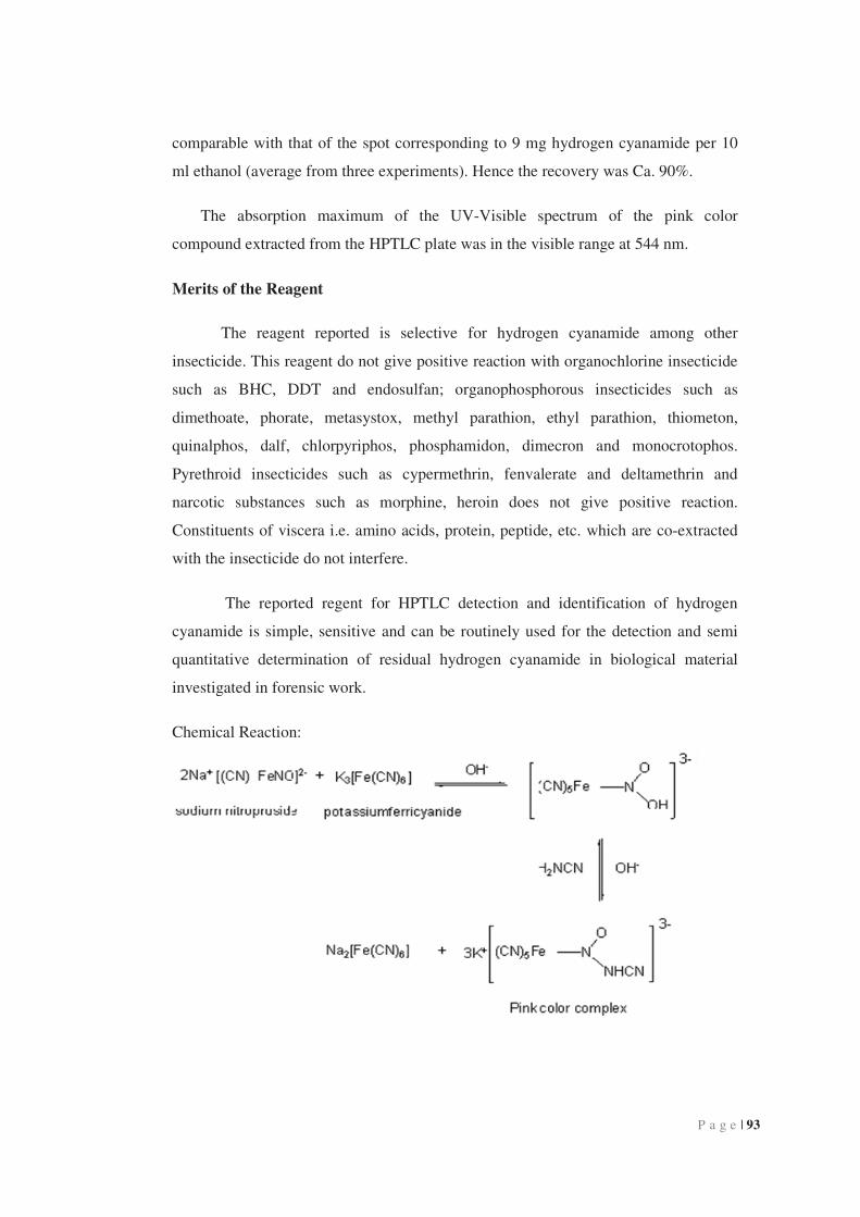
Chemical Reaction:
P a g e | 94
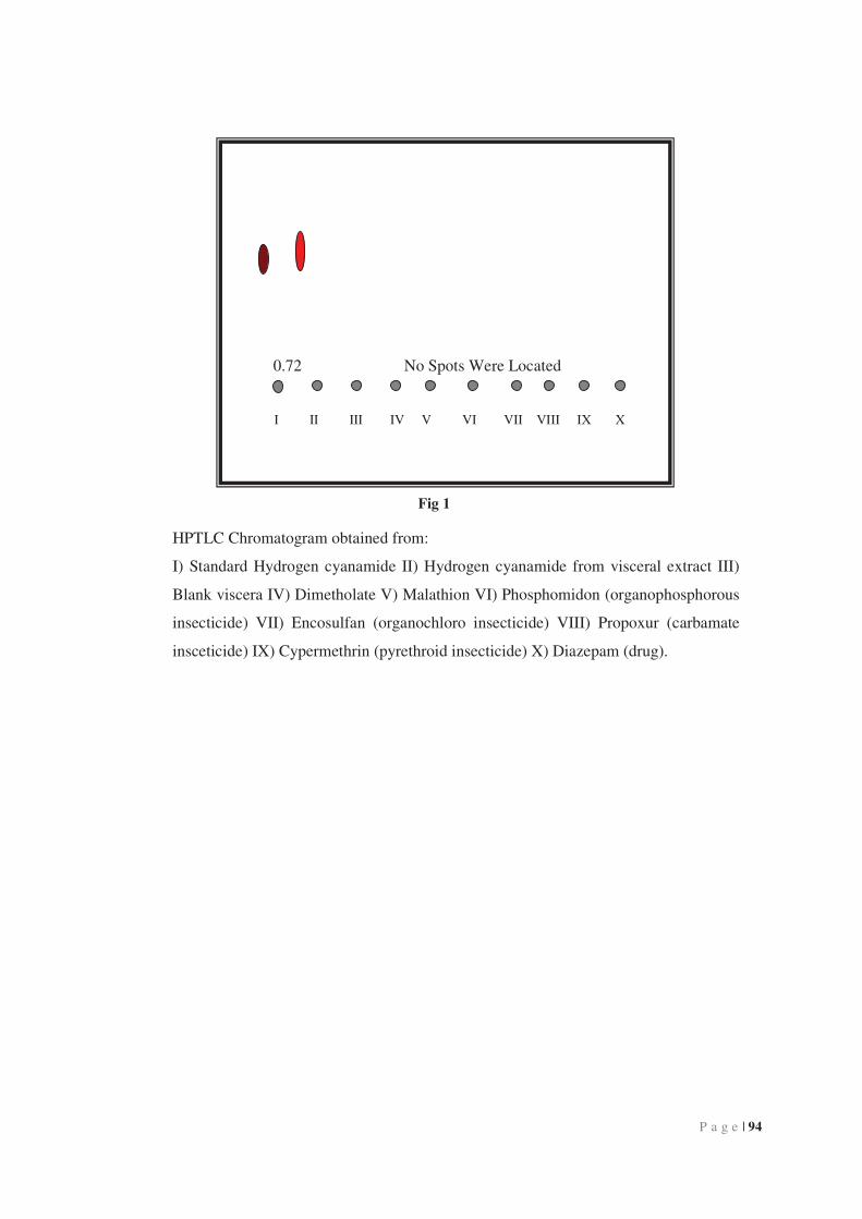
0.72 No Spots Were Located
I II III IV V VI VII VIII IX X
Fig 1
HPTLC Chromatogram obtained from:
I) Standard Hydrogen cyanamide II) Hydrogen cyanamide from visceral extract III)
Blank viscera IV) Dimetholate V) Malathion VI) Phosphomidon (organophosphorous
insecticide) VII) Encosulfan (organochloro insecticide) VIII) Propoxur (carbamate
insceticide) IX) Cypermethrin (pyrethroid insecticide) X) Diazepam (drug).

P a g e | 95
Fig. 2 Color photograph of HPTLC Chromatogram obtained from:
I) Standard hydrogen cyanamide II) Hydrogen cyanamide from visceral extract III)
Blank viscera IV) Dimethoate v) Malathion VI) Phosphomidon (organophosphorous
insecticide) VII) Encosulfan (organochloro insecticide) VIII) Propoxur (carbamate
insecticide) IX) Cypermethrin (pyrethroid insecticide) X) Diazepam (drug).

P a g e | 96
3C.6 References
1. Hathaway, G. J.; Proctor, N. H.; Hughes, J. P., Proctor and Hughe’s chemical
hazards of the workplace, New York, Ed. 4, John Wile & Sons, Inc., 1996,
170.
2. Fritz, Feigl; Spot Test in Organic analysis, Ed. 7, Elsevier Publications Pvt.
Ltd., New Delhi, 1980, 592.
3. Merck; The Merck Index, Merck Rathway, N. J., 1988, Ed. 13, 2713.
4. Egon, Stahl, Thin layer Chromatography, Indian Reprint, Ed. 2, 2005, 551.
5. Wanjie., Wanjie international Co. Ltd., HTML document, 2005.
6. Randerath, K., Thin-layer chromatography, Academic press, New York, 1965,
176.
7. Fritz, feigl, Spot Test in Inorganic analysis, Ed. 6., Elsevier Publication Pvt.
Ltd., New Delhi, 1972, 476.
8. Sicilia, D., Analyst, 1999, 124, 615.
9. Deitrich, R. A.; Troxell, P. A., Biochem Parmacol, 1976, 25, 2733.
10. Kaur, P.; Upadhya, S.; Gupta,V. K., Analyst , 1987, 112, 1681.
11. Agrawal, V.; Cherian, L.; Gupta, V. K.; International Journal of
Environmental Analytical Chemistry, 1991, 45, 235.
12. Amlathe, S.; Gupta, V. K., Journal of Analytical Chemistry, 1990, 338, 615.
13. Rubio, S.; Gomer, A.; Valcarcel, M., Talenta, 1984, 31, 783.
14. Dolizine, T. W.; Espositro, G. G.; Rinehart, D. S., Analytical Chemistry, 1982,
54, 470.
15. Fielden, P. R.; Smith, S. J.; Analyst, 1986, 111, 695.
16. Endecrott, B. R., Journal of Analytical Toxicology, 1996, 20, 189.
17. Miralles, E., Analyst, 1997, 122, 553.
18. Sicilia, D.; Talenta, 1991, 38, 1147.