chapter human bone and dental histology in an...
TRANSCRIPT

CHAPTER 33Human Bone and Dental Histologyin an Archaeological Context
Justyna J. Miszkiewicz1 and Patrick Mahoney21Australian National University, Canberra, ACT, Australia 2University of Kent, Canterbury,United Kingdom
3.1 INTRODUCTION
3.2 BONE
3.3 TEETH
3.4 TECHNICAL CONSIDERATIONS
3.5 HUMAN SKELETAL HISTOLOGY IN MEDIEVAL CANTERBURY,UK: SHORT STUDY
3.5.1 Materials and Methods
3.5.2 Results
3.5.3 Discussion and Summary
3.6 CONCLUSIONS
ACKNOWLEDGMENTS
REFERENCES
3.1 INTRODUCTION
Histological studies of ancient human skeletons can reveal biologicalprocesses and structures that underlie skeletal growth and adaptationin individuals and populations from the archeological past. This infor-mation can be accessed from bones and teeth, which provide two dif-ferent types of microstructural data. Human bone reflects indicators ofits metabolic activity which remodels the skeleton throughout our life-span, adapting, and responding to external and internal factors and sti-muli (such as physical activity, dietary change, disease, aging). Toothenamel structures, on the other hand, serve as a permanent record ofskeletal growth during childhood, because once formed enamel doesnot remodel. The aim of this chapter is to provide an overview of
Human Remains: Another Dimension. DOI: http://dx.doi.org/10.1016/B978-0-12-804602-9.00004-7© 2017 Elsevier Inc. All rights reserved.
Thompson, Tim, and David Errickson. Human Remains: Another Dimension, edited by Tim Thompson, and David Errickson, Elsevier Science, 2017. ProQuest Ebook Central, http://ebookcentral.proquest.com/lib/anu/detail.action?docID=4808408.Created from anu on 2017-04-10 21:08:09.
Cop
yrig
ht ©
201
7. E
lsev
ier
Sci
ence
. All
right
s re
serv
ed.
histological approaches to archeological human adult bone and juve-nile teeth. We then apply this methodology to a British Medieval pop-ulation from Canterbury (11th�16th centuries AD) revealing aspects oftheir skeletal biology.
3.2 BONE
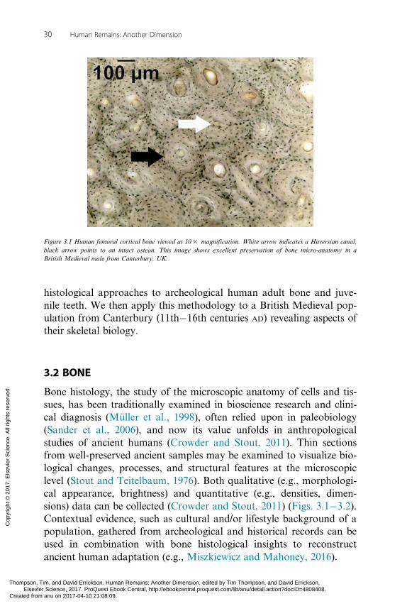
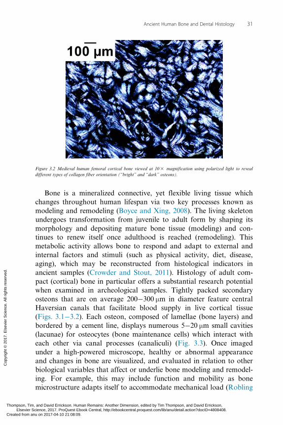
Bone histology, the study of the microscopic anatomy of cells and tis-sues, has been traditionally examined in bioscience research and clini-cal diagnosis (Müller et al., 1998), often relied upon in paleobiology(Sander et al., 2006), and now its value unfolds in anthropologicalstudies of ancient humans (Crowder and Stout, 2011). Thin sectionsfrom well-preserved ancient samples may be examined to visualize bio-logical changes, processes, and structural features at the microscopiclevel (Stout and Teitelbaum, 1976). Both qualitative (e.g., morphologi-cal appearance, brightness) and quantitative (e.g., densities, dimen-sions) data can be collected (Crowder and Stout, 2011) (Figs. 3.1�3.2).Contextual evidence, such as cultural and/or lifestyle background of apopulation, gathered from archeological and historical records can beused in combination with bone histological insights to reconstructancient human adaptation (e.g., Miszkiewicz and Mahoney, 2016).
Figure 3.1 Human femoral cortical bone viewed at 103 magnification. White arrow indicates a Haversian canal,black arrow points to an intact osteon. This image shows excellent preservation of bone micro-anatomy in aBritish Medieval male from Canterbury, UK.
30 Human Remains: Another Dimension
Thompson, Tim, and David Errickson. Human Remains: Another Dimension, edited by Tim Thompson, and David Errickson, Elsevier Science, 2017. ProQuest Ebook Central, http://ebookcentral.proquest.com/lib/anu/detail.action?docID=4808408.Created from anu on 2017-04-10 21:08:09.
Cop
yrig
ht ©
201
7. E
lsev
ier
Sci
ence
. All
right
s re
serv
ed.
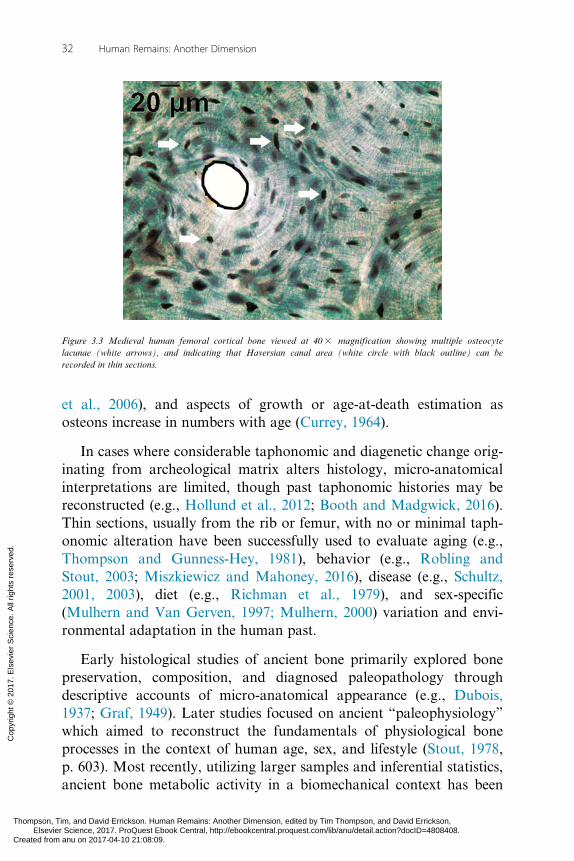
Bone is a mineralized connective, yet flexible living tissue whichchanges throughout human lifespan via two key processes known asmodeling and remodeling (Boyce and Xing, 2008). The living skeletonundergoes transformation from juvenile to adult form by shaping itsmorphology and depositing mature bone tissue (modeling) and con-tinues to renew itself once adulthood is reached (remodeling). Thismetabolic activity allows bone to respond and adapt to external andinternal factors and stimuli (such as physical activity, diet, disease,aging), which may be reconstructed from histological indicators inancient samples (Crowder and Stout, 2011). Histology of adult com-pact (cortical) bone in particular offers a substantial research potentialwhen examined in archeological samples. Tightly packed secondaryosteons that are on average 200�300 μm in diameter feature centralHaversian canals that facilitate blood supply in live cortical tissue(Figs. 3.1�3.2). Each osteon, composed of lamellae (bone layers) andbordered by a cement line, displays numerous 5�20 μm small cavities(lacunae) for osteocytes (bone maintenance cells) which interact witheach other via canal processes (canaliculi) (Fig. 3.3). Once imagedunder a high-powered microscope, healthy or abnormal appearanceand changes in bone are visualized, and evaluated in relation to otherbiological variables that affect or underlie bone modeling and remodel-ing. For example, this may include function and mobility as bonemicrostructure adapts itself to accommodate mechanical load (Robling
Figure 3.2 Medieval human femoral cortical bone viewed at 103 magnification using polarized light to revealdifferent types of collagen fiber orientation (“bright” and “dark” osteons).
31Ancient Human Bone and Dental Histology
Thompson, Tim, and David Errickson. Human Remains: Another Dimension, edited by Tim Thompson, and David Errickson, Elsevier Science, 2017. ProQuest Ebook Central, http://ebookcentral.proquest.com/lib/anu/detail.action?docID=4808408.Created from anu on 2017-04-10 21:08:09.
Cop
yrig
ht ©
201
7. E
lsev
ier
Sci
ence
. All
right
s re
serv
ed.
et al., 2006), and aspects of growth or age-at-death estimation asosteons increase in numbers with age (Currey, 1964).
In cases where considerable taphonomic and diagenetic change orig-inating from archeological matrix alters histology, micro-anatomicalinterpretations are limited, though past taphonomic histories may bereconstructed (e.g., Hollund et al., 2012; Booth and Madgwick, 2016).Thin sections, usually from the rib or femur, with no or minimal taph-onomic alteration have been successfully used to evaluate aging (e.g.,Thompson and Gunness-Hey, 1981), behavior (e.g., Robling andStout, 2003; Miszkiewicz and Mahoney, 2016), disease (e.g., Schultz,2001, 2003), diet (e.g., Richman et al., 1979), and sex-specific(Mulhern and Van Gerven, 1997; Mulhern, 2000) variation and envi-ronmental adaptation in the human past.
Early histological studies of ancient bone primarily explored bonepreservation, composition, and diagnosed paleopathology throughdescriptive accounts of micro-anatomical appearance (e.g., Dubois,1937; Graf, 1949). Later studies focused on ancient “paleophysiology”which aimed to reconstruct the fundamentals of physiological boneprocesses in the context of human age, sex, and lifestyle (Stout, 1978,p. 603). Most recently, utilizing larger samples and inferential statistics,ancient bone metabolic activity in a biomechanical context has been
Figure 3.3 Medieval human femoral cortical bone viewed at 403 magnification showing multiple osteocytelacunae (white arrows), and indicating that Haversian canal area (white circle with black outline) can berecorded in thin sections.
32 Human Remains: Another Dimension
Thompson, Tim, and David Errickson. Human Remains: Another Dimension, edited by Tim Thompson, and David Errickson, Elsevier Science, 2017. ProQuest Ebook Central, http://ebookcentral.proquest.com/lib/anu/detail.action?docID=4808408.Created from anu on 2017-04-10 21:08:09.
Cop
yrig
ht ©
201
7. E
lsev
ier
Sci
ence
. All
right
s re
serv
ed.

investigated (e.g., Pfeiffer et al., 2006; Miszkiewicz and Mahoney,2016). Using principles of bone functional adaptation in relation to dif-ferent mechanical loading regimes, changes in density and geometricproperties of bone histology features have been measured and inter-preted in the light of information about past behavior (e.g., Roblingand Stout, 2003; Miszkiewicz and Mahoney, 2016). For example,human femora from a physically active group had higher density ofosteons when compared to sedentary humans from ancient Peru(Robling and Stout, 2003).
Analytical approaches range from counting or measuring osteons (his-tomorphometry) in images captured under normal transmitted light (e.g.,Miszkiewicz, 2015) (Figs. 3.1�3.2), qualitative evaluation of collagen fiberorientation (CFO) under polarized light (histomorphology) (e.g., Schultz,2001) (Fig. 3.2) where osteon birefringent “rings” can be scored (Skedroset al., 2013), to the use of geographical information systems in mappingcortical bone variation (e.g., Rose et al., 2012; Gocha and Agnew, 2016).One may be more suitable over the other depending on research questionsposed. For example, reconstructing populational trends, such as genderdivision in labor or the effect of socioeconomic status on bone health,from microstructural variation has been achieved using femoral corticalhistomorphometry (e.g., Mulhern, 2000; Miszkiewicz and Mahoney,2016) because it allows for localized bone functional adaptation to bemeasured. Mechanical load adaptation at the osteon level may be betterunderstood from CFO at sites of lower limb tension or compression,because mechanical properties of individual osteons are largely determinedby the arrangement of lamellar collagen fibers (Goldman et al., 2003).
3.3 TEETH
The secretory stage of tooth enamel growth commences in deciduousincisors, early in the second trimester (Kraus and Jordan, 1965;Mahoney, 2015). The first molar is the only permanent tooth to com-mence enamel growth in utero, usually a few weeks before birth(Mahoney, 2008), but this varies greatly between individuals. Growthcontinues after birth in all deciduous and permanent tooth types, fin-ishing in permanent third molar crowns in the late childhood to earlyteenage years (Reid and Dean, 2006; Mahoney, 2015).
Although tooth enamel continues to mature after the crown hasformed, it does not remodel, unlike bone. Thus, tooth crowns preserve
33Ancient Human Bone and Dental Histology
Thompson, Tim, and David Errickson. Human Remains: Another Dimension, edited by Tim Thompson, and David Errickson, Elsevier Science, 2017. ProQuest Ebook Central, http://ebookcentral.proquest.com/lib/anu/detail.action?docID=4808408.Created from anu on 2017-04-10 21:08:09.
Cop
yrig
ht ©
201
7. E
lsev
ier
Sci
ence
. All
right
s re
serv
ed.

within their structure a permanent record of their own growth, in theform of incremental markings. These markings develop as ameloblastsdifferentiate along the dentin�enamel junction (DEJ), and move awayfrom the DEJ, as tooth crowns increase in height and thickness (Boyde,1989). Ameloblasts are cells that secrete enamel matrix proteins.Ameloblast differentiation and secretion is not consistent, which pro-duces incremental markings in the form of cross striations (Fig. 3.4)and Retzius lines (Fig. 3.5). Cross striations occur at a rate that corre-sponds to a circadian rhythm (Lacruz et al., 2012; Zheng et al., 2013).
Figure 3.4 Cross striations (white arrows) in tooth enamel viewed at 403 magnification. Dashed white lineindicates prism direction.
Figure 3.5 Retzius lines (white arrows) in tooth enamel viewed at 103 magnification using polarized light.
34 Human Remains: Another Dimension
Thompson, Tim, and David Errickson. Human Remains: Another Dimension, edited by Tim Thompson, and David Errickson, Elsevier Science, 2017. ProQuest Ebook Central, http://ebookcentral.proquest.com/lib/anu/detail.action?docID=4808408.Created from anu on 2017-04-10 21:08:09.
Cop
yrig
ht ©
201
7. E
lsev
ier
Sci
ence
. All
right
s re
serv
ed.

Retzius lines occur with a periodicity that ranges between 4 and 12days when compared between humans (Mahoney et al., 2016). A thirdtype of incremental marking is a neonatal line, a prominent markingthat occurs at birth (e.g., Sabel et al., 2008). The neonatal line subdi-vides enamel growth into pre- and postnatal periods.
Several macroscopic methods are available to reconstruct toothenamel growth (e.g., Liversidge et al., 1993; Liversidge and Molleson,2004). Histological methods rely upon imaging microstructure, in thinsections, to reconstruct growth by counting and measuring cross stria-tions and Retzius lines. Histological analyses of incremental markingsin archeological samples of modern human deciduous teeth have beenused to document the timing and rate of prenatal enamel growth(Mahoney, 2015), estimate age-at-death (Boyde, 1964), explore the evo-lution of modern human life history (Mahoney, 2015), and reveal bior-hythms underlying enamel and bone growth (Mahoney et al., 2016).
3.4 TECHNICAL CONSIDERATIONS
Samples removed for histological analyses of bone can be sufficientlysmall (0.5�1 cm in height), making the technique minimally invasive(e.g., Pfeiffer et al., 2006; Miszkiewicz and Mahoney, 2016). Dentalhistology studies can also rely on sampling one tooth per individual(e.g., Mahoney, 2008). Accepted standards and ethics for destructivesampling should be followed (Mays et al., 2013). Increasingly efficientand cost-effective ancient skeletal sample preparation techniques andways of equipping laboratories are available (De Boer et al., 2012,2013; Paine, 2007), encouraging the inclusion of histology in bioarch-aeological schools and research centers. Extracted skeletal samplesrequire embedding in resin or plastic and are then usually cut on a pre-cision low speed saw, ground, and polished (Bancroft and Gamble,2002) to yield final thin sections that can be stored for future analyses.These normally range from 50 to 100 μm in thickness (Crowder andStout, 2011). A high-powered microscope (e.g., Olympus BX, or lessexpensive CX series) is necessary to achieve a relatively high magnifi-cation (603 to 1003 ) if cell-related features (e.g., osteocyte lacunae)are to be studied, though a range of 103 to 403 magnification suf-fices for the examination of osteons, and their features. Images of thinsections are captured with a mounted microscope camera (e.g.,Olympus DP, or less expensive CS series). Data are recorded andquantified using an imaging software with a range of drawing and
35Ancient Human Bone and Dental Histology
Thompson, Tim, and David Errickson. Human Remains: Another Dimension, edited by Tim Thompson, and David Errickson, Elsevier Science, 2017. ProQuest Ebook Central, http://ebookcentral.proquest.com/lib/anu/detail.action?docID=4808408.Created from anu on 2017-04-10 21:08:09.
Cop
yrig
ht ©
201
7. E
lsev
ier
Sci
ence
. All
right
s re
serv
ed.

point counting tools (e.g., CELL Live Biology Imaging, OlympusStream, or free and open access ImageJ).
Given that bone undergoes remodeling, the selection of regions ofinterest (ROIs) within a thin section should be consistent to ensure rep-licability of studies, meaningful data comparisons, and appropriateevaluation of research questions. Different techniques have beenreported, including the use of an eyepiece grid micrometer (e.g.,Mulhern and Van Gerven, 1997), evaluation of bone strips (Roblingand Stout, 2003), subparts of the cortex (Miszkiewicz, 2015;Miszkiewicz and Mahoney, 2016), examining countable osteons (e.g.,Pfeiffer et al., 2006) or entire thin section (Thompson and Gunness-Hey, 1981). However, localized taphonomic and diagenetic changemay complicate study design. Though it has been shown that differentROI selection procedures yield matching results (Villa and Lynnerup,2010), it is recommended that a minimum of 25�50 osteons should bestudied per thin section (Crowder and Stout, 2011).
Standard histological sectioning of teeth relies upon capturing one2D plane that travels through the tip of the enamel cusp, the tip of thedentin horn, and into the most cervical enamel. Mineralization of teeth(and in particular enamel) is much higher compared to bone, whichmeans that dental samples are less susceptible to diagenetic and tapho-nomic change (Hollund et al., 2015). The thickness of sections willvary depending upon the degree of mineralization within a crown.Usually, sections of deciduous teeth are between 70 and 120 μm inthickness. Cross striations and Retzius lines are recorded at a magnifi-cation of 203 to 603 . The region examined within a tooth crownwill depend upon the variable that is recorded. Cross striations can beused to calculate the rate of enamel secretion in cuspal (occlusal)enamel, which is usually subdivided into inner, mid, and outer regions.Retzius lines can be used to calculate periodicity of lateral enamelregions. Image capturing and recording is undertaken with appropriatesoftware such as Olympus DP and CELL Live Biology Imaging.
3.5 HUMAN SKELETAL HISTOLOGY IN MEDIEVALCANTERBURY, UK: SHORT STUDY
The Skeletal Biology Research Centre at the University of Kent(Canterbury, UK) curates several hundred human skeletons that
36 Human Remains: Another Dimension
Thompson, Tim, and David Errickson. Human Remains: Another Dimension, edited by Tim Thompson, and David Errickson, Elsevier Science, 2017. ProQuest Ebook Central, http://ebookcentral.proquest.com/lib/anu/detail.action?docID=4808408.Created from anu on 2017-04-10 21:08:09.
Cop
yrig
ht ©
201
7. E
lsev
ier
Sci
ence
. All
right
s re
serv
ed.

include subadults and adults, males and females, and individuals witha range of pathologies. The collection represents a late Medieval(11th�16th centuries AD) British population from high-status St.Gregory’s Priory and adjacent low-status cemetery in Canterbury (seeHicks and Hicks, 2001; Miszkiewicz, 2012). As revealed through arche-ological excavations, the high-status group had few and rich burialslocated inside the Priory (Hicks and Hicks, 2001). Historical evidenceidentifies the adjacent cemetery to be designated for poor and sick pea-sants and those who could not afford burial in the higher status site(Brent, 1879). Over the past few years we have been exploring thebioarchaeology of these individuals using skeletal histology.
Recently, we reported significant variation in femoral bone micro-structure between the two distinct burial groups (Miszkiewicz andMahoney, 2016). We demonstrated that differences in histomorpho-metric parameters (such as secondary osteon density, area and dimeterof transverse cross-sectional osteon and vascular surfaces) correspondedwith adult lifestyle information for each status groups. When comparedto those of low status, high-status adults had increased osteon densitieswith larger osteon and Haversian canal surfaces, indicating goodhealth, nutrition, but sedentary lifestyle. Our interpretations were basedon basic engineering principles of bone microstructure functional adap-tation which indicate the deposition of smaller osteons with increasingload (e.g., van Oers et al., 2008), and dietary influences on bone remo-deling which suggest an increase in bone density with surplus of calories(e.g., Richman et al., 1979; Paine and Brenton, 2006).
Dental histology research on juveniles from this archeological siterevealed that deciduous enamel growth commenced in deciduous inci-sors early in the second trimester and finished around the end of thefirst postnatal year in second molars (Mahoney, 2015). Enamel growthappears to be coordinated, to some extent, with primary bone growth(Mahoney et al., 2016). An underlying biorhythm correspondswith greater production of enamel and primary bone matrix.
In this brief study, we extend the above findings to demonstrate theapplication of histology images from archeological bones and teeth:
1. We compare femoral histology data within the low-status cemeterybetween two adult age groups to explore ancient aging and behav-ior. Though all low-status individuals usually undertook physically
37Ancient Human Bone and Dental Histology
Thompson, Tim, and David Errickson. Human Remains: Another Dimension, edited by Tim Thompson, and David Errickson, Elsevier Science, 2017. ProQuest Ebook Central, http://ebookcentral.proquest.com/lib/anu/detail.action?docID=4808408.Created from anu on 2017-04-10 21:08:09.
Cop
yrig
ht ©
201
7. E
lsev
ier
Sci
ence
. All
right
s re
serv
ed.

active occupations (Dyer, 1989), we predict bone microstructure toreflect adaptation to larger and more frequent mechanical loadresulting from more regular and strenuous activities in the youngermales (Dyer, 2000, 2002) when compared to the middle-agedmales.
2. Using cross striations, Retzius and neonatal lines in teeth, we com-pare the proportion of pre- and postnatal enamel growth in decid-uous maxillary molars.
3.5.1 Materials and MethodsA total of 199 femora from adult British Medieval males fromCanterbury were selected for a histomorphometric analysis. Age-at-death and sex were estimated following standard anthropologicalmethods based on gross skeletal anatomy examination (Buikstra andUbelaker, 1994). Two age groups of “young” 20�34 (n5 44) and“middle-aged” 35�50 years old (n5 155) individuals were created.Osteon population density (OPD as #/mm2) and Haversian canalarea (H.Ar in μm2) were recorded in images of thin sections removedfrom the posterior femur (see Miszkiewicz, 2015 and Miszkiewiczand Mahoney, 2016) and compared between the young and middle-aged adults using a Mann�Whitney U test accounting for unequalsample size.
Forty-eight deciduous maxillary molars (dm1, dm2) from juvenilesrecovered in Medieval Canterbury were sectioned using standard histo-logical methods (see Mahoney, 2015; and new data here). Total crownformation times, subdivided by the proportion of pre- and postnatalenamel in these tooth types, were calculated from daily enamel secre-tion rates, Retzius periodicity, and measures of enamel thickness com-bined with the location of the neonatal line.
3.5.2 ResultsBoth OPD (n5 186, U5 3813.000, p5 0.006) and H.Ar (n5 199,U5 4200.000, p5 0.019) were significantly higher in middle-agedmales when compared to the younger group (Fig. 3.6). The totalcrown formation time was 415 days for dm1 and 539 days for dm2.Thirty-three percent of dm1 enamel and 15% of dm2 enamel formedbefore birth (Fig. 3.7).
38 Human Remains: Another Dimension
Thompson, Tim, and David Errickson. Human Remains: Another Dimension, edited by Tim Thompson, and David Errickson, Elsevier Science, 2017. ProQuest Ebook Central, http://ebookcentral.proquest.com/lib/anu/detail.action?docID=4808408.Created from anu on 2017-04-10 21:08:09.
Cop
yrig
ht ©
201
7. E
lsev
ier
Sci
ence
. All
right
s re
serv
ed.

3.5.3 Discussion and SummaryOur aim in this brief study was to demonstrate that skeletal micro-structure data can be recorded in images captured from thin sectionsand used to reconstruct aspects of ancient human life in the past. Wefound an increase in OPD (#/mm2) from young to middle-aged adultsin agreement with previous ancient bone histology research (e.g.,Thompson and Gunness-Hey, 1981) and attribute it to age-relatedincrease in osteons (Currey, 1964). As predicted, we also observed theyoung males to have smaller Haversian canals (μm2) indicating
Figure 3.6 Graph illustrating OPD and Haversian canal area differences between the two age groups of males.
Figure 3.7 Pre- and postnatal enamel growth in a maxillary deciduous first molar. Black arrow points to theneonatal line.
39Ancient Human Bone and Dental Histology
Thompson, Tim, and David Errickson. Human Remains: Another Dimension, edited by Tim Thompson, and David Errickson, Elsevier Science, 2017. ProQuest Ebook Central, http://ebookcentral.proquest.com/lib/anu/detail.action?docID=4808408.Created from anu on 2017-04-10 21:08:09.
Cop
yrig
ht ©
201
7. E
lsev
ier
Sci
ence
. All
right
s re
serv
ed.

adaptation to larger strains (Miszkiewicz and Mahoney, 2016), andthus corresponding with the age-specific differences in Medieval life-styles and occupations (Bennett and Hollister, 2006; Dyer, 1989, 2000,2002). For example, younger Medieval peasant males tended to engagein agricultural occupations that involved heavy load carrying subject-ing their lower limbs to rigorous walking and additional stress originat-ing from the carried weight (Gransden, 1972). While the older peasantmales also engaged in substantial walking, their load carrying responsi-bilities would have been reduced (if not abandoned completely) due tohealth deterioration, aging, and age group specific division of labor(Bennett and Hollister, 2006). However, we emphasize that the intri-cate processes of bone growth require cautious interpretations ofancient human past and, ideally, comparisons with experimental orcontemporary data.
The average proportion of enamel growth in this sample of decid-uous maxillary molars is slightly greater when compared to samples ofmandibular molars from Medieval Canterbury (Mahoney, 2011;dm15 29%, dm25 16%). This is most likely due to slight differences inenamel initiation times in utero, in order to facilitate slightly thickermaxillary molar enamel within the total crown formation period.Knowledge of the proportions of pre- and postnatal enamel growthcan be used to assess changes in growth trajectories in ancient com-pared to modern day populations. They can also contribute to age-at-death estimations in a bioarchaeological and forensic context. Takentogether, our results reveal insights into skeletal growth and adaptationin this Medieval population.
3.6 CONCLUSIONS
Histological analyses of ancient human skeletons can reveal the under-lying mechanisms that facilitate morphological change during growth,aging, and disease. Imaging and analyzing these microscopic structuresholds great potential for revealing the skeletal biology of past humanpopulations.
ACKNOWLEDGMENTS
We thank Tim Thompson for inviting us to contribute to this volume and tworeviewers for invaluable feedback. The Royal Society funded equipment (PM), bone
40 Human Remains: Another Dimension
Thompson, Tim, and David Errickson. Human Remains: Another Dimension, edited by Tim Thompson, and David Errickson, Elsevier Science, 2017. ProQuest Ebook Central, http://ebookcentral.proquest.com/lib/anu/detail.action?docID=4808408.Created from anu on 2017-04-10 21:08:09.
Cop
yrig
ht ©
201
7. E
lsev
ier
Sci
ence
. All
right
s re
serv
ed.

histology data were collected during a PhD studentship (JJM) funded by the School ofAnthropology and Conservation, University of Kent. Elle Grono (Geoarchaeology,Australian National University) facilitated microscope imaging in Figs. 3.1 and 3.3.
REFERENCESBancroft, J.D., Gamble, M., 2002. Theory and Practice of Histological Techniques. ChurchillLivingstone, London.
Bennett, J., Hollister, C.W., 2006. Medieval Europe: A Short History. McGraw-Hill, New York.
Booth, T.J., Madgwick, R., 2016. New evidence for diverse secondary burial practices in IronAge Britain: a histological case study. J. Archaeol. Sci. 67, 14�24.
Boyce, B.F., Xing, L., 2008. Functions of RANKL/RANK/OPG in bone modeling and remodel-ing. Arch. Biochem. Biophys. 473, 139�146.
Boyde, A., 1964. Estimation of age at death of young human skeletal remains from incrementallines in dental enamel. Third International Meeting in Forensic Immunology, Medicine,Pathology and Toxicology, Plenary Session 11A. 16�24 April 1963, London. Excerpta Med IntCongr Ser, vol. 80.
Boyde, A., 1989. Enamel. In: Berkovitz, B.K.B., Boyde, A., FrankRM, Hohling, H.J., Moxham, B.J.,Nalbandian, J., Tonge, C.H. (Eds.), Teeth. Handbook of Microscopic Anatomy. Springer-Verlag,Berlin, pp. 309�473.
Brent, J., 1879. Canterbury in the Olden Time. Simpkin, Marshall and Co, London.
Buikstra, J.E., Ubelaker, D.H. 1994. Standards for Data Collection from Human SkeletalRemains. Arkansas Archaeology Survey, Fayetteville.
Crowder, C., Stout, S., 2011. Bone Histology: An Anthropological Perspective. CRC Press, BocaRaton, FL.
Currey, J.D., 1964. Some effects of ageing in human Haversian systems. J. Anat. 98 (1), 69�75.
De Boer, H.H., Aarents, M.J., Maat, G.J.R., 2012. Staining ground sections of natural dry bonetissue for microscopy. Int. J. Osteoarchaeol. 22, 379�386.
De Boer, H.H., Aarents, M.J., Maat, G.J.R., 2013. Manual for the preparation and staining ofembedded natural dry bone tissue sections for microscopy. Int. J. Osteoarchaeol. 23, 83�93.
Dubois, E., 1937. The osteone arrangement of the thigh-bone compacta of man identical withthat first found of Pithecanthropus. Proc. R. Acad. Amsterdam 38, 850�852.
Dyer, C., 1989. Standards of Living in the Later Middle Ages: Social Change in England,1200�1520. Cambridge University Press.
Dyer, C., 2000. Everyday Life in Medieval England. Cambridge University Press.
Dyer, C., 2002. Making a Living in the Middle Ages. Yale University Press.
Gocha, T.P., Agnew, A.M., 2016. Spatial variation in osteon population density at the humanfemoral midshaft: histomorphometric adaptations to habitual load environment. J. Anat. 228 (5),733�745.
Goldman, H.M., Bromage, T.G., Thomas, C.D.L., Clement, J.G., 2003. Preferred collagen fiberorientation in the human mid-shaft femur. Anat. Rec. 272A, 434�445.
Graf, W., 1949. Preserved histological structures in Egyptian mummy tissues and ancient Swedishskeletons. Cells Tissues Organs 8, 236�250.
Gransden, A., 1972. Childhood and youth in Mediaeval England. Nottingham Medieval Studies16 (1), 3�19.
41Ancient Human Bone and Dental Histology
Thompson, Tim, and David Errickson. Human Remains: Another Dimension, edited by Tim Thompson, and David Errickson, Elsevier Science, 2017. ProQuest Ebook Central, http://ebookcentral.proquest.com/lib/anu/detail.action?docID=4808408.Created from anu on 2017-04-10 21:08:09.
Cop
yrig
ht ©
201
7. E
lsev
ier
Sci
ence
. All
right
s re
serv
ed.
Hicks, M., Hicks, A., 2001. St. Gregory’s Priory, Northgate, Canterbury Excavations 1988-1991(p. Volume II). Canterbury Archaeological Trust Ltd.
Hollund, H.I., Jans, M.M.E., Collins, M.J., Kars, H., Joosten, I., Kars, S.M., 2012. What hap-pened here? Bone histology as a tool in decoding the postmortem histories of archaeological bonefrom Castricum, the Netherlands. Int. J. Osteoarchaeol. 22 (5), 537�548.
Hollund, H.I., Arts, N., Jans, M.M.E., Kars, H., 2015. Are teeth better? Histological characteri-zation of diagenesis in archaeological bone�tooth pairs and a discussion of the consequences forarchaeometric sample selection and analyses. Int. J. Osteoarchaeol. 25 (6), 901�911.
Kraus, B., Jordan, R., 1965. The Human Dentition Before Birth. Lea & Febiger, Philadelphia.
Lacruz, R.S., Hacia, J.G., Bromage, T.G., Boyde, A., Lei, Y., Xu, Y., et al., 2012. The circadianclock modulates enamel development. J. Biol. Rhythms 27 (3), 237�245.
Liversidge, H.M., Molleson, T.I., 2004. Variation in crown and root formation and eruption ofhuman deciduous teeth. Am. J. Phys. Anthropol. 123 (2), 172�180.
Liversidge, H.M., Dean, M.C., Molleson, T.I., 1993. Increasing human tooth length betweenbirth and 5.4 years. Am. J. Phys. Anthropol. 90, 307�313.
Mahoney, P., 2008. Intraspecific variation in M 1 enamel development in modern humans: impli-cations for human evolution. J. Hum. Evol. 55, 131�147.
Mahoney, P., 2011. Human deciduous mandibular molar incremental enamel development. Am.J. Phys. Anthropol. 144, 204�214.
Mahoney, P., 2015. Dental fast track: prenatal enamel growth, incisor eruption, and weaning inhuman infants. Am. J. Phys. Anthropol. 156, 407�421.
Mahoney, P., Miszkiewicz, J.J., Pitfield, R., Schlecht, S.H., Deter, C., Guatelli-Steinberg, D.,2016. Biorhythms, deciduous enamel thickness, and primary bone growth in modern human chil-dren: a test of the Havers�Halberg Oscillation hypothesis. J. Anat. 228, 919�928.
Mays, S., Elders, J., Humphrey, L., White, W., Marshall, P., 2013. Science and the Dead:A Guidelines for the Destructive Sampling of Archaeological Human Remains for ScientificAnalysis. Advisory Panel on the Archaeology of Burials in England. English Heritage.
Miszkiewicz, J.J., 2012. Linear enamel hypoplasia and age-at-death at Medieval (11th�16th cen-turies) St. Gregory’s Priory and Cemetery, Canterbury, UK. Int. J. Osteoarchaeol. 25, 79�87.
Miszkiewicz, J.J., 2015. Investigating histomorphometric relationships at the human femoral mid-shaft in a biomechanical context. J. Bone Miner. Metab. 34, 179�192.
Miszkiewicz, J.J., Mahoney, P., 2016. Ancient human bone microstructure and behaviour inmedieval England: comparisons between two socio-economic groups. Anat. Rec. 299, 42�59.
Mulhern, D.M., 2000. Rib remodeling dynamics in a skeletal population from Kulubnarti,Nubia. Am. J. Phys. Anthropol. 111, 519�530.
Mulhern, D.M., Van Gerven, D.P., 1997. Patterns of femoral bone remodeling dynamics in aMedieval Nubian population. Am. J. Phys. Anthropol. 104, 133�146.
Müller, R., Van Campenhout, H., Van Damme, B., Van der Perre, G., Dequeker, J., Hildebrand,T., et al., 1998. Morphometric analysis of human bone biopsies: a quantitative structural compar-ison of histological sections and micro-computed tomography. Bone 23, 59�66.
Paine, R.R., 2007. How to equip a basic histological lab for the anthropological assessment ofhuman bone and teeth. J. Anthropol. Sci. 85, 213�219.
Paine, R.R., Brenton, B.P., 2006. Dietary health does affect histological age assessment: an evalu-ation of the Stout and Paine (1992) age estimation equation using secondary osteons from the rib.J. Forensic Sci. 51, 489�492.
Pfeiffer, S., Crowder, C., Harrington, L., Brown, M., 2006. Secondary osteon and Haversiancanal dimensions as behavioral indicators. Am. J. Phys. Anthropol. 468, 460�468.
42 Human Remains: Another Dimension
Thompson, Tim, and David Errickson. Human Remains: Another Dimension, edited by Tim Thompson, and David Errickson, Elsevier Science, 2017. ProQuest Ebook Central, http://ebookcentral.proquest.com/lib/anu/detail.action?docID=4808408.Created from anu on 2017-04-10 21:08:09.
Cop
yrig
ht ©
201
7. E
lsev
ier
Sci
ence
. All
right
s re
serv
ed.
Reid, D.J., Dean, M.C., 2006. Variation in modern human enamel formation times. J. Hum.Evol. 50, 329�346.
Richman, E.A., Ortner, D.J., Schulter-Ellis, F.P., 1979. Differences in intracortical bone remodel-ing in three aboriginal American populations: possible dietary factors. Calcif. Tissue Int. 28,209�214.
Robling, A.G., Stout, S.D., 2003. Histomorphology, geometry, and mechanical loading in pastpopulations. In: Agarwal, S.C., Stout, S.D. (Eds.), Bone Loss and Osteoporosis: AnAnthropological Perspective. Kluwer Academic/Plenum Publishers, New York, pp. 189�206.
Robling, A.G., Castillo, A.B., Turner, C.H., 2006. Biomechanical and molecular regulation ofbone remodeling. Annu. Rev. Biomed. Eng. 8, 455�498.
Rose, D.C., Agnew, A.M., Gocha, T.P., Stout, S.D., Field, J.S., 2012. Technical note: the use ofgeographical information systems software for the spatial analysis of bone microstructure. Am. J.Phys. Anthropol. 148, 648�654.
Sabel, N., Johansson, C., Kuhnisch, J., Robertson, A., Steiniger, F., Nore�n, J.G., et al., 2008.Neonatal lines in the enamel of primary teeth—a morphological and scanning electron micro-scopic investigation. Arch. Oral. Biol. 53, 954�963.
Sander, P.M., Mateus, O., Laven, T., Knötschke, N., 2006. Bone histology indicates insulardwarfism in a new Late Jurassic sauropod dinosaur. Nature 441, 739�741.
Schultz, M., 2001. Paleohistopathology of bone: a new approach to the study of ancient diseases.Am. J. Phys. Anthropol. 116, 106�147.
Schultz, M., 2003. Light microscopic analysis in skeletal paleopathology. In: Ortner, D.J. (Ed.),Identification of Pathological Conditions in Human Skeletal Remains. Elsevier Science, London,pp. 73�109.
Skedros, J.G., Keenan, K.E., Williams, T.J., Kiser, C.J., 2013. Secondary osteon size and colla-gen/lamellar organization (“osteon morphotypes”) are not coupled, but potentially adapt indepen-dently for local strain mode or magnitude. J. Struct. Biol. 181 (2), 95�107.
Stout, S.D., 1978. Histological structure and its preservation in ancient bone. Curr. Anthropol.19, 601�604.
Stout, S.D., Teitelbaum, S.L., 1976. Histological analysis of undecalcified thin sections of archeo-logical bone. Am. J. Phys. Anthropol. 44, 263�269.
Thompson, D.D., Gunness-Hey, M., 1981. Bone mineral-osteon analysis of Yupik-Inupiaq skele-tons. Am. J. Phys. Anthropol. 55, 1�7.
van Oers, R.F., Ruimerman, R., van Rietbergen, B., Hilbers, P.A., Huiskes, R., 2008. Relatingosteon diameter to strain. Bone 43, 476�482.
Villa, C., Lynnerup, N., 2010. Technical note: a stereological analysis of the cross-sectional vari-ability of the femoral osteon population. Am. J. Phys. Anthropol. 142, 491�496.
Zheng, L., Seon, Y.J., Mourão, M.A., Schnell, S., Kim, D., Harada, H., et al., 2013. Circadianrhythms regulate amelogenesis. Bone 55, 158�165.
43Ancient Human Bone and Dental Histology
Thompson, Tim, and David Errickson. Human Remains: Another Dimension, edited by Tim Thompson, and David Errickson, Elsevier Science, 2017. ProQuest Ebook Central, http://ebookcentral.proquest.com/lib/anu/detail.action?docID=4808408.Created from anu on 2017-04-10 21:08:09.
Cop
yrig
ht ©
201
7. E
lsev
ier
Sci
ence
. All
right
s re
serv
ed.

This page intentionally left blank
Thompson, Tim, and David Errickson. Human Remains: Another Dimension, edited by Tim Thompson, and David Errickson, Elsevier Science, 2017. ProQuest Ebook Central, http://ebookcentral.proquest.com/lib/anu/detail.action?docID=4808408.Created from anu on 2017-04-10 21:08:09.
Cop
yrig
ht ©
201
7. E
lsev
ier
Sci
ence
. All
right
s re
serv
ed.