chapter effects in food-producing animals
TRANSCRIPT
Chapter 5. Effects in food-producing animals 59
chapter 5.
Effects in food-producing animals
Summary
Unexplained disease outbreaks in farm and domestic animals have suggested the likely presence of mycotoxins in feeds for many years. The manifestations of mycotoxicoses in the field are frequently nondescript and potentially have many contrib-uting factors, which are often difficult to define. Nevertheless, toxigenic moulds were implicated in, and sometimes proven to be the cause of, animal disease in field outbreaks long before the toxins were discovered. The development of methods for the chemical analysis of mycotoxins in feeds and animal tissues has contributed to an improved understanding of the dose–response relationships of farm animal diseases associated with exposure to aflatoxins, fumonisins, ochratoxin A, deoxynivalenol, zearalenone, and
ergot alkaloids. In all cases the effect of mycotoxins on animal performance is potentially a major problem for farmers regardless of their scale of operation. Reduced growth, decreased egg and milk production, lower reproductive efficiency, and increased susceptibility to stress are all potentially devastating consequences of mycotoxin exposure. Thus, being aware of the outward signs that might signal the involvement of a mycotoxin in an animal performance problem is the first step to minimizing potential adverse impacts. The target organ affected can provide important clues to involvement of a specific mycotoxin, in which case understanding the toxicokinetics and toxicology will assist in minimizing the cost and maximizing the effectiveness of interventions. The primary objective of this chapter is to provide information that will aid in the field identification of the
possible involvement of a mycotoxin in an animal production problem. In conjunction with the information provided in the other chapters, this information will assist farmers in making decisions that will minimize losses due to diseases induced by mycotoxins.
1. Introduction
In this chapter, we discuss the effects in farm and domestic animals of the most economically important feedborne mycotoxins. The chapter begins with a summary of field outbreaks, followed by sections that describe the toxicokinetics and metabolism of each mycotoxin or group of mycotoxins. Of the trichothecenes, only deoxynivalenol is covered in depth; however, it should be recognized that the predominant trichothecene encountered in con-
CH
AP
TER
5
60
taminated feeds may differ in different geographical locations (Starkey et al., 2007; Miller, 2008; Sugita-Konishi and Nakajima, 2010).
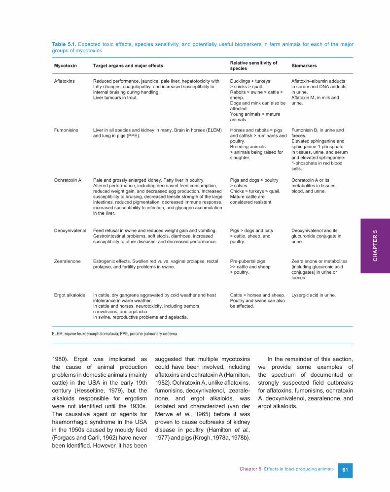
Additional information relevant to the toxicology in animals can be found in Chapter 6, which focuses on effects and toxicology in humans. Potential interventions to prevent field outbreaks or minimize their adverse effects are described in Chapter 9. Numerous extensive review articles and monographs provide additional information on effects in farm animals (WHO, 2001, 2011; CAST, 2003; Cousin et al., 2005; Roberts et al., 2005; Fink-Gremmels and Malekinejad, 2007; Morgavi and Riley, 2007a, 2007b; Voss et al., 2007; Fribourg et al., 2009; Pestka, 2010a, 2010b; Eaton et al., 2010). To aid in the field identification of the main observations characterizing exposure to a particular mycotoxin, Table 5.1 summarizes the expected toxic effects, species sensitivity, and potentially useful biomarkers in farm animals for each of the major groups of mycotoxins.
2. Field outbreaks
The interest of the scientific and regulatory communities in mycotoxins began in earnest when aflatoxins were found to be potent carcinogens that occurred in several important food and feed commodities. However, even in the absence of any known causal agent, unexplained disease outbreaks in farm and domestic animals, which often involved mortality or evidence of acute toxicity, served as an indicator of the likely presence of mycotoxins in foodstuffs (Forgacs and Carll, 1962).
Definitively linking a disease outbreak in the field to a specific mycotoxin is very difficult (Hamilton, 1982), even though a great deal of experimental evidence exists to characterize the potential of mycotoxins and mouldy foodstuffs
to cause animal disease. A major problem for the veterinarian in the field is that disease expression is seldom pathognomonic, i.e. with a sign or symptom that is so characteristic of a disease as to be diagnostic. Instead, multiple interacting factors often occur that can modify the expression of toxicity. Thus, the manifestations of mycotoxicoses in the field are frequently nondescript and have many potential contributing factors, which are often difficult to define.
An attempt to apply Koch’s postulates to demonstrate causality requires a bioassay that reproduces the disease when a pure compound is used. Unfortunately, reproducing the exact conditions that existed in the field is confounded by the potential presence of multiple contributing factors. These include, but are not limited to, environmental stress, multiple toxigenic fungi and mycotoxins, nutrient/vitamin deficiencies, infectious agents, and pre-existing conditions. These co-occurring factors can influence the clinical signs, severity, and progression of the disease (CAST, 2003) in ways that can confound both diagnosis and replication of the observed adverse effects. Also, mycotoxins in feeds are not evenly distributed (see Chapter 3); therefore, reproducing disease at the dosages found in feed samples analysed from field outbreaks can be difficult. It has been suggested that 100–200 kg or more of suspect feed should be saved for confirmatory studies in experimental animals (Osweiler, 2000). In addition, experimental confirmation can require a large number of animals if the incidence of the disease in the suspected field outbreak is low.
Despite these difficulties, toxi-genic moulds were implicated as the cause of animal disease in field outbreaks long before the toxins were discovered. This was
the case for aflatoxins, fumonisins, deoxynivalenol, zearalenone, and ergot alkaloids, where mould-contaminated feed was associated with disease outbreaks before the toxins were identified. For example, in the 1960s the outbreak of a disease known as turkey “X” disease led to the discovery of aflatoxins. However, before this disease was reported, outbreaks of liver cancer in farm-raised rainbow trout fed diets containing cottonseed (Butler, 1974) and of liver toxicity in pigs and cattle fed maize contaminated with Aspergillus flavus were documented in 1935 and 1953, respectively (Raisbeck et al., 1991). Equine leukoencephalomalacia (ELEM) was first linked to mouldy maize in 1891 (Haliburton and Buck, 1986). ELEM is now known to be caused by fumonisins. Effects from the consumption of maize on which Fusarium verticillioides had been cultured were the first indication that mouldy maize might cause porcine pulmonary oedema (PPE) syndrome (Kriek et al., 1981). This was confirmed only after the discovery of fumonisins in 1988 and after the cause of outbreaks of PPE in the USA in 1989–1990 was confirmed by inducing PPE with pure fumonisin B1 (Marasas, 2001).
Feed refusal syndrome in the USA, most likely caused by deoxynivalenol and other trichothecenes, led to an embargo of United States barley by Germany in the 1930s (Hamilton, 1982). The association between consumption of mouldy feed and estrogenism in pigs has been known since 1928 (McNutt et al., 1928), and estrogenism has been attributed to consumption of feeds contaminated with zearalenone. The biological activity of ergot (the sclerotia of Claviceps spp.) was known in China more than 5000 years ago even though its involvement in animal disease was probably not reported until the Middle Ages (Christensen,
Chapter 5. Effects in food-producing animals 61
1980). Ergot was implicated as the cause of animal production problems in domestic animals (mainly cattle) in the USA in the early 19th century (Hesseltine, 1979), but the alkaloids responsible for ergotism were not identified until the 1930s. The causative agent or agents for haemorrhagic syndrome in the USA in the 1950s caused by mouldy feed (Forgacs and Carll, 1962) have never been identified. However, it has been
suggested that multiple mycotoxins could have been involved, including aflatoxins and ochratoxin A (Hamilton, 1982). Ochratoxin A, unlike aflatoxins, fumonisins, deoxynivalenol, zearale-none, and ergot alkaloids, was isolated and characterized (van der Merwe et al., 1965) before it was proven to cause outbreaks of kidney disease in poultry (Hamilton et al., 1977) and pigs (Krogh, 1978a, 1978b).
In the remainder of this section, we provide some examples of the spectrum of documented or strongly suspected field outbreaks for aflatoxins, fumonisins, ochratoxin A, deoxynivalenol, zearalenone, and ergot alkaloids.
Mycotoxin Target organs and major effects Relative sensitivity of species Biomarkers
Aflatoxins Reduced performance, jaundice, pale liver, hepatotoxicity with fatty changes, coagulopathy, and increased susceptibility to internal bruising during handling.Liver tumours in trout.
Ducklings > turkeys > chicks > quail.Rabbits > swine > cattle > sheep.Dogs and mink can also be affected.Young animals > mature animals.
Aflatoxin–albumin adducts in serum and DNA adducts in urine.Aflatoxin M1 in milk and urine.
Fumonisins Liver in all species and kidney in many. Brain in horses (ELEM) and lung in pigs (PPE).
Horses and rabbits > pigs and catfish > ruminants and poultry.Breeding animals > animals being raised for slaughter.
Fumonisin B1 in urine and faeces.Elevated sphinganine and sphinganine-1-phosphate in tissues, urine, and serum and elevated sphinganine-1-phosphate in red blood cells.
Ochratoxin A Pale and grossly enlarged kidney. Fatty liver in poultry. Altered performance, including decreased feed consumption, reduced weight gain, and decreased egg production. Increased susceptibility to bruising, decreased tensile strength of the large intestines, reduced pigmentation, decreased immune response, increased susceptibility to infection, and glycogen accumulation in the liver.
Pigs and dogs > poultry > calves.Chicks > turkeys > quail.Mature cattle are considered resistant.
Ochratoxin A or its metabolites in tissues, blood, and urine.
Deoxynivalenol Feed refusal in swine and reduced weight gain and vomiting. Gastrointestinal problems, soft stools, diarrhoea, increased susceptibility to other diseases, and decreased performance.
Pigs > dogs and cats > cattle, sheep, and poultry.
Deoxynivalenol and its glucuronide conjugate in urine.
Zearalenone Estrogenic effects. Swollen red vulva, vaginal prolapse, rectal prolapse, and fertility problems in swine.
Pre-pubertal pigs >> cattle and sheep > poultry.
Zearalenone or metabolites (including glucuronic acid conjugates) in urine or faeces.
Ergot alkaloids In cattle, dry gangrene aggravated by cold weather and heat intolerance in warm weather.In cattle and horses, neurotoxicity, including tremors, convulsions, and agalactia.In swine, reproductive problems and agalactia.
Cattle > horses and sheep.Poultry and swine can also be affected.
Lysergic acid in urine.
Table 5.1. Expected toxic effects, species sensitivity, and potentially useful biomarkers in farm animals for each of the major groups of mycotoxins
ELEM, equine leukoencephalomalacia; PPE, porcine pulmonary oedema.
CH
AP
TER
5
62
2.1 Aflatoxins
Turkey “X” disease was responsible for the deaths of > 100 000 turkeys in the United Kingdom in 1960. Mortality was also documented in ducks, chickens, pheasant, calves, and pigs (Butler, 1974). In the USA and elsewhere, field outbreaks causing mortality have been well documented in turkeys, laying hens, pigs, cattle, rainbow trout, and dogs (Butler, 1974; Hamilton, 1982). In the case of poultry, pigs, and farm-raised trout, large numbers of animals were involved (Butler, 1974; Hamilton, 1982). Acute toxicity is easily recognized, but the more subtle effects are probably of greater concern to farmers. For example, decreased rates of weight gain, decreased milk or egg production, increased susceptibility to bruising during processing of poultry and pigs, underpigmentation of meat in poultry, and altered immune function have all been associated with exposure in the field (Hamilton, 1982; Raisbeck et al., 1991). Pet food recalls due to aflatoxin contamination resulting in liver toxicity and death in dogs are not uncommon in the USA.
2.2 Fumonisins
In the USA, field outbreaks of ELEM caused by mouldy maize have been reported for more than 100 years (Haliburton and Buck, 1986). It was reported that about 5000 horses died of “mouldy corn poisoning” in Illinois in 1934–1935 (Haliburton and Buck, 1986). The disease has also been reported in South America, Hungary, China, Greece, France, Mexico, New Caledonia, Egypt, South Africa, and Germany (Magnol et al., 1983; Haliburton and Buck, 1986; Laurent et al., 1998; Rosiles et al., 1998). After identifying Fusarium verticillioides (then known as F. moniliforme) as the predominant fungal contaminant of mouldy maize that had caused
cases of ELEM in Egypt, Wilson and Maronpot (1971) reproduced ELEM in horses by feeding them maize on which this fungus had been grown. Kriek et al. (1981) induced PPE, a disease that has been known in Hungary since 1950 (Fazekas et al., 1998), by feeding pigs maize on which F. verticillioides had been grown. After isolating and chemically characterizing the fumonisins, Mara-sas et al. (1988) induced ELEM in a horse by using purified fumonisin B1. Serendipitously, in 1989–1990 there were numerous outbreaks of ELEM and PPE syndrome in the USA. In 1990, PPE was induced using pure fumonisin B1 (Harrison et al., 1990). Field outbreaks of PPE have not occurred in the USA since the early 1990s, but reports of ELEM have persisted. In poultry, reduced performance associated with feeds contaminated with fumonisins has been reported, but the effects seldom involve increased mortality (WHO, 2000).
2.3 Ochratoxin A
In pigs, mycotoxic nephropathy was first described in 1928 in Denmark and was reproduced experimentally using mouldy barley or oats (Krogh, 1992). This disease has also been reported in Norway, Sweden, Ireland, Finland, Germany, Hungary, Poland, the former Serbia and Montenegro, and Bulgaria (Krogh, 1978a, 1978b; Marquardt and Frohlich, 1992; Stoev et al., 1998). The main clinical signs are renal dysfunction and oedema. In pigs, excessive thirst (polydipsia) and passage of large volumes of urine (polyuria) are characteristic signs of this disease under field conditions. The association of ochratoxin A (OTA) with field outbreaks of nephropathy in poultry was first reported in 1975 (Krogh, 1992) and has been reported in many countries since then (Marquardt and Frohlich, 1992).
Outbreaks of mycotoxic nephropathy have also been reported in horses (Krogh, 1978b), and there are a few suspected cases in ruminants (Raisbeck et al., 1991). The first well-documented and confirmed case of ochratoxicosis occurred in young turkeys in the south-eastern USA (Hamilton et al., 1977). In 1976, about 80% of the marketed turkeys in North Carolina displayed signs of ochratoxicosis, and all cases involved maize contaminated with OTA (Schaeffer and Hamilton, 1986). In a turkey house with 16 000 turkeys, mortality was 59%. The first indication of intoxication was feed refusal (Schaeffer and Hamilton, 1986). Death was most commonly seen in younger birds, whereas older birds developed infections with Escherichia coli (air-sacculitis) that did not respond to antibiotics. Interestingly, in outbreaks in chickens, feed refusal did not occur, whereas poor pigmentation was common (Schaeffer and Hamilton, 1986). Other field cases in chickens have reported decreased egg production and eggshell quality, increased susceptibility to intestinal rupture during processing, decreased bone strength, and haemorrhagic episodes (Schaeffer and Hamilton, 1986). In all species, effects on the kidney were apparent.
2.4 Deoxynivalenol
Trichothecenes have been implicated in feed refusal by cattle, pigs, and chickens; however, pigs appear to be the most sensitive to deoxynivalenol (DON) (Rotter et al., 1996; Haschek et al., 2002). Outbreaks in animals of “red mould poisoning” from cereals in Japan as far back as 1890 (Miller, 2008) led to the discovery of DON. It was first isolated in Japan and was originally called Rd-toxin (Moorooka et al., 1972). It was given the name vomitoxin (Vesonder et al.,
Chapter 5. Effects in food-producing animals 63
1973) after its isolation from maize contaminated with F. graminearum because it is associated with emesis in pigs. Numerous confirmed cases have been reported of feed refusal or reduced feed intake in pigs consuming feeds contaminated with DON (Osweiler, 2000). The most frequently observed effect of DON in farm animals is feed refusal, which may explain why toxic effects are to a great extent self-limiting (Osweiler, 2000). There have been unconfirmed reports of feed refusal in dogs, cats, and rabbits consuming pet foods naturally contaminated with DON (Bohm and Razzazi-Fazeli, 2005). In ruminants, field disease outbreaks attributed to DON are rare (Raisbeck et al., 1991).
2.5 Zearalenone
Estrogenism in pigs consuming mouldy feed was first reported in 1928 (McNutt et al., 1928); since then, field outbreaks of reproductive problems, including vulvovaginitis and anestrus, and enlargement of the mammae in males have been attributed to consumption of feeds contaminated with zearalenone (ZEA) (Aucock et al., 1980; Raisbeck et al., 1991), which was previously called F-2 toxin (Mirocha et al., 1968). Field outbreaks of estrogenic syndrome in pigs have been reported in North America, Europe, Africa, Asia, and Australia (Christensen, 1979). ZEA-induced abortions in pigs have also been reported and are probably a result of embryo implantation failure (Osweiler et al., 1986). Suggestions that reproductive problems in ruminants can be attri-buted to ZEA are considered to be controversial (Raisbeck et al., 1991). The semisynthetic ZEA analogue zeranol is used in cattle as a growth promoter, and its estrogenic effects have caused reduced performance in bulls (Raisbeck et al., 1991).
Reproductive problems have been reported in sheep grazing on grasses contaminated with ZEA (CAST, 2003), and ZEA was found to interfere with the induction of parturition by oxytocin in gilts and sows (Alexopoulos, 2001). In maize contaminated with ZEA, co-contamination with DON is likely.
2.6 Ergot alkaloids
Poisoning resulting from the con-sumption of foods contaminated by ergots, which are fungal sclerotia that replace grass seeds, has been known since the Middle Ages, but it was not until the 20th century that ergot alkaloids from Claviceps purpurea were determined to be the causative agent (Barger, 1931). Ergot alkaloids associated with or suspected to be involved in diseases of farm animals include ergoline alkaloids, which contain the lysergic acid ring structure, and ergopeptine alkaloids, which contain a tripeptide moiety. In farm animals, ergot poisoning is usually associated with grazing on seed heads of grasses contaminated with ergots of Claviceps spp. or grasses colonized by groups of fungi that occur as endophytic symbionts (Neotyphodium spp. and Epichloë spp.; Roberts et al., 2005) or with the consumption of feeds contaminated with ergot (CAST, 2003). Outbreaks of ergot poisoning have also been reported in pigs and cattle consuming sorghum-based feeds. Consumption of feeds based on small grains, such as rye and barley, or foraging on infected grasses contaminated with ergots has caused lameness or necrosis of ears, tails, and feet in cattle (CAST, 2003). Endophytes (Neotyphodium spp. and Epichloë spp.) that occur in pasture grasses such as tall fescue and ryegrass are known to produce ergot alkaloids, which have caused a diverse array of toxic syndromes in grazing animals, including reproductive
problems and neurological effects in horses (staggers, “drunkenness”, and sleep) and reduced reproductive performance, decreased milk produc-tion, and reduced growth in cattle (Cross, 2003; White et al., 2003). In cattle and sheep, increased body temperature due to peripheral vasoconstriction is often seen in animals that have ingested grasses infected by endophytes (Cross, 2003). Field cases of fescue foot (necrosis of the extremities) in cattle and laminitis in horses have been reported to be due to peripheral vasoconstriction caused by ergot alkaloids (Cross, 2003). Environmental conditions appear to influence the vasoconstrictive effects of ergot alkaloids in grasses; episodes of fescue foot are most common in autumn and early winter, and the condition known as summer slump is most common in warm weather (Raisbeck et al., 1991).
3. Toxicokinetics andmetabolism
Mycotoxins are chemically diverse, and therefore their uptake, distribution, metabolism, and excretion are equally diverse. Galtier (1998) summarized the kinetics and biological fate of most of the economically important mycotoxins. Uptake from the gastrointestinal tract depends to a large extent on the water solubility or lipophilicity of the particular mycotoxin. Metabolism by microbes in the gut can also have a profound effect on uptake and toxicity. Some mycotoxins, like OTA, are well absorbed, whereas others, like fumonisin B1, are very poorly absorbed. Binding of mycotoxins to plasma proteins can influence uptake, distribution, and the half-life in the blood. Again, some mycotoxins, including OTA, are tightly bound, whereas others, including fumonisin B1, are not bound. Metabolism and excretion are important considerations
CH
AP
TER
5
64
for determining whether residues will remain in tissues and also for developing biomarkers for exposure. For example, aflatoxin B1 is extensively metabolized in the liver and elsewhere, and significant amounts can be excreted as metabolites in milk. For DON, most of the absorbed toxin is excreted in urine, whereas only very small amounts of fumonisin B1 are excreted in urine. Fumonisin B1 is not metabolized in the liver or other tissues and therefore is recovered intact or partially hydrolysed (probably by microbes in the gastrointestinal tract) in faeces. Although mycotoxin residues can be carried over into milk and eggs (Galtier, 1998), only aflatoxin M1 and OTA have been detected naturally in cow’s milk.
3.1 Aflatoxins
3.1.1 Absorption
Aflatoxin B1 absorption, distribution, and elimination is rapid. Aflatoxin B1 is well absorbed; it accumulates in the liver and is extensively metabolized in the liver and other tissues. The binding of metabolites to macromolecules, including proteins and nucleic acids, also occurs soon after absorption. Unbound water-soluble metabolites are excreted in urine and other fluids, and the parent compound is excreted in faeces; however, some metabolites bound to nucleic acids can persist for relatively long periods in tissues.
3.1.2 Gastrointestinal metabolism
Little published information is avail-able about the ability of micro-organisms in the gastrointestinal tract to metabolize or bind aflatoxins. It is known, however, that certain bacteria can bind aflatoxin B1 in vitro, and it has been suggested that they may aid intestinal excretion (Oatley et al., 2000).
3.1.3 Bioavailability
Aflatoxin B1 is rapidly absorbed from the small intestines, and the rate of absorption is much higher in suckling and young animals than in adults (Kumagai, 1985). The process of absorption from the small intestines is passive and complete. For example, the time needed to reduce aflatoxin B1 in the intestinal lumen by 50% is about 7 minutes (Ramos and Hernández, 1996). Aflatoxins B1 and B2 are more rapidly absorbed than are aflatoxins G1 and G2, ensuring that their bioavailability is high (Ramos and Hernández, 1996). Aflatoxin B1 is also absorbed slowly through the skin, and intratracheal absorption is more rapid than via the oral route (IARC, 1993a). Once absorbed, aflatoxin B1 is non-covalently bound to albumin and is transported to other tissues.
3.1.4 Distribution
Early distribution studies focused on extraction of the parent compounds or non-polar aflatoxin metabolites and ignored the water-soluble metabolites (Busby and Wogan, 1981a). In sheep and other animals, aflatoxin B1 is immediately transported to the liver after absorption from the gut (Wilson et al., 1985). The half-life of aflatoxin B1 in plasma after intravenous dosing is < 1 hour (Wong and Hsieh, 1980), whereas after intratracheal or oral dosing it is about 90 hours (Coulombe and Sharma, 1985). As with absorption from the small intestines, aflatoxin B1 is rapidly taken up by the liver with a half-life of < 5 minutes (Busby and Wogan, 1981a). Aflatoxin B1 is widely distributed in the body, with most accumulating in the liver, kidney, and lung. Maximal levels in tissues, and especially the liver, are reached quickly, and the relative retention of aflatoxin B1 is greater than that of aflatoxin B2 (Busby and Wogan, 1981a; IARC,
1993a). Low levels of aflatoxin B1 and aflatoxin M1 (< 1 µg/kg) were detected in the liver and kidneys of lactating cows fed diets containing 1250 µg/kg aflatoxin B1 for 2 weeks (Busby and Wogan, 1981a). Pigs fed diets containing 100–400 µg/kg aflatoxin B1 for 4 weeks had detectable levels of aflatoxins B1 and M1 in the liver, muscle, kidney, and blood. At 400 µg/kg aflatoxin B1 in feed, the detected levels of aflatoxins B1 and M1 were as high as 4 µg/kg and 1.5 µg/kg, respectively (Busby and Wogan, 1981a). In chickens, maximal levels were attained in plasma and the liver within 6 hours after oral dosing, and levels declined rapidly thereafter (Hirano et al., 1994). After either a single oral dose or daily oral dosing for 2 weeks, aflatoxin residues were detected in various organs and in muscle tissue in laying hens (Busby and Wogan, 1981a). Aflatoxins can cross the placenta and accumulate in developing fetuses (IARC, 1993a). Unmetabolized aflatoxin B1 can accumulate in tissues rich in melanin pigment and the upper respiratory tract (Larsson and Tjälve, 1993).
3.1.5 Excretion
After intraperitoneal or intravenous dosing of aflatoxin B1, excretion is rapid and most of the dose is eliminated in faeces and urine (Wong and Hsieh, 1980; Busby and Wogan, 1981a). The relative retention of aflatoxin B1 is greater than that of aflatoxin B2 due to greater urinary excretion of aflatoxin B2 (Busby and Wogan, 1981a). A large percentage of the intraperitoneal dose is recovered in bile (Busby and Wogan, 1981a). In sheep, aflatoxin residues, mainly aflatoxin M1, are excreted primarily in urine within 48 hours after oral dosing (Busby and Wogan, 1981a). Aflatoxin M1 is the main unconjugated metabolite of aflatoxin B1 excreted in the milk of sheep,
Chapter 5. Effects in food-producing animals 6565
goats, and cows. In sheep and cattle, aflatoxin B1 is the main component in faeces. In laying hens, aflatoxin B1 excretion is primarily in faeces and is maximal 24 hours after a single oral dose, a finding similar to that seen in laboratory animals dosed intravenously (Wong and Hsieh, 1980). In laying hens, the body half-life of aflatoxin B1 was 67 hours after a single oral dose (Busby and Wogan, 1981a).
3.1.6 Transmission
Approximately 1–15% (IARC, 2002a) of the aflatoxin B1 dose is recovered as aflatoxin M1 in the milk of sheep, goats, and cows (IARC, 1993a). The conversion rate is > 1% when the aflatoxin B1 dose is low (IARC, 2002a). Extensive evidence has been found for the lactational transfer of aflatoxin M1 and its accumulation in the livers and lungs of offspring (IARC, 1993a). Low levels of aflatoxin metabolites can be detected in the liver and other tissues several weeks after animals are exposed to high levels of aflatoxin B1 (Busby and Wogan, 1981a; Coulombe and Sharma, 1985). However, the levels retained in edible tissues are generally low; feed-to-tissue ratios range from 800:1 to 14 000:1 (IARC, 2002a). Aflatoxin B1 can be detected in eggs of laying hens fed diets containing aflatoxin B1, with a feed-to-tissue ratio ranging from 2200:1 to 5000:1 (Oliveira et al., 2000; IARC, 2002a). With the exception of milk, transmission to edible animal products should pose little health risk to consumers.
3.1.7 Metabolism
The metabolism of aflatoxin B1 has been extensively reviewed (IARC, 1993a, 2002a; see also Chapter 6). Briefly, in the liver and other tissues, aflatoxin B1 is metabolized in
microsomal systems to aflatoxins P1, M1, and Q1 and, most importantly, the highly reactive aflatoxin B1-8,9-epoxide. In the liver, cytochrome P450 enzymes are responsible for activation of aflatoxins B1, M1, and P1, all of which can form nucleic acid adducts or undergo conjugation to glutathione, conversion to dihydrodiols, or binding to serum proteins or other macromolecules. Aflatoxin M1 is the main unconjugated metabolite in the urine of cows, pigs, and sheep. In rodents, and presumably in farm animals, aflatoxin B1–nucleic acid adducts are also found in urine, and 80% of the depurinating adducts are excreted within 48 hours after dosing. A close correlation has been established between levels of adducts in urine and levels in the liver (IARC, 1993a), and correlations have also been observed between dietary intake and levels of adducts in urine and serum (IARC, 2002a).
The relative sensitivity of animals to the toxic effects of aflatoxin B1 is closely linked to differences in metabolism among species (IARC, 2002a). The susceptibility of animals to aflatoxin B1 toxicity and carcinogenicity depends to a large extent on the type of metabolites produced and the rate of formation and detoxification of the aflatoxin B1-8,9-epoxide. Risk factors contributing to an individual’s sensitivity to liver tumours and hepatotoxicity include level of exposure to aflatoxin B1; expression of enzymes in the aflatoxin activation and detoxification pathways; nutritional status; co-exposure to other mycotoxins, es-pecially fumonisin; and exposure to infectious agents (see Chapter 6). Evidence suggests that the critical factor determining species sensitivity is the rate at which the aflatoxin B1-8,9-epoxide can be conjugated to glutathione by glutathione S-transferase (GST) (Eaton et
al., 2010). For example, domestic turkeys, one of the most susceptible species to aflatoxicosis, are known to be deficient in the GST that mediates detoxification of the aflatoxin B1-8,9-epoxide (Klein et al., 2000), and that deficiency results in high levels of hepatic aflatoxin B1 epoxidation (Yip and Coulombe, 2006; Rawal et al., 2009).
3.2 Fumonisins
3.2.1 Absorption
In most animals, fumonisin B1 absorption, distribution, and elim-ination is rapid. Fumonisins are poorly absorbed, and although some evidence exists that fumonisins can be partially metabolized in the gut, metabolism by the liver or other tissues has not been convincingly demonstrated (WHO, 2000, 2001, 2012; IARC, 2002b; Shephard et al., 2007; Voss et al., 2007).
3.2.2 Gastrointestinal metabolism
Microbial metabolism most likely occurs in the gut of monogastric animals, because partially hydrolysed fumonisin B1 (lacking one tricarballylic acid side chain) and, to a lesser extent, fully hydrolysed fumonisin B1 (lacking both side chains) were recovered in faeces but not in bile of vervet monkeys (WHO, 2000). Most (60–90%) of the total fumonisin B1 found in ruminant faeces was present as the partially hydrolysed form. In non-ruminants, the parent compound was the dominant species present (WHO, 2000). However, studies in pigs have reported significant amounts of fully hydrolysed and partially hydrolysed fumonisin B1 in faeces and tissues (Fodor et al., 2008). Whether the hydrolysed fumonisin B1 was produced in the tissues was not determined.
CH
AP
TER
5
66
3.2.3 Bioavailability
In all animals studied, including pigs, laying hens, turkey poults, ducks, and dairy cows, the fumonisin absorption that does occur is rapid and the quantity of fumonisin B1 detected in plasma and tissues after oral administration is very low (negligible to < 4% of dose) (WHO, 2000, 2001, 2012; IARC, 2002b; Fodor et al., 2008; Tardieu et al., 2008, 2009). The bioavailability of fumonisin B2 may be less than that of fumonisin B1. Feeding studies have shown that, in the liver and kidney, fumonisin B1 is accumulated to a much greater extent than expected based on the relative amounts in feed of fumonisin B1, fumonisin B2 (Riley and Voss, 2006; Fodor et al., 2008; Gazzotti et al., 2010), and fumonisin B3 (Riley and Voss, 2006). Although diets containing predominantly fumonisin B2 from culture material induced liver toxicity in both rats and horses (Riley et al., 1997; Voss et al., 1998), pure fumonisin B2 did not induce liver toxicity in mice in one feeding study (Howard et al., 2002). In a cultured intestinal epithelial cell model, hydrolysed fumonisin B1, but not fumonisin B1, was found to cross the monolayer (primarily from basolateral to apical), suggesting a carrier-mediated efflux process (De Angelis et al., 2005).
3.2.4 Distribution
Although fumonisins are distributed to most tissues, the liver and kidney retain the highest concentrations of the absorbed material in all animals studied (reviewed in Voss et al., 2007). Fumonisin B1 persists in the kidney much longer than in plasma or the liver, and in male Sprague Dawley (Riley and Voss, 2006) and Wistar rats (Martinez-Larranaga et al., 1999), the levels of fumonisin B1 in the kidney can be 10 times the amount in the liver. Radiolabeled fumonisin has
been detected in the brains of pigs (Prelusky et al., 1996a), but little or no fumonisin has been detected in the brain tissue of horses, although the brain is a known target organ (Haschek et al., 2002). Until recently it was believed that fumonisins could not cross the placenta and enter the developing embryo (WHO, 2001). However, recent studies have detected [14C]fumonisin B1 in embryos and placentas, an observation confirmed by the presence of elevated levels of free sphinganine, a biomarker for fumonisin inhibition of ceramide synthase (Gelineau-van Waes et al., 2005).
3.2.5 Excretion
After intraperitoneal or intravenous dosing of fumonisin B1, initial elim-ination from tissues is rapid, with no evidence of metabolism (Shephard et al., 2007; Voss et al., 2007), but extensive enterohepatic circulation occurs (Prelusky et al., 1996a). After oral dosing, peak plasma levels occur within 1 hour to several hours. Several studies using different routes of exposure and different animal species have shown that fumonisins are excreted primarily in faeces, either unchanged or with loss of one or both of the tricarballylic acid side chains. Low levels of fumonisin B1 can be detected in the urine of animals exposed experimentally to fumonisin, including rabbits (Orsi et al., 2009), rats (Cai et al., 2007), pigs (Fodor et al., 2008; Dilkin et al., 2010), horses (Tumbleson et al., 2003), and vervet monkeys (Shephard et al., 2007). In pigs, < 1% of the oral dose is recovered in urine (Prelusky et al., 1996a; Dilkin et al., 2010). It has been estimated that pigs exposed to dietary fumonisin B1 at 2–3 mg/kg body weight (bw) in feed would require a withdrawal period of at least 2 weeks for the fumonisin B1 to be eliminated from the liver and kidney (Prelusky et al., 1996a, 1996b).
Several studies have confirmed this finding using the persistence of free sphinganine as a biomarker in the kidney and liver to show that although fumonisin B1 is rapidly eliminated, the biomarker remains elevated for a much longer period. Although the half-life after oral dosing is not known, the oral half-life is probably between 8 hours and 48 hours based on what is known from the parenteral routes, the time required to reach peak levels in plasma (1–7 hours) after gavage, and the estimated time for complete clearance from the liver and kid-ney (2 weeks) (WHO, 2000).
3.2.6 Transmission
Little evidence exists to suggest significant transfer of fumonisins through milk (WHO, 2000). No fumonisin B1 was detected in the milk of lactating sows fed diets containing nonlethal levels of fumonisin B1, and no evidence was found of toxicosis in their suckling pigs. In a study with lactating cows administered fumonisin B1 intravenously, the carry-over of fumonisin B1 into the milk was either very small or not detected. The fact that very little fumonisin B1 is retained in any tissue, milk, or eggs has led to the conclusion that fumonisin residues in food products derived from animals are insufficient to render them injurious to consumers (WHO, 2000). However, in a study in weaned piglets fed diets containing fumonisin B1 at 10 mg/kg or 30 mg/kg diet, the livers contained 306 μg/kg or 830 μg/kg of fumonisin B1, respectively, after 28 days (Dilkin et al., 2003). At the higher level of contamination, a 70 kg person would need to consume 170 g of liver to exceed the Joint WHO/FAO Expert Committee on Food Additives (JECFA) provisional maximum tolerable daily intake (PMTDI) of 2 μg/kg bw/day. Pigs fed diets containing low levels (1.66 mg/animal/day) of fumonisins for 7 weeks
Chapter 5. Effects in food-producing animals 6767
had detectable, but much lower, levels of fumonisins (15–43 μg/kg) in the liver (Gazzotti et al., 2010). Levels of fumonisin detected in the muscle of pigs or poultry orally dosed with fumonisin were either very low or below the detection limits (Prelusky et al., 1996a; Tardieu et al., 2008).
3.2.7 Metabolism
Fumonisins do not appear to be metabolized in vitro or in vivo by animal tissues (WHO, 2001), even though they are clearly excreted in bile, and hydrolysed and partially hydrolysed fumonisin B1 has been reported in tissues (Fodor et al., 2008). The source of hydrolysed fumonisins in tissues is unknown. However, formation of hydrolysed fumonisin during alkaline processing (nixtamalization) and by microbial metabolism in the gastrointestinal tract has been demonstrated (see Section 3.2.2). One study suggested that a cytochrome P450 isoform is capable of producing a fumonisin B1 metabolite (Marvasi et al., 2006), but no convincing evidence has been reported of in vivo or in vitro metabolism by cytochrome P450, the microsomal esterase, or any other microsomal enzyme (WHO, 2001, 2012). However, studies have shown that cytochrome P450 activity can be altered as a result of the inhibition of the enzyme ceramide synthase by fumonisin (WHO, 2001). Both B fumonisins and their hydrolysed counterparts can react with ceramide synthase; hydrolysed fumonisin B1 is a substrate for this enzyme, produc-ing the compound N-palmitoyl-AP1 (Seiferlein et al., 2007). Studies using hydrolysed fumonisin B1 have shown that it is much less toxic than the parent compound (Howard et al., 2002; Collins et al., 2006; Voss et al., 2009; Grenier et al., 2012).
Even though little evidence has been found for metabolism of B fumonisins in tissues, chemical
acylation of the free primary amino group prevents toxicity and reduces the ability of the fumonisin to inhibit ceramide synthase (Norred et al., 1997, 2001). The two tricarballylic acid side chains are also important for toxicity, as demonstrated by reduced or no toxicity, or evidence for disruption of sphingolipid metabolism, when fumonisin is hydrolysed (Howard et al., 2002; Collins et al., 2006; Voss et al., 2009; Grenier et al., 2012).
3.3 Ochratoxin A
3.3.1 Absorption
Comprehensive reviews of the pharmacokinetics and metabolism of OTA are available (Marquardt and Frohlich, 1992; WHO, 2001; Dietrich et al., 2005; Pfohl-Leszkowicz and Manderville, 2007). OTA is rapidly absorbed. The half-life in plasma depends on the extent of binding to plasma proteins. OTA is widely distributed; in pigs, it is accumulated in the kidney and other tissues and can occur in edible tissues. OTA and its metabolites are reabsorbed by the kidney and excreted in urine and also undergo enterohepatic circulation and excretion in faeces. The potential exists for extensive metabolism by microbes to less toxic metabolites in the gastrointestinal tract. Ochratoxins A and B can also be metabolized by cytochrome P450 enzymes in various tissues.
3.3.2 Gastrointestinal metabolism
In cows and sheep, OTA is degraded by the rumen flora and protozoa (WHO, 2001). However, some portion is not metabolized and can accumulate in serum, tissues, and milk (IARC, 1993b; Höhler et al., 1999). The enzymes responsible for the metabolism are carboxypeptidase A and chymotrypsin. Antibiotics that inhibit
the intestinal microbial population can also reduce the hydrolysis of OTA to its non-toxic metabolite, ochratoxin α, leading to increased blood concentrations of OTA (WHO, 2001). In pigs, ochratoxin B is metabolized much more efficiently to ochratoxin β than OTA is to ochratoxin α. The rate of disappearance of OTA from rumen fluids and its gastrointestinal metabolism depend on diet (Müller et al., 2001) and are affected by the amount of feed concentrates (feed additives with a high nutrient density) added to the diet (Höhler et al., 1999).
3.3.3 Bioavailability
OTA is well absorbed from the gastrointestinal tract, presumably from the small intestines, and it is also well absorbed from the lungs, with a calculated bioavailability of 98% (WHO, 2001). Bioavailability from the oral route is reported as ranging from 40% to 70% in chickens, rabbits, and pigs. Absorbed OTA is rapidly and tightly bound to serum proteins and in that form is only slowly transferred from the bloodstream to the liver and kidney. The high affinity of OTA for serum albumin results in a higher concentration in plasma than in the gut (Kumagai, 1988). The maximal concentration in serum after a single oral dose is attained in < 1 hour in chickens and in 10–48 hours in pigs (WHO. 2001). The plasma half-life of OTA is quite variable, ranging from a few hours in chickens to 5 days in pigs.
3.3.4 Distribution
In pigs, chickens, and goats, the relative tissue distribution of OTA is usually kidney > liver or muscle > fat (WHO, 2001; Biró et al., 2002; Dietrich et al., 2005). In rabbits, the relative distribution is kidney > liver > mammary gland > muscle (Ferrufino-Guardia et al., 2000). OTA has been shown to cross the blood–brain
CH
AP
TER
5
68
barrier in rats and the placenta in rats and pigs (WHO, 2001). The rate of disappearance of OTA from the blood is much slower than that from the kidney or liver and other tissues, indicating the importance of binding to serum proteins and enterohepatic recirculation in the overall fate of OTA once it has been absorbed. In cells, OTA is accumulated via a multispecific organic anion transporter (O’Brien and Dietrich, 2005). The accumulation and persistence of OTA in the kidney is due to its reabsorption by the organic anion transporter (Zingerle et al., 1997; Welborn et al., 1998). The ability of the kidney to accumulate OTA plays an important role in its nephrotoxicity. In pigs fed OTA for 1 month, the half-life of residues in the kidney, liver, and muscle was 3.5–4.5 days (Busby and Wogan, 1981b).
3.3.5 Excretion
Elimination of OTA is primarily via the urine, but significant amounts can also be excreted in faeces. OTA is eliminated slowly because it undergoes enterohepatic recir-culation, is reabsorbed by the kidney, and binds tightly to serum protein (WHO, 2001). The extent of albumin binding also markedly decreases the uptake of OTA by transporters (Bow et al., 2006). The elimination half-life is much shorter for ochratoxins B and C than for OTA and its metabolites (Li et al., 1997).
3.3.6 Transmission
OTA has been shown to be transferred efficiently to milk in rodents and rabbits (WHO, 2001). At high levels of exposure, OTA can accumulate in eggs of chickens and quail (WHO, 2001). Detectable levels have been reported in pig kidney and liver, blood products, and other meat products for human consumption (WHO, 2001; CAST, 2003).
3.3.7 Metabolism
In addition to gastrointestinal degra-dation by microbes, microsomal preparations from rabbits and pigs containing various cytochrome P450 enzymes can oxidize OTA to less toxic hydroxyochratoxin A (WHO, 2001). At least 20 OTA derivatives have been identified after incubation with liver microsomes or cultured cells, and hydroxylated metabolites have been detected in pig kidney (Pfohl-Leszkowicz and Manderville, 2007). Metabolism by cyclo-oxygenases and other oxidative enzymes can produce reactive oxygen species, leading to oxidative damage to tissues. OTA has been suggested to induce DNA adducts (see Pfohl-Leszkowicz and Manderville, 2007, and Chapter 6).
3.4 Deoxynivalenol
3.4.1 Absorption
Depending on geographical location, the predominant trichothecenes may be DON, nivalenol (NIV), or one of their acetylated precursors. However, DON is the focus of this section. For more information specific to NIV and acetylated derivatives of DON and NIV, see Pestka (2010a) and Sugita-Konishi and Nakajima (2010).
Absorption, distribution, and elim-ination of DON are rapid after either oral or parenteral dosing. No evidence has been found for DON accumulation in tissues or transmission to eggs or milk at the DON levels normally encountered in animal feed (Prelusky et al., 1994). The potential for extensive metabolism in the gastrointestinal tract via de-epoxidation reactions results in the formation of de-epoxy DON (DOM-1) (reviewed in WHO, 2001; Pestka, 2010a, 2010b).
3.4.2 Gastrointestinalmetabolism
A great deal of information has been published about the ability of microorganisms in the gastrointestinal tract to metabolize DON. In some studies, incubation with cultures or extracts from the gastrointestinal tract has resulted in extensive conversion of DON to the de-epoxy metabolite, DOM-1 (WHO, 2001). Near-complete de-epoxidation has been shown using intestinal contents from pigs and chickens and bovine rumen fluid. Pigs lacking the ability to carry out intestinal de-epoxidation can acquire the ability through contact with faeces from pigs capable of making the de-epoxy metabolite (Eriksen et al., 2003). It has also been shown that de-acetylation of 3-acetylDON to DON occurs in the pig gastrointestinal tract before DON is absorbed. Whereas the de-epoxides of DON and NIV are less cytotoxic than the parent compounds, de-epoxidation, unlike de-acetylation, appears to occur primarily in the distal portion of the digestive tract, where DON absorption is low (Eriksen et al., 2003).
3.4.3 Bioavailability
DON is rapidly absorbed from the gastrointestinal tract in sheep, cows, and pigs (WHO, 2001). In sheep, the bioavailability is low (< 10%). Bioavailability in cows also appears to be low, whereas one study estimated that the systemic bioavailability in pigs was 55%. After a single intragastric dose in pigs, the peak plasma level occurred at 15–30 minutes and the plasma half-life was 7.1 hours. The plasma half-life after intravenous dosing was 3.9 hours in pigs and 100–125 minutes in sheep. When pigs were fed diets containing 3-acetylDON for 3 days, DON was detected in plasma 20 minutes after the first feeding (Eriksen et al., 2003). The maximal plasma level occurred
Chapter 5. Effects in food-producing animals 6969
2.8 hours after feeding, suggesting that absorption started in the stomach or the upper part of the duodenum.
3.4.4 Distribution
The little information available on the distribution of DON in tissues suggests that this compound is rapidly and widely distributed but is not accumulated. In chickens, 3 hours after a single oral dose of radiolabelled DON, the relative accumulation was bile >> kidney > blood/plasma > liver >> other tissues (WHO, 2001). At later time points (72 and 96 hours), very little or no DON was detected. In pigs, 3 hours after a single intravenous dose, DON was distributed to all tissues examined and the relative accumulation was kidney > plasma > liver > fat >> other tissues (WHO, 2001). At 24 hours after dosing, the levels in all tissues were reduced by > 90% relative to those at 3 hours after dosing.
3.4.5 Excretion
DON is rapidly eliminated in urine and faeces in chickens, sheep, and pigs (WHO, 2001). In chickens, 79%, 92%, and 98% of an oral dose could be accounted for in excreta at 24, 48, and 72 hours after dosing, respectively. In sheep, 36 hours after a single oral dose, 6.9%, 0.11%, and 64% of the dose was recovered in urine, bile, and faeces, respectively. In ewes, 91% and 6% of a single intravenous dose was recovered in urine and bile, respectively, after 24 hours,and a similar result was seen in pigs. Pigs consuming wheat naturally contaminated with DON excreted 50–62% of the DON in their urine (Goyarts and Dänicke, 2006). In a feeding study with 3-acetylDON, 45% and 2% of the dose was recovered in urine and faeces, respectively, 48 hours after the pigs were taken off the contaminated diets, and the remainder of the dose was unaccounted for (Eriksen et al., 2003).
3.4.6 Transmission
DON was not detected in milk from cows fed a diet containing maize naturally contaminated with DON at up to 12 mg/kg dry diet for 10 weeks (WHO, 2001). Low levels of DON (< 1.7 mg/egg) were detected in eggs from chickens fed diets containing DON at 5.5 mg/kg diet for 65 days. Other studies also indicate that transmission to milk is small in sheep and cows. Transmission of DON to edible animal products should pose little health risk to consumers.
3.4.7 Metabolism
No evidence has been found that DON is metabolized by microsomal enzymes. However, evidence for glucuronide conjugation in sheep and pigs is well documented (WHO, 2001; Eriksen et al., 2003). In some studies with sheep, the glucuronide conjugate of DON can account for a large percentage of the total plasma DON. In pigs fed diets containing 3-acetylDON, 42% and 33% of the DON in plasma and urine, respectively, was conjugated to the glucuronide (Eriksen et al., 2003). In that study, a significant portion of the DON in faeces was the de-epoxide.
3.5 Zearalenone
3.5.1 Absorption
ZEA is rapidly absorbed and elim-inated. It is metabolized in the liver (and possibly the intestinal mucosa) and excreted in urine and faeces as the glucuronide after considerable enterohepatic recirculation. It is metabolized by rumen microbes. Accumulation in tissues is minimal. Comprehensive reviews of the toxicokinetics of ZEA in animals are available (Fink-Gremmels and Malekinejad, 2007; Zinedine et al., 2007).
3.5.2 Gastrointestinalmetabolism
ZEA can be degraded in the rumen. In both sheep and cattle, rumen fluid reduces ZEA to its more easily excreted metabolites, α- and β-zearalenol (Raisbeck et al., 1991).
3.5.3 Bioavailability
ZEA is poorly absorbed from the gut of chickens (Christensen, 1979). For example, in chickens, 12 hours after oral dosing, 0.08% of the dose was recovered in tissues and organs, whereas 99% was recovered in excreta, bile, and the digestive tract plus its contents (Christensen, 1979). Peak plasma concentrations occurred 2–6 hours after a single oral dose in broilers (Bernhoft et al., 2001). Oral bioavailability may be much greater in other species; for example, in rats the oral bioavailability was reported as 2.7% of the dose (Shin et al., 2009). In pigs orally dosed with ZEA, the half-life in plasma was 87 hours (Biehl et al., 1993) due to extensive enterohepatic recirculation.
3.5.4 Distribution
In rats orally dosed with ZEA, only very low levels were detected in tissues (Christensen, 1979). The liver contained the highest levels of ZEA of the tissues examined (Christensen, 1979; Bernhoft et al., 2001). ZEA and its metabolite α-zearalenol can cross the placenta and enter the fetus (Bernhoft et al., 2001).
3.5.5 Excretion
The half-life of ZEA in pigs dosed either intravenously or orally was 87 hours (Biehl et al., 1993). When bile was removed, the half-life was reduced to 3 hours, indicating
CH
AP
TER
5
70
the substrates for these enzymes include natural steroid hormones (reviewed in Fink-Gremmels and Malekinejad, 2007). The intestinal mucosa may also actively reduce ZEA to α-zearalenol and mediate conjugation to glucuronic acid (Biehl et al., 1993). The rapid conversion of ZEA to the more easily excreted α- and β-zearalenol derivatives in cattle, along with microbial metabolism in the rumen, could explain the relative resistance of cattle to the reproductive effects of ZEA, compared with pigs (Raisbeck et al., 1991).
3.6 Ergot alkaloids
3.6.1 Absorption
The absorption and metabolism of ergot alkaloids may be quite different in monogastrics and ruminants. Water-soluble alkaloids appear to be more strongly absorbed than lipophilic ones, and rumen microbes appear to play an important role in the release and intestinal availability of soluble alkaloids in the rumen (Hill, 2005; Ayer et al., 2009). The more soluble alkaloids, such as lysergic acid, are excreted in urine, and the less soluble alkaloids can undergo enterohepatic recirculation and are excreted primarily in bile. Transmission is unlikely, and accu-mulation in tissues, if any, is low. Once absorbed, ergot alkaloids such as ergotamine can be metabolized by cytochrome P450 enzymes.
3.6.2 Gastrointestinalmetabolism
Studies in sheep and cattle suggest that rumen microorganisms can degrade plant material and release the more soluble ergoline alkaloids (Hill, 2005; Ayer et al., 2009). The small amounts of less soluble ergopeptine alkaloids (ergonovine and ergovaline) present in rumen
fluids are quickly degraded by rumen microorganisms to metabolites such as lysergic acid (Moyer et al., 1993; Hill, 2005; Ayer et al., 2009). In cattle, the main ergot alkaloid found in urine is lysergic acid (Stuedemann et al., 1998). Gastrointestinal metabolism in monogastrics is probably limited (Hill, 2005).
3.6.3 Bioavailability
Gastrointestinal absorption of ergot alkaloids is low (< 5%), and clearance is rapid (Haschek et al., 2002). However, physiological effects can be persistent, suggesting either that metabolites are tightly bound or that an undiscovered reservoir exists in the body. The bioavailability of ergot alkaloids appears to be a function of solubility. The ergopeptine alkaloids are less soluble than the ergoline alkaloids and when administered orally must be dissolved in lipophilic carriers or chemically modified to improve solubility (Hill, 2005). In sheep rumen and omasal tissue, transport of alkaloids appears to be an active process, and lysergic acid and lysergol are transported much more effectively than are ergopeptine alkaloids (Hill et al., 2001).
3.6.4 Distribution
Ergot alkaloids are widely distributed, as evidenced by the fact that most of the physiological effects involve direct interaction with dopamine receptors in the peripheral vasculature and, in some cases, in the brain and other neuronal tissue (see Section 4.6).
3.6.5 Excretion
Once ergot alkaloids are absorbed, clearance is very rapid. For ex-ample, intravenously administered ergovaline was shown to have a plasma half-life of 24 minutes in sheep (Jaussaud et al., 1998) and a half-life of 56 minutes in horses (Bony
that enterohepatic cycling of ZEA (principally as the glucuronide) is extensive in pigs. Excretion in bile is rapid in broilers; peak concentrations occur 2–6 hours after a single oral dose (Dänicke et al., 2001). In chickens orally dosed with ZEA, 75% of the administered dose was recovered in excreta after 24 hours (Christensen, 1979). Zearalenol can be detected as the glucuronide in urine and faeces of pigs and in excreta of chickens (Christensen, 1979; CAST, 2003). α-Zearalenol glucuronide appears to be the major metabolite detected in pig urine and serum after prolonged exposure to ZEA (Dänicke et al., 2005).
3.5.6 Transmission
ZEA is poorly transferred to milk as zearalenol derivatives (< 1% of dose) and may induce signs of estrogenism in female piglets (Osweiler, 2000). In the liver, α-zearalenol is detected more frequently than β-zearalenol. Trace amounts of ZEA and its metabolites can be detected in muscle tissues of pigs fed oats contaminated with ZEA; however, residues do not persist (Zöllner et al., 2002).
3.5.7 Metabolism
After considerable enterohepatic re-circulation, ZEA is metabolized in the liver and excreted in urine and faeces as the parent compound, the metabolites α-zearalenol and/or β-zearalanol, or their respective glucuronide conjugates (reviewed in Fink-Gremmels and Malekinejad, 2007; Zinedine et al., 2007). The enzymes responsible for the conversion of ZEA to α- and β-zearalenol are 3-α- and 3-β-hydroxysteroid dehydrogenase, respectively. These two microsomal enzymes are important in steroid metabolism, so ZEA has the potential to disrupt steroid metabolism because
Chapter 5. Effects in food-producing animals 7171
et al., 2001). Ergopeptine alkaloids are excreted in bile and ergoline alkaloids in urine (Hill et al., 2001). In cattle grazing on tall fescue infected by endophytes, the urine contained approximately 96% of the total ergot alkaloid excreted, and after 2 days of grazing on grass free of endophytes, the urinary alkaloids were reduced to control levels (Stuedemann et al., 1998). The maximal levels of ergot alkaloids in urine were attained within 48 hours, and the main alkaloid was lysergic acid.
3.6.6 Transmission
There are no reports that ergot alkaloids are transferred to milk or accumulate in tissues.
3.6.7 Metabolism
Ergopeptine alkaloids are metab-olized in the liver via cytochrome P450 enzymes such as CYP3A, and hydroxylated metabolites are excreted in bile (Moubarak and Rosenkrans, 2000; Haschek et al., 2002). Ergoline alkaloids (i.e. lysergic acid), which are more water soluble, are excreted in urine (Hill, 2005; Ayer et al., 2009) and faeces in amounts greater than that consumed, suggesting that ergovaline and possibly other ergot alkaloids are metabolized in tissues to lysergic acid (Schultz et al., 2006).
4. Toxicological effects
Only a few known mycotoxins pose a measurable health risk to farm animals, for several reasons. First, a fundamental tenet of toxicology is “the dose makes the poison”. Thus, even though farm animals are exposed to mycotoxins every day through their feed, the dose is usually insufficient to make the contaminated feed acutely poisonous. Second, the doses and routes of exposure used in controlled laboratory experiments
cannot model the uncontrolled exposure of farm animals to naturally contaminated feeds and foods in the field, where multiple factors contribute to the expression of disease. Thus, the potential for tox- icity revealed in controlled experi-ments is often not predictive of the levels of exposure in feeds associated with suspected field outbreaks of disease. One explanation for the difficulty in equating dose–response in laboratory studies with dose–response in the field is the inability to identify all the environmental, nutritional, and genetic factors that contribute to disease expression. Nevertheless, information gained from in vitro studies and studies with laboratory animals is predictive of the possible contribution of mycotoxins in altering immune function (Bondy and Pestka, 2000), thereby contributing to unexplained animal diseases and performance problems in farm animals (Osweiler, 2000). Several excellent reviews have documented the toxicology of mycotoxins in farm animals and provided extensive descriptions of the clinical manifestations (Oltjen, 1979; Richard and Thurston, 1986; Raisbeck et al., 1991; WHO, 2001; Haschek et al., 2002; CAST, 2003; Cousin et al., 2005; O’Brien and Dietrich, 2005; Fink-Gremmels and Malekinejad, 2007; Pestka, 2007, 2010a, 2010b; Pfohl-Leszkowicz and Manderville, 2007; Voss et al., 2007; Zinedine et al., 2007; Steyn et al., 2009). In this section, we describe the main clinical signs in farm animals exposed to the levels of mycotoxins encountered in field outbreaks. We also present postulated responses to low levels of mycotoxins.
4.1 Aflatoxins
The overt symptoms of aflatoxin poisoning are not definitive. Animals do not eat well and therefore have
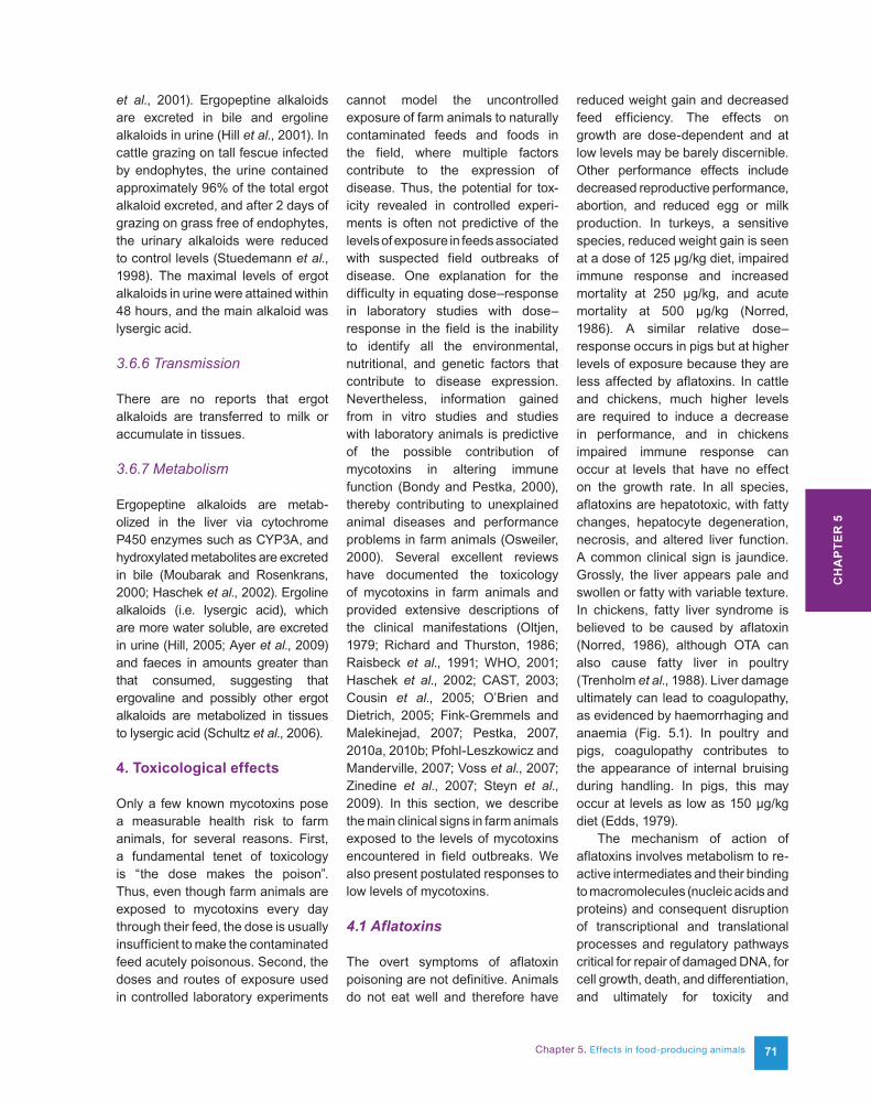
reduced weight gain and decreased feed efficiency. The effects on growth are dose-dependent and at low levels may be barely discernible. Other performance effects include decreased reproductive performance, abortion, and reduced egg or milk production. In turkeys, a sensitive species, reduced weight gain is seen at a dose of 125 µg/kg diet, impaired immune response and increased mortality at 250 µg/kg, and acute mortality at 500 µg/kg (Norred, 1986). A similar relative dose–response occurs in pigs but at higher levels of exposure because they are less affected by aflatoxins. In cattle and chickens, much higher levels are required to induce a decrease in performance, and in chickens impaired immune response can occur at levels that have no effect on the growth rate. In all species, aflatoxins are hepatotoxic, with fatty changes, hepatocyte degeneration, necrosis, and altered liver function. A common clinical sign is jaundice. Grossly, the liver appears pale and swollen or fatty with variable texture. In chickens, fatty liver syndrome is believed to be caused by aflatoxin (Norred, 1986), although OTA can also cause fatty liver in poultry (Trenholm et al., 1988). Liver damage ultimately can lead to coagulopathy, as evidenced by haemorrhaging and anaemia (Fig. 5.1). In poultry and pigs, coagulopathy contributes to the appearance of internal bruising during handling. In pigs, this may occur at levels as low as 150 µg/kg diet (Edds, 1979).
The mechanism of action of aflatoxins involves metabolism to re-active intermediates and their binding to macromolecules (nucleic acids and proteins) and consequent disruption of transcriptional and translational processes and regulatory pathways critical for repair of damaged DNA, for cell growth, death, and differentiation, and ultimately for toxicity and
CH
AP
TER
5
72
carcinogenicity (Wild and Gong, 2010; Eaton et al., 2010; Kensler et al., 2011). Some evidence has been found that aflatoxin B1 produces reactive oxygen species, resulting in oxidation of DNA bases (Guindon et al., 2007).
The response of an animal to aflatoxin depends to a large extent on the rate of metabolism and the type of metabolites that are produced (see Section 3.1.7 and Chapter 6). For example, quail and turkeys are sensitive to aflatoxin toxicity and have a high rate of epoxide formation and a low rate of glutathione conjugation. In resistant species, even if the rate of epoxidation is high, a high rate of glutathione conjugation is protective. However, the resulting clinical signs are similar although the dose dependence may be quite different. Aflatoxin adducts in urine and blood of farm animals may be very useful as a biomarker for exposure during suspected field outbreaks (Riley et al., 2011).
4.2 Fumonisins
Consumption of feeds contaminated with fumonisins is a proven cause of two farm animal diseases and a suspected cause of others. ELEM is a fatal neurotoxic disease that occurs only in equids (horses and related species). The disease is characterized by the presence of liquefactive necrotic lesions in the white matter of the brain; the grey matter may also be involved (WHO, 2000). All aspects of the disease can be reproduced experimentally. The brain lesions are caused by vasogenic cerebral oedema (Haschek et al., 2002; Foreman et al., 2004) and are accompanied by increased protein in the cerebrospinal fluid and other changes consistent with vasogenic cerebral oedema (Smith et al., 2002; Foreman et al., 2004). Early symptoms include lethargy,
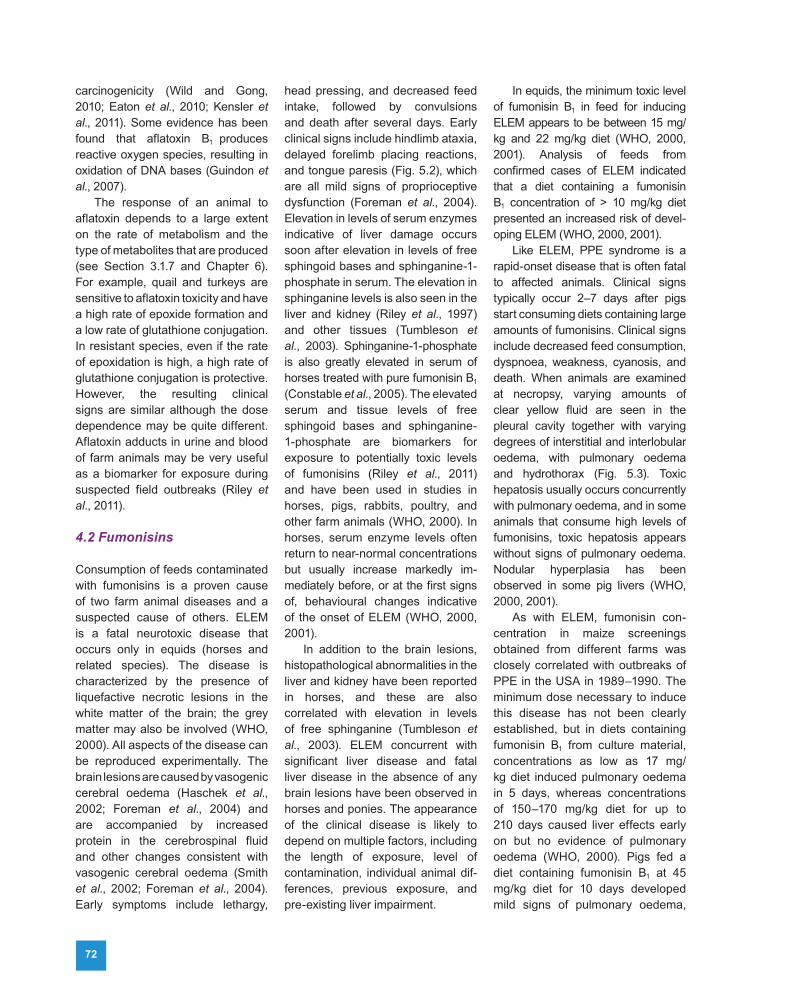
head pressing, and decreased feed intake, followed by convulsions and death after several days. Early clinical signs include hindlimb ataxia, delayed forelimb placing reactions, and tongue paresis (Fig. 5.2), which are all mild signs of proprioceptive dysfunction (Foreman et al., 2004). Elevation in levels of serum enzymes indicative of liver damage occurs soon after elevation in levels of free sphingoid bases and sphinganine-1-phosphate in serum. The elevation in sphinganine levels is also seen in the liver and kidney (Riley et al., 1997) and other tissues (Tumbleson et al., 2003). Sphinganine-1-phosphate is also greatly elevated in serum of horses treated with pure fumonisin B1 (Constable et al., 2005). The elevated serum and tissue levels of free sphingoid bases and sphinganine-1-phosphate are biomarkers for exposure to potentially toxic levels of fumonisins (Riley et al., 2011) and have been used in studies in horses, pigs, rabbits, poultry, and other farm animals (WHO, 2000). In horses, serum enzyme levels often return to near-normal concentrations but usually increase markedly im-mediately before, or at the first signs of, behavioural changes indicative of the onset of ELEM (WHO, 2000, 2001).
In addition to the brain lesions, histopathological abnormalities in the liver and kidney have been reported in horses, and these are also correlated with elevation in levels of free sphinganine (Tumbleson et al., 2003). ELEM concurrent with significant liver disease and fatal liver disease in the absence of any brain lesions have been observed in horses and ponies. The appearance of the clinical disease is likely to depend on multiple factors, including the length of exposure, level of contamination, individual animal dif-ferences, previous exposure, and pre-existing liver impairment.
In equids, the minimum toxic level of fumonisin B1 in feed for inducing ELEM appears to be between 15 mg/kg and 22 mg/kg diet (WHO, 2000, 2001). Analysis of feeds from confirmed cases of ELEM indicated that a diet containing a fumonisin B1 concentration of > 10 mg/kg diet presented an increased risk of devel-oping ELEM (WHO, 2000, 2001).
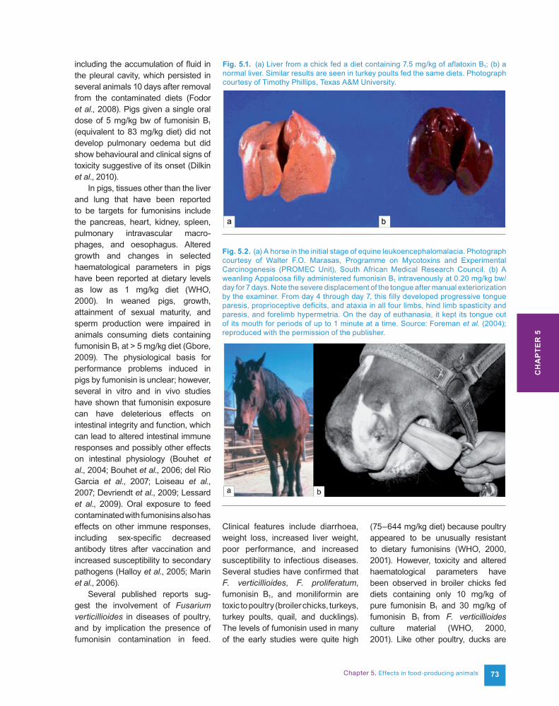
Like ELEM, PPE syndrome is a rapid-onset disease that is often fatal to affected animals. Clinical signs typically occur 2–7 days after pigs start consuming diets containing large amounts of fumonisins. Clinical signs include decreased feed consumption, dyspnoea, weakness, cyanosis, and death. When animals are examined at necropsy, varying amounts of clear yellow fluid are seen in the pleural cavity together with varying degrees of interstitial and interlobular oedema, with pulmonary oedema and hydrothorax (Fig. 5.3). Toxic hepatosis usually occurs concurrently with pulmonary oedema, and in some animals that consume high levels of fumonisins, toxic hepatosis appears without signs of pulmonary oedema. Nodular hyperplasia has been observed in some pig livers (WHO, 2000, 2001).
As with ELEM, fumonisin con-centration in maize screenings obtained from different farms was closely correlated with outbreaks of PPE in the USA in 1989–1990. The minimum dose necessary to induce this disease has not been clearly established, but in diets containing fumonisin B1 from culture material, concentrations as low as 17 mg/kg diet induced pulmonary oedema in 5 days, whereas concentrations of 150–170 mg/kg diet for up to 210 days caused liver effects early on but no evidence of pulmonary oedema (WHO, 2000). Pigs fed a diet containing fumonisin B1 at 45 mg/kg diet for 10 days developed mild signs of pulmonary oedema,
Chapter 5. Effects in food-producing animals 73
including the accumulation of fluid in the pleural cavity, which persisted in several animals 10 days after removal from the contaminated diets (Fodor et al., 2008). Pigs given a single oral dose of 5 mg/kg bw of fumonisin B1 (equivalent to 83 mg/kg diet) did not develop pulmonary oedema but did show behavioural and clinical signs of toxicity suggestive of its onset (Dilkin et al., 2010).
In pigs, tissues other than the liver and lung that have been reported to be targets for fumonisins include the pancreas, heart, kidney, spleen, pulmonary intravascular macro-phages, and oesophagus. Altered growth and changes in selected haematological parameters in pigs have been reported at dietary levels as low as 1 mg/kg diet (WHO, 2000). In weaned pigs, growth, attainment of sexual maturity, and sperm production were impaired in animals consuming diets containing fumonisin B1 at > 5 mg/kg diet (Gbore, 2009). The physiological basis for performance problems induced in pigs by fumonisin is unclear; however, several in vitro and in vivo studies have shown that fumonisin exposure can have deleterious effects on intestinal integrity and function, which can lead to altered intestinal immune responses and possibly other effects on intestinal physiology (Bouhet et al., 2004; Bouhet et al., 2006; del Rio Garcia et al., 2007; Loiseau et al., 2007; Devriendt et al., 2009; Lessard et al., 2009). Oral exposure to feed contaminated with fumonisins also has effects on other immune responses, including sex-specific decreased antibody titres after vaccination and increased susceptibility to secondary pathogens (Halloy et al., 2005; Marin et al., 2006).
Several published reports sug-gest the involvement of Fusarium verticillioides in diseases of poultry, and by implication the presence of fumonisin contamination in feed.
Clinical features include diarrhoea, weight loss, increased liver weight, poor performance, and increased susceptibility to infectious diseases. Several studies have confirmed that F. verticillioides, F. proliferatum, fumonisin B1, and moniliformin are toxic to poultry (broiler chicks, turkeys, turkey poults, quail, and ducklings). The levels of fumonisin used in many of the early studies were quite high
(75–644 mg/kg diet) because poultry appeared to be unusually resistant to dietary fumonisins (WHO, 2000, 2001). However, toxicity and altered haematological parameters have been observed in broiler chicks fed diets containing only 10 mg/kg of pure fumonisin B1 and 30 mg/kg of fumonisin B1 from F. verticillioides culture material (WHO, 2000, 2001). Like other poultry, ducks are
Fig. 5.1. (a) Liver from a chick fed a diet containing 7.5 mg/kg of aflatoxin B1; (b) a normal liver. Similar results are seen in turkey poults fed the same diets. Photograph courtesy of Timothy Phillips, Texas A&M University.
Fig. 5.2. (a) A horse in the initial stage of equine leukoencephalomalacia. Photograph courtesy of Walter F.O. Marasas, Programme on Mycotoxins and Experimental Carcinogenesis (PROMEC Unit), South African Medical Research Council. (b) A weanling Appaloosa filly administered fumonisin B1 intravenously at 0.20 mg/kg bw/day for 7 days. Note the severe displacement of the tongue after manual exteriorization by the examiner. From day 4 through day 7, this filly developed progressive tongue paresis, proprioceptive deficits, and ataxia in all four limbs, hind limb spasticity and paresis, and forelimb hypermetria. On the day of euthanasia, it kept its tongue out of its mouth for periods of up to 1 minute at a time. Source: Foreman et al. (2004); reproduced with the permission of the publisher.
CH
AP
TER
5
74
relatively resistant to the toxic effects of fumonisins (Tran et al., 2005). However, increased mortality and toxicity have been observed in ducks force-fed diets containing 20 mg/kg of fumonisin B1 (Tardieu et al., 2009). As in pigs, fumonisin can also alter immune response and susceptibility to infectious agents in poultry (Tessari et al., 2006; Deshmukh et al., 2007).
Other farm animals that have been studied using pure fumonisins, contaminated maize screenings, or maize culture material from F. verticillioides include carp, catfish, lambs, goats, trout, cattle, mink, and rabbits. Rabbits are especially sensitive to nephrotoxicity (Voss et al., 2001; Ewuola, 2009). In all cases where toxicity was evident, it involved the liver and/or kidney or the equivalent organs in fish.
Disruption of lipid metabolism appears to be the underlying mechanism by which fumonisins cause toxicity to animals (WHO, 2000, 2001). The initial mechanism of action of fumonisin is inhibition of ceramide synthase, a key enzyme in the de novo sphingolipid biosynthesis
pathway (Wang et al., 1991). Fumonisins are structurally similar to sphingoid bases and especially 1-deoxysphinganine, which lacks a hydroxyl group on carbon 1 (Zitomer et al., 2009). Sphingoid bases are essential components of the chemical backbone of all sphingolipids in animals. When toxicity associated with fumonisins is observed in laboratory or farm animals, the onset and severity of the pathology is closely correlated with evidence of disrupted sphingolipid metabolism (Riley et al., 1996, 2001; WHO, 2000, 2001, 2012; Riley and Voss, 2006; Voss et al., 2011). Disrupted sphingolipid metabolism can also be evident at dosages that do not cause overt toxicity (NTP, 2001). This is especially true in resistant species such as ducks (Tardieu et al., 2006). The major biochemical and cellular consequences resulting from blockage of ceramide biosynthesis are the accumulation of free sphingoid bases and sphingoid base 1-phosphates (Riley and Voss, 2006; Zitomer et al., 2009), the depletion of more complex sphingolipids (Voss et al., 2009), and the global disruption of lipid metabolism (WHO, 2001). The changes in concentrations of impor-tant lipid mediators lead ultimately to perturbation of the signalling pathways and altered regulatory and physiological processes (Lemmer et al., 1999; Bondy et al., 2000; Merrill et al., 2001), which are the basis for the observed clinical signs associated with the diseases induced by fumonisins. ELEM is associated with alterations in cardiovascular function induced by sphingolipids, i.e. deregulation of cerebral arteries responsible for autoregulation of blood flow to the horse’s brain (Haschek et al., 2002; Foreman et al., 2004). PPE is hypothesized to be a result of acute left-sided heart failure as a consequence of inhibition of L-type calcium channels induced by sphingoid bases
(Haschek et al., 2002). The elevation of free sphingoid bases and sphingoid base 1-phosphates and the depletion of more complex sphingolipids in tissues, serum, and urine have proven to be useful biomarkers for exposure and the effects of fumonisin in farm animals (Riley et al., 2011).
4.3 Ochratoxin A
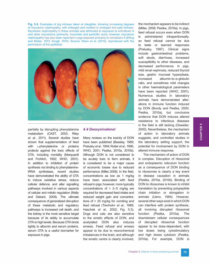
The primary effect of OTA in all farm animals is nephrotoxicity. In pigs and poultry, the proximal tubules are mainly affected and the kidney is pale and grossly enlarged (Fig. 5.4).
Fatty liver can occur in poultry. The most sensitive indicator of acute ochratoxicosis in chickens is the reduction in total serum proteins and albumin. A decrease in phosphoenolpyruvate carboxykinase in the kidney is a sensitive and specific indicator in pigs (Krogh, 1992; Marquardt and Frohlich, 1992). In pigs, large increases in levels of proteins excreted in urine are indicative of glomerular proteinuria and correlate with histological obser-vations of renal damage.
In poultry and pigs, exposure to OTA at lower levels can result in altered performance, including decreased feed consumption and reduced weight gain, and at higher levels can result in delayed response to immunization and increased susceptibility to infection (Stoev et al., 2000a, 2000b). Other effects in poultry include decreased egg production, coagulopathy (in-creased susceptibility to bruising during processing), decreased bone strength, decreased tensile strength of the large intestines, underpigmentation, and glycogen accumulation in the liver.
The mechanism of action in farm animals is unclear. However, the structural similarity of OTA to phenylalanine and the fact that it inhibits many enzymes and processes that are dependent on phenylalanine strongly suggest that OTA acts at least
Fig. 5.3. (Left) A lung with severe oede-ma from a pig with porcine pulmonary oedema after being fed fumonisin-contaminated feed and (right) a normal lung. Photograph courtesy of Wanda Haschek, University of Illinois.
Chapter 5. Effects in food-producing animals 75
partially by disrupting phenylalanine metabolism (CAST, 2003; Riley et al., 2011). Several studies have shown that supplementation of feed with L-phenylalanine or proteins protects against the toxic effects of OTA, including mortality (Marquardt and Frohlich, 1992; WHO, 2001). In addition to inhibition of protein synthesis via binding to phenylalanine-tRNA synthetase, recent studies have demonstrated the ability of OTA to induce oxidative stress, reduce cellular defence, and alter signalling pathways involved in various aspects of cellular and mitotic regulation (Mally and Dekant, 2009). The ultimate consequence of generalized disruption of these metabolic and regulatory pathways is increased cell death, and the kidney is the most sensitive target because of its ability to accumulate OTA to high levels. Because OTA binds tightly to albumin and serum proteins, serum OTA is a useful biomarker for exposure in pigs.
4.4 Deoxynivalenol
Many reviews on the toxicity of DON have been published (Beasley, 1989; Prelusky et al., 1994; Rotter et al., 1996, WHO, 2001; Pestka, 2010a, 2010b). Although DON is not considered to be acutely toxic to farm animals, it is considered to be a major cause of economic losses due to reduced performance (Miller, 2008). In the field, concentrations as low as 1 mg/kg have been associated with feed refusal in pigs; however, more typically concentrations of > 2–5 mg/kg are required for decreased feed intake and reduced weight gain and concentra-tions of > 20 mg/kg for vomiting and feed refusal (Trenholm et al., 1988; Haschek et al., 2002; Fig. 5.5).Dogs and cats are also sensitive to the emetic effects of DON, and acetylated DON also induces emesis. Feed refusal and emesis appear to be due to neurochemical imbalances in the brain, and although the emetic centre is clearly involved,
the mechanism appears to be indirect (Miller, 2008; Pestka, 2010a). In pigs, feed refusal occurs even when DON is administered intraperitoneally, so feed refusal cannot be due to taste or learned responses (Prelusky, 1997). Clinical signs include gastrointestinal problems, soft stools, diarrhoea, increased susceptibility to other diseases, and decreased performance. In pigs, mild renal nephrosis, reduced thyroid size, gastric mucosal hyperplasia, increased albumin-to-α-globulin ratio, and sometimes mild changes in other haematological parameters have been reported (WHO, 2001). Numerous studies in laboratory animals have demonstrated alter-ations in immune function induced by DON (Bondy and Pestka, 2000; Pestka, 2010a), but conclusive evidence that DON induces altered resistance to infectious diseases in the field is still lacking (Osweiler, 2000). Nevertheless, the mechanism of action in laboratory animals suggests, and controlled studies in the laboratory setting support, the potential for involvement by DON in altered immune response.
The mechanism of action of DON is complex. Disruption of ribosomal and endoplasmic reticulum function as a consequence of DON binding to ribosomes is clearly a key event in disease causation in animals (Pestka, 2010a, 2010b). Binding of DON to ribosomes is known to inhibit translation by preventing polypeptide chain initiation or elongation in animals (Ueno, 1984). However, several other ways exist in which DON can interfere with protein synthesis, all involving disrupted ribosomal function (Pestka, 2010a). The downstream cellular consequences of disrupted ribosomal function appear to be dose-dependent, with low doses being cytostimulatory and high doses cytotoxic (Pestka, 2010a). For example, DON is
Fig. 5.4. Examples of pig kidneys taken at slaughter, showing increasing degrees of mycotoxic nephropathy, with enlarged and mottled or enlarged and pale kidneys. Mycotoxic nephropathy in these animals was attributed to exposure to ochratoxin A and other mycotoxins (primarily, fumonisin and penicillic acid); however, mycotoxic nephropathy has also been reported in pigs exposed primarily to ochratoxin A (Elling and Moller, 1973; Krogh, 1974). Source: Stoev et al. (2010); reproduced with the permission of the publisher.
CH
AP
TER
5
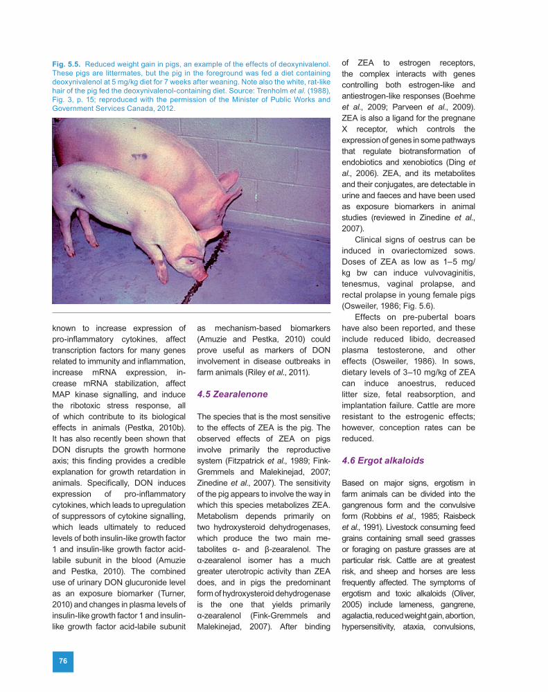
76
known to increase expression of pro-inflammatory cytokines, affect transcription factors for many genes related to immunity and inflammation, increase mRNA expression, in-crease mRNA stabilization, affect MAP kinase signalling, and induce the ribotoxic stress response, all of which contribute to its biological effects in animals (Pestka, 2010b). It has also recently been shown that DON disrupts the growth hormone axis; this finding provides a credible explanation for growth retardation in animals. Specifically, DON induces expression of pro-inflammatory cytokines, which leads to upregulation of suppressors of cytokine signalling, which leads ultimately to reduced levels of both insulin-like growth factor 1 and insulin-like growth factor acid-labile subunit in the blood (Amuzie and Pestka, 2010). The combined use of urinary DON glucuronide level as an exposure biomarker (Turner, 2010) and changes in plasma levels of insulin-like growth factor 1 and insulin-like growth factor acid-labile subunit
as mechanism-based biomarkers (Amuzie and Pestka, 2010) could prove useful as markers of DON involvement in disease outbreaks in farm animals (Riley et al., 2011).
4.5 Zearalenone
The species that is the most sensitive to the effects of ZEA is the pig. The observed effects of ZEA on pigs involve primarily the reproductive system (Fitzpatrick et al., 1989; Fink-Gremmels and Malekinejad, 2007; Zinedine et al., 2007). The sensitivity of the pig appears to involve the way in which this species metabolizes ZEA. Metabolism depends primarily on two hydroxysteroid dehydrogenases, which produce the two main me-tabolites α- and β-zearalenol. The α-zearalenol isomer has a much greater uterotropic activity than ZEA does, and in pigs the predominant form of hydroxysteroid dehydrogenase is the one that yields primarily α-zearalenol (Fink-Gremmels and Malekinejad, 2007). After binding
of ZEA to estrogen receptors, the complex interacts with genes controlling both estrogen-like and antiestrogen-like responses (Boehme et al., 2009; Parveen et al., 2009). ZEA is also a ligand for the pregnane X receptor, which controls the expression of genes in some pathways that regulate biotransformation of endobiotics and xenobiotics (Ding et al., 2006). ZEA, and its metabolites and their conjugates, are detectable in urine and faeces and have been used as exposure biomarkers in animal studies (reviewed in Zinedine et al., 2007).
Clinical signs of oestrus can be induced in ovariectomized sows. Doses of ZEA as low as 1–5 mg/kg bw can induce vulvovaginitis, tenesmus, vaginal prolapse, and rectal prolapse in young female pigs (Osweiler, 1986; Fig. 5.6).
Effects on pre-pubertal boars have also been reported, and these include reduced libido, decreased plasma testosterone, and other effects (Osweiler, 1986). In sows, dietary levels of 3–10 mg/kg of ZEA can induce anoestrus, reduced litter size, fetal reabsorption, and implantation failure. Cattle are more resistant to the estrogenic effects; however, conception rates can be reduced.
4.6 Ergot alkaloids
Based on major signs, ergotism in farm animals can be divided into the gangrenous form and the convulsive form (Robbins et al., 1985; Raisbeck et al., 1991). Livestock consuming feed grains containing small seed grasses or foraging on pasture grasses are at particular risk. Cattle are at greatest risk, and sheep and horses are less frequently affected. The symptoms of ergotism and toxic alkaloids (Oliver, 2005) include lameness, gangrene, agalactia, reduced weight gain, abortion, hypersensitivity, ataxia, convulsions,
Fig. 5.5. Reduced weight gain in pigs, an example of the effects of deoxynivalenol. These pigs are littermates, but the pig in the foreground was fed a diet containing deoxynivalenol at 5 mg/kg diet for 7 weeks after weaning. Note also the white, rat-like hair of the pig fed the deoxynivalenol-containing diet. Source: Trenholm et al. (1988), Fig. 3, p. 15; reproduced with the permission of the Minister of Public Works and Government Services Canada, 2012.
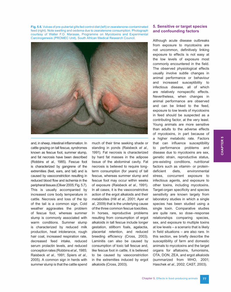
Chapter 5. Effects in food-producing animals 77
and, in sheep, intestinal inflammation. In cattle grazing on tall fescue, syndromes known as fescue foot, summer slump, and fat necrosis have been described (Robbins et al., 1985). Fescue foot is characterized by gangrene of the extremities (feet, ears, and tail) and is caused by vasoconstriction resulting in reduced blood flow and ischemia in the peripheral tissues (Oliver 2005; Fig. 5.7). This is usually accompanied by increased core body temperature in cattle. Necrosis and loss of the tip of the tail is a common sign. Cold weather aggravates the problem of fescue foot, whereas summer slump is commonly associated with warm conditions. Summer slump is characterized by reduced milk production, heat intolerance, rough hair coat, increased respiration rate, decreased feed intake, reduced serum prolactin levels, and reduced conception rates (Robbins et al., 1985; Raisbeck et al., 1991; Spiers et al., 2005). A common sign in herds with summer slump is that the cattle spend
much of their time seeking shade or standing in ponds (Raisbeck et al., 1991). Fat necrosis is characterized by hard fat masses in the adipose tissue of the abdominal cavity. Fat necrosis is believed to require long-term consumption (for years) of tall fescue, whereas summer slump and fescue foot may occur within weeks of exposure (Raisbeck et al., 1991). In all cases, it is the vasoconstrictive action of the ergot alkaloids and their metabolites (Hill et al., 2001; Ayer et al., 2009) that is the underlying cause of the three common fescue toxicities. In horses, reproductive problems resulting from consumption of ergot alkaloids in tall fescue include longer gestation, stillborn foals, agalactia, placental retention, and reduced breeding efficiency (Cross, 2003). Laminitis can also be caused by consumption of toxic tall fescue and, like fescue foot in cattle, it is believed to be caused by vasoconstriction in the extremities induced by ergot alkaloids (Cross, 2003).
5. Sensitive or target species and confounding factors
Although acute disease outbreaks from exposure to mycotoxins are not uncommon, definitively linking exposure to effects is not easy at the low levels of exposure most commonly encountered in the field. The observed physiological effects usually involve subtle changes in animal performance or behaviour and increased susceptibility to infectious disease, all of which are relatively nonspecific effects. Nevertheless, when changes in animal performance are observed and can be linked to the feed, exposure to low levels of mycotoxins in feed should be suspected as a contributing factor, at the very least. Young animals are more sensitive than adults to the adverse effects of mycotoxins, in part because of a higher metabolic rate. Factors that can influence susceptibility to performance problems and disease due to mycotoxins are sex, genetic strain, reproductive status, pre-existing conditions, nutritional factors such as vitamin- or protein-deficient diets, environmental stress, concurrent exposure to infectious agents, and exposure to other toxins, including mycotoxins. Target-organ specificity and species sensitivity are known largely from laboratory studies in which a single species has been studied using a single toxin. Comparative studies are quite rare, so dose–response relationships comparing species, sex, and exposure to multiple toxins at low levels – a scenario that is likely in field situations – are also rare. In this section, we briefly describe the susceptibility of farm and domestic animals to mycotoxins and the target organs for aflatoxins, fumonisins, OTA, DON, ZEA, and ergot alkaloids (summarized from WHO, 2001; Haschek et al., 2002; CAST, 2003).
Fig. 5.6. Vulvas of pre-pubertal gilts fed control diet (left) or zearalenone-contaminated feed (right). Note swelling and oedema due to zearalenone consumption. Photograph courtesy of Walter F.O. Marasas, Programme on Mycotoxins and Experimental Carcinogenesis (PROMEC Unit), South African Medical Research Council.
CH
AP
TER
5
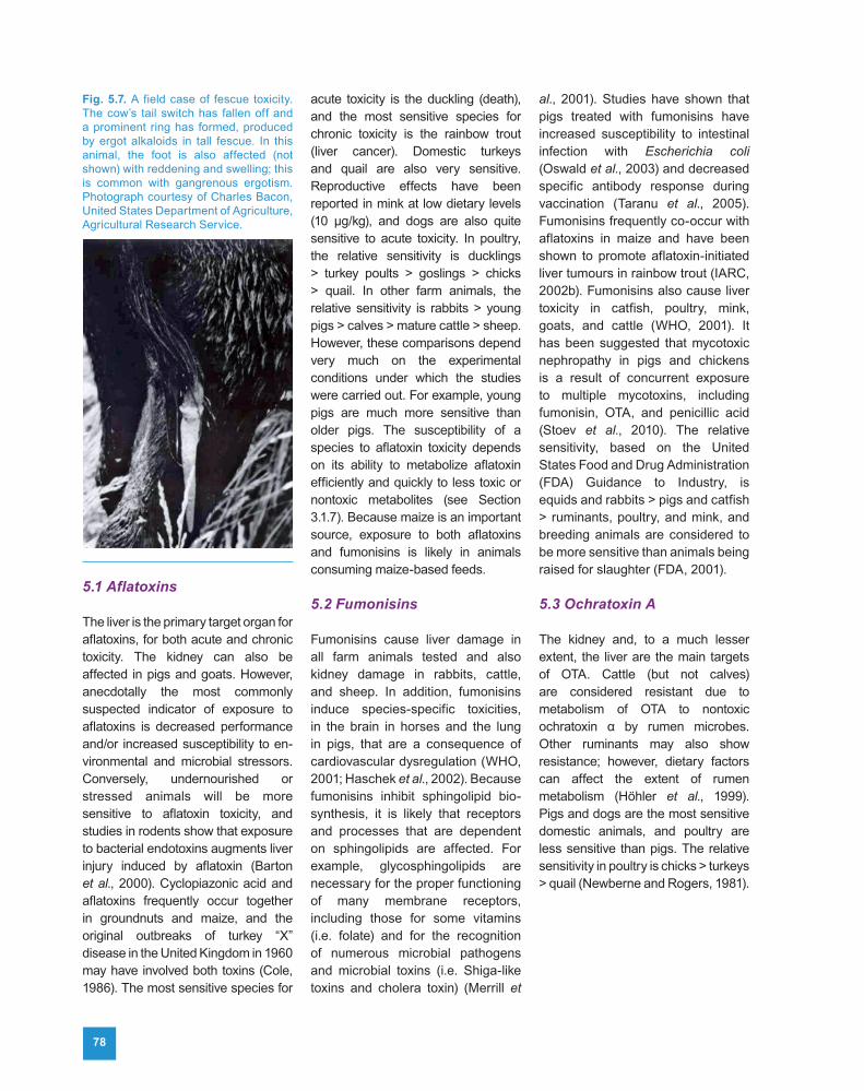
78
5.1 Aflatoxins
The liver is the primary target organ for aflatoxins, for both acute and chronic toxicity. The kidney can also be affected in pigs and goats. However, anecdotally the most commonly suspected indicator of exposure to aflatoxins is decreased performance and/or increased susceptibility to en-vironmental and microbial stressors. Conversely, undernourished or stressed animals will be more sensitive to aflatoxin toxicity, and studies in rodents show that exposure to bacterial endotoxins augments liver injury induced by aflatoxin (Barton et al., 2000). Cyclopiazonic acid and aflatoxins frequently occur together in groundnuts and maize, and the original outbreaks of turkey “X” disease in the United Kingdom in 1960 may have involved both toxins (Cole, 1986). The most sensitive species for
acute toxicity is the duckling (death), and the most sensitive species for chronic toxicity is the rainbow trout (liver cancer). Domestic turkeys and quail are also very sensitive. Reproductive effects have been reported in mink at low dietary levels (10 µg/kg), and dogs are also quite sensitive to acute toxicity. In poultry, the relative sensitivity is ducklings > turkey poults > goslings > chicks > quail. In other farm animals, the relative sensitivity is rabbits > young pigs > calves > mature cattle > sheep. However, these comparisons depend very much on the experimental conditions under which the studies were carried out. For example, young pigs are much more sensitive than older pigs. The susceptibility of a species to aflatoxin toxicity depends on its ability to metabolize aflatoxin efficiently and quickly to less toxic or nontoxic metabolites (see Section 3.1.7). Because maize is an important source, exposure to both aflatoxins and fumonisins is likely in animals consuming maize-based feeds.
5.2 Fumonisins
Fumonisins cause liver damage in all farm animals tested and also kidney damage in rabbits, cattle, and sheep. In addition, fumonisins induce species-specific toxicities, in the brain in horses and the lung in pigs, that are a consequence of cardiovascular dysregulation (WHO, 2001; Haschek et al., 2002). Because fumonisins inhibit sphingolipid bio-synthesis, it is likely that receptors and processes that are dependent on sphingolipids are affected. For example, glycosphingolipids are necessary for the proper functioning of many membrane receptors, including those for some vitamins (i.e. folate) and for the recognition of numerous microbial pathogens and microbial toxins (i.e. Shiga-like toxins and cholera toxin) (Merrill et
al., 2001). Studies have shown that pigs treated with fumonisins have increased susceptibility to intestinal infection with Escherichia coli (Oswald et al., 2003) and decreased specific antibody response during vaccination (Taranu et al., 2005). Fumonisins frequently co-occur with aflatoxins in maize and have been shown to promote aflatoxin-initiated liver tumours in rainbow trout (IARC, 2002b). Fumonisins also cause liver toxicity in catfish, poultry, mink, goats, and cattle (WHO, 2001). It has been suggested that mycotoxic nephropathy in pigs and chickens is a result of concurrent exposure to multiple mycotoxins, including fumonisin, OTA, and penicillic acid (Stoev et al., 2010). The relative sensitivity, based on the United States Food and Drug Administration (FDA) Guidance to Industry, is equids and rabbits > pigs and catfish > ruminants, poultry, and mink, and breeding animals are considered to be more sensitive than animals being raised for slaughter (FDA, 2001).
5.3 Ochratoxin A
The kidney and, to a much lesser extent, the liver are the main targets of OTA. Cattle (but not calves) are considered resistant due to metabolism of OTA to nontoxic ochratoxin α by rumen microbes. Other ruminants may also show resistance; however, dietary factors can affect the extent of rumen metabolism (Höhler et al., 1999). Pigs and dogs are the most sensitive domestic animals, and poultry are less sensitive than pigs. The relative sensitivity in poultry is chicks > turkeys > quail (Newberne and Rogers, 1981).
Fig. 5.7. A field case of fescue toxicity. The cow’s tail switch has fallen off and a prominent ring has formed, produced by ergot alkaloids in tall fescue. In this animal, the foot is also affected (not shown) with reddening and swelling; this is common with gangrenous ergotism. Photograph courtesy of Charles Bacon, United States Department of Agriculture, Agricultural Research Service.
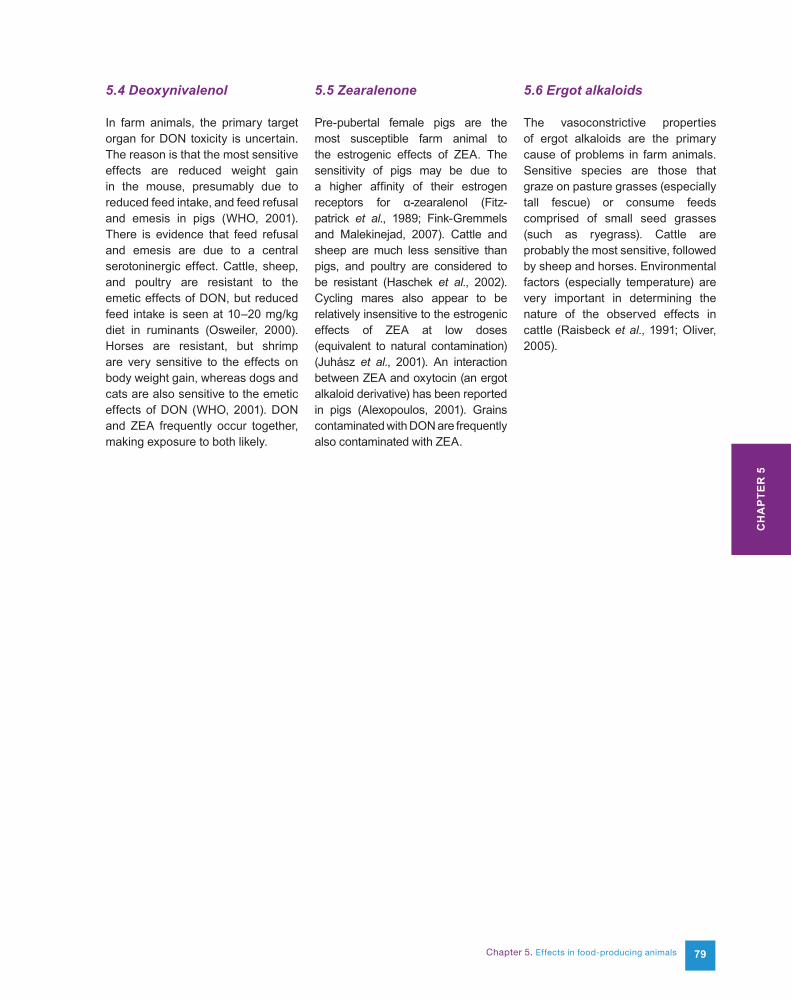
Chapter 5. Effects in food-producing animals 79
5.4 Deoxynivalenol
In farm animals, the primary target organ for DON toxicity is uncertain. The reason is that the most sensitive effects are reduced weight gain in the mouse, presumably due to reduced feed intake, and feed refusal and emesis in pigs (WHO, 2001). There is evidence that feed refusal and emesis are due to a central serotoninergic effect. Cattle, sheep, and poultry are resistant to the emetic effects of DON, but reduced feed intake is seen at 10–20 mg/kg diet in ruminants (Osweiler, 2000). Horses are resistant, but shrimp are very sensitive to the effects on body weight gain, whereas dogs and cats are also sensitive to the emetic effects of DON (WHO, 2001). DON and ZEA frequently occur together, making exposure to both likely.
5.5 Zearalenone
Pre-pubertal female pigs are the most susceptible farm animal to the estrogenic effects of ZEA. The sensitivity of pigs may be due to a higher affinity of their estrogen receptors for α-zearalenol (Fitz-patrick et al., 1989; Fink-Gremmels and Malekinejad, 2007). Cattle and sheep are much less sensitive than pigs, and poultry are considered to be resistant (Haschek et al., 2002). Cycling mares also appear to be relatively insensitive to the estrogenic effects of ZEA at low doses (equivalent to natural contamination) (Juhász et al., 2001). An interaction between ZEA and oxytocin (an ergot alkaloid derivative) has been reported in pigs (Alexopoulos, 2001). Grains contaminated with DON are frequently also contaminated with ZEA.
5.6 Ergot alkaloids
The vasoconstrictive properties of ergot alkaloids are the primary cause of problems in farm animals. Sensitive species are those that graze on pasture grasses (especially tall fescue) or consume feeds comprised of small seed grasses (such as ryegrass). Cattle are probably the most sensitive, followed by sheep and horses. Environmental factors (especially temperature) are very important in determining the nature of the observed effects in cattle (Raisbeck et al., 1991; Oliver, 2005).
CH
AP
TER
5
80
References
Bondy GS, Barker MG, Lombaert GA et al. (2000). A comparison of clinical, histopathological and cell-cycle markers in rats receiving the fungal toxins fumonisin B1 or fumonisin B2 by intraperitoneal injection. Food Chem Toxicol, 38:873–886. doi:10.1016/S0278-6915(00)00084-3 PMID:11039321
Bondy GS, Pestka JJ (2000). Immunomod-ulation by fungal toxins. J Toxicol Environ Health B Crit Rev, 3:109–143. PMID:10834078
Bony S, Durix A, Leblond A, Jaussaud P (2001). Toxicokinetics of ergovaline in the horse after an intravenous administration. Vet Res, 32:509–513. doi:10.1051/vetres:2001142 PMID:11592620
Bouhet S, Hourcade E, Loiseau N et al. (2004). The mycotoxin fumonisin B1 alters the proliferation and barrier function of porcine intestinal epithelial cells. Toxicol Sci, 77:165–171.
Bouhet S, Le Dorze E, Peres S et al. (2006). Mycotoxin fumonisin B1 selectively down-regulates the basal IL-8 expression in pig intestine: in vivo and in vitro studies. Food Chem Toxicol, 44:1768–1773. PMID:16843581
Bow DA, Perry JL, Simon JD, Pritchard JB (2006). The impact of plasma protein binding on the renal transport of organic anions. J Pharmacol Exp Ther, 316:349–355. doi:10.1124/jpet.105.093070 PMID:16195420
Busby WF, Wogan GN (1981a). Aflatoxins. In: Shank RC, ed. Mycotoxins and N-Nitroso Compounds: Environmental Risks, Vol. 1. Boca Raton, FL: CRC Press, pp. 3–28.
Busby WF, Wogan GN (1981b). Ochratoxins. In: Shank RC, ed. Mycotoxins and N-Nitroso Compounds: Environmental Risks, Vol. 1. Boca Raton, FL: CRC Press, pp. 129–136.
Butler WH (1974). Aflatoxins. In: Purchase IFH, ed. Mycotoxins. New York: Elsevier Scientific Publishing, pp. 1–28.
Cai Q, Tang L, Wang JS (2007). Validation of fumonisin biomarkers in F344 rats. Toxicol Appl Pharmacol, 225:28–39. doi:10.1016/j.taap.2007.06.027 PMID:17904604
CAST (2003). Mycotoxins: Risks in Plant, Animal, and Human Systems. Ames, IA: Council for Agricultural Science and Technology (Task Force Report No. 139).
Christensen CM (1979). Zearalenone. In: Shimoda W, ed. Conference on Mycotoxins in Animal Feeds and Grains Related to Animal Health. Springfield, VA: United States Department of Commerce, National Technical Information Service, pp. 1–79.
Christensen CM (1980). Ergots. In: Shimoda W, ed. Conference on Mycotoxins in Animal Feeds and Grains Related to Animal Health. Springfield, VA: United States Department of Commerce, National Technical Information Service, pp. 1–44.
Cole RJ (1986). Etiology of turkey “X” disease in retrospect: a case for the involvement of cyclopiazonic acid. Mycotoxin Res, 2:3–7. doi:10.1007/BF03191956
Collins TF, Sprando RL, Black TN et al. (2006). Effects of aminopentol on in utero development in rats. Food Chem Toxicol, 44:161–169. doi:10.1016/j.fct.2005.06.009 PMID:16081197
Constable PD, Riley RT, Waggoner AL et al. (2005). Serum sphingosine-1-phosphate and sphinganine-1-phosphate are elevated in horses exposed to fumonisin B1. AOAC International Midwest Section Final Program, pp. 63–64.
Coulombe RA Jr, Sharma RP (1985). Clearance and excretion of intratracheally and orally administered aflatoxin B1 in the rat. Food Chem Toxicol, 23:827–830. doi:10.1016/0278-6915(85)90283-2 PMID:3930357
Cousin MA, Riley RT, Pestka JJ (2005). Foodborne mycotoxins: chemistry, biology, ecology, and toxicology. In: Fratamico PM, Bhunia AK, eds. Foodborne Pathogens: Microbiology and Molecular Biology. Norfolk, UK: Horizon Scientific Press, pp. 163–226.
Cross DL (2003). Ergot alkaloid toxicity. In: White JF Jr, Bacon CW, Hywel-Jones NL, Spatafora JW, eds. Clavicipitalean Fungi. New York: Marcel Dekker, pp. 475–494.
Dänicke S, Swiech E, Buraczewska L, Ueberschär KH (2005). Kinetics and metabolism of zearalenone in young female pigs. J Anim Physiol Anim Nutr (Berl), 89:268–276. doi:10.1111/j.1439-0396.2005.00516.x PMID:15972077
Dänicke S, Ueberschär KH, Halle I et al. (2001). Excretion kinetics and metabolism of zearalenone in broilers in dependence on a detoxifying agent. Arch Tierernahr, 55:299–313. doi:10.1080/17450390109386199 PMID:12357591
De Angelis I, Friggè G, Raimondi F et al. (2005). Absorption of fumonisin B1 and aminopentol on an in vitro model of intestinal epithelium; the role of P-glycoprotein. Toxicon, 45:285–291. doi:10.1016/j.toxicon.2004.10.015 PMID:15683866
del Rio Garcia JC, Moreno Ramos C, Pinton P et al. (2007). Evaluation of the cytotoxicity of aflatoxin B1 in intestinal cells. Rev Iberoam Micol, 24:136–141. PMID:17604433
Deshmukh S, Asrani RK, Ledoux DR et al. (2007). Pathologic changes in extrahepatic organs and agglutinin response to Salmonella gallinarum infection in Japanese quail fed Fusarium verticillioides culture material containing known levels of fumonisin B1. Avian Dis, 51:705–712. doi:10.1637/0005-2086(2007)51[705:PCIEOA]2. 0.CO;2 PMID:17992930
Alexopoulos C (2001). Association of Fusarium mycotoxicosis with failure in applying an induction of parturition program with PGF2alpha and oxytocin in sows. Theriogenology, 55:1745–1757. doi:10.1016/S0093-691X(01)00517-9 PMID:11393224
Amuzie CJ, Pestka JJ (2010). Suppression of insulin-like growth factor acid-labile subunit expression–a novel mechanism for deoxynivalenol-induced growth retardation. Toxicol Sci, 113:412–421. doi:10.1093/toxsci/kfp225 PMID:19805407
Aucock HW, Marasas WF, Meyer CJ, Chalmers P (1980). Field outbreaks of hyperoestrogenism (vulvo-vaginitis) in pigs consuming maize infected by Fusarium graminearum and contaminated with zearalenone. J S Afr Vet Assoc, 51:163–166. PMID:6455520
Ayer AW, Hill NS, Rottinghaus GE et al. (2009). Ruminal metabolism and transport of tall fescue ergot alkaloids. Crop Sci, 49:2309–2316. doi:10.2135/cropsci2009.01.0018
Barger G (1931). Ergot and Ergotism. London: Gurney and Jackson.
Barton CC, Hill DA, Yee SB et al. (2000). Bacterial lipopolysaccharide exposure augments aflatoxin B1-induced liver injury. J Toxicol Sci, 55:444–452. doi:10.1093/toxsci/55.2.444
Beasley VR (1989). Trichothecene Mycotoxins: Pathophysiologic Effects, Vols 1, 2. Boca Raton, FL: CRC Press.
Bernhoft A, Behrens GH, Ingebrigtsen K et al. (2001). Placental transfer of the estrogenic mycotoxin zearalenone in rats. Reprod Toxicol, 15:545–550. doi:10.1016/S0890-6238(01)00159-9 PMID:11780962
Biehl ML, Prelusky DB, Koritz GD et al. (1993). Biliary excretion and enterohepatic cycling of zearalenone in immature pigs. Toxicol Appl Pharmacol, 121:152–159. doi:10.1006/taap.1993.1140 PMID:8337696
Biró K, Solti L, Barna-Vetró I et al. (2002). Tissue distribution of ochratoxin A as determined by HPLC and ELISA and histopathological effects in chickens. Avian Pathol, 31:141–148. doi:10.1080/03079450120118621 PMID:12396358
Boehme K, Simon S, Mueller SO (2009). Gene expression profiling in Ishikawa cells: a fingerprint for estrogen active compounds. Toxicol Appl Pharmacol, 236:85–96. doi:10.1016/j.taap.2009.01.006 PMID:19371625
Bohm J, Razzazi-Fazeli E (2005). Effects of mycotoxins on domestic pet species. In: Diaz DE, ed. The Mycotoxin Blue Book. Nottingham, UK: Nottingham University Press, pp. 77–91.
Chapter 5. Effects in food-producing animals 81
Devriendt B, Gallois M, Verdonck F et al. (2009). The food contaminant fumonisin B1 reduces the maturation of porcine CD11R1+ intestinal antigen presenting cells and antigen-specific immune responses, leading to a prolonged intestinal ETEC infection. Vet Res, 40:40. doi:10.1051/vetres/2009023 PMID:19389343
Dietrich DR, Heussner AH, O’Brien E (2005). Ochratoxin A: comparative pharmacokinetics and toxicological implications (experimental and domestic animals and humans). Food Addit Contam, 22 Suppl 1:45–52. doi:10.1080/02652030500309327 PMID:16332621
Dilkin P, Direito G, Simas MMS et al. (2010). Toxicokinetics and toxicological effects of single oral dose of fumonisin B1 containing Fusarium verticillioides culture material in weaned piglets. Chem Biol Interact, 185:157–162. doi:10.1016/j.cbi.2010.03.025 PMID:20338158
Dilkin P, Zorzete P, Mallmann CA et al. (2003). Toxicological effects of chronic low doses of aflatoxin B1 and fumonisin B1-containing Fusarium moniliforme culture material in weaned piglets. Food Chem, 41:1345–1353. doi:10.1016/S0278-6915(03)00137-6
Ding X, Lichti K, Staudinger JL (2006). The mycoestrogen zearalenone induces CYP3A through activation of the pregnane X receptor. Toxicol Sci, 91:448–455. doi:10.1093/toxsci/kfj163 PMID:16547076
Eaton DL, Beima KM, Bammler TK et al. (2010). Hepatotoxic mycotoxins. In: Roth RA, Ganey PE, eds. Comprehensive Toxicology, Vol. 9: Hepatic Toxicology, 2nd ed. Amsterdam: Elsevier, pp. 527–569.
Edds GT (1979). Biological effects of aflatoxins in swine. In: Oltjen RJ, ed. Interactions of Mycotoxins in Animal Production. Washington, DC: National Academy of Sciences, pp. 67–76.
Elling F, Moller T (1973). Mycotoxic nephropathy in pigs. Bull World Health Organ, 49:411–418. PMID:4546872
Eriksen GS, Pettersson H, Lindberg JE (2003). Absorption, metabolism and excretion of 3-acetyl DON in pigs. Arch Tierernahr, 57:335–345. doi:10.1080/00039420310001607699 PMID:14620907
Ewuola EO (2009). Organ traits and histopathology of rabbits fed varied levels of dietary fumonisin B1. J Anim Physiol Anim Nutr (Berl), 93:726–731. doi:10.1111/j.1439-0396.2008.00862.x PMID:19138352
Fazekas B, Bajmocy E, Glavits R et al. (1998). Fumonisin B1 contamination of maize and experimental acute fumonisin toxicosis in pigs. Zentralbl Veterinarmed B, 45:171–181. doi:10.1111/j.1439-0450.1998.tb00780.x
FDA (2001). Guidance for Industry: Fumonisin Levels in Human Foods and Animal Feeds; Final Guidance. Washington, DC: Center for Food Safety and Applied Nutrition, United States Food and Drug Administration. Avai lable at ht tp: / /w w w.fda.gov/Food /GuidanceComplianceRegulatoryInformation/GuidanceDocuments/ ChemicalContaminantsand Pesticides/ucm109231.htm.
Ferrufino-Guardia EV, Tangni EK, Larondelle Y, Ponchaut S (2000). Transfer of ochratoxin A during lactation: exposure of suckling via the milk of rabbit does fed a naturally-contaminated feed. Food Addit Contam, 17:167–175. doi:10.1080/026520300283522 PMID:10793848
Fink-Gremmels J, Malekinejad H (2007). Biochemical mechanisms and clinical effects associated with exposure to the mycoestrogen zearalenone. Anim Feed Sci Technol, 137:326–341. doi:10.1016/j.anifeedsci.2007.06.008
Fitzpatrick DW, Picken CA, Murphy LC, Buhr MM (1989). Measurement of the relative binding affinity of zearalenone, alpha-zearalenol and beta-zearalenol for uterine and oviduct estrogen receptors in swine, rats and chickens: an indicator of estrogenic potencies. Comp Biochem Physiol C, 94:691–694.
Fodor J, Balogh K, Weber M et al. (2008). Absorption, distribution and elimination of fumonisin B1 metabolites in weaned piglets. Food Addit Contam Part A Chem Anal Control Expo Risk Assess, 25:88–96. doi:10.1080/02652030701546180
Foreman JH, Constable PD, Waggoner AL et al. (2004). Neurologic abnormalities and cerebrospinal fluid changes in horses administered fumonisin B1 intravenously. J Vet Intern Med, 18:223–230. PMID:15058775
Forgacs J, Carll WT (1962). Mycotoxicoses. In: Brandly CA, Jungherr EL, eds. Advances in Veterinary Sciences. New York: Academic Press, pp. 274–382.
Fribourg HA, Hannaway DB, West CP, eds (2009). Tall Fescue for the Twenty-first Century. Madison, WI: American Society of Agronomy, Crop Science Society of America, Soil Science Society of America (Agronomy Monograph 53).
Galtier P (1998). Biological fate of mycotoxins in animals. Rev Med Vet, 149:549–554.
Gazzotti T, Zironi E, Lugoboni B et al. (2010). Analysis of fumonisins B1, B2 and their hydrolyzed metabolites in pig liver by LC-MS/MS. Food Chem, 125:1379–1384. doi:10.1016/j.foodchem.2010.10.009
Gbore FA (2009). Growth performance and puberty attainment in growing pigs fed dietary fumonisin B1. J Anim Physiol Anim Nutr (Berl), 93:761–767. doi:10.1111/j.1439-0396.2008.00866.x PMID:19175462
Gbore FA, Yinusa RI, Salleh B (2010). Evaluation of subchronic dietary fumonisin B1
on nutrient digestibility and growth performance of rats. Afr J Biotechnol, 9:6442–6447.
Gelineau-van Waes J, Starr L, Maddox JR et al. (2005). Maternal fumonisin exposure and risk for neural tube defects: disruption of sphingolipid metabolism and folate transport in an in vivo mouse model. Birth Defects Res A Clin Mol Teratol, 73:487–497. doi:10.1002/bdra.20148 PMID:15959874
Goyarts T, Dänicke S (2006). Bioavailability of the Fusarium toxin deoxynivalenol (DON) from naturally contaminated wheat for the pig. Toxicol Lett, 163:171–182. doi:10.1016/j.toxlet.2005.10.007 PMID:16326049
Grenier G, Bracarense AP, Schwartz HE et al. (2012). The low intestinal and hepatic toxicity of hydrolyzed fumonisin B1 correlates with its inability to alter metabolism of sphingolipids. Biochem Pharmacol, 83:1465–1473. PMID:22366513 doi:10.1016/j.bcp.2012.02.007
Guindon KA, Bedard LL, Massey TE (2007). Elevation of 8-hydroxydeoxyguanosine in DNA from isolated mouse lung cells following in vivo treatment with aflatoxin B1. Toxicol Sci, 98:57–62. doi:10.1093/toxsci/kfm073 PMID:17400578
Haliburton JC, Buck WB (1986). Equine leucoencephalomalacia: an historical review. In: Richard JL, Thurston JR, eds. Diagnosis of Mycotoxicoses. Dordrecht: Martinus Nijhoff Publishers, pp. 75–80.
Halloy DJ, Gustin PG, Bouhet S, Oswald IP (2005). Oral exposure to culture material extract containing fumonisins predisposes swine to the development of pneumonitis caused by Pasteurellamultocida. Toxicology, 213:34–44. doi:10.1016/j.tox.2005.05.012 PMID:15979225
Hamilton PB (1982). Mycotoxins and farm animals. Refu Vet, 39:17–45.
Hamilton PB, Huff WE, Harris JR (1977). Field episodes of ochratoxicosis in poultry. Poult Sci, 56:1719.
Harrison LR, Colvin BM, Greene JT et al. (1990). Pulmonary edema and hydrothorax in swine produced by fumonisin B1, a toxic metabolite of Fusarium moniliforme. J Vet Diagn Invest, 2:217–221. doi:10.1177/104063879000200312 PMID:2094448
Haschek WM, Voss KA, Beasley VR (2002). Selected mycotoxins affecting animal and human health. In: Handbook of Toxicologic Pathology, Vol. 1, 2nd ed. San Diego, CA: Academic Press, pp. 645–699.
Hesseltine CW (1979). Introduction, definition and history of mycotoxins of importance to animal production. In: Oltjen RJ, ed. Interactions of Mycotoxins in Animal Production. Washington, DC: National Academy of Sciences, pp. 3–18.
Hill NS (2005). Absorption of ergot alkaloids in the ruminant. In: Roberts CA, West CP, Spiers D, eds. Neotyphodium in Cool-Season Grasses. Ames, IA: Blackwell Publishing, pp. 271–290.
Hill NS, Thompson FN, Stuedemann JA et al. (2001). Ergot alkaloid transport across ruminant gastric tissues. J Anim Sci, 79:542–549. PMID:11219466
Hirano K, Adachi Y, Ishibashi S (1994). Possible role of bovine serum albumin for the prevention of aflatoxin B1-absorption from the intestinal tract in young chicks. J Vet Med Sci, 56:281–286. doi:10.1292/jvms.56.281 PMID:8075216
Höhler D, Südekum KH, Wolffram S et al. (1999). Metabolism and excretion of ochratoxin A fed to sheep. J Anim Sci, 77:1217–1223. PMID:10340589
Howard PC, Couch LH, Patton RE et al. (2002). Comparison of the toxicity of several fumonisin derivatives in a 28-day feeding study with female B6C3F(1) mice. Toxicol Appl Pharmacol, 185:153–165. doi:10.1006/taap.2002.9529 PMID:12498732
CH
AP
TER
5
82
IARC (1993a). Aflatoxins B1, B2, G1, G2, M1. In: Some Naturally Occurring Substances: Food Items and Constituents, Heterocyclic Aromatic Amines and Mycotoxins. Lyon: International Agency for Research on Cancer (IARC Monographs on the Evaluation of Carcinogenic Risks to Humans, Vol. 56), pp. 245–395.
IARC (1993b). Ochratoxin A. In: Some Naturally Occurring Substances: Food Items and Constituents, Heterocyclic Aromatic Amines and Mycotoxins. Lyon: International Agency for Research on Cancer (IARC Monographs on the Evaluation of Carcinogenic Risks to Humans, Vol. 56), pp. 489–521.
IARC (2002a). Aflatoxins. In: Some Traditional Herbal Medicines, Some Mycotoxins, Naphtha-lene and Styrene. Lyon: International Agency for Research on Cancer (IARC Monographs on the Evaluation of Carcinogenic Risks to Humans, Vol. 82), pp. 171–300.
IARC (2002b). Fumonisin B1. In: Some Traditional Herbal Medicines, Some Myco-toxins, Naphthalene and Styrene. Lyon: International Agency for Research on Cancer (IARC Monographs on the Evaluation of Carcinogenic Risks to Humans, Vol. 82), pp. 275–366.
Jaussaud P, Durix A, Videmann B et al. (1998). Rapid analysis of ergovaline in ovine plasma using high-performance liquid chromatography with fluorimetric detection. J Chromatogr A, 815:147–153. do i :10.1016/S0021-9673(98)00002-8 PMID:9718715
Juhász J, Nagy P, Kulcsár M et al. (2001). Effect of low-dose zearalenone exposure on luteal function, follicular activity and uterine oedema in cycling mares. Acta Vet Hung, 49:211–222. doi:10.1556/AVet.49.2001.2.11 PMID:11402650
Kensler TW, Roebuck BD, Wogan GN, Groopman JD (2011). Aflatoxin: a 50-year odyssey of mechanistic and translational toxicology. Toxicol Sci, 120 Suppl 1:S28–S48. doi:10.1093/toxsci/kfq283 PMID:20881231
Klein PJ, Buckner R, Kelly J, Coulombe RA Jr (2000). Biochemical basis for the extreme sensitivity of turkeys to aflatoxin B1. Toxicol Appl Pharmacol, 165:45–52. doi:10.1006/taap.2000.8926 PMID:10814552
Kriek NPJ, Kellerman TS, Marasas WFO (1981). A comparative study of the toxicity of Fusarium verticillioides (= F. moniliforme) to horses, primates, pigs, sheep and rats. Onderstepoort J Vet Res, 48:129–131. PMID:7312307
Krogh P (1974). Mycotoxic nephropathy. In: Purchase IFH, ed. Mycotoxins. New York: Elsevier Scientific Publishing, pp. 419–428.
Krogh P (1978a). Causal associations of mycotoxic nephropathy. Acta Pathol Microbiol Scand B Microbiol Immunol, 269 Suppl:1–28.
Krogh P (1978b). Mycotoxic nephropathy. In: Wyllie TD, Morehouse LG, eds. Mycotoxic Fungi, Mycotoxins, Mycotoxicoses, Vol. 2. New York: Marcel Dekker, pp. 236–255.
Krogh P (1992). Role of ochratoxin in disease causation. Food Chem Toxicol, 30:213–224. doi:10.1016/0278-6915(92)90036-K PMID:1618445
Kumagai S (1985). Ochratoxin A: plasma concentration and excretion into bile and urine in albumin-deficient rats. Food Chem Toxicol, 23:941–943. doi:10.1016/0278-6915(85)90112-7 PMID:4065769
Kumagai S (1988). Effects of plasma ochratoxin A and luminal pH on the jejunal absorption of ochratoxin A in rats. Food Chem Toxicol, 26:753–758. doi:10.1016/0278-6915(88)90210-4 PMID:3209137
Larsson P, Tjälve H (1993). Distribution and metabolism of aflatoxin B1 in the marmoset monkey (Callithrix jacchus). Carcinogenesis, 14:1–6. doi:10.1093/carcin/14.1.1 PMID:8425254
Laurent D, Costa R, Modesto J et al. (1998). Fumonisin: a major problem in New Caledonia. Rev Med Vet, 149:702.
Lemmer ER, de la Motte Hall P, Omori N et al. (1999). Histopathology and gene expression changes in rat liver during feeding of fumonisin B1, a carcinogenic mycotoxin produced by Fusarium moniliforme. Carcinogenesis, 20:817–824. doi:10.1093/carcin/20.5.817 PMID:10334199
Lessard M, Boudry G, Sève B et al. (2009). Intestinal physiology and peptidase activity in male pigs are modulated by consumption of corn culture extracts containing fumonisins. J Nutr, 139:1303–1307. doi:10.3945/jn.109.105023 PMID:19474154
Li S, Marquardt RR, Frohlich AA et al. (1997). Pharmacokinetics of ochratoxin A and its metabolites in rats. Toxicol Appl Pharmacol, 145:82–90. doi:10.1006/taap.1997.8155 PMID:9221827
Loiseau N, Debrauwer L, Sambou T et al. (2007). Fumonisin B1 exposure and its selective effect on porcine jejunal segment: sphingolipids, glycolipids and trans-epithelial passage disturbance. Biochem Pharmacol, 74:144–152. doi:10.1016/j.bcp.2007.03.031 PMID:17499218
Magnol JP, LeBars J, Quere J-P (1983). Leucoencephalomalacie toxique chez le cheval [Toxic leukoencephalomalacia of the horse]. Rev Med Vet (Toulouse), 134:297–299.
Mally A, Dekant W (2009). Mycotoxins and the kidney: modes of action for renal tumor formation by ochratoxin A in rodents. Mol Nutr Food Res, 53:467–478. doi:10.1002/mnfr.200800149 PMID:19072886
Marasas WF (2001). Discovery and occurrence of the fumonisins: a historical perspective. Environ Health Perspect, 109 Suppl 2:239–243. PMID:11359691
Marasas WFO, Kellerman TS, Gelderblom WCA et al. (1988). Leukoencephalomalacia in a horse induced by fumonisin B1 isolated from Fusarium moniliforme. Onderstepoort J Vet Res, 55:197–203. PMID:3217091
Marin DE, Taranu I, Pascale F et al. (2006). Sex-related differences in the immune response of weanling piglets exposed to low doses of fumonisin extract. Br J Nutr, 95:1185–1192. doi:10.1079/BJN20061773 PMID:16768843
Marquardt RR, Frohlich AA (1992). A review of recent advances in understanding ochratoxicosis. J Anim Sci, 70:3968–3988. PMID:1474034
Martinez-Larranaga MR, Anadon A, Diaz MJ et al. (1999). Toxicokinetics and oral bioavailability of fumonisin B1. Vet Hum Toxicol, 41:357–362. PMID:10592940
Marvasi L, Marin D, Viadere JL et al. (2006). Interaction between fumonisin B1 and pig liver cytochromes P450. In: Njapau H, Trujillo S, van Egmond HP, Park DL, eds. Mycotoxins and Phycotoxins: Advances in Determination, Toxicology and Exposure Management. Wageningen: Wageningen Academic Publish-ers, pp. 135–143.
McNutt SH, Purwin P, Murray C (1928). Vulvovaginitis in swine. J Am Vet Med Assoc, 73:484–492.
Merrill AH Jr, Sullards MC, Wang E et al. (2001). Sphingolipid metabolism: roles in signal transduction and disruption by fumonisins. Environ Health Perspect, 109 Suppl 2:283–289. PMID:11359697
Miller JD (2008). Mycotoxins in small grains: old problem, new challenges. Food Addit Contam, 25:219–230. doi:10.1080/02652030701744520
Mirocha CJ, Christensen CM, Nelson GH (1968). Physiologic activity of some fungal estrogens produced by Fusarium. Cancer Res, 28:2319–2322. PMID:5749422
Moorooka N, Uratsuji N, Yoshizawa T, Yamamoto H (1972). Studies on the toxic substances in barley infected with Fusarium spp. Shokuhin Eiseigaku Zasshi, 13:368–375. doi:10.3358/shokueishi.13.368
Morgavi DP, Riley RT (2007a). Fusarium and their toxins: mycology, occurrence, toxicity, control and economic impact. Anim Feed Sci Technol, 137:199–374. doi:10.1016/j.anifeedsci.2007.06.001
Morgavi DP, Riley RT (2007b). An historical overview of field disease outbreaks known or suspected to be caused by consumption of feeds contaminated with Fusarium toxins. Anim Feed Sci Technol, 137:201–212. doi:10.1016/j.anifeedsci.2007.06.002
Moubarak AS, Rosenkrans CF Jr (2000). Hepatic metabolism of ergot alkaloids in beef cattle by cytochrome P450. Biochem Biophys Res Commun, 274:746–749. doi:10.1006/bbrc.2000.3210 PMID:10924348
Moyer JL, Hill NS, Martin SA, Agee CS (1993). Degradation of ergoline alkaloids during in vitro ruminal digestion of tall fescue forage. Crop Sci, 33:264–266. doi:10.2135/cropsci1993.0011183X003300020009x
Müller HM, Müller K, Steingass H (2001). Effect of feeding regime on the metabolism of ochratoxin A during the in vitro incubation in buffered rumen fluid from cows. Arch Tierernahr, 54:265–279. doi:10.1080/17450390109381984 PMID:11921850
Newberne PM, Rogers AE (1981). Animal toxicity and major environmental mycotoxins. In: Shank RC, ed. Mycotoxins and N-Nitroso Compounds, Vol. 2. Boca Raton, FL: CRC Press, pp. 51–106.
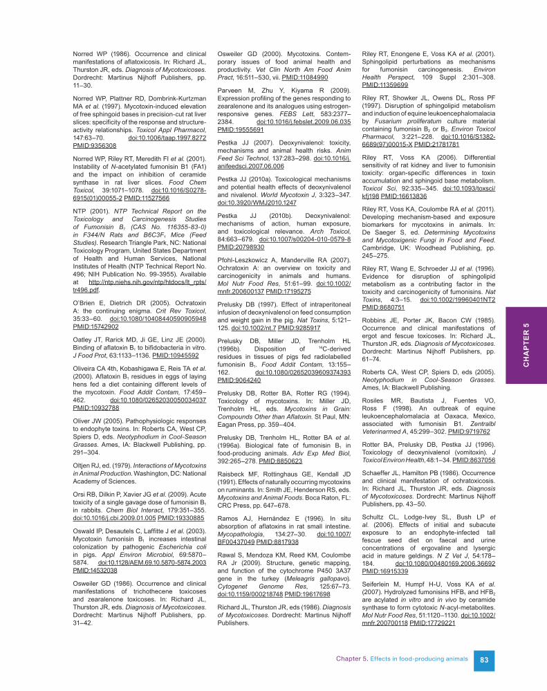
Chapter 5. Effects in food-producing animals 83
Norred WP (1986). Occurrence and clinical manifestations of aflatoxicosis. In: Richard JL, Thurston JR, eds. Diagnosis of Mycotoxicoses. Dordrecht: Martinus Nijhoff Publishers, pp. 11–30.
Norred WP, Plattner RD, Dombrink-Kurtzman MA et al. (1997). Mycotoxin-induced elevation of free sphingoid bases in precision-cut rat liver slices: specificity of the response and structure-activity relationships. Toxicol Appl Pharmacol, 147:63–70. doi:10.1006/taap.1997.8272 PMID:9356308
Norred WP, Riley RT, Meredith FI et al. (2001). Instability of N-acetylated fumonisin B1 (FA1) and the impact on inhibition of ceramide synthase in rat liver slices. Food Chem Toxicol, 39:1071–1078. doi:10.1016/S0278-6915(01)00055-2 PMID:11527566
NTP (2001). NTP Technical Report on the Toxicology and Carcinogenesis Studies of Fumonisin B1 (CAS No. 116355-83-0) in F344/N Rats and B6C3F1 Mice (Feed Studies). Research Triangle Park, NC: National Toxicology Program, United States Department of Health and Human Services, National Institutes of Health (NTP Technical Report No. 496; NIH Publication No. 99-3955). Available at http://ntp.niehs.nih.gov/ntp/htdocs/lt_rpts/tr496.pdf.
O’Brien E, Dietrich DR (2005). Ochratoxin A: the continuing enigma. Crit Rev Toxicol, 35:33–60. doi:10.1080/10408440590905948 PMID:15742902
Oatley JT, Rarick MD, Ji GE, Linz JE (2000). Binding of aflatoxin B1 to bifidobacteria in vitro. J Food Prot, 63:1133–1136. PMID:10945592
Oliveira CA 4th, Kobashigawa E, Reis TA et al. (2000). Aflatoxin B1 residues in eggs of laying hens fed a diet containing different levels of the mycotoxin. Food Addit Contam, 17:459–462. doi:10.1080/02652030050034037 PMID:10932788
Oliver JW (2005). Pathophysiologic responses to endophyte toxins. In: Roberts CA, West CP, Spiers D, eds. Neotyphodium in Cool-Season Grasses. Ames, IA: Blackwell Publishing, pp. 291–304.
Oltjen RJ, ed. (1979). Interactions of Mycotoxins in Animal Production. Washington, DC: National Academy of Sciences.
Orsi RB, Dilkin P, Xavier JG et al. (2009). Acute toxicity of a single gavage dose of fumonisin B1
in rabbits. Chem Biol Interact, 179:351–355. doi:10.1016/j.cbi.2009.01.005 PMID:19330885
Oswald IP, Desautels C, Laffitte J et al. (2003). Mycotoxin fumonisin B1 increases intestinal colonization by pathogenic Escherichia coli in pigs. Appl Environ Microbiol, 69:5870–5874. doi:10.1128/AEM.69.10.5870-5874.2003 PMID:14532038
Osweiler GD (1986). Occurrence and clinical manifestations of trichothecene toxicoses and zearalenone toxicoses. In: Richard JL, Thurston JR, eds. Diagnosis of Mycotoxicoses. Dordrecht: Martinus Nijhoff Publishers, pp. 31–42.
Osweiler GD (2000). Mycotoxins. Contem-porary issues of food animal health and productivity. Vet Clin North Am Food Anim Pract, 16:511–530, vii. PMID:11084990
Parveen M, Zhu Y, Kiyama R (2009). Expression profiling of the genes responding to zearalenone and its analogues using estrogen-responsive genes. FEBS Lett, 583:2377–2384. doi:10.1016/j.febslet.2009.06.035 PMID:19555691
Pestka JJ (2007). Deoxynivalenol: toxicity, mechanisms and animal health risks. Anim Feed Sci Technol, 137:283–298. doi:10.1016/j.anifeedsci.2007.06.006
Pestka JJ (2010a). Toxicological mechanisms and potential health effects of deoxynivalenol and nivalenol. World Mycotoxin J, 3:323–347. doi:10.3920/WMJ2010.1247
Pestka JJ (2010b). Deoxynivalenol: mechanisms of action, human exposure, and toxicological relevance. Arch Toxicol, 84:663–679. doi:10.1007/s00204-010-0579-8 PMID:20798930
Pfohl-Leszkowicz A, Manderville RA (2007). Ochratoxin A: an overview on toxicity and carcinogenicity in animals and humans. Mol Nutr Food Res, 51:61–99. doi:10.1002/mnfr.200600137 PMID:17195275
Prelusky DB (1997). Effect of intraperitoneal infusion of deoxynivalenol on feed consumption and weight gain in the pig. Nat Toxins, 5:121–125. doi:10.1002/nt.7 PMID:9285917
Prelusky DB, Miller JD, Trenholm HL (1996b). Disposition of 14C-derived residues in tissues of pigs fed radiolabelled fumonisin B1. Food Addit Contam, 13:155–162. doi:10.1080/02652039609374393 PMID:9064240
Prelusky DB, Rotter BA, Rotter RG (1994). Toxicology of mycotoxins. In: Miller JD, Trenholm HL, eds. Mycotoxins in Grain: Compounds Other than Aflatoxin. St Paul, MN: Eagan Press, pp. 359–404.
Prelusky DB, Trenholm HL, Rotter BA et al. (1996a). Biological fate of fumonisin B1 in food-producing animals. Adv Exp Med Biol, 392:265–278. PMID:8850623
Raisbeck MF, Rottinghaus GE, Kendall JD (1991). Effects of naturally occurring mycotoxins on ruminants. In: Smith JE, Henderson RS, eds. Mycotoxins and Animal Foods. Boca Raton, FL: CRC Press, pp. 647–678.
Ramos AJ, Hernández E (1996). In situ absorption of aflatoxins in rat small intestine. Mycopathologia, 134:27–30. doi:10.1007/BF00437049 PMID:8817938
Rawal S, Mendoza KM, Reed KM, Coulombe RA Jr (2009). Structure, genetic mapping, and function of the cytochrome P450 3A37 gene in the turkey (Meleagris gallopavo). Cytogenet Genome Res, 125:67–73. doi:10.1159/000218748 PMID:19617698
Richard JL, Thurston JR, eds (1986). Diagnosis of Mycotoxicoses. Dordrecht: Martinus Nijhoff Publishers.
Riley RT, Enongene E, Voss KA et al. (2001). Sphingolipid perturbations as mechanisms for fumonisin carcinogenesis. Environ Health Perspect, 109 Suppl 2:301–308. PMID:11359699
Riley RT, Showker JL, Owens DL, Ross PF (1997). Disruption of sphingolipid metabolism and induction of equine leukoencephalomalacia by Fusarium proliferatum culture material containing fumonisin B2 or B3. Environ Toxicol Pharmacol, 3:221–228. doi:10.1016/S1382-6689(97)00015-X PMID:21781781
Riley RT, Voss KA (2006). Differential sensitivity of rat kidney and liver to fumonisin toxicity: organ-specific differences in toxin accumulation and sphingoid base metabolism. Toxicol Sci, 92:335–345. doi:10.1093/toxsci/kfj198 PMID:16613836
Riley RT, Voss KA, Coulombe RA et al. (2011). Developing mechanism-based and exposure biomarkers for mycotoxins in animals. In: De Saeger S, ed. Determining Mycotoxins and Mycotoxigenic Fungi in Food and Feed. Cambridge, UK: Woodhead Publishing, pp. 245–275.
Riley RT, Wang E, Schroeder JJ et al. (1996). Evidence for disruption of sphingolipid metabolism as a contributing factor in the toxicity and carcinogenicity of fumonisins. Nat Toxins, 4:3–15. doi:10.1002/19960401NT2 PMID:8680751
Robbins JE, Porter JK, Bacon CW (1985). Occurrence and clinical manifestations of ergot and fescue toxicoses. In: Richard JL, Thurston JR, eds. Diagnosis of Mycotoxicoses. Dordrecht: Martinus Nijhoff Publishers, pp. 61–74.
Roberts CA, West CP, Spiers D, eds (2005). Neotyphodium in Cool-Season Grasses. Ames, IA: Blackwell Publishing.
Rosiles MR, Bautista J, Fuentes VO, Ross F (1998). An outbreak of equine leukoencephalomalacia at Oaxaca, Mexico, associated with fumonisin B1. Zentralbl Veterinarmed A, 45:299–302. PMID:9719762
Rotter BA, Prelusky DB, Pestka JJ (1996). Toxicology of deoxynivalenol (vomitoxin). J Toxicol Environ Health, 48:1–34. PMID:8637056
Schaeffer JL, Hamilton PB (1986). Occurrence and clinical manifestation of ochratoxicosis. In: Richard JL, Thurston JR, eds. Diagnosis of Mycotoxicoses. Dordrecht: Martinus Nijhoff Publishers, pp. 43–50.
Schultz CL, Lodge-Ivey SL, Bush LP et al. (2006). Effects of initial and subacute exposure to an endophyte-infected tall fescue seed diet on faecal and urine concentrations of ergovaline and lysergic acid in mature geldings. N Z Vet J, 54:178–184. doi:10.1080/00480169.2006.36692 PMID:16915339
Seiferlein M, Humpf H-U, Voss KA et al. (2007). Hydrolyzed fumonisins HFB1 and HFB2
are acylated in vitro and in vivo by ceramide synthase to form cytotoxic N-acyl-metabolites. Mol Nutr Food Res, 51:1120–1130. doi:10.1002/mnfr.200700118 PMID:17729221
CH
AP
TER
5
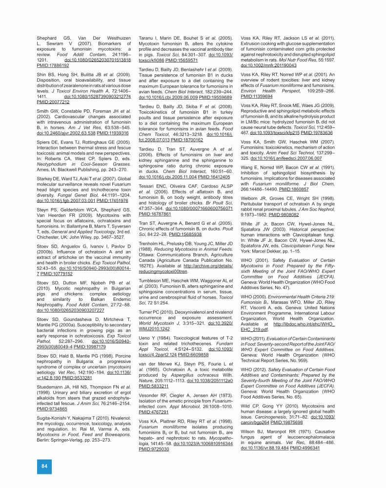
84
Shephard GS, Van Der Westhuizen L, Sewram V (2007). Biomarkers of exposure to fumonisin mycotoxins: a review. Food Addit Contam, 24:1196–1201. doi:10.1080/02652030701513818 PMID:17886192
Shin BS, Hong SH, Bulitta JB et al. (2009). Disposition, oral bioavailability, and tissue distribution of zearalenone in rats at various dose levels. J Toxicol Environ Health A, 72:1406–1411. doi:10.1080/15287390903212774 PMID:20077212
Smith GW, Constable PD, Foreman JH et al. (2002). Cardiovascular changes associated with intravenous administration of fumonisin B1 in horses. Am J Vet Res, 63:538–545. doi:10.2460/ajvr.2002.63.538 PMID:11939316
Spiers DE, Evans TJ, Rottinghaus GE (2005). Interaction between thermal stress and fescue toxicosis: animal models and new perspectives. In: Roberts CA, West CP, Spiers D, eds. Neotyphodium in Cool-Season Grasses. Ames, IA: Blackwell Publishing, pp. 243–270.
Starkey DE, Ward TJ, Aoki T et al. (2007). Global molecular surveillance reveals novel Fusarium head blight species and trichothecene toxin diversity. Fungal Genet Biol, 44:1191–1204. doi:10.1016/j.fgb.2007.03.001 PMID:17451976
Steyn PS, Gelderblom WCA, Shephard GS, Van Heerden FR (2009). Mycotoxins with special focus on aflatoxins, ochratoxins and fumonisins. In: Ballantyne B, Marrs T, Syversen T, eds. General and Applied Toxicology, 3rd ed. Chichester, UK: John Wiley, pp. 3467–3527.
Stoev SD, Anguelov G, Ivanov I, Pavlov D (2000b). Influence of ochratoxin A and an extract of artichoke on the vaccinal immunity and health in broiler chicks. Exp Toxicol Pathol, 52:43–55. doi:10.1016/S0940-2993(00)80014-7 PMID:10779152
Stoev SD, Dutton MF, Njobeh PB et al. (2010). Mycotic nephropathy in Bulgarian pigs and chickens: complex aetiology and similarity to Balkan Endemic Nephropathy. Food Addit Contam, 27:72–88. doi:10.1080/02652030903207227
Stoev SD, Goundasheva D, Mirtcheva T, Mantle PG (2000a). Susceptibility to secondary bacterial infections in growing pigs as an early response in ochratoxicosis. Exp Toxicol Pathol, 52:287–296. doi:10.1016/S0940-2993(00)80049-4 PMID:10987179
Stoev SD, Hald B, Mantle PG (1998). Porcine nephropathy in Bulgaria: a progressive syndrome of complex or uncertain (mycotoxin) aetiology. Vet Rec, 142:190–194. doi:10.1136/vr.142.8.190 PMID:9533281
Stuedemann JA, Hill NS, Thompson FN et al. (1998). Urinary and biliary excretion of ergot alkaloids from steers that grazed endophyte-infected tall fescue. J Anim Sci, 76:2146–2154. PMID:9734865
Sugita-Konishi Y, Nakajima T (2010). Nivalenol: the mycology, occurrence, toxicology, analysis and regulation. In: Rai M, Varma A, eds. Mycotoxins in Food, Feed and Bioweapons. Berlin: Springer-Verlag, pp. 253–273.
Taranu I, Marin DE, Bouhet S et al. (2005). Mycotoxin fumonisin B1 alters the cytokine profile and decreases the vaccinal antibody titer in pigs. Toxicol Sci, 84:301–307. doi:10.1093/toxsci/kfi086 PMID:15659571
Tardieu D, Bailly JD, Benlashehr I et al. (2009). Tissue persistence of fumonisin B1 in ducks and after exposure to a diet containing the maximum European tolerance for fumonisins in avian feeds. Chem Biol Interact, 182:239–244. doi:10.1016/j.cbi.2009.06.009 PMID:19559689
Tardieu D, Bailly JD, Skiba F et al. (2008). Toxicokinetics of fumonisin B1 in turkey poults and tissue persistence after exposure to a diet containing the maximum European tolerance for fumonisins in avian feeds. Food Chem Toxicol, 46:3213–3218. doi:10.1016/j.fct.2008.07.013 PMID:18700162
Tardieu D, Tran ST, Auvergne A et al. (2006). Effects of fumonisins on liver and kidney sphinganine and the sphinganine to sphingosine ratio during chronic exposure in ducks. Chem Biol Interact, 160:51–60. doi:10.1016/j.cbi.2005.11.004 PMID:16412405
Tessari ENC, Oliveira CAF, Cardoso ALSP et al. (2006). Effects of aflatoxin B1 and fumonisin B1 on body weight, antibody titres and histology of broiler chicks. Br Poult Sci, 47:357–364. doi:10.1080/00071660600756071 PMID:16787861
Tran ST, Auvergne A, Benard G et al. (2005). Chronic effects of fumonisin B1 on ducks. Poult Sci, 84:22–28. PMID:15685938
Trenholm HL, Prelusky DB, Young JC, Miller JD (1988). Reducing Mycotoxins in Animal Feeds. Ottawa: Communications Branch, Agriculture Canada (Agriculture Canada Publication No. 1827E). Available at http://archive.org/details/reducingmycotoxi00tren.
Tumbleson ME, Haschek WM, Waggoner AL et al. (2003). Fumonisin B1 alters sphinganine and sphingosine concentrations in serum, tissue, urine and cerebrospinal fluid of horses. Toxicol Sci, 72 S1:254.
Turner PC (2010). Deoxynivalenol and nivalenol occurrence and exposure assessment. World Mycotoxin J, 3:315–321. doi:10.3920/WMJ2010.1242
Ueno Y (1984). Toxicological features of T-2 toxin and related trichothecenes. Fundam Appl Toxicol, 4:S124–S132. doi:10.1093/toxsci/4.2part2.124 PMID:6609858
van der Merwe KJ, Steyn PS, Fourie L et al. (1965). Ochratoxin A, a toxic metabolite produced by Aspergillus ochraceus Wilh. Nature, 205:1112–1113. doi:10.1038/2051112a0 PMID:5833211
Vesonder RF, Ciegler A, Jensen AH (1973). Isolation of the emetic principle from Fusarium-infected corn. Appl Microbiol, 26:1008–1010. PMID:4767291
Voss KA, Plattner RD, Riley RT et al. (1998). Fusarium moniliforme isolates producing fumonisins B2 or B3 but not fumonisin B1, are hepato- and nephrotoxic to rats. Mycopatho-logia, 141:45–58. doi:10.1023/A:1006810916344 PMID:9725030
Voss KA, Riley RT, Jackson LS et al. (2011). Extrusion cooking with glucose supplementation of fumonisin contaminated corn grits protected against nephrotoxicity and disrupted sphingolipid metabolism in rats. Mol Nutr Food Res, 55:1597. doi:10.1002/mnfr.201190043
Voss KA, Riley RT, Norred WP et al. (2001). An overview of rodent toxicities: liver and kidney effects of Fusarium moniliforme and fumonisins. Environ Health Perspect, 109:259–266. PMID:11359694
Voss KA, Riley RT, Snook ME, Waes JG (2009). Reproductive and sphingolipid metabolic effects of fumonisin B1 and its alkaline hydrolysis product in LM/Bc mice: hydrolyzed fumonisin B1 did not cause neural tube defects. Toxicol Sci, 112:459–467. doi:10.1093/toxsci/kfp215 PMID:19783636
Voss KA, Smith GW, Haschek WM (2007). Fumonisins: toxicokinetics, mechanism of action and toxicity. Anim Feed Sci Technol, 137:299–325. doi:10.1016/j.anifeedsci.2007.06.007
Wang E, Norred WP, Bacon CW et al. (1991). Inhibition of sphingolipid biosynthesis by fumonisins. Implications for diseases associated with Fusarium moniliforme. J Biol Chem, 266:14486–14490. PMID:1860857
Welborn JR, Groves CE, Wright SH (1998). Peritubular transport of ochratoxin A by single rabbit renal proximal tubules. J Am Soc Nephrol, 9:1973–1982. PMID:9808082
White JF Jr, Bacon CW, Hywel-Jones NL, Spatafora JW (2003). Historical perspective: human interactions with Clavicipitalean fungi. In: White JF Jr, Bacon CW, Hywel-Jones NL, Spatafora JW, eds. Clavicipitalean Fungi. New York: Marcel Dekker, pp. 1–15.
WHO (2001). Safety Evaluation of Certain Mycotoxins in Food: Prepared by the Fifty-sixth Meeting of the Joint FAO/WHO Expert Committee on Food Additives (JECFA). Geneva: World Health Organization (WHO Food Additives Series, No. 47).
WHO (2000). Environmental Health Criteria 219: Fumonisin B1. Marasas WFO, Miller JD, Riley RT, Visconti A, eds. Geneva: United Nations Environment Programme, International Labour Organization, World Health Organization. Available at http://libdoc.who.int/ehc/WHO_EHC_219.pdf.
WHO (2011). Evaluation of Certain Contaminants in Food: Seventy-second Report of the Joint FAO/WHO Expert Committee on Food Additives. Geneva: World Health Organization (WHO Technical Report Series, No. 959).
WHO (2012). Safety Evaluation of Certain Food Additives and Contaminants: Prepared by the Seventy-fourth Meeting of the Joint FAO/WHO Expert Committee on Food Additives (JECFA). Geneva: World Health Organization (WHO Food Additives Series, No. 65).
Wild CP, Gong YY (2010). Mycotoxins and human disease: a largely ignored global health issue. Carcinogenesis, 31:71–82. doi:10.1093/carcin/bgp264 PMID:19875698
Wilson BJ, Maronpot RR (1971). Causative fungus agent of leucoencephalomalacia in equine animals. Vet Rec, 88:484–486. doi:10.1136/vr.88.19.484 PMID:4996341

Chapter 5. Effects in food-producing animals 85
Wilson R, Ziprin R, Ragsdale S, Busbee D (1985). Uptake and vascular transport of ingested afla-toxin. Toxicol Lett, 29:169–176. doi:10.1016/0378-4274(85)90038-4 PMID:3937298
Wong ZA, Hsieh DPH (1980). The comparative metabolism and toxicokinetics of aflatoxin B1
in the monkey, rat, and mouse. Toxicol Appl Pharmacol, 55:115–125. doi:10.1016/0041-008X(80)90227-6 PMID:7423498
Yip SS, Coulombe RA Jr (2006). Molecular cloning and expression of a novel cytochrome P450 from turkey liver with aflatoxin B1 oxidizing activity. Chem Res Toxicol, 19:30–37. doi:10.1021/tx050233+ PMID:16411653
Zinedine A, Soriano JM, Moltó JC, Mañes J (2007). Review on the toxicity, occurrence, metabolism, detoxification, regulations and intake of zearalenone: an oestrogenic myco-toxin. Food Chem Toxicol, 45:1–18. doi:10.1016/j.fct.2006.07.030 PMID:17045381
Zingerle M, Silbernagl S, Gekle M (1997). Reabsorption of the nephrotoxin ochratoxin A along the rat nephron in vivo. J Pharmacol Exp Ther, 280:220–224. PMID:8996199
Zitomer NC, Mitchell T, Voss KA et al. (2009). Ceramide synthase inhibition by fumonisin B1
causes accumulation of 1-deoxysphinganine: a novel category of bioactive 1-deoxysphingoid bases and 1-deoxydihydroceramides biosyn-thesized by mammalian cell lines and animals. J Biol Chem, 284:4786–4795. doi:10.1074/jbc.M808798200 PMID:19095642
Zöllner P, Jodlbauer J, Kleinova M et al. (2002). Concentration levels of zearalenone and its metabolites in urine, muscle tissue, and liver samples of pigs fed with mycotoxin-contaminated oats. J Agric Food Chem, 50:2494–2501. doi:10.1021/jf0113631 PMID:11958611
CH
AP
TER
5
