chapter chapter i ii i -...
TRANSCRIPT

IntroductionIntroductionIntroductionIntroduction
Chapter Chapter Chapter Chapter I I I I

Stress can be defined as any environmental factor capable of inducing a
potentially injurious strain in living organisms. A biological strain is any change in
physiological process and functional activity of enzymes of metabolism under
stress (Zlatev et. al., 2012). Plants are frequently exposed to many stresses, such
as, drought, low temperature, salt, flooding, heat, oxidative stress and heavy metal
toxicity, while growing in nature. These stresses are of two types - biotic and
abiotic. Biotic stress involves damage to plants by other living organisms, such as,
bacteria, viruses, fungi, parasites, harmful insects and weeds, while, abiotic stress
is a negative impact of non-living factors on plants in specific environment, such
as, light, temperature, water availability, nutrients, and soil composition. Abiotic
stress affects plant metabolism, physiological and biochemical processes and
disrupt cellular homeostasis. Amongst abiotic stresses, osmotic stress, drought and
salinity are the most severe problems in worldwide agricultural production (Saha
et. al., 2010).
Osmotic stress means insufficient water availability i.e. water deficit, which
limits plant growth and development and hence productivity (Zhu et. al., 1997).
The first response of virtually all the plants to acute water deficit is the closure of
stomata to prevent the transpirational water loss (Manfield et. al., 1990). Closure
of stomata may result from direct evaporation of water from the guard cells with
no metabolic involvement. It is a major temporary adaptive change that prevents
further water loss from the plants. When water deficit becomes too intense or
prolonged, plants can wilt, cells can undergo shrinkage and this may lead to
mechanical constraint on cellular membranes. The normal bilayer structure of
membrane due to removal of water disrupts and becomes porous when desiccated.
This in turn impairs the functioning of ions and transporters as well as membrane
associated enzymes (Bowler, 1992). Further, due to cell shrinkage and a decline in
cellular volume, cellular content becomes viscous, therefore increasing the
probability of protein–protein interaction leading to their aggregation and
denaturation (Mahajan et. al., 2005). Increased concentration of solutes may also
exceed toxic levels, which may be deleterious for the functioning of the enzymes

ncluding the enzymes required for photosynthetic machinery (Hoekstra et. al.,
2001; Mahajan et. al., 2005; Farooq et. al., 2009).
Water deficit influences many physiological responses in plants. Thus,
decrease in germination rate, relative water content, fresh weight, dry weight, root
length and shoot length due to water stress have been reported in several plant
systems (Sharma et. al., 2004; Nayyar and Gupta, 2006 and Jamwal et. al., 2012).
Osmotic stress has been reported to stimulate respiration in pea protoplasts
(Saradadevi and Raghavendra, 1994; Dwivedi et. al., 2003). A major effect of
water deficit is reduction in photosynthesis, which arises by a decrease in leaf
expansion, impaired photosynthetic machinery, premature leaf senescence and thus
associated with reduction in food production (Wahid and Rasul, 2005). The effects
can be direct due to decreased CO2 availability caused by diffusion limitations
through the stomatal closure and the mesophyll conductance (Flexas et. al., 2007)
or indirect due to oxidative stress. Decline in intracellular CO2 levels also results in
the over-reduction of components within the electron transport chain and the
electrons get transferred to oxygen at photosystem I. Further, the steady state levels
of major photosystem II mRNAs and proteins, half lives of D1 and D2 proteins, as
well as of psb-A and psb–D mRNAs have been shown to decline with increasing
water stress in barley leaves (Yuan et. al., 2005). Furthermore, changes in the
structure of developing chloroplast by water stress which decreased the net
photosynthetic rate and reduction in the contents and activity of ribulose-1,5-
bisphosphate carboxylase / oxygenase by water stress have been reported
(Bourque et. al., 1975; Chaitanya et. al., 2003; Zgalla et. al., 2005; Saibo et. al.,
2009).
Chlorophyll (Chl) molecules play a central role in the photosynthetic
apparatus by capturing light and directing the energy towards the photosystems.
Chlorophyll is a tetrapyrrole macro cycle containing Mg, a phytol chain, and a
characteristic fifth ring. Higher plants have two chlorophyll species, chlorophyll a
and b (Figure 1). Chlorophyll metabolism is a highly coordinated process that is
executed via a series of cooperative reactions catalyzed by several enzymes (Beale,

Figure 1. Structure of Chlorophyll a and b

1999). The major site of chlorophyll biosynthesis in plants is plastid, which is
regulated at several steps (Masuda and Fujita, 2008; Mochizuki et. al., 2010).
Earlier steps of the pathway are common with biosynthesis of other tetrapyrrole
derivatives like heme, phtyochromes, phycobilins etc. Outline of the key steps
involved in chlorophyll biosynthesis is presented in Figure 2.
The synthesis of 5-amino levulinic acid (ALA) is the first committed step of
chlorophyll biosynthesis, and is therefore a key control point in the regulation of
chlorophyll formation. ALA, the universal precursor contributes all the carbon and
nitrogen atoms in chlorophyll molecule is synthesized by two pathways (Panek and
O’Brian, 2002; Schoefs and Bertrand, 2005). In yeast, some bacteria and animals, a
single enzyme ALA synthase catalyzes the condensation of glycine and succinyl
Co A to form ALA through Shemin pathway (Avissar et. al., 1989; Tanaka et. al.,
2011). In higher plants, algae and cynobacteria the intact 5-carbon skeleton of
glutamate is converted into ALA by a multienzyme system involving three steps
(Kannangara et. al., 1988; Bollivar, 2006). These are depicted in Figure 3 (Tanaka
et. al., 2011). These steps include ligation of glutamate to tRNAGlu
catalysed by
glutamyl tRNA synthetase, the reduction of glutamate to glutamate-1-
semialdehyde by glutamyl tRNA reductase (GluTR) and a final transamination
step mediated by glutamate-1-semialdehyde aminotransferase (GSAT)
(Kannangara et. al.,1988; Tanaka et. al., 2011). GluTR is the main target of
regulatory mechanism modulating ALA formation and thus responding to a wide
range of stimuli, whereas GSAT responds only weakly. GluTR mRNA levels
correlate well with chlorophyll synthetic activity in higher plants. When etiolated
seedlings are exposed to light, GluTR mRNA levels and ALA synthesis rise in
parallel, while, on inhibition of GluTR mRNA production using antisense RNA,
chlorophyll levels decrease in Arabdiopsis (Kumar and Soll, 2000; Ujwal, 2002).
On the other hand, in water-stressed Oryza sativa seedlings protein / transcript
abundance of GSAT increased, while, the ALA content decreased (Mohanty et. al.,
2006). The authors suggested inactivation of GSAT enzyme by post-translational
modification under stress condition. Further, reduced synthesis of ALA in chilled
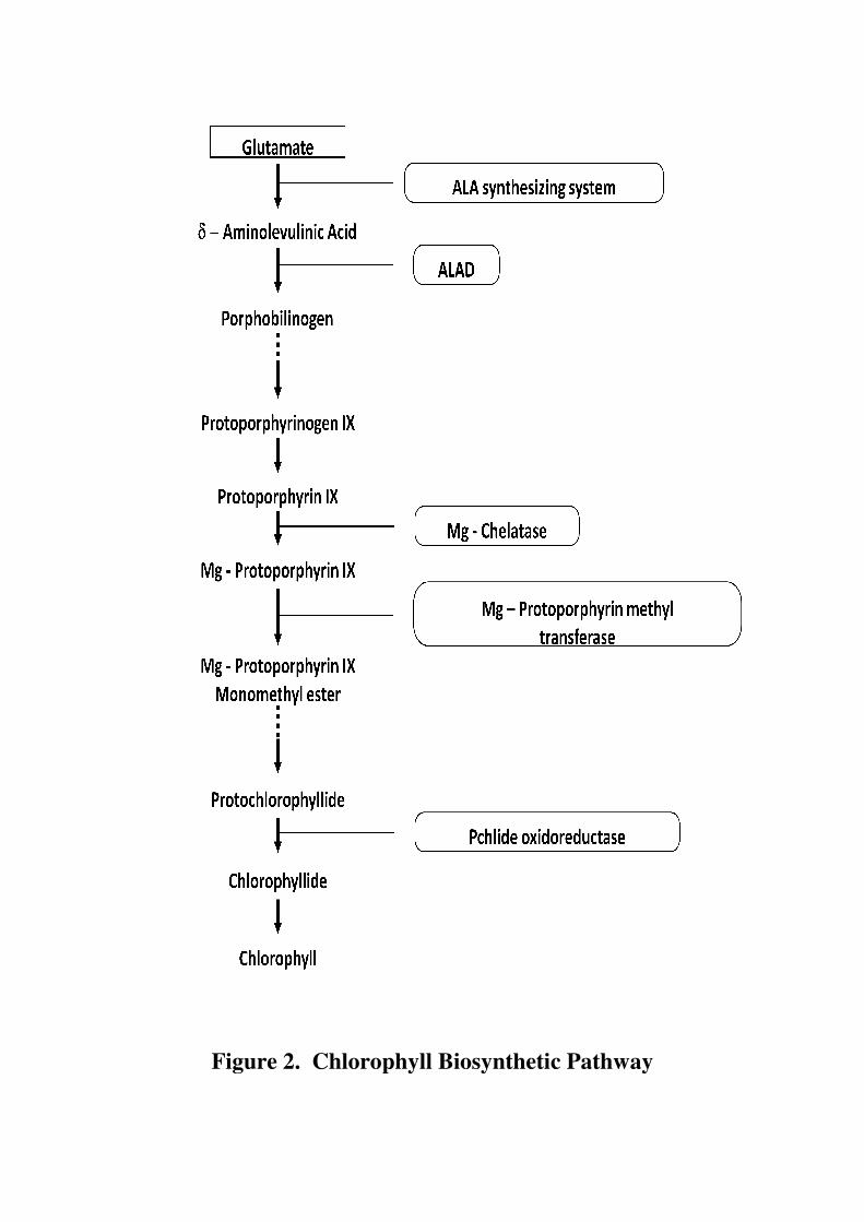
Figure 2. Chlorophyll Biosynthetic Pathway
Figure 2. Chlorophyll Biosynthetic Pathway
Figure 2. Chlorophyll Biosynthetic Pathway

Figure 3. The three step reaction of 5
(Reference: Tanaka et. al., 2011).
The three step reaction of 5-aminolevulenic acid (ALA) synthesis from glutamate
2011).
aminolevulenic acid (ALA) synthesis from glutamate

and heat stressed Cucumis sativa seedlings has been reported by Tewari and
Tripathy (1998).
δ-Aminolevulinic acid dehydratase (ALAD; E.C 4.2.1.24), one of the earlier
enzymes of the pathway, catalyzes the asymmetric condensation of two molecules
of ALA with the release of two H2O molecules leading to the formation of the
basic unit of tetrapyrroles, the porphobilinogen (PBG). In several systems the
enzyme plays a major role in regulating the chlorophyll biosynthesis (Naito et. al.,
1980; Prasad et. al., 1989; Padmaja et. al., 1990; Prasad and Prasad, 1990).
ALADs from different sources are metalloenzymes that utilize a variety of divalent
and monovalent cations (Jaffe, 2000). The plant enzyme requires Mg2+
for
enzymatic activity. ALAD is a substrate modulated enzyme (Tchuinmogne et. al.,
1989) and levulinic acid, a structural analogue of ALA, is a competitive inhibitor
of the enzyme (Senior et. al., 1996). Decline in ALAD activity and its gene
expression in water stressed rice seedlings (Dalal and Tripathy, 2012) and chill and
heat-stressed Cucumis sativa and Triticum aestivum seedlings (Tewari and
Tripathy, 1998; Mohanty et al. 2006) has been reported. Decreased availability of
the substrate ALA of the enzyme could negatively regulate the gene expression in
water stress conditions (Dalal and Tripathy, 2012). Inhibition of ALAD activity by
cadmium, mercury and arsenic has been reported in excised greening maize leaf
segments (Jain and Gadre, 2004; Sarangthem et. al., 2011; Gupta et. al., 2013).
Porphobilinogen deaminase (PBGD; EC 2.5.1.61) condenses four PBG
molecules to form the first tetrapyrrole, hydroxymethylbilane, which further forms
uroporphyrinogen III through the nonphysiological product uroporphyrinogen I
(Battersby et. al., 1979). The enzyme is found to be highly specific for PBG and
requires thiol compounds for activity (Bogorad, 1958). Decline in the rate of
synthesis of protochlorophyllide (Pchlide) and Chlorophyllide (Chlide) due to loss
of ALAD and PBGD activity has been suggested in senescent barley leaves
(Hukmani and Tripathy, 1994). Inhibition of PBGD activity by chill and heat
stress has been demonstrated in etiolated T. aestivum seedlings (Tewari and
Tripathy, 1998). Down regulation of transcript abundance is suggested as the cause
of reduced PBGD activity in water-stressed O. sativa seedlings (Dalal and
Tripathy, 2012). In subsequent steps urogen III is converted to Protoporphyrin IX,
which chelates with Mg2+
to form Mg Protoporphyrin IX, which is then converted
to Pchlide. Pchlide is converted to Chlide and then to chlorophylls. Changes in
chlorophyll a /b ratio and carotenoids due to drought stress have been reported in
Hordeum vulgare L. (Anjum et. al., 2003) and O. sativa (Sikuku et. al., 2010).
Reduction in chlorophyll content due to drought has been shown in Helianthus
annuus L. (Kiani et. al., 2008), Vaccinium myrtillus L. (Tahkokorpi et. al., 2007),
Gossypium hirsutum L. (Massacci et. al., 2008), Catharanthus roseus L. (Jaleel et.
al., 2008), Oryza. sativa (Chutia and Borah, 2012). Regulation of the levels of
chlorophyll branch intermediates is extremely important since most of these
molecules are strong photosensitizers. When present in excess they produce
reactive oxygen species causing oxidative damage or cell death (Mock et. al.,
2002).
Plants have many adaptive mechanisms contributing to water stress
resistance. One of them is osmotic adjustment (OA), which involves the net
accumulation of solutes in cells in response to a fall in the water potential of the
environment (Zhang et. al., 1999). Organic compounds that function as solutes
(osmoprotectants / osmolytes) include amino acids, such as, proline, sugar
alcohols, such as, mannitol, and quarternary ammonium compounds, such as,
glycine-betanine (GB). Accumulation of proline (Viegas and Silvera, 1997 and
Kumar et. al., 2012) and glycine- betaine (Pollard and Wyn Jones, 1979; Sairam et.
al., 2002) has been reported in several plant systems as a result of osmotic stress.
Overproduction of proline in response to salinity and osmotic stress has also been
shown in a genetically modified cyanobacterium, Nostoc muscorum (Bhargava,
2006). It influences protein solvation and preserves the quarternary structure of
complex proteins, maintains membrane integrity under dehydration stress and
reduces oxidation of lipid membranes or photoinhibition (Demiral and Turkan,
2004). Furthermore, it also contributes in stabilizing sub-cellular structures,
scavenging free radicals, and buffering cellular redox potential during stress
conditions (Ashraf and Foolad, 2007; Anjum et. al., 2011). Besides proline,
abundant levels of other amino acids, such as, asparagine, glutamate and γ-amino
benzoic acid have also been reported in response to drought (Alia et.al, 2001; Diaz
et. al., 2005; Asharaf et. al., 2007; Chutia and Borah, 2012).
One of the most common responses of plants to water deficit leads to
enhanced generation of reactive oxygen species (ROS), such as, H2O2, superoxide
(O2• −
) and hydroxyl (OH•) radicals due to disruption of cellular homeostasis
(Mittler, 2002; Hu et. al., 2008; Han et. al., 2009; Mishra et. al., 2011; Srivastava
et. al., 2011). All ROS are extremely harmful to organisms at high concentrations.
When the level of ROS exceeds the defense mechanisms, a cell is said to be in a
state of “oxidative stress.” The enhanced production of ROS during environmental
stresses can pose a threat to cells by causing peroxidation of lipids, oxidation of
proteins, damage to nucleic acids, enzyme inhibition, activation of programmed
cell death (PCD) pathway and ultimately leading to death of the cells (Mittler,
2002; Meriga et. al., 2004; Maheshwari and Dubey, 2009; Mishra et. al., 2011).
Drought-induced overproduction of ROS increases the content of
malondialdehyde (MDA) due to membrane lipid peroxidation. Thus, the content of
MDA has been considered an indicator of oxidative damage (Moller et. al., 2007).
H2O2 is generated in normal metabolism via the Mehler reaction in chloroplasts,
electron transport chain in mitochondria and photorespiration in peroxisomes.
Increment in MDA and H2O2 concentrations in the water stressed cuttings of
Populus cathayana L., P. kangdingensis has been shown (Yang and Miao, 2010).
Further, in Pisum sativum L. levels of lipid peroxidation increased two to four fold
with an increase in the drought stress, and this was highly correlated with protein
oxidation peroxidation (Moran et. al., 1994).The ability of higher plants to
scavenge the toxic effects of ROS seems to be very important determinant of their
tolerance to these stresses. Production and scavenging of ROS in plants is depicted
in Figure 4, (da Silva et. al., 2013). Antioxidants are the first line of defense
against free radical damage. They are critical for maintaining optimum health of
plants cells. There are several antioxidant enzymes like superoxide dismutase
(SOD), catalase (CAT), ascorbate peroxidase (APX), dehydroascorbate reductase

Figure 4. Production and scavenging of ROS in plants (reference: da Silva
Production and scavenging of ROS in plants (reference: da Silva Production and scavenging of ROS in plants (reference: da Silva et. al., 2013)
(DHAR) and glutathione reductase (GR), and non enzymatic antioxidants, such as,
ascorbate (ASC), glutathione (GSH) which are effectively involved in scavenging
of ROS in plants.
Superoxide dismutase (SOD; EC 1.15.1.1) is the first enzyme in the ROS
detoxifying process, which catalyzes the dismutation of superoxide radical (O• −
)
into oxygen and hydrogen peroxide (H2O2).
2O• −
+ 2O• −
+ 2H+ H2O2 + O2
Three isozymes of SOD copper/zinc SOD (Cu/Zn-SOD), manganese SOD (Mn-
SOD), and iron SOD (Fe-SOD) are reported in plants (Fridovich, 1989; Racchi et.
al., 2001). All forms of SOD are nuclear encoded and targeted to their respective
sub cellular compartments by an amino terminal targeting sequence (Bowler et. al.,
1992). Fe-SOD is localized in chloroplast where the major site of O2• −
production
is the thylakoid membrane-bound primary electron acceptor of PSI. SOD activity
has been reported to increase in several plants exposed to various stresses, such as,
salinity and heavy metals (Kukreja et. al., 2005; Zlatev et. al., 2006; Wang et. al.,
2008; Mishra et. al., 2011).
Catalase (CAT, EC 1.11.1.6) was the first enzyme to be discovered and
characterized in antioxidant enzymes. CAT is a tetrameric heme containing
enzyme with the potential to directly dismutase H2O2 into H2O and O2 and is
indispensable for ROS detoxification during stressed conditions (Garg and
Manchanda, 2009).
2H2O2 O2 + 2H2O
The enzyme function is to remove the H2O2 generated in peroxisomes by oxidases
involved in β-oxidation of fatty acids, photorespiration, purine catabolism and
during oxidative stress (Mittler, 2002; Vellosillo et. al., 2010). Increase in CAT
activity is an adaptive trait possibly helping to overcome the damage to tissue
metabolism by reducing toxic levels of H2O2. Increased CAT activity in T.

aestivum (Simova- Stoilova et. al., 2010) and in P. asperata (Yang et. al., 2008)
has been shown under drought stress.
Guaiacol Peroxidase (Gu-POX, E.C. 1.11.1.7), a heme containing protein,
preferably oxidizes aromatic electron donor, such as guaiacol and pyragallol at the
expense of H2O2. Many isoenzymes of Gu-POX exist in plant tissues localized in
vacuoles, the cell wall, and the cytosol (Asada, 1992). It is involved in large
number of biological and physiological processes including plant growth and
differentiation, cell elongation, cross linking of cell wall polysaccharides,
lignifications, wound healing, phenolic oxidation, flavonoids degradation and
defense against pathogen. It can function as effective quencher of reactive
intermediary forms of O2 and peroxy radicals under stressed conditions
(Vangronsveld et. al., 1994). Increased Gu-POX activity under drought stress
conditions in various plants, like Glycyrrhiza uralensis L. (Pan et. al., 2006),
Helianthus cultivar (Gunes et. al., 2008) and P. cathayana L. (Xiao et. al., 2008)
has been reported.
Ascorbate peroxidase (APX, E.C. 1.1.11.1) is a central component of
ascorbate – glutathione cycle (ASC-GSH cycle), and plays an essential role in the
control of intracellular ROS levels.
ASC + H2O2 2DHA + 2H2O
It is a member of Class I super family of heme peroxidases and is regulated by
redox signals and H2O2. Five distinct isoenzymes of APX have been found in
cytosolic, stromal, thylakoidal, mitochondrial and peroxisomal locations (Nakano
and Asada, 1987; Jimenez et. al., 1997; Madhusudan et. al., 2003). These isoforms
respond differently to metabolic and environmental signals (Kubo et. al., 1995).
Significant increase in APX activity was noted under water stress in three cultivars
of Phaseolus vulgaris L. (Zaltev et. al., 2006) and P. asperata L. (Yang et. al.,
2008).
Glutathione reductase (GR, E.C. 1.6.4.2), a NADPH-dependent enzyme
catalyzes the reduction of GSSG to GSH and thus, maintains high cellular
GSH/GSSG ratio.
NADPH + GSSG NADP+ + 2GSH
GR belongs to a group of flavoenzymes and contains an essential disulfide group
(Ghisla and Massey, 1989). It is located in the chloroplasts, cytosol, mitochondria,
and peroxisomes (Edwards et. al., 1990). In chloroplast, GSH and GR are involved
in detoxification of H2O2 generated by Mehler reaction. Eyidogan and Oz (2005)
reported increased GR activity in the leaf tissue of Cicer arietinum L. under salt
stress.
Nonenzymic components of the antioxidative defense system include the
major cellular redox buffers, ascorbate and glutathione (γ-glutamyl-cysteinyl-
glycine) as well as tocopherol, carotenoids, and phenolic compounds. They
perform multiple roles in defense and as enzyme cofactor, thus, regulate plant
growth and development by modulating processes from mitosis and cell elongation
to senescence and cell death (Pinto and Gara, 2004). Ascorbate is the most
abundant, low molecular weight antioxidant that reacts with OH•, O2
• −, and
1O2
and plays a key role in defense against oxidative stress (Chen and Gallie, 2003).
Ascorbate removes H2O2 via ASC-GSH cycle (Pinto et. al., 2004), where two
molecules of ASC are utilized to reduce H2O2 to water with concomitant
generation of monodehydro ascorbate (MDHA). MDHA is a radical with a short
life time and can spontaneously dismutate into dehydroascorbate (DHA) and ASC
or is reduced to ASC by NADPH dependent enzyme monodehydro ascorbate
reductase (MDHAR) (Miyake and Asada, 1994). ASC level has been reported to
alter in response to various stresses, such as, water deficit, low or high temperature
and metal stress (Sharma and Dubey, 2005; Radyuk et. al., 2009; Maheshwari and
Dubey, 2009; Mishra et. al., 2011; Srivastava et. al., 2011).
Reduced glutathione occurs abundantly in plant tissues and is localized in
all cell compartments like cytosol, endoplasmic reticulum, vacuoles, mitochondria,
chloroplasts, peroxisomes as well as in apoplast (Jimenez et. al., 1997). GSH reacts
chemically with O2•−
, OH•, H2O2 and, therefore, can function directly as a free
radical scavenger. It can protect macromolecules (i.e. proteins, lipids, DNA) either
by the formation of adducts directly with reactive electrophiles (glutathiolation) or
by acting as proton donor in the presence of ROS or organic free radicals, yielding
oxidized glutathione (GSSG) (Asada et. al., 1994). It can participate in
regeneration of another potential antioxidant ASC, via the ASC-GSH cycle. GSH
recycles ASC from it’s oxidized to reduced form by the enzyme dehydroascorbate
reductase (DHAR) (Loewus, 1988). The role of GSH in the antioxidative defense
system provides a rationale for its use as a stress marker. Srivastava et. al. (2005)
reported an appreciable decline in glutathione reductase (GR) activity and GSH
pool under Cu stress and significant increase under salt stress.
Sorbitol is a low molecular weight 6 carbon alditol which has been used to
impose osmotic stress on leaf slices, protoplast or chloroplast. It is a direct product
of photosynthesis in mature leaves, in parallel with sucrose, and both serve similar
functions, such as, translocation of carbon skeletons and energy between sources
and sink organs. Increased transport of polyols, both in the xylem and phloem
occurs frequently as a result of salt or drought stress (Noiraud et. al., 2001). In
addition to sorbitol, polyethylene glycol (PEG), a neutral osmotically active
polymer, is most frequently used in plant water deficit studies to induce
dehydration by decreasing the water potential of nutrient solution. PEG 6000
cannot enter the pores of plant cells and thus, cause cytorrhysis rather than
plasmolysis (Oertli, 1985); therefore, PEG solutions mimic dry soil more closely
(Verslues et. al., 1998).
The present study aimed at analyzing the effect of osmotic stress induced by
sorbitol and PEG-6000 on overall growth and biochemical parameters, related to
growth and stress response, enzymes of chlorophyll biosynthesis, such as, ALA
synthesizing activity, ALAD as well as non- enzymatic and enzymatic components

of andioxidative system in dark and light grown maize leaf segments with a view
to elucidate the mechanistic details. The study was planned with the following
objectives:
� To analyze the effect of sorbitol and PEG-6000 on overall growth and
biochemical parameters, such as, RWC, total protein, total RNA, DNA,
proline, chlorophylls and carotenoids.
� To analyze the effect of sorbitol and PEG-6000 on δ amino levulinic acid
content and δ-aminolevulinic acid synthesizing activity, a key step
controlling chlorophyll biosynthesis.
� To analyze the effect of sorbitol and PEG-6000 on δ-amino levulinic acid
dehydratase activity, one of the earlier enzymes of chlorophyll
biosynthesis.
� To analyze the effect of sorbitol and PEG-6000 on enzymatic antioxidants,
such as, superoxide dismutase, catalase, guaiacol peroxidase, ascorbate
peroxidase and glutathione reductase.
� To analyze the effect of sorbitol and PEG-6000 on non enzymatic
antioxidants, such as, total ascorbate and GSH.
It is envisaged that such a study will provide useful information about the effect of
osmotic stress induced by sorbitol and PEG-6000 on key steps of chlorophyll
biosynthesis and antioxidative system for further advanced studies.