chapter cells: basic units of life on earth - wiley: · pdf filechapter 1 cells: basic units...
TRANSCRIPT
KEY KNOWLEDGE
This chapter is designed to enable students to: ■ appreciate the scope of life on planet Earth ■ understand that cells are the basic units of structure and function of living organisms
■ understand and apply the concept of surface-area-to-volume ratio ■ list the de� ning characteristics of prokaryotic and eukaryotic cells ■ recognise the plasma membrane as the boundary separating the cell from its external environment
■ describe the various modes of transport across the plasma membrane.
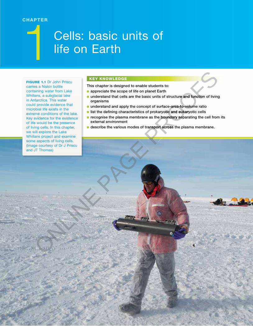
FIGURE 1.1 Dr John Priscu carries a Niskin bottle containing water from Lake Whillans, a subglacial lake in Antarctica. This water could provide evidence that microbial life exists in the extreme conditions of the lake. Key evidence for the existence of life would be the presence of living cells. In this chapter, we will explore the Lake Whillans project and examine some aspects of living cells. (Image courtesy of Dr J Priscu and JT Thomas)
1 Cells: basic units of life on Earth
CHAPTER
CHAPTER1Cells: basic units of life on EarthSearching for lifeCells: the basic units of life Prokaryotes: no nuclear envelope!Plasma membrane: the gatekeeperFunctions of the plasma membrane Crossing the plasma membrane SCIENTIST AT WORKBiochallengeChapter revie
ONLINE P
AGE PROOFS
PROOFSunderstand that cells are the basic units of structure and function of living
PROOFSunderstand that cells are the basic units of structure and function of living
understand and apply the concept of surface-area-to-volume ratio
PROOFSunderstand and apply the concept of surface-area-to-volume ratiolist the de� ning characteristics of prokaryotic and eukaryotic cells
PROOFSlist the de� ning characteristics of prokaryotic and eukaryotic cellsrecognise the plasma membrane as the boundary separating the cell from its
PROOFSrecognise the plasma membrane as the boundary separating the cell from its
PROOFS
PROOFS
describe the various modes of transport across the plasma membrane.
PROOFS
describe the various modes of transport across the plasma membrane.
PROOFS
NATURE OF BIOLOGY 12
Searching for lifeLife abounds on planet Earth. In every habitat on this planet where life exists, living organisms are built of one or more cells.
Living organisms can exist only where: • an energy source is available that can be trapped and utilised by an organism
for metabolic processes that maintain its living state• liquid water is available to allow biochemical reactions to occur, and to dis-
solve chemicals and transport them both within cells and to and from cells• the chemical building blocks required for life are available for use by an
organism in cellular repair, growth and reproduction. � ese chemical building blocks include carbon, oxygen, nitrogen and hydrogen, and each is able to form chemical bonds with other elements (see Odd fact). Carbon, in particular, is the most versatile chemical building block, as it can bond with many other elements, forming a variety of complex biomolecules, including long chains.
• stable environmental conditions exist within the range of tolerance of an organism, such as pressure, temperature, light intensity, pH and salinity. Where these conditions are met, living organisms can use energy to perform
the complex set of chemical transformations (metabolic activities) within their cells that sustains their living state. � ese activities include not only capturing energy but also taking up nutrients and water and removing wastes, so that their internal environment is kept within narrow limits.
Provided the above conditions can be met, life is possible even in extreme and hostile environments, such as:• around superheated hydrothermal vents at crushing pressures deep in the
mid-ocean (the world record holder is an archaeon that survives at high pressure and temperatures of 122 °C)
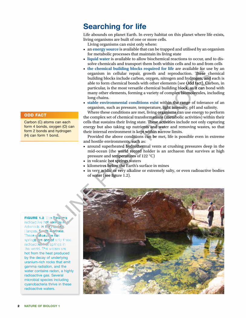
• in volcanic hot springs waters• kilometres below the Earth’s surface in mines• in very acidic or very alkaline or extremely salty, or even radioactive bodies
of water (see � gure 1.2).
FIGURE 1.2 The Paralana radioactive hot springs near Arkaroola in the Flinders Ranges, South Australia. These radioactive hot springs are one of only three radioactive hot springs in the world. The waters are hot from the heat produced by the decay of underlying uranium-rich rocks that emit gamma radiation, and the water contains radon, a highly radioactive gas. Several microbial species including cyanobacteria thrive in these radioactive waters.
ODD FACT
Carbon (C) atoms can each form 4 bonds, oxygen (O) can form 2 bonds and hydrogen (H) can form 1 bond.
ONLINE
ONLINE
ONLINE
ONLINE
The Paralana
ONLINE
The Paralana radioactive hot springs near
ONLINE
radioactive hot springs near Arkaroola in the Flinders
ONLINE
Arkaroola in the Flinders Ranges, South Australia.
ONLINE
Ranges, South Australia.
ONLINE
These radioactive hot ONLINE
These radioactive hot springs are one of only three ONLIN
E
springs are one of only three radioactive hot springs in ONLIN
E
radioactive hot springs in the world. The waters are ONLIN
E
the world. The waters are ONLINE P
AGE Provided the above conditions can be met, life is possible even in extreme
PAGE Provided the above conditions can be met, life is possible even in extreme and hostile environments, such as:
PAGE and hostile environments, such as:around superheated hydrothermal vents at crushing pressures deep in the
PAGE around superheated hydrothermal vents at crushing pressures deep in the mid-ocean (the world record holder is an archaeon that survives at high
PAGE mid-ocean (the world record holder is an archaeon that survives at high pressure and temperatures of 122
PAGE pressure and temperatures of 122 in volcanic hot springs waters
PAGE in volcanic hot springs waterskilometres below the Earth’s surface in mines
PAGE kilometres below the Earth’s surface in minesin very acidic or very alkaline or extremely salty, or even radioactive bodies
PAGE in very acidic or very alkaline or extremely salty, or even radioactive bodies of water (see � gure 1.2). PAGE of water (see � gure 1.2).
PROOFS are available for use by an
PROOFS are available for use by an
organism in cellular repair, growth and reproduction. � ese chemical
PROOFSorganism in cellular repair, growth and reproduction. � ese chemical building blocks include carbon, oxygen, nitrogen and hydrogen, and each is
PROOFSbuilding blocks include carbon, oxygen, nitrogen and hydrogen, and each is able to form chemical bonds with other elements (see Odd fact). Carbon, in
PROOFSable to form chemical bonds with other elements (see Odd fact). Carbon, in particular, is the most versatile chemical building block, as it can bond with
PROOFSparticular, is the most versatile chemical building block, as it can bond with many other elements, forming a variety of complex biomolecules, including
PROOFSmany other elements, forming a variety of complex biomolecules, including
within the range of tolerance of an
PROOFSwithin the range of tolerance of an
as pressure, temperature, light intensity, pH and salinity.
PROOFSas pressure, temperature, light intensity, pH and salinity.
Where these conditions are met, living organisms can use energy to perform
PROOFSWhere these conditions are met, living organisms can use energy to perform
the complex set of chemical transformations (metabolic activities) within their
PROOFSthe complex set of chemical transformations (metabolic activities) within their cells that sustains their living state. � ese activities include not only capturing
PROOFScells that sustains their living state. � ese activities include not only capturing energy but also taking up nutrients and water and removing wastes, so that PROOFS
energy but also taking up nutrients and water and removing wastes, so that their internal environment is kept within narrow limits. PROOFS
their internal environment is kept within narrow limits. Provided the above conditions can be met, life is possible even in extreme PROOFS
Provided the above conditions can be met, life is possible even in extreme
3CHAPTER 1 Cells: basic units of life on Earth
Organisms that live in these extreme environments are termed extremophiles. Most commonly, they are unicellular microbes — bacteria and archaea. Until the late 1970s all microbes were classi�ed as bacteria. However, the microbiologist Carl Woese (1928–2012) was the �rst to recog-nise that, based on many biochemical di�erences, the group once known as ‘bacteria’ included two di�erent groups of microbes. Members of this new group of microbes were given the label ‘archaea’ to di�erentiate them from classical bacteria.
Given the existence of extremophiles in many harsh environments, one group of scientists set out to answer the question:
Could living organisms thrive under nearly a kilometre of ice sheet in Antarctica in a frigid environment, in complete darkness and having been isolated from direct contact with the atmosphere, probably for thousands of years?
�e answer to this question was to be found at Lake Whillans.
Reaching Lake WhillansIt is 28 January 2013. A scientist walks across the icy surface of Antarctica car-rying a specialised container, known as a Niskin bottle, with its precious con-tents (refer to �gure 1.1). (A Niskin bottle is a specialised water sampler that consists of an open tube with valves at each end that can be closed by remote control.) �e scientist is Dr John Priscu, chief scientist for the WISSARD (Whil-lans Ice Stream Subglacial Access Research Drilling) project. �e Niskin bottle that he carries contains a sample of water collected from Lake Whillans.
Collecting water from a lake sounds like an easy task — just toss an empty container on a rope into a lake and pull the container out, full of water. How-ever, this was not the case at Lake Whillans.
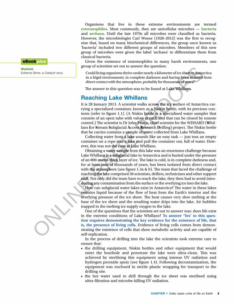
Obtaining a water sample from this lake was an enormous challenge because Lake Whillans is a subglacial lake in Antarctica and is buried under the pressure of an 800-metre-thick layer of ice. �e lake is cold, is in complete darkness and, for at least tens of thousands of years, has been isolated from direct contact with the atmosphere (see �gure 1.3a & b). �e team that faced the challenge of reaching the lake comprised 50 scientists, drillers, technicians and other support sta�. Not only did the team have to reach the lake, they then had to avoid intro-ducing any contamination from the surface or the overlying ice into the lake.
How can subglacial water lakes exist in Antarctica? �e water in these lakes remains liquid because of the �ow of heat from the Earth’s interior and the overlying pressure of the ice sheet. �e heat causes very slow melting at the base of the ice sheet and the resulting water drips into the lake. Air bubbles trapped in the melting ice supply oxygen to the lake.
One of the questions that the scientists set out to answer was: does life exist in the extreme conditions of Lake Whillans? To answer ‘Yes’ to this ques-tion requires demonstrating the key evidence for the existence of life, that is, the presence of living cells. Evidence of living cells comes from demon-strating the existence of cells that show metabolic activity and are capable of self-replication.
In the process of drilling into the lake the scientists took extreme care to ensure that: • the drilling equipment, Niskin bottles and other equipment that would
enter the borehole and penetrate the lake were ultra-clean. �is was achieved by sterilising this equipment using intense UV radiation and hydrogen peroxide spray (see �gure 1.4). Following decontamination, the equipment was enclosed in sterile plastic wrapping for transport to the drilling site.
• the hot water used to drill through the ice sheet was sterilised using ultra-�ltration and microbe-killing UV radiation.
Weblink Extreme Slime: a Catalyst story
ONLINE reaching the lake comprised 50 scientists, drillers, technicians and other support
ONLINE reaching the lake comprised 50 scientists, drillers, technicians and other support
sta�. Not only did the team have to reach the lake, they then had to avoid intro
ONLINE sta�. Not only did the team have to reach the lake, they then had to avoid intro
ducing any contamination from the surface or the overlying ice into the lake.
ONLINE ducing any contamination from the surface or the overlying ice into the lake. How can subglacial water lakes exist in Antarctica? �e water in these lakes
ONLINE How can subglacial water lakes exist in Antarctica? �e water in these lakes
remains liquid because of the �ow of heat from the Earth’s interior and the
ONLINE
remains liquid because of the �ow of heat from the Earth’s interior and the overlying pressure of the ice sheet. �e heat causes very slow melting at the
ONLINE
overlying pressure of the ice sheet. �e heat causes very slow melting at the
PAGE that he carries contains a sample of water collected from Lake Whillans.
PAGE that he carries contains a sample of water collected from Lake Whillans.Collecting water from a lake sounds like an easy task — just toss an empty
PAGE Collecting water from a lake sounds like an easy task — just toss an empty container on a rope into a lake and pull the container out, full of water. How
PAGE container on a rope into a lake and pull the container out, full of water. However, this was not the case at Lake Whillans.
PAGE ever, this was not the case at Lake Whillans.
PAGE Obtaining a water sample from this lake was an enormous challenge because
PAGE Obtaining a water sample from this lake was an enormous challenge because
Lake Whillans is a subglacial lake in Antarctica and is buried under the pressure
PAGE Lake Whillans is a subglacial lake in Antarctica and is buried under the pressure of an 800-metre-thick layer of ice. �e lake is cold, is in complete darkness and,
PAGE of an 800-metre-thick layer of ice. �e lake is cold, is in complete darkness and, for at least tens of thousands of years, has been isolated from direct contact
PAGE for at least tens of thousands of years, has been isolated from direct contact with the atmosphere (see �gure 1.3a & b). �e team that faced the challenge of PAGE with the atmosphere (see �gure 1.3a & b). �e team that faced the challenge of reaching the lake comprised 50 scientists, drillers, technicians and other support PAGE
reaching the lake comprised 50 scientists, drillers, technicians and other support sta�. Not only did the team have to reach the lake, they then had to avoid introPAGE
sta�. Not only did the team have to reach the lake, they then had to avoid intro
PROOFSCould living organisms thrive under nearly a kilometre of ice sheet in Antarctica
PROOFSCould living organisms thrive under nearly a kilometre of ice sheet in Antarctica in a frigid environment, in complete darkness and having been isolated from
PROOFSin a frigid environment, in complete darkness and having been isolated from direct contact with the atmosphere, probably for thousands of years?
PROOFSdirect contact with the atmosphere, probably for thousands of years?
�e answer to this question was to be found at Lake Whillans.
PROOFS�e answer to this question was to be found at Lake Whillans.
It is 28 January 2013. A scientist walks across the icy surface of Antarctica car
PROOFSIt is 28 January 2013. A scientist walks across the icy surface of Antarctica carrying a specialised container, known as a Niskin bottle, with its precious con
PROOFSrying a specialised container, known as a Niskin bottle, with its precious contents (refer to �gure 1.1). (A Niskin bottle is a specialised water sampler that
PROOFStents (refer to �gure 1.1). (A Niskin bottle is a specialised water sampler that consists of an open tube with valves at each end that can be closed by remote
PROOFS
consists of an open tube with valves at each end that can be closed by remote control.) �e scientist is Dr John Priscu, chief scientist for the WISSARD (PROOFS
control.) �e scientist is Dr John Priscu, chief scientist for the WISSARD (ccess PROOFS
ccess RPROOFS
Research PROOFS
esearch that he carries contains a sample of water collected from Lake Whillans.PROOFS
that he carries contains a sample of water collected from Lake Whillans.Collecting water from a lake sounds like an easy task — just toss an empty PROOFS
Collecting water from a lake sounds like an easy task — just toss an empty
NATURE OF BIOLOGY 14
WESTANTARCTICA
LakeElisworth
LakeWhillans
LakeVostok
RonneIce Shelf
RossIce Shelf
EAST ANTARCTICA
South Pole
(a) 1000 km
500 miles
800 m
(b)
FIGURE 1.3 (a) Lake Whillans is located near the edge of the Ross Ice Shelf, 640 km from the South Pole. The lake is one of hundreds of subglacial lakes that have been identi� ed in Antarctica using techniques such as air-borne radar and satellite-based radar altimetry. (b) Lake Whillans lies under an ice sheet that is 800 m thick.
Why were these precautionary steps taken? � ese rigorous sterile precautions were designed to prevent any cells from the surface or the overlying ice reaching the lake. � is ensured that any living cells found in the samples from the lake originated from the lake itself and were not introduced contaminant cells from the surface.
Pressurised hot water was used to drill through the 800 m of ice overlying Lake Whillans. After seven days of drilling, the last layers of ice were broken
ONLINE
ONLINE
ONLINE
ONLINE
ONLINE
ONLINE
ONLINE
ONLINE
ONLINE
ONLINE
ONLINE
Lake Whillans
ONLINE
Lake Whillans is located near the edge of the
ONLINE
is located near the edge of the Ross Ice Shelf, 640 km from
ONLINE
Ross Ice Shelf, 640 km from the South Pole. The lake is
ONLINE
the South Pole. The lake is one of hundreds of subglacial
ONLINE
one of hundreds of subglacial lakes that have been identi� ed
ONLINE
lakes that have been identi� ed
ONLINE
in Antarctica using techniques ONLINE
in Antarctica using techniques such as air-borne radar and ONLIN
E
such as air-borne radar and satellite-based radar altimetry. ONLIN
E
satellite-based radar altimetry. Lake Whillans lies under an ONLIN
E
Lake Whillans lies under an ONLINE P
AGE
PAGE
PAGE
PAGE
PAGE
PAGE
PAGE
PAGE
PAGE
PAGE
PAGE
PAGE
PAGE
PAGE
PAGE
PAGE
PAGE
PAGE
PAGE
PAGE
PAGE
PAGE
PAGE
PAGE
PAGE
PAGE
PAGE
PAGE
PAGE
PAGE
PAGE
PAGE
PAGE
PAGE
PAGE
PAGE
PAGE
PAGE
PAGE
PAGE
PAGE
PAGE
PAGE
PAGE
PAGE
PAGE
PAGE
PAGE
PAGE
PAGE
PAGE
PAGE PROOFS
PROOFS
PROOFS
PROOFS
PROOFS
PROOFS
PROOFS
PROOFS
PROOFS
PROOFS
PROOFS
PROOFS
PROOFS
PROOFS
PROOFS
PROOFS
PROOFS
PROOFS
PROOFS
PROOFS
PROOFS
PROOFS
PROOFS
PROOFS
PROOFS
PROOFS
PROOFS
PROOFS
PROOFS
PROOFS
PROOFS
PROOFS
PROOFS
PROOFS
PROOFS
PROOFS
PROOFS
PROOFS
PROOFS
PROOFS
PROOFS
PROOFS
PROOFS
PROOFSWhillans
PROOFSWhillans
PROOFS
PROOFS
PROOFS
PROOFS
PROOFS
PROOFS
PROOFS
PROOFS
PROOFS
PROOFS
PROOFS
PROOFS
PROOFS
PROOFS
PROOFS
PROOFS
PROOFS
5CHAPTER 1 Cells: basic units of life on Earth
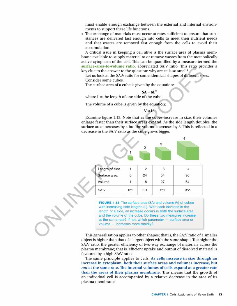
through and the lake was reached through a 60-centimetre-wide borehole. A Niskin bottle was inserted down the borehole into the water to obtain a water sample. The valves on the Niskin bottle were remotely closed and the bottle was raised to the surface (see � gure 1.5a). Had the valves on the Niskin bottle closed? Was there a sample of lake water in the bottle? A quick check showed that the valves had closed, trapping the � rst water sample from Lake Whillans.
In total, the scienti� c team collected about 30 litres of lake water and eight samples of sediment from the lake bottom. � ese samples would allow the scientists to discover if living organisms were present in Lake Whillans.
� e � rst sample of lake water from the Niskin bottle was carefully carried to a tem-porary � eld laboratory. Scientists extracted samples of lake water and began their examination (see � gure 1.5b).
FIGURE 1.5 (a) Scientists Brent Christner (left) and John Priscu (right) retrieve the � rst water-sampling Niskin bottle from the borehole in subglacial Lake Whillans. (Image courtesy of JT Thomas) (b) Scientists processing water samples from Lake Whillans in the � eld laboratory
(a) (b)
Signs of life under the ice?� e � rst test was to add a DNA-sensitive dye to a sample of the lake water. DNA is the genetic material of all cells. If cells were present in the lake water, this DNA-sensitive dye would reveal them as glowing green dots when viewed under a microscope. Imagine the scientists’ delight when this test gave a posi-tive result (see � gure 1.6a). � e presence of cells was a strong indication that microbial life existed in Lake Whillans . . . but were they living cells?
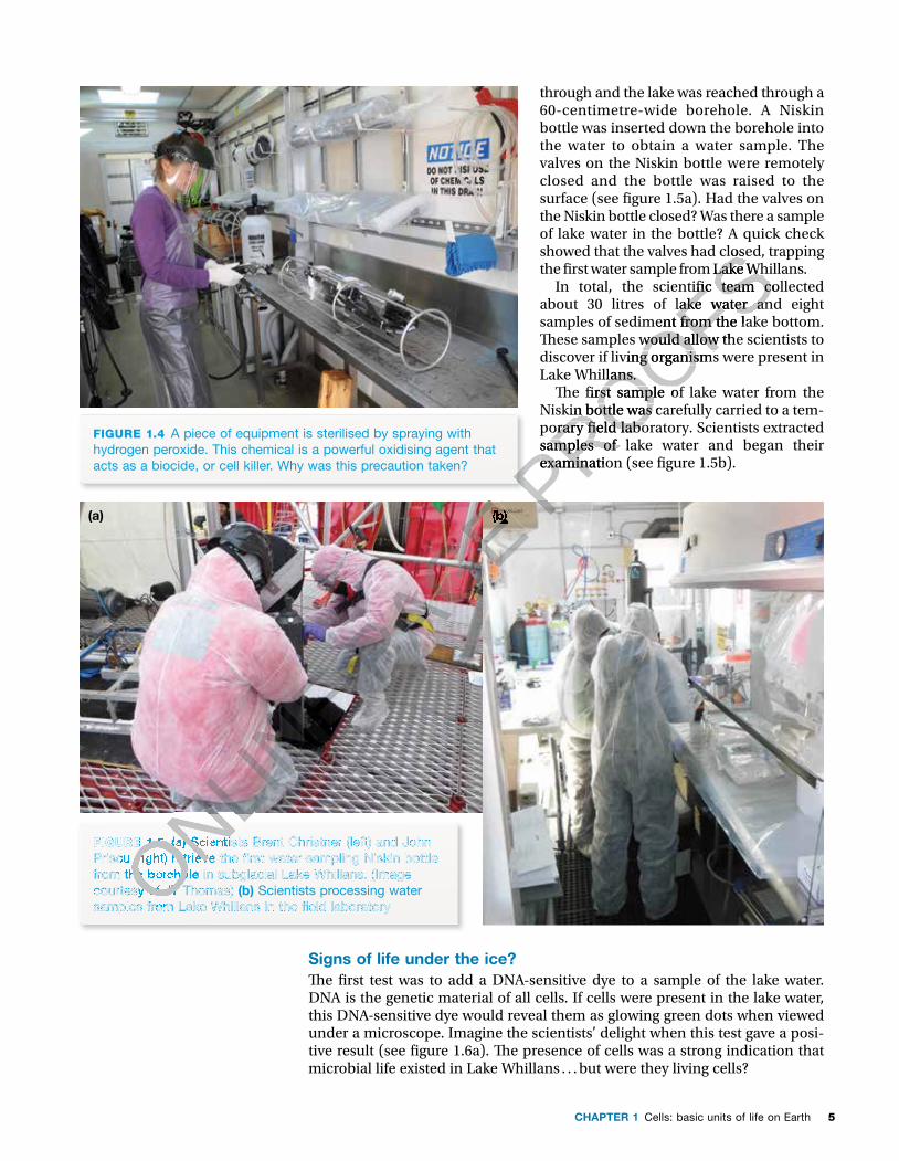
FIGURE 1.4 A piece of equipment is sterilised by spraying with hydrogen peroxide. This chemical is a powerful oxidising agent that acts as a biocide, or cell killer. Why was this precaution taken?
ONLINE
ONLINE
ONLINE
FIGURE 1.5
ONLINE
FIGURE 1.5 (a)
ONLINE
(a) Scientists Brent Christner (left) and John
ONLINE
Scientists Brent Christner (left) and John Priscu (right) retrieve the � rst water-sampling Niskin bottle ONLIN
E
Priscu (right) retrieve the � rst water-sampling Niskin bottle from the borehole in subglacial Lake Whillans. (Image ONLIN
E
from the borehole in subglacial Lake Whillans. (Image courtesy of JT Thomas) ONLIN
E
courtesy of JT Thomas) samples from Lake Whillans in the � eld laboratoryONLIN
E
samples from Lake Whillans in the � eld laboratoryONLINE
ONLINE P
AGE
PAGE
PAGE (b)
PAGE (b) PROOFSshowed that the valves had closed, trapping
PROOFSshowed that the valves had closed, trapping the � rst water sample from Lake Whillans.
PROOFSthe � rst water sample from Lake Whillans.In total, the scienti� c team collected
PROOFSIn total, the scienti� c team collected about 30 litres of lake water and eight
PROOFSabout 30 litres of lake water and eight samples of sediment from the lake bottom.
PROOFSsamples of sediment from the lake bottom. � ese samples would allow the scientists to
PROOFS� ese samples would allow the scientists to discover if living organisms were present in
PROOFSdiscover if living organisms were present in Lake Whillans.
PROOFSLake Whillans.
� e � rst sample of lake water from the
PROOFS� e � rst sample of lake water from the
Niskin bottle was carefully carried to a tem-
PROOFSNiskin bottle was carefully carried to a tem-porary � eld laboratory. Scientists extracted
PROOFSporary � eld laboratory. Scientists extracted samples of lake water and began their
PROOFSsamples of lake water and began their examination (see � gure 1.5b). PROOFS
examination (see � gure 1.5b). PROOFS
PROOFS
PROOFS
PROOFS

NATURE OF BIOLOGY 16
Scanning electron microscopy of the lake water samples showed that the microbial cells varied in shape and included rod-shaped, curved and spher-ical microbial cells. Figure 1.6b, for example, shows a spherical microbial cell against a background of sediment particles.
However, it was necessary to con� rm that the cells were living. Living cells carry out a diverse range of metabolic activities, including uptake of nutrients, and syn-thesising DNA and proteins. When cells from samples of lake water were exposed to thymidine, one of the building blocks of DNA, the cells took up this compound and incorporated it into their DNA. � is observation con� rmed that the cells were living and undergoing cell division. Similar con� rmation that the cells were meta-bolically active came from showing that they were synthesising proteins.
Active cell division of the lake microbes was revealed when samples of lake water were plated onto a nutrient medium and incubated. Individual microbial cells, too small to be seen with an unaided eye, underwent multiple cycles of cell division and produced visible colonies comprising millions of cells (see � gure 1.6c).
FIGURE 1.6 (a) Epi� uorescence microscopy image of DNA-containing microbial cells (green) from the subglacial Lake Whillans water sample (Image courtesy of Dr A Purcell) (b) Scanning electron microscope image showing a coccoid-shaped microbial cell with an attached sediment particle from the subglacial Lake Whillans water column (Image courtesy of Trista Vick-Majors, Priscu Research Group, Montana State University) (c) Microbial colonies produced by multiple divisions of microbial cells from subglacial Lake Whillans. Different colours and shapes of the colonies indicate different microbial species. (Image courtesy of Dr B Christner)
(a) (b)
(c)
After showing that living microbial cells existed in Lake Whillans, the scientists then set out to identify the di� erent species. Back in the United States they used DNA sequencing techniques and identi� ed more than 3900 di� erent microbial species — bacteria and archaea — as part of the living community. � e discovery of a living microbial community that obtains energy and the chemical building blocks required for life in the cold, dark environ-ment of subglacial Lake Whillans is signi� cant because:• it provides the � rst unequivocal evidence of a complex ecosystem in a sub-
glacial lake under the Antarctic ice sheet• it highlights the possibility that life might exist beyond planet Earth, such as
on distant ice-covered bodies in our solar system that conceal oceans below their frozen surfaces.
ONLINE
ONLINE
ONLINE
Epi� uorescence microscopy image of DNA-containing microbial cells (green) from the subglacial
ONLINE
Epi� uorescence microscopy image of DNA-containing microbial cells (green) from the subglacial Lake Whillans water sample (Image courtesy of Dr A Purcell)
ONLINE
Lake Whillans water sample (Image courtesy of Dr A Purcell) coccoid-shaped microbial cell with an attached sediment particle from the subglacial Lake Whillans water column
ONLINE
coccoid-shaped microbial cell with an attached sediment particle from the subglacial Lake Whillans water column (Image courtesy of Trista Vick-Majors, Priscu Research Group, Montana State University)
ONLINE
(Image courtesy of Trista Vick-Majors, Priscu Research Group, Montana State University) produced by multiple divisions of microbial cells from subglacial Lake Whillans. Different colours and shapes of the
ONLINE
produced by multiple divisions of microbial cells from subglacial Lake Whillans. Different colours and shapes of the colonies indicate different microbial species. (Image courtesy of Dr B Christner)
ONLINE
colonies indicate different microbial species. (Image courtesy of Dr B Christner)
ONLINE
ONLINE P
AGE PROOFS
living and undergoing cell division. Similar con� rmation that the cells were meta-
PROOFSliving and undergoing cell division. Similar con� rmation that the cells were meta-bolically active came from showing that they were synthesising proteins.
PROOFSbolically active came from showing that they were synthesising proteins.Active cell division of the lake microbes was revealed when samples of lake water
PROOFSActive cell division of the lake microbes was revealed when samples of lake water were plated onto a nutrient medium and incubated. Individual microbial cells, too
PROOFSwere plated onto a nutrient medium and incubated. Individual microbial cells, too small to be seen with an unaided eye, underwent multiple cycles of cell division
PROOFSsmall to be seen with an unaided eye, underwent multiple cycles of cell division and produced visible colonies comprising millions of cells (see � gure 1.6c).
PROOFSand produced visible colonies comprising millions of cells (see � gure 1.6c).
PROOFS
PROOFS
PROOFS

7CHAPTER 1 Cells: basic units of life on Earth
Life beyond Earth?� e discovery of a diverse microbial ecosystem hidden deep under the Antarctic ice sheet and away from sunlight raises the possibility that life may exist beyond planet Earth. Possible locations for extraterrestrial life include ice-covered moons that circle planets in our solar system. One such location is Europa, one of the large moons of the giant planet Jupiter.
Europa, with a diameter of 3144 kilometres, is a little smaller than Earth’s moon (see � gure 1.7a). Europa is a frozen world with an ice-covered surface, many kilometres thick and criss-crossed by long fractures (see � gure 1.7b). In the period from 1996–99 the Galileo spacecraft made 11 � ybys past Europa, capturing high-resolution images of the moon’s surface and using its remote-sensing instruments to gain information about Europa. Galileo gathered strong evidence of a possible sub-surface ocean of salty water on Europa that is sandwiched between the moon’s icy surface and its underlying rocky core. � e surface of Europa constantly stretches and relaxes in tidal movements as it moves in an elliptical orbit around Jupiter every 3.5 days. � e constant � exing of the surface generates heat that would keep a sub-surface ocean in the liquid state.
FIGURE 1.7 (a) Europa, a moon of Jupiter. This composite image, taken by the Galileo spacecraft, shows the long fractures on the moon’s ice-covered surface. This ice may conceal what is regarded as ‘perhaps the most promising place in our solar system beyond Earth to look for present-day environments that are suitable for life’ (NASA Media Release, www.jpl.nasa.gov/news/news.php?feature=4386). (b) Close-up of the surface of Europa showing its fractured ice surface. The blue–white areas are pure water ice, while the ice in the reddish bands is mixed with salts such as magnesium sulfate or with sulfuric acid.
(a) (b)
Dr Robert Pappalardo, a scientist from NASA, has described Europa as ‘the most likely place to � nd life beyond Earth’. He continued:
We think Europa is the most likely place for being habitable because of its relatively thin ice shell, its liquid ocean and that fact that it is in contact with the rock below which is geologically active . . . Europa has the right ingredients for life: it has water and the right chemical elements, as well as an environ-ment that is probably stable over time.
Source: As cited in � e Independent, 15 February 2013.
ODD FACT
The Galileo spacecraft was deliberately plunged into Jupiter’s crushing atmosphere on 21 September 2003.
NASA: National Aeronautic and Space Administration
ONLINE
ONLINE
ONLINE
(a)
ONLINE
(a) Europa, a moon of Jupiter. This composite image, taken by the
ONLINE
Europa, a moon of Jupiter. This composite image, taken by the fractures on the moon’s ice-covered surface. This ice may conceal what is regarded as ‘perhaps the most promising
ONLINE
fractures on the moon’s ice-covered surface. This ice may conceal what is regarded as ‘perhaps the most promising
ONLINE
ONLINE
place in our solar system beyond Earth to look for present-day environments that are suitable for life’ (NASA Media
ONLINE
place in our solar system beyond Earth to look for present-day environments that are suitable for life’ (NASA Media Release, www.jpl.nasa.gov/news/news.php?feature
ONLINE
Release, www.jpl.nasa.gov/news/news.php?featureice surface. The blue–white areas are pure water ice, while the ice in the reddish bands is mixed with salts such as ONLIN
E
ice surface. The blue–white areas are pure water ice, while the ice in the reddish bands is mixed with salts such as magnesium sulfate or with sulfuric acid.ONLIN
E
magnesium sulfate or with sulfuric acid.ONLINE
ONLINE
ONLINE P
AGE PROOFS
many kilometres thick and criss-crossed by long fractures (see � gure 1.7b). In
PROOFSmany kilometres thick and criss-crossed by long fractures (see � gure 1.7b). In
spacecraft made 11 � ybys past Europa,
PROOFS spacecraft made 11 � ybys past Europa,
capturing high-resolution images of the moon’s surface and using its remote-
PROOFScapturing high-resolution images of the moon’s surface and using its remote-sensing instruments to gain information about Europa.
PROOFSsensing instruments to gain information about Europa. Galileo
PROOFSGalileo gathered
PROOFSgathered strong evidence of a possible sub-surface ocean of salty water on Europa that
PROOFSstrong evidence of a possible sub-surface ocean of salty water on Europa that is sandwiched between the moon’s icy surface and its underlying rocky core.
PROOFSis sandwiched between the moon’s icy surface and its underlying rocky core. � e surface of Europa constantly stretches and relaxes in tidal movements
PROOFS� e surface of Europa constantly stretches and relaxes in tidal movements as it moves in an elliptical orbit around Jupiter every 3.5 days. � e constant
PROOFSas it moves in an elliptical orbit around Jupiter every 3.5 days. � e constant � exing of the surface generates heat that would keep a sub-surface ocean in
PROOFS� exing of the surface generates heat that would keep a sub-surface ocean in
PROOFS

NATURE OF BIOLOGY 18
What might the future bring? If the presence of a sub-surface ocean on Europa is proved and if the conditions appear suitable for life, an unmanned probe might be sent to land on this moon, drill down to its ocean, sample its waters and look for signs of life. What would the most powerful signs of life be? Yes, cells.
KEY IDEAS
■ On planet Earth, life exists in hostile and extreme environments and the organisms that survive there are termed extremophiles.
■ For life to exist, a set of conditions must be met, including the availability of a source of energy and the presence of liquid water.
■ Living cells have been found in a subglacial lake in Antarctica under hundreds of metres of ice sheet.
■ The discovery of a diverse microbial ecosystem in a subglacial lake in Antarctica raises the possibility that life might exist under the surface of ice-covered moons in our solar system.
■ Critical direct evidence of life (as we know it) is the presence of metabolically active cells.
QUICK CHECK
1 List the conditions that must be met for life to exist.2 In drilling into Lake Whillans, great care was taken to ensure that the equipment
that entered the borehole was sterilised. Why was this precaution taken?3 What was the �rst evidence that indicated that it was possible life existed in
Lake Whillans? 4 Identify the follow-up experiment that con�rmed this �nding.5 What kinds of organism live in the Lake Whillans ecosystem?6 Why is Europa, one of the moons of planet Jupiter, of interest as a possible
location for life beyond planet Earth?
Cells: the basic units of life Cells are the basic structural and functional units of life, and all living organisms are built of one or more cells. Cells, with only a very few excep-tions, are too small to be seen with an unaided eye. �eir existence was not recognised until after the development of the �rst simple microscopes. �is enabled the �rst observations of cells to be made in the 1660s. However, the recognition of cells as the basic unit of life did not occur until almost 200 years later.
Cells: how big?Cells are typically microscopic (not visible with an unaided eye). Only a few single cells are large enough to be seen with an unaided human eye, for example, human egg cells with diameters about 0.1 mm and the common amoeba (Amoeba proteus), a unicellular organism with an average size ranging from 0.25 to 0.75 mm. (You would see an amoeba as about the size of a full stop on this page.) Contrast this with one of the smallest bacteria, Plelagibacter ubique, consisting of a cell just 0.2 µm diameter. How many of these bacteria could �t across an amoeba that is 0.5 mm wide?• Most animal cells fall within the size range of 10 to 40 μm. Among the
smallest human cells are red blood cells with diameters for normal cells in the range of 6 to 8 μm.
Weblink NASA video — Europa
Unit 1 Cells: structural units of lifeConcept summary and practice questions
AOS 1
Topic 1
Concept 1
1 millimetre (mm) = 1000 micrometres (µm)
1 micrometre (µm) = 1000 nanometres (nm)
Unit 1 Cell sizeConcept summary and practice questions
AOS 1
Topic 1
Concept 4
ONLINE Cells: the basic units of life
ONLINE Cells: the basic units of life
Cells are the basic structural and functional units of life, and all living
ONLINE
Cells are the basic structural and functional units of life, and all living organisms are built of one or more cells.
ONLINE
organisms are built of one or more cells.
ONLINE
tions, are too small to be seen with an unaided eye. �eir existence was not
ONLINE
tions, are too small to be seen with an unaided eye. �eir existence was not recognised until after the development of the �rst simple microscopes. �is
ONLINE
recognised until after the development of the �rst simple microscopes. �is
ONLINE
ONLINE
Concept summary
ONLINE
Concept summaryand practice
ONLINE
and practice questions
ONLINE
questions
ONLINE
1 millimetre (mm)
ONLINE
1 millimetre (mm) =
ONLINE
=
ONLINE
1000 micrometres (ONLIN
E
1000 micrometres (µONLINE
µm)ONLINE
m)
1 micrometre (ONLINE
1 micrometre (µONLINE
µm) ONLINE
m) 1000 nanometres (nm)ONLIN
E
1000 nanometres (nm)
PAGE
PAGE
PAGE List the conditions that must be met for life to exist.
PAGE List the conditions that must be met for life to exist.In drilling into Lake Whillans, gr
PAGE In drilling into Lake Whillans, grthat entered the borehole was sterilised. Why was this precaution taken?
PAGE that entered the borehole was sterilised. Why was this precaution taken?What was the �rst evidence that indicated that it was possible life existed in
PAGE What was the �rst evidence that indicated that it was possible life existed inLake Whillans?
PAGE Lake Whillans? Identify the follow-up experiment that con�rmed this �nding.
PAGE Identify the follow-up experiment that con�rmed this �nding.What kinds of or
PAGE What kinds of organism live in the Lake Whillans ecosystem?
PAGE ganism live in the Lake Whillans ecosystem?
Why is Eur
PAGE Why is Europa, one of the moons of planet Jupiter, of interest as a possible
PAGE opa, one of the moons of planet Jupiter, of interest as a possible
location for life beyond planet Earth?
PAGE location for life beyond planet Earth?
Cells: the basic units of life PAGE
Cells: the basic units of life
PROOFS
PROOFS
PROOFSFor life to exist, a set of conditions must be met, including the availability
PROOFSFor life to exist, a set of conditions must be met, including the availability
Living cells have been found in a subglacial lake in Antarctica under
PROOFSLiving cells have been found in a subglacial lake in Antarctica under
The discovery of a diverse microbial ecosystem in a subglacial lake in
PROOFSThe discovery of a diverse microbial ecosystem in a subglacial lake in Antarctica raises the possibility that life might exist under the surface of
PROOFSAntarctica raises the possibility that life might exist under the surface of
Critical direct evidence of life (as we know it) is the presence of
PROOFSCritical direct evidence of life (as we know it) is the presence of
PROOFS
PROOFS
PROOFS
List the conditions that must be met for life to exist.PROOFS
List the conditions that must be met for life to exist.eat care was taken to ensure that the equipment PROOFS
eat care was taken to ensure that the equipment

9CHAPTER 1 Cells: basic units of life on Earth
• Plant cells typically fall in the range of 10 to 100 μm.• Microbial cells, both bacterial and archaeal, are much smaller than plant
and animal cells. Most bacterial cells have diameters in the range of 0.4 to 2.0 µm and 0.5 to 5 µm in length. On average, microbial cells are about 10 times smaller than plant and animal cells, with sizes typically in the few micrometres range.
A non-living microworld exists beyond that of microbes. � is is occupied by viruses that are non-cellular particles that are generally regarded as belonging to the grey area between living and non-living. Why? Because viruses do not have a cellular structure, they cannot carry out metabolic activities in isolation and they cannot self-replicate. (Viruses can replicate only inside and with the assistance of living cells.) Viruses range in diameter from 20 to 300 nano-metres (nm); for example, the cold-causing rhinovirus is about 30 nm in diameter and the measles-causing virus is about 220 nm in diameter. Figure 1.8 shows a sample of the range of sizes seen in selected cells. (Other non-cellular structures are included for size comparison.)
Microbial cells are relatively much smaller than the cells of animals and plants, so some animal bac-terial infections can involve the invasion of bacterial cells into the cells of the host, where they multiply. Examine � gure 1.9 of a human lung � broblast and note the presence of numerous bacterial cells in a single cell. � is image highlights the size di� erence between microbial cells and the cells of animals (and plants).
FIGURE 1.9 Transmission electron microscope (TEM) image of a lung � broblast infected with many bacterial cells (shown as small dark circular and ovoid shapes). The bacteria are Legionella pneumophila, the cause of several infections in people, including Legionnaires’ disease.
Human egg130 μm
Sperm cell60 × 5 μm
(a)
(b)
(c)
Yeast cell3 × 4 μm
Mitochondrion4 × 0.8 μm
Mitochondrion
Ribosome30 nm
In�uenza virus130 nm
Measles virus220 nm
HIV130 nm
Skin cell30 μm
Red blood cell8 μm
Yeast cellE coli bacterium3 × 0.6 μm
Red blood cell
FIGURE 1.8 Diagrams, at increasing levels of magni� cation, showing cells, cell organelles and viruses. Note the extreme differences in size. (a) Some human cells showing variation in cell size (b) A bacterial cell with a mitochondrion, a cell organelle and other small cells shown for comparison (c) A mitochondrion compared with some viruses
ONLINE
ONLINE
ONLINE
ONLINE
ONLINE
Mitochondrion
ONLINE
Mitochondrion
Ribosome
ONLINE
Ribosome
In�uenza virusONLINE
In�uenza virus
130 nmONLINE
130 nmONLINE
ONLINE
ONLINE
ONLINE
ONLINE
ONLINE
ONLINE
ONLINE
ONLINE
ONLINE
ONLINE
ONLINE
ONLINE
ONLINE
ONLINE
ONLINE
ONLINE
ONLINE
ONLINE
ONLINE
ONLINE
ONLINE
ONLINE
ONLINE P
AGE terial infections can involve the invasion of bacterial
PAGE terial infections can involve the invasion of bacterial cells into the cells of the host, where they multiply.
PAGE cells into the cells of the host, where they multiply. Examine � gure 1.9 of a human lung � broblast and
PAGE Examine � gure 1.9 of a human lung � broblast and note the presence of numerous bacterial cells in a
PAGE note the presence of numerous bacterial cells in a single cell. � is image highlights the size di� erence
PAGE single cell. � is image highlights the size di� erence between microbial cells and the cells of animals (and
PAGE between microbial cells and the cells of animals (and plants).
PAGE plants).
PAGE PROOFS
belonging to the grey area between living and non-
PROOFSbelonging to the grey area between living and non-living. Why? Because viruses do not have a cellular
PROOFSliving. Why? Because viruses do not have a cellular structure, they cannot carry out metabolic activities in
PROOFSstructure, they cannot carry out metabolic activities in isolation and they cannot self-replicate. (Viruses can
PROOFSisolation and they cannot self-replicate. (Viruses can replicate only inside and with the assistance of living
PROOFSreplicate only inside and with the assistance of living cells.) Viruses range in diameter from 20 to 300 nano-
PROOFScells.) Viruses range in diameter from 20 to 300 nano-metres (nm); for example, the cold-causing rhinovirus
PROOFSmetres (nm); for example, the cold-causing rhinovirus is about 30 nm in diameter and the measles-causing
PROOFSis about 30 nm in diameter and the measles-causing virus is about 220 nm in diameter. Figure 1.8 shows
PROOFSvirus is about 220 nm in diameter. Figure 1.8 shows a sample of the range of sizes seen in selected cells.
PROOFSa sample of the range of sizes seen in selected cells. (Other non-cellular structures are included for size
PROOFS(Other non-cellular structures are included for size comparison.)
PROOFScomparison.)
Microbial cells are relatively much smaller than PROOFS
Microbial cells are relatively much smaller than the cells of animals and plants, so some animal bac-PROOFS
the cells of animals and plants, so some animal bac-terial infections can involve the invasion of bacterial PROOFS
terial infections can involve the invasion of bacterial cells into the cells of the host, where they multiply. PROOFS
cells into the cells of the host, where they multiply.

NATURE OF BIOLOGY 110
Cells: all sorts of shapes� ere is no � xed shape for cells. Cells vary in shape and their shapes often re� ect their functions. Figure 1.10 shows some examples of cell shapes. Scan this � gure and note that some cells are thin and � attened, others are column-shaped, yet others are spherical.
FIGURE 1.10 Examples of variations in cell shape: (a) star-shaped (b) spherical (c) columnar (d) � at (e) elongated (f) disc-shaped (g) cuboidal
(a) Star-shaped (e.g. motor neuron cells)
(e) Elongated (e.g. human smooth muscle cells)
(f) Disc-shaped (e.g. human red blood cells)
(g) Cuboidal (e.g. human kidney cells)
(b) Spherical (e.g. egg cells)
(c) Columnar (e.g. gut cells)
(d) Flat (e.g. skin cells)
Look at � gure 1.10a. Note the long axon that is a distinctive feature of motor neuron cells. � ese cells transmit nerve impulses from a person’s spinal cord to voluntary muscles throughout the body. In this case, the shape of the nerve cell is � tted to its conductive function. Can you estimate the approximate length of a motor neuron that has its cell body in the lower spinal cord with its axon reaching to your big toe?
Look at � gure 1.10e. Note the spindle-shaped smooth muscle cells. Smooth muscle cells contain special proteins that criss-cross the cell, and when these proteins contract the smooth muscle � bres shorten. � e spindle shape of these cells is suited to their contractile function. Bundles of smooth muscle cells are found in the gut wall, in the walls of blood vessels, in ducts of secretory glands and in the wall of the uterus. � ese bundles of smooth muscle cells can generate sustained involuntary contractions in these organs.
Microbial cells also vary in shape (see � gure 1.11). Note that some bac-teria are rod-shaped, such as the gut-dwelling bacterium Escherichia coli; some are corkscrew-shaped, such as Borrelia burgdorferi, the caus-ative agent of Lyme disease; while others are more or less spherical, such as Streptococcus pneumonia, the cause of many infections, including pneumonia.
ODD FACT
Motor neurons in animals, such as the giant squid (Architeuthis sp.), may be as long as 12 metres.
ONLINE
ONLINE
ONLINE
ONLINE
Look at � gure 1.10a. Note the long axon that is a distinctive feature of motor
ONLINE
Look at � gure 1.10a. Note the long axon that is a distinctive feature of motor neuron cells. � ese cells transmit nerve impulses from a person’s spinal cord
ONLINE
neuron cells. � ese cells transmit nerve impulses from a person’s spinal cord
ONLINE
ONLINE
ONLINE
ODD FACT
ONLINE
ODD FACT
Motor neurons in animals, ONLINE
Motor neurons in animals, ONLINE
such as the giant squid ONLINE
such as the giant squid Architeuthis ONLIN
E
Architeuthis sp.), may be as ONLINE
sp.), may be as
PAGE
PAGE
PAGE Examples of variations in cell shape: PAGE Examples of variations in cell shape: (a)PAGE
(a) star-shaped PAGE star-shaped PAGE
(f) Disc-shaped (e.g. human red blood
PAGE (f) Disc-shaped (e.g. human red blood
PAGE PROOFS
(c) Columnar (e.g. gut
PROOFS(c) Columnar (e.g. gut (d) Flat (e.g. skin cells)
PROOFS(d) Flat (e.g. skin cells)
PROOFS
PROOFS

11CHAPTER 1 Cells: basic units of life on Earth
2.5 μm
(b)
2.2 μm
(a)
2.9 μm
(c)
FIGURE 1.11 Bacterial cells come in many shapes. Some are (a) rod-shaped bacilli (singular: bacillus) (b) spiral-shaped and (c) spherical cocci (singular: coccus).
Not all cells have a � xed shape. For example, some cells are able to move actively, and these self-propelled cells do not have � xed shapes because their outer boun-dary is their � exible plasma membrane. So, as these cells move, their shapes change. Examples of cells capable of active self-propelled movement include:• cancer cells that migrate into capillaries and move around the body when a
malignant tumour undergoes metastasis (see � gure 1.12a). � e thread-like protrusions (known as � lopodia) that fold out from the plasma membrane of cancer cells make a cancer cell self-mobile and able to migrate from a primary tumour and invade other tissues.
• white blood cells that can squeeze from capillaries into the surrounding tissues where they travel to attack infectious microbes (refer to � gure 1.21, p. 22)
• amoebas as they move across surfaces (see � gure 1.12b).Some other cells that have a � xed shape because of the presence of a rigid
cell wall outside their plasma membranes can self-propel. However, this ability depends on the presence of cilia or � agella to power their movement. For example, the green alga, Chlamydomonas sp., moves due to the beating of its two � agella (see � gure 1.12c).
ONLINE
ONLINE
ONLINE FIGURE 1.11
ONLINE FIGURE 1.11 (singular: bacillus)
ONLINE (singular: bacillus)
ONLINE P
AGE
PAGE
PAGE
PAGE PROOFS
PROOFS
PROOFS

NATURE OF BIOLOGY 112
Cells: why so small? Why are cells microscopically small? Would it be more e� cient to have a larger macroscopic unit to carry out cellular processes rather than many smaller units occupying the same space? To answer these questions we need to look at the concept of surface-area-to-volume ratio.
Surface-area-to-volume ratioEvery living cell must maintain its internal environment within a narrow range of conditions, such as pH and the concentrations of ions and chemical com-pounds. At the same time, a cell must carry out a variety of functions that are essential for life. � ese functions include trapping a source of energy, obtaining the chemical building blocks needed for cellular repair, growth and reproduc-tion, taking up water and nutrients, and removing wastes. • � ese essential functions require a constant exchange of material between
the cell and its external environment. • � e site of exchange where materials are moved into or out of a cell is the
plasma membrane, also termed the cell membrane. � e plasma membrane
FIGURE 1.12 (a) Cancer cells. Note the many thread-like projections (� lopodia) that enable these cancer cells to be mobile or self-propelling. The ability to move is an important factor in the spread of a malignant cancer. (b) Outlines showing the changing shape of an amoeba as it moves (c) Scanning EM image of Chlamydomonas reinhardtii, a single-celled green alga. Note the presence of two � agella that make this organism able to self-propel. What is the reason for the � xed shape of this organism? Like all algae, this organism has a rigid cell wall that de� nes its shape.
(a)1
4 5
2 3
(b)
(c)
ONLINE
ONLINE P
AGE
PAGE
PAGE FIGURE 1.12
PAGE FIGURE 1.12 like projections (� lopodia) that enable these cancer cells
PAGE like projections (� lopodia) that enable these cancer cells to be mobile or self-propelling. The ability to move is
PAGE to be mobile or self-propelling. The ability to move is
PAGE
PAGE PROOFS
PROOFS
PROOFS
PROOFS
PROOFS
PROOFS
PROOFS
PROOFS
PROOFS
PROOFS
PROOFS
PROOFS
PROOFS
PROOFS
PROOFS
PROOFS
PROOFS
PROOFS
PROOFS
PROOFS
PROOFS
PROOFS
PROOFS
PROOFS
PROOFS
PROOFS
PROOFS
PROOFS
PROOFS
PROOFS
PROOFS
PROOFS
PROOFS
PROOFS
PROOFS
PROOFS
PROOFS
PROOFS
PROOFS
PROOFS
PROOFS
PROOFS
PROOFS
PROOFS
PROOFS
PROOFS
PROOFS
PROOFS
PROOFS
PROOFS
PROOFS
PROOFS
PROOFS
PROOFS
PROOFS
PROOFS
PROOFS
PROOFS
PROOFS
PROOFS
PROOFS
PROOFS
PROOFS
PROOFS
PROOFS
PROOFS
PROOFS
PROOFS
PROOFS
PROOFS
PROOFS
PROOFS
PROOFS
PROOFS
PROOFS
PROOFS
PROOFS
PROOFS
PROOFS
PROOFS
PROOFS
PROOFS
PROOFS
PROOFS
PROOFS
PROOFS
PROOFS
PROOFS
PROOFS
PROOFS
PROOFS
PROOFS
PROOFS
PROOFS
PROOFS
PROOFS
PROOFS
PROOFS
PROOFS
PROOFS
PROOFS
PROOFS
PROOFS
PROOFS
PROOFS
PROOFS
PROOFS
PROOFS
PROOFS
PROOFS
PROOFS
PROOFS
PROOFS
PROOFS
PROOFS
PROOFS
PROOFS
PROOFS
PROOFS
PROOFS
PROOFS
PROOFS
PROOFS
PROOFS
PROOFS
PROOFS
PROOFS
PROOFS
PROOFS
PROOFS
PROOFS
PROOFS
PROOFS
PROOFS
PROOFS
PROOFS
PROOFS
PROOFS
PROOFS
PROOFS
PROOFS
PROOFS
PROOFS
PROOFS
PROOFS
PROOFS
PROOFS
PROOFS
PROOFS
PROOFS
PROOFS
PROOFS
PROOFS
PROOFS
PROOFS
PROOFS
PROOFS
PROOFS
PROOFS
PROOFS
PROOFS
PROOFS
PROOFS
PROOFS
PROOFS
PROOFS
PROOFS
PROOFS
PROOFS
PROOFS
PROOFS
PROOFS
PROOFS
PROOFS
PROOFS
PROOFS
PROOFS
PROOFS
PROOFS
PROOFS
PROOFS
PROOFS
PROOFS
PROOFS
PROOFS
PROOFS
PROOFS
PROOFS
PROOFS
PROOFS
PROOFS
PROOFS
PROOFS
PROOFS
PROOFS
PROOFS
PROOFS
PROOFS
PROOFS
PROOFS
PROOFS
PROOFS
PROOFS
PROOFS
PROOFS
PROOFS
PROOFS
PROOFS
PROOFS
PROOFS
PROOFS
PROOFS
PROOFS
PROOFS
PROOFS
PROOFS
PROOFS
PROOFS
PROOFS
PROOFS
PROOFS
PROOFS
PROOFS
PROOFS
PROOFS
PROOFS
PROOFS
PROOFS
PROOFS
PROOFS
PROOFS
PROOFS
PROOFS
PROOFS
PROOFS
PROOFS
PROOFS
PROOFS
PROOFS
PROOFS
PROOFS
PROOFS
PROOFS
PROOFS
PROOFS
PROOFS
PROOFS
PROOFS
PROOFS
PROOFS
PROOFS
PROOFS
PROOFS
PROOFS
PROOFS
PROOFS
PROOFS
PROOFS
PROOFS
PROOFS
PROOFS
PROOFS
PROOFS
PROOFS
PROOFS
PROOFS
PROOFS
PROOFS
PROOFS
PROOFS
PROOFS
PROOFS
PROOFS
PROOFS
PROOFS
PROOFS
PROOFS
PROOFS
PROOFS
PROOFS
PROOFS
PROOFS
PROOFS
PROOFS
PROOFS
PROOFS
PROOFS
PROOFS
PROOFS
PROOFS
PROOFS
PROOFS
PROOFS
PROOFS
PROOFS
PROOFS
PROOFS
PROOFS
PROOFS
PROOFS
PROOFS
PROOFS
PROOFS
PROOFS
PROOFS
PROOFS
PROOFS
PROOFS
PROOFS
PROOFS
PROOFS
PROOFS
PROOFS
PROOFS
PROOFS
PROOFS
PROOFS
PROOFS
PROOFS
PROOFS
PROOFS
PROOFS
PROOFS
PROOFS
PROOFS
PROOFS
PROOFS
PROOFS
PROOFS
PROOFS
PROOFS
PROOFS
PROOFS
PROOFS
PROOFS
PROOFS
PROOFS
PROOFS
PROOFS
PROOFS
PROOFS
PROOFS
PROOFS
PROOFS
PROOFS
PROOFS
PROOFS
PROOFS
PROOFS
PROOFS
PROOFS
PROOFS
PROOFS
PROOFS
PROOFS
PROOFS
PROOFS
PROOFS
PROOFS
PROOFS
PROOFS
PROOFS
PROOFS
PROOFS
PROOFS
PROOFS
PROOFS
PROOFS
PROOFS
PROOFS
PROOFS
PROOFS
PROOFS
PROOFS
PROOFS
PROOFS
PROOFS
PROOFS
PROOFS
PROOFS
PROOFS
PROOFS
PROOFS
PROOFS
PROOFS
PROOFS
PROOFS
PROOFS
PROOFS
PROOFS
PROOFS
PROOFS
PROOFS
PROOFS
PROOFS
PROOFS
PROOFS
PROOFS
PROOFS
PROOFS
PROOFS
PROOFS
PROOFS
PROOFS
PROOFS
PROOFS
PROOFS
PROOFS
PROOFS
PROOFS
PROOFS
PROOFS
PROOFS
PROOFS
PROOFS
PROOFS
PROOFS
PROOFS
PROOFS
PROOFS
PROOFS
PROOFS
PROOFS
PROOFS
PROOFS
PROOFS
PROOFS
PROOFS
PROOFS
PROOFS
PROOFS
PROOFS
PROOFS
PROOFS
PROOFS
PROOFS
PROOFS
PROOFS
PROOFS
PROOFS
PROOFS
PROOFS
PROOFS
PROOFS
PROOFS
PROOFS
PROOFS
PROOFS
PROOFS
PROOFS
PROOFS
PROOFS
PROOFS
PROOFS
PROOFS
PROOFS
PROOFS
PROOFS
PROOFS
PROOFS
PROOFS
PROOFS
PROOFS
PROOFS
PROOFS
PROOFS
PROOFS
PROOFS
PROOFS
PROOFS
PROOFS
PROOFS
PROOFS
PROOFS
PROOFS
PROOFS
PROOFS
PROOFS
PROOFS
PROOFS
PROOFS
PROOFS
PROOFS
PROOFS
PROOFS
PROOFS
PROOFS
PROOFS
PROOFS
PROOFS
PROOFS
PROOFS
PROOFS
PROOFS
PROOFS
PROOFS
PROOFS
PROOFS
PROOFS
PROOFS
PROOFS
PROOFS
PROOFS
PROOFS
PROOFS
PROOFS
PROOFS
PROOFS
PROOFS
PROOFS
PROOFS
PROOFS
PROOFS
PROOFS
PROOFS
PROOFS
PROOFS
PROOFS
PROOFS
PROOFS
PROOFS
PROOFS
PROOFS
PROOFS
PROOFS
PROOFS
PROOFS
PROOFS
PROOFS
PROOFS
PROOFS
PROOFS
PROOFS
PROOFS
PROOFS
PROOFS
PROOFS
PROOFS
PROOFS
PROOFS
PROOFS
PROOFS
PROOFS
PROOFS
PROOFS
PROOFS
PROOFS
PROOFS
PROOFS
PROOFS
PROOFS
PROOFS
PROOFS
PROOFS
PROOFS
PROOFS
PROOFS
PROOFS
PROOFS
PROOFS
PROOFS
PROOFS
PROOFS
PROOFS
PROOFS
PROOFS
PROOFS
PROOFS
PROOFS
PROOFS
PROOFS
PROOFS
PROOFS
PROOFS
PROOFS
PROOFS
PROOFS
PROOFS
PROOFS
PROOFS
PROOFS
PROOFS
PROOFS
PROOFS
PROOFS
PROOFS
PROOFS
PROOFS
PROOFS
PROOFS
PROOFS
PROOFS
PROOFS
PROOFS
PROOFS
PROOFS
PROOFS
PROOFS
PROOFS
PROOFS
PROOFS
PROOFS
PROOFS
PROOFS
PROOFS
PROOFS
PROOFS
PROOFS
PROOFS
PROOFS
PROOFS
PROOFS
PROOFS
PROOFS
PROOFS
PROOFS
PROOFS
PROOFS
PROOFS
PROOFS
PROOFS
PROOFS
PROOFS
PROOFS
PROOFS
PROOFS
PROOFS
PROOFS
PROOFS
PROOFS
PROOFS
PROOFS
PROOFS
PROOFS
PROOFS
PROOFS
PROOFS
PROOFS
PROOFS
PROOFS
PROOFS
PROOFS
PROOFS
PROOFS
PROOFS
PROOFS
PROOFS
PROOFS
PROOFS
PROOFS
PROOFS
PROOFS
PROOFS
PROOFS
PROOFS
PROOFS
PROOFS
PROOFS
PROOFS
PROOFS
PROOFS
PROOFS
PROOFS
PROOFS
PROOFS
PROOFS
PROOFS
PROOFS
PROOFS
PROOFS
PROOFS
PROOFS
PROOFS
PROOFS
PROOFS
PROOFS
PROOFS
PROOFS
PROOFS
PROOFS
PROOFS
PROOFS
PROOFS
PROOFS
PROOFS
PROOFS
PROOFS
PROOFS
PROOFS
PROOFS
PROOFS
PROOFS
PROOFS
PROOFS
PROOFS
PROOFS
PROOFS
PROOFS
PROOFS
PROOFS
PROOFS
PROOFS
PROOFS
PROOFS
PROOFS
PROOFS
PROOFS
PROOFS
PROOFS
PROOFS
PROOFS
PROOFS
PROOFS
PROOFS
PROOFS
PROOFS
PROOFS
PROOFS
PROOFS
PROOFS
PROOFS
PROOFS
PROOFS
PROOFS
PROOFS
PROOFS
PROOFS
PROOFS
PROOFS
PROOFS
PROOFS
PROOFS
PROOFS
PROOFS
PROOFS
PROOFS
PROOFS
PROOFS
PROOFS
PROOFS
PROOFS
PROOFS
PROOFS
PROOFS
PROOFS
PROOFS
PROOFS
PROOFS
PROOFS
PROOFS
PROOFS
PROOFS
PROOFS
PROOFS
PROOFS
PROOFS
PROOFS
PROOFS
PROOFS
PROOFS
PROOFS
PROOFS
PROOFS
PROOFS
PROOFS
PROOFS
PROOFS
PROOFS
PROOFS
PROOFS
PROOFS
PROOFS
PROOFS
PROOFS
PROOFS
PROOFS
PROOFS
PROOFS
PROOFS
PROOFS
PROOFS
PROOFS
PROOFS
PROOFS
PROOFS
PROOFS
PROOFS
PROOFS
PROOFS
PROOFS
PROOFS
PROOFS
PROOFS
PROOFS
PROOFS
PROOFS
PROOFS
PROOFS
PROOFS
PROOFS
PROOFS
PROOFS
PROOFS
PROOFS
PROOFS
PROOFS
PROOFS
PROOFS
PROOFS
PROOFS
PROOFS
PROOFS
PROOFS
PROOFS
PROOFS
PROOFS
PROOFS
PROOFS
PROOFS
PROOFS
PROOFS
PROOFS
PROOFS
PROOFS
PROOFS
PROOFS
PROOFS
PROOFS
PROOFS
PROOFS
PROOFS
PROOFS
PROOFS
PROOFS
PROOFS
PROOFS
PROOFS
PROOFS
PROOFS
PROOFS
PROOFS
PROOFS
PROOFS
PROOFS
PROOFS
PROOFS
PROOFS
PROOFS
PROOFS
PROOFS
PROOFS
PROOFS
PROOFS
PROOFS
PROOFS
PROOFS
PROOFS
PROOFS
PROOFS
PROOFS
PROOFS
PROOFS
PROOFS
PROOFS
PROOFS
PROOFS
PROOFS
PROOFS
PROOFS
PROOFS
PROOFS
PROOFS
PROOFS
PROOFS
PROOFS
PROOFS
PROOFS
PROOFS
PROOFS
PROOFS
PROOFS
PROOFS
PROOFS
PROOFS
PROOFS
PROOFS
PROOFS
PROOFS
PROOFS
PROOFS
PROOFS
PROOFS
PROOFS
PROOFS
PROOFS
PROOFS
PROOFS
PROOFS
PROOFS
PROOFS
PROOFS
PROOFS
PROOFS
PROOFS
PROOFS
PROOFS
PROOFS
PROOFS
PROOFS
PROOFS
PROOFS
PROOFS
PROOFS
PROOFS
PROOFS
PROOFS
PROOFS
PROOFS
PROOFS
PROOFS
PROOFS
PROOFS
PROOFS
PROOFS
PROOFS
PROOFS
PROOFS
PROOFS
PROOFS
PROOFS
PROOFS
PROOFS
PROOFS
PROOFS
PROOFS
PROOFS
PROOFS
PROOFS
PROOFS
PROOFS
PROOFS
PROOFS
PROOFS
PROOFS
PROOFS
PROOFS
PROOFS
PROOFS
PROOFS
PROOFS
PROOFS
PROOFS
PROOFS
PROOFS
PROOFS
PROOFS
PROOFS
PROOFS
PROOFS
PROOFS
PROOFS
PROOFS
PROOFS
PROOFS
PROOFS
PROOFS
PROOFS
PROOFS
PROOFS
PROOFS
PROOFS
PROOFS
PROOFS
PROOFS
PROOFS
PROOFS
PROOFS
PROOFS
PROOFS
PROOFS
PROOFS
PROOFS
PROOFS
PROOFS
PROOFS
PROOFS
PROOFS
PROOFS
PROOFS
PROOFS
PROOFS
PROOFS
PROOFS
PROOFS
PROOFS
PROOFS
PROOFS
PROOFS
PROOFS
PROOFS
PROOFS
PROOFS
PROOFS
PROOFS
PROOFS
PROOFS
PROOFS
PROOFS
PROOFS
PROOFS
PROOFS
PROOFS
PROOFS
PROOFS
PROOFS
PROOFS
PROOFS
PROOFS
PROOFS
PROOFS
PROOFS
PROOFS
PROOFS
PROOFS
PROOFS
PROOFS
PROOFS
PROOFS
PROOFS
PROOFS
PROOFS
PROOFS
PROOFS
PROOFS
PROOFS
PROOFS
PROOFS
PROOFS
PROOFS
PROOFS
PROOFS
PROOFS
PROOFS
PROOFS
PROOFS
PROOFS
PROOFS
PROOFS
PROOFS
PROOFS
PROOFS
PROOFS
PROOFS
PROOFS
PROOFS
PROOFS
PROOFS
PROOFS
PROOFS
PROOFS
PROOFS
PROOFS
PROOFS
PROOFS
PROOFS
PROOFS
PROOFS
PROOFS
PROOFS
PROOFS
PROOFS
PROOFS
PROOFS
PROOFS
PROOFS
PROOFS
PROOFS
PROOFS
PROOFS
PROOFS
PROOFS
PROOFS
PROOFS
PROOFS
PROOFS
PROOFS
PROOFS
PROOFS
PROOFS
PROOFS
PROOFS
PROOFS
PROOFS
PROOFS
PROOFS
PROOFS
PROOFS
PROOFS
PROOFS
PROOFS
PROOFS
PROOFS
PROOFS
PROOFS
PROOFS
PROOFS
PROOFS
PROOFS
PROOFS
PROOFS
PROOFS
PROOFS
PROOFS
PROOFS
PROOFS
PROOFS
PROOFS
PROOFS
PROOFS
PROOFS
PROOFS
PROOFS
PROOFS
PROOFS
PROOFS
PROOFS
PROOFS
PROOFS
PROOFS
PROOFS
PROOFS
PROOFS
PROOFS
PROOFS
PROOFS
PROOFS
PROOFS
PROOFS
PROOFS
PROOFS
PROOFS
PROOFS
PROOFS
PROOFS
PROOFS
PROOFS
PROOFS
PROOFS
PROOFS
PROOFS
PROOFS
PROOFS
PROOFS
PROOFS
PROOFS
PROOFS
PROOFS
PROOFS
PROOFS
PROOFS
PROOFS
PROOFS
PROOFS
PROOFS
PROOFS
PROOFS
PROOFS
PROOFS
PROOFS
PROOFS
PROOFS
PROOFS
PROOFS
PROOFS
PROOFS
PROOFS
PROOFS
PROOFS
PROOFS
PROOFS
PROOFS
PROOFS
PROOFS
PROOFS
PROOFS
PROOFS
PROOFS
PROOFS
PROOFS
PROOFS
PROOFS
PROOFS
PROOFS
PROOFS
PROOFS
PROOFS
PROOFS
PROOFS
PROOFS
PROOFS
PROOFS
PROOFS
PROOFS
PROOFS
PROOFS
PROOFS
PROOFS
PROOFS
PROOFS
PROOFS
PROOFS
PROOFS
PROOFS
PROOFS
PROOFS
PROOFS
PROOFS
PROOFS
PROOFS
PROOFS
PROOFS
PROOFS
PROOFS
PROOFS
PROOFS
PROOFS
PROOFS
PROOFS
PROOFS
PROOFS
PROOFS
PROOFS
PROOFS
PROOFS
PROOFS
PROOFS
PROOFS
PROOFS
PROOFS
PROOFS
PROOFS
PROOFS
PROOFS
PROOFS
PROOFS
PROOFS
PROOFS
PROOFS
PROOFS
PROOFS
PROOFS
PROOFS
PROOFS
PROOFS
PROOFS
PROOFS
PROOFS
PROOFS
PROOFS
PROOFS
PROOFS
PROOFS
PROOFS
PROOFS
PROOFS
PROOFS
PROOFS
PROOFS
PROOFS
PROOFS
PROOFS
PROOFS
PROOFS
PROOFS
PROOFS
PROOFS
PROOFS
PROOFS
PROOFS
PROOFS
PROOFS
PROOFS
PROOFS
PROOFS
PROOFS
PROOFS
PROOFS
PROOFS
PROOFS
PROOFS
PROOFS
PROOFS
PROOFS
PROOFS
PROOFS
PROOFS
PROOFS
PROOFS
PROOFS
PROOFS
PROOFS
PROOFS
PROOFS
PROOFS
PROOFS
PROOFS
PROOFS
PROOFS
PROOFS
PROOFS
PROOFS
PROOFS
PROOFS
PROOFS
PROOFS
PROOFS
PROOFS
PROOFS
PROOFS
PROOFS
PROOFS
PROOFS
PROOFS
PROOFS
PROOFS
PROOFS
PROOFS
PROOFS
PROOFS
PROOFS
PROOFS
PROOFS
PROOFS
PROOFS
PROOFS
PROOFS
PROOFS
PROOFS
PROOFS
PROOFS
PROOFS
PROOFS
PROOFS
PROOFS
PROOFS
PROOFS
PROOFS
PROOFS
PROOFS
PROOFS
PROOFS
PROOFS
PROOFS
PROOFS
PROOFS
PROOFS
PROOFS
PROOFS
PROOFS
PROOFS
PROOFS
PROOFS
PROOFS
PROOFS
PROOFS
PROOFS
PROOFS
PROOFS
PROOFS
PROOFS
PROOFS
PROOFS
PROOFS
PROOFS
PROOFS
PROOFS
PROOFS
PROOFS
PROOFS
PROOFS
PROOFS
PROOFS
PROOFS
PROOFS
PROOFS
PROOFS
PROOFS
PROOFS
PROOFS
PROOFS
PROOFS
PROOFS
PROOFS
PROOFS
PROOFS
PROOFS
PROOFS
PROOFS
PROOFS
PROOFS
PROOFS
PROOFS
PROOFS
PROOFS
PROOFS
PROOFS
PROOFS
PROOFS
PROOFS
PROOFS
PROOFS
PROOFS
PROOFS
PROOFS
PROOFS
PROOFS
PROOFS
PROOFS
PROOFS
PROOFS
PROOFS
PROOFS
PROOFS
PROOFS
PROOFS
PROOFS
PROOFS
PROOFS
PROOFS
PROOFS
PROOFS
PROOFS
PROOFS
PROOFS
PROOFS
PROOFS
PROOFS
PROOFS
PROOFS
PROOFS
PROOFS
PROOFS
PROOFS
PROOFS
PROOFS
PROOFS
PROOFS
PROOFS
PROOFS
PROOFS
PROOFS
PROOFS
PROOFS
PROOFS
PROOFS
PROOFS
PROOFS
PROOFS
PROOFS
PROOFS
PROOFS
PROOFS
PROOFS
PROOFS
PROOFS
PROOFS
PROOFS
PROOFS
PROOFS
PROOFS
PROOFS
PROOFS
PROOFS
PROOFS
PROOFS
PROOFS
PROOFS
PROOFS
PROOFS
PROOFS
PROOFS
PROOFS
PROOFS
PROOFS
PROOFS
PROOFS
PROOFS
PROOFS
PROOFS
PROOFS
PROOFS
PROOFS
PROOFS
PROOFS
PROOFS
PROOFS
PROOFS
PROOFS
PROOFS
PROOFS
PROOFS
PROOFS
PROOFS
PROOFS
PROOFS
PROOFS
PROOFS
PROOFS
PROOFS
PROOFS
PROOFS
PROOFS
PROOFS
PROOFS
PROOFS5
PROOFS5
13CHAPTER 1 Cells: basic units of life on Earth
must enable enough exchange between the external and internal environ-ments to support these life functions.
• � e exchange of materials must occur at rates su� cient to ensure that sub-stances are delivered fast enough into cells to meet their nutrient needs and that wastes are removed fast enough from the cells to avoid their accumulation. A critical issue in keeping a cell alive is the surface area of plasma mem-
brane available to supply material to or remove wastes from the metabolically active cytoplasm of the cell. � is can be quanti� ed by a measure termed the surface-area-to-volume ratio, abbreviated SA:V ratio. � is ratio provides a key clue to the answer to the question: why are cells so small?
Let us look at the SA:V ratio for some identical shapes of di� erent sizes.Consider some cubes. � e surface area of a cube is given by the equation:
SA = 6L2
where L = the length of one side of the cube
� e volume of a cube is given by the equation:
V = L3
Examine � gure 1.13. Note that as the cubes increase in size, their volumes enlarge faster than their surface areas expand. As the side length doubles, the surface area increases by 4 but the volume increases by 8. � is is re� ected in a decrease in the SA:V ratio as the cube grows bigger.
Length of side 1
1
Surface area 6
Volume 1
SA:V 6:1
2
2
24
8
3:1
3
3
54
27
2:1
4
4
96
64
3:2
FIGURE 1.13 The surface area (SA) and volume (V) of cubes with increasing side lengths (L). With each increase in the length of a side, an increase occurs in both the surface area and the volume of the cube. Do these two measures increase at the same rate? If not, which parameter — surface area or volume — increases more rapidly?
� is generalisation applies to other shapes; that is, the SA:V ratio of a smaller object is higher than that of a larger object with the same shape. � e higher the SA:V ratio, the greater e� ciency of two-way exchange of materials across the plasma membrane; that is, e� cient uptake and output of dissolved material is favoured by a high SA:V ratio.
� e same principle applies to cells. As cells increase in size through an increase in cytoplasm, both their surface areas and volumes increase, but not at the same rate. � e internal volumes of cells expand at a greater rate than the areas of their plasma membrane. � is means that the growth of an individual cell is accompanied by a relative decrease in the area of its plasma membrane.
ONLINE
ONLINE
ONLINE
ONLINE P
AGE decrease in the SA:V ratio as the cube grows bigger.
PAGE decrease in the SA:V ratio as the cube grows bigger.
1
PAGE 1
PAGE Length of side 1PAGE Length of side 1
Surface area 6PAGE
Surface area 6PAGE PROOFS
, abbreviated SA:V ratio. � is ratio provides a
PROOFS, abbreviated SA:V ratio. � is ratio provides a
key clue to the answer to the question: why are cells so small?
PROOFSkey clue to the answer to the question: why are cells so small?Let us look at the SA:V ratio for some identical shapes of di� erent sizes.
PROOFSLet us look at the SA:V ratio for some identical shapes of di� erent sizes.
� e surface area of a cube is given by the equation:
PROOFS� e surface area of a cube is given by the equation:
the length of one side of the cube
PROOFS the length of one side of the cube
� e volume of a cube is given by the equation:
PROOFS� e volume of a cube is given by the equation:
=
PROOFS= L
PROOFS L3
PROOFS3
Examine � gure 1.13. Note that as the cubes increase in size, their volumes
PROOFSExamine � gure 1.13. Note that as the cubes increase in size, their volumes
enlarge faster than their surface areas expand. As the side length doubles, the PROOFS
enlarge faster than their surface areas expand. As the side length doubles, the surface area increases by 4 but the volume increases by 8. � is is re� ected in a PROOFS
surface area increases by 4 but the volume increases by 8. � is is re� ected in a decrease in the SA:V ratio as the cube grows bigger. PROOFS
decrease in the SA:V ratio as the cube grows bigger.
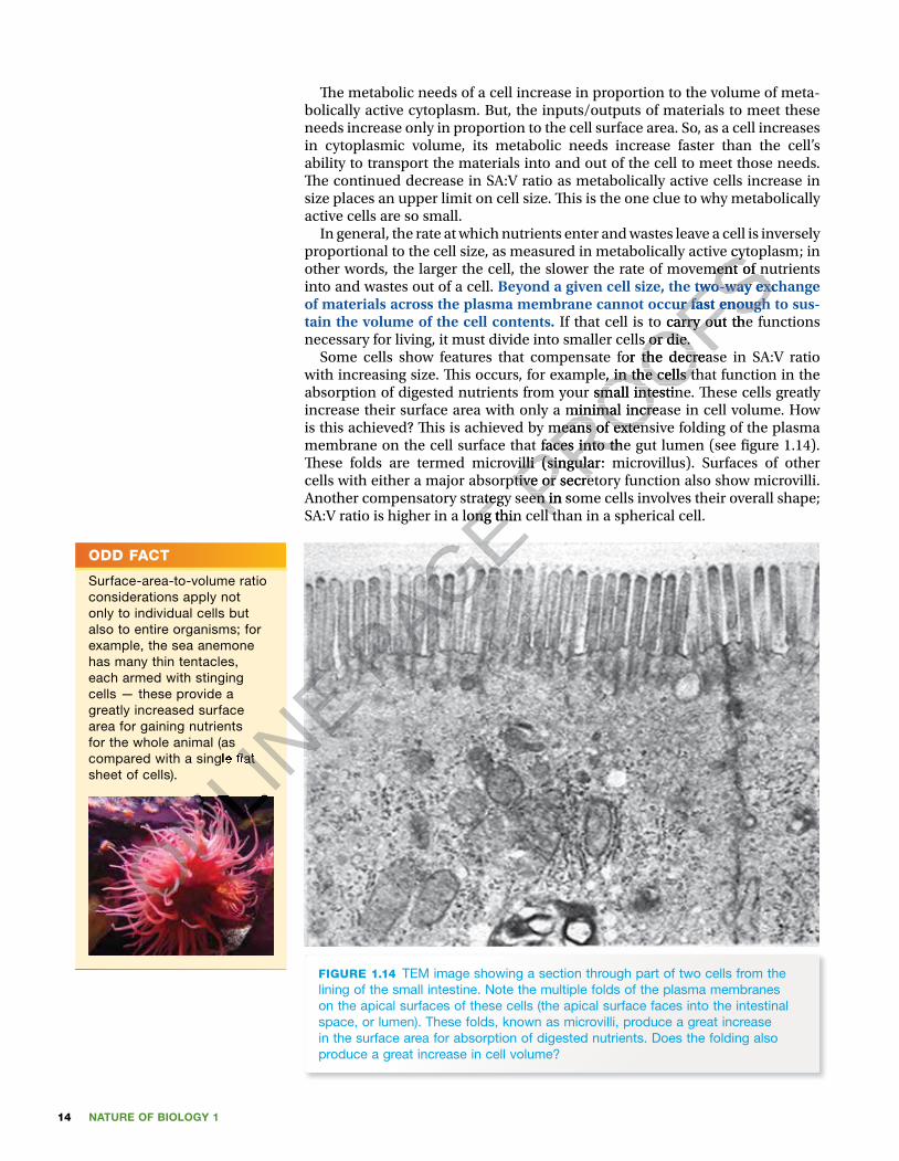
NATURE OF BIOLOGY 114
� e metabolic needs of a cell increase in proportion to the volume of meta-bolically active cytoplasm. But, the inputs/outputs of materials to meet these needs increase only in proportion to the cell surface area. So, as a cell increases in cytoplasmic volume, its metabolic needs increase faster than the cell’s ability to transport the materials into and out of the cell to meet those needs. � e continued decrease in SA:V ratio as metabolically active cells increase in size places an upper limit on cell size. � is is the one clue to why metabolically active cells are so small.
In general, the rate at which nutrients enter and wastes leave a cell is inversely proportional to the cell size, as measured in meta bolically active cytoplasm; in other words, the larger the cell, the slower the rate of movement of nutrients into and wastes out of a cell. Beyond a given cell size, the two-way exchange of materials across the plasma membrane cannot occur fast enough to sus-tain the volume of the cell contents. If that cell is to carry out the functions necessary for living, it must divide into smaller cells or die.
Some cells show features that compensate for the decrease in SA:V ratio with increasing size. � is occurs, for example, in the cells that function in the absorption of digested nutrients from your small intestine. � ese cells greatly increase their surface area with only a minimal increase in cell volume. How is this achieved? � is is achieved by means of extensive folding of the plasma membrane on the cell surface that faces into the gut lumen (see � gure 1.14). � ese folds are termed microvilli (singular: microvillus). Surfaces of other cells with either a major absorptive or secretory function also show microvilli. Another compensatory strategy seen in some cells involves their overall shape; SA:V ratio is higher in a long thin cell than in a spherical cell.
FIGURE 1.14 TEM image showing a section through part of two cells from the lining of the small intestine. Note the multiple folds of the plasma membranes on the apical surfaces of these cells (the apical surface faces into the intestinal space, or lumen). These folds, known as microvilli, produce a great increase in the surface area for absorption of digested nutrients. Does the folding also produce a great increase in cell volume?
ODD FACT
Surface-area-to-volume ratio considerations apply not only to individual cells but also to entire organisms; for example, the sea anemone has many thin tentacles, each armed with stinging cells — these provide a greatly increased surface area for gaining nutrients for the whole animal (as compared with a single � at sheet of cells).
ONLINE
ONLINE
ONLINE
compared with a single � at
ONLINE
compared with a single � at
ONLINE P
AGE Another compensatory strategy seen in some cells involves their overall shape;
PAGE Another compensatory strategy seen in some cells involves their overall shape; SA:V ratio is higher in a long thin cell than in a spherical cell.
PAGE SA:V ratio is higher in a long thin cell than in a spherical cell.
PAGE PROOFS
proportional to the cell size, as measured in meta bolically active cytoplasm; in
PROOFSproportional to the cell size, as measured in meta bolically active cytoplasm; in other words, the larger the cell, the slower the rate of movement of nutrients
PROOFSother words, the larger the cell, the slower the rate of movement of nutrients Beyond a given cell size, the two-way exchange
PROOFSBeyond a given cell size, the two-way exchange of materials across the plasma membrane cannot occur fast enough to sus-
PROOFSof materials across the plasma membrane cannot occur fast enough to sus- If that cell is to carry out the functions
PROOFS If that cell is to carry out the functions necessary for living, it must divide into smaller cells or die.
PROOFSnecessary for living, it must divide into smaller cells or die.
Some cells show features that compensate for the decrease in SA:V ratio
PROOFSSome cells show features that compensate for the decrease in SA:V ratio
with increasing size. � is occurs, for example, in the cells that function in the
PROOFSwith increasing size. � is occurs, for example, in the cells that function in the absorption of digested nutrients from your small intestine. � ese cells greatly
PROOFSabsorption of digested nutrients from your small intestine. � ese cells greatly increase their surface area with only a minimal increase in cell volume. How
PROOFSincrease their surface area with only a minimal increase in cell volume. How is this achieved? � is is achieved by means of extensive folding of the plasma
PROOFSis this achieved? � is is achieved by means of extensive folding of the plasma membrane on the cell surface that faces into the gut lumen (see � gure 1.14).
PROOFSmembrane on the cell surface that faces into the gut lumen (see � gure 1.14). � ese folds are termed microvilli (singular: microvillus). Surfaces of other PROOFS
� ese folds are termed microvilli (singular: microvillus). Surfaces of other cells with either a major absorptive or secretory function also show microvilli. PROOFS
cells with either a major absorptive or secretory function also show microvilli. Another compensatory strategy seen in some cells involves their overall shape; PROOFS
Another compensatory strategy seen in some cells involves their overall shape; SA:V ratio is higher in a long thin cell than in a spherical cell.PROOFS
SA:V ratio is higher in a long thin cell than in a spherical cell.

15CHAPTER 1 Cells: basic units of life on Earth
When he examined thin slices of cork using a simple microscope, the Englishman Robert Hooke (1635–1703) became the � rst person to see and record evidence of cells. What Hooke saw, using his simple microsope, were not living cells, but the cell walls of dead and empty plant cells (see � gure 1.15b). In his book Micrographia, published in 1665, Hooke used the word ‘cell’ to describe these structural units. ‘Cell’ is short for the Latin word cellula that means ‘little compartment’. Hooke’s observations were sig-ni� cant because he was the � rst person to recognise that the plant material had an organised structure that was built of small units, visible only through a microscope.
FIGURE 1.15 (a) The microscope built by Robert Hooke that he used to make his � rst observations of ‘little compartments’, or cells (b) First drawing made by Hooke in 1665 of ‘cells’ from a thin piece of cork. Were these living cells?
(a) (b)
In the 1670s, a Dutch cloth merchant, Anton van Leeuwenhoek (1632–1723), built a simple microscope and was then the � rst person to see and record living cells that he called ‘little animacules’ (see � gure 1.16). � ese cells were crescent-shaped bacteria present in a scraping of plaque from between his teeth.
However, until the nineteenth century the � ndings of Hooke and Leeuwenhoek were regarded as iso-lated and unrelated observations.
Early in 1838, a German botanist, Matthias Schleiden (1804–81), carried out microscopic examin-ations of a variety of plant tissues and came to the con-clusion that cells were the basic structural unit of all plants. Early in the following year, a German physiol-ogist, � eodor Schwann, (1810–82) having examined various animal tissues recognised that animals were
built of cells. At that time, however, plant and animal cells were not seen as having much in common, so no link was made between these separate observ-ations of plant and animal cells; they were regarded as separate worlds. � eodor Schwann also discovered the digestive enzyme pepsin and that yeast was a living organism, and introduced the term ‘metabolism’.
FIGURE 1.16 (a) The simple microscope built by Leeuwenhoek that revealed ‘little animacules’. The specimen was placed on top of the pin, the microscope was held up to the eye and viewed through a quartz lens just 2 mm wide. (b) Some of the ‘little animacules’ seen by Leeuwenhoek — these were various bacteria.
(a)
(b)
SEEING, THEN INTERPRETING CELLS
(continued)
ONLINE
ONLINE
ONLINE The microscope built by Robert Hooke
ONLINE The microscope built by Robert Hooke
that he used to make his � rst observations of ‘little
ONLINE that he used to make his � rst observations of ‘little
First drawing made by
ONLINE First drawing made by
Hooke in 1665 of ‘cells’ from a thin piece of cork. Were
ONLINE
Hooke in 1665 of ‘cells’ from a thin piece of cork. Were
ONLINE
In the 1670s, a Dutch cloth merchant, Anton van
ONLINE
In the 1670s, a Dutch cloth merchant, Anton van Leeuwenhoek (1632–1723), built a simple microscope
ONLINE
Leeuwenhoek (1632–1723), built a simple microscope and was then the � rst person to see and record living
ONLINE
and was then the � rst person to see and record living cells that he called ‘little animacules’ (see � gure 1.16).
ONLINE
cells that he called ‘little animacules’ (see � gure 1.16). � ese cells were crescent-shaped bacteria present in a
ONLINE
� ese cells were crescent-shaped bacteria present in a scraping of plaque from between his teeth.
ONLINE
scraping of plaque from between his teeth. However, until the nineteenth century the � ndings ONLIN
E
However, until the nineteenth century the � ndings ONLINE
of Hooke and Leeuwenhoek were regarded as iso-ONLINE
of Hooke and Leeuwenhoek were regarded as iso-lated and unrelated observations. ONLIN
E
lated and unrelated observations.
PAGE
PAGE
PAGE The microscope built by Robert Hooke PAGE The microscope built by Robert Hooke
that he used to make his � rst observations of ‘little PAGE
that he used to make his � rst observations of ‘little PAGE
PAGE
PAGE PROOFS
organism, and introduced the term ‘metabolism’.
PROOFSorganism, and introduced the term ‘metabolism’.
PROOFS
PROOFS
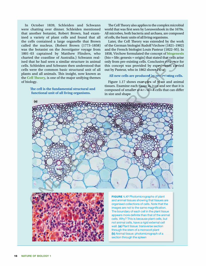
NATURE OF BIOLOGY 116
In October 1839, Schleiden and Schwann were chatting over dinner. Schleiden mentioned that another botanist, Robert Brown, had exam-ined a variety of plant cells and found that all the cells contained a large organelle that Brown called the nucleus. (Robert Brown (1773–1858) was the botanist on the Investigator voyage from 1801–03 captained by Matthew Flinders, who charted the coastline of Australia.) Schwann real-ised that he had seen a similar structure in animal cells. Schleiden and Schwann then understood that cells were the common basic structural unit of all plants and all animals. � is insight, now known as the Cell � eory, is one of the major unifying themes of biology.
� e cell is the fundamental structural and functional unit of all living organisms.
� e Cell � eory also applies to the complex microbial world that was � rst seen by Leeuwenhoek in the 1670s. All microbes, both bacteria and archaea, are composed of cells, the basic units of all living organisms.
Later, the Cell � eory was extended by the work of the German biologist Rudolf Virchow (1821–1902) and the French biologist Louis Pasteur (1822–95). In 1858, Virchow formulated the concept of biogenesis (bio = life; genesis = origin) that stated that cells arise only from pre-existing cells. Conclusive evidence for this concept was provided by experiments carried out by Pasteur, who in 1862 showed that:
All new cells are produced by pre-existing cells.
Figure 1.17 shows examples of plant and animal tissues. Examine each tissue in turn and see that it is composed of smaller units called cells that can di� er in size and shape.
FIGURE 1.17 Photomicrographs of plant and animal tissues showing that tissues are organised collections of cells. Note that the images are not to the same magni� cation. The boundary of each cell in the plant tissue appears more de� nite than that of the animal cells. Why? This is because plant cells, but not animal cells, have a rigid external cell wall. (a) Plant tissue: transverse section through the stem of a monocot plant (b) Animal tissue: photomicrograph of a section through the spleen
(b)
(a)
ONLINE
ONLINE
(b)ONLINE
(b)ONLINE
ONLINE P
AGE PROOFS origin) that stated that cells arise
PROOFS origin) that stated that cells arise
only from pre-existing cells. Conclusive evidence for
PROOFSonly from pre-existing cells. Conclusive evidence for this concept was provided by experiments carried
PROOFSthis concept was provided by experiments carried out by Pasteur, who in 1862 showed that:
PROOFSout by Pasteur, who in 1862 showed that:
All new cells are produced by pre-existing cells.
PROOFSAll new cells are produced by pre-existing cells.
Figure 1.17 shows examples of plant and animal
PROOFSFigure 1.17 shows examples of plant and animal
tissues. Examine each tissue in turn and see that it is
PROOFStissues. Examine each tissue in turn and see that it is
PROOFScomposed of smaller units called cells that can di� er
PROOFScomposed of smaller units called cells that can di� er in size and shape.
PROOFSin size and shape.
PROOFS

17CHAPTER 1 Cells: basic units of life on Earth
KEY IDEAS
■ Cells are the basic structural and functional units of life. ■ Cells are typically too small to be seen by an unaided eye. ■ The unit of measurement used for cell size is the micrometre (µm), one millionth of a metre.
■ Microbial cells are much smaller than plant and animal cells. ■ The metabolic needs of a cell are determined by its metabolically active cytoplasmic volume.
■ The ability of a cell to meet its metabolic needs is determined by the surface area of the cell.
■ As a cell increases in size, its internal volume expands at a greater rate than the area of its plasma membrane.
■ The surface-area-to-volume ratio (SA:V ratio) of a smaller object is higher than that of a larger object with the same shape.
■ The continued decrease in SA:V ratio as metabolically active cells increase in size places an upper limit on cell size.
QUICK CHECK
7 Identify whether each of the following statements is true or false.a Cells are typically too small to be seen with an unaided eye.b Bacterial cells are typically larger than animal cells.c Viral particles are smaller than microbial cells.d As a given shape increases in size, its surface-area-to-volume ratio
increases.e Beyond a given cell size, the two-way exchange of materials across the
cell surface cannot occur at a rate suf�cient to meet the needs of a cell. 8 Two spheres (A and B) have different diameters, with A being larger than B.
Which has the higher SA:V ratio?
Prokaryotes: no nuclear envelope!�e remarkable living community discovered deep under the Antarctic ice sheet in subglacial Lake Whillans consists of microbes belonging to two di�erent classi�cation groups (bacteria and archaea). �e cells of all these microbes can be readily distinguished from the cells of the other major groups of living organisms: fungi, plants and animals. �e key distinguishing feature of archaea and bacteria is that their cells lack a membrane-bound nucleus (see �gure 1.18a). Cells with this characteristic are described as prokaryotic cells and organisms displaying this feature are called prokaryotes. Prokaryotes are generally assumed to be the oldest existing form of life on planet Earth. �e absence of a distinct nucleus does not mean that prokary-otes, such as archaea and bacteria, lack genetic material. Like all other kinds of organism, archaea and bacteria have DNA in their cells, but the DNA in prokar-yotic cells is dispersed, not enclosed within a separate membrane-bound compartment.
In contrast, the cells of all other organisms — protists, fungi, plants and ani-mals — have a de�nite nucleus (see �gure 1.18b). �e nucleus is enclosed by a double membrane, called the nuclear envelope. Organisms with this feature are termed eukaryotes and their cells are described as being eukaryotic. �e nucleus of a eukaryotic cell contains DNA, the genetic material of cells. In addition, eukaryotic cells contain many membrane-bound cell organelles that are not present in prokaryotic cells (see table 1.1).
ODD FACT
A single cell lining the small intestine may have up to 10 000 microvilli on its apical surface facing into the gut lumen. How would this affect the surface area available for absorption of digested nutrients, compared with a cell with no microvilli?
Unit 1 ProkaryotesConcept summary and practice questions
AOS 1
Topic 1
Concept 2
ONLINE Prokaryotes: no nuclear envelope!
ONLINE Prokaryotes: no nuclear envelope!
�e remarkable living community discovered deep under the Antarctic ice
ONLINE
�e remarkable living community discovered deep under the Antarctic ice sheet in subglacial Lake Whillans consists of microbes belonging to two
ONLINE
sheet in subglacial Lake Whillans consists of microbes belonging to two di�erent classi�cation groups (bacteria and archaea). �e cells of all these
ONLINE
di�erent classi�cation groups (bacteria and archaea). �e cells of all these
ONLINE
ONLINE
ONLINE
ONLINE
Prokaryotes
ONLINE
ProkaryotesConcept summary
ONLINE
Concept summary and practice
ONLINE
and practice questions
ONLINE
questions
ONLINE P
AGE
PAGE
PAGE e typically too small to be seen with an unaided eye.
PAGE e typically too small to be seen with an unaided eye.e typically larger than animal cells.
PAGE e typically larger than animal cells.Viral particles ar
PAGE Viral particles are smaller than microbial cells.
PAGE e smaller than microbial cells.As a given shape incr
PAGE As a given shape increases in size, its surface-area-to-volume ratio
PAGE eases in size, its surface-area-to-volume ratio
Beyond a given cell size, the two-way exchange of materials acr
PAGE Beyond a given cell size, the two-way exchange of materials acrcell surface cannot occur at a rate suf�cient to meet the needs of a cell.
PAGE cell surface cannot occur at a rate suf�cient to meet the needs of a cell.
wo spheres (A and B) have different diameters, with A being larger than B.
PAGE wo spheres (A and B) have different diameters, with A being larger than B.
Which has the higher SA:V ratio?
PAGE Which has the higher SA:V ratio?
Prokaryotes: no nuclear envelope!PAGE
Prokaryotes: no nuclear envelope!
PROOFS
PROOFS
PROOFSThe ability of a cell to meet its metabolic needs is determined by the
PROOFSThe ability of a cell to meet its metabolic needs is determined by the
As a cell increases in size, its internal volume expands at a greater rate
PROOFSAs a cell increases in size, its internal volume expands at a greater rate
The surface-area-to-volume ratio (SA:V ratio) of a smaller object is higher
PROOFSThe surface-area-to-volume ratio (SA:V ratio) of a smaller object is higher than that of a larger object with the same shape.
PROOFSthan that of a larger object with the same shape.The continued decrease in SA:V ratio as metabolically active cells increase
PROOFSThe continued decrease in SA:V ratio as metabolically active cells increase
PROOFS
PROOFS
PROOFS
Identify whether each of the following statements is true or false.PROOFS
Identify whether each of the following statements is true or false.e typically too small to be seen with an unaided eye.PROOFS
e typically too small to be seen with an unaided eye.e typically larger than animal cells.PROOFS
e typically larger than animal cells.

NATURE OF BIOLOGY 118
FIGURE 1.18 Images of cells (not to same magni� cation) (a) Image of prokaryotic cells. One is in the process of dividing. Note that the cells do not have a discrete nucleus enclosed within a membrane. Instead, the genetic material (stained orange) is dispersed. (b) A confocal � uorescence microscope view of human breast cells, examples of eukaryotic cells. The position of the nucleus that encloses the genetic material is shown by the discrete blue area within each cell. The cells have been treated with special stains to highlight two different cytoskeleton proteins: vimentin (green), found in cells within cancerous tissue, and keratin (red).
(a) (b)
Comparing prokaryotes with eukaryotesFigure 1.19 shows the structure of a typical prokaryotic cell in comparison with a eukaryotic cell. Note that a prokaryotic cell has a simple architecture in con-trast to a eukaryotic cell that has a more elaborate structure owing to the pres-ence of many membrane-enclosed compartments within the cell.
FIGURE 1.19 Diagram showing a basic comparison of a prokaryotic cell (left) and a eukaryotic cell (right). The key distinction between these cells is the presence in the eukaryotic cell of only membrane-bound organelles, in particular the nucleus that contains the genetic material, DNA. (Cells are not drawn to scale.)
Table 1.1 outlines a comparison between the structures of prokaryotic and eukaryotic cells. � e critical di� erence is the absence of membrane-enclosed organelles in prokaryotes, in contrast to eukaryotic cells. We will explore details of eukaryotic cells in chapter 2.
In general, prokaryotic cells are about 10 times smaller than eukaryotic cells. However, size is not an absolute distinction; there are some rare exceptions:• Large prokaryotic cells exist, such as the giant bacterium, � iomargarita
namibiensis, (0.1 × 0.3 mm in diameter) that lives in the muddy sea � oor o� the coast of Namibia.
• Relatively small eukaryotic cells exist, such as the single-celled green alga, Ostreococcus tauri that is just 0.8 µm in diameter.
pro = before + karyon = kernel, nucleus
eu = well, good + karyon = kernel, nucleus
ONLINE
ONLINE
ONLINE
ONLINE
ONLINE
ONLINE
ONLINE
ONLINE
ONLINE
ONLINE
The key distinction between
ONLINE
The key distinction between these cells is the presence
ONLINE
these cells is the presence in the eukaryotic cell of only
ONLINE
in the eukaryotic cell of only membrane-bound organelles,
ONLINE
membrane-bound organelles, in particular the nucleus that
ONLINE
in particular the nucleus that contains the genetic material,
ONLINE
contains the genetic material,
ONLINE
ONLINE
ONLINE
DNA. (Cells are not drawn to
ONLINE
DNA. (Cells are not drawn to
ONLINE P
AGE Comparing prokaryotes with eukaryotes
PAGE Comparing prokaryotes with eukaryotesFigure 1.19 shows the structure of a typical prokaryotic cell in comparison with
PAGE Figure 1.19 shows the structure of a typical prokaryotic cell in comparison with a eukaryotic cell. Note that a prokaryotic cell has a simple architecture in con-
PAGE a eukaryotic cell. Note that a prokaryotic cell has a simple architecture in con-trast to a eukaryotic cell that has a more elaborate structure owing to the pres-
PAGE trast to a eukaryotic cell that has a more elaborate structure owing to the pres-ence of many membrane-enclosed compartments within the cell.
PAGE ence of many membrane-enclosed compartments within the cell.
PAGE
PAGE
PAGE
PAGE
PAGE
PAGE
PAGE
PAGE
PAGE
PAGE
PAGE
PAGE
PAGE
PAGE
PAGE
PAGE PROOFS
PROOFS
PROOFS Image of prokaryotic cells. One is in the process of
PROOFS Image of prokaryotic cells. One is in the process of
dividing. Note that the cells do not have a discrete nucleus enclosed within a membrane. Instead, the genetic material
PROOFSdividing. Note that the cells do not have a discrete nucleus enclosed within a membrane. Instead, the genetic material
A confocal � uorescence microscope view of human breast cells, examples of
PROOFS A confocal � uorescence microscope view of human breast cells, examples of
eukaryotic cells. The position of the nucleus that encloses the genetic material is shown by the discrete blue area within
PROOFSeukaryotic cells. The position of the nucleus that encloses the genetic material is shown by the discrete blue area within each cell. The cells have been treated with special stains to highlight two different cytoskeleton proteins: vimentin
PROOFSeach cell. The cells have been treated with special stains to highlight two different cytoskeleton proteins: vimentin
PROOFS
PROOFS
Comparing prokaryotes with eukaryotesPROOFS
Comparing prokaryotes with eukaryotes

19CHAPTER 1 Cells: basic units of life on Earth
TABLE 1.1 Comparison of prokaryotic and eukaryotic cells.
Feature Prokaryote Eukaryote
size small: typically ~1–2 µm diameter larger: typically in range 10–100 µm
chromosomes present as singlecircular DNA molecule
present as multiple linearDNA molecules
ribosomes present: small size (70s) present: large size (80s)
plasma membrane present present
cell wall present and chemically complex present in plants, fungi, and some protists, but chemically simple; absent in animal cells
membrane-bound nucleus absent present
membrane-bound cell organelles absent present; e.g. lysosomes, mitochondria
cytoskeleton absent present
In general, prokaryotic cells are unicellular and the great majority of eukary-otes are multicellular. However, it cannot be assumed for certain that a unicel-lular organism is a prokaryote (either a bacterium or an archaean), because a number of eukaryotes are unicellular. Unicellular eukaryotes are protists, such as Amoeba, Paramecium and Euglena, some algae such as Chlorella and the diatoms, and fungi such as yeasts.
While there are some di�erences in aspects of the structure of eukaryotic and prokaryotic cells, there are many similarities in their structures and func-tioning. �ese common features re�ect the inter-connectedness of life forms.
For example, both prokaryotic and eukaryotic cells:• have DNA as their genetic material; however, in comparison with eukary-
otes, prokaryotes have only about 0.001 times the amount of DNA• have plasma membranes that selectively control the entry and exit of dis-
solved materials into and out of the cell• use the same chemical building blocks including carbon, nitrogen, oxygen,
hydrogen and phosphorus, to build the organic molecules that form their structure and enable their function
• produce proteins through the same mechanism (transcription of DNA and translation of mRNA on ribosomes)
• use ATP as their source of energy to drive the energy-requiring activities of their cells.
One or more compartments?What is strikingly di�erent is that every prokaryotic cell is a single compart-ment, with no further subdivisions of the cell. �is is in contrast to eukaryotic cells that are organised internally into various compartments, each enclosed by a membrane. Because of the multi-compartmental structure of eukaryotic cells, their ultrastructure is more complex than that of prokaryotic cells.
Table 1.2 shows the relative volumes of the di�erent compartments in a eukaryotic cell, a liver cell.
TABLE 1.2 Relative volumes of the major compartments within a liver cell
Intracellular compartment Percentage of total cell volume∗
cytosol 54mitochondria 22rough endoplasmic reticulum 9smooth endoplasmic reticulum 6nucleus 6lysosomes, peroxisomes, endosomes 3
∗More than half of the cell volume is occupied by the cytosol.
Unit 1 Cell organellesConcept summary and practice questions
AOS 1
Topic 1
Concept 5
Unit 1 EukaryotesConcept summary and practice questions
AOS 1
Topic 1
Concept 3ONLINE structure and enable their function
ONLINE structure and enable their function
•
ONLINE • produce proteins through the same mechanism (transcription of DNA and
ONLINE produce proteins through the same mechanism (transcription of DNA and translation of mRNA on ribosomes)
ONLINE translation of mRNA on ribosomes)
•
ONLINE
• use ATP as their source of energy to drive the energy-requiring activities of
ONLINE use ATP as their source of energy to drive the energy-requiring activities of their cells.
ONLINE their cells.
O
ONLINE
O
ONLINE
ONLINE
ONLINE
ONLINE
ONLINE
ONLINE
ONLINE
ONLINE
ONLINE
ONLINE
ONLINE
ONLINE
ONLINE
ONLINE
ONLINE
E
ONLINE
Eukaryotes
ONLINE
ukaryotesConcept summary
ONLINE
Concept summaryand practice
ONLINE
and practice questions
ONLINE
questions1
ONLINE
1
ONLINE
Topic ONLINE
Topic 1ONLINE
1ONLINE
oncept ONLINE
oncept 3ONLINE
3ONLINE P
AGE While there are some di�erences in aspects of the structure of eukaryotic
PAGE While there are some di�erences in aspects of the structure of eukaryotic and prokaryotic cells, there are many similarities in their structures and func
PAGE and prokaryotic cells, there are many similarities in their structures and functioning. �ese common features re�ect the inter-connectedness of life forms.
PAGE tioning. �ese common features re�ect the inter-connectedness of life forms. For example, both prokaryotic and eukaryotic cells:
PAGE For example, both prokaryotic and eukaryotic cells:have DNA as their genetic material; however, in comparison with eukary
PAGE have DNA as their genetic material; however, in comparison with eukaryotes, prokaryotes have only about 0.001 times the amount of DNA
PAGE otes, prokaryotes have only about 0.001 times the amount of DNAhave plasma membranes that selectively control the entry and exit of dis
PAGE have plasma membranes that selectively control the entry and exit of dissolved materials into and out of the cell
PAGE solved materials into and out of the celluse the same chemical building blocks including carbon, nitrogen, oxygen,
PAGE use the same chemical building blocks including carbon, nitrogen, oxygen, hydrogen and phosphorus, to build the organic molecules that form their PAGE hydrogen and phosphorus, to build the organic molecules that form their structure and enable their functionPAGE
structure and enable their functionproduce proteins through the same mechanism (transcription of DNA and PAGE
produce proteins through the same mechanism (transcription of DNA and
PROOFS
PROOFS
PROOFSprotists, but chemically simple;
PROOFSprotists, but chemically simple; absent in animal cells
PROOFSabsent in animal cells
present; e.g. lysosomes,
PROOFSpresent; e.g. lysosomes, mitochondria
PROOFSmitochondria
present
PROOFSpresent
In general, prokaryotic cells are unicellular and the great majority of eukary
PROOFSIn general, prokaryotic cells are unicellular and the great majority of eukary
otes are multicellular. However, it cannot be assumed for certain that a unicel
PROOFSotes are multicellular. However, it cannot be assumed for certain that a unicellular organism is a prokaryote (either a bacterium or an archaean), because a
PROOFSlular organism is a prokaryote (either a bacterium or an archaean), because a number of eukaryotes are unicellular. Unicellular eukaryotes are protists, such
PROOFSnumber of eukaryotes are unicellular. Unicellular eukaryotes are protists, such
Euglena
PROOFS
Euglena, some algae such as
PROOFS
, some algae such as diatoms, and fungi such as yeasts. PROOFS
diatoms, and fungi such as yeasts.While there are some di�erences in aspects of the structure of eukaryotic PROOFS
While there are some di�erences in aspects of the structure of eukaryotic and prokaryotic cells, there are many similarities in their structures and funcPROOFS
and prokaryotic cells, there are many similarities in their structures and func

NATURE OF BIOLOGY 120
�e multicompartment structure of a eukaryotic cell enables it to main-tain di�erent conditions within each membrane-enclosed compartment that are suitable for the particular function of that compartment. �ink about a house that is subdivided into rooms with di�erent functions: you shower in the bathroom, not in the kitchen; the stove is in the kitchen, not in the bedroom. A eukaryotic cell can be likened to a house — its many com-partments are like di�erent rooms where di�erent tasks are carried out. (In chapter 2, we will explore these various compartments in eukaryotic cells.)
KEY IDEAS
■ Prokaryotic cells lack a membrane-bound nucleus, and organisms lacking a nuclear envelope are termed prokaryotes — bacteria and archaea.
■ Prokaryotes differ from eukaryotes in the absence of membrane-bound cell organelles of any kind.
■ Eukaryotic cells are typically about ten times larger than prokaryotic cells. ■ Eukaryotic cells have a membrane-bound nucleus in addition to other membrane-bound organelles.
■ Organisms built of cells that have a nucleus enclosed within a nuclear envelope are termed eukaryotes — protists, fungi, plants and animals.
QUICK CHECK
9 Identify whether each of the following statements is true or false.a Prokaryotes are unicellular organisms, comprising bacteria and archaea.b The presence of a membrane-bound nucleus in its cells provides
evidence that an organism is a eukaryote. c All eukaryotes are multicellular organisms.d The various compartments within eukaryotic cells would be expected to
have identical conditions.10 List two similarities between prokaryotic and eukaryotic cells.11 A unicellular organism was found in a sample of pond water. Is it
reasonable to conclude that this organism must be either a bacterium or an archaeon? Brie�y explain.
Plasma membrane: the gatekeeper�e cells of all living organisms have a boundary that separates their internal environment from the external environment of their surroundings. From single-celled organisms, such as amoebae or bacteria, to multicellular organ-isms, such as mushrooms, palm trees and human beings, each of their cells has an active boundary called the plasma membrane, also known as the cell membrane.
�e plasma membrane forms the outer boundary of the living compartment of every cell. Within this compartment, conditions can be established that di�er from those in the external environment and that support the living state. �e plasma membrane can exclude some substances from entering the cell, while permitting entry of other substances and elimination of yet other sub-stances. Without such a boundary, life could not exist, and indeed could not have evolved.
�e plasma membrane boundary can be thought of as a busy gatekeeper selec-tively controlling the entry and exit of materials into and out of cells. As such, the plasma membrane is said to be semipermeable or selectively permeable,
Unit 1 Structure of the plasma membraneConcept summary and practice questions
AOS 1
Topic 2
Concept 1 ONLINE
ONLINE
ONLINE 10
ONLINE 10
11
ONLINE 11
reasonable to conclude that this organism must be either a bacterium or
ONLINE reasonable to conclude that this organism must be either a bacterium or
an archaeon? Brie�y explain.
ONLINE
an archaeon? Brie�y explain.
ONLINE
ONLINE
ONLINE
ONLINE
ONLINE
ONLINE
ONLINE
ONLINE
ONLINE
ONLINE
ONLINE
ONLINE
ONLINE
ONLINE
1
ONLINE
1
ONLINE
S
ONLINE
Structure of
ONLINE
tructure of the plasma
ONLINE
the plasmamembraneONLIN
E
membraneConcept summary ONLIN
E
Concept summary and practice ONLIN
E
and practice
O ONLINE
OS ONLINE
S 1 ONLINE
1 ONLINE
Topic ONLINE
Topic 2 ONLINE
2 ONLINE
ONLINE P
AGE
PAGE
PAGE
PAGE Identify whether each of the following statements is true or false.
PAGE Identify whether each of the following statements is true or false.
okaryotes are unicellular organisms, comprising bacteria and archaea.
PAGE okaryotes are unicellular organisms, comprising bacteria and archaea.
The pr
PAGE The presence of a membrane-bound nucleus in its cells provides
PAGE esence of a membrane-bound nucleus in its cells provides
evidence that an organism is a eukaryote.
PAGE evidence that an organism is a eukaryote. All eukaryotes ar
PAGE All eukaryotes ar
d
PAGE d The various compartments within eukaryotic cells would be expected to
PAGE The various compartments within eukaryotic cells would be expected tohave identical conditions.PAGE have identical conditions.
List two similarities between prPAGE
List two similarities between prA unicellular orPAGE
A unicellular or
PROOFS
PROOFS
PROOFS
PROOFS
PROOFSProkaryotic cells lack a membrane-bound nucleus, and organisms lacking
PROOFSProkaryotic cells lack a membrane-bound nucleus, and organisms lacking a nuclear envelope are termed prokaryotes — bacteria and archaea.
PROOFSa nuclear envelope are termed prokaryotes — bacteria and archaea.Prokaryotes differ from eukaryotes in the absence of membrane-bound
PROOFSProkaryotes differ from eukaryotes in the absence of membrane-bound
Eukaryotic cells are typically about ten times larger than prokaryotic cells.
PROOFSEukaryotic cells are typically about ten times larger than prokaryotic cells. Eukaryotic cells have a membrane-bound nucleus in addition to other
PROOFSEukaryotic cells have a membrane-bound nucleus in addition to other
Organisms built of cells that have a nucleus enclosed within a nuclear
PROOFSOrganisms built of cells that have a nucleus enclosed within a nuclear envelope are termed eukaryotes — protists, fungi, plants and animals.
PROOFS
envelope are termed eukaryotes — protists, fungi, plants and animals.
PROOFS
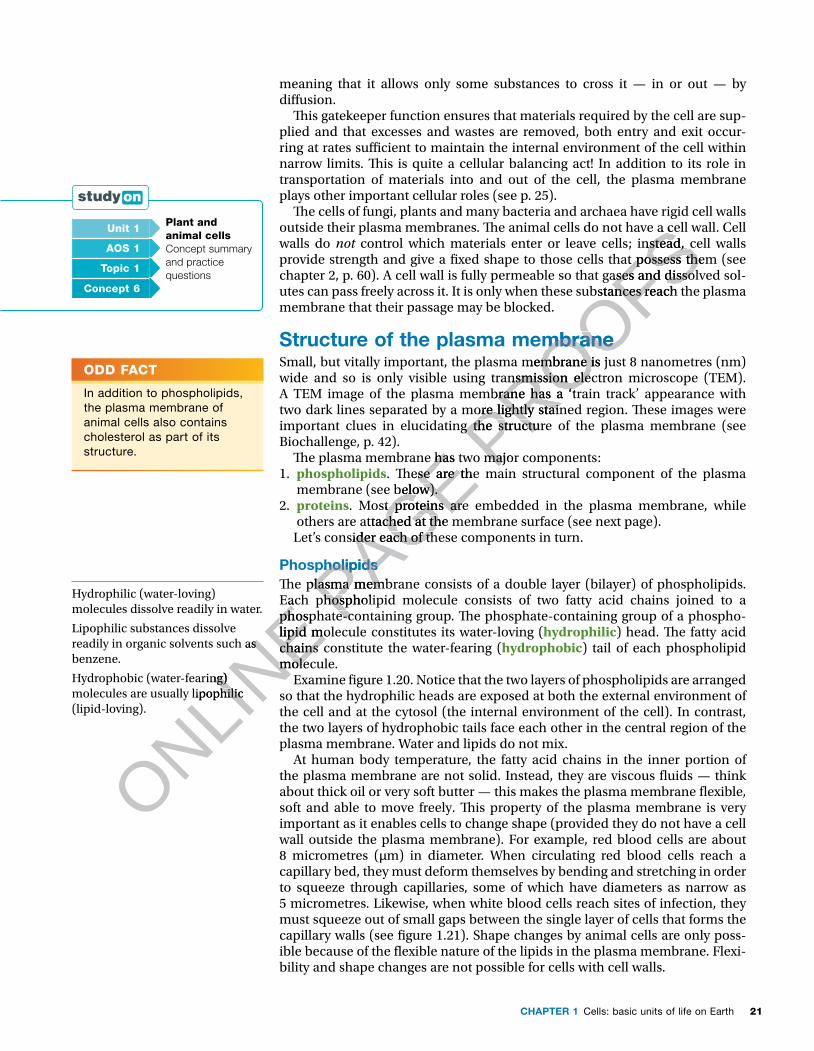
21CHAPTER 1 Cells: basic units of life on Earth
meaning that it allows only some substances to cross it — in or out — by di�usion.
�is gatekeeper function ensures that materials required by the cell are sup-plied and that excesses and wastes are removed, both entry and exit occur-ring at rates su�cient to maintain the internal environment of the cell within narrow limits. �is is quite a cellular balancing act! In addition to its role in transportation of materials into and out of the cell, the plasma membrane plays other important cellular roles (see p. 25).
�e cells of fungi, plants and many bacteria and archaea have rigid cell walls outside their plasma membranes. �e animal cells do not have a cell wall. Cell walls do not control which materials enter or leave cells; instead, cell walls provide strength and give a �xed shape to those cells that possess them (see chapter 2, p. 60). A cell wall is fully permeable so that gases and dissolved sol-utes can pass freely across it. It is only when these substances reach the plasma membrane that their passage may be blocked.
Structure of the plasma membraneSmall, but vitally important, the plasma membrane is just 8 nanometres (nm) wide and so is only visible using transmission electron microscope (TEM). A TEM image of the plasma membrane has a ‘train track’ appearance with two dark lines separated by a more lightly stained region. �ese images were important clues in elucidating the structure of the plasma membrane (see Biochallenge, p. 42).
�e plasma membrane has two major components:1. phospholipids. �ese are the main structural component of the plasma
membrane (see below).2. proteins. Most proteins are embedded in the plasma membrane, while
others are attached at the membrane surface (see next page). Let’s consider each of these components in turn.
Phospholipids�e plasma membrane consists of a double layer (bilayer) of phospholipids. Each phospholipid molecule consists of two fatty acid chains joined to a phosphate-containing group. �e phosphate-containing group of a phospho-lipid molecule constitutes its water-loving (hydrophilic) head. �e fatty acid chains constitute the water-fearing (hydrophobic) tail of each phospholipid molecule.
Examine �gure 1.20. Notice that the two layers of phospholipids are arranged so that the hydrophilic heads are exposed at both the external environment of the cell and at the cytosol (the internal environment of the cell). In contrast, the two layers of hydrophobic tails face each other in the central region of the plasma membrane. Water and lipids do not mix.
At human body temperature, the fatty acid chains in the inner portion of the plasma membrane are not solid. Instead, they are viscous �uids — think about thick oil or very soft butter — this makes the plasma membrane �exible, soft and able to move freely. �is property of the plasma membrane is very important as it enables cells to change shape (provided they do not have a cell wall outside the plasma membrane). For example, red blood cells are about 8 micrometres (μm) in diameter. When circulating red blood cells reach a capillary bed, they must deform themselves by bending and stretching in order to squeeze through capillaries, some of which have diameters as narrow as 5 micrometres. Likewise, when white blood cells reach sites of infection, they must squeeze out of small gaps between the single layer of cells that forms the capillary walls (see �gure 1.21). Shape changes by animal cells are only poss-ible because of the �exible nature of the lipids in the plasma membrane. Flexi-bility and shape changes are not possible for cells with cell walls.
ODD FACT
In addition to phospholipids, the plasma membrane of animal cells also contains cholesterol as part of its structure.
Hydrophilic (water-loving) molecules dissolve readily in water.
Lipophilic substances dissolve readily in organic solvents such as benzene.
Hydrophobic (water-fearing) molecules are usually lipophilic (lipid-loving).
Unit 1 Plant and animal cellsConcept summary and practice questions
AOS 1
Topic 1
Concept 6
ONLINE Each phospholipid molecule consists of two fatty acid chains joined to a
ONLINE Each phospholipid molecule consists of two fatty acid chains joined to a
phosphate-containing group. �e phosphate-containing group of a phospho
ONLINE phosphate-containing group. �e phosphate-containing group of a phospho
lipid molecule constitutes its water-loving (
ONLINE lipid molecule constitutes its water-loving (
chains constitute the water-fearing (
ONLINE
chains constitute the water-fearing (molecule.
ONLINE
molecule.
readily in organic solvents such as
ONLINE
readily in organic solvents such as
Hydrophobic (water-fearing)
ONLINE
Hydrophobic (water-fearing) molecules are usually lipophilic
ONLINE
molecules are usually lipophilic
PAGE �e plasma membrane has two major components:
PAGE �e plasma membrane has two major components:. �ese are the main structural component of the plasma
PAGE . �ese are the main structural component of the plasma membrane (see below).
PAGE membrane (see below).
. Most proteins are embedded in the plasma membrane, while
PAGE . Most proteins are embedded in the plasma membrane, while
others are attached at the membrane surface (see next page).
PAGE others are attached at the membrane surface (see next page).
PAGE Let’s consider each of these components in turn.
PAGE Let’s consider each of these components in turn.
Phospholipids
PAGE Phospholipids�e plasma membrane consists of a double layer (bilayer) of phospholipids. PAGE �e plasma membrane consists of a double layer (bilayer) of phospholipids. Each phospholipid molecule consists of two fatty acid chains joined to a PAGE
Each phospholipid molecule consists of two fatty acid chains joined to a phosphate-containing group. �e phosphate-containing group of a phosphoPAGE
phosphate-containing group. �e phosphate-containing group of a phospho
PROOFSoutside their plasma membranes. �e animal cells do not have a cell wall. Cell
PROOFSoutside their plasma membranes. �e animal cells do not have a cell wall. Cell
control which materials enter or leave cells; instead, cell walls
PROOFS control which materials enter or leave cells; instead, cell walls provide strength and give a �xed shape to those cells that possess them (see
PROOFSprovide strength and give a �xed shape to those cells that possess them (see chapter 2, p. 60). A cell wall is fully permeable so that gases and dissolved sol
PROOFSchapter 2, p. 60). A cell wall is fully permeable so that gases and dissolved solutes can pass freely across it. It is only when these substances reach the plasma
PROOFSutes can pass freely across it. It is only when these substances reach the plasma
Structure of the plasma membrane
PROOFSStructure of the plasma membraneSmall, but vitally important, the plasma membrane is just 8 nanometres (nm)
PROOFSSmall, but vitally important, the plasma membrane is just 8 nanometres (nm) wide and so is only visible using transmission electron microscope (TEM).
PROOFSwide and so is only visible using transmission electron microscope (TEM). A TEM image of the plasma membrane has a ‘train track’ appearance with
PROOFSA TEM image of the plasma membrane has a ‘train track’ appearance with two dark lines separated by a more lightly stained region. �ese images were
PROOFS
two dark lines separated by a more lightly stained region. �ese images were important clues in elucidating the structure of the plasma membrane (see PROOFS
important clues in elucidating the structure of the plasma membrane (see
�e plasma membrane has two major components:PROOFS
�e plasma membrane has two major components:
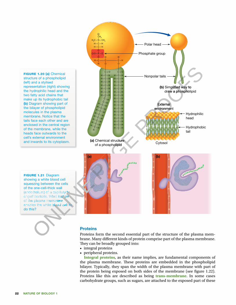
NATURE OF BIOLOGY 122
(a) (b)
FIGURE 1.21 Diagram showing a white blood cell squeezing between the cells of the one-cell-thick wall (endothelium) of a capillary to engulf bacteria. What feature of the plasma membrane enables the white blood cell to do this?
ProteinsProteins form the second essential part of the structure of the plasma mem-brane. Many di� erent kinds of protein comprise part of the plasma membrane. � ey can be broadly grouped into:• integral proteins • peripheral proteins.
Integral proteins, as their name implies, are fundamental components of the plasma membrane. � ese proteins are embedded in the phospholipid bilayer. Typically, they span the width of the plasma membrane with part of the protein being exposed on both sides of the membrane (see � gure 1.22). Proteins like this are described as being trans-membrane. In some cases carbohydrate groups, such as sugars, are attached to the exposed part of these
(a) Chemical structure of a phospholipid
C = O
H – C – H
H – C – H
H
C – H
H
C
H – C – H
H – C – H
H – C – H
H – C – H
H – C – H
H – C – H
H – C – H
H – C – H
H – C – H
H – C – H
H – C – H
H – C – H
H – C – H
H – C – H
H – C – H
C – HC – HH – C – HH – C – HH – C – HH – C – HH – C – HH – C – HH – C – HH – C – H
H
C = O
O
H – C – H
H –
H – C – H
H – C – H
H – C – H
O
O – P – O
O
H – C – H
H – C – H
H – C – H
H3C – N – CH3
CH3
H – C – H
H – C – H
O
C
H
+
–
Nonpolar tails
Phosphate group
(b) Simpli�ed way to draw a phospholipid
Polar head
Hydrophilichead
Hydrophobictail
Externalenvironment
Cytosol
FIGURE 1.20 (a) Chemical structure of a phospholipid (left) and a stylised representation (right) showing the hydrophilic head and the two fatty acid chains that make up its hydrophobic tail (b) Diagram showing part of the bilayer of phospholipid molecules in the plasma membrane. Notice that the tails face each other and are enclosed in the central region of the membrane, while the heads face outwards to the cell’s external environment and inwards to its cytoplasm.
ONLINE
ONLINE
ONLINE
ONLINE
ONLINE
ONLINE
ONLINE
ONLINE
ONLINE
ONLINE
ONLINE
ONLINE
(endothelium) of a capillary to
ONLINE
(endothelium) of a capillary to engulf bacteria. What feature
ONLINE
engulf bacteria. What feature of the plasma membrane
ONLINE
of the plasma membrane enables the white blood cell to
ONLINE
enables the white blood cell to
ONLINE
ONLINE P
AGE
PAGE
PAGE
PAGE
PAGE
PAGE
PAGE
PAGE
PAGE
PAGE
PAGE
PAGE Chemical structure
PAGE Chemical structure of a phospholipid
PAGE of a phospholipid
PAGE PROOFS
PROOFS
PROOFS Simpli�ed way to
PROOFS Simpli�ed way to
draw a phospholipid
PROOFS draw a phospholipid
PROOFS
PROOFS
PROOFS
PROOFS
PROOFS
PROOFS
PROOFS
PROOFS
PROOFS
PROOFS
PROOFS
PROOFS
PROOFS
PROOFS
PROOFS
PROOFS
PROOFS
PROOFS
PROOFS
PROOFS
PROOFS
PROOFS
PROOFS
PROOFS
PROOFS
PROOFS
PROOFS
PROOFS
PROOFS
PROOFS
PROOFS
PROOFS
PROOFS
PROOFS
PROOFS
PROOFS
PROOFS
PROOFS
PROOFS
PROOFS
PROOFS
PROOFS
PROOFS
PROOFS
PROOFS
PROOFS
PROOFS
PROOFS
PROOFS
PROOFS
PROOFS
PROOFS
PROOFS
PROOFS
PROOFS
PROOFS
PROOFS
PROOFS
PROOFS
PROOFS
PROOFS
PROOFS
PROOFS
PROOFS
PROOFS
PROOFS
PROOFS
PROOFS
PROOFS
PROOFS
PROOFS
PROOFS
PROOFS
PROOFS
PROOFS
PROOFS
PROOFS
PROOFS
PROOFS
PROOFS
PROOFS
PROOFS
PROOFS
PROOFS
PROOFS
PROOFS
PROOFS
PROOFS
PROOFS
PROOFS
PROOFS
PROOFS
PROOFS
PROOFS
PROOFS
PROOFS
PROOFS
PROOFS
PROOFS
PROOFS
PROOFS
PROOFS
PROOFSExternal
PROOFSExternal
environment
PROOFSenvironment
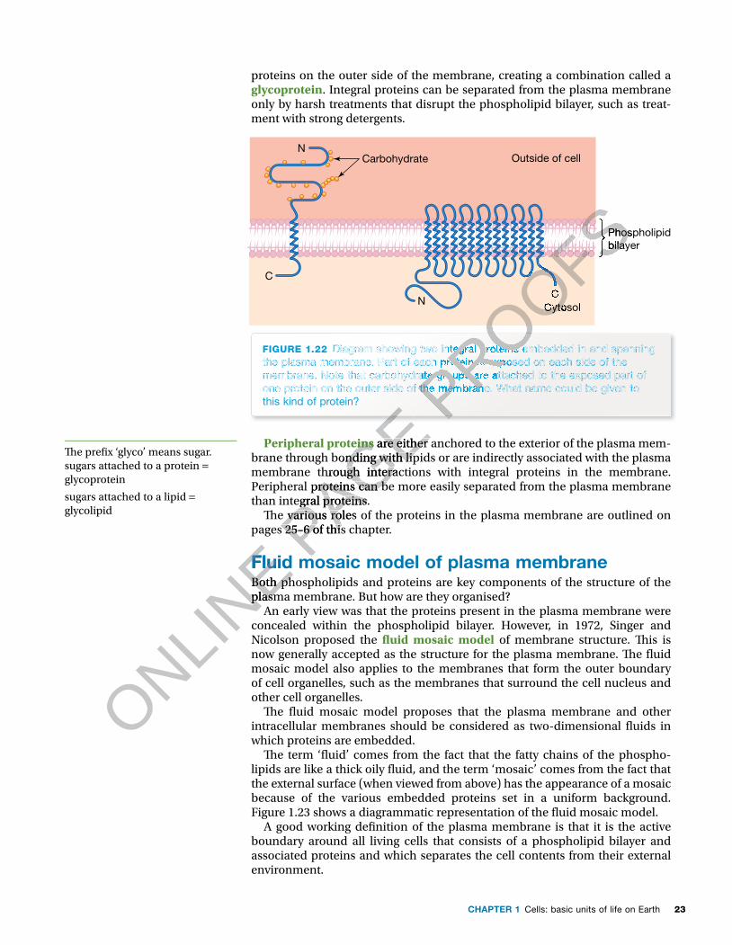
23CHAPTER 1 Cells: basic units of life on Earth
proteins on the outer side of the membrane, creating a combination called a glycoprotein. Integral proteins can be separated from the plasma membrane only by harsh treatments that disrupt the phospholipid bilayer, such as treat-ment with strong detergents.
FIGURE 1.22 Diagram showing two integral proteins embedded in and spanning the plasma membrane. Part of each protein is exposed on each side of the membrane. Note that carbohydrate groups are attached to the exposed part of one protein on the outer side of the membrane. What name could be given to this kind of protein?
Carbohydrate Outside of cell
CytosolC
C
N
N
Phospholipidbilayer
Peripheral proteins are either anchored to the exterior of the plasma mem-brane through bonding with lipids or are indirectly associated with the plasma membrane through interactions with integral proteins in the membrane. Peripheral proteins can be more easily separated from the plasma membrane than integral proteins.
� e various roles of the proteins in the plasma membrane are outlined on pages 25–6 of this chapter.
Fluid mosaic model of plasma membraneBoth phospholipids and proteins are key components of the structure of the plasma membrane. But how are they organised?
An early view was that the proteins present in the plasma membrane were concealed within the phospholipid bilayer. However, in 1972, Singer and Nicolson proposed the � uid mosaic model of membrane structure. � is is now generally accepted as the structure for the plasma membrane. � e � uid mosaic model also applies to the membranes that form the outer boundary of cell organelles, such as the membranes that surround the cell nucleus and other cell organelles.
� e � uid mosaic model proposes that the plasma membrane and other intracellular membranes should be considered as two-dimensional � uids in which proteins are embedded.
� e term ‘� uid’ comes from the fact that the fatty chains of the phospho-lipids are like a thick oily � uid, and the term ‘mosaic’ comes from the fact that the external surface (when viewed from above) has the appearance of a mosaic because of the various embedded proteins set in a uniform background. Figure 1.23 shows a diagrammatic representation of the � uid mosaic model.
A good working de� nition of the plasma membrane is that it is the active boundary around all living cells that consists of a phospholipid bilayer and associated proteins and which separates the cell contents from their external environment.
� e pre� x ‘glyco’ means sugar.sugars attached to a protein = glycoprotein
sugars attached to a lipid = glycolipid
ONLINE Fluid mosaic model of plasma membrane
ONLINE Fluid mosaic model of plasma membrane
Both phospholipids and proteins are key components of the structure of the
ONLINE
Both phospholipids and proteins are key components of the structure of the plasma membrane. But how are they organised?
ONLINE
plasma membrane. But how are they organised?
PAGE
PAGE
PAGE
PAGE
PAGE Peripheral proteins
PAGE Peripheral proteins are either anchored to the exterior of the plasma mem-
PAGE are either anchored to the exterior of the plasma mem-
brane through bonding with lipids or are indirectly associated with the plasma
PAGE brane through bonding with lipids or are indirectly associated with the plasma membrane through interactions with integral proteins in the membrane.
PAGE membrane through interactions with integral proteins in the membrane. Peripheral proteins can be more easily separated from the plasma membrane
PAGE Peripheral proteins can be more easily separated from the plasma membrane than integral proteins.
PAGE than integral proteins.
� e various roles of the proteins in the plasma membrane are outlined on PAGE � e various roles of the proteins in the plasma membrane are outlined on
pages 25–6 of this chapter.PAGE pages 25–6 of this chapter.
Fluid mosaic model of plasma membranePAGE
Fluid mosaic model of plasma membrane
PROOFS
PROOFS
PROOFSDiagram showing two integral proteins embedded in and spanning
PROOFSDiagram showing two integral proteins embedded in and spanning
the plasma membrane. Part of each protein is exposed on each side of the
PROOFSthe plasma membrane. Part of each protein is exposed on each side of the membrane. Note that carbohydrate groups are attached to the exposed part of
PROOFS
membrane. Note that carbohydrate groups are attached to the exposed part of one protein on the outer side of the membrane. What name could be given to PROOFS
one protein on the outer side of the membrane. What name could be given to PROOFS
PROOFS
PROOFS
PROOFS
PROOFS
PROOFS
PROOFS
PROOFS
PROOFS
PROOFS
PROOFS
PROOFS
PROOFS
PROOFS
PROOFS
PROOFS
PROOFS
PROOFS
PROOFS
PROOFS
PROOFS
PROOFS
PROOFS
PROOFS
PROOFS
PROOFS
PROOFS
PROOFS
PROOFS
PROOFS
PROOFS
PROOFS
PROOFS
PROOFS
PROOFS
PROOFS
PROOFS
PROOFS
PROOFS
PROOFS
PROOFS
PROOFS
PROOFS
PROOFS
PROOFS
PROOFS
PROOFS
PROOFS
PROOFS
PROOFS
PROOFSCytosol
PROOFSCytosol
C
PROOFSC
Phospholipid
PROOFSPhospholipidbilayer
PROOFSbilayer
PROOFS
PROOFS
PROOFS
PROOFS
PROOFS
PROOFS
PROOFS

NATURE OF BIOLOGY 124
Carbohydrate
Integralprotein
Exterior
CytosolIntegralprotein
Hydrophobiccore
Glycoprotein Glycolipid
Lea�ets
Phospholipidbilayer
Phospholipid
Fatty acidtails
Peripheral protein
Peripheralproteins
Hydrophilicpolar head
FIGURE 1.23 Diagram showing the � uid mosaic model of membrane structure. Note the bilayer of phospholipids. What are the two components of the phospholipids? Note the integral proteins, some of which extend through the bilayer and are exposed at both the outer and the inner surface of the membrane. Do any of these proteins have carbohydrate chains attached to their exposed regions? Note the peripheral proteins that are more loosely associated with the membrane.
KEY IDEAS
■ A major role of the plasma membrane of a cell is to act as a gatekeeper that controls the entry and exit of materials into and out of the cell.
■ The major structural component of plasma membrane is a bilayer of phospholipid molecules, each with a hydrophilic head and hydrophobic tail.
■ The fatty acid chains within the plasma membrane confer � exibility on the plasma membrane.
■ Proteins comprise the other essential component of the plasma membrane; these are both integral proteins and peripheral proteins.
■ The � uid mosaic model of membrane structure is currently accepted as the best description of the structure of the plasma membrane (and other cellular membranes).
QUICK CHECK
12 What are the two major components of a plasma membrane?13 Identify whether each of the following statements is true or false.
a The plasma membrane is present as a boundary in all living cells.b The plasma membrane consists of layers of proteins in which
phospholipids are embedded.c A key role of the plasma membrane is the control of transport of
materials into or out of cells.d Trans-membrane proteins span the width of the plasma membrane.
14 What is a glycoprotein?15 What part of a plasma membrane is responsible for its � exibility?16 Brie� y outline the � uid mosaic model of the plasma membrane.
ONLINE
ONLINE ■
ONLINE ■ The fatty acid chains within the plasma membrane confer � exibility on the
ONLINE The fatty acid chains within the plasma membrane confer � exibility on the
plasma membrane.
ONLINE plasma membrane. ■
ONLINE ■ Proteins comprise the other essential component of the plasma
ONLINE
Proteins comprise the other essential component of the plasma
PAGE
PAGE
PAGE are exposed at both the outer and the inner surface of the membrane. Do any of these proteins have carbohydrate chains
PAGE are exposed at both the outer and the inner surface of the membrane. Do any of these proteins have carbohydrate chains attached to their exposed regions? Note the peripheral proteins that are more loosely associated with the membrane.
PAGE attached to their exposed regions? Note the peripheral proteins that are more loosely associated with the membrane.
PAGE
PAGE
PAGE
PAGE
PAGE KEY IDEAS
PAGE KEY IDEAS
A major role of the plasma membrane of a cell is to act as a gatekeeper
PAGE A major role of the plasma membrane of a cell is to act as a gatekeeper that controls the entry and exit of materials into and out of the cell.
PAGE that controls the entry and exit of materials into and out of the cell.The major structural component of plasma membrane is a bilayer of PAGE The major structural component of plasma membrane is a bilayer of phospholipid molecules, each with a hydrophilic head and hydrophobic tail.PAGE
phospholipid molecules, each with a hydrophilic head and hydrophobic tail.The fatty acid chains within the plasma membrane confer � exibility on the PAGE
The fatty acid chains within the plasma membrane confer � exibility on the
PROOFSFatty acid
PROOFSFatty acidtails
PROOFStails
polar head
PROOFSpolar head
PROOFS
PROOFS
PROOFS
PROOFS
PROOFS
Diagram showing the � uid mosaic model of membrane structure. Note the bilayer of phospholipids. What PROOFS
Diagram showing the � uid mosaic model of membrane structure. Note the bilayer of phospholipids. What are the two components of the phospholipids? Note the integral proteins, some of which extend through the bilayer and PROOFS
are the two components of the phospholipids? Note the integral proteins, some of which extend through the bilayer and are exposed at both the outer and the inner surface of the membrane. Do any of these proteins have carbohydrate chains PROOFS
are exposed at both the outer and the inner surface of the membrane. Do any of these proteins have carbohydrate chains attached to their exposed regions? Note the peripheral proteins that are more loosely associated with the membrane.PROOFS
attached to their exposed regions? Note the peripheral proteins that are more loosely associated with the membrane.PROOFS

25CHAPTER 1 Cells: basic units of life on Earth
Functions of the plasma membrane �e plasma membrane carries out several important functions for a cell. �e plasma membrane:1. is an active and selective boundary2. denotes cell identity3. receives external signals 4. transports materials.
The active boundary�e plasma membrane forms the active boundary of a cell, separating the cell from its external environment and from other cells; it allows the passage of some substances only. �e plasma membrane forms the boundary of a com-partment in which the internal environment of a living cell can be held within a narrow range of conditions that are di�erent from those of the external environment.
Within the cell, similar membranes form the active boundaries of cell organ-elles, including the nucleus, the endoplasmic reticulum, the Golgi apparatus and lysosomes. In other cell organelles, such as mitochondria and chloroplasts, membranes form both the external boundary and part of the internal structure. Because of the presence of their membrane boundaries, membrane-bound cell organelles can maintain internal environments that di�er from those in the surrounding cytosol and can perform di�erent functions. (Refer to chapter 2 for more detail.)
Cell identityGlycoproteins on the outer plasma membrane function as cell surface markers, also known as antigens or cell identity tags. Each cell type has a di�erent combination of surface markers. In mammals, these markers enable the immune system to identify these cells as ‘self ’ and distinguish them from foreign cells. Glycolipids on the plasma membrane play a role in tissue recognition.
Receiving external signalsCells receive signals from their external environment. In the case of a mul-ticellular organism, the signal may originate from another cell within that organism. In the case of unicellular organisms the signal may come from other organisms in its neighbourhood or from its external environ-ment. �ese external signals are often chemical compounds, for example, hormones.
Trans-membrane proteins on the outer surface of the plasma membrane are the receptors for these signals, and each cell has many di�erent kinds of receptor protein. �e signal binds to the receptor protein and this binding alters the shape of the receptor protein and starts a speci�c response in the cell. For example, Saccharomyces cereviseae is a single-celled yeast used in winemaking, brewing and bread making. During one stage of the yeast life cycle, haploid yeast cells exist in one of two di�erent mating types, desig-nated ‘a’ and ‘α ’ (alpha). When one type is ready to mate, it releases a small chemical, called a mating factor, into its environment. �e mating factor is a signal to nearby yeast cells of the other mating type that it is ready to mate (see �gure 1.24). �e signal from an a-type yeast cell can be received by receptors on the surface of the plasma membrane of yeast cells of the other mating type. �e signal binds to a speci�c receptor and causes a change in the behaviour in the receiver yeast cell — it changes its shape and moves
ODD FACT
The human blood group A and B antigens are present on red blood cells as glycolipids and as glycoproteins — they differ by one sugar group.
ONLINE recognition.
ONLINE recognition.
Receiving external signals
ONLINE Receiving external signals
Cells receive signals from their external environment. In the case of a mul
ONLINE
Cells receive signals from their external environment. In the case of a multicellular organism, the signal may originate from another cell within
ONLINE
ticellular organism, the signal may originate from another cell within
ONLINE
ONLINE P
AGE surrounding cytosol and can perform di�erent functions. (Refer to chapter 2
PAGE surrounding cytosol and can perform di�erent functions. (Refer to chapter 2
Cell identity
PAGE Cell identityGlycoproteins on the outer plasma membrane function as
PAGE Glycoproteins on the outer plasma membrane function as
, also known as antigens or cell identity tags. Each cell type has a
PAGE , also known as antigens or cell identity tags. Each cell type has a
di�erent combination of surface markers. In mammals, these markers enable
PAGE di�erent combination of surface markers. In mammals, these markers enable
PAGE the immune system to identify these cells as ‘self ’ and distinguish them
PAGE the immune system to identify these cells as ‘self ’ and distinguish them from foreign cells. Glycolipids on the plasma membrane play a role in tissue PAGE from foreign cells. Glycolipids on the plasma membrane play a role in tissue recognition.PAGE
recognition.
PROOFS�e plasma membrane forms the active boundary of a cell, separating the cell
PROOFS�e plasma membrane forms the active boundary of a cell, separating the cell from its external environment and from other cells; it allows the passage of
PROOFSfrom its external environment and from other cells; it allows the passage of some substances only. �e plasma membrane forms the boundary of a com
PROOFSsome substances only. �e plasma membrane forms the boundary of a compartment in which the internal environment of a living cell can be held within
PROOFSpartment in which the internal environment of a living cell can be held within a narrow range of conditions that are di�erent from those of the external
PROOFSa narrow range of conditions that are di�erent from those of the external
Within the cell, similar membranes form the active boundaries of cell organ
PROOFSWithin the cell, similar membranes form the active boundaries of cell organ
, including the nucleus, the endoplasmic reticulum, the Golgi apparatus
PROOFS, including the nucleus, the endoplasmic reticulum, the Golgi apparatus
and lysosomes. In other cell organelles, such as mitochondria and chloroplasts,
PROOFSand lysosomes. In other cell organelles, such as mitochondria and chloroplasts, membranes form both the external boundary and part of the internal structure.
PROOFSmembranes form both the external boundary and part of the internal structure. Because of the presence of their membrane boundaries, membrane-bound
PROOFS
Because of the presence of their membrane boundaries, membrane-bound cell organelles can maintain internal environments that di�er from those in the PROOFS
cell organelles can maintain internal environments that di�er from those in the surrounding cytosol and can perform di�erent functions. (Refer to chapter 2 PROOFS
surrounding cytosol and can perform di�erent functions. (Refer to chapter 2
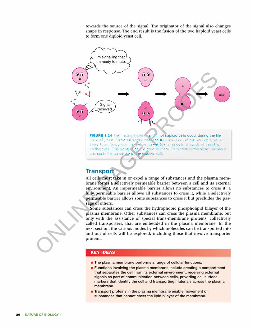
NATURE OF BIOLOGY 126
towards the source of the signal. � e originator of the signal also changes shape in response. � e end result is the fusion of the two haploid yeast cells to form one diploid yeast cell.
FIGURE 1.24 Two mating types (a and α ) of haploid cells occur during the life cycle of yeast. Chemical signals released by a yeast cell of one mating type can travel to surface protein receptors on neighbouring cells of yeasts of the other mating type. This signal is an invitation to mate. Reception of the signal causes a change in the behaviour of the receiver cell.
I’m signalling thatI’m ready to mate.
Signalreceived.
α
aa
α
a
a/α
α
TransportAll cells must take in or expel a range of substances and the plasma mem-brane forms a selectively permeable barrier between a cell and its external environment. An impermeable barrier allows no substances to cross it; a fully permeable barrier allows all substances to cross it, while a selectively permeable barrier allows some substances to cross it but precludes the pas-sage of others.
Some substances can cross the hydrophobic phospholipid bilayer of the plasma membrane. Other substances can cross the plasma membrane, but only with the assistance of special trans-membrane proteins, collectively called transporters, that are embedded in the plasma membrane. In the next section, the various modes by which molecules can be transported into and out of cells will be explored, including those that involve transporter proteins.
KEY IDEAS
■ The plasma membrane performs a range of cellular functions. ■ Functions involving the plasma membrane include creating a compartment that separates the cell from its external environment, receiving external signals as part of communication between cells, providing cell surface markers that identify the cell and transporting materials across the plasma membrane.
■ Transport proteins in the plasma membrane enable movement of substances that cannot cross the lipid bilayer of the membrane.
ONLINE environment. An impermeable barrier allows no substances to cross it; a
ONLINE environment. An impermeable barrier allows no substances to cross it; a
fully permeable barrier allows all substances to cross it, while a selectively
ONLINE fully permeable barrier allows all substances to cross it, while a selectively
permeable barrier allows some substances to cross it but precludes the pas-
ONLINE permeable barrier allows some substances to cross it but precludes the pas-
sage of others.
ONLINE
sage of others. Some substances can cross the hydrophobic phospholipid bilayer of the
ONLINE
Some substances can cross the hydrophobic phospholipid bilayer of the plasma membrane. Other substances can cross the plasma membrane, but
ONLINE
plasma membrane. Other substances can cross the plasma membrane, but
PAGE
PAGE
PAGE cycle of yeast. Chemical signals released by a yeast cell of one mating type can
PAGE cycle of yeast. Chemical signals released by a yeast cell of one mating type can travel to surface protein receptors on neighbouring cells of yeasts of the other
PAGE travel to surface protein receptors on neighbouring cells of yeasts of the other mating type. This signal is an invitation to mate. Reception of the signal causes a
PAGE mating type. This signal is an invitation to mate. Reception of the signal causes a change in the behaviour of the receiver cell.
PAGE change in the behaviour of the receiver cell.
PAGE Transport
PAGE TransportAll cells must take in or expel a range of substances and the plasma mem-PAGE All cells must take in or expel a range of substances and the plasma mem-brane forms a selectively permeable barrier between a cell and its external PAGE brane forms a selectively permeable barrier between a cell and its external environment. An impermeable barrier allows no substances to cross it; a PAGE
environment. An impermeable barrier allows no substances to cross it; a fully permeable barrier allows all substances to cross it, while a selectively PAGE
fully permeable barrier allows all substances to cross it, while a selectively
PROOFS
PROOFS
PROOFS
Two mating types (a and PROOFS
Two mating types (a and αPROOFS
α ) of haploid cells occur during the life PROOFS
) of haploid cells occur during the life α ) of haploid cells occur during the life αPROOFS
α ) of haploid cells occur during the life αcycle of yeast. Chemical signals released by a yeast cell of one mating type can PROOFS
cycle of yeast. Chemical signals released by a yeast cell of one mating type can travel to surface protein receptors on neighbouring cells of yeasts of the other PROOFS
travel to surface protein receptors on neighbouring cells of yeasts of the other PROOFS
PROOFS
PROOFS
PROOFSα
PROOFSα
PROOFS

27CHAPTER 1 Cells: basic units of life on Earth
QUICK CHECK
17 Is the plasma membrane impermeable, selectively permeable or fully permeable?
18 Identify two functions of the plasma membrane.19 What kind of proteins act as cell identity tags?20 What advantage might result from creating several membrane-enclosed
compartments within a cell? 21 What is the role of receptor proteins in the plasma membrane?22 Give an example of a problem that arises from the malfunction of a protein
transporter in the plasma membrane.
Crossing the plasma membrane Generally, substances entering or exiting a cell are in aqueous solution. Several factors determine whether or not dissolved substances can di�use down their concentration gradients across the phospholipid bilayer of the plasma membrane (see table 1.3).
TABLE 1.3 Factors affecting the ease with which substances can diffuse across the plasma membrane
molecular size Smaller molecules cross more easily than larger molecules; however, very large molecules (macromolecules), such as proteins and nucleic acids, cannot cross the plasma membrane.
presence of net charge (+ or −) Gases, such as CO2 and O2, and small uncharged molecules, such as urea and ethanol, can cross the plasma membrane. In contrast, mineral ions, such as Na+, K+, Cl− cannot cross because they are repelled by the hydrophobic lipid component of the plasma membrane.
solubility in lipid solvents Lipophilic molecules can cross easily, but hydrophilic molecules, such as glucose, cannot cross because they are repelled by the hydrophobic lipid component of the plasma membrane.
direction of concentration gradient
Movement down a gradient (from a region of higher to a region of lower concentration of a substance) does not require an input of energy and can occur by di�usion. Movement against a gradient cannot occur by di�usion.
From this table, it may be seen that the smaller the molecule and the more lipophilic (lipid-loving) it is, the more easily it can di�use across the plasma membrane down its concentration gradient. �is is summarised in �gure 1.25.
It is apparent that many substances are prevented from crossing the plasma membrane because they are repelled by the hydrophobic lipid component of the plasma membrane and/or because their concentration gradients are in the wrong direction. However, at any time, many substances are entering or leaving cells. �ese include substances that cannot cross the hydrophobic lipid bilayer of the plasma membrane, such as glucose, sodium ions and calcium ions. Yet movement of these substances across the plasma membrane is essen-tial for life. So, it may be concluded that simple di�usion down a concentra-tion gradient across the phospholipid bilayer cannot be the only means by
ONLINE
ONLINE solubility in lipid solvents Lipophilic molecules can cross easily, but
ONLINE solubility in lipid solvents Lipophilic molecules can cross easily, but PAGE
PAGE (macromolecules), such as proteins and nucleic
PAGE (macromolecules), such as proteins and nucleic acids, cannot cross the plasma membrane.
PAGE acids, cannot cross the plasma membrane.
presence of net charge (
PAGE presence of net charge (+
PAGE +
PAGE or
PAGE or −
PAGE −)
PAGE )
solubility in lipid solvents Lipophilic molecules can cross easily, but PAGE
solubility in lipid solvents Lipophilic molecules can cross easily, but
PROOFS
PROOFS
PROOFSoblem that arises from the malfunction of a protein
PROOFSoblem that arises from the malfunction of a protein
Crossing the plasma membrane
PROOFSCrossing the plasma membrane Generally, substances entering or exiting a cell are in aqueous solution.
PROOFSGenerally, substances entering or exiting a cell are in aqueous solution. Several factors determine whether or not dissolved substances can di�use
PROOFSSeveral factors determine whether or not dissolved substances can di�use down their concentration gradients across the phospholipid bilayer of the
PROOFSdown their concentration gradients across the phospholipid bilayer of the
fecting the ease with which substances can diffuse across
PROOFSfecting the ease with which substances can diffuse across
PROOFS
PROOFS
PROOFS
Smaller molecules cross more easily than larger PROOFS
Smaller molecules cross more easily than larger molecules; however, very large molecules PROOFS
molecules; however, very large molecules (macromolecules), such as proteins and nucleic PROOFS
(macromolecules), such as proteins and nucleic
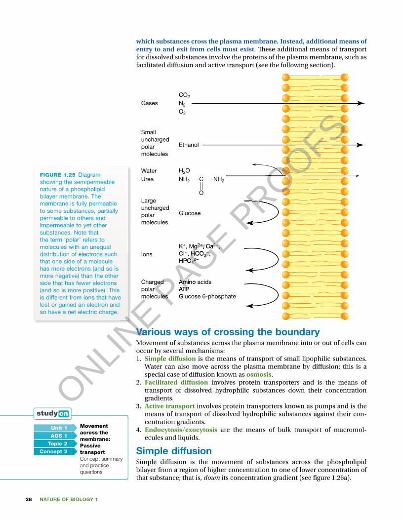
NATURE OF BIOLOGY 128
which substances cross the plasma membrane. Instead, additional means of entry to and exit from cells must exist. � ese additional means of transport for dissolved substances involve the proteins of the plasma membrane, such as facilitated di� usion and active transport (see the following section).
Gases
Smallunchargedpolarmolecules
Water
Largeunchargedpolarmolecules
Chargedpolarmolecules
Amino acidsATPGlucose 6-phosphate
Glucose
Ethanol
CO2
H2ONH2
K+, Mg2+, Ca2+,CI−, HCO3
−,HPO4
2−
NH2C
O
N2
O2
Ions
UreaFIGURE 1.25 Diagram showing the semipermeable nature of a phospholipid bilayer membrane. The membrane is fully permeable to some substances, partially permeable to others and impermeable to yet other substances. Note that the term ‘polar’ refers to molecules with an unequal distribution of electrons such that one side of a molecule has more electrons (and so is more negative) than the other side that has fewer electrons (and so is more positive). This is different from ions that have lost or gained an electron and so have a net electric charge.
Various ways of crossing the boundaryMovement of substances across the plasma membrane into or out of cells can occur by several mechanisms:1. Simple di� usion is the means of transport of small lipophilic substances.
Water can also move across the plasma membrane by di� usion; this is a special case of di� usion known as osmosis.
2. Facilitated di� usion involves protein transporters and is the means of transport of dissolved hydrophilic substances down their concentration gradients.
3. Active transport involves protein transporters known as pumps and is the means of transport of dissolved hydrophilic substances against their con-centration gradients.
4. Endocytosis/exocytosis are the means of bulk transport of macromol-ecules and liquids.
Simple diffusionSimple di� usion is the movement of substances across the phospholipid bilayer from a region of higher concentration to one of lower concentration of that substance; that is, down its concentration gradient (see � gure 1.26a).
Unit 1 Movement across the membrane: Passive transportConcept summary and practice questions
AOS 1
Topic 2
Concept 2
ONLINE molecules
ONLINE molecules
ONLINE
ONLINE
ONLINE
Various ways of crossing the boundary
ONLINE
Various ways of crossing the boundaryMovement of substances across the plasma membrane into or out of cells can
ONLINE
Movement of substances across the plasma membrane into or out of cells can
PAGE Charged
PAGE Chargedpolar PAGE polarmoleculesPAGE
molecules
Amino acids
PAGE Amino acidsATPPAGE ATPGlucose 6-phosphatePAGE
Glucose 6-phosphate
, Mg
PAGE , Mg2
PAGE 2+
PAGE +, Ca
PAGE , Ca2
PAGE 2+
PAGE +,
PAGE ,
, HCO
PAGE , HCO3
PAGE 3
−
PAGE −,
PAGE ,
HPO
PAGE HPO4
PAGE 42
PAGE 2−
PAGE −
PROOFS
PROOFS
PROOFS
PROOFS
PROOFS
PROOFS
PROOFS
PROOFS
PROOFS
PROOFS
PROOFS
PROOFS
PROOFS
PROOFS
PROOFS
PROOFS
PROOFS
PROOFS
PROOFS
PROOFS
PROOFS
PROOFS
PROOFS
PROOFS
PROOFS
PROOFS
PROOFS
PROOFS
PROOFS
PROOFS
PROOFS
PROOFS
PROOFS
PROOFS
PROOFS
PROOFS
PROOFS
PROOFS
PROOFS
PROOFS
PROOFS

29CHAPTER 1 Cells: basic units of life on Earth
Movement down a concentration gradient by simple di� usion does not require any input of energy. It is the gradient that drives the di� usion (like letting a ball roll down a slope). � e end point of simple di� usion is reached when equal concentrations of the substance are reached on both sides of the plasma membrane.
Substances that move easily across the plasma membrane by simple dif-fusion are small lipophilic molecules that can dissolve in the lipid bilayer. Among these substances are steroid hormones, alcohol and lipophilic drugs.
Figure 1.26b shows the stages in simple di� usion of a dissolved substance (X) across a plasma membrane. Its molecules are in constant random motion, some colliding with the membrane. If the concentration of substance X out-side the cell is greater than that inside the cell, more movement of X into the cell will occur compared with movement in the opposite direction. � is will produce a net movement of substance X into the cell. Net movement stops when collisions on both sides of the membrane equalise. � is occurs when the concentrations of X on both sides of the membrane are equal.
FIGURE 1.26 (a) Simple diffusion involves the movement of substances through the phospholipid bilayer of the plasma membrane. The direction of movement is down the concentration gradient of the diffusing substance (from high to low concentration). Does this process require an input of energy? (b) Stages of simple diffusion: (i) At the start, substance X starts to move into the cell because of random movement that results in some collisions with the membrane. (ii) Midway, molecules of substance X are moving both into and out of the cell, but the net movement is from outside to inside. (iii) When the concentration of X is equal on each side of the membrane, the number of collisions on either side of the membrane is equal and the net movement of molecules of substance X stops. Does this mean that collisions of molecules of substance X with the membrane stop?
Diffusion
(a)
Start Midway End(i)
(b)
(ii) (iii)
Outside Inside Outside Inside Outside Inside
Osmosis: a special case of diffusion Osmosis is a special case of di� usion that relates to the movement of solvents and, in biological systems, that solvent is water.
Osmosis can be de� ned as the net movement of water across a semipermeable membrane from a solution of lesser solute concentration to one of greater solute concentration. A condition for osmosis is that the membrane must be permeable to water but not to the solute molecules. Solutions that have a high concentration of dissolved solute have a lower concentration of water, and vice versa. � e net movement of water molecules in osmosis from a solution of high water con-centration to one of lower concentration is known as osmotic � ow.
ONLINE
ONLINE
ONLINE FIGURE 1.26
ONLINE FIGURE 1.26 the phospholipid bilayer of the plasma membrane. The direction of movement
ONLINE the phospholipid bilayer of the plasma membrane. The direction of movement is down the concentration gradient of the diffusing substance (from high to low
ONLINE is down the concentration gradient of the diffusing substance (from high to low concentration). Does this process require an input of energy?
ONLINE concentration). Does this process require an input of energy? diffusion: (i) At the start, substance X starts to move into the cell because of random
ONLINE diffusion: (i) At the start, substance X starts to move into the cell because of random
ONLINE P
AGE
PAGE
PAGE
PAGE
PAGE
PAGE
PAGE
PAGE
PAGE
PAGE
PAGE
PAGE
PAGE
PAGE
PAGE
PAGE
PAGE
PAGE
PAGE
PAGE
FIGURE 1.26 PAGE
FIGURE 1.26 PAGE
PAGE (ii)
PAGE (ii)
Outside Inside Outside Inside Outside InsidePAGE Outside Inside Outside Inside Outside Inside
PROOFS(X) across a plasma membrane. Its molecules are in constant random motion,
PROOFS(X) across a plasma membrane. Its molecules are in constant random motion, some colliding with the membrane. If the concentration of substance X out-
PROOFSsome colliding with the membrane. If the concentration of substance X out-side the cell is greater than that inside the cell, more movement of X into the
PROOFSside the cell is greater than that inside the cell, more movement of X into the cell will occur compared with movement in the opposite direction. � is will
PROOFScell will occur compared with movement in the opposite direction. � is will produce a net movement of substance X into the cell. Net movement stops
PROOFSproduce a net movement of substance X into the cell. Net movement stops when collisions on both sides of the membrane equalise. � is occurs when the
PROOFSwhen collisions on both sides of the membrane equalise. � is occurs when the concentrations of X on both sides of the membrane are equal.
PROOFSconcentrations of X on both sides of the membrane are equal.
PROOFS
PROOFS
PROOFS
PROOFS
PROOFS
PROOFS
PROOFS
PROOFS
PROOFS
PROOFS
PROOFS
PROOFS
PROOFS
PROOFS
PROOFS
PROOFS
PROOFS
PROOFS
PROOFS
PROOFS
PROOFS
PROOFS
PROOFS
PROOFS
PROOFS
PROOFS
PROOFS
PROOFS
PROOFS
PROOFS
PROOFS
PROOFS
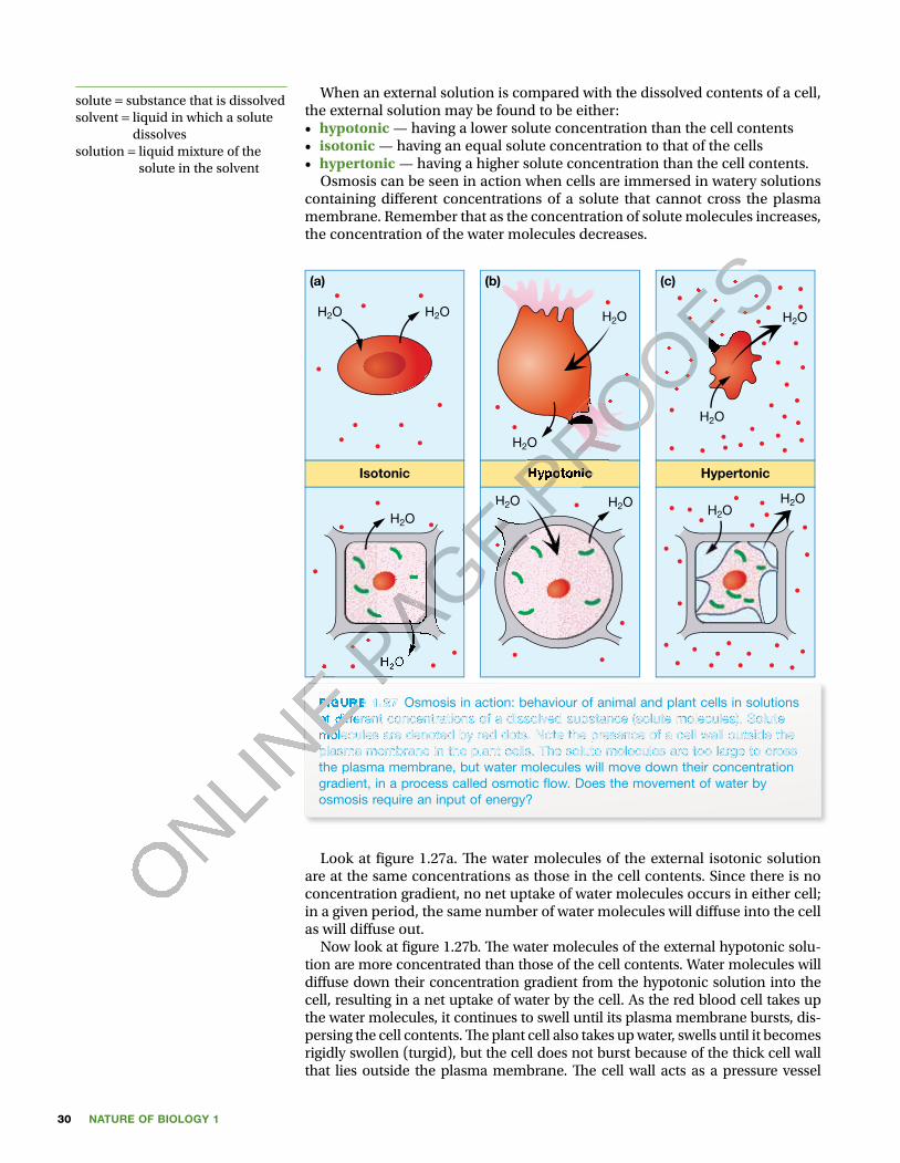
NATURE OF BIOLOGY 130
When an external solution is compared with the dissolved contents of a cell, the external solution may be found to be either:• hypotonic — having a lower solute concentration than the cell contents• isotonic — having an equal solute concentration to that of the cells• hypertonic — having a higher solute concentration than the cell contents.
Osmosis can be seen in action when cells are immersed in watery solutions containing di� erent concentrations of a solute that cannot cross the plasma membrane. Remember that as the concentration of solute molecules increases, the concentration of the water molecules decreases.
Isotonic Hypotonic Hypertonic
H2O H2O H2O H2O
H2O
H2O
H2O
H2O
H2OH2O H2OH2O
(a) (b) (c)
FIGURE 1.27 Osmosis in action: behaviour of animal and plant cells in solutions of different concentrations of a dissolved substance (solute molecules). Solute molecules are denoted by red dots. Note the presence of a cell wall outside the plasma membrane in the plant cells. The solute molecules are too large to cross the plasma membrane, but water molecules will move down their concentration gradient, in a process called osmotic � ow. Does the movement of water by osmosis require an input of energy?
Look at � gure 1.27a. � e water molecules of the external isotonic solution are at the same concentrations as those in the cell contents. Since there is no concentration gradient, no net uptake of water molecules occurs in either cell; in a given period, the same number of water molecules will di� use into the cell as will di� use out.
Now look at � gure 1.27b. � e water molecules of the external hypotonic solu-tion are more concentrated than those of the cell contents. Water molecules will di� use down their concentration gradient from the hypotonic solution into the cell, resulting in a net uptake of water by the cell. As the red blood cell takes up the water molecules, it continues to swell until its plasma membrane bursts, dis-persing the cell contents. � e plant cell also takes up water, swells until it becomes rigidly swollen (turgid), but the cell does not burst because of the thick cell wall that lies outside the plasma membrane. � e cell wall acts as a pressure vessel
solute = substance that is dissolvedsolvent = liquid in which a solute
dissolvessolution = liquid mixture of the
solute in the solvent
ONLINE
ONLINE
ONLINE
ONLINE
ONLINE
ONLINE FIGURE 1.27
ONLINE FIGURE 1.27
of different concentrations of a dissolved substance (solute molecules). Solute
ONLINE
of different concentrations of a dissolved substance (solute molecules). Solute molecules are denoted by red dots. Note the presence of a cell wall outside the
ONLINE
molecules are denoted by red dots. Note the presence of a cell wall outside the plasma membrane in the plant cells. The solute molecules are too large to cross
ONLINE
plasma membrane in the plant cells. The solute molecules are too large to cross
ONLINE P
AGE
PAGE
PAGE
PAGE
PAGE
PAGE
PAGE
PAGE
PAGE
PAGE
PAGE
PAGE
PAGE
PAGE
PAGE
PAGE
PAGE
PAGE
PAGE
PAGE
PAGE
PAGE
PAGE
PAGE
PAGE
PAGE
PAGE
PAGE
PAGE
PAGE
PAGE
PAGE
PAGE
PAGE
PAGE
PAGE
PAGE
PAGE
PAGE
PAGE
PAGE
PAGE
PAGE
PAGE
PAGE
PAGE
PAGE
PAGE
PAGE
PAGE
PAGE
PAGE
PAGE
PAGE
PAGE
PAGE
PAGE
PAGE
PAGE
PAGE
PAGE
PAGE
PAGE
PAGE
PAGE
PAGE
PAGE
PAGE
PAGE
PAGE
PAGE
PAGE
PAGE
PAGE
PAGE
PAGE
PAGE
PAGE
PAGE
PAGE
PAGE
PAGE
PAGE
PAGE
PAGE
PAGE
PAGE
PAGE
PAGE
PAGE
PAGE
PAGE
PAGE
PAGE
PAGE
PAGE
PAGE
PAGE
PAGE
PAGE
PAGE
PAGE
PAGE
PAGE
PAGE
PAGE
PAGE
PAGE
PAGE
PAGE
PAGE
PAGE
PAGE
PAGE
PAGE
PAGE
PAGE
PAGE
PAGE
PAGE
PAGE
PAGE
PAGE
PAGE
PAGE
PAGE
PAGE
PAGE
PAGE
PAGE
PAGE
PAGE
PAGE
PAGE
PAGE
PAGE
PAGE
PAGE
PAGE
PAGE
PAGE
PAGE
PAGE
PAGE
PAGE
PAGE
PAGE
PAGE
PAGE
PAGE
PAGE
PAGE
PAGE
PAGE
PAGE
PAGE
PAGE
PAGE
PAGE
PAGE
PAGE
PAGE
PAGE
PAGE
PAGE
PAGE
PAGE
PAGE
PAGE
PAGE
PAGE
PAGE
PAGE
PAGE
PAGE
PAGE
PAGE
PAGE
PAGE
PAGE
PAGE
PAGE
PAGE
PAGE
PAGE
PAGE
PAGE
PAGE
PAGE
PAGE
PAGE
PAGE
PAGE
PAGE
PAGE
PAGE
PAGE
PAGE
PAGE
PAGE
PAGE
PAGE
HPAGE
H2PAGE
2OPAGE
O
H
PAGE H
PAGE
PAGE
PAGE
PAGE
PAGE
PAGE
PAGE
PAGE
PAGE
PAGE
PAGE
PAGE
PAGE
PAGE
PAGE
PAGE
PAGE
PAGE
PAGE PROOFS
PROOFS
PROOFS
HypotonicPROOFS
HypotonicPROOFS
PROOFS
PROOFS
PROOFS
PROOFS
PROOFS
PROOFS
PROOFS
PROOFS
PROOFS
PROOFS
PROOFS
PROOFS
PROOFS
PROOFS
PROOFS
PROOFS
PROOFS
PROOFS
PROOFS
PROOFS
PROOFS
PROOFS
PROOFS
PROOFS
PROOFS
PROOFS
PROOFS
PROOFS
PROOFS
PROOFS
PROOFS
PROOFS
PROOFS
PROOFS
PROOFS
PROOFS
PROOFS
PROOFS
PROOFS
PROOFS
PROOFS
PROOFS
PROOFS
PROOFS
PROOFS
PROOFS
PROOFS
PROOFS
PROOFS
PROOFS
PROOFS
PROOFS
PROOFS
PROOFS
PROOFS
PROOFS
PROOFS
PROOFS
PROOFS
PROOFS
PROOFS
PROOFS
PROOFS
PROOFS
PROOFS
PROOFS
PROOFS
PROOFS
PROOFS
PROOFS
PROOFS
PROOFS
PROOFS
PROOFS
PROOFS
PROOFS
PROOFS
PROOFS
PROOFS
PROOFS
PROOFS
PROOFS
PROOFS
PROOFS
PROOFS
PROOFS
PROOFS
PROOFS
PROOFS
PROOFS
PROOFS
PROOFS
PROOFS
PROOFS
PROOFS

31CHAPTER 1 Cells: basic units of life on Earth
preventing the plasma membrane from swelling to a point of bursting. Net entry of water molecules into the plant cell � nally stops as a result of the increasing out-ward pressure of the cell contents that opposes the net inward � ow of water.
Finally look at � gure 1.27c. � e water molecules of the external hypertonic solution are at a lower concentration than those in the cell contents. Water molecules will di� use down their concentration gradient from the cells into the external solution, resulting in a net loss of water from the cells. � e red blood cell shrinks, becoming crenated. � e plant cell within its plasma mem-brane shrinks away from its cell wall.
Looking at water movement Water is a major constituent of all living organisms and cells are about 50 to 70 per cent water. � e adult human body is about 60 per cent water overall, with the water content of di� erent tissues varying: brain is about 73 per cent water, lungs more than 80 per cent and bones about 30 per cent water.
Examples of the movement of water by osmosis follow.The freshwater protozoansProtozoans are single-celled organisms with an outer plasma membrane boundary. One example is Paramecium caudata. Paramecium caudata lives in fresh water, a habitat that is hypotonic relative to the cell contents (cytosol) of this organism. Consequently, water constantly � ows by osmosis from the high concentration outside the cell to the low concentration inside the cell. Look at � gure 1.28 and note the ducts surrounding a contractile vacuole. � ese ducts collect water from the cytosol and move it into the contractile vacuole. From there, it is forced to the outside when the vacuole contracts in an energy-requiring process. Would you expect this to be a continuous process? Why?
Food vacuole
Cilia
NucleusContractile
vacuole
FIGURE 1.28 Drawing of Paramecium caudata, a single-celled organism that lives in fresh water. A constant � ow of water by osmosis occurs from the external fresh water into the cell. Ducts surrounding the contractile vacuole carry the water to the vacuole which then forces the water out of the cell. Can you predict what would happen if the contractile vacuole stopped operating?
The salted meatPreserving means either slowing down the multiplication of microbes that cause food spoilage (e.g. by chilling or freezing), or inhibiting or killing the microbes that are responsible for food spoilage (e.g. by salting).
Salt (sodium chloride) is a highly e� ective food preservative, commonly used to preserve meat, particularly before freezing was possible. In the past for long sea voyages, meat was preserved by building up layers of meat and salt in wooden barrels (see � gure 1.29). Another technique was to immerse meat in a solution of up to 20 per cent salt. For the long voyage of Christopher Columbus
ODD FACT
Seawater is hypertonic to the cells of the human body. Instead of quenching thirst, drinking seawater results in a person becoming dehydrated.
ONLINE
ONLINE
ONLINE
ONLINE
ONLINE
ONLINE
ONLINE
ONLINE
ONLINE
ONLINE
ONLINE
ONLINE
ONLINE
ONLINE
ONLINE
ONLINE
ONLINE
ONLINE
ONLINE
ONLINE
ONLINE
ONLINE
ONLINE
ONLINE
ONLINE
ONLINE
ONLINE
ONLINE
ONLINE
ONLINE
ONLINE
ONLINE
ONLINE
ONLINE
ONLINE
ONLINE
ONLINE
ONLINE
ONLINE
ONLINE
ONLINE
ONLINE
ONLINE
ONLINE
ONLINE
ONLINE
ONLINE
ONLINE
ONLINE
ONLINE
ONLINE
ONLINE
ONLINE
ONLINE
ONLINE
ONLINE
ONLINE
ONLINE
ONLINE
ONLINE
ONLINE
ONLINE
ONLINE
ONLINE
ONLINE
ONLINE
ONLINE
ONLINE
ONLINE
ONLINE
ONLINE
ONLINE
ONLINE
ONLINE
ONLINE
ONLINE
ONLINE
ONLINE
ONLINE
ONLINE
ONLINE
ONLINE
ONLINE
ONLINE
ONLINE
ONLINE
ONLINE
ONLINE
ONLINE
ONLINE
ONLINE
ONLINE
ONLINE
ONLINE
ONLINE
ONLINE
ONLINE
ONLINE
ONLINE P
AGE ducts collect water from the cytosol and move it into the contractile vacuole.
PAGE ducts collect water from the cytosol and move it into the contractile vacuole. From there, it is forced to the outside when the vacuole contracts in an energy-
PAGE From there, it is forced to the outside when the vacuole contracts in an energy-requiring process. Would you expect this to be a continuous process? Why?
PAGE requiring process. Would you expect this to be a continuous process? Why?
PAGE
PAGE
PAGE
PAGE
PAGE
PAGE
PAGE
PAGE
PAGE
PAGE
PAGE
PAGE
PAGE
PAGE
PAGE
PAGE
PAGE
PAGE
PAGE
PAGE
PAGE
PAGE
PAGE
PAGE
PAGE
PAGE
PAGE
PAGE
PAGE
PAGE
PAGE
PAGE
PAGE
PAGE
PAGE
PAGE
PAGE
PAGE
PAGE
PAGE
PAGE
PAGE
PAGE
PAGE
PAGE
PAGE
PAGE
PAGE
PAGE
PAGE
PAGE
PAGE
PAGE
PAGE
PAGE
PAGE
PAGE
PAGE
PAGE
PAGE
PAGE
PAGE
PAGE
PAGE
PAGE
PAGE
PAGE
PAGE
PAGE
PAGE
PAGE
PAGE
PAGE
PAGE
PAGE
PAGE
PAGE
PAGE
PAGE
PAGE
PAGE
PAGE
PAGE
PAGE
PAGE
PAGE
PAGE
PAGE
PAGE
PAGE
PAGE
PAGE
PAGE
PAGE
PAGE
PAGE
PAGE
PAGE
PAGE
PAGE
PAGE
PAGE
PAGE
PAGE
PAGE
PAGE
PAGE
PAGE
PAGE
PAGE
PAGE
PAGE
PAGE
PAGE
PAGE
PAGE
PAGE
PAGE
PAGE
PAGE
PAGE
PAGE
PAGE
PAGE
PAGE
PAGE
PAGE
PAGE
PAGE
PAGE
PAGE
PAGE
PAGE
PAGE
PAGE
PAGE
PAGE
PAGE
PAGE
PAGE
PAGE
PAGE
PAGE
PAGE
PAGE
PAGE
PAGE
PAGE
PAGE
PAGE
PAGE
PAGE
PAGE
PAGE
PAGE
PAGE
PAGE
PAGE
PAGE
PAGE
PAGE
PAGE
PAGE
PAGE
PAGE
PAGE
PAGE
PAGE
PAGE
PAGE
PAGE
PAGE
PAGE
PAGE
PAGE
PAGE
PAGE
PAGE
PAGE
PAGE
PAGE
PAGE
PAGE
PAGE
PAGE
PAGE
PAGE
PAGE
PAGE
PAGE
PAGE
PAGE
PAGE
PAGE
PAGE
PAGE
PAGE
PAGE
PAGE
PAGE
PAGE
PAGE
PAGE
PAGE
PAGE
PAGE
PAGE
PAGE
PAGE
PAGE
PAGE
PAGE
PAGE
PAGE
PAGE
PAGE
PAGE
PAGE
PAGE
PAGE
PAGE
PAGE
PAGE
PAGE
PAGE
PAGE
PAGE
PAGE
PAGE
PAGE
PAGE
PAGE
PAGE
PAGE
PAGE
PAGE
PAGE
PAGE
PAGE
PAGE
PAGE
PAGE
PAGE
PAGE
PAGE
PAGE
PAGE
PAGE
PAGE
PAGE
PAGE
PAGE
PAGE
PAGE
PAGE
PAGE
PAGE
PAGE
PAGE
PAGE
PAGE
PAGE
PAGE
PAGE
PAGE
PAGE
PAGE
PAGE
PAGE
PAGE
PAGE
PAGE
PAGE
PAGE
PAGE
PAGE
PAGE
PAGE
PAGE
PAGE
PAGE
PAGE
PAGE
PAGE
PAGE
PAGE
PAGE
PAGE
PAGE
PAGE
PAGE
PAGE
PAGE
PAGE
PAGE
PAGE
PAGE
PAGE
PAGE
PAGE
PAGE
PAGE
PAGE
PAGE
PAGE
PAGE
PAGE
PAGE
PAGE
PAGE
PAGE
PAGE
PAGE
PAGE
PAGE
PAGE
PAGE
PAGE
PAGE
PAGE
PAGE
PAGE
PAGE
PAGE
PAGE
PAGE
PAGE
PAGE
PAGE
PAGE
PAGE
PAGE
PAGE
PAGE
PAGE
PAGE
PAGE
PAGE
PAGE
PAGE
PAGE
PAGE
PAGE
PAGE
PAGE
PAGE
PAGE
PAGE
PAGE
PAGE
PAGE
PAGE
PAGE
PAGE
PAGE
PAGE
PAGE
PAGE
PAGE
PAGE
PAGE
PAGE
PAGE
PAGE
PAGE
PAGE
PAGE
PAGE
PAGE
PAGE
PAGE
PAGE
PAGE
PAGE
PAGE
PAGE
PAGE
PAGE
PAGE
PAGE
PAGE
PAGE
PAGE
PAGE
PAGE
PAGE PROOFSWater is a major constituent of all living organisms and cells are about 50 to
PROOFSWater is a major constituent of all living organisms and cells are about 50 to 70 per cent water. � e adult human body is about 60 per cent water overall,
PROOFS70 per cent water. � e adult human body is about 60 per cent water overall, with the water content of di� erent tissues varying: brain is about 73 per cent
PROOFSwith the water content of di� erent tissues varying: brain is about 73 per cent water, lungs more than 80 per cent and bones about 30 per cent water.
PROOFSwater, lungs more than 80 per cent and bones about 30 per cent water.
Examples of the movement of water by osmosis follow.
PROOFSExamples of the movement of water by osmosis follow.
Protozoans are single-celled organisms with an outer plasma membrane
PROOFSProtozoans are single-celled organisms with an outer plasma membrane
Paramecium caudata. Paramecium caudata
PROOFSParamecium caudata. Paramecium caudata
in fresh water, a habitat that is hypotonic relative to the cell contents (cytosol)
PROOFSin fresh water, a habitat that is hypotonic relative to the cell contents (cytosol) of this organism. Consequently, water constantly � ows by osmosis from the
PROOFSof this organism. Consequently, water constantly � ows by osmosis from the high concentration outside the cell to the low concentration inside the cell.
PROOFS
high concentration outside the cell to the low concentration inside the cell. Look at � gure 1.28 and note the ducts surrounding a contractile vacuole. � ese PROOFS
Look at � gure 1.28 and note the ducts surrounding a contractile vacuole. � ese ducts collect water from the cytosol and move it into the contractile vacuole. PROOFS
ducts collect water from the cytosol and move it into the contractile vacuole. From there, it is forced to the outside when the vacuole contracts in an energy-PROOFS
From there, it is forced to the outside when the vacuole contracts in an energy-

NATURE OF BIOLOGY 132
in 1492 from Spain to the Americas, the food carried on board his ships was olive oil, wine, sea biscuits and salted meat.
Why does salt work as a preservative? Salt draws water out of microbial cells on the sur-face of the meat by osmosis. Dissolved salt on the meat surface is hypertonic to the micro-bial cell contents so that the water � ows down its concentration gradient out of these cells. � is osmotic � ow of water dehydrates the cells of the microbes that cause food spoilage, thus preserving the meat. Until refrigeration became available in the 1880s, salting con-tinued to be used for the preservation of meat on long sea voyages.The crying albatrossSeawater is hypertonic to the extracellular � uid of marine birds, such as the wandering albatross (Diomedea exulans). � ese birds drink only seawater to supply their water requirements, but in so doing, they take in large amounts of salt, a situation that could lead to fatal dehydration in a person. In these birds, salt from the intake of seawater moves from the gut into the bloodstream and the
extracellular � uid (ECF) that bathes the body cells. � e higher concentration of salt in the ECF relative to that in the body cells leads to the movement of water molecules from the body cells into the ECF by osmosis. � e loss of water from the cells stimulates salt-secreting glands in the bird’s head to remove the excess salt (see � gure 1.30) and keep the internal environment of the cells within narrow limits. � e cells in these glands take up salt from the bird’s body � uids and produce a secretion with a very high concentration of salt — more salty than the bird’s body � uid and even saltier than seawater. � is � uid passes down ducts that lead to the bird’s nostrils from where it is discharged.
Salt-secretinggland
Ducts
Nostril withsalt secretions
FIGURE 1.30 Diagram showing the position of the two salt-secreting glands in the head of an albatross. The highly salty excretion from the glands is removed by dripping from the bird’s nostrils.
FIGURE 1.29 Barrels of salted pork. Meat preserved with salt was carried on board sailing ships to provide rations during long voyages at sea. How does salt preserve meat?
ODD FACT
The � rst successful refrigerated shipment of meat left Dunedin in New Zealand in February 1882 en route to England.
ONLINE salty than the bird’s body � uid and even saltier than seawater. � is � uid passes
ONLINE salty than the bird’s body � uid and even saltier than seawater. � is � uid passes
down ducts that lead to the bird’s nostrils from where it is discharged.
ONLINE down ducts that lead to the bird’s nostrils from where it is discharged.PAGE
PAGE from the gut into the bloodstream and the
PAGE from the gut into the bloodstream and the extracellular � uid (ECF) that bathes the body cells. � e higher concentration
PAGE extracellular � uid (ECF) that bathes the body cells. � e higher concentration of salt in the ECF relative to that in the body cells leads to the movement of
PAGE of salt in the ECF relative to that in the body cells leads to the movement of water molecules from the body cells into the ECF by osmosis. � e loss of water
PAGE water molecules from the body cells into the ECF by osmosis. � e loss of water from the cells stimulates salt-secreting glands in the bird’s head to remove
PAGE from the cells stimulates salt-secreting glands in the bird’s head to remove the excess salt (see � gure 1.30) and keep the internal environment of the cells
PAGE the excess salt (see � gure 1.30) and keep the internal environment of the cells within narrow limits. � e cells in these glands take up salt from the bird’s body
PAGE within narrow limits. � e cells in these glands take up salt from the bird’s body � uids and produce a secretion with a very high concentration of salt — more PAGE � uids and produce a secretion with a very high concentration of salt — more PAGE salty than the bird’s body � uid and even saltier than seawater. � is � uid passes PAGE salty than the bird’s body � uid and even saltier than seawater. � is � uid passes down ducts that lead to the bird’s nostrils from where it is discharged.PAGE
down ducts that lead to the bird’s nostrils from where it is discharged.PAGE
PAGE PROOFS
� is osmotic � ow of water dehydrates the
PROOFS� is osmotic � ow of water dehydrates the cells of the microbes that cause food spoilage,
PROOFScells of the microbes that cause food spoilage, thus preserving the meat. Until refrigeration
PROOFSthus preserving the meat. Until refrigeration became available in the 1880s, salting con-
PROOFSbecame available in the 1880s, salting con-tinued to be used for the preservation of meat
PROOFStinued to be used for the preservation of meat on long sea voyages.
PROOFSon long sea voyages.The crying albatross
PROOFSThe crying albatrossSeawater is hypertonic to the extracellular
PROOFSSeawater is hypertonic to the extracellular � uid of marine birds, such as the wandering
PROOFS� uid of marine birds, such as the wandering albatross (
PROOFSalbatross (Diomedea exulans
PROOFSDiomedea exulans
drink only seawater to supply their water
PROOFSdrink only seawater to supply their water requirements, but in so doing, they take in
PROOFSrequirements, but in so doing, they take in large amounts of salt, a situation that could PROOFS
large amounts of salt, a situation that could lead to fatal dehydration in a person. In these PROOFS
lead to fatal dehydration in a person. In these birds, salt from the intake of seawater moves PROOFS
birds, salt from the intake of seawater moves from the gut into the bloodstream and the PROOFS
from the gut into the bloodstream and the

33CHAPTER 1 Cells: basic units of life on Earth
The dehydrated personA person su�ering from a prolonged bout of diarrhoea is severely dehydrated and may need to be admitted to hospital. Normally, the cells lining the small and large intestine absorb the large volume of �uid and dissolved salts that enter the gut daily (see Odd fact). Several bacterial infections, including Staphylococcus sp., can inhibit this absorption. As a result, most of the �uid and the dissolved salts pass into the large intestine and are expelled from the body in large volumes of watery diarrhoea, resulting in dehydration and salt loss. Worsening dehydration is serious because it produces a decrease in blood volume that, if untreated, may lead to cardiovascular failure.
Initial treatment of severe dehydration is replacement of the lost �uid and salts. �e rehydration therapy involves an isotonic solution of saline (salt) solu-tion plus glucose. �is solution can be administered either orally (by mouth) or by direct infusion into the bloodstream (by intravenous infusion, or IV).
Why does this treatment act as a rehydration therapy? Glucose stimulates the absorption of sodium by the gut cells. �e uptake of sodium and glucose from the gut into the extracellular �uid creates a hypertonic internal environ-ment that causes water to move by osmosis from the gut across the cells lining the gut, and so returns water to the extracellular �uid and from there to the body cells.
Facilitated diffusionFacilitated di�usion is an example of protein-mediated transport. Facilitated di�usion is so named because the di�usion across the membrane is enabled or facilitated by special protein transporters in the plasma membrane.
Like simple di�usion, facilitated di�usion does not require an input of energy. Like simple di�usion, facilitated di�usion moves substances down their concentration gradients. However, facilitated di�usion of dissolved sub-stances requires the action of protein transporters that are embedded in the cell membrane.
Facilitated di�usion enables molecules that cannot di�use across the phospholipid bilayer to move across the plasma membrane through the agency of transporter proteins. �ese transporters are either channel proteins or carrier proteins. Are transporters required in simple di�usion? (Refer to �gure 1.31.)
Channel proteinsOne group of transport proteins involved in facilitated di�usion are the channel proteins. Each channel protein is trans-membrane and has a central water-�lled pore through which dissolved substances can pass down their con-centration gradient. Di�erent channel proteins are speci�c for the di�usion of charged particles and polar molecules.
Each channel protein consists of a narrow water-�lled pore in the plasma membrane through which substances can move down their concentration gradients (see �gure 1.31b). By providing water-�lled pores, channel proteins create a hydrophilic passage across the plasma membrane that bypasses the phospholipid bilayer and facilitates the di�usion of charged particles, such as sodium and potassium ions, and small polar molecules.
Carrier proteinsAnother group of membrane proteins involved in facilitated di�usion are car-rier proteins. Carrier proteins are speci�c, with each kind of carrier enabling the di�usion of one kind of molecule across the plasma membrane. After binding to its speci�c cargo molecule, the carrier protein undergoes a change in shape as it delivers its cargo to the other side of the plasma mem-brane (see �gure 1.31c).
ODD FACT
Each day about 2 litres of �uid are taken into the gut in food and drinks. This is increased by a further volume of about 7 litres from secretions, including those of the salivary glands, stomach, liver and pancreas.
In the case of ions, which carry either + or − charge(s), it is more correct to say that, rather than moving down their concentration gradients, they move down their electrochemical gradients.
ODD FACT
Channel proteins known as aquaporins are trans-membrane proteins that are speci�c for the facilitated diffusion of water molecules.
ONLINE agency of transporter proteins.
ONLINE agency of transporter proteins.
or
ONLINE or carrier proteins
ONLINE carrier proteins
�gure 1.31.)
ONLINE �gure 1.31.)
Channel proteins
ONLINE
Channel proteinsOne group of transport proteins involved in facilitated di�usion are the
ONLINE
One group of transport proteins involved in facilitated di�usion are the
PAGE Facilitated di�usion is an example of protein-mediated transport. Facilitated
PAGE Facilitated di�usion is an example of protein-mediated transport. Facilitated di�usion is so named because the
PAGE di�usion is so named because the by special protein transporters in the plasma membrane.
PAGE by special protein transporters in the plasma membrane. Like simple di�usion, facilitated di�usion does
PAGE Like simple di�usion, facilitated di�usion does
energy. Like simple di�usion, facilitated di�usion moves substances down
PAGE energy. Like simple di�usion, facilitated di�usion moves substances down
PAGE their concentration gradients.
PAGE their concentration gradients. stances requires the action of protein transporters that are embedded in the
PAGE stances requires the action of protein transporters that are embedded in the cell membrane.
PAGE cell membrane.
Facilitated di�usion enables molecules that cannot di�use across the
PAGE Facilitated di�usion enables molecules that cannot di�use across the
phospholipid bilayer to move across the plasma membrane through the PAGE phospholipid bilayer to move across the plasma membrane through the agency of transporter proteins. PAGE
agency of transporter proteins. carrier proteinsPAGE
carrier proteins
PROOFSInitial treatment of severe dehydration is replacement of the lost �uid and
PROOFSInitial treatment of severe dehydration is replacement of the lost �uid and salts. �e rehydration therapy involves an isotonic solution of saline (salt) solu
PROOFSsalts. �e rehydration therapy involves an isotonic solution of saline (salt) solution plus glucose. �is solution can be administered either orally (by mouth)
PROOFStion plus glucose. �is solution can be administered either orally (by mouth) or by direct infusion into the bloodstream (by intravenous infusion, or IV).
PROOFSor by direct infusion into the bloodstream (by intravenous infusion, or IV). Why does this treatment act as a rehydration therapy? Glucose stimulates
PROOFSWhy does this treatment act as a rehydration therapy? Glucose stimulates
the absorption of sodium by the gut cells. �e uptake of sodium and glucose
PROOFSthe absorption of sodium by the gut cells. �e uptake of sodium and glucose from the gut into the extracellular �uid creates a hypertonic internal environ
PROOFSfrom the gut into the extracellular �uid creates a hypertonic internal environment that causes water to move by osmosis from the gut across the cells lining
PROOFSment that causes water to move by osmosis from the gut across the cells lining the gut, and so returns water to the extracellular �uid and from there to the
PROOFSthe gut, and so returns water to the extracellular �uid and from there to the
Facilitated di�usion is an example of protein-mediated transport. Facilitated PROOFS
Facilitated di�usion is an example of protein-mediated transport. Facilitated di�usionPROOFS
di�usion
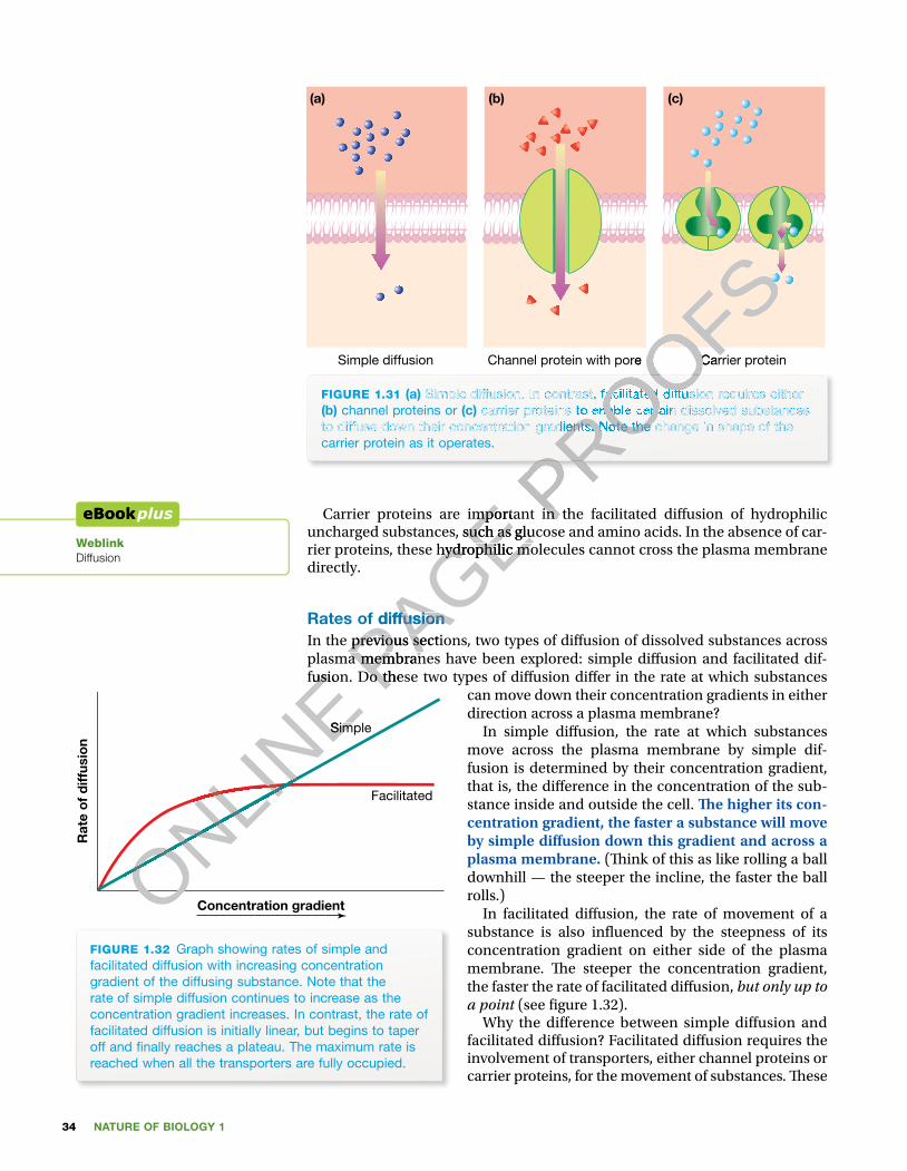
NATURE OF BIOLOGY 134
Simple diffusion Carrier proteinChannel protein with pore
(a) (b) (c)
FIGURE 1.31 (a) Simple diffusion. In contrast, facilitated diffusion requires either (b) channel proteins or (c) carrier proteins to enable certain dissolved substances to diffuse down their concentration gradients. Note the change in shape of the carrier protein as it operates.
Carrier proteins are important in the facilitated di� usion of hydrophilic uncharged substances, such as glucose and amino acids. In the absence of car-rier proteins, these hydrophilic molecules cannot cross the plasma membrane directly.
Rates of diffusionIn the previous sections, two types of di� usion of dissolved substances across plasma membranes have been explored: simple di� usion and facilitated dif-fusion. Do these two types of di� usion di� er in the rate at which substances
can move down their concentration gradients in either direction across a plasma membrane?
In simple di� usion, the rate at which substances move across the plasma membrane by simple dif-fusion is determined by their concentration gradient, that is, the di� erence in the concentration of the sub-stance inside and outside the cell. � e higher its con-centration gradient, the faster a substance will move by simple di� usion down this gradient and across a plasma membrane. (� ink of this as like rolling a ball downhill — the steeper the incline, the faster the ball rolls.)
In facilitated di� usion, the rate of movement of a substance is also in� uenced by the steepness of its concentration gradient on either side of the plasma membrane. � e steeper the concentration gradient, the faster the rate of facilitated di� usion, but only up to a point (see � gure 1.32).
Why the difference between simple diffusion and facilitated di� usion? Facilitated di� usion requires the involvement of transporters, either channel proteins or carrier proteins, for the movement of substances. � ese
WeblinkDiffusion
Concentration gradient
Rat
e o
f d
iffus
ion
Simple
Facilitated
FIGURE 1.32 Graph showing rates of simple and facilitated diffusion with increasing concentration gradient of the diffusing substance. Note that the rate of simple diffusion continues to increase as the concentration gradient increases. In contrast, the rate of facilitated diffusion is initially linear, but begins to taper off and � nally reaches a plateau. The maximum rate is reached when all the transporters are fully occupied.
ONLINE fusion. Do these two types of di� usion di� er in the rate at which substances
ONLINE fusion. Do these two types of di� usion di� er in the rate at which substances
ONLINE
ONLINE
ONLINE Simple
ONLINE Simple
PAGE Carrier proteins are important in the facilitated di� usion of hydrophilic
PAGE Carrier proteins are important in the facilitated di� usion of hydrophilic uncharged substances, such as glucose and amino acids. In the absence of car-
PAGE uncharged substances, such as glucose and amino acids. In the absence of car-rier proteins, these hydrophilic molecules cannot cross the plasma membrane
PAGE rier proteins, these hydrophilic molecules cannot cross the plasma membrane
Rates of diffusion
PAGE Rates of diffusionIn the previous sections, two types of di� usion of dissolved substances across PAGE In the previous sections, two types of di� usion of dissolved substances across plasma membranes have been explored: simple di� usion and facilitated dif-PAGE plasma membranes have been explored: simple di� usion and facilitated dif-fusion. Do these two types of di� usion di� er in the rate at which substances PAGE
fusion. Do these two types of di� usion di� er in the rate at which substances
PROOFS
PROOFSCarrier protein
PROOFSCarrier proteinChannel protein with pore
PROOFSChannel protein with pore
PROOFS
PROOFS
PROOFS Simple diffusion. In contrast, facilitated diffusion requires either
PROOFS Simple diffusion. In contrast, facilitated diffusion requires either
carrier proteins to enable certain dissolved substances
PROOFS carrier proteins to enable certain dissolved substances
to diffuse down their concentration gradients. Note the change in shape of the
PROOFSto diffuse down their concentration gradients. Note the change in shape of the
PROOFS
Carrier proteins are important in the facilitated di� usion of hydrophilic PROOFS
Carrier proteins are important in the facilitated di� usion of hydrophilic
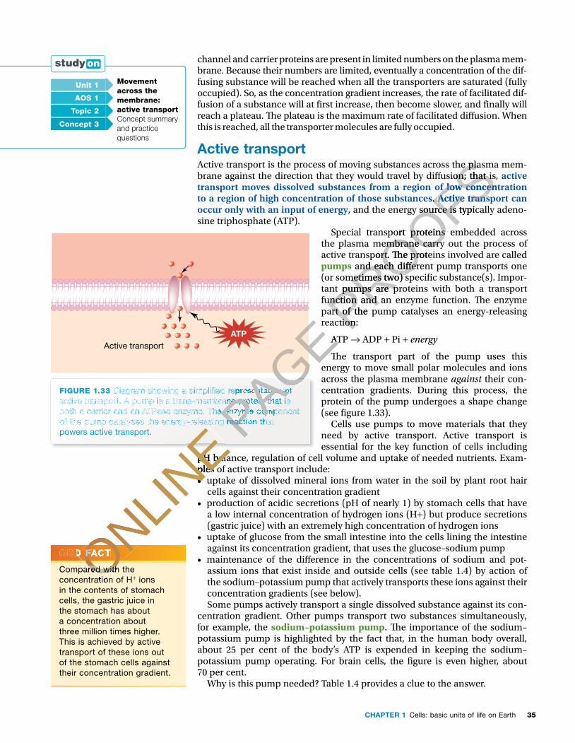
35CHAPTER 1 Cells: basic units of life on Earth
channel and carrier proteins are present in limited numbers on the plasma mem-brane. Because their numbers are limited, eventually a concentration of the dif-fusing substance will be reached when all the transporters are saturated (fully occupied). So, as the concentration gradient increases, the rate of facilitated dif-fusion of a substance will at � rst increase, then become slower, and � nally will reach a plateau. � e plateau is the maximum rate of facilitated di� usion. When this is reached, all the transporter molecules are fully occupied.
Active transportActive transport is the process of moving substances across the plasma mem-brane against the direction that they would travel by di� usion; that is, active transport moves dissolved substances from a region of low concentration to a region of high concentration of those substances. Active transport can occur only with an input of energy, and the energy source is typically adeno-sine triphosphate (ATP).
Special transport proteins embedded across the plasma membrane carry out the process of active transport. � e proteins involved are called pumps and each di� erent pump transports one (or sometimes two) speci� c substance(s). Impor-tant pumps are proteins with both a transport function and an enzyme function. � e enzyme part of the pump catalyses an energy-releasing reaction:
ATP → ADP + Pi + energy
� e transport part of the pump uses this energy to move small polar molecules and ions across the plasma membrane against their con-centration gradients. During this process, the protein of the pump undergoes a shape change (see � gure 1.33).
Cells use pumps to move materials that they need by active transport. Active transport is essential for the key function of cells including
pH balance, regulation of cell volume and uptake of needed nutrients. Exam-ples of active transport include:• uptake of dissolved mineral ions from water in the soil by plant root hair
cells against their concentration gradient • production of acidic secretions (pH of nearly 1) by stomach cells that have
a low internal concentration of hydrogen ions (H+) but produce secretions (gastric juice) with an extremely high concentration of hydrogen ions
• uptake of glucose from the small intestine into the cells lining the intestine against its concentration gradient, that uses the glucose–sodium pump
• maintenance of the di� erence in the concentrations of sodium and pot-assium ions that exist inside and outside cells (see table 1.4) by action of the sodium–potassium pump that actively transports these ions against their concentration gradients (see below). Some pumps actively transport a single dissolved substance against its con-
centration gradient. Other pumps transport two substances simultaneously, for example, the sodium–potassium pump. � e importance of the sodium–potassium pump is highlighted by the fact that, in the human body overall, about 25 per cent of the body’s ATP is expended in keeping the sodium–potassium pump operating. For brain cells, the � gure is even higher, about 70 per cent.
Why is this pump needed? Table 1.4 provides a clue to the answer.
FIGURE 1.33 Diagram showing a simpli� ed representation of active transport. A pump is a trans-membrane protein that is both a carrier and an ATPase enzyme. The enzyme component of the pump catalyses the energy-releasing reaction that powers active transport.
Active transportATP
Unit 1 Movement across themembrane:active transportConcept summary and practice questions
AOS 1
Topic 2
Concept 3
ODD FACT
Compared with the concentration of H+ ions in the contents of stomach cells, the gastric juice in the stomach has about a concentration about three million times higher. This is achieved by active transport of these ions out of the stomach cells against their concentration gradient.
ONLINE
pH balance, regulation of cell volume and uptake of needed nutrients. Exam-
ONLINE
pH balance, regulation of cell volume and uptake of needed nutrients. Exam-ples of active transport include:
ONLINE
ples of active transport include:•
ONLINE
•
ONLINE
ONLINE of the pump catalyses the energy-releasing reaction that
ONLINE of the pump catalyses the energy-releasing reaction that
ONLINE
ONLINE
ONLINE
ONLINE
ODD FACTONLINE
ODD FACT
Compared with the ONLINE
Compared with the concentration of HONLIN
E
concentration of H
PAGE reaction:
PAGE reaction:
ATP
PAGE ATP
PAGE
PAGE Diagram showing a simpli� ed representation of
PAGE Diagram showing a simpli� ed representation of
active transport. A pump is a trans-membrane protein that is
PAGE active transport. A pump is a trans-membrane protein that is both a carrier and an ATPase enzyme. The enzyme component PAGE both a carrier and an ATPase enzyme. The enzyme component of the pump catalyses the energy-releasing reaction that PAGE of the pump catalyses the energy-releasing reaction that PAGE
PAGE PROOFS
Active transport is the process of moving substances across the plasma mem-
PROOFSActive transport is the process of moving substances across the plasma mem-brane against the direction that they would travel by di� usion; that is,
PROOFSbrane against the direction that they would travel by di� usion; that is, transport moves dissolved substances from a region of low concentration
PROOFStransport moves dissolved substances from a region of low concentration to a region of high concentration of those substances
PROOFSto a region of high concentration of those substances.
PROOFS. Active transport can
PROOFSActive transport can , and the energy source is typically adeno-
PROOFS, and the energy source is typically adeno-
Special transport proteins embedded across
PROOFSSpecial transport proteins embedded across
the plasma membrane carry out the process of
PROOFSthe plasma membrane carry out the process of active transport. � e proteins involved are called
PROOFSactive transport. � e proteins involved are called
and each di� erent pump transports one
PROOFS and each di� erent pump transports one
(or sometimes two) speci� c substance(s). Impor-
PROOFS(or sometimes two) speci� c substance(s). Impor-tant pumps are proteins with both a transport
PROOFS
tant pumps are proteins with both a transport function and an enzyme function. � e enzyme PROOFS
function and an enzyme function. � e enzyme part of the pump catalyses an energy-releasing PROOFS
part of the pump catalyses an energy-releasing reaction: PROOFS
reaction:
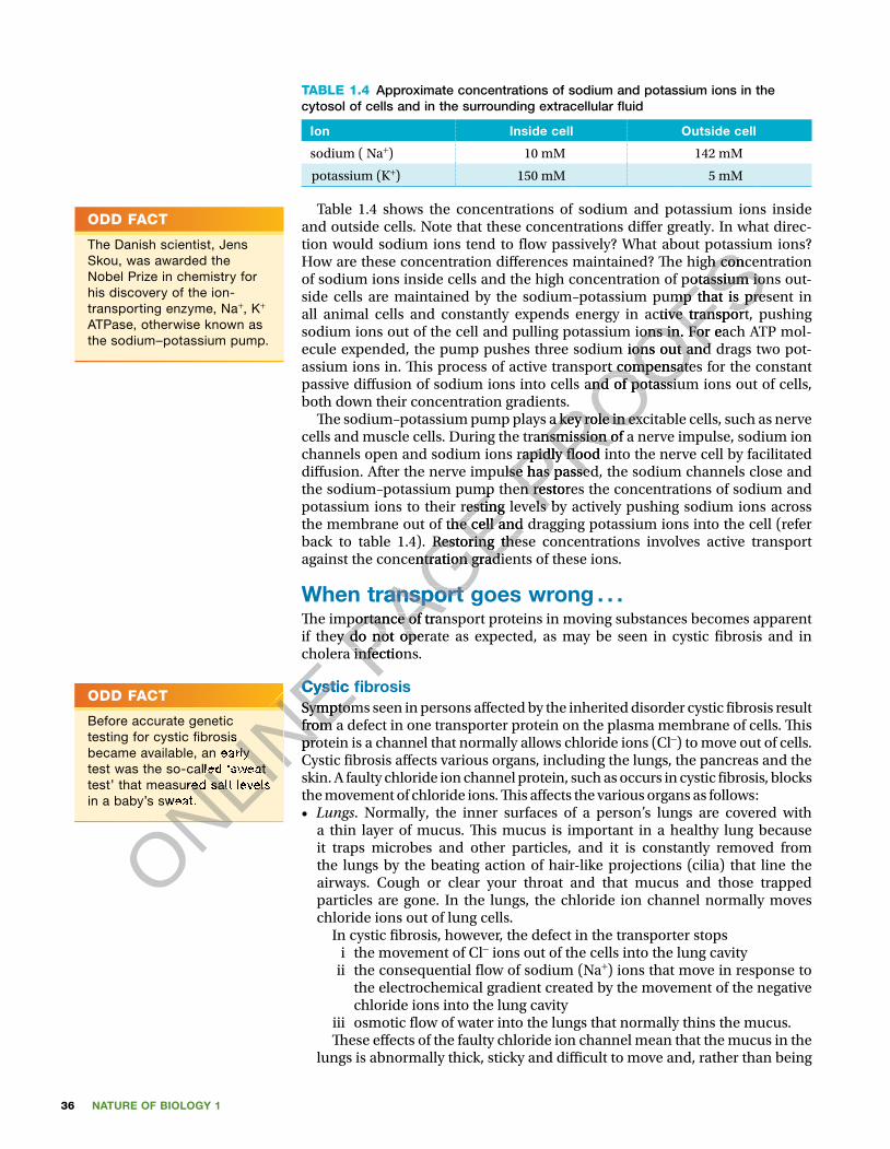
NATURE OF BIOLOGY 136
TABLE 1.4 Approximate concentrations of sodium and potassium ions in the cytosol of cells and in the surrounding extracellular �uid
Ion Inside cell Outside cell
sodium ( Na+) 10 mM 142 mM
potassium (K+) 150 mM 5 mM
Table 1.4 shows the concentrations of sodium and potassium ions inside and outside cells. Note that these concentrations di�er greatly. In what direc-tion would sodium ions tend to �ow passively? What about potassium ions? How are these concentration di�erences maintained? �e high concentration of sodium ions inside cells and the high concentration of potassium ions out-side cells are maintained by the sodium–potassium pump that is present in all animal cells and constantly expends energy in active transport, pushing sodium ions out of the cell and pulling potassium ions in. For each ATP mol-ecule expended, the pump pushes three sodium ions out and drags two pot-assium ions in. �is process of active transport compensates for the constant passive di�usion of sodium ions into cells and of potassium ions out of cells, both down their concentration gradients.
�e sodium–potassium pump plays a key role in excitable cells, such as nerve cells and muscle cells. During the transmission of a nerve impulse, sodium ion channels open and sodium ions rapidly �ood into the nerve cell by facilitated di�usion. After the nerve impulse has passed, the sodium channels close and the sodium–potassium pump then restores the concentrations of sodium and potassium ions to their resting levels by actively pushing sodium ions across the membrane out of the cell and dragging potassium ions into the cell (refer back to table 1.4). Restoring these concentrations involves active transport against the concentration gradients of these ions.
When transport goes wrong . . .�e importance of transport proteins in moving substances becomes apparent if they do not operate as expected, as may be seen in cystic �brosis and in cholera infections.
Cystic �brosisSymptoms seen in persons a�ected by the inherited disorder cystic �brosis result from a defect in one transporter protein on the plasma membrane of cells. �is protein is a channel that normally allows chloride ions (Cl−) to move out of cells. Cystic �brosis a�ects various organs, including the lungs, the pancreas and the skin. A faulty chloride ion channel protein, such as occurs in cystic �brosis, blocks the movement of chloride ions. �is a�ects the various organs as follows: • Lungs. Normally, the inner surfaces of a person’s lungs are covered with
a thin layer of mucus. �is mucus is important in a healthy lung because it traps microbes and other particles, and it is constantly removed from the lungs by the beating action of hair-like projections (cilia) that line the airways. Cough or clear your throat and that mucus and those trapped particles are gone. In the lungs, the chloride ion channel normally moves chloride ions out of lung cells. In cystic �brosis, however, the defect in the transporter stops
i the movement of Cl− ions out of the cells into the lung cavity ii the consequential �ow of sodium (Na+) ions that move in response to
the electrochemical gradient created by the movement of the negative chloride ions into the lung cavity
iii osmotic �ow of water into the lungs that normally thins the mucus. �ese e�ects of the faulty chloride ion channel mean that the mucus in the
lungs is abnormally thick, sticky and di�cult to move and, rather than being
ODD FACT
The Danish scientist, Jens Skou, was awarded the Nobel Prize in chemistry for his discovery of the ion-transporting enzyme, Na+, K+ ATPase, otherwise known as the sodium–potassium pump.
ODD FACT
Before accurate genetic testing for cystic �brosis became available, an early test was the so-called ‘sweat test’ that measured salt levels in a baby’s sweat.
ONLINE cholera infections.
ONLINE cholera infections.
Cystic �brosis
ONLINE Cystic �brosis
Symptoms seen in persons a�ected by the inherited disorder cystic �brosis result
ONLINE
Symptoms seen in persons a�ected by the inherited disorder cystic �brosis result from a defect in one transporter protein on the plasma membrane of cells. �is
ONLINE
from a defect in one transporter protein on the plasma membrane of cells. �is protein is a channel that normally allows chloride ions (Cl
ONLINE
protein is a channel that normally allows chloride ions (ClCystic �brosis a�ects various organs, including the lungs, the pancreas and the
ONLINE
Cystic �brosis a�ects various organs, including the lungs, the pancreas and the
ONLINE
ONLINE
ONLINE
ONLINE
ONLINE
ONLINE
testing for cystic �brosis
ONLINE
testing for cystic �brosis became available, an early
ONLINE
became available, an early test was the so-called ‘sweat
ONLINE
test was the so-called ‘sweat test’ that measured salt levels
ONLINE
test’ that measured salt levels in a baby’s sweat.
ONLINE
in a baby’s sweat.
PAGE the sodium–potassium pump then restores the concentrations of sodium and
PAGE the sodium–potassium pump then restores the concentrations of sodium and potassium ions to their resting levels by actively pushing sodium ions across
PAGE potassium ions to their resting levels by actively pushing sodium ions across the membrane out of the cell and dragging potassium ions into the cell (refer
PAGE the membrane out of the cell and dragging potassium ions into the cell (refer back to table 1.4). Restoring these concentrations involves active transport
PAGE back to table 1.4). Restoring these concentrations involves active transport against the concentration gradients of these ions.
PAGE against the concentration gradients of these ions.
When transport goes wrong
PAGE When transport goes wrong�e importance of transport proteins in moving substances becomes apparent
PAGE �e importance of transport proteins in moving substances becomes apparent if they do not operate as expected, as may be seen in cystic �brosis and in PAGE if they do not operate as expected, as may be seen in cystic �brosis and in cholera infections. PAGE
cholera infections.
PROOFStion would sodium ions tend to �ow passively? What about potassium ions?
PROOFStion would sodium ions tend to �ow passively? What about potassium ions? How are these concentration di�erences maintained? �e high concentration
PROOFSHow are these concentration di�erences maintained? �e high concentration of sodium ions inside cells and the high concentration of potassium ions out
PROOFSof sodium ions inside cells and the high concentration of potassium ions outside cells are maintained by the sodium–potassium pump that is present in
PROOFSside cells are maintained by the sodium–potassium pump that is present in all animal cells and constantly expends energy in active transport, pushing
PROOFSall animal cells and constantly expends energy in active transport, pushing sodium ions out of the cell and pulling potassium ions in. For each ATP mol
PROOFSsodium ions out of the cell and pulling potassium ions in. For each ATP molecule expended, the pump pushes three sodium ions out and drags two pot
PROOFSecule expended, the pump pushes three sodium ions out and drags two potassium ions in. �is process of active transport compensates for the constant
PROOFSassium ions in. �is process of active transport compensates for the constant passive di�usion of sodium ions into cells and of potassium ions out of cells,
PROOFSpassive di�usion of sodium ions into cells and of potassium ions out of cells, both down their concentration gradients.
PROOFSboth down their concentration gradients.
�e sodium–potassium pump plays a key role in excitable cells, such as nerve
PROOFS�e sodium–potassium pump plays a key role in excitable cells, such as nerve
cells and muscle cells. During the transmission of a nerve impulse, sodium ion
PROOFScells and muscle cells. During the transmission of a nerve impulse, sodium ion channels open and sodium ions rapidly �ood into the nerve cell by facilitated
PROOFS
channels open and sodium ions rapidly �ood into the nerve cell by facilitated di�usion. After the nerve impulse has passed, the sodium channels close and PROOFS
di�usion. After the nerve impulse has passed, the sodium channels close and the sodium–potassium pump then restores the concentrations of sodium and PROOFS
the sodium–potassium pump then restores the concentrations of sodium and potassium ions to their resting levels by actively pushing sodium ions across PROOFS
potassium ions to their resting levels by actively pushing sodium ions across
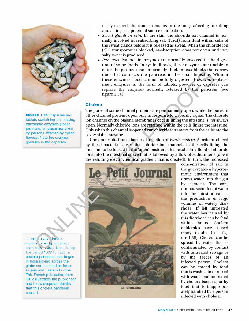
37CHAPTER 1 Cells: basic units of life on Earth
easily cleared, the mucus remains in the lungs a� ecting breathing and acting as a potential source of infection.
• Sweat glands in skin. In the skin, the chloride ion channel is nor-mally involved in reabsorbing salt (NaCl) from � uid within cells of the sweat glands before it is released as sweat. When the chloride ion (Cl–) transporter is blocked, re-absorption does not occur and very salty sweat is produced.
• Pancreas. Pancreatic enzymes are normally involved in the diges-tion of some foods. In cystic � brosis, these enzymes are unable to enter the gut because abnormally thick mucus blocks the narrow duct that connects the pancreas to the small intestine. Without these enzymes, food cannot be fully digested. However, replace-ment enzymes in the form of tablets, powders or capsules can replace the enzymes normally released by the pancreas (see � gure 1.34).
Cholera� e pores of some channel proteins are permanently open, while the pores in other channel proteins open only in response to a speci� c signal. � e chloride ion channel on the plasma membranes of cells lining the intestine is not always open. Normally chloride ions are retained within the cells lining the intestine. Only when this channel is opened can chloride ions move from the cells into the cavity of the intestine.
Cholera results from a bacterial infection of Vibrio cholera. A toxin produced by these bacteria causes the chloride ion channels in the cells lining the intestine to be locked in the ‘open’ position. � is results in a � ood of chloride ions into the intestinal space that is followed by a � ow of sodium ions (down the resulting electrochemical gradient that is created). In turn, the increased
concentration of salt in the gut creates a hyperos-motic environment that draws water into the gut by osmosis. � e con-tinuous secretion of water into the intestine causes the production of large volumes of watery diar-rhoea. If left untreated the water loss caused by this diarrhoea can be fatal within hours. Cholera epidemics have caused many deaths (see � g-ure 1.35). Cholera can be spread by water that is contaminated by contact with untreated sewage or by the faeces of an infected person. Cholera can be spread by food that is washed in or mixed with water contaminated by cholera bacteria, or by food that is inappropri-ately handled by a person infected with cholera.
FIGURE 1.34 Capsules and tablets containing the missing pancreatic enzymes (lipase, protease, amylase) are taken by persons affected by cystic � brosis. Note the enzyme granules in the capsules.
FIGURE 1.35 Cholera epidemics and pandemics have taken many lives. During the period 1899 to 1923, a cholera pandemic that began in India spread across the globe and reached as far as Russia and Eastern Europe. This French publication from 1912 illustrates the public fear and the widespread deaths that this cholera pandemic caused.
ONLINE
ONLINE
ONLINE
ONLINE
FIGURE 1.35 ONLINE
FIGURE 1.35 Cholera ONLINE
Cholera epidemics and pandemics ONLIN
E
epidemics and pandemics have taken many lives. During ONLIN
E
have taken many lives. During the period 1899 to 1923, a ONLIN
E
the period 1899 to 1923, a ONLINE P
AGE Cholera results from a bacterial infection of
PAGE Cholera results from a bacterial infection of by these bacteria causes the chloride ion channels in the cells lining the
PAGE by these bacteria causes the chloride ion channels in the cells lining the intestine to be locked in the ‘open’ position. � is results in a � ood of chloride
PAGE intestine to be locked in the ‘open’ position. � is results in a � ood of chloride ions into the intestinal space that is followed by a � ow of sodium ions (down
PAGE ions into the intestinal space that is followed by a � ow of sodium ions (down the resulting electrochemical gradient that is created). In turn, the increased
PAGE the resulting electrochemical gradient that is created). In turn, the increased
PAGE PROOFS
enter the gut because abnormally thick mucus blocks the narrow
PROOFSenter the gut because abnormally thick mucus blocks the narrow duct that connects the pancreas to the small intestine. Without
PROOFSduct that connects the pancreas to the small intestine. Without these enzymes, food cannot be fully digested. However, replace-
PROOFSthese enzymes, food cannot be fully digested. However, replace-ment enzymes in the form of tablets, powders or capsules can
PROOFSment enzymes in the form of tablets, powders or capsules can replace the enzymes normally released by the pancreas (see
PROOFSreplace the enzymes normally released by the pancreas (see
� e pores of some channel proteins are permanently open, while the pores in
PROOFS� e pores of some channel proteins are permanently open, while the pores in other channel proteins open only in response to a speci� c signal. � e chloride
PROOFSother channel proteins open only in response to a speci� c signal. � e chloride ion channel on the plasma membranes of cells lining the intestine is
PROOFSion channel on the plasma membranes of cells lining the intestine is open. Normally chloride ions are retained within the cells lining the intestine.
PROOFS
open. Normally chloride ions are retained within the cells lining the intestine. Only when this channel is opened can chloride ions move from the cells into the PROOFS
Only when this channel is opened can chloride ions move from the cells into the
Cholera results from a bacterial infection of PROOFS
Cholera results from a bacterial infection of by these bacteria causes the chloride ion channels in the cells lining the PROOFS
by these bacteria causes the chloride ion channels in the cells lining the

NATURE OF BIOLOGY 138
Bulk transport of solids and liquidsTo this point, we have been concerned with move-ment of dissolved substances across the plasma membrane. In addition, small solid particles and liquids in bulk can be moved across the plasma membrane into or out of cells. Figure 1.36 gives a summary of how bulk material can enter cells.
Endocytosis: getting inSolid particles can be taken into a cell. For example, one kind of white blood cell is able to engulf a disease-causing bacteria cell and enclose it within a lysosome sac where it is destroyed. Unicel-lular protists, such as Amoeba and Paramecium, obtain their energy for living in the form of rela-tively large ‘food’ particles, which they engulf and enclose within a sac where the food is digested (see � gure 1.37a).
If material is solid If material is �uid
Endocytosisbulk transport of
material into a cell
phagocytosisfrom the Greek
phagos = ‘eating’and cyto = ‘cell’
pinocytosisfrom the Greek
pinus = ‘drinking’and cyto = ‘cell’
the process is called the process is called
FIGURE 1.36 Endocytosis — a summary
Cytosol
Phagocyticvesicle
Outside cell(b)
Lipidbilayer
FIGURE 1.37 (a) Transport of a solid food particle across the membrane of an Amoeba (b) Endocytosis occurs when part of the plasma membrane forms around food particles to form a phagocytic vesicle (or phagosome). This vesicle then moves into the cytosol where it fuses with a lysosome, a bag of digestive enzymes. (Believe it or not, this fused structure is called a phago-lysosome.)The same digestive process can also occur to microbes.
Pseudopods
Food particle
(a)
Lysosomes containingdigestive enzymes
Entrapment
Amoeba
Engulfment
Digestion
Absorption
Foodvacuole
Digestedfood
Absorbedfood
Food
ONLINE
ONLINE
ONLINE
FIGURE 1.37
ONLINE
FIGURE 1.37 (a)
ONLINE
(a) Transport
ONLINE
Transport of a solid food particle across
ONLINE
of a solid food particle across the membrane of an Amoeba ONLIN
E
the membrane of an Amoeba Endocytosis occurs when ONLIN
E
Endocytosis occurs when part of the plasma membrane ONLIN
E
part of the plasma membrane forms around food particles to ONLIN
E
forms around food particles to ONLINE
ONLINE
ONLINE
ONLINE
ONLINE
ONLINE P
AGE
PAGE
PAGE
PAGE
PAGE
PAGE
PAGE Entrapment PAGE Entrapment
EngulfmentPAGE Engulfment
PROOFSSolid particles can be taken into a cell. For example,
PROOFSSolid particles can be taken into a cell. For example, one kind of white blood cell is able to engulf a
PROOFSone kind of white blood cell is able to engulf a disease-causing bacteria cell and enclose it within
PROOFSdisease-causing bacteria cell and enclose it within sac where it is destroyed. Unicel-
PROOFS sac where it is destroyed. Unicel-
lular protists, such as
PROOFSlular protists, such as Amoeba
PROOFSAmoeba and
PROOFSand
obtain their energy for living in the form of rela-
PROOFSobtain their energy for living in the form of rela-tively large ‘food’ particles, which they engulf and
PROOFStively large ‘food’ particles, which they engulf and enclose within a sac where the food is digested (see
PROOFSenclose within a sac where the food is digested (see � gure 1.37a).
PROOFS� gure 1.37a).
PROOFS

39CHAPTER 1 Cells: basic units of life on Earth
Note how part of the plasma membrane encloses the material to be trans-ported and then pinches o� to form a membranous vesicle that moves into the cytosol (� gure 1.37b). � is process of bulk transport of material into a cell is called endocytosis. When the material being transported is a solid food par-ticle, the type of endocytosis is called phagocytosis.
Although some cells are capable of phagocytosis, most cells are not. Most eukaryotic cells rely on pinocytosis, a form of endocytosis that involves mat-erial that is in solution being transported into cells. � e process of endocy-tosis is an energy-requiring process and requires an input of ATP.
Exocytosis: getting out Bulk transport out of cells (such as the export of material from the Golgi com-plex, discussed in chapter 2, p. 67) is called exocytosis. In exocytosis, vesicles formed within a cell fuse with the plasma membrane before the contents of the vesicles are released from the cell (see � gure 1.38). If the released mat-erial is a product of the cell (e.g. the contents of a Golgi vesicle), then ‘secreted from the cell’ is the phrase generally used. If the released material is a waste product after digestion of some matter taken into the cell, ‘voided from the cell’ is generally more appropriate. � e process of exocytosis requires an input of energy in the form of ATP.
Cytosol
1. Vesicle with materialfrom Golgi complex
to be exported
2. Vesicle fuses withplasma membrane
3. Vesicle expels contentsinto the extracellular
�uid
Outside cell
Lipidbilayer
FIGURE 1.38 Exocytosis (bulk transport out of cells) occurs when vesicles within the cytosol fuse with the plasma membrane and vesicle contents are released from the cell.
KEY IDEAS
■ Simple diffusion moves dissolved substances across the plasma membrane down their concentration gradient and requires no input of energy.
■ Osmosis is a special case of diffusion, being the movement of water across the plasma membrane down its concentration gradient.
■ Facilitated diffusion moves dissolved substances across the plasma membrane down their concentration gradients, but this movement occurs through involvement of transport proteins, either channel or carrier proteins, and requires no input of energy.
■ Active transport moves dissolved substances across the plasma membrane against the concentration gradient, a process that can occur only via the action of protein pumps.
■ Active transport requires an input of energy that commonly comes from ATP, catalysed by the ATPase enzyme that is part of some protein pumps.
■ Endocytosis is the bulk transport of material into cells; if solids are being moved, the process is termed phagocytosis and, if liquids, the process is termed pinocytosis.
■ Exocytosis is the bulk movement of materials via secretory vesicles out of cells.
eLessonPhagocytosiseles-2444
ONLINE
ONLINE 1. Vesicle with material
ONLINE 1. Vesicle with materialfrom Golgi complex
ONLINE from Golgi complex
to be exported
ONLINE to be exported
ONLINE
ONLINE
ONLINE
ONLINE
ONLINE
ONLINE KEY IDEAS
ONLINE KEY IDEAS
PAGE
PAGE
PAGE
PAGE
PAGE
PAGE
PAGE
PAGE
PAGE
PAGE
PAGE
PAGE
PAGE
PAGE
PAGE
PAGE
PAGE
PAGE
PAGE
PAGE
PAGE
PAGE
PAGE
PAGE
PAGE
PAGE
PAGE
PAGE
PAGE
PAGE
PAGE
PAGE
PAGE
PAGE
PAGE
PAGE
PAGE
PAGE
PAGE
PAGE
PAGE
PAGE
PAGE
PAGE
PAGE
PAGE
PAGE
PAGE
PAGE
PAGE
PAGE
PAGE
PAGE
PAGE
PAGE
PAGE
PAGE
PAGE
PAGE
PAGE
PAGE
PAGE
PAGE
PAGE
PAGE
PAGE
PAGE
PAGE
PAGE
PAGE
PAGE
PAGE
PAGE
PAGE
PAGE
PAGE
PAGE
PAGE
PAGE
PAGE
PAGE
PAGE
PAGE
PAGE
PAGE
PAGE
PAGE
PAGE
PAGE
PAGE
PAGE
PAGE
PAGE
PAGE
PAGE
PAGE
PAGE
PAGE
PAGE
PAGE
PAGE
PAGE
PAGE
PAGE
PAGE
PAGE
PAGE
PAGE
PAGE
PAGE
PAGE
PAGE
PAGE
PAGE
PAGE
PAGE
PAGE
PAGE
PAGE
PAGE
PAGE
PAGE
PAGE
PAGE
PAGE
PAGE
PAGE
PAGE
PAGE
PAGE
PAGE
PAGE
PAGE
PAGE
PAGE
PAGE
PAGE
PAGE
PAGE
PAGE
PAGE
PAGE
PAGE
PAGE
PAGE
PAGE
PAGE
PAGE
PAGE
PAGE
PAGE
PAGE
PAGE
PAGE
PAGE
PAGE
PAGE
PAGE
PAGE
PAGE
PAGE
PAGE
PAGE
PAGE
PAGE
PAGE
PAGE
PAGE
PAGE
PAGE
PAGE
PAGE
PAGE
PAGE
PAGE
PAGE
PAGE
PAGE
PAGE
PAGE
PAGE
PAGE
PAGE
PAGE
PAGE
PAGE
PAGE
PAGE
PAGE
PAGE
PAGE
1. Vesicle with materialPAGE
1. Vesicle with materialfrom Golgi complexPAGE
from Golgi complexPAGE
PAGE
PAGE
PAGE
PAGE
PAGE
PAGE
PAGE
PAGE
PAGE
PAGE
PAGE
PAGE
PAGE
PAGE
PAGE
PAGE
PAGE
PAGE
PAGE
PAGE
PAGE
PAGE
PAGE PROOFSBulk transport out of cells (such as the export of material from the Golgi com-
PROOFSBulk transport out of cells (such as the export of material from the Golgi com-In exocytosis, vesicles
PROOFSIn exocytosis, vesicles formed within a cell fuse with the plasma membrane
PROOFSformed within a cell fuse with the plasma membrane before the contents of
PROOFSbefore the contents of the vesicles are released from the cell (see � gure 1.38). If the released mat-
PROOFSthe vesicles are released from the cell (see � gure 1.38). If the released mat-erial is a product of the cell (e.g. the contents of a Golgi vesicle), then ‘secreted
PROOFSerial is a product of the cell (e.g. the contents of a Golgi vesicle), then ‘secreted from the cell’ is the phrase generally used. If the released material is a waste
PROOFSfrom the cell’ is the phrase generally used. If the released material is a waste product after digestion of some matter taken into the cell, ‘voided from the
PROOFSproduct after digestion of some matter taken into the cell, ‘voided from the cell’ is generally more appropriate. � e process of exocytosis requires an input
PROOFScell’ is generally more appropriate. � e process of exocytosis requires an input
PROOFS
PROOFS
PROOFS
PROOFS
PROOFS
PROOFS
PROOFS
PROOFS
PROOFS
PROOFS
PROOFS
PROOFS
PROOFS
PROOFS
PROOFS
PROOFS
PROOFS
PROOFS
PROOFS
PROOFS
PROOFS
PROOFS
PROOFS
PROOFS
PROOFS
PROOFS
PROOFS
PROOFS
PROOFS
PROOFS
PROOFS
PROOFS
PROOFS
PROOFS

NATURE OF BIOLOGY 140
QUICK CHECK
23 What is the process by which bulk materials are exported out of cells? 24 Consider passive diffusion and facilitated diffusion:
a Identify one difference between these processes.b Identify one similarity that they share.
25 Identify one difference between diffusion and active transport.26 Which transport process relies on the involvement of either a carrier or a
channel protein?27 By which process do cells of the stomach lining manage to move
hydrogen ions out of the cells to produce a highly acidic gastric secretion? 28 What process is involved in the movement of water down its concentration
gradient and across a layer of cells from outside the body to inside?
ONLINE P
AGE PROOFS
PROOFS
PROOFSocess do cells of the stomach lining manage to move
PROOFSocess do cells of the stomach lining manage to move
hydrogen ions out of the cells to produce a highly acidic gastric secretion?
PROOFShydrogen ions out of the cells to produce a highly acidic gastric secretion?
ocess is involved in the movement of water down its concentration
PROOFSocess is involved in the movement of water down its concentration gradient and across a layer of cells from outside the body to inside?
PROOFSgradient and across a layer of cells from outside the body to inside?

41CHAPTER 1 Cells: basic units of life on Earth
SCIENTIST AT WORK
Jonica Newby — TV Science ReporterI’ll be honest with you. When I chose to focus on science in high school, I did it because I thought it was so much easier than the pain of creative writing. Now though, as a TV science reporter/producer, I have to write creatively and still understand the science! Argh!
But of course, I love it; it’s the best of both worlds. I have the privilege of being one of the reporters on ABC television’s longstanding TV science pro-gram, Catalyst. It’s an amazing and sometimes crazy job. What are some of the things I’ve done in the last couple of years? I’ve lain inside a vertical wind tunnel to see what wind speed it would take to make me � y; I sourced motion activated infra-red cameras to see what animals were visiting my yard at night; I jumped out of a plane with a parachute to see how fear interferes with language centres of the brain; I staged a full scale mock cyclone in an unsuspecting family’s house to show how climate change might bring cyclones as far south as the Gold Coast; and I got bitten by a tick while � lming — you guessed it — how not to be bitten by a tick!
Although the � lming days are probably the most exciting — and stressful — they only take up a small portion of the job. What I’m doing most of the time is ringing scientists to talk about what they do for a living, and then trying to come up with a way to make a film about it, complete with the scripted words and images. My science background is cru-cial, because I’m basi-cally getting a crash course in anything from Quantum physics to the neurobiology of fear. (‘Ok Jonica, you have one week to understand every-thing there is about the science of Mam-malian Meat Allergy. Go!’). And all the rest is creative.
� e process goes like this. We have an idea for a story. � en we (me and a TV researcher) research it quickly.
� en I have to sit down and write a shoot script — and it’s still a painful process being creative, I can tell you. � en we organise the shoot. On shoot days, I’m the � lm director as well as the onscreen presenter; and that’s why it’s pretty stressful, trying to keep everyone on schedule, working with cameramen and sound technicians, and encouraging scientists who may not have been on TV before. � en we take the content back to base and look at every single camera tape many times. From these I have to write an edit script. � en I give this edit script to the editor, who assembles the rough cut. We then work together to make decisions about shortening this, rewriting that, and eventually it becomes the story you can see on TV. Phew! It may be only a few minutes of broadcast, but it takes weeks to months of full time work to prepare every piece.
And in case you’re wondering what my ‘Dr’ stands for, I trained originally as a veterinarian. Yes indeed — plenty of looking after cows, and vacci-nating dogs and cats for me. I did that for about 3 years after I graduated. And it was great but in the end, despite my fear of the creative side of life, I was drawn to be more creative and so I set about on the long path to the job I have now. It’s not for everyone, and there are very few jobs like mine, but it just goes to show, you never know where a good grounding in science will take you. Flying inside a wind tunnel, maybe . . . that was fun!
FIGURE 1.39 Jonica Newby on a shoot
ONLINE
cial, because I’m basi-
ONLINE
cial, because I’m basi-cally getting a crash
ONLINE
cally getting a crash course in anything
ONLINE
course in anything from Quantum physics
ONLINE
from Quantum physics to the neurobiology
ONLINE
to the neurobiology of fear. (‘Ok Jonica,
ONLINE
of fear. (‘Ok Jonica, you have one week
ONLINE
you have one week to understand every-ONLIN
E
to understand every-thing there is about ONLIN
E
thing there is about the science of Mam-ONLIN
E
the science of Mam-ONLINE P
AGE Although the � lming days are probably the most
PAGE Although the � lming days are probably the most
exciting — and stressful — they only take up a small
PAGE exciting — and stressful — they only take up a small portion of the job. What I’m doing most of the time
PAGE portion of the job. What I’m doing most of the time is ringing scientists to talk about what they do for a
PAGE is ringing scientists to talk about what they do for a
3 years after I graduated. And it was great but in
PAGE 3 years after I graduated. And it was great but in the end, despite my fear of the creative side of life,
PAGE the end, despite my fear of the creative side of life, I was drawn to be more creative and so I set about
PAGE I was drawn to be more creative and so I set about on the long path to the job I have now. It’s not for
PAGE on the long path to the job I have now. It’s not for everyone, and there are very few jobs like mine, but
PAGE everyone, and there are very few jobs like mine, but it just goes to show, you never know where a good
PAGE it just goes to show, you never know where a good grounding in science will take you. Flying inside a
PAGE grounding in science will take you. Flying inside a wind tunnel, maybe . . . that was fun!
PAGE wind tunnel, maybe . . . that was fun!
PAGE PROOFS
have been on TV before. � en we take the content back
PROOFShave been on TV before. � en we take the content back to base and look at every single camera tape many
PROOFSto base and look at every single camera tape many times. From these I have to write an edit script. � en
PROOFStimes. From these I have to write an edit script. � en I give this edit script to the editor, who assembles the
PROOFSI give this edit script to the editor, who assembles the rough cut. We then work together to make decisions
PROOFSrough cut. We then work together to make decisions about shortening this, rewriting that, and eventually it
PROOFSabout shortening this, rewriting that, and eventually it becomes the story you can see on TV. Phew! It may be
PROOFSbecomes the story you can see on TV. Phew! It may be only a few minutes of broadcast, but it takes weeks to
PROOFSonly a few minutes of broadcast, but it takes weeks to months of full time work to prepare every piece.
PROOFSmonths of full time work to prepare every piece.
And in case you’re wondering what my ‘Dr’
PROOFSAnd in case you’re wondering what my ‘Dr’
stands for, I trained originally as a veterinarian. Yes
PROOFSstands for, I trained originally as a veterinarian. Yes indeed — plenty of looking after cows, and vacci-
PROOFS
indeed — plenty of looking after cows, and vacci-nating dogs and cats for me. I did that for about PROOFS
nating dogs and cats for me. I did that for about 3 years after I graduated. And it was great but in PROOFS
3 years after I graduated. And it was great but in the end, despite my fear of the creative side of life, PROOFS
the end, despite my fear of the creative side of life,

42 NATURE OF BIOLOGY 1
BIOCHALLENGE
Exploring the plasma membrane
1 The plasma membrane has been described as being like a ‘train track’. This was because the � rst images of the plasma membrane showed it as two dark lines separated by a lighter region. Figure 1.40 shows part of the plasma membranes of two adjoining cells. The plasma membranes have been sectioned so that their surfaces are oriented horizontally at right angles into the plane of this page.
FIGURE 1.40 Plasma membrane
a How thick is the plasma membrane in nanometres? In micrometres?
b What kind of microscope was needed to produce the image in � gure 1.40?
c What are the ‘rails’ of the train track composed of?d What is present in the space between the rails?
2 Key information about the nature of the plasma membrane came from an experiment carried out in 1925 by two Dutch scientists. They took a known number of red blood cells and, based on the average size of these cells, they estimated their
combined surface area. Then they extracted only the lipid from the plasma membrane of these cells and allowed it to spread out on a water surface where it formed a monolayer or single layer of molecules. (Remember, lipids will not mix with water!) To their surprise, the scientists found that the area of the lipid monolayer on the water surface was twice the combined surface area of the red blood cells that were the source of the lipid.
Consider this � nding and suggest what key information this result provided about the structure of the plasma membrane.
3 In 1970, Frye and Edidin carried out an experiment in which they took a human cell and a mouse cell and fused them to form a human–mouse hybrid cell. They showed the distribution of the surface proteins on the plasma membrane of each cell by using anti-human and anti-mouse antibodies labelled with a different � uorescent dye. A red dye showed the positions of the surface proteins on the membrane of the human cell. A green dye showed the positions of the surface proteins on the membrane of the mouse cell.
Figure 1.41a shows the initial observation immediately after the fusion of the two cells. After 40 minutes, the researchers carried out a second observation and their � ndings are shown in � gure 1.41b.
From the results of this experiment, which of the following is it reasonable to conclude?
a Surface proteins are � xed in position on the plasma membrane.
b Surface proteins from each cell type have fused.c Surface proteins can move laterally across the plasma
membrane.
4 True or false?
The results of this experiment provide support for the � uid mosaic model of membrane structure. Brie� y explain.
Human cell Mouse cell
Hybrid cell
Hybrid cell
Humanprotein
Humanprotein
Fusion
Mouseprotein
40 minutesof incubation
Mouseprotein
(a) (b)
FIGURE 1.41 (a) Start of experiment (b) 40 minutes later
ONLINE What is present in the space between the rails?
ONLINE What is present in the space between the rails?
Key information about the nature of the plasma membrane
ONLINE Key information about the nature of the plasma membrane
came from an experiment carried out in 1925 by two Dutch
ONLINE
came from an experiment carried out in 1925 by two Dutch scientists. They took a known number of red blood cells and,
ONLINE
scientists. They took a known number of red blood cells and, based on the average size of these cells, they estimated their
ONLINE
based on the average size of these cells, they estimated their
ONLINE
ONLINE
ONLINE
ONLINE
ONLINE
ONLINE
ONLINE
ONLINE
ONLINE
ONLINE
ONLINE
ONLINE
ONLINE
ONLINE
ONLINE
ONLINE
ONLINE
Human cell Mouse cell
ONLINE
Human cell Mouse cell
ONLINE
ONLINE
ONLINE
ONLINE
ONLINE
ONLINE
ONLINE
ONLINE
ONLINE
ONLINE
ONLINE P
AGE How thick is the plasma membrane in nanometres? In
PAGE How thick is the plasma membrane in nanometres? In
What kind of microscope was needed to produce the
PAGE What kind of microscope was needed to produce the
What are the ‘rails’ of the train track composed of?PAGE What are the ‘rails’ of the train track composed of? What is present in the space between the rails?PAGE
What is present in the space between the rails?
membrane of the mouse cell.
PAGE membrane of the mouse cell.
Figure 1.41a shows the initial observation immediately
PAGE Figure 1.41a shows the initial observation immediately after the fusion of the two cells. After 40 minutes, the
PAGE after the fusion of the two cells. After 40 minutes, the researchers carried out a second observation and their
PAGE researchers carried out a second observation and their � ndings are shown in � gure 1.41b.
PAGE � ndings are shown in � gure 1.41b.
From the results of this experiment, which of the following
PAGE From the results of this experiment, which of the following is it reasonable to conclude?
PAGE is it reasonable to conclude?
PROOFSwith water!) To their surprise, the scientists found that the
PROOFSwith water!) To their surprise, the scientists found that the area of the lipid monolayer on the water surface was twice
PROOFSarea of the lipid monolayer on the water surface was twice the combined surface area of the red blood cells that were
PROOFSthe combined surface area of the red blood cells that were
Consider this � nding and suggest what key information this
PROOFSConsider this � nding and suggest what key information this result provided about the structure of the plasma membrane.
PROOFSresult provided about the structure of the plasma membrane.
In 1970, Frye and Edidin carried out an experiment in
PROOFSIn 1970, Frye and Edidin carried out an experiment in which they took a human cell and a mouse cell and fused
PROOFSwhich they took a human cell and a mouse cell and fused them to form a human–mouse hybrid cell. They showed
PROOFSthem to form a human–mouse hybrid cell. They showed the distribution of the surface proteins on the plasma
PROOFSthe distribution of the surface proteins on the plasma membrane of each cell by using anti-human and anti-
PROOFSmembrane of each cell by using anti-human and anti-mouse antibodies labelled with a different � uorescent
PROOFSmouse antibodies labelled with a different � uorescent dye. A red dye showed the positions of the surface
PROOFSdye. A red dye showed the positions of the surface proteins on the membrane of the human cell. A green
PROOFS
proteins on the membrane of the human cell. A green dye showed the positions of the surface proteins on the PROOFS
dye showed the positions of the surface proteins on the membrane of the mouse cell. PROOFS
membrane of the mouse cell.
Figure 1.41a shows the initial observation immediately PROOFS
Figure 1.41a shows the initial observation immediately

43CHAPTER 1 Cells: basic units of life on Earth
Unit 1 Cell size, structure and function
Sit topic test
AOS 1
Topic 1
Chapter review
Key wordsactive transportaquaporinsarchaeabacteria biogenesiscarrier proteinscell membranecell surface markersCell � eory channel proteinsendocytosis
eukaryoteseukaryoticexocytosisextremophilesfacilitated di� usion � uid mosaic modelglycoproteinhydrophilic hydrophobichypertonichypotonic
integral proteinsisotonic lysosomenuclear envelopeosmosisperipheral proteinsphospholipidspinocytosisplasma membraneprokaryotesprokaryotic
proteinspumpsreceptorsselectively permeablesemipermeablesimple di� usionsodium–potassium pumpsurface-area-to-volume
ratiotrans-membranevesicle
Questions 1 Making connections ➜ � e key words listed above
can also be called concepts. Concepts can be related to one another by using linking words or phrases to form propositions. For example, the concept ‘compound light microscope’ can be linked to the concept ‘lenses’ by the linking phrase ‘contains at least two’ to form a proposition. An arrow shows the sense of the relationship: when several concepts are related in a meaningful way, a concept map is
formed. Because concepts can be related in many di� erent ways, there is no single, correct concept map. Figure 1.42 shows one concept map containing some of the key words and other terms from this chapter. Use at least six of the key words above to make a concept map relating to the movement of substances across a cell membrane. You may use other words in drawing your map.
Lens/es
Simplemicroscope
Visiblelight
Compoundmicroscope
Electronmicroscope
Lightmicroscope
Ultravioletlight
Microscope
Specialglass
has only one
aremade
of
can be
can be
uses
uses has shorterwavelength than
can be
can be
has atleast two
FIGURE 1.42 Example of a concept map
ONLINE
ONLINE
ONLINE
ONLINE
ONLINE
ONLINE
ONLINE
ONLINE
ONLINE
ONLINE
ONLINE
ONLINE
ONLINE
ONLINE
ONLINE
ONLINE
ONLINE
ONLINE
ONLINE
ONLINE
ONLINE
ONLINE
ONLINE
ONLINE
ONLINE
ONLINE
ONLINE Lens/es
ONLINE Lens/es
ONLINE
has at
ONLINE
has athas at
ONLINE
has atleast two
ONLINE
least twoleast two
ONLINE
least two
PAGE least two’ to form a proposition. An arrow shows the
PAGE least two’ to form a proposition. An arrow shows the sense of the relationship: when several concepts are
PAGE sense of the relationship: when several concepts are related in a meaningful way, a concept map is
PAGE related in a meaningful way, a concept map is
map. Figure 1.42 shows one concept map containing
PAGE map. Figure 1.42 shows one concept map containing some of the key words and other terms from this
PAGE some of the key words and other terms from this chapter.
PAGE chapter. Use at least six of the key words above to make
PAGE Use at least six of the key words above to make a concept map relating to the movement of
PAGE a concept map relating to the movement of substances across a cell membrane. You may use
PAGE substances across a cell membrane. You may use
PAGE
PAGE
PAGE
arePAGE
are
PROOFSselectively permeable
PROOFSselectively permeablesemipermeable
PROOFSsemipermeablesimple di� usion
PROOFSsimple di� usionsodium–potassium pump
PROOFSsodium–potassium pumpsurface-area-to-volume
PROOFSsurface-area-to-volume
ratio
PROOFSratio
trans-membrane
PROOFStrans-membranevesicle
PROOFSvesicle
formed. Because concepts can be related in many
PROOFS
formed. Because concepts can be related in many di� erent ways, there is no single, correct concept PROOFS
di� erent ways, there is no single, correct concept map. Figure 1.42 shows one concept map containing PROOFS
map. Figure 1.42 shows one concept map containing some of the key words and other terms from this PROOFS
some of the key words and other terms from this

44 NATURE OF BIOLOGY 1
2 Applying your knowledge and understanding ➜ Sterile saline (NaCl) solutions, with or without glucose, may be used to treat a person in certain circumstances. � e treatment may be delivered either orally or directly into a vein by intravenous infusion. � e normal salinity level of body cells and the surrounding extracellular � uid is 0.9 per cent sodium chloride. Fluids that might be administered include:
■ normal-strength saline solution with 0.9 per cent saline, with solutes in balance with normal body � uids making the solution isotonic
■ half-strength saline solution with 0.45 per cent salt, with fewer electrolytes making it hypotonic to body � uids; or
■ double-strength saline, with greater than 0.9 per cent dissolved solutes, making it hypertonic to body � uids.
FIGURE 1.43 Infusion of an intravenous saline drip is performed by experienced medical professionals who decide which saline strength should be used, whether the solution should also include glucose, and calculate the correct rate of � ow for the particular patient.
A patient (MM) is in urgent need of treatment following blood loss. To increase the circulating blood volume and raise the blood pressure, an emergency treatment while waiting on blood typing results, might be the intravenous infusion of a saline solution.a Would you expect the saline solution selected for
this purpose to be: i isotonic ii hypotonic iii hypertonic?
Explain your decision.
b � e treatment was given intravenously. Would it be equally e� ective if given by mouth (orally)? Brie� y explain.
3 Another patient (NN) is su� ering from severe dehydration and salt loss. A possible treatment in this case involves administration of a sterile saline solution.a Would you predict the saline solution used in this
case to be: i isotonic ii hypotonic iii double strength?
Explain your decision.b Is glucose likely to be included with the saline?
Explain. 4 Another patient (PP) is immobilised in bed
recovering from an operation. PP is given an infusion of an intravenous saline drip in order to prevent edema; that is, an accumulation of excess extracellular � uid in his body tissues. Would you predict the saline solution to be:
i normal strength ii half strength iii double strength?
Explain your decision. 5 Applying your understanding➜ Consider the
information in table 1.5.
TABLE 1.5 Data for three different shapes, each having the same volume. (Where necessary, � gures have been rounded.)
Cell Shape DimensionsSurface
area VolumeSA:V ratio
A � at sheet 10 × 10 × 0.1 204 10 20.4
B cube 2.15 × 2.15 × 2.15 28 10 2.8
C sphere diameter: 1.67 22 10 2.2
a If these shapes represented cells, which cell (A, B or C) would be most e� cient in moving required materials into and removing wastes from the cell? Explain.
b Which cell would be least e� cient? Explain.c Can you suggest a biological consequence of your
conclusion?d Identify one way in which a cell might
retain its overall shape, but greatly increase its surface area with a minimal increase in volume. (Clue: � is strategy is used by cells involved in absorption of material, such as those lining the small intestine.)
ONLINE
ONLINE
ONLINE
Infusion of an intravenous saline drip is
ONLINE
Infusion of an intravenous saline drip is performed by experienced medical professionals who
ONLINE
performed by experienced medical professionals who decide which saline strength should be used, whether
ONLINE
decide which saline strength should be used, whether the solution should also include glucose, and calculate
ONLINE
the solution should also include glucose, and calculate the correct rate of � ow for the particular patient.
ONLINE
the correct rate of � ow for the particular patient.
ONLINE
ONLINE
A patient (MM) is in urgent need of treatment ONLINE
A patient (MM) is in urgent need of treatment following blood loss. To increase the circulating ONLIN
E
following blood loss. To increase the circulating blood volume and raise the blood pressure, an ONLIN
E
blood volume and raise the blood pressure, an
PAGE
PAGE iii double strength?
PAGE iii double strength? Explain your decision.
PAGE Explain your decision. 5
PAGE 5 Applying your understanding
PAGE Applying your understandinginformation in table 1.5.
PAGE information in table 1.5.
TABLE 1.5
PAGE TABLE 1.5
PROOFS Is glucose likely to be included with the saline?
PROOFS Is glucose likely to be included with the saline?
Another patient (PP) is immobilised in bed
PROOFS Another patient (PP) is immobilised in bed
recovering from an operation. PP is given an
PROOFSrecovering from an operation. PP is given an infusion of an intravenous saline drip in order to
PROOFSinfusion of an intravenous saline drip in order to
PROOFSprevent edema; that is, an accumulation of excess
PROOFSprevent edema; that is, an accumulation of excess extracellular � uid in his body tissues.
PROOFSextracellular � uid in his body tissues. Would you predict the saline solution to be:
PROOFS
Would you predict the saline solution to be: i normal strengthPROOFS
i normal strength ii half strength PROOFS
ii half strength iii double strength? PROOFS
iii double strength? Explain your decision. PROOFS
Explain your decision.

45CHAPTER 1 Cells: basic units of life on Earth
6 Analysing information and drawing conclusions ➜ Consider the data in table 1.6.
TABLE 1.6 Data for two sets of cells of identical shape but of decreasing sizes.
Cell Shape DimensionsSurface
area VolumeSA:V ratio
P �at sheet 10 × 10 × 0.1 204 10 20.4
Q �at sheet 5 × 5 × 0.05 51 1.25 40.8
R �at sheet 1 × 1 × 0.01 2.04 0.01 204
H sphere diameter: 10 314.2 523.6 0.6
K sphere diameter: 5 78.5 65.5 1.2
L sphere diameter: 1.0 3.14 0.52 6
Note: relative to the �rst shape in each set, the dimensions of other members of the set are scaled down by a factor of 2 and by a factor of 10.
a A student stated the same shape scaled down should retain the same surface-area-to-volume ratio, the student’s reason being ‘the shapes stay the same’. Do you agree with this student? Explain your decision.
b With regard to the information in table 1.6, identify how scaling a shape (up or down) a�ects the SA:V ratio of a given shape by completing the following sentences:
i If the size of a given shape is doubled, its SA:V ratio is . . .
ii If the size of a given shape is halved, its SA:V ratio is . . .
c A particular shape has an SA:V ratio of 10. i What would happen to this ratio if this shape
were scaled up by a factor of 5? ii What would happen to this ratio if this shape
were scaled down by a factor of 2? d Sphere (M) has a diameter of 0.5 units. Refer to
table 1.6 and predict its SA:V ratio. e Consider a di�erent shape, such as a cube or a
pyramid, that is changed in scale. Would its SA:V ratio be expected to follow a similar or a di�erent pattern to that shown by the �at sheets and the spheres?
7 Communicating understanding ➜ Two cells (P and Q) have the same volume, but the surface area of cell P is 10 times greater than that of cell Q.a Placed in the same environment, which cell
would be expected to take up dissolved material at a greater rate? Why?
b What might reasonably be inferred about the shapes of these two cells?
c Which measure — surface area or volume — determines the rate at which essential materials can be supplied to a cell?
d Which measure — surface area or volume — determines the needs of a cell for essential materials?
e Brie�y explain why the surface-area-to-volume ratio provides a clue as to why cells are microscopically small?
8 Analysing information and drawing conclusions ➜ Identify the following statements as true or false. For (d) and (h) only, brie�y justify your choice.a Osmotic �ow of water occurs from a region of
high to low solute concentration. b Simple di�usion does not require the
involvement of transporter proteins.c Facilitated di�usion requires the involvement of
a protein pump.d �e movement by di�usion of charged ions,
such as Na+ and Cl−, across the plasma membrane is blocked by the lipid bilayer in the middle of the plasma membrane.
e Water is moved into and out of cells by active transport.
f Solid particles cannot cross the plasma membrane.
g Plant cells immersed in a hypertonic solution would be expected to burst.
9 Applying your understanding ➜ Sucrose cannot cross the plasma membranes of red blood cells, but glucose can. Red blood cells are immersed in the following solutions:
■ a hypertonic sucrose solution ■ a hypertonic glucose solution ■ a hypotonic sucrose solution ■ a hypotonic glucose solution.
a Which solution would be expected to cause the greatest water loss and shrinkage of the red blood cells? Explain.
b Which solution, if any, might cause the red blood cells to burst? Explain.
10 Making valid comparisons ➜a Name the two types of di�usion that are
involved in the movement of dissolved substances across the plasma membrane.
b Identify two similarities in these two types of di�usion.
c Identify how these two types of di�usion di�er.d A student stated ‘Surely two types of di�usion
are unnecessary’. Indicate whether you agree or disagree with this statement and give a reason for your decision.
e Identify one key di�erence between di�usion and active transport of a substance.
11 Analysing information and drawing conclusions ➜ Suggest a possible explanation for the following observations:a Proteins can move laterally across the plasma
membrane.
ONLINE articular shape has an SA:V ratio of 10.
ONLINE articular shape has an SA:V ratio of 10.
hat would happen to this ratio if this shape
ONLINE hat would happen to this ratio if this shape
were scaled up by a factor of 5?
ONLINE
were scaled up by a factor of 5? hat would happen to this ratio if this shape
ONLINE
hat would happen to this ratio if this shape were scaled down by a factor of 2?
ONLINE
were scaled down by a factor of 2? phere (M) has a diameter of 0.5 units. Refer to
ONLINE
phere (M) has a diameter of 0.5 units. Refer to table 1.6 and predict its SA:V ratio.
ONLINE
table 1.6 and predict its SA:V ratio. onsider a di�erent shape, such as a cube or a
ONLINE
onsider a di�erent shape, such as a cube or a pyramid, that is changed in scale. Would its SA:V
ONLINE
pyramid, that is changed in scale. Would its SA:V ratio be expected to follow a similar or a di�erent
ONLINE
ratio be expected to follow a similar or a di�erent pattern to that shown by the �at sheets and the ONLIN
E
pattern to that shown by the �at sheets and the spheres? ONLIN
E
spheres? Comm ONLIN
E
Communicating understanding ONLINE
unicating understanding and Q) have the same volume, but the surface area ONLIN
E
and Q) have the same volume, but the surface area
PAGE identify how scaling a shape (up or down) a�ects
PAGE identify how scaling a shape (up or down) a�ects the SA:V ratio of a given shape by completing the
PAGE the SA:V ratio of a given shape by completing the
ize of a given shape is doubled, its SA:V
PAGE ize of a given shape is doubled, its SA:V
ize of a given shape is halved, its SA:V PAGE ize of a given shape is halved, its SA:V
articular shape has an SA:V ratio of 10. PAGE
articular shape has an SA:V ratio of 10.
g
PAGE gwould be expected to burst.
PAGE would be expected to burst.9
PAGE 9 A
PAGE Applying your understanding
PAGE pplying your understanding cross the plasma membranes of red blood cells,
PAGE cross the plasma membranes of red blood cells, but glucose can. Red blood cells are immersed in
PAGE but glucose can. Red blood cells are immersed in
PROOFSw of water occurs from a region of
PROOFSw of water occurs from a region of
high to low solute concentration.
PROOFShigh to low solute concentration. imple di�usion does not require the
PROOFSimple di�usion does not require the involvement of transporter proteins.
PROOFSinvolvement of transporter proteins.acilitated di�usion requires the involvement of
PROOFSacilitated di�usion requires the involvement of
vement by di�usion of charged ions,
PROOFSvement by di�usion of charged ions,
and Cl
PROOFS and Cl−
PROOFS−, across the plasma
PROOFS, across the plasma
membrane is blocked by the lipid bilayer in the
PROOFSmembrane is blocked by the lipid bilayer in the middle of the plasma membrane.
PROOFSmiddle of the plasma membrane.
ater is moved into and out of cells by active
PROOFSater is moved into and out of cells by active
transport.
PROOFStransport.
PROOFS
olid particles cannot cross the plasma PROOFS
olid particles cannot cross the plasma membrane.PROOFS
membrane.lant cells immersed in a hypertonic solution PROOFS
lant cells immersed in a hypertonic solution would be expected to burst.PROOFS
would be expected to burst.
46 NATURE OF BIOLOGY 1
b A person with cystic �brosis is at high risk of lung infections.
c Lipophilic substances cross the plasma membrane by simple di�usion, but not charged particles.
d A baby with cystic �brosis produces abnormally salty sweat.
e Persons with a cholera infection su�er severe diarrhoea.
12 Making valid predictions based on your knowledge ➜ An arti�cial membrane, composed of a phospholipid bilayer only, was manufactured. Its behaviour was compared with that of a natural plasma membrane. Predict if these two membranes might behave in a similar or a di�erent manner when tested for their ability to allow the following dissolved substances to cross them:
■ small lipophilic substances ■ charged particles, such as sodium ions ■ glucose ■ proteins.
Brie�y justify each of your decisions. 13 Performing calculations ➜ �e width of an
average head hair from a Caucasian is about 0.6 mm. Refer back to �gure 1.11 and estimate about how many red blood cells could �t across the width of such a hair.
14 Making an estimation ➜ Use your knowledge of the structure of the plasma membrane to suggest which of the following dimensions is most likely to designate the width and length of one phospholipid molecule:
■ 0.9 × 3.4 nm ■ 0.9 × 3.4 µm OR 0.9 × 3.4 mm.
Brie�y explain your choice. 15 Refer to �gure 1.30 on page 32.
a By what process do the cells in the salt gland produce a secretion with a much higher concentration than that in the cytosol of the gland cells?
b Does this process require an input of energy?c Would you predict that a salt-secreting
mechanism might also be present in: ■ marine turtles ■ sea snakes ■ freshwater crocodiles?
Brie�y explain your decisions. 16 Applying your knowledge and understanding
➜ Nerve impulses involve several movements
of sodium ions in di�erent directions across the plasma membrane of a nerve cell as follows: a Before a nerve impulse occurs, sodium ions
are more concentrated in the extracellular �uid outside the cell than inside the cell. By what means does the cell maintain this di�erence?
b During transmission of the nerve impulse, sodium ions �ood into the nerve cell from the extracellular �uid. By what means do these ions enter the cell?
c After the impulse has passed, the original concentration of sodium ions is restored to its high concentration outside the cell by a process that moves sodium ions out of the cell. By what means does this restoration occur?
17 Discussion question ➜ In Haiti, a disastrous earthquake in January 2010 killed and injured hundreds of thousands of people, left even more homeless, destroyed buildings and damaged infrastructure including roads, telecommunications, water supplies and water treatment plants. Homeless people sought shelter in camps that soon became overcrowded. Cholera infections broke out and developed into an epidemic. By February 2014, nearly 700 000 cases of cholera had been reported, resulting in more than 8000 deaths.a What is the causative agent of cholera?b What causes the particular e�ects of a cholera
infection? c By what means is cholera spread?d What possible health consequences would
be expected from the destruction of the water supplies and the re-housing of large numbers of people in temporary camps?
e In an attempt to halt the spread of cholera the United Nations and its partners worked with the Haitian Government to introduce measures including:
■ repair of water treatment plants ■ distribution of water puri�cation tablets and
hygiene kits to families ■ distribution of oral rehydration salts packs to
treatment centres ■ introduction of community health education
programs ■ introduction of oral vaccines for cholera ■ introduction of rapid diagnosis tests to
distinguish cholera from diarrhoea.Consider each of these measures in turn and discuss how each might contribute to slowing and stopping the spread of cholera.
ONLINE 3.4 mm.
ONLINE 3.4 mm.
Brie�y explain your choice.
ONLINE
Brie�y explain your choice. efer to �gure 1.30 on page 32.
ONLINE
efer to �gure 1.30 on page 32.y what process do the cells in the salt gland
ONLINE
y what process do the cells in the salt gland produce a secretion with a much higher
ONLINE
produce a secretion with a much higher concentration than that in the cytosol of the
ONLINE
concentration than that in the cytosol of the gland cells?
ONLINE
gland cells? oes this process require an input of energy?
ONLINE
oes this process require an input of energy?ould you predict that a salt-secreting
ONLINE
ould you predict that a salt-secreting mechanism might also be present in:
ONLINE
mechanism might also be present in:■ ONLIN
E
■ marine turtlesONLINE
marine turtlessea snakesONLIN
E
sea snakesfreshwater crocodiles?ONLIN
E
freshwater crocodiles?
PAGE Use your knowledge
PAGE Use your knowledge
of the structure of the plasma membrane to
PAGE of the structure of the plasma membrane to suggest which of the following dimensions is most
PAGE suggest which of the following dimensions is most likely to designate the width and length of one PAGE likely to designate the width and length of one
nearly 700
PAGE nearly 700resulting in more than 8000 deaths.
PAGE resulting in more than 8000 deaths.a
PAGE a W
PAGE What is the causative agent of cholera?
PAGE hat is the causative agent of cholera?What is the causative agent of cholera?W
PAGE What is the causative agent of cholera?Wb
PAGE b W
PAGE What causes the particular e�ects of a cholera
PAGE hat causes the particular e�ects of a cholera What causes the particular e�ects of a cholera W
PAGE What causes the particular e�ects of a cholera Winfection?
PAGE infection?
c
PAGE c
PROOFSer the impulse has passed, the original
PROOFSer the impulse has passed, the original
concentration of sodium ions is restored to its
PROOFSconcentration of sodium ions is restored to its high concentration outside the cell by a process
PROOFShigh concentration outside the cell by a process that moves sodium ions out of the cell. By what
PROOFSthat moves sodium ions out of the cell. By what means does this restoration occur?
PROOFSmeans does this restoration occur? In Haiti, a disastrous
PROOFS In Haiti, a disastrous
earthquake in January 2010 killed and injured
PROOFSearthquake in January 2010 killed and injured hundreds of thousands of people, left even more
PROOFShundreds of thousands of people, left even more homeless, destroyed buildings and damaged
PROOFShomeless, destroyed buildings and damaged infrastructure including roads, telecommunications,
PROOFSinfrastructure including roads, telecommunications, water supplies and water treatment plants.
PROOFSwater supplies and water treatment plants. Homeless people sought shelter in camps that soon
PROOFSHomeless people sought shelter in camps that soon became overcrowded. Cholera infections broke out PROOFS
became overcrowded. Cholera infections broke out PROOFS
and developed into an epidemic. By February 2014, PROOFS
and developed into an epidemic. By February 2014, nearly 700PROOFS
nearly 700 PROOFS
000 cPROOFS
000 cresulting in more than 8000 deaths.PROOFS
resulting in more than 8000 deaths.