chapter 7: the nervous system - amazon s3 courses/medical terminology... · neurons, or nerve...
TRANSCRIPT
Medical Terminology
Chapter 7: The Nervous System
CHAPTER OBJECTIVESThis chapter covers the Nervous System and the combining forms, terms, and abbreviations used in building words that relate to it. Upon completion of this chapter, you should be able:
• Name the parts of the nervous system and discuss their function
• Define combining forms used in building words that relate to the nervous system
• Identify the meaning of related abbreviations
• Name the common diagnoses, laboratory tests, and clinical procedures used in treating the nervous system
• Define the major pathological conditions, surgical terms and pharmacological agents relating to the nervous system

INTRODUCTION
The picture you have in your mind of the nervous system probably includes the brain, the nervous tissue contained within the cranium, and the spinal cord, the extension of nervous tissue within the vertebral column. That suggests it is made of two organs—and you may not even think of the spinal cord as an organ—but the nervous system is a very complex structure. Within the brain, many different and separate regions are responsible for many different and separate functions. It is as if the nervous system is composed of many organs that all look similar and can only be differentiated using tools such as the microscope or electrophysiology.
The nervous system is the major controlling, regulatory, and communicating system in the body. It is the center of all mental activity including thought, learning, and memory. Together with the endocrine system, the nervous system is responsible for regulating and maintaining homeostasis. Through its receptors, the nervous system keeps us in touch with our environment, both external and internal.
SensoryIntegrativeMotor
The various activities of the nervous system can be grouped together as three general, overlapping functions:
FUNCTION
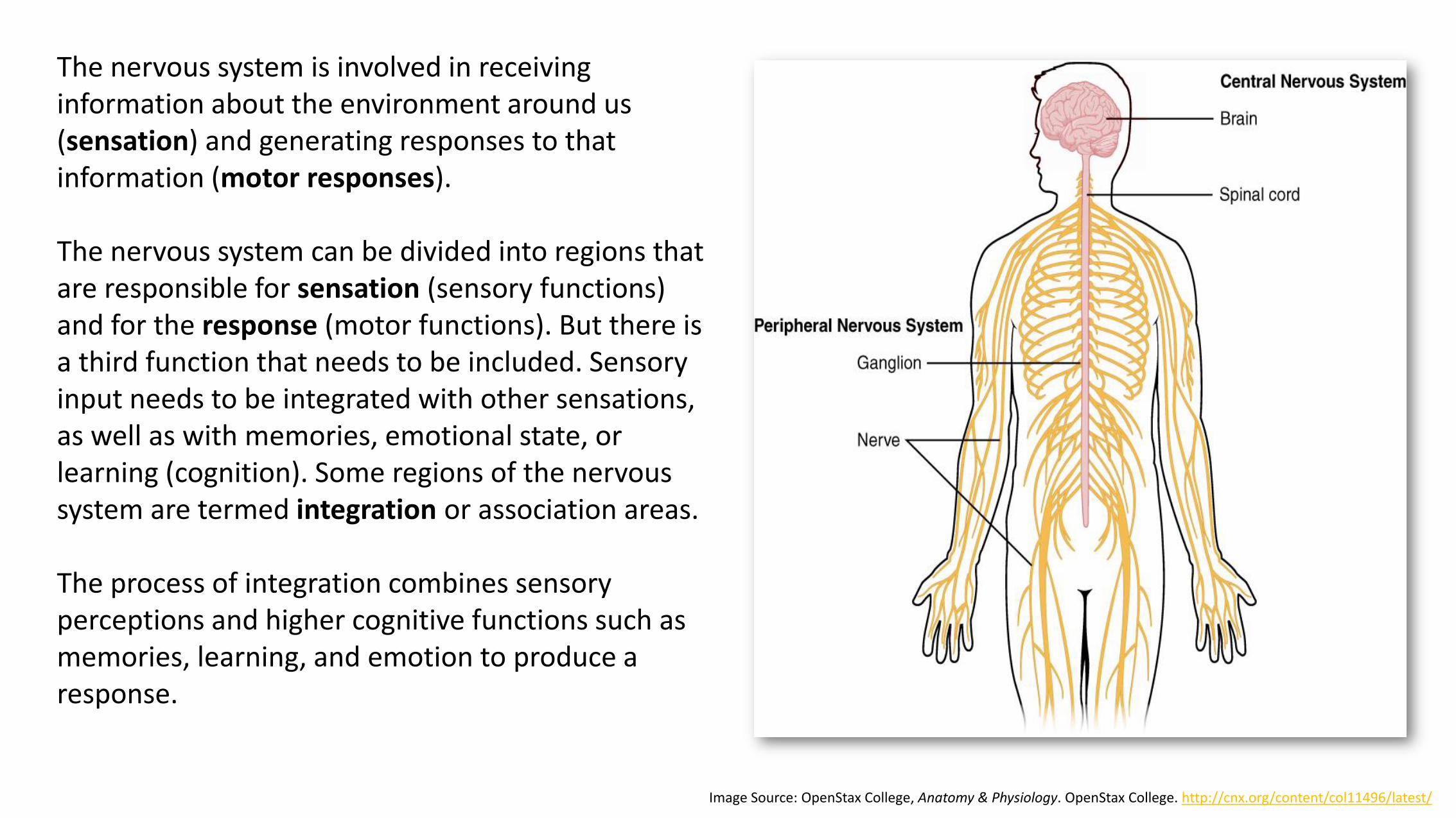
The nervous system is involved in receiving information about the environment around us (sensation) and generating responses to that information (motor responses).
The nervous system can be divided into regions that are responsible for sensation (sensory functions) and for the response (motor functions). But there is a third function that needs to be included. Sensory input needs to be integrated with other sensations, as well as with memories, emotional state, or learning (cognition). Some regions of the nervous system are termed integration or association areas.
The process of integration combines sensory perceptions and higher cognitive functions such as memories, learning, and emotion to produce a response.
Image Source: OpenStax College, Anatomy & Physiology. OpenStax College. http://cnx.org/content/col11496/latest/
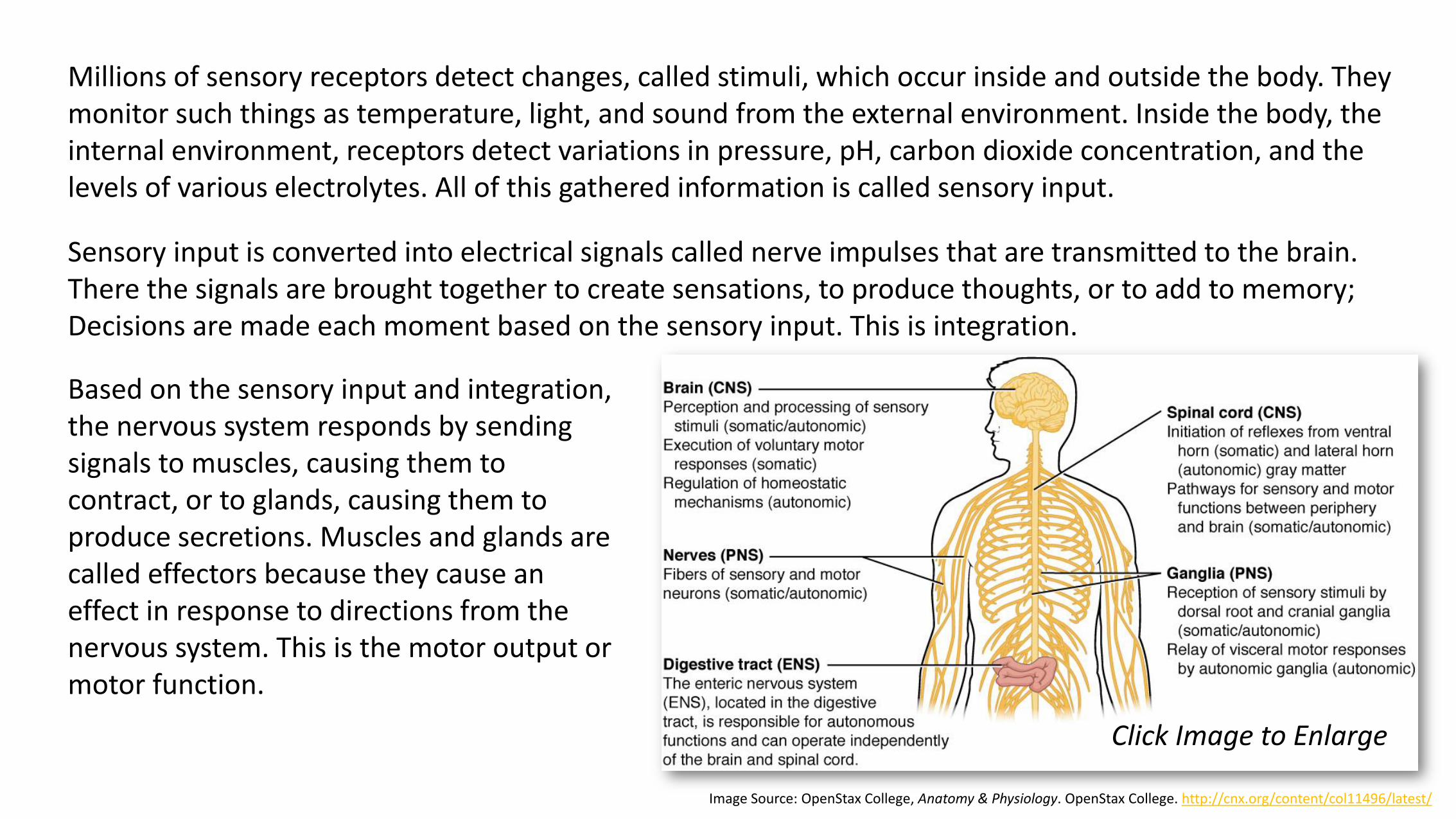
Millions of sensory receptors detect changes, called stimuli, which occur inside and outside the body. They monitor such things as temperature, light, and sound from the external environment. Inside the body, the internal environment, receptors detect variations in pressure, pH, carbon dioxide concentration, and the levels of various electrolytes. All of this gathered information is called sensory input.
Image Source: OpenStax College, Anatomy & Physiology. OpenStax College. http://cnx.org/content/col11496/latest/
Click Image to Enlarge
Sensory input is converted into electrical signals called nerve impulses that are transmitted to the brain. There the signals are brought together to create sensations, to produce thoughts, or to add to memory; Decisions are made each moment based on the sensory input. This is integration.
Based on the sensory input and integration, the nervous system responds by sending signals to muscles, causing them to contract, or to glands, causing them to produce secretions. Muscles and glands are called effectors because they cause an effect in response to directions from the nervous system. This is the motor output or motor function.
NERVE TISSUE
Nervous tissue is composed of two types of cells, neurons and glial cells. Neurons are the primary type of cell that most anyone associates with the nervous system. They are responsible for the computation and communication that the nervous system provides. They are electrically active and release chemical signals to target cells. Glial cells, or glia, are known to play a supporting role for nervous tissue. Ongoing research pursues an expanded role that glial cells might play in signaling, but neurons are still considered the basis of this function. Neurons are important, but without glial support they would not be able to perform their function.
NEURONS
Neurons, or nerve cells, carry out the functions of the nervous system by conducting nerve impulses.
They are highly specialized and amitotic. This means that if a neuron is destroyed, it cannot be replaced
because neurons do not go through mitosis. Each neuron has three basic parts: cell body (soma), one or
more dendrites, and a single axon.
The main part of a neuron is the cell body, which is also known as the soma (soma =“body”). The cell
body contains the nucleus and most of the major organelles. But what makes neurons special is that
they have many extensions of their cell membranes, which are generally referred to as processes.

Neurons are usually described as having one, and only one, axon—a fiber that emerges from the cell body and projects to target cells. That single axon can branch repeatedly to communicate with many target cells (conducts nerve pulses away from the body). It is the axon that propagates the nerve impulse, which is communicated to one or more cells. The axon is covered by a fatty tissue called the myelin sheath. This protective sheath prevents the nerve impulse from transmitting in the wrong direction.
Image Source: OpenStax College, Anatomy & Physiology. OpenStax College. http://cnx.org/content/col11496/latest/
The other processes of the neuron are dendrites,
which receive information from other neurons at
specialized areas of contact called synapses. The
dendrites are usually highly branched processes,
providing locations for other neurons to
communicate with the cell body (conduct nerve
impulses towards the body). Information flows
through a neuron from the dendrites, across the
cell body, and down the axon. This gives the
neuron a polarity—meaning that information
flows in this one direction. Click Image to Enlarge

Looking at nervous tissue, there are regions that predominantly contain cell bodies and regions that are largely composed of just axons. These two regions within nervous system structures are often referred to as gray matter (the regions with many cell bodies and dendrites) or white matter (the regions with many axons). Gray matter is not necessarily gray. It can be pinkish because of blood content, or even slightly tan, depending on how long the tissue has been preserved. But white matter is white because axons are insulated by a lipid-rich substance called myelin. Lipids can appear as white (“fatty”) material, much like the fat on a raw piece of chicken or beef. Actually, gray matter may have that color ascribed to it because next to the white matter, it is just darker—hence, gray.
Image Source: OpenStax College, Anatomy & Physiology. OpenStax College. http://cnx.org/content/col11496/latest/
Functionally, neurons are classified as afferent, efferent, or interneurons (association neurons) according to the direction in which they transmit impulses relative to the central nervous system.
Afferent, or sensory, neurons carry impulses from peripheral sense receptors to the CNS. They usually have long dendrites and relatively short axons.
Efferent, or motor, neurons transmit impulses from the CNS to effector organs such as muscles and glands. Efferent neurons usually have short dendrites and long axons.
Interneurons, or association neurons, are located entirely within the CNS in which they form the connecting link between the afferent and efferent neurons. They make more complex types of reflexes possible and have short dendrites and may have either a short or long axon.
GLIAL CELLS
Glial cells, or neuroglia or simply glia, are the other type of cell found in nervous tissue. They do not conduct nerve impulses but instead are considered to be supporting cells, and many functions are directed at helping neurons complete their function for communication. The name glia comes from the Greek word that means “glue.” They are far more numerous than neurons and, unlike neurons, are capable of mitosis. Each of the three types of cells serve a different purpose.
Astrocytes (star-shaped) are supporting cells for the neurons in the central nervous system. Some ways in which they support neurons in the central nervous system are by maintaining the concentration of chemicals in the extracellular space, removing excess signaling molecules, reacting to tissue damage, and contributing to the blood-brain barrier (BBB). The blood-brain barrier is a physiological barrier that keeps many substances that circulate in the rest of the body from getting into the central nervous system, restricting what can cross from circulating blood into the CNS.
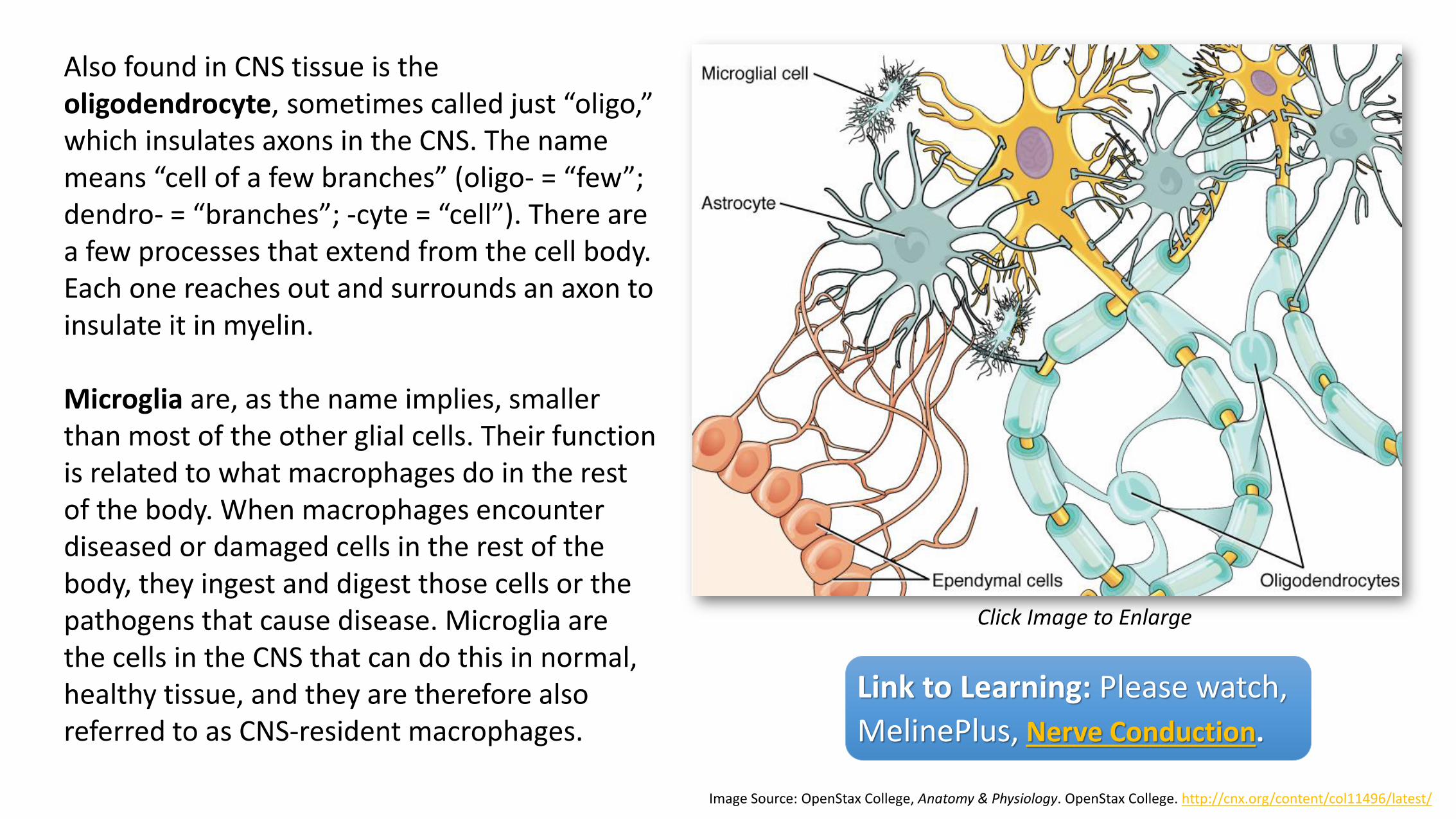
Also found in CNS tissue is the oligodendrocyte, sometimes called just “oligo,” which insulates axons in the CNS. The name means “cell of a few branches” (oligo- = “few”; dendro- = “branches”; -cyte = “cell”). There are a few processes that extend from the cell body. Each one reaches out and surrounds an axon to insulate it in myelin.
Microglia are, as the name implies, smaller than most of the other glial cells. Their function is related to what macrophages do in the rest of the body. When macrophages encounter diseased or damaged cells in the rest of the body, they ingest and digest those cells or the pathogens that cause disease. Microglia are the cells in the CNS that can do this in normal, healthy tissue, and they are therefore also referred to as CNS-resident macrophages.
Image Source: OpenStax College, Anatomy & Physiology. OpenStax College. http://cnx.org/content/col11496/latest/
Click Image to Enlarge
Link to Learning: Please watch,
MelinePlus, Nerve Conduction.
For an overview of what you have learned, please watch, Neurons or Nerve Cells – Structure, function and types of neurons.

THE CENTRAL AND PERIPHERAL NERVOUS SYSTEM
The nervous system can be divided into two major regions: the central and peripheral nervous systems. The central nervous system (CNS) is the brain and spinal cord, and the organs of the peripheral nervous system (PNS) are the nerves and ganglia.
THE CENTRAL NERVOUS SYSTEM
The CNS consists of the brain and spinal cord, which are located in the dorsal body cavity. The brain is surrounded by the cranium, and the spinal cord is protected by the vertebrae. The brain is continuous with the spinal cord at the foramen magnum. In addition to bone, the CNS is surrounded by connective tissue membranes, called meninges, and a series of interconnected, fluid-filled cavities. The cavities are called ventricles and the fluid is cerebrospinal fluid (CSF).
MENINGES
There are three layers of meninges around the brain and spinal cord. The outer layer, the dura mater, is
tough white fibrous connective tissue. The middle layer of meninges is arachnoid, which resembles a
cobweb in appearance, is a thin layer with numerous threadlike strands that attach it to the innermost
layer. The space under the arachnoid, the subarachnoid space, is filled with cerebrospinal fluid and
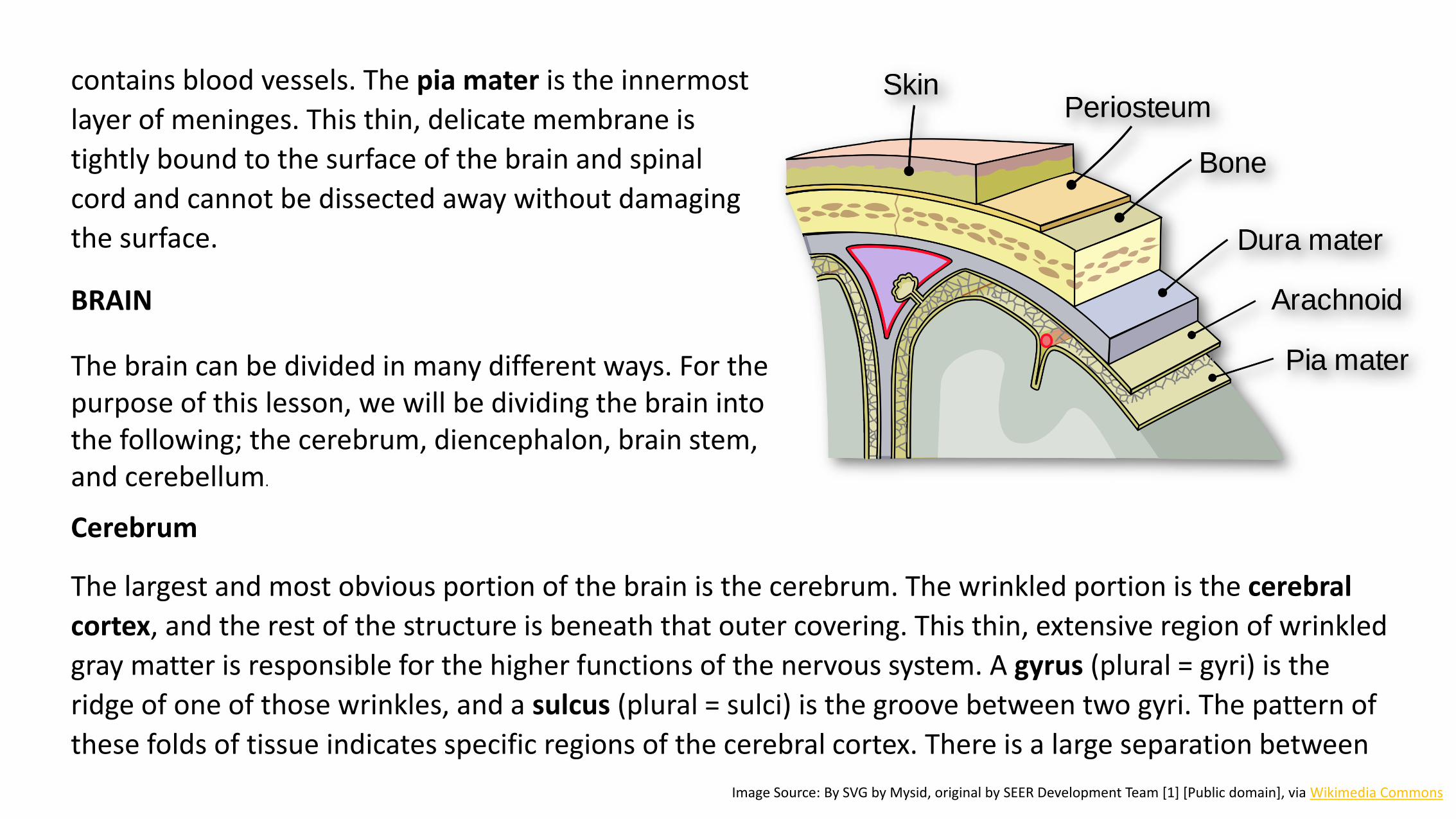
contains blood vessels. The pia mater is the innermost
layer of meninges. This thin, delicate membrane is
tightly bound to the surface of the brain and spinal
cord and cannot be dissected away without damaging
the surface.
Cerebrum
The largest and most obvious portion of the brain is the cerebrum. The wrinkled portion is the cerebral
cortex, and the rest of the structure is beneath that outer covering. This thin, extensive region of wrinkled
gray matter is responsible for the higher functions of the nervous system. A gyrus (plural = gyri) is the
ridge of one of those wrinkles, and a sulcus (plural = sulci) is the groove between two gyri. The pattern of
these folds of tissue indicates specific regions of the cerebral cortex. There is a large separation between
Image Source: By SVG by Mysid, original by SEER Development Team [1] [Public domain], via Wikimedia Commons
BRAIN
The brain can be divided in many different ways. For the purpose of this lesson, we will be dividing the brain into the following; the cerebrum, diencephalon, brain stem, and cerebellum.
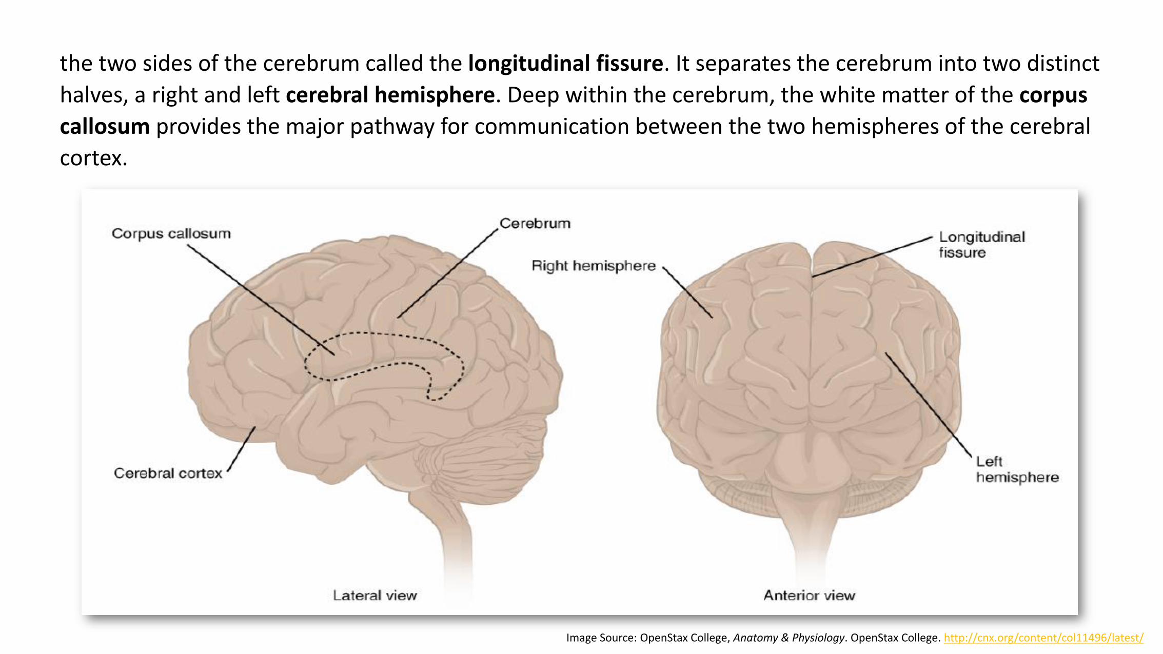
the two sides of the cerebrum called the longitudinal fissure. It separates the cerebrum into two distinct
halves, a right and left cerebral hemisphere. Deep within the cerebrum, the white matter of the corpus
callosum provides the major pathway for communication between the two hemispheres of the cerebral
cortex.
Image Source: OpenStax College, Anatomy & Physiology. OpenStax College. http://cnx.org/content/col11496/latest/

• Frontal lobe: associated with voluntary
motor functions.
• Parietal lobe: associated with the general
sensations associated with the body as
well as taste.
• Occipital lobe: associated with vision.
• Temporal lobe: associated with primary
auditory sensation, memory and emotion.
Each cerebral hemisphere is divided
into five lobes, four of which have the
same name as the bone over them:
A fifth lobe, the insula or Island of Reil, lies deep within the lateral sulcus.
Image Source: OpenStax CNX, The Brain and Spinal Cord. OpenStax College. https://cnx.org/contents/_Io4zP0c@7/The-Brain-and-Spinal-Cord
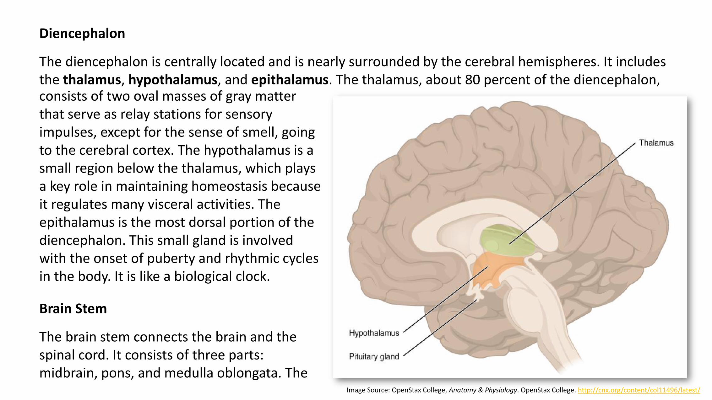
Diencephalon
The diencephalon is centrally located and is nearly surrounded by the cerebral hemispheres. It includes the thalamus, hypothalamus, and epithalamus. The thalamus, about 80 percent of the diencephalon,
Image Source: OpenStax College, Anatomy & Physiology. OpenStax College. http://cnx.org/content/col11496/latest/
consists of two oval masses of gray matter that serve as relay stations for sensory impulses, except for the sense of smell, going to the cerebral cortex. The hypothalamus is a small region below the thalamus, which plays a key role in maintaining homeostasis because it regulates many visceral activities. The epithalamus is the most dorsal portion of the diencephalon. This small gland is involved with the onset of puberty and rhythmic cycles in the body. It is like a biological clock.
Brain Stem
The brain stem connects the brain and the spinal cord. It consists of three parts: midbrain, pons, and medulla oblongata. The
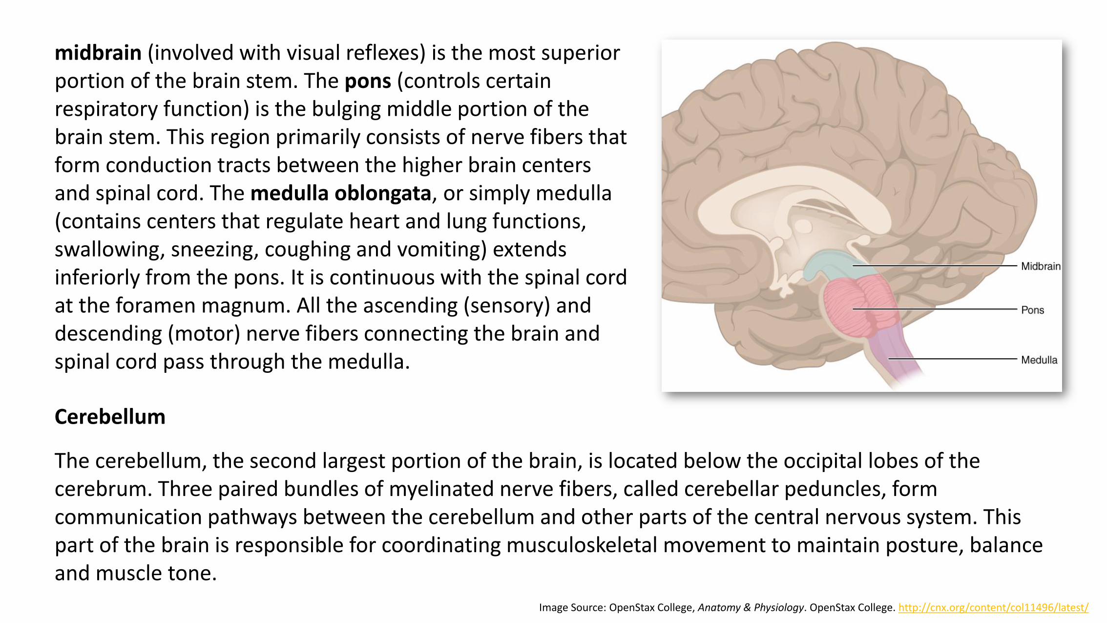
Image Source: OpenStax College, Anatomy & Physiology. OpenStax College. http://cnx.org/content/col11496/latest/
midbrain (involved with visual reflexes) is the most superior portion of the brain stem. The pons (controls certain respiratory function) is the bulging middle portion of the brain stem. This region primarily consists of nerve fibers that form conduction tracts between the higher brain centers and spinal cord. The medulla oblongata, or simply medulla (contains centers that regulate heart and lung functions, swallowing, sneezing, coughing and vomiting) extends inferiorly from the pons. It is continuous with the spinal cord at the foramen magnum. All the ascending (sensory) and descending (motor) nerve fibers connecting the brain and spinal cord pass through the medulla.
Cerebellum
The cerebellum, the second largest portion of the brain, is located below the occipital lobes of the cerebrum. Three paired bundles of myelinated nerve fibers, called cerebellar peduncles, form communication pathways between the cerebellum and other parts of the central nervous system. This part of the brain is responsible for coordinating musculoskeletal movement to maintain posture, balance and muscle tone.

SPINAL CORD
The spinal cord extends from the foramen magnum at the base of the skull to the level of the first lumbar vertebra. The cord is continuous with the medulla oblongata at the foramen magnum. Like the brain, the spinal cord is surrounded by bone, meninges, and cerebrospinal fluid.
The spinal cord is divided into 31 segments with each segment giving rise to a pair of spinal nerves. At the distal end of the cord, many spinal nerves extend beyond the conus medullaris to form a collection that resembles a horse's tail. This is the cauda equina. In cross section, the spinal cord appears oval in shape.
The spinal cord has two main functions:
• Serving as a conduction pathway for impulses going to and from the brain. Sensory impulses travel to the brain on ascending tracts in the cord. Motor impulses travel on descending tracts.
• Serving as a reflex center. The reflex arc is the functional unit of the nervous system. Reflexes are responses to stimuli that do not require conscious thought and consequently, they occur more quickly than reactions that require thought processes. For example, with the withdrawal reflex, the reflex action withdraws the affected part before you are aware of the pain. Many reflexes are mediated in the spinal cord without going to the higher brain centers.

Please watch, 3D Medical Animation – Central Nervous System, for an overview on what you have learned so far.
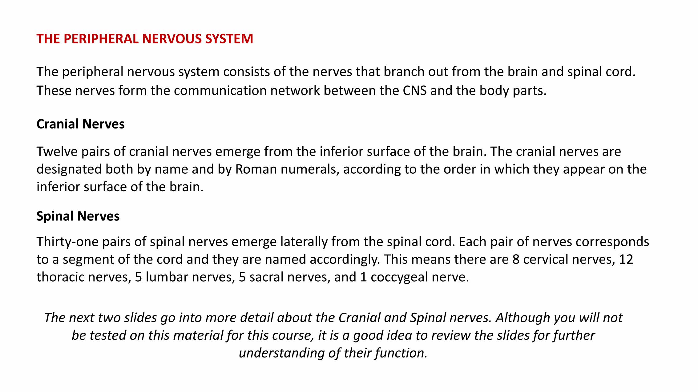
THE PERIPHERAL NERVOUS SYSTEM
The peripheral nervous system consists of the nerves that branch out from the brain and spinal cord.
These nerves form the communication network between the CNS and the body parts.
Cranial Nerves
Twelve pairs of cranial nerves emerge from the inferior surface of the brain. The cranial nerves are designated both by name and by Roman numerals, according to the order in which they appear on the inferior surface of the brain.
Spinal Nerves
Thirty-one pairs of spinal nerves emerge laterally from the spinal cord. Each pair of nerves corresponds to a segment of the cord and they are named accordingly. This means there are 8 cervical nerves, 12 thoracic nerves, 5 lumbar nerves, 5 sacral nerves, and 1 coccygeal nerve.
The next two slides go into more detail about the Cranial and Spinal nerves. Although you will not be tested on this material for this course, it is a good idea to review the slides for further
understanding of their function.
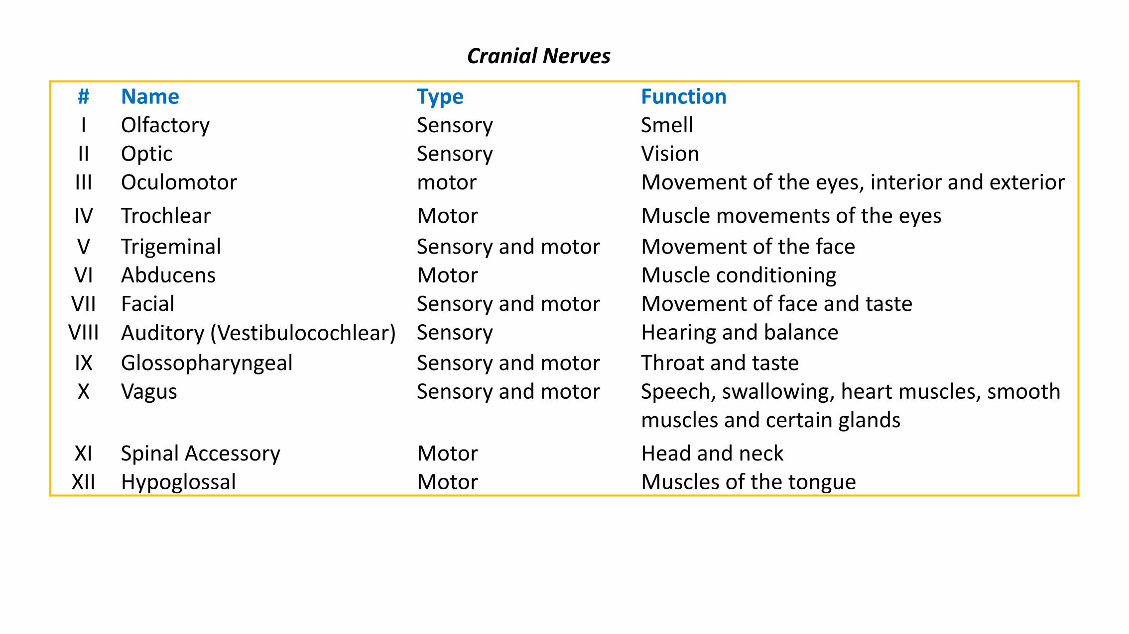
# Name Type FunctionI Olfactory Sensory SmellII Optic Sensory VisionIII Oculomotor motor Movement of the eyes, interior and exterior
IV Trochlear Motor Muscle movements of the eyes
V Trigeminal Sensory and motor Movement of the faceVI Abducens Motor Muscle conditioningVII Facial Sensory and motor Movement of face and tasteVIII Auditory (Vestibulocochlear) Sensory Hearing and balance
IX Glossopharyngeal Sensory and motor Throat and tasteX Vagus Sensory and motor Speech, swallowing, heart muscles, smooth
muscles and certain glands
XI Spinal Accessory Motor Head and neckXII Hypoglossal Motor Muscles of the tongue
Cranial Nerves

Each spinal nerve is connected to the spinal cord by a dorsal root and a ventral root. The spinal nerves exit the spinal cord and pass through the intervertebral foramen, then divide into branches called plexus.
• Cervical plexus - serves the head, neck and shoulders
• Brachial plexus - serves the chest, shoulders, arms and hands
• Lumbar plexus - serves the back, abdomen, groin, thighs, knees, and calves
• Sacral plexus - serves the pelvis, buttocks, genitals, thighs, calves, and feet
• Coccygeal plexus - serves a small region over the coccyx
Image Source: By OpenStax College [CC BY 3.0 (http://creativecommons.org/licenses/by/3.0)], via Wikimedia Commons
Click Image to Enlarge

The peripheral nerves can further be divided into two subsystems mostly on the basis of a functional difference in responses.
The somatic nervous system, also called the somatomotor or somatic efferent nervous system, supplies motor impulses to the skeletal muscles. Because these nerves permit conscious control of the skeletal muscles, it is sometimes called the voluntary nervous system.
The autonomic nervous system (involuntary nervous system) is a visceral efferent system, which means it sends motor impulses to the visceral organs (internal organs such as the heart, stomach and intestines). It functions automatically and continuously, without conscious effort, to innervate smooth muscle, cardiac muscle, and glands. It is concerned with heart rate, breathing rate, blood pressure, body temperature, and other visceral activities that work together to maintain homeostasis.
The autonomic nervous system has two parts, the sympathetic division and the parasympathetic division. The sympathetic nervous system is often considered the "fight or flight" system, while the parasympathetic nervous system is often considered the "rest and digest" or "feed and breed" system. In many cases, both of these systems have "opposite" actions where one system activates a physiological response and the other inhibits it.
Link to Learning: Please read: Merck Manuals: Nerves,
for an overview of the somatic and autonomic system.
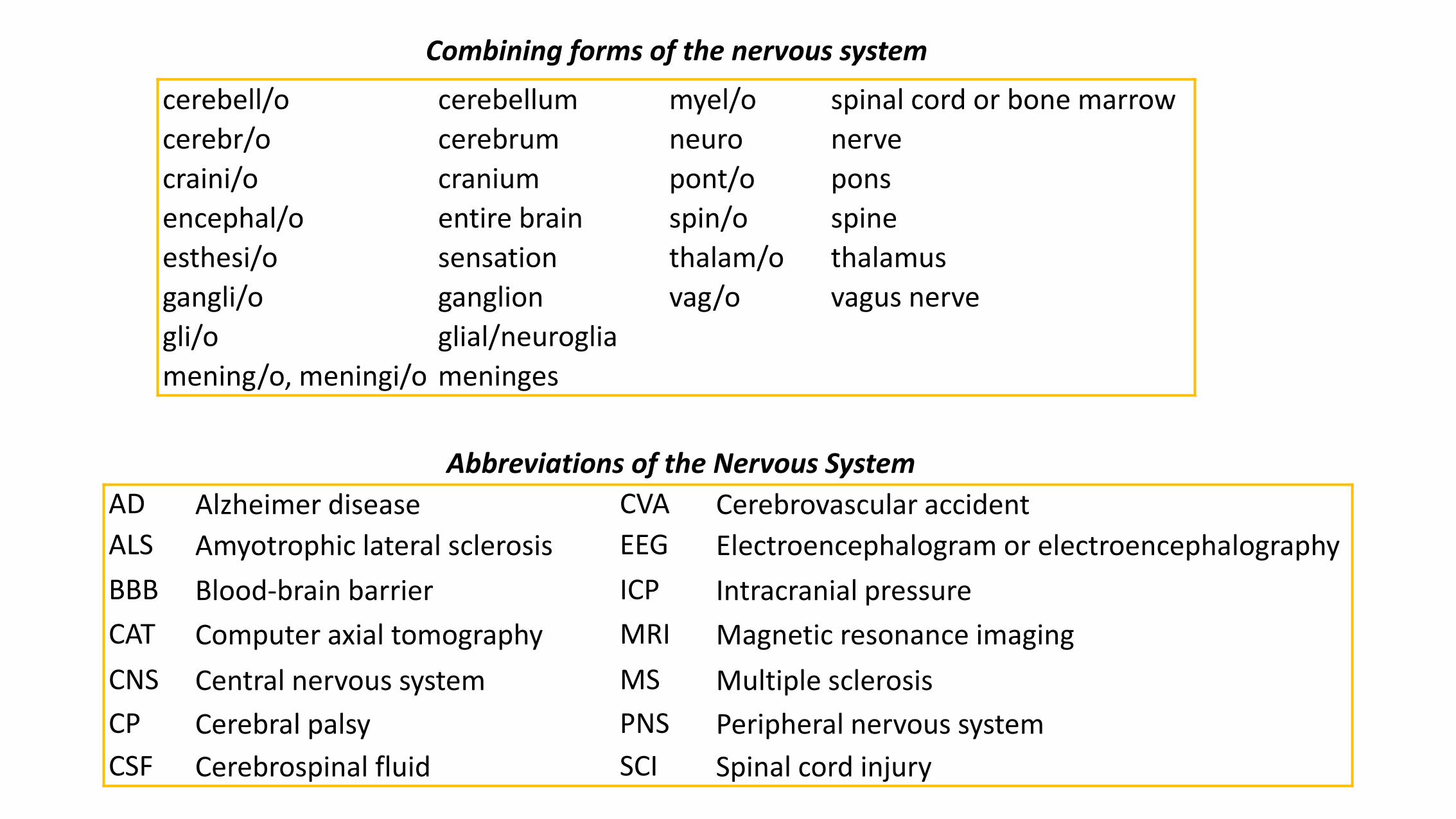
Combining forms of the nervous system
cerebell/o cerebellum myel/o spinal cord or bone marrow
cerebr/o cerebrum neuro nerve
craini/o cranium pont/o pons
encephal/o entire brain spin/o spine
esthesi/o sensation thalam/o thalamus
gangli/o ganglion vag/o vagus nerve
gli/o glial/neuroglia
mening/o, meningi/o meninges
Abbreviations of the Nervous System
AD Alzheimer disease CVA Cerebrovascular accident
ALS Amyotrophic lateral sclerosis EEG Electroencephalogram or electroencephalography
BBB Blood-brain barrier ICP Intracranial pressure
CAT Computer axial tomography MRI Magnetic resonance imaging
CNS Central nervous system MS Multiple sclerosis
CP Cerebral palsy PNS Peripheral nervous system
CSF Cerebrospinal fluid SCI Spinal cord injury
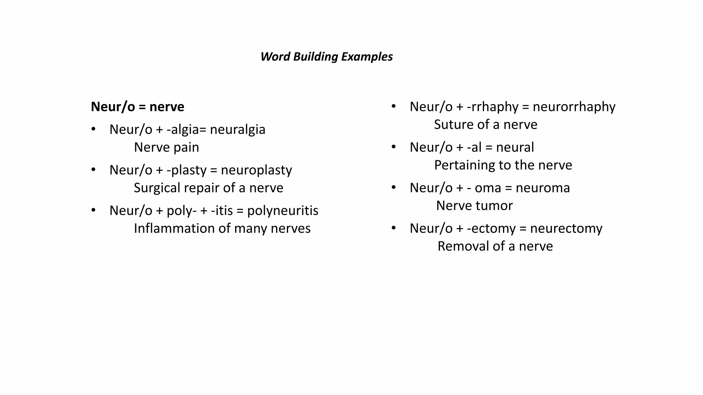
Word Building Examples
Neur/o = nerve
• Neur/o + -algia= neuralgiaNerve pain
• Neur/o + -plasty = neuroplastySurgical repair of a nerve
• Neur/o + poly- + -itis = polyneuritisInflammation of many nerves
• Neur/o + -rrhaphy = neurorrhaphySuture of a nerve
• Neur/o + -al = neuralPertaining to the nerve
• Neur/o + - oma = neuromaNerve tumor
• Neur/o + -ectomy = neurectomyRemoval of a nerve

TEST YOUR KNOWLEDGECombine the following word parts and then define the created word’s meaning. Click the image to compare your answers.
1. Cephal/o (head) + -algia = ?
2. Cerebell/o + -ar = ?
3. Cerebr/o + spin/o + -al = ?
4. Encephal/o + -itis = ?
5. Mening/o + -eal = ?
6. Neur/o + -pathy = ?
Answer:
1. Cephalagia = head pain2. Cerebellar = pertaining to cerebellum3. Cerebrospinal = pertaining to the cerebrum and spine4. Encephalitis = brain inflammation5. Meningeal = pertaining to the meninges6. Neuropathy = nerve disease
DIAGNOSTIC
Neurology is the branch of medicine that is concerned with the study and treatment of disorders of the
nervous system. The physician who specializes in neurology is called a neurologist. Neurologists treat
disorders of the brain, spinal cord and nerves. Depending on the diagnoses of the neurologist, he/she may
refer a patient to a neurosurgeon as neurologists do not do surgery.
Evaluating and diagnosing damage to the nervous system is complicated and complex. Many of the same
symptoms occur in different combinations among the different disorders. The two main components of a
neurologic examination are the medical history and the physical examination. If necessary, diagnostic
procedures are done to confirm the diagnosis or exclude other possible disorders.
• Arteriogram (angiogram): a procedure that provides a scan of arteries going to and through the brain. It is used to detect blockages of the arteries or veins.
• Biopsy: involves the removal and examination of a small piece of tissue from the body. Muscle or nerve biopsies are used to diagnose neuromuscular disorders and may also reveal if a person is a carrier of a defective gene that could be passed on to children.
• Brain scans: are imaging techniques used to diagnose tumors, blood vessel malformations, or
hemorrhage in the brain. These scans are used to study organ function or injury or disease to tissue or
muscle. Types of brain scans include: computed tomography, magnetic resonance imaging, and
positron emission tomography.
• Cerebrospinal fluid analysis (spinal tap): involves the removal of a small amount of the fluid that
protects the brain and spinal cord. The fluid is tested to detect any bleeding or brain hemorrhage,
diagnose infection to the brain and/or spinal cord, identify some cases of multiple sclerosis and other
neurological conditions, and measure intracranial pressure.
• Computed tomography: also known as a CT scan, is a noninvasive, painless process used to produce
rapid, clear, two-dimensional images of organs, bones, and tissues. Neurological CT scans are used to
view the brain and spine. They can detect bone and vascular irregularities, certain brain tumors and
cysts, herniated discs, epilepsy, encephalitis, spinal stenosis (narrowing of the spinal canal), a blood
clot or intracranial bleeding in patients with stroke, brain damage from head injury, and other
disorders.
• Electroencephalogram (EEG) -- a procedure that records the brain's continuous electrical activity by
means of electrodes attached to the scalp. EEG is used to help diagnose certain seizure disorders,
brain tumors, brain damage from head injuries, inflammation of the brain and/or spinal cord, alcoholism,
certain psychiatric disorders, and metabolic and degenerative disorders that affect the brain.
• Electromyogram (EMG): a procedure that measures and records electrical activity from the muscles and nerves with mild electrical shocks to stimulate the nerves. It is used to diagnose nerve and muscle dysfunction and spinal cord disease.
• Evoked potentials: procedures that record the brain's electrical response to visual, auditory, and sensory stimuli. These tests are used to assess sensory nerve problems and confirm neurological conditions including multiple sclerosis, brain tumor, acoustic neuroma (small tumors of the inner ear), and spinal cord injury.
• Magnetic resonance imaging (MRI): uses computer-generated radio waves and a powerful magnetic
field to produce detailed images of body structures including tissues, organs, bones, and
nerves. Neurological uses include the diagnosis of brain and spinal cord tumors, eye disease,
inflammation, infection, and vascular irregularities that may lead to stroke. MRI can also detect and
monitor degenerative disorders such as multiple sclerosis and can document brain injury from trauma.
• Myelography: involves the injection of a water- or oil-based contrast dye into the spinal canal to
enhance x-ray imaging of the spine. Myelograms are used to diagnose spinal nerve injury, herniated
discs, fractures, back or leg pain, and spinal tumors.

• Positron emission tomography (PET scans): provide two- and three-dimensional pictures of brain
activity by measuring radioactive isotopes that are injected into the bloodstream. PET scans of the brain
are used to detect or highlight tumors and diseased tissue, measure cellular and/or tissue metabolism,
show blood flow, evaluate patients who have seizure disorders that do not respond to medical therapy
and patients with certain memory disorders, and determine brain changes following injury or drug
abuse, among other uses.
• Polysomnogram: measures brain and body activity during sleep. It is performed over one or more
nights at a sleep center. Electrodes are pasted or taped to the patient’s scalp, eyelids, and/or
chin. Throughout the night and during the various wake/sleep cycles, the electrodes record brain waves,
eye movement, breathing, leg and skeletal muscle activity, blood pressure, and heart rate. Results are
then used to identify any characteristic patterns of sleep disorders, including restless legs syndrome,
periodic limb movement disorder, insomnia, and breathing disorders such as obstructive sleep apnea.
• Single photon emission computed tomography (SPECT): a nuclear imaging test involving blood flow to
tissue, is used to evaluate certain brain functions. A radioactive isotope, which binds to chemicals that
flow to the brain, is injected intravenously into the body. Areas of increased blood flow will collect more
of the isotope.
• Ultrasound imaging: also called ultrasound scanning or sonography, uses high-frequency sound waves
to obtain images inside the body. Neurosonography (ultrasound of the brain and spinal column)
analyzes blood flow in the brain and can diagnose stroke, brain tumors, hydrocephalus (build-up of
cerebrospinal fluid in the brain), and vascular problems. It can also identify or rule out inflammatory
processes causing pain.
Link to Learning: Please read: White-Wilson
Medical Center: Neurological Examination.
PATHOLOGICAL TERMS
When something goes wrong with a part of your nervous system, you can have trouble moving, speaking,
swallowing, breathing, or learning. You can also have problems with your memory, senses, or mood.
Neurological disorders can be caused by:
• Injuries: such as trauma to the spinal cord or brain.
• Vascular disorders: such as stroke, transient ischemic attack (TIA), subarachnoid hemorrhage,
subdural hemorrhage and hematoma, and extradural hemorrhage.
• Infections: such as meningitis, encephalitis, polio, and epidural abscess.
• Structural disorders: such as brain or spinal cord injury, Bell's palsy, cervical spondylosis, carpal tunnel syndrome, brain or spinal cord tumors, peripheral neuropathy, and Guillain-Barre syndrome.
• Functional disorders: such as headache, epilepsy, dizziness, and neuralgia.
• Degeneration: such as Parkinson's disease, multiple sclerosis, amyotrophic lateral sclerosis (ALS), Huntington's chorea, and Alzheimer's disease.
According to the US National Library of Medicine, there are over 600 neurological diseases and disorders, the most common are listed below.
• Agnosia: the inability to process sensory information.
• Alzheimer's Disease: progressive degenerative disorder of the brain that affects the ability to carry out daily activities of living.
• Amnesia: a deficit in memory caused by brain damage, disease, or psychological trauma.
• Amyotrophic Lateral Sclerosis (ALS): also known as Lou Gehrig's disease. A degenerative disease of neurons resulting in progressive weakness.
• Aneurysm: is a localized, blood-filled balloon-like bulge in the wall of a blood vessel.

• Aphasia: loss of speech.
• Apraxia: a motor disorder caused by damage to the brain, resulting in the inability to properly use familiar objects.
• Ataxia: a term for a group of disorders that affect co-ordination, balance and speech. Usually results from disorders of the cerebellum or spinal cord.
• Bell's Palsy: damage to facial nerve resulting in paralysis & drooping on one side of face.
• Brain contusion: bruising on the surface of the brain.
• Carpal tunnel syndrome: occurs when the median nerve, which runs from the forearm into the palm of the hand, becomes pressed or squeezed at the wrist.
• Cerebral Palsy: brain damage before or during birth resulting in poor muscle control & spasticity.
• Cerbrovascular accident: decreased blood supply to brain caused by clot / hemorrhage (stroke).
• Coma: a state of unconsciousness in which a person: cannot be awakened; fails to respond normally to painful stimuli, light, or sound; lacks a normal wake-sleep cycle; and does not initiate voluntary actions.
• Concussion: the most minor form of brain injury. Technically, a concussion is a short loss of normal brain function in response to a head injury, but people often use it to describe any minor injury to the head or brain.
• Dementia: a broad category of brain diseases that cause a long term and often gradual decrease in the ability to think and remember that is great enough to affect a person's daily functioning.
• Encephalitis: severe inflammation of the brain commonly caused by virus (complication of flu).
• Epilepsy: abnormal electrical impulses in brain that cause bursts of excitement and can result in seizures.
• Huntington disease: a hereditary neurological disease that is characterized by irregular and involuntary movements of the muscles and progressive loss of cognitive ability.
• Hydrocephalus: excessive fluid within the brain.
• Meningitis: inflammation of the covering of the brain and spinal cord (meninges).
• Multiple Sclerosis: an auto-immune disorder that causes destruction of myelin sheath resulting in episodic tremors, weakness, mood swings & vision changes.
• Myasthenia gravis: chronic autoimmune neuromuscular disease characterized by varying degrees of weakness of the skeletal (voluntary) muscles of the body.
• Narcolepsy: a chronic neurological disorder involving the loss of the brain's ability to regulate sleep-wake cycles.
• Neuralgia: nerve pain caused by an irritated or damaged nerve.
• Paraplegia: paralysis from the waist down usually the result of damage / trauma to the spinal cord.
• Parkinson's disease: degeneration of nerve cells in brain that control movement, resulting in tremors.
• Quadriplegia: paralysis from the shoulders down (respiratory assist), usually a result of damage / trauma to spinal cord.
• Sciatica: inflammation of sciatic nerve that results in pain in thigh and leg (longest nerve in the body).
• Seizure Disorders: disturbance in brain function causing uncontrollable contractions in muscles.
• Shingles: a viral disease affecting the peripheral nerves resulting in a painful rash on the skin.
• Somnambulism: sleep walking.
• Spina Bifida: birth defect where the spinal cord is not completely enclosed.
• Tay-Sachs disease: is a rare autosomal recessive genetic disorder that causes a progressive deterioration of the central nervous system and eventually death.
• Tourette syndrome (TS): a neurological disorder characterized by repetitive, stereotyped, involuntary movements and vocalizations called tics.
• Transient ischemic attack (TIA): a “mini” stroke that comes and goes quickly. It happens when the blood supply to part of the brain stops briefly. Symptoms of a TIA are like other stroke symptoms, but do not last as long.

SURGICAL TERMS:Some treatments for nervous system diseases and disorders include surgery, most of which are performed by a neurosurgeon, a specialist who performs surgery on the nervous system, especially the spinal cord and brain.
• Cordotomy: removal of part of the spinal cord in order to relieve pain.
• Craniectomy: removing part of the skull in order to relieve pressure.
• Craniotomy: a surgical operation in which a bone flap is temporarily removed from the skull to access the brain.
• Lobectomy: is the surgical removal of a lobe of an organ. It most often refers to the removal of a section of a lung. It may also refer to the liver, brain, thyroid gland, and other organs.
• Lobotomy: an incision into the brain that severs the connections in the brain's prefrontal lobe.
• Neurectomy: surgical removal of all or part of a nerve.
• Neuroplasty: surgical repair of the nerves.
• Neurorrhaphy: the suturing of severed nerves.
• Trephination: a circular incision into the skull.
PHARMACOLOGICAL TERMS:
• Analgesics: alleviate pain with no effect on the cause.
• Anorexiants: suppress appetite.
• Anticonvulsants: prevent or reduce the severity and frequency of seizures in various types of epilepsy.
• Anxiolytics, sedatives and hypnotics: treat anxiety and insomnia.
• Depressants or relaxants: slow down the activity of the CNS, which often results in the user feeling less pain, more relaxed and sleepy.
• Muscle relaxants: reduce tension in muscles.
• Narcotics: treat severe pain.
• Stimulants: speed up the activity of a person's central nervous system (CNS) including the brain. They are used to treat attention-deficit hyperactivity disorder (ADHD) and narcolepsy.
Treatment for the nervous system depends on the cause of the problem.

PRONUNCIATION
Practice pronouncing each term, then click the audio icon to hear it.
Analgesic(an-al-JE-zik)
An agent that relieves pain.
Astrocytoma(AS-tro-si-TOE-ma)
A nerve-tissue tumor composed of astrocytes.
Craniectomy(kray-nee-EK-toe-me)
The surgical removal of a portion of the skull.
Cerebellitis(ser-e-bell-EYE-tis)
Inflammation of the cerebellum.
Dysphasia(dys-FAY-zhe-a)
Loss of or deficiency in the power to use or understand language as a result of injury to or disease of the brain.
Embolus(EM-bo-lus)
An abnormal particle (as an air bubble or clot) circulating in the blood.
Encephalitis(en-sef-a-LIE-tis)
Inflammation of the brain.
Audio and Definition Source: MedlinePlus Medical Dictionary, https://www.nlm.nih.gov/medlineplus/mplusdictionary.html, public domain.
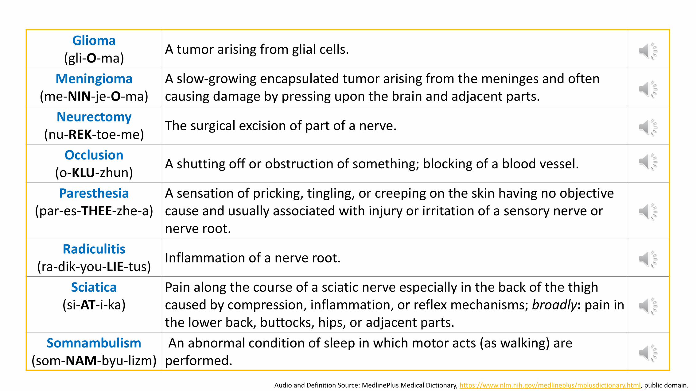
Glioma(gli-O-ma)
A tumor arising from glial cells.
Meningioma(me-NIN-je-O-ma)
A slow-growing encapsulated tumor arising from the meninges and often causing damage by pressing upon the brain and adjacent parts.
Neurectomy(nu-REK-toe-me)
The surgical excision of part of a nerve.
Occlusion(o-KLU-zhun)
A shutting off or obstruction of something; blocking of a blood vessel.
Paresthesia(par-es-THEE-zhe-a)
A sensation of pricking, tingling, or creeping on the skin having no objective cause and usually associated with injury or irritation of a sensory nerve or nerve root.
Radiculitis(ra-dik-you-LIE-tus)
Inflammation of a nerve root.
Sciatica(si-AT-i-ka)
Pain along the course of a sciatic nerve especially in the back of the thigh caused by compression, inflammation, or reflex mechanisms; broadly: pain in the lower back, buttocks, hips, or adjacent parts.
Somnambulism(som-NAM-byu-lizm)
An abnormal condition of sleep in which motor acts (as walking) are performed.
Audio and Definition Source: MedlinePlus Medical Dictionary, https://www.nlm.nih.gov/medlineplus/mplusdictionary.html, public domain.
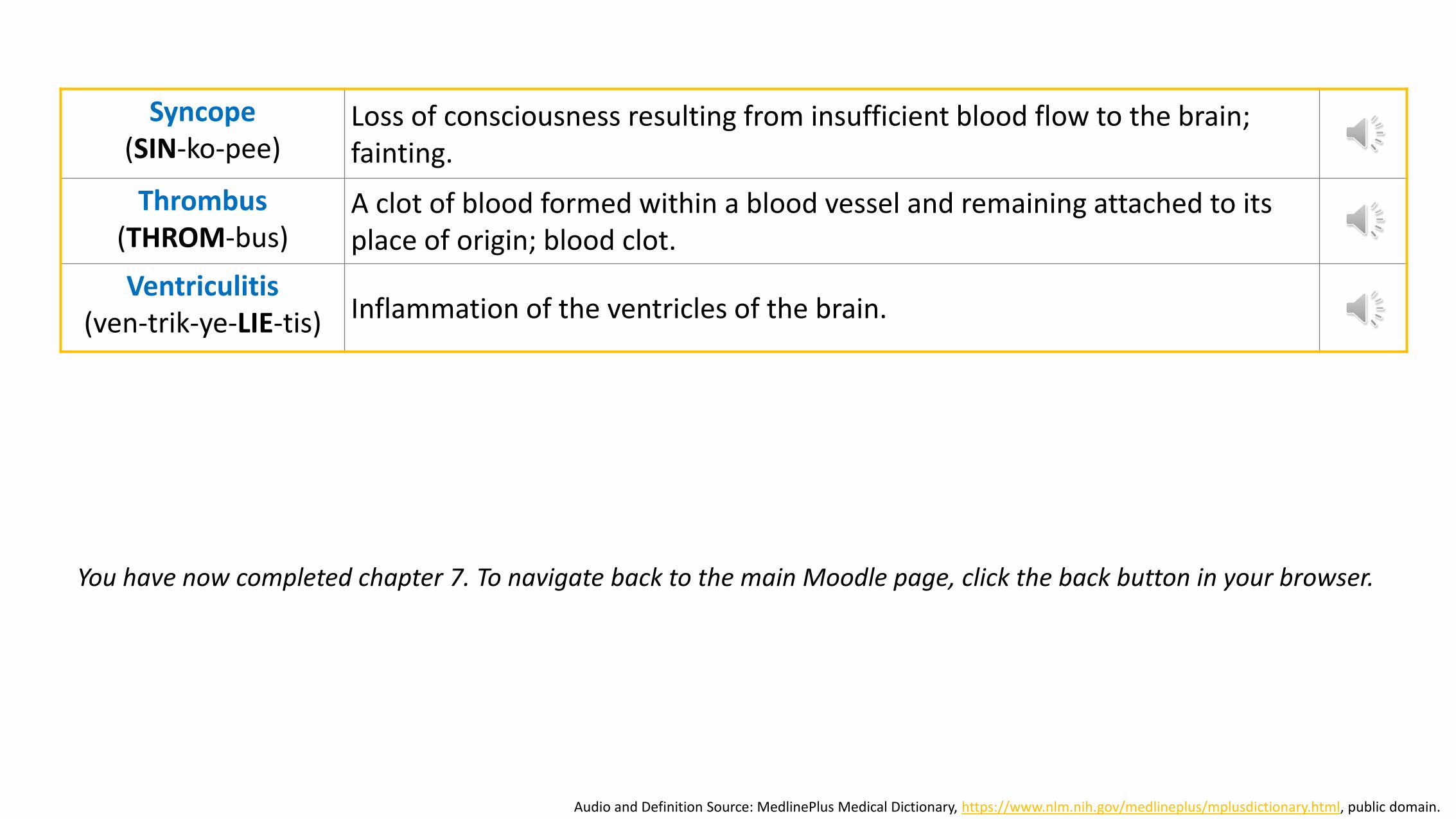
Syncope(SIN-ko-pee)
Loss of consciousness resulting from insufficient blood flow to the brain; fainting.
Thrombus(THROM-bus)
A clot of blood formed within a blood vessel and remaining attached to its place of origin; blood clot.
Ventriculitis(ven-trik-ye-LIE-tis) Inflammation of the ventricles of the brain.
Audio and Definition Source: MedlinePlus Medical Dictionary, https://www.nlm.nih.gov/medlineplus/mplusdictionary.html, public domain.
You have now completed chapter 7. To navigate back to the main Moodle page, click the back button in your browser.