chapter 6 seeley - · pdf file6-1 chapter 6 skeletal system: ... 6-4 hyaline cartilage ......
TRANSCRIPT

1
6-1
Chapter 6Skeletal System:
Bones and Bone Tissue
6-2
Skeletal System Functions• Support. Bone is hard and rigid; cartilage is
flexible yet strong. Cartilage in nose, external ear, thoracic cage and trachea. Ligaments- bone to bone
• Protection. Skull around brain; ribs, sternum, vertebrae protect organs of thoracic cavity
• Movement. Produced by muscles on bones, via tendons. Ligaments allow some movement between bones but prevent excessive movement
• Storage. Ca and P. Stored then released as needed. Fat stored in marrow cavities
• Blood cell production. Bone marrow that gives rise to blood cells and platelets

2
6-3
Components of Skeletal System
• Bone• Cartilage: three types
– Hyaline– Fibrocartilage– Elastic
6-4
Hyaline Cartilage• Consists of specialized cells that produce matrix
– Chondroblasts: form matrix– Chondrocytes: surrounded by matrix; are in lacunae
• Matrix. Collagen fibers for strength, proteoglycans for resiliency• Perichondrium. Double-layered C.T. sheath. Covers cartilage except at
articulations– Inner. More delicate, has fewer fibers, contains chondroblasts– Outer. Blood vessels and nerves penetrate. No blood vessels inside.– (Articular cartilage. Covers bones at joints; has no perichondrium)
• Growth– Appositional. New chondrocytes and new matrix at the periphery– Interstitial. Chondrocytes within the tissue divide and add more
matrix between the cells.

3
6-5
Bone Shapes• Long
– Ex. Upper and lower limbs
• Short– Ex. Carpals and tarsals
• Flat– Ex. Ribs, sternum,
skull, scapulae• Irregular
– Ex. Vertebrae, facial
6-6
Long Bone Structure
• Diaphysis– Shaft– Compact bone
• Epiphysis– End of the bone– Cancellous bone
• Epiphyseal plate: growth plate– Hyaline cartilage; present until growth
stops• Epiphyseal line: bone stops growing in
length • Medullary cavity: In children medullary
cavity is red marrow, gradually changes to yellow in limb bones and skull
• Red marrow & Yellow marrow

4
6-7
Long Bone Structure,
cont.
• Periosteum– Outer is fibrous – Inner is single layer of bone cells
including osteoblasts, osteoclasts and osteochondral progenitor cells
– Fibers of tendon become continuous with fibers of periosteum.
– Sharpey’s fibers: some periosteal fibers penetrate through the periosteum and into the bone. Strengthen attachment of tendon to bone.
• Endosteum. Similar to inner layer of periosteum. Lines all internal spaces including spaces in cancellous bone.
6-8
Flat, Short, Irregular Bones• Flat Bones
– No diaphyses, epiphyses– Sandwich of cancellous
between compact bone• Short and Irregular Bone
– Compact bone that surrounds cancellous bone center; similar to structure of epiphyses of long bones
– No diaphyses and not elongated
• Some flat and irregular bones of skull have sinuses lined by mucous membranes.

5
6-9
Bone Histology• Bone matrix. Like reinforced concrete. Rebar is
collagen fibers, cement is hydroxyapetite– Organic: collagen and proteoglycans– Inorganic: hydroxyapetite. CaPO4 crystals
• Bone cells (see following slides for particulars)– Osteoblasts– Osteocytes– Osteoclasts– (Stem cells or osteochondral progenitor cells)
• (Woven bone: collagen fibers randomly oriented)• (Lamellar bone: mature bone in sheets)• Cancellous bone: trabeculae• Compact bone: dense
6-10
Bone Matrix
• If mineral removed, bone is too bendable• If collagen removed, bone is too brittle

6
6-11
Bone Cells• Osteoblasts
– Formation of bone through ossification or osteogenesis. Collagen produced by E.R. and golgi. Released by exocytosis. Precursors of hydroxyapetite stored in vesicles, then released by exocytosis.
– Ossification: formation of bone by osteoblasts. Osteoblasts communicate through gap junctions. Cells surround themselves by matrix.
6-12
Osteocytes• Osteocytes. Mature bone
cells. Stellate. Surrounded by matrix, but can make small amounts of matrix to maintain it.– Lacunae: spaces occupied
by osteocyte cell body– Canaliculi: canals occupied
by osteocyte cell processes– Nutrients diffuse through
tiny amount of liquid surrounding cell and filling lacunae and canaliculi. Then can transfer nutrients from one cell to the next through gap junctions.

7
6-13
Osteoclasts and Stem Cells• Osteoclasts. Resorption of bone
– Ruffled border: where cell membrane borders bone and resorption is taking place.
– H ions pumped across membrane, acid forms, eats away bone.
– Release enzymes that digest the bone.– Derived from monocytes (which are formed from stem
cells in red bone marrow) + others=multinucleated
• Stem Cells. Mesenchyme (Osteochondral Progenitor Cells) become chondroblasts or osteoblasts.
6-14
Cancellous (Spongy) Bone
• Trabeculae: interconnecting rods or plates of bone. Like scaffolding. – Spaces filled with marrow. – Covered with endosteum.– Oriented along stress lines

8
6-15
Compact Bone• Central or Haversian canals:
parallel to long axis• Lamellae: concentric,
circumferential, interstitial• Osteon or Haversian system:
central canal, contents, associated concentric lamellae and osteocytes
• Perforating or Volkmann’s canal: perpendicular to long axis.
• Canaliculi—tiny canals• Circumferential & interstitial
lamellae (between osteons)
6-16
Bone Development: Two Types
• Intramembranous ossification– Takes place in membrane connective tissue– Fontanels—large membrane-covered spaces
• Endochondral ossification– Takes place in cartilage
• Both methods of ossification– Produce woven bone that is then remodeled

9
6-17
Endochondral Ossification
6-18
Endochondral Ossification
10
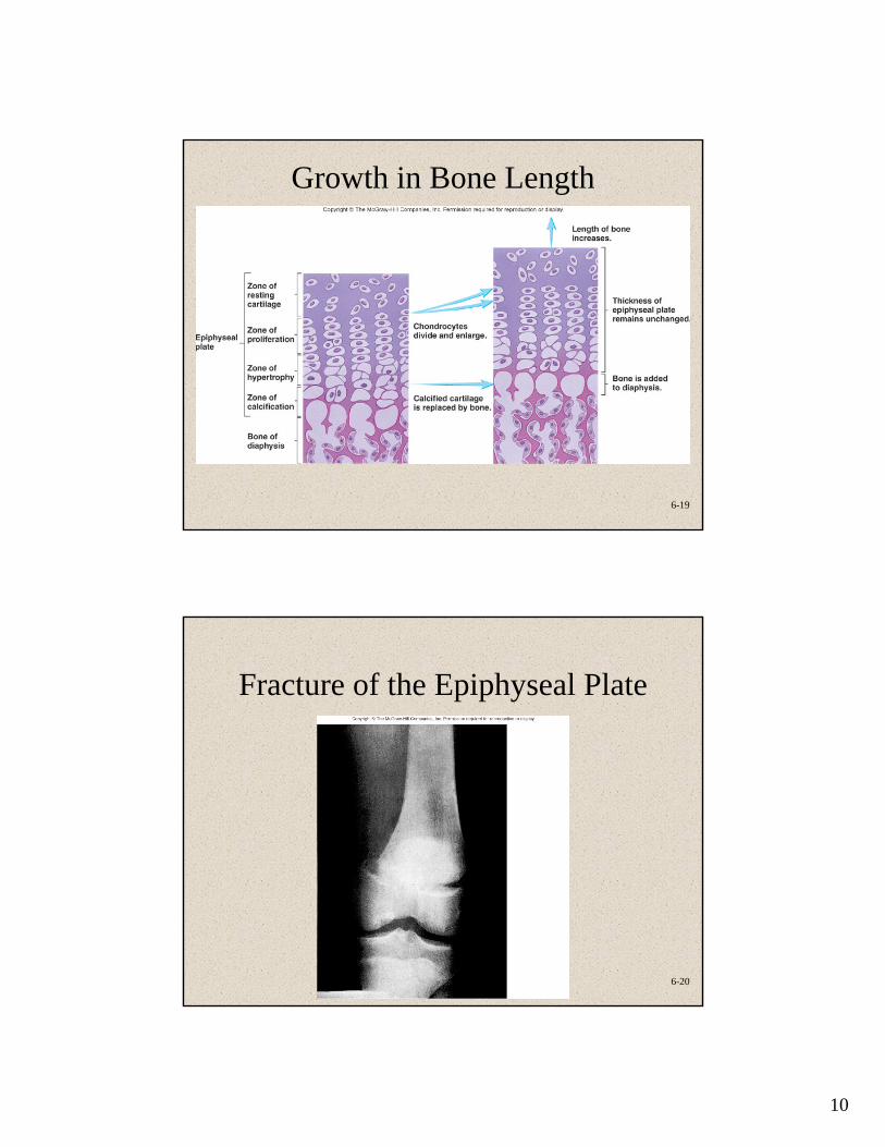
6-19
Growth in Bone Length
6-20
Fracture of the Epiphyseal Plate

11
6-21
Growth in Bone Width
6-22
Factors Affecting Bone Growth• Size and shape of a bone determined genetically but can be
modified and influenced by nutrition and hormones• Nutrition
– Lack of calcium, protein and other nutrients during growth and development can cause bones to be small
– Vitamin D—for absorption of Ca from intestines• Can be eaten or manufactured in the body• Rickets: lack of vitamin D during childhood• Osteomalacia: lack of vitamin D during adulthood leading to
softening of bones– Vitamin C—for collagen synthesis by osteoblasts
• Scurvy: deficiency of vitamin C• Lack of vitamin C also causes wounds not to heal, lose teeth
– Hormones --pituitary(growth), thyroid, and sex

12
6-23
Bone Remodeling• Converts woven bone into lamellar
bone
• Involved in bone growth, changes in bone shape, adjustments in bone due to stress, bone repair, and Ca ion regulation
• Relative thickness of bone changes as bone grows. Bone constantly removed by osteoclasts and new bone formed by osteoblasts.
6-24
Bone Repair 1. Hematoma formation. Localized mass of blood released from blood vessels but confined within an organ or space. Clot formation.
2. Callus formation. Callus: mass of tissue that forms at a fracture site and connects the broken ends of the bone. – Internal- blood vessels grow into
clot in hematoma. – External- collar around opposing
ends. Collar stabilizes two pieces.

13
6-25
Bone Repair, cont.
3. Callus ossification. Callus replaced by woven, cancellous bone
4. Bone remodeling. Replacement of cancellous bone and damaged material by compact bone. Sculpting of site by osteoclasts
6-26
Calcium Homeostasis

14
6-27
Effects of Aging on Skeletal System• Bone matrix decreases. More brittle due to lack of
collagen; but also less hydroxyapetite. • . African Americans and Hispanics have higher
bone masses than Caucasians and Asians. Rate of bone loss increases 10 fold after menopause. Cancellous bone lost first, then compact.
• Increased bone fractures• Bone loss causes deformity, loss of height, pain,
stiffness– Stooped posture– Loss of teeth
6-28
Bone Fractures
• Open (compound)- bone break with open wound. Bone may be sticking out of wound.
• Closed (simple)- Skin not broken• --------------------------------------• Incomplete- doesn’t extend
across the bone. Complete- does• Greenstick: incomplete fracture
Hairline: incomplete where two sections of bone do not separate. Common in skull fractures
• Comminuted fractures: complete with break into more than two pieces
• Impacted-crunched together