chapter 6 catalysis and protein folding in extreme ... · dependence of reaction rate on...
TRANSCRIPT
Chapter 6Catalysis and Protein Folding in ExtremeTemperature Environments
Charles Gerday
6.1 Introduction
The diversity of life partially derives from the various characteristics of naturalenvironments encountered on planet Earth suggesting that an organism, embeddedin an environment exhibiting specific properties, will benefit from physiologicaland biochemical adjustments tending to optimize the adequacy between thisenvironment and the overall characteristics of the organism. Some environmentsare extreme meaning that as such they are not suitable for organisms such ashuman being or Escherichia coli which are commonly exposed to moderatetemperatures, pH close to neutrality, atmospheric oxygen, low salinity and lowpressure. In this context, the temperature parameter is crucial for survival since itconsiderably affects the reaction rates of the numerous chemical reactions thatoccur in any living organism. The relation is in fact exponential, has been definedin 1899 by the Swedish chemist Svante Arrhenius, and takes the form:k = A.exp-Ea/RT in which k is the reaction rate, A is a pre-exponential term thatnotably depends on the activation entropy of the reaction (Collins et al. 2007), Ea
is known as the activation energy, that renders the reaction possible, R is the gasconstant (8.314 J mol-1 K-1) and T is the absolute temperature in Kelvin. Thedependence of reaction rate on temperature is abolished in organisms such asmammals which have chosen to keep their temperature more or less constantindependently of that of the environment. This is a very costly choice since itusually consumes about 75 % of the energy absorbed by the organism under theform of food, in such a way that in winter some of these homeotherms cannotmaintain their temperature constant due to lack of food, and enter hibernation. For
C. Gerday (&)Laboratory of Biochemistry, University of Liège, Institute of Chemistry,B-4000 Liège, Belgiume-mail: [email protected]
C. Verde and G. di Prisco (eds.), Adaptation and Evolution in Marine Environments,Volume 2, From Pole to Pole, DOI: 10.1007/978-3-642-27349-0_6,� Springer-Verlag Berlin Heidelberg 2013
89
those organisms that do not have the capacity to keep their temperature constantthe problem of the environmental temperature is more acute. Indeed, low tem-peratures render reaction rates too slow to sustain life whereas high temperaturescan accelerate them to an extent that would lead to production of unwanted or anexcess of some metabolites due to the differential action of temperature on reactionrates via the activation energy.
In nature three types of organisms have been defined as a function of thetemperature of their usual environment, the psychrophiles that thrive in environ-ments characterized by temperatures close or below the freezing point of water, themesophiles, confronted to moderate temperatures and the thermophiles that areexposed to temperatures which in some cases exceed that of the boiling point ofwater. In the context of this paper, the mesophiles do not pose acute problem ofreaction rate and will be only consider as a reference.
6.2 The Thermophiles
On Earth, natural environments of temperatures exceeding 60 �C are alwaysassociated with volcanic activity and are either terrestrial such as geysers, hotsprings and solfataric fields, or marine such as deep-sea vents. Typical tempera-tures in hot springs are around 80 �C (Brock 1967) whereas in hydrothermal ventsthe temperatures of fluids that are liberated in the cold waters of the ocean’sbottom can approach 400 �C. Of course, due to the rapid dilution of these hotfluids, a local gradient of temperatures is rapidly created and cover temperaturesfrom about 0 to 350 �C. Amazingly these environments are richly populated anddisplay a large biodiversity of organisms from fish to hyperthermophilic bacteria;this is mainly due to the large concentration of gases and mineral nutrients presentin these venting fluids. Temperatures as high as 350 �C are not compatible with thetype of life we know and actually the record is held by an archaea, known as strain121, which displays an upper growth temperature of 121 �C (Kashefi and Lovley2003). Hyperthermophiles have been recognized in 23 genera most of them beingarchaea but some bacteria such as Thermotoga maritima or Thermus thermophilusalso display high apparent optimum temperature close to 80 �C. At these tem-peratures the question of the permeability of the cytoplasmic membrane and inparticular the selective proton permeability, that generates the proton gradientwhich usually constitutes the essential part of the metabolic energy under the formof ATP, becomes acute. It appears however that the proton permeability is ratherkept constant in microorganisms characterized by different growth temperaturesand this phenomenon is known as ‘‘homeo-proton permeability adaptation’’ (vande Vossenberg et al. 1995). This is, in general, achieved through various adap-tations of the membrane composition tending to increase its rigidity in response toany increase in temperature of the environment (Gerday 2011). Other crucialcomponents of the cell are proteins which are first very sensitive to the deleteriouseffect of heat on their structure and among them the enzymes which in addition
90 C. Gerday
have to adjust their catalytic efficiency in order to normalize reaction rates andrender them more or less comparable to those that occur in mesophilic organisms.
6.2.1 The Stability Problem
Proteins are stabilized by weak bonds which have different sensitivities to tem-perature that depend on enthalpic and entropic terms. Van der Waals bonds areessentially electrostatic interactions that can implicate any type of atoms and thatare formed between transient dipoles or transient dipoles and induced dipoles. Theenergy associated with these interactions is weak and strongly depends on thedistance between the atoms involved. It includes an attractive and a repulsive termwhen atoms are located beyond or at a distance shorter than the so called van derWaals contact distance respectively. The empirical equation of the energy changeis: DE = A/r12-B/r6 in which r is the distance between groups A and B. In thecase of proteins the thermodependence of these bonds varies according to the typeof dipoles involved. For example the van der Waals interaction between permanentdipole and temporary dipoles is considered to be independent of the temperature(Ross and Subramanian 1981).
Hydrogen bonds occur between polar groups and can be defined as a particulartype of electrostatic interactions, in which a hydrogen atom is shared between aproton donor and a proton acceptor acting as a base: Ad-….H….Bd+. The energyassociated with these bonds will depend on the distance between A and B, on thegeometric arrangement of the bond, a linear geometry being optimal, and on therelative acidity of A an B, the strongest bond being encountered where the pKa
’sare similar. These bonds are formed with a negative modification of enthalpy andwill therefore be sensitive to any increase of temperature.
Hydrophobic interactions are formed between lateral chains of aliphatic andaromatic amino acids. These chains are poorly soluble in water especially aroundroom temperature. When they are transferred to water they affect the organizationof the hydrogen bonds leading to significant losses in translational and rotationalentropy of water molecules, a process unfavourable in terms of free energy.Clathrate-type cages are formed around the hydrophobic groups and this induces anegative modification of the entropy leading itself to a high energy level of thesesystems, even if the enthalpic term is negative probably due to favourable van derWaals interactions formed between the atoms of the hydrophobic groups and thesolvent. They therefore tend to evolve towards a more favourable energy state informing bonds and clusters that are mainly located in the core of the proteins. Theenergy change associated with the formation of these bonds can be expressed bythe following equation: DGHPH = DHHPH - TDSHPH, in which HPH stands forhydrophobic. This equation can be decomposed into the following terms:DGHPH = DHvdW ? DHHyd - TDSHPH, in which DHvdW corresponds to theenthalpy changes associated with the formation of van der Waals interactions,DHHyd to the positive enthalpy change capable to induce appropriate disorders in
6 Catalysis and Protein Folding 91
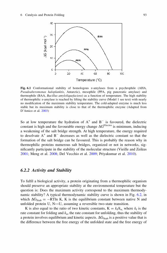
the organization of water molecules surrounding each individual hydrophobicgroups and TDSHPH the entropic change, at a given temperature, associated withthe formation of a hydrophobic interaction, which includes a large and positivecontribution DSHyd resulting from the release of water molecules from the indi-vidual groups. One can often read that a hydrophobic interaction is entropicallydriven due to the large disorder resulting from the release of water molecules, butthis is forgetting that both the enthalpic and entropic terms strongly vary with theenvironmental temperature. At low temperature, DHHyd is largely positive and thisis detrimental to the formation of the bond whereas at high temperaturethe amplitude of this term decreases and tends to zero favouring the formation ofthe bond. At low and moderate temperatures, TDSHPH is largely positive due to therelease of water (high value of DSHyd), whereas at high temperature DSHyd tends tozero due to large perturbation of the water molecules surrounding each individualgroups in such a way that at high temperature a hydrophobic interaction isweakened and can be assimilated to a simple van der Waals interaction(Priyakumar et al. 2010). This has led to the conclusion that the hydrophobic effectis maximum at moderate temperatures and all proteins from psychrophilic, mes-ophilic and thermophilic organisms show a maximum thermodynamic stabilityclose to room temperature (Kumar et al. 2002). This is clearly illustrated inFig. 6.1 in which the thermodynamic stability curves of three a-amylases frompsychrophilic, mesophilic and thermophilic organisms show that the maximumstability is close to 20 �C for the three proteins (Collins et al. 2008).
Salt bridges are formed between permanently charged side chains of oppositesigns such as carboxylic and amino groups. As in the case of hydrophobic inter-actions, an electrostatic interaction first requires dehydration of individual chargedgroups, thus the energy changes associated with these interactions are rathercomplex and also fluctuate as a function of temperature. The general equationcorresponding to the energy change associated with the formation of an electro-static interaction between two groups of opposite sign, A+, B-, can be written as:
DGTotal ¼ DGDesol þ DGElectro þ DGvdW þ DGProt
in which DGDesol correspond to the energy (unfavourable) necessary to dehydrateA+ and B-, DGElectro to the favourable decrease of free energy resulting from theformation of the salt bridge, DGvdWto changes related to new van der Waalsinteractions induced by the formation of a salt bridge and DGProt to any otherinteractions in the protein also induced by the salt bridge. The strength of the bondalso strongly depends on the temperature. Indeed, DGDesolcan be decomposed intoDHDesol-TDSDesol, DSDesolcan be considered as negligible so that DGDesol is equalto: DHDehyd A+ ? DHDehyd B — DHHyd(A+B-), this term is largely unfavourabledue to the energy required to dehydrate A+ and B-, especially at low temperature,and therefore should be compensated by a favourable energy, DGElectro, associatedwith the formation of the bridge, and which is equal roughly to ZA+ZB-.e2/D.dA+B-
in which ZA+ and ZB-are the charge of the ions; e, the charge of the electron; Dthe dielectric constant of the medium, and dA+B- the distance between A+ and B-.
92 C. Gerday
So at low temperature the hydration of A+ and B- is favoured, the dielectricconstant is high and the favourable energy change DGElectro is minimum, inducinga weakening of the salt bridge strength. At high temperature, the energy requiredto desolvate A+ and B- decreases as well as the dielectric constant so that theformation of the salt bridge can be favoured. This is probably the reason why inthermophilic proteins numerous salt bridges, organized or not in networks, sig-nificantly participate in the stability of the molecular structure (Vieille and Zeikus2001; Meng et al. 2008; Del Vecchio et al. 2009; Priyakumar et al. 2010).
6.2.2 Activity and Stability
To fulfil a biological activity, a protein originating from a thermophilic organismshould preserve an appropriate stability at the environmental temperature but thequestion is: Does the maximum activity correspond to the maximum thermody-namic stability? A typical thermodynamic stability curve is shown in Fig. 6.2, inwhich DGStab = -RTln K, K is the equilibrium constant between native N andunfolded protein U, N$U, assuming a reversible two-state transition.
K is also equal to the ratio of two kinetic constants, K = kf/ku, where kf is therate constant for folding and ku, the rate constant for unfolding, thus the stability ofa protein involves equilibrium and kinetic aspects. DGStab is a positive value that isthe difference between the free energy of the unfolded state and the free energy of
Fig. 6.1 Conformational stability of homologous a-amylases from a psychrophile (AHA,Pseudoalteromonas haloplanktis, Antarctic), mesophile (PPA, pig pancreatic amylase) andthermophile (BAA, Bacillus amyloliquefaciens) as a function of temperature. The high stabilityof thermophilic a-amylase is reached by lifting the stability curve (Model 1 see text) with nearlyno modification of the maximum stability temperature. The cold-adapted enzyme is much lessstable but its maximum stability is close to that of the thermophilic enzyme (Adapted fromD’Amico et al. 2003)
6 Catalysis and Protein Folding 93
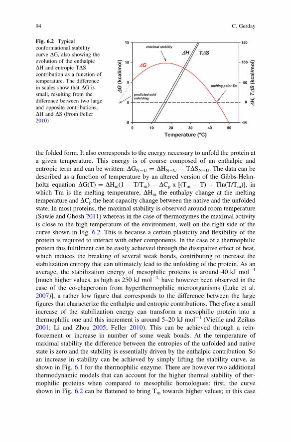
the folded form. It also corresponds to the energy necessary to unfold the protein ata given temperature. This energy is of course composed of an enthalpic andentropic term and can be written: DGN-U = DHN-U - TDSN-U. The data can bedescribed as a function of temperature by an altered version of the Gibbs-Helm-holtz equation DG(T) = DHm(1 - T/Tm) - DCp x [(Tm - T) ? Tln(T/Tm)], inwhich Tm is the melting temperature, DHm the enthalpy change at the meltingtemperature and DCp the heat capacity change between the native and the unfoldedstate. In most proteins, the maximal stability is observed around room temperature(Sawle and Ghosh 2011) whereas in the case of thermozymes the maximal activityis close to the high temperature of the environment, well on the right side of thecurve shown in Fig. 6.2. This is because a certain plasticity and flexibility of theprotein is required to interact with other components. In the case of a thermophilicprotein this fulfilment can be easily achieved through the dissipative effect of heat,which induces the breaking of several weak bonds, contributing to increase thestabilization entropy that can ultimately lead to the unfolding of the protein. As anaverage, the stabilization energy of mesophilic proteins is around 40 kJ mol-1
[much higher values, as high as 250 kJ mol-1, have however been observed in thecase of the co-chaperonin from hyperthermophilic microorganisms (Luke et al.2007)], a rather low figure that corresponds to the difference between the largefigures that characterize the enthalpic and entropic contributions. Therefore a smallincrease of the stabilization energy can transform a mesophilic protein into athermophilic one and this increment is around 5–20 kJ mol-1 (Vieille and Zeikus2001; Li and Zhou 2005; Feller 2010). This can be achieved through a rein-forcement or increase in number of some weak bonds. At the temperature ofmaximal stability the difference between the entropies of the unfolded and nativestate is zero and the stability is essentially driven by the enthalpic contribution. Soan increase in stability can be achieved by simply lifting the stability curve, asshown in Fig. 6.1 for the thermophilic enzyme. There are however two additionalthermodynamic models that can account for the higher thermal stability of ther-mophilic proteins when compared to mesophilic homologues: first, the curveshown in Fig. 6.2 can be flattened to bring Tm towards higher values; in this case
Fig. 6.2 Typicalconformational stabilitycurve DG, also showing theevolution of the enthalpicDH and entropic TDScontribution as a function oftemperature. The differencein scales show that DG issmall, resulting from thedifference between two largeand opposite contributions,DH and DS (From Feller2010)
94 C. Gerday
the lesser dependence of DGN-U on temperature would result in a lower magnitudeof DHN - U and DSN - U and of DCp, the difference between the specific heat ofthe unfolded and native form respectively. This decrease, in the case of DCp, canpresumably originate from a more discrete exposure of hydrophobic groups onunfolding; second, the whole curve can be displaced to the right, so that theamplitude of the maximal stability of the thermophilic protein would be similar tothat of the mesophilic enzyme, but this maximal value will be shifted towardshigher temperatures, meaning that the thermophilic protein would be more stableat higher temperatures (Beadle et al. 1999). This is the result of a reduced loss inentropy for the folding transition. This arises from a decrease in entropy of theunfolding state, induced by increase in Pro residues and decrease in Gly residues,or by residual structures subsisting in the unfolded state. Often, a combination ofincreased DG (first model) and reduced DCp (second model) is used in nature toachieve higher Tm, but more than 70 % of the thermophilic proteins rely on anincrease in DG, independently or in combination with an increase of Tm (Razviand Scholtz 2006). This has been somehow contradicted by a recent study on 116proteins from mesophiles and thermophiles, that indicates that there is a significantcorrelation between increased Tm and reduced entropy change upon folding (thirdmodel) (Sawle and Ghosh 2011). In hyperthermophiles, a combination of the threemodels seems to be preferred (Luke et al. 2007). Nevertheless, knowing that only afew alterations of the amino-acid composition (inducing either additional bonds orchanges in conformational or local entropy) can secure a good stability at hightemperature, one can easily understand that the modifications at the level of the3-dimensional structure will also be rather discrete. So it is rather difficult, evenknowing the 3D structure, to clearly identify the parameters involved in the sta-bilization of a thermophilic protein. As shown in Fig. 6.2, when the dissipativeeffect of heat becomes too large, the DG stability curve crosses the zero line. Thisis the melting point, where the ratio between the native and unfolded form is equalto one. It is also worth mentioning that on the left side of the curve both theenthalpic and entropic contribution change sign. We will come back to that later.So, an appropriate flexibility, local or global, of the molecular edifice is necessaryto secure catalytic efficiency. That is the reason why, in many cases, thermophilicenzymes show very low levels of efficiency at room temperature. By contrast, as inthe case of adenylsuccinate synthetase from Methanococcus jannaschii, at hightemperature up to 85 �C, the enzyme displays catalytic rates comparable to that ofmesophilic counterparts around room temperature (Vemparala et al. 2011). Unlikeits mesophilic counterpart, the catalytic function of this thermophilic enzyme onlyimplies the movement of two loops out of five for ligand binding and catalysis. Thestrategy consists in a pre-arrangement of catalytic residues, an increase in polarresidues and number and stability of salt bridges that induce an increased rigidityassociated with fewer loop movements. Most thermophilic proteins unfold irre-versibly, and so the previous equations cannot be applied. Their stability can beevaluated through the kinetic stability that expresses the rate at which the proteincollapses. This is a function of the free energy of activation DG* that represents thebarrier which has to be overcome to eventually proceed up to denaturation. This
6 Catalysis and Protein Folding 95
activation energy is high in thermophilic proteins that generally unfold at slowrates. Some differences are however observed when one compared the unfoldingcharacteristics of thermophilic proteins from archaea and bacteria. The unfoldingrate of thermophilic proteins from bacteria is in general faster than that of ther-mophilic counterparts from archaea. Archaea and bacteria have diverged at anearly state of evolution and thermophilic archaea presumably originated in hotenvironments, whereas thermophilic bacteria recolonized at later-stage hot envi-ronments. The difference in the unfolding rate of hyperthermostable proteinsseems to reflect the characteristics of the environments from which the microor-ganism originates. It has indeed been proposed that proteins originating from hotenvironments are more compact and display a more hydrophobic core than pro-teins issued from bacteria that recolonized at a later stage and which are notablystabilized by salt bridges. As an example, the core of RNases from archaea such asSulfolobus tokodaii and Thermococcus kodakarensis are richer in hydrophobicresidues than that of RNases from bacteria such as Thermus thermophilus. Thesuperslow unfolding of the enzymes from archaea, when compared to the fasterrate of homologous enzymes from bacteria, is due to the strong effect of hydro-phobic interactions on unfolding rate (Okada et al. 2010). This again underlines thefact that various strategies are used by microorganisms to adapt to an extremeenvironment and that the specificity of the adaptation strategy largely depends onthe evolutionary history of the organisms.
6.2.3 Folding at High Temperature
Although folding of proteins can spontaneously be successful, the particularenvironment of the cell and the high protein concentration, around 400 mg/ml, canmake folding inefficient and error-prone. In addition, as moderately elevatedtemperatures favour hydrophobic interactions, the folding of proteins in thermo-philic organisms implies the control of aggregation processes of nascent unfoldedforms and, within the cell, of a favourable equilibrium between N and U, viewedas a homeostatic control of the protein concentrations. This is achieved through theproduction of molecular chaperones, a multitude of proteins that act in variouscellular activities such as folding, refolding of misfolded proteins, protein aggre-gation, multimeric protein assembly, protein hydrolysis, and protein transport.Some of these chaperones are permanent hosts of the cell, others are inducedfollowing a stress such as a heat shock, that is the reason why they are usuallyknown as heat-shock proteins. They take any non-native conformation of proteinsin charge up to their successful final destination. They can act as ‘‘holdases,’’ instabilizing non-native conformation, as ‘‘foldases’’, in assisting correct folding,and as ‘‘unfoldases’’, when contributing to unfold misfolded proteins (Hoffmannet al. 2010). Protein misfolding is a very important process that can occur in a cell;it is associated with more than 30 human diseases, including some of the mostdebilitating such as Alzheimer’s, Parkinson’s and Creutzfeldt–Jakob’s diseases
96 C. Gerday
(Tartaglia et al. 2010). For the sake of comparison between the factors involved inthe folding of proteins from thermophiles and psychrophiles, we will summarizethe role of some of the most studied chaperones, such as the so-called chaperonins(GroEL/GroES), practically ubiquitous in all living organisms; Dnak, known asHsp 70 in eukaryotes, which is the major cytosolic chaperone in E. coli; and TF thetrigger factor, the only ribosome-associated chaperone in bacteria.
6.2.3.1 GroEL/GroES
Chaperonins are subdivided into two and possibly three groups differing instructure and distribution. Group I is formed by the GroEL and GroES complexesin which GroES acts as a co-chaperone. The crystal structure of GroEL has beensolved in 1994 (Braig et al. 1994). The protein is made of 60-kDa (Hsp60) subunitsand consists of two heptameric rings placed back to back to form a barrel structure.They are found mainly in eubacteria, and in eukaryotic organelles such as mito-chondria and chloroplasts. Roughly, the unfolded polypeptide chain is first trappedinto the central cavity through hydrophobic interactions, then ATP binding induceslarge conformational changes that notably renders the cavity much more hydro-philic and triggers the rapid binding of the co-chaperone GroES (Hsp10) to one ofthe ring and that acts as a lid. The sequestration of the polypeptide chain into thehydrophilic cavity forces exposed hydrophobic residues to cluster to form a coreand that is followed by protein folding. ATP hydrolysis by the ATP-bound ring isfollowed by the release of GroES and of the encapsulated protein. Group II is morecomplex than group I (Yébenes et al. 2011). Although they are also formed by tworings, GroES is replaced by a protruding rather flexible helical extension that canact as a lid essential for encapsulation of the unfolded protein. The closure of thelid is also ATP driven. The rings are made of octameric or nonameric structuresformed by association of one, two or three different subunits. Group-II chapero-nins, also known as thermosomes, are found in archaea and in the cytosol ofeukaryotes. The crystal structure of the closed conformation of the chaperoninfrom Thermoplasma acidophilum has been solved in 1998 (Ditzel et al. 1998).Other structures and notably the crystal structure of the open state are nowavailable (Huo et al. 2010) and a mechanism of folding chamber closure has beenproposed (Zhang et al. 2010). Group-III chaperonins have been identified in car-boxydotrophic bacteria; they are conserved in the genome of 11 bacteria (Techt-mann and Robb 2010). They are distinct from Group-II chaperonins because theircoding genes are associated with the Hsp 70 operon that includes dnaK, suggestingco-regulation and functional relationship between the two systems. They alsocontain a built-in lid and are composed of two octameric rings, unlike bacterialchaperonin. They form a monophyletic clade and can originate from an ancientlateral gene transfer from the archaea into an ancestral bacterium.
6 Catalysis and Protein Folding 97
6.2.3.2 DnaK/DnaJ/GrpE
DnaK, known as Hsp70 in eukaryotes, is involved in various cellular processes: itprevents protein aggregation, assists in protein disaggregation and in folding andrefolding of improperly folded proteins in an ATP-dependent manner. It is asso-ciated with co-factors DnaJ (Hsp40) and GrpE (nucleotide exchange factor) thatrespectively enhance, in E. coli, the ATPase rate of DnaK and significantlyaccelerates the nucleotide exchange. They play a role in minimizing aggregationand in enhancing the solubility of unfolded and also recombinant proteins. DnaKalso plays an important role in targeting aggregated proteins to proteases. Dnakappears as monomer, dimer or higher-ordered oligomer, whereas GrpE is ahomodimer and is the only component of the DnaK operon system that shows athermal transition in the range of the temperatures experienced by the microor-ganism. For example, in T. thermophilus (growth temperature of 70–75 �C) itdisplays a fully reversible thermal transition at 90 �C and an irreversible onearound 100 �C, compared to 48 �C and 75–80 �C in E. coli. As in the case of theDnaK system from E. coli, GrpE is considered as the thermosensor of the DnaKsystem that prevents thermal denaturation of substrate proteins. Its homodimericstructure has been solved by Nakamura et al. (2010). In binding to DnaK in a 2:1stoichiometry, it accelerates ADP/ATP exchange by 80,000-fold. Amazingly, inT. thermophilus, DnaJTh does not apparently stimulate the hydrolysis rate of ATPby DnaK nor the binding of the nucleotide, these roles being taken in charge solelyby GrpETh (Schlee and Reinstein 2002). In T. thermophilus Dnak displays highthermal stability in the absence or presence of ADP, as well as DnaJ(Tm = 100 �C). The DnaK system is present in all mesophilic archaea and only insome thermophilic archaea. The system has been recently characterized fromMethanothermobacter thermoautotrophicus with growth temperature ranging from40–70 �C. As in mesophilic and thermophilic bacteria, GrpEMt shows a reversiblethermal transition in the physiologically relevant temperature range and preventsthe release of the substrate from the DnaK complex, but the significant differenceis that both DnaK and Dnaj also display transition in the same temperature range.So GrpE cannot be really considered in this case as a thermosensor, since thatimplies that both Dnak and Dnaj would still be in a native state at the transitiontemperature of GrpE. The system seems to be optimized in preventing heat-induced protein aggregation (Popp and Reinstein 2009).
6.2.3.3 The Trigger Factor
The trigger factor is found in bacteria and chloroplasts and is the only chaperoneassociated with ribosomes. In archaea, some, not structurally related, similarfactors, do also exist. It is the first chaperone, encountered by nascent polypeptidechains, that forms a stoichiometric complex with the ribosome. It is localized at theribosomal exit of the newly built polypeptide chain. It may also act as an inde-pendent chaperone, since stable complexes with a large variety of full-length
98 C. Gerday
proteins were found, suggesting that the trigger factor plays an important role inthe stabilization of native-like substrate prior to their incorporation into multimericassemblies such as the ribosomal edifice (Martinez-Hackert and Hendrickson2009). It is found in the cytosol in rather high concentration mainly in a dimericform that can interact with folding intermediates that can be rescued by thechaperone system DnaK-DnaJ-GrpE (Liu et al. 2005). In E. coli, the trigger factoris a 48 kDa protein. Its 3D structure has been solved at 2.7-Ångström resolutionas well as the structure of its ribosomal binding domain in complex with thelarge ribosomal subunit of Haloarcula marismortui (Ferbitz et al. 2004). Thestructure resembles a crouching dragon made of three domains, the N-terminaldomain (1–149), or tail of the dragon, carries a specific motif (GFRxGxxP)involved in ribosome binding, the central domain (aa246–432) or carboxyl-ter-minal part is the chaperone module binding nascent polypeptide chains and thehead of the dragon-like structure (aa150–245) has an activity of peptidyl-prolylcis/trans isomerase. The PPIase forms a first module, whereas a second module ismade of the N- and C-terminal domains, forming a crevice that provides a pro-tective envelope over the peptide exit site on the ribosome. It has been proposedthat a hydrophobic pocket in the PPIase domain assists the folding of the nascentprotein bound to the chaperone site in a substrate dependent manner (Liu et al.2010). The three dimensional structure of the N- and C-terminal domain of thetrigger factor from Thermotoga maritima has been solved by X-ray crystallogra-phy and oligomerization and interactions have been studied (Martinez-Hackert andHendrickson 2007). Despite low sequence identity with the E. coli homologues[22 % for the N-terminal domain (TFN) and 12 % for the C-terminal domain(TFC)], the secondary-structure elements match up surprisingly well. The structureof the Tm TFN is also very similar to those observed in other eubacteria such asV. cholerae and D. radiodurans as well as to the structure of Hsp33s and othersmall heat-shock proteins. This domain exists in solution as a mixture of mono-mers and dimers. The structure of the Tm TFC domain is also very similar to that ofE. coli, but significantly diverges from the homologous domain from V. cholerae.Its structure is also close to that of the periplasmic chaperone SurA and a proteincalled mpn555. The Tm TFC domain can form dimers as in TFC from E. coli.Heteromeric assemblies between the N- and C-domains of the trigger factor fromT. maritima were also observed (Martinez-Hackert and Hendrickson 2007).Independently of binding to ribosomes and ribosome-nascent chains, the triggerfactor is thought to also interact with cytosolic proteins, favouring complexassemblies. The dimeric form of the trigger factor could represent an inactivestorage form of unfolded substrates (holdase function). The peptidyl-prolyl cis/trans isomerase activity of TF accelerates proline-limited refolding, but TF alsodisplays an in vitro chaperone activity in a concentration dependent manner. Itapparently cooperates with the two ATP-dependent chaperones, the DnaK andGroeS systems that are parts of the stress-induced misfolding and aggregation ofcellular proteins. According to a generally accepted sequence of events, nascentpolypeptide chains first interact with ribosome-bound TF then, upon release, some
6 Catalysis and Protein Folding 99
chains spontaneously fold whereas some other, about one third, need furtherassistance by the GroeL and DnaK systems.
6.2.4 Partial Conclusion
As recently stated (Tartaglia et al. 2010), the maintenance of protein solubility thatprevents misfolding and aggregation is essential for an appropriate function withinthe cell. Proteins can be classified into three groups as a function of their pro-pensity to aggregate; class I has a lower aggregation propensity than class II andclass III which are largely chaperone dependent. The number of highly solubleproteins decreases from class I to class III and there is an inverse relationshipbetween cytosol abundance and GroEL requirement. Class I and class III have alow Dnak requirement in contrast to class II; this is correlated with the stronghydrophobic character of exposed regions in class II, that use the DnaK system forfolding, whereas class III uses Dnak for preventing aggregation and GroEL forfolding. Class-III proteins are characterized by high b-sheet content that contrib-utes to display residual structured elements on unfolding that seem to be preferredby the GroEL system. Out of 1,158 cytosolic proteins investigated, 59 % belong toclass I and are GroEL independent, 25.6 % can be grouped in class II, the othersbelong to class III and only contains a weak percentage (2 %) of essential proteins,in agreement with their propensity to misfold. When compared to the othercytosolic proteins, class-III proteins have lower degree of hydrophobicity andhigher flexibility. These characteristics impose a high selective pressure on theamino-acid sequence of proteins and it has been possible to predict, from thesequence with a significant accuracy of 90 %, the respective requirement forthe GroEl or DnaK systems (Tartaglia et al. 2010).
6.3 The Psychrophiles
Psychrophiles are organisms living in permanently cold habitats and that areexposed to temperatures often well below the freezing point of water, such as inpermafrost zones or in the brine veins of sea water (Gilichinski et al. 2007;Deming 2007). Cold environments are also by far the most abundant on Earthsince they include polar regions, deep sea (below a depth of 1,000 m), glaciers, seaand freshwater ice, permanent snows and permafrost zones. All these habitats havebeen colonized by microorganisms, as well as by invertebrates and lower verte-brates in the case of the oceans for example. So, in contrast to what was initiallyexpected, the diversity of life in these regions is high and one can raise thequestion of how these organisms maintain appropriate metabolic rates despite thenegative effect of low temperatures on reaction rates. Low temperatures have inaddition the tendency to freeze some macromolecular structures and we have seen
100 C. Gerday
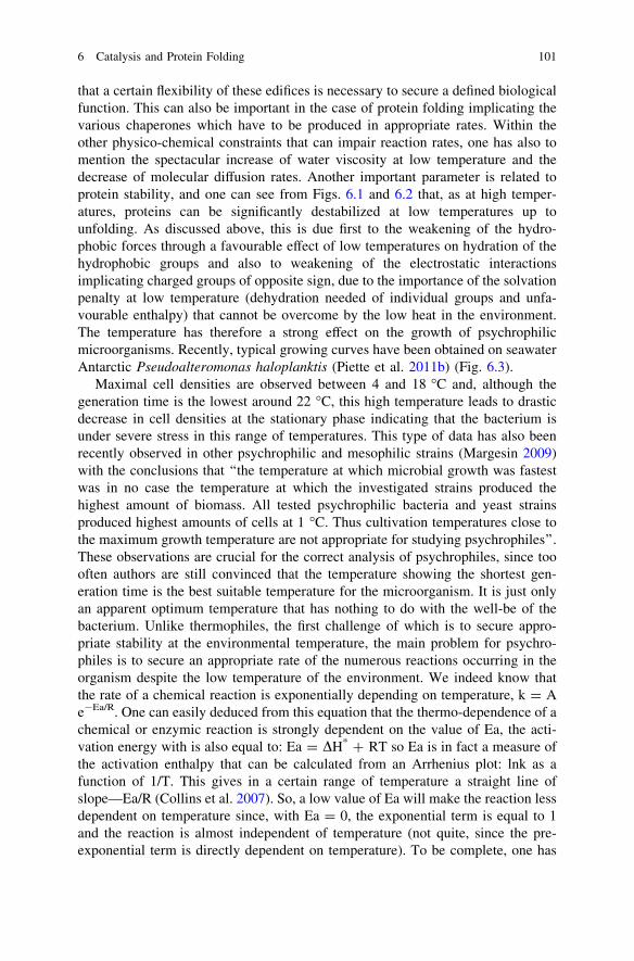
that a certain flexibility of these edifices is necessary to secure a defined biologicalfunction. This can also be important in the case of protein folding implicating thevarious chaperones which have to be produced in appropriate rates. Within theother physico-chemical constraints that can impair reaction rates, one has also tomention the spectacular increase of water viscosity at low temperature and thedecrease of molecular diffusion rates. Another important parameter is related toprotein stability, and one can see from Figs. 6.1 and 6.2 that, as at high temper-atures, proteins can be significantly destabilized at low temperatures up tounfolding. As discussed above, this is due first to the weakening of the hydro-phobic forces through a favourable effect of low temperatures on hydration of thehydrophobic groups and also to weakening of the electrostatic interactionsimplicating charged groups of opposite sign, due to the importance of the solvationpenalty at low temperature (dehydration needed of individual groups and unfa-vourable enthalpy) that cannot be overcome by the low heat in the environment.The temperature has therefore a strong effect on the growth of psychrophilicmicroorganisms. Recently, typical growing curves have been obtained on seawaterAntarctic Pseudoalteromonas haloplanktis (Piette et al. 2011b) (Fig. 6.3).
Maximal cell densities are observed between 4 and 18 �C and, although thegeneration time is the lowest around 22 �C, this high temperature leads to drasticdecrease in cell densities at the stationary phase indicating that the bacterium isunder severe stress in this range of temperatures. This type of data has also beenrecently observed in other psychrophilic and mesophilic strains (Margesin 2009)with the conclusions that ‘‘the temperature at which microbial growth was fastestwas in no case the temperature at which the investigated strains produced thehighest amount of biomass. All tested psychrophilic bacteria and yeast strainsproduced highest amounts of cells at 1 �C. Thus cultivation temperatures close tothe maximum growth temperature are not appropriate for studying psychrophiles’’.These observations are crucial for the correct analysis of psychrophiles, since toooften authors are still convinced that the temperature showing the shortest gen-eration time is the best suitable temperature for the microorganism. It is just onlyan apparent optimum temperature that has nothing to do with the well-be of thebacterium. Unlike thermophiles, the first challenge of which is to secure appro-priate stability at the environmental temperature, the main problem for psychro-philes is to secure an appropriate rate of the numerous reactions occurring in theorganism despite the low temperature of the environment. We indeed know thatthe rate of a chemical reaction is exponentially depending on temperature, k = Ae-Ea/R. One can easily deduced from this equation that the thermo-dependence of achemical or enzymic reaction is strongly dependent on the value of Ea, the acti-vation energy with is also equal to: Ea = DH* ? RT so Ea is in fact a measure ofthe activation enthalpy that can be calculated from an Arrhenius plot: lnk as afunction of 1/T. This gives in a certain range of temperature a straight line ofslope—Ea/R (Collins et al. 2007). So, a low value of Ea will make the reaction lessdependent on temperature since, with Ea = 0, the exponential term is equal to 1and the reaction is almost independent of temperature (not quite, since the pre-exponential term is directly dependent on temperature). To be complete, one has
6 Catalysis and Protein Folding 101
also to mention that the rate of an enzymic reaction is also dependent on animportant term, the transmission coefficient j, expressed in the temperaturedependence of the rate of catalysis given by an equation proposed by Eyring andsimilar to the Arrhenius law: kcat = j kBT/h e-DG*/RT (Marx et al. 2006; Feller2010) or its more generalized expression c(T) (Garcia-Viloca et al. 2004), thatexpresses the possibility that the activated state has some probability to return toits ground state (re-crossing the activation barrier). Often neglected, this term isnot equal to one in the case of reactions occurring at low temperature, and notablydepends on the viscosity of the medium.
6.3.1 Enzyme Activity at Low Temperatures
In living organisms, most reactions occurring in an organism are catalyzed byenzymes and from the discussion above one can see that one way to cope with thenegative effect of low temperatures would be to lower the activation energy of theenzyme, either by decreasing the activation enthalpy or by increase the activationentropy since DG* = DH* - TDS*. The activation parameters have been calcu-lated for a few psychrophilic enzymes and compared to those of mesophiliccounterparts (D’Amico et al. 2003; Marx et al. 2006; Coquelle et al. 2007; Bjelic
Fig. 6.3 Growth of theAntarctic psychrophilicbacteriumPseudoalteromonashaloplanktis as a function oftemperature. a shows thethermodependence of thegeneration time compared tothat of mesophilic E. coliRR1 (dashed curve). b showsthe growth curve at 4 �C(open circles), 18 �C (blackdots) and 26 �C (blacksquares). The shortestdoubling time alsocorresponds to thetemperature that gives thelowest cell density (FromPiette et al. 2011b)
102 C. Gerday

et al. 2008). In all cases, and in agreement with the higher catalytic efficiency, alower activation energy DG* was recorded for psychrophilic enzymes approxi-mately in the range of a decrease of 2–10 %. This lower activation energy sys-tematically results from a large decrease of the activation enthalpy often higherthan 20 % however compensated by a much more negative activation entropy. Thelower activation enthalpy has been attributed to the lower number of weak bonds,enthalpy-driven, that have to be broken to reach the activated state. The loweractivation enthalpy also depicts the lower temperature dependence of the catalyticactivity of these enzymes. With regards to the much more negative value of theactivation entropy the figures are also consistent with the idea that the ground stateof the cold-adapted enzyme has a rather loose structure, that needs a larger re-ordering to be transformed into a well-organized transition state (Lonhienne et al.2001; Feller 2010). The consequence is that the lower activation enthalpy iscounterbalanced by a less favourable entropy change on activation, so that theactivation energy is higher than what would be predicted by the difference in theactivation enthalpy. That is not really a problem since for example in the case of a-amylase from an Antarctic bacterium a decrease of activation energy by only 2 %allows to increase the rate of catalysis three fold at 10 �C when compared to themesophilic counterpart (Collins et al. 2007). The lower stability and the morenegative activation entropy suggests, as stated above, that cold-adapted enzymesdisplay higher flexibility when compared to their mesophilic counterparts andintuitively this can indeed counteract the freezing effect of low temperatures on the3D structure of macromolecules and preserve some plasticity for accommodationof substrates and release of products. Numerous techniques have been applied forthe evaluation of the relative flexibility of protein structure (for a more detaileddiscussion see Collins et al. 2008) such as fluorescence quenching, neutron scat-tering and molecular dynamics. Fluorescence quenching has been successfullyapplied to several psychrophilic and mesophilic enzymes: Ca2+–Zn2+ proteases(Chessa et al. 2000), xylanases (Collins et al. 2003), DNA ligases (Georlette et al.2003), a-amylases (D’Amico et al. 2003), cellulases (Sonan et al. 2007) and zincmetalloproteases (Xie et al. 2009) and from these experiments one can concludewith a certain degree of confidence that psychrophilic enzymes display a moreflexible structure. As recently stated (Bjelic et al. 2008), this flexibility is notprobably localized systematically at the level of the active sites, in general highlyconserved in thermal homologues, but rather in other important sites more or lessremote from the active site, but which, in any case, allow to decrease the activationenergy of the enzyme by facilitating access to the active site. As shown by thelarge difference in thermal stability existing between the two main domains of acold-adapted phosphoglycerate kinase (Bentahir et al. 2000), flexibility does notindeed concerns the whole structure but only crucial parts of it. Later it wasdemonstrated (Zecchinon et al. 2005) that the two stability domains of this cold-adapted phosphoglycerate kinase do not match the well known N- and C-domains,since the heat-stable domain is made of only 80 residues of the C-domain,including the nucleotide binding site. The remaining part of the protein forms theheat-labile domain and it was proposed that some rigidity of a crucial part of the
6 Catalysis and Protein Folding 103
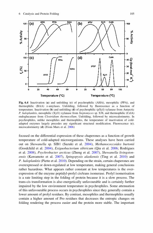
protein was necessary to preserve appropriate affinity of the cold-adapted enzymefor its substrate, since these intracellular enzymes are probably working far fromsubstrate saturation. An important characteristic of cold-adapted enzymes thatconcerns the activity is that thermal inactivation always precedes any structuralchanges that can be detected either by fluorescence or by microcalorimetry.Figure 6.4 indeed concerns, on the left side, a-amylases from psychrophilic,mesophilic and thermophilic counterparts, and on the right side psychrophilic,mesophilic and thermophilic glycoside hydrolases. In mesophilic and thermophilicenzymes, the unfolding thermal transitions strictly match the apparent optimum ofactivity whereas, in cold-adapted enzymes, the loss of activity largely precedesany apparent changes in the 3D structure. These data can have two explanations.First, the active site of cold-adapted enzymes can be altered to some extent withoutsignificant modification of the structure; it has indeed been shown in cold-adapteda-amylase that the active site is the first structural element that unfolds. Second, itis also possible that the enzyme–substrate complex is the first structure to bealtered by heat simply by weakening of the interactions between the two partners.In any case these data favour the idea that indeed the low stability of cold-adaptedenzymes is undoubtedly related to increase of the flexibility of crucial domains,counterbalancing in this way the freezing effect of low temperatures on structureand reaction rates. This can have additional consequences at least in some cases,e.g. better accessibility of the active site that can also induce broadening ofthe specificity at least in multi-substrate enzymes (Tsigos et al. 1998; Smalås et al.2000).
6.3.2 Folding at Low Temperatures
As discussed above, in thermophiles protein folding is often assisted by severalcellular tools that prevent aggregation, prepare the protein for proper folding andeliminate misfolded assemblies. The folding of proteins at low temperature canalso be impaired by an improper adaptation of the participating enzymes, bypossible decrease of hydrophobic and electrostatic interactions (salt bridges) at lowtemperatures, and by physical constrains such as the important increase in vis-cosity of the aqueous intracellular space. It has been shown that the intrinsicfolding rate constants of psychrophilic, mesophilic and thermophilic proteins aresimilar (Piette et al. 2011a) at a given temperature, meaning that at low temper-ature psychrophilic proteins should fold at much slower rates than mesophilic orthermophilic counterparts at their usual environmental temperatures. We have seenthat in thermophiles many proteins require assistance for proper folding under theform of chaperones and, from the analysis of the genome sequence of a highnumber of psychrophiles (Casanueva et al. 2010), it is clear that psychrophiles alsohave the potential to express all types of chaperones, but the question is to knowwhether they really express all of them or only specific chaperones at low tem-peratures. To answer these questions, a limited number of studies have recently
104 C. Gerday
focused on the differential expression of these chaperones as a function of growthtemperature of cold-adapted microorganisms. These analyses have been carriedout on Shewanella sp. SIB1 (Suzuki et al. 2004), Methanococcoides burtonii(Goodchild et al. 2004), Exiguobacterium sibiricum (Qiu et al. 2006; Rodrigueset al. 2008), Psychrobacter arcticus (Zheng et al. 2007), Shewanella livingston-ensis (Kawamoto et al. 2007), Spingopyxis alaskensis (Ting et al. 2010) andP. haloplanktis (Piette et al. 2010). Depending on the strain, certain chaperones areoverexpressed or down-regulated at low temperature, making general conclusionsrather hazardous. What appears rather constant at low temperatures is the over-expression of the enzyme peptidyl-prolyl cis/trans isomerase. Prolyl isomerisationis a rate limiting step in the folding of protein because it is a slow process. Thetrans-cis transformation is also energetically unfavourable and is certainly furtherimpaired by the low environment temperature in psychrophiles. Some attenuationof this unfavourable process occurs in psychrophiles since they generally contain alower amount of prolyl residues. By contrast, mesophiles and thermophiles usuallycontain a higher amount of Pro residues that decreases the entropic changes onfolding rendering the process easier and the protein more stable. The important
Fig. 6.4 Inactivation (a) and unfolding (c) of psychrophilic (AHA), mesophilic (PPA), andthermophilic (BAA) a-amylases. Unfolding, followed by fluorescence as a function oftemperature. Inactivation (b) and unfolding (d) of psychrophilic (pXyl) xylanase from AntarcticP. haloplanktis, mesophilic (Xyl1) xylanase from Steptomyces sp. S38, and thermophilic (CelA)endoglucanase from Clostridium thermocellum. Unfolding, followed by microcalorimetry. Inpsychrophiles, unlike mesophiles and thermophiles, the temperature of inactivation of cold-adapted enzymes largely precedes any significant structural modification. Fluorescence (c);microcalorimetry (d) (From Marx et al. 2006)
6 Catalysis and Protein Folding 105

chaperone GroEL is down regulated in some cold-adapted species as well as theassociated factors GroES and GrpE, a similar trend is also observed in the case ofthe DnaK system. As in peptidyl-prolyl cis/trans isomerase, the trigger factor isalso generally overexpressed except in for P. arcticus. Not forgetting the possi-bility that the apparent differential expression of chaperones in organisms living atlow temperatures could simply reflect the evolutionary history, it is also possiblethat the differences observed in the temperature regulation of the expression ofchaperones as a function of temperature could be due to differences in method-ology. In some cases indeed, some authors use, as upper growth temperature, thetemperature of optimum growth rate. But these temperatures, as stated above,induce a severe stress, since maximal growth rates are just kinetic effects whichhave nothing to do with the physiological state of the bacterium (Margesin 2009).
It seems clear that, in psychrophiles, the trigger factor that interacts with nas-cent polypeptide chains is essential for securing proper folding of cold-adaptedproteins, probably in association with peptidyl-prolyl cis/trans isomerase (Pietteet al. 2011a). The down-regulation of the other chaperones can possibly be relatedto the fact that low temperatures reduce to some extent the risk of aggregation andof misfolding. In support of this, it has been also observed that overexpression of acold trigger factor represses expression of other chaperones when E. coli is grownat low temperatures (Kandror and Goldberg 1997).
6.4 Conclusions
The rate of growth of a microorganism is related to the rate of the metabolicreactions, most of them being catalyzed by enzymes that require proper foldingand stability. That does not mean that the temperature of maximum growth rate isthe best possible temperature for the microorganism, since these temperatures, dueto the deleterious effect of heat, induce partial unfolding or overproduction ofunwanted metabolites due to the differential effect of heat on enzyme activitythrough largely different values of activation energies. A delicate balance hastherefore to be found between conflicting parameters. In thermophiles, althoughhigh temperatures favour reaction rates, they also induce unfolding of molecularstructures or misfolding notably due to uncontrolled hydrophobic forces. Proteinsin thermophiles have therefore slightly modified their structure to resist to the hightemperature of their environments, while the folding of a high number of theiressential proteins is assisted by a large set of folding helpers named chaperones.The maximum stability of a protein is however not suitable in living organismssince a certain plasticity and flexibility of the molecular structures are required tosecure the necessary interactions between partners in a very crowded cell. Inpsychrophiles, one can consider that the main problem is to secure appropriatemetabolic fluxes by acting on the activity or relative abundance of enzymesinvolved in catalysis. In the history of evolution, the strategy to produce higheramounts of catalysts is not the cheapest way in terms of energy costs, so in general
106 C. Gerday
the reaction-rate problem has been solved through progressive evolution towardsenzymes displaying lower activation energy and in consequence lower thermaldependence of the activity that allows the organisms to be transiently exposed tounusually low or high temperatures. Therefore a continuum in the adaptation ofcold-adapted microorganisms, depending on their evolutionary history, is expec-ted, and subdivisions such as those introduced in the form of terms such as psy-chrotolerants or psychrotrophs appear devoid of any interest. Tentatively, also theapparent simplification or limitation of the tools rendering appropriate the foldingof proteins at low temperature is correlated to reduction of the physico-chemicalconstrains that can counteract the folding process and possibly also to the energeticcare to limit protein synthesis to the minimum required.
References
Beadle BM, Baase WA, Wilson DB, Gilkes NR, Shoichet BK (1999) Comparing the thermody-namic stabilities of a related thermophilic and mesophilic enzyme. Biochemistry 38:2570–2576
Bentahir M, Feller G, Aittaleb M, Lamotte-Brasseur J, Himri T, Chessa J-P, Gerday C (2000)Structural, kinetic and calorimetric characterization of the cold-active phosphoglyceratekinase from the Antarctic Pseudomonas sp. TACII18. J Biol Chem 275:11147–11153
Bjelic S, Brandsdal BO, Aqvist J (2008) Cold adaptation of enzyme reaction rates. Biochemistry47:10049–10057
Braig K, Otwinowski Z, Hedge R, Boisvert DC, Joachimiak A, Horwich AL, Sigler PB (1994)The crystal-structure of the bacterial chaperonin groel at 2.8-Ångstrom. Nature 371:578–586
Brock TD (1967) Microorganisms adapted to high temperatures. Nature 214:882–885Casanueva A, Tuffin M, Cary C, Cowan DA (2010) Molecular adaptations to psychrophily: the
impact of ‘omic’ technologies. Trends Microbiol 18:374–381Chessa J-P, Petrescu I, Bentahir M, Van Beeumen J, Gerday C (2000) Purification, physico-
chemical characterization and sequence of a heat labile alkaline metalloprotease isolated froma psychrophilic Pseudomonas species. Biochim Biophys Acta 1479:265–274
Collins T, Meeuwis MA, Gerday C, Feller G (2003) Activity, stability and flexibility inglycosidases adapted to extreme thermal environments. J Mol Biol 328:419–428
Collins T, D’Amico S, Marx J-C, Feller G, Gerday C (2007) Cold-adapted enzymes. In: Gerday C,Glansdorff N (eds) Physiology and Biochemistry of extremophiles. ASM Press, Washington,pp 165–170
Collins T, Roulling F, Piette F, Marx J-C, Feller G, Gerday C, D’Amico S (2008) Fundamentalsof cold-adapted enzymes. In: Margesin R, Schinner F, Marx J-C, Gerday C (eds)Psychrophiles, from biodiversity to biotechnology. Springer-Verlag, Berlin, pp 211–227
Coquelle N, Fioravanti E, Weik M, Vellieux F, Madern D (2007) Activity, stability and structural studiesof lactate dehydrogenases adapted to extreme thermal environments. J Mol Biol 374:547–562
D’Amico S, Marx J-C, Gerday C, Feller G (2003) Activity-stability relationships in extremophilicenzymes. J Biol Chem 278:7891–7896
Del Vecchio P, Elias M, Merone L, Graziano G, Dupuy J, Mandrich L, Carullo P, Fournier B,Rochu D, Rossi M, Masson P, Chabriere E, Manco G (2009) Structural determinants of thehigh thermal stability of SsoPox from the hyperthermophilic archaeon Sulfolobus solfataricus.Extremophiles 13:461–470
Deming JW (2007) Life in ice formation at very low temperatures. In: Gerday C, Glansdorff N(eds) Physiology and biochemistry of extremophiles. ASM Press, Washington, pp 133–144
Ditzel L, Löwe J, Stock D, Stetter KO, Huber H, Huber R, Steinbacher S (1998) Crystal structureof the thermosome, the archaeal chaperonin and homolog of CCT. Cell 93:125–138
6 Catalysis and Protein Folding 107

Feller G (2010) Protein stability and enzyme activity at extreme biological temperatures. J PhysCondens Matter 22:32101–321018
Ferbitz L, Maier T, Patzelt H, Bukau B, Deurling E, Ban N (2004) Trigger factor in complex withthe ribosome forms a molecular cradle for nascent proteins. Nature 431:590–596
Garcia-Viloca M, Gao J, Karplus L, Truhlar DG (2004) How enzymes work: analysis by modernrate theory and computer simulations. Science 303:186–195
Georlette D, Damien B, Blaise V, Depierreux E, Uversky VN, Gerday C, Feller G (2003)Structural and functional adaptations to extreme temperatures in psychrophilic, mesophilicand thermophilic DNA ligases. J Biol Chem 278:37015–37023
Gerday C (2011) Life at the extreme of temperature. In: Storz G, Hengge R (eds) Bacterial stressresponse. ASM Press, Washington, pp 425–444
Gilichinski D, Vishnivetskaya M, Petrova M, Spirina E, Mamykin V, Rivkina E (2007) Bacteriain permafrost. In: Margesin R, Schinner F, Marx J-C, Gerday C (eds) Psychrophiles: frombiodiversity to biotechnology. Springer-Verlag, Berlin, pp 83–102
Goodchild A, Saunders NF, Erlan H, Raftery M, Guilhaus M, Curmi PM, Cavicchioli R (2004) Aproteomic determination of cold adaptation in the Antarctic archaeon, Methanococcoidesburtonii. Mol Microbiol 53:309–321
Hoffmann A, Bukau B, Kramer G (2010) Structure and function of the molecular chaperone,trigger factor. Biochim Biophys Acta 1803:650–661
Huo Y, Hu Z, Zhang K, Wang L, Zhai Y, Zhou Q, Lander G, Zhu J, He Y, Pang X, Xu W,Bartlam M, Don Z, Sun F (2010) Crystal structure of group II chaperonin in the open state.Structure 18:1270–1279
Kandror O, Goldberg AL (1997) Trigger factor is induced upon cold shock and enhances viabilityof Escherichia coli at low temperatures. Proc Natl Acad Sci U S A 94:4978–4981
Kashefi K, Lovley DR (2003) Extending the upper temperature limit for life. Science 301:934Kawamoto J, Kurihara T, Kitagawa M, Kato I, Esaki N (2007) proteomic studies of an Antarctic
cold-adapted bacterium, Shewanella livingstonensis Ac 10, for global identification of cold-inducible proteins. Extremophiles 10:819–826
Kumar S, Tsai C-J, Nussinov R (2002) Maximal stabilities of reversible two-state proteins.Biochemistry 41:5359–5374
Li WF, Zhou PL (2005) Structural features of thermozymes. Biotechnol Adv 23:271–281Liu CP, Perrett S, Zhou JM (2005) Dimeric trigger factor stably binds folding-competent
intermediates and cooperates with the DnaK-DnaJ-GrpE chaperone system to allow refolding.J Biol Chem 280:13315–13320
Liu CP, Zhou QM, Fan DJ, Zhou JM (2010) PPIase domain of trigger factor acts as auxiliarychaperone site to assist the folding of protein substrates bound to the crevice of trigger factor.Int J Biochem Cell Biol 42:890–901
Lonhienne T, Gerday C, Feller G (2001) Psychrophilic enzymes: revisiting the thermodynamicparameters of activation may explain local flexibility. Biochim Biophys Acta 1543:1–10
Luke KA, Higgins CL, Wittung-Stafshede P (2007) Thermodynamic stability and folding ofproteins from hyperthermophilic organisms. FEBS J 274:4023–4033
Margesin R (2009) Effect of temperature on growth parameters of psychrophilic bacteria andyeasts. Extremophiles 13:257–262
Martinez-Hackert E, Hendrickson WA (2007) Structure of and interactions between domains oftrigger factor from Thermotoga maritima. Acta Crystallogr Sect D63:536–547
Martinez-Hackert E, Hendrickson WA (2009) Promiscuous substrate recognition in folding andassembly activities of the trigger factor chaperone. Cell 138:923–934
Marx J-C, Collins T, D’Amico S, Feller G, Gerday C (2006) Cold-adapted enzymes from marineAntarctic microorganisms. Mar Biotechnol 9:293–304
Meng G, Xia-Yu X, Xian-Ming P (2008) Salt bridges in the hyperthermophilic protei Ssh10b areresilient to temperature increases. J Biol Chem 283:31690–31696
Nakamura A, Takumi K, Miki K (2010) Crystal structure of a thermophilic GrpE protein: insightinto thermosensing function for the DnaK chaperone system. J Mol Biol 396:1000–1011
108 C. Gerday

Okada J, Okamoto T, Mukaiyama A, Tadokoro T, You D-J, Chon H, Koga Y, Takano K, Kanaya S(2010) Evolution and thermodynamics of the slow unfolding of hyperstable monomericproteins. BMC Evol Biol 10:207–218
Piette F, D’Amico S, Struvay C, Mazzuchelli G, Renaut J, Tutino ML, Danchin A, Leprince P, Feller G(2010) Proteomics of life at low temperatures: trigger factor is the primary chaperone in theAntarctic bacterium Pseudoalteromonas haloplanktis TAC 125. Mol Microbiol 76:120–132
Piette F, D’Amico S, Mazzucchelli G, Danchin A, Leprince P, Feller G (2011b) Life in the cold: aproteomic study of cold-repressed proteins in the Antarctic bacterium Pseudoalteromonashaloplanktis TAC125. Appl Environ Microbiol 77:3881–3883
Piette F, Struvay C, Feller G (2011a) The protein folding challenge in psychrophiles: facts andcurrent issues. Environ Microbiol 13:1924–1933
Popp SL, Reinstein J (2009) Functional characterization of the DnaK chaperone system from thearchaeon Methanothermobacter thermoautrophicus DH. FEBS Lett 583:573–578
Priyakumar UD, Ramakrishna S, Nagarjuna KR, Reddy SK (2010) Structural and energeticdeterminants of thermal stability and hierarchical unfolding pathways of hyperthermophilicproteins, Sac7d and Sso7d. J Phys Chem B 114:1707–1718
Qiu Y, Kathariou S, Lubman DM (2006) Proteomic analysis of cold adaptation in a Siberianpermafrost bacterium Exiguobacterium sibiricum 255–15 by two-dimensional liquid separa-tion coupled with mass spectrometry. Proteomics 6:5221–5233
Razvi A, Scholtz JM (2006) Lessons in stability from thermophilic proteins. Protein Sci 15:1569–1578Rodrigues DF, Ivanova N, He Z, Huebner M, Zhou J, Tiedje JM (2008) Architecture of thermal
adaptation in an Exiguobacterium sibiricum strain isolated from 3 million year old permafrost:a genome and transcriptome approach. BMC Genomics 9:547
Ross PD, Subramanian S (1981) Thermodynamics of protein association reactions: forcescontributing to stability. Biochemistry 20:3096–3102
Sawle L, Ghosh K (2011) How do thermophilic proteins and proteosomes withstand hightemperature? Biophys J 101:217–227
Schlee S, Reinstein J (2002) The DnaK/ClpB chaperone system from Thermus thermophilus. CellMol Life Sci 59:1598–1606
Smalås AO, Leiros HK, Os V, Willassen NP (2000) Cold adapted enzymes. Biotechnol Annu Rev6:1–57
Sonan GK, Receveur-Bréchot V, Duez C, Aghajari N, Czjzek M, Haser R, Gerday C (2007) Thelinker region plays a key role in the adaptation to cold of the cellulose from an Antarcticbacterium. Biochem J 407:293–302
Suzuki Y, Haruki M, Takano K, Morikawa M, Kanaya S (2004) Possible involvement of anFKBP family member protein from a psychrotrophic bacterium, Shewanella sp. SIB1 in coldadaptation. Eur J Biochem 271:1372–1381
Tartaglia GG, Dobson CM, Hartl FU, Vendruscolo M (2010) Physicochemical determinants ofchaperone requirements. J Mol Biol 400:579–588
Techtmann SM, Robb FT (2010) Archaeal-like chaperonins in bacteria. Proc Natl Acad Sci U SA 107:20269–20274
Ting L, Williams TJ, Cowley MJ, Lauro FM, Guilhaus M, Raftery MJ, Cavicchioli R (2010) Coldadaptation in the marine bacterium, Shingopyxis alaskensis assessed using quantitativeproteomics. Environ Microbiol 12:2658–2676
Tsigos I, Velonia K, Smonou I, Bouriotis V (1998) Purification and characterization of an alcoholdehydrogenase from the Antarctic psychrophile Moraxella sp. TAE 123. Eur J Biochem254:356–362
Van de Vossenberg JL, Ubbink-Kok T, Elferink MG, Driessen AJ, Konings WN (1995) Ionpermeability of the cytoplasmic membrane limits the maximum growth temperatures ofbacteria and archaea. Mol Microbiol 18:925–932
Vemparala S, Mehrotra S, Balaram H (2011) Role of loop dynamics in thermal stability ofmesophilic and thermophilic adenylsuccinate synthetase: a molecular dynamics and normalmode analysis study. Biochim Biophys Acta 1814:630–637
6 Catalysis and Protein Folding 109
Vieille C, Zeikus G (2001) Hyperthermophilic enzymes: sources, uses, and molecularmechanisms for thermostability. Microbiol Mol Biol Rev 65:1–43
Xie B-B, Bian F, Chen X-L, He H-L, Guo J, Gao X, Zeng Y-X, Chen B, Zhou B-C, Zhang Y-Z(2009) Cold adaptation of zinc metalloprotease in the thermolysin family from deep sea andArctic sea ice bacteria revealed by catalytic and structural properties and molecular dynamics.J Biol Chem 284:9257–9269
Yébenes H, Mesa P, Munoz IG, Montoya G, Valpuesta JM (2011) Chaperonins: two rings forfolding. Trends Biochem Sci 36:424–432
Zecchinon L, Oriol A, Netzel U, Svennberg J, Gerardin-Otthiers N, Feller G (2005) Stabilitydomains, substrate-induced conformational changes and hinge-bending motions in a psychro-philic phosphoglycerate kinase. A microcalorimetric study. J Biol Chem 280:1307–41314
Zhang J, Baker ML, Schröder GF, Douglas NR, Reissman S, Jakane J, Dougherty M, Fuc J,Levitt M, Ludtke SJ, Frydman J, Chiu W (2010) Mechanisms of folding chamber closure in agroup II chaperonin. Nature 463:379–383
Zheng S, Ponder MA, Shih JY, Yiedje JM, Thomashow MF, Lubman DM (2007) A proteomicanalysis of Psychrobacter arcticus 273–4 adaptation to low temperature and salinity using a2-D liquid mapping approach. Electrophoresis 28:467–488
110 C. Gerday