chapter 6: bones and skeletal tissues bone anatomylwcmrstaylor.yolasite.com/resources/chapter 6 -...
TRANSCRIPT
Chapter 6:
Bones and Skeletal Tissues
Bone Anatomy
Bone Textures
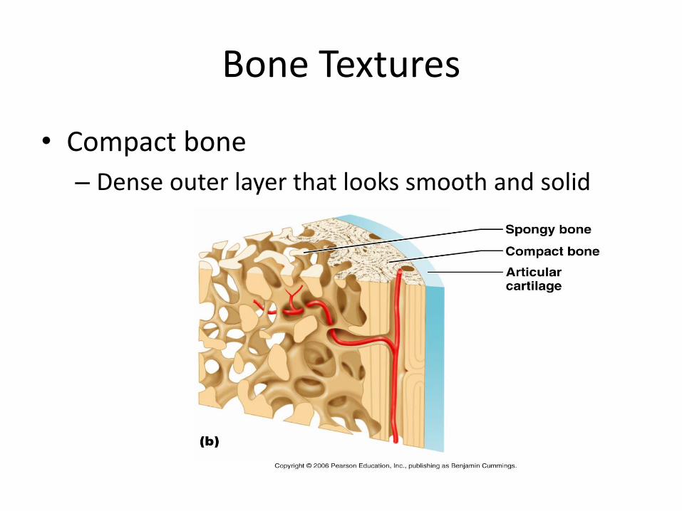
• Compact bone
– Dense outer layer that looks smooth and solid
Bone Textures
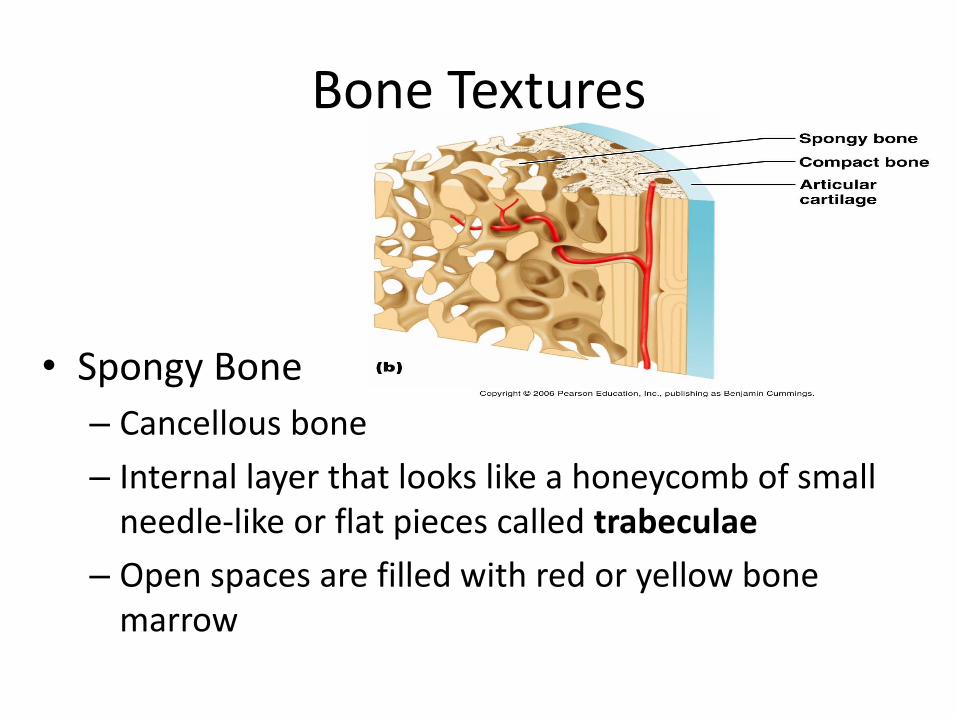
• Spongy Bone
– Cancellous bone
– Internal layer that looks like a honeycomb of small needle-like or flat pieces called trabeculae
– Open spaces are filled with red or yellow bone marrow
Structure of a Typical Long Bone
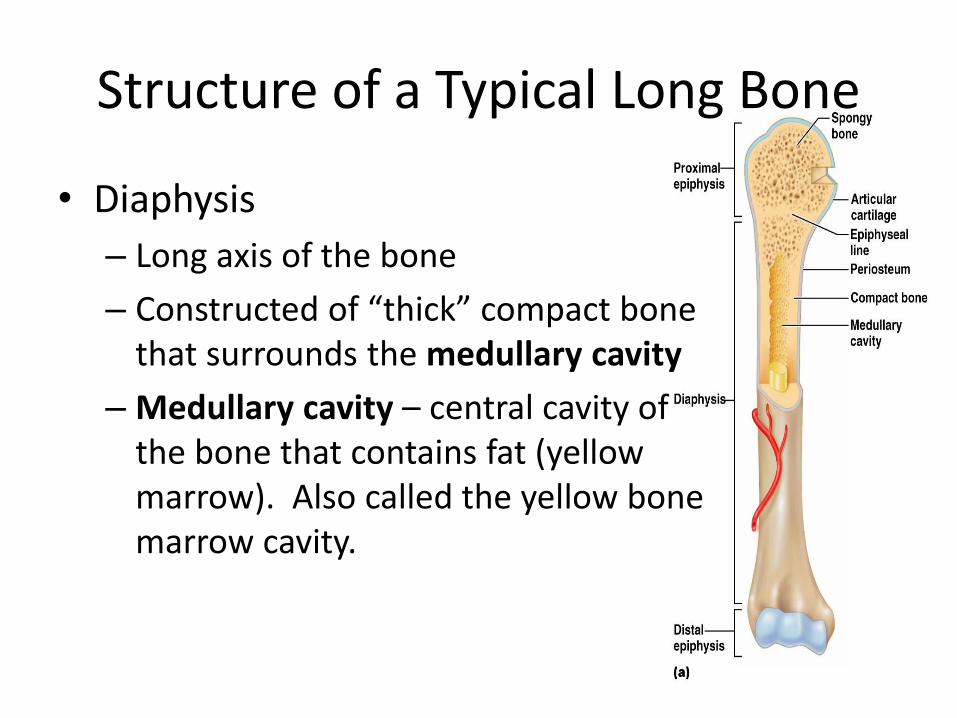
• Diaphysis
– Long axis of the bone
– Constructed of “thick” compact bone that surrounds the medullary cavity
– Medullary cavity – central cavity of the bone that contains fat (yellow marrow). Also called the yellow bone marrow cavity.
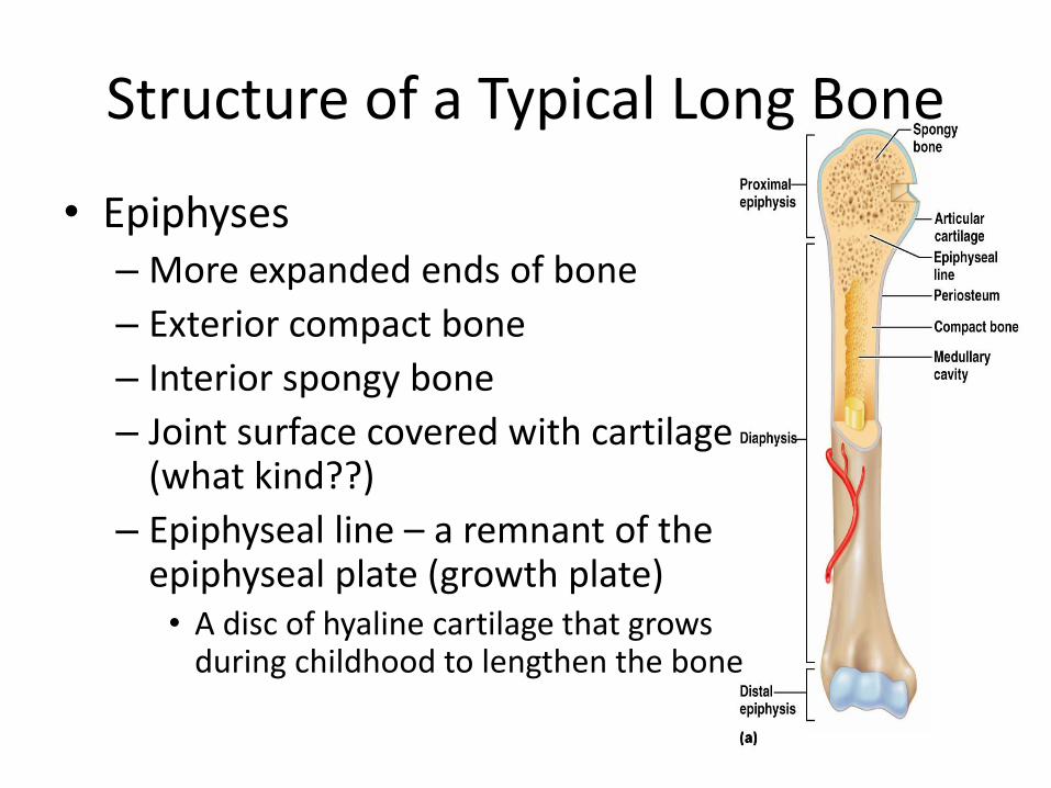
Structure of a Typical Long Bone
• Epiphyses – More expanded ends of bone
– Exterior compact bone
– Interior spongy bone
– Joint surface covered with cartilage (what kind??)
– Epiphyseal line – a remnant of the epiphyseal plate (growth plate) • A disc of hyaline cartilage that grows
during childhood to lengthen the bone
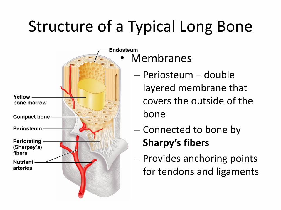
Structure of a Typical Long Bone
• Membranes
– Periosteum – double layered membrane that covers the outside of the bone
– Connected to bone by Sharpy’s fibers
– Provides anchoring points for tendons and ligaments
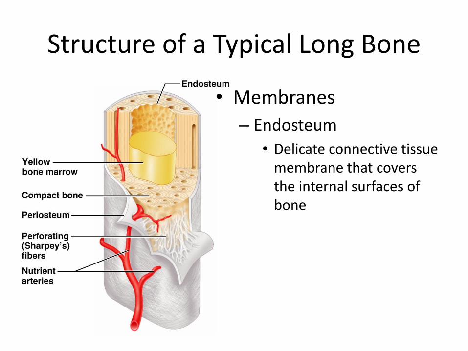
Structure of a Typical Long Bone
• Membranes
– Endosteum
• Delicate connective tissue membrane that covers the internal surfaces of bone
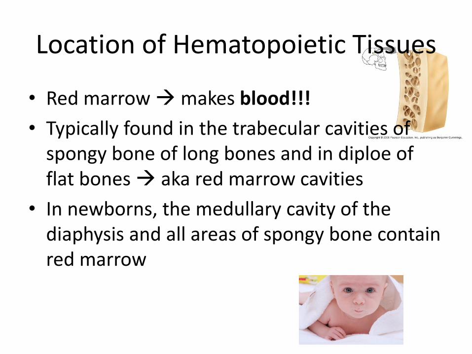
Location of Hematopoietic Tissues
• Red marrow makes blood!!!
• Typically found in the trabecular cavities of spongy bone of long bones and in diploe of flat bones aka red marrow cavities
• In newborns, the medullary cavity of the diaphysis and all areas of spongy bone contain red marrow
Location of Hematopoietic Tissues
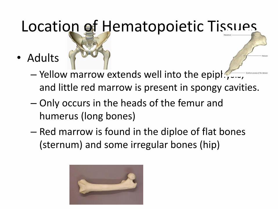
• Adults
– Yellow marrow extends well into the epiphysis, and little red marrow is present in spongy cavities.
– Only occurs in the heads of the femur and humerus (long bones)
– Red marrow is found in the diploe of flat bones (sternum) and some irregular bones (hip)
Location of Hematopoietic Tissues
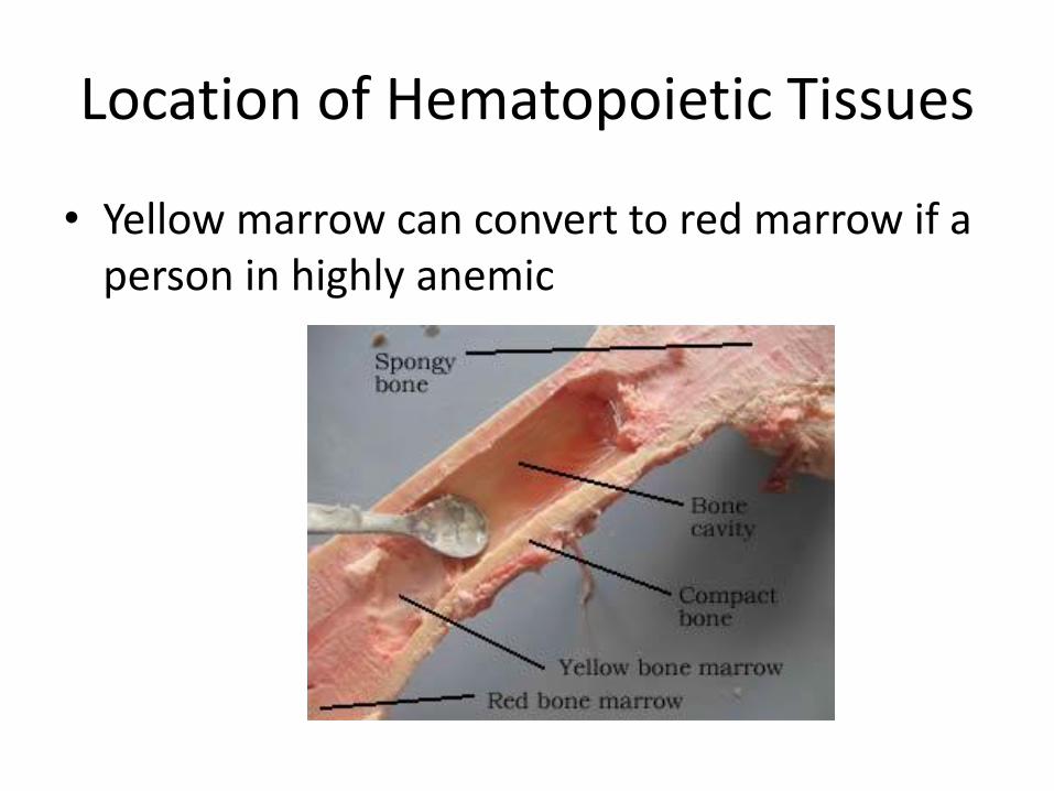
• Yellow marrow can convert to red marrow if a person in highly anemic
Microscopic Anatomy of Bone
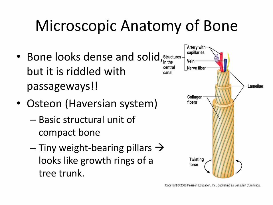
• Bone looks dense and solid, but it is riddled with passageways!!
• Osteon (Haversian system)
– Basic structural unit of compact bone
– Tiny weight-bearing pillars looks like growth rings of a tree trunk.
Microscopic Anatomy of Bone
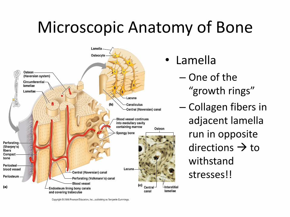
• Lamella
– One of the “growth rings”
– Collagen fibers in adjacent lamella run in opposite directions to withstand stresses!!
Microscopic Anatomy of Bone
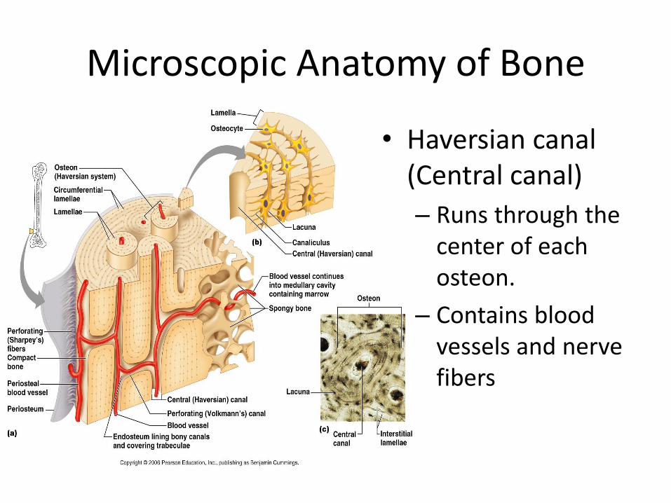
• Haversian canal (Central canal)
– Runs through the center of each osteon.
– Contains blood vessels and nerve fibers
Microscopic Anatomy of Bone
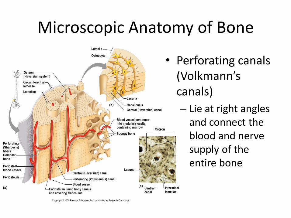
• Perforating canals (Volkmann’s canals)
– Lie at right angles and connect the blood and nerve supply of the entire bone
Microscopic Anatomy of Bone
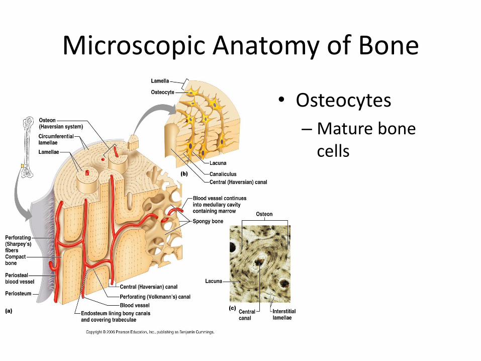
• Osteocytes
– Mature bone cells

Microscopic Anatomy of Bone
• Lacunae
– Osteocytes reside here. Lie at the junctions of lamellae
Microscopic Anatomy of Bone
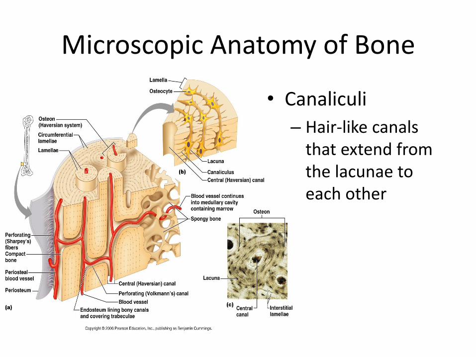
• Canaliculi
– Hair-like canals that extend from the lacunae to each other