chapter 44 · fig. 44-7 ducts nostril with salt secretions nasal salt gland an example is in salt...
TRANSCRIPT

Chapter 44
Osmoregulation and Excretion

Copyright © 2008 Pearson Education Inc., publishing as Pearson Benjamin Cummings
Overview: A Balancing Act
• Physiological systems of animals operate in a fluid
environment
• Relative concentrations of water and solutes must
be maintained within fairly narrow limits
• Osmoregulation regulates solute concentrations
and balances the gain and loss of water
• Osmoregulation is based largely on controlled
movement of solutes between internal fluids and
the external environment

Copyright © 2008 Pearson Education Inc., publishing as Pearson Benjamin Cummings
• Freshwater animals show adaptations that
reduce water uptake and conserve solutes
• Desert and marine animals face desiccating
environments that can quickly deplete body
water
• Excretion gets rid of nitrogenous metabolites
and other waste products

Copyright © 2008 Pearson Education Inc., publishing as Pearson Benjamin Cummings
Osmosis and Osmolarity
• Cells require a balance between osmotic gain and loss of water
• Osmolarity, the solute concentration of a solution, determines the movement of water across a selectively permeable membrane
• If two solutions are isoosmotic, the movement of water is equal in both directions
• If two solutions differ in osmolarity, the net flow of water is from the hypoosmotic to the hyperosmotic solution

Fig. 44-2
Selectively permeable membrane
Net water flow
Hyperosmotic side Hypoosmotic side
Water
Solutes

Copyright © 2008 Pearson Education Inc., publishing as Pearson Benjamin Cummings
Marine Animals
• Most marine invertebrates are osmoconformers
• Most marine vertebrates and some invertebrates are osmoregulators
• Marine bony fishes are hypoosmotic to sea water
• They lose water by osmosis and gain salt by diffusion and from food
• They balance water loss by drinking seawater and excreting salts

Fig. 44-4
Excretion of salt ions from gills
Gain of water and salt ions from food
Osmotic water loss through gills and other parts of body surface
Uptake of water and some ions in food
Uptake of salt ions by gills
Osmotic water gain through gills and other parts of body surface
Excretion of large amounts of water in dilute urine from kidneys
Excretion of salt ions and small amounts of water in scanty urine from kidneys
Gain of water and salt ions from drinking seawater
(a) Osmoregulation in a saltwater fish (b) Osmoregulation in a freshwater fish

Copyright © 2008 Pearson Education Inc., publishing as Pearson Benjamin Cummings
Freshwater Animals
• Freshwater animals constantly take in water by
osmosis from their hypoosmotic environment
• They lose salts by diffusion and maintain water
balance by excreting large amounts of dilute
urine
• Salts lost by diffusion are replaced in foods and
by uptake across the gills

Copyright © 2008 Pearson Education Inc., publishing as Pearson Benjamin Cummings
Animals That Live in Temporary Waters
•Some aquatic
invertebrates in
temporary ponds lose
almost all their body
water and survive in a
dormant state
•This adaptation is called
anhydrobiosis

Copyright © 2008 Pearson Education Inc., publishing as Pearson Benjamin Cummings
Land Animals
• Land animals manage water budgets by
drinking and eating moist foods and using
metabolic water
• Desert animals get major water savings from
simple anatomical features and behaviors such
as a nocturnal life style
Copyright © 2008 Pearson Education Inc., publishing as Pearson Benjamin Cummings
Transport Epithelia in Osmoregulation
• Animals regulate the composition of body fluid
that bathes their cells
• Transport epithelia are specialized epithelial
cells that regulate solute movement
• They are essential components of osmotic
regulation and metabolic waste disposal

Fig. 44-7
Ducts
Nostril with salt secretions
Nasal salt gland
An example is in salt glands of marine birds, which
remove excess sodium chloride from the blood

Copyright © 2008 Pearson Education Inc., publishing as Pearson Benjamin Cummings
Concept 44.2: An animal’s nitrogenous wastes reflect its phylogeny and habitat
• The type and quantity of an animal’s waste
products may greatly affect its water balance
• Among the most important wastes are
nitrogenous breakdown products of proteins
and nucleic acids
• Some animals convert toxic ammonia (NH3) to
less toxic compounds prior to excretion
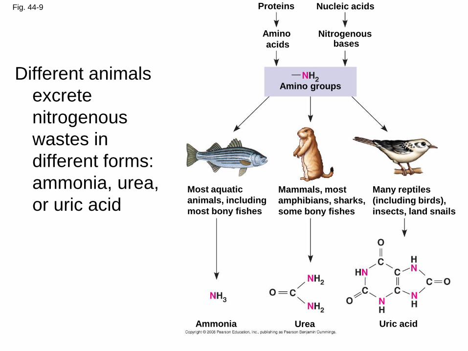
Fig. 44-9
Many reptiles
(including birds),
insects, land snails
Ammonia Uric acid Urea
Most aquatic
animals, including
most bony fishes
Mammals, most
amphibians, sharks,
some bony fishes
Nitrogenous bases
Amino
acids
Proteins Nucleic acids
Amino groups Different animals
excrete
nitrogenous
wastes in
different forms:
ammonia, urea,
or uric acid

Copyright © 2008 Pearson Education Inc., publishing as Pearson Benjamin Cummings
Excretory Processes
• Most excretory systems produce urine by
refining a filtrate derived from body fluids
• Key functions of most excretory systems:
– Filtration: pressure-filtering of body fluids
– Reabsorption: reclaiming valuable solutes
– Secretion: adding toxins and other solutes
from the body fluids to the filtrate
– Excretion: removing the filtrate from the
system

Fig. 44-11
Tubule
Tubules of protonephridia
Cilia
Interstitial fluid flow
Opening in body wall
Nucleus of cap cell
Flame bulb
Tubule cell
A protonephridium is a network of dead-end
tubules connected to external openings
The smallest branches of the network are capped by
a cellular unit called a flame bulb
These tubules excrete a dilute fluid and function in
osmoregulation

Copyright © 2008 Pearson Education Inc., publishing as Pearson Benjamin Cummings
Malpighian Tubules
• In insects and other terrestrial arthropods,
Malpighian tubules remove nitrogenous
wastes from hemolymph and function in
osmoregulation
• Insects produce a relatively dry waste matter,
an important adaptation to terrestrial life

Fig. 44-13
Rectum
Digestive tract
Hindgut Intestine
Malpighian tubules
Rectum
Feces and urine
HEMOLYMPH
Reabsorption
Midgut (stomach)
Salt, water, and nitrogenous
wastes

Fig. 44-14ab
Posterior vena cava
Renal artery and vein
Urinary bladder
Ureter
Aorta
Urethra
(a) Excretory organs and major associated blood vessels
(b) Kidney structure Section of kidney from a rat 4 mm
Kidney
Ureter
Renal medulla
Renal cortex
Renal pelvis
Kidneys
Kidneys, the excretory organs of vertebrates,
function in both excretion and osmoregulation
The mammalian kidney has two distinct regions: an
outer renal cortex and an inner renal medulla

Copyright © 2008 Pearson Education Inc., publishing as Pearson Benjamin Cummings
Structure of the Mammalian Excretory System
• The mammalian excretory system centers on paired kidneys, which are also the principal site of water balance and salt regulation
• Each kidney is supplied with blood by a renal artery and drained by a renal vein
• Urine exits each kidney through a duct called the ureter
• Both ureters drain into a common urinary bladder, and urine is expelled through a urethra

Fig. 44-14a
Posterior vena cava
Renal artery and vein
Urinary bladder
Ureter
Aorta
Urethra
(a) Excretory organs and major associated blood vessels
Kidney

Copyright © 2008 Pearson Education Inc., publishing as Pearson Benjamin Cummings
Filtration of the Blood
•The nephron, the
functional unit of the
vertebrate kidney, consists
of a single long tubule and
a ball of capillaries called
the glomerulus
•There are approximately 1
million nephrons in each
human kidney
•Bowman’s capsule
surrounds and receives
filtrate from the glomerulus

Copyright © 2008 Pearson Education Inc., publishing as Pearson Benjamin Cummings
Filtration of the Blood
• Filtration occurs as blood pressure forces fluid
from the blood in the glomerulus into the lumen
of Bowman’s capsule
• Filtration of small molecules is nonselective
• The filtrate contains salts, glucose, amino
acids, vitamins, nitrogenous wastes, and other
small molecules
Copyright © 2008 Pearson Education Inc., publishing as Pearson Benjamin Cummings
Pathway of the Filtrate
• From Bowman’s capsule, the filtrate passes
through three regions of the nephron: the
proximal tubule, the loop of Henle, and the
distal tubule
• Fluid from several nephrons flows into a
collecting duct, all of which lead to the renal
pelvis, which is drained by the ureter

Copyright © 2008 Pearson Education Inc., publishing as Pearson Benjamin Cummings
Cortical nephrons are confined to the renal cortex, while juxtamedullary nephrons have loops of Henle that descend into the renal medulla

Copyright © 2008 Pearson Education Inc., publishing as Pearson Benjamin Cummings
Blood Vessels Associated with the Nephrons
• Each nephron is supplied with blood by an
afferent arteriole, a branch of the renal artery
that divides into the capillaries
• The capillaries converge as they leave the
glomerulus, forming an efferent arteriole
• The vessels divide again, forming the
peritubular capillaries, which surround the
proximal and distal tubules

Copyright © 2008 Pearson Education Inc., publishing as Pearson Benjamin Cummings
Solute Gradients and Water Conservation
• Urine is much more concentrated than blood
• The cooperative action and precise
arrangement of the loops of Henle and
collecting ducts are largely responsible for the
osmotic gradient that concentrates the urine
• NaCl and urea contribute to the osmolarity of
the interstitial fluid, which causes reabsorption
of water in the kidney and concentrates the
urine

Copyright © 2008 Pearson Education Inc., publishing as Pearson Benjamin Cummings
The Two-Solute Model
• In the proximal tubule, filtrate volume decreases, but its osmolarity remains the same
• The countercurrent multiplier system involving the loop of Henle maintains a high salt concentration in the kidney
• This system allows the vasa recta to supply the kidney with nutrients, without interfering with the osmolarity gradient
• Considerable energy is expended to maintain the osmotic gradient between the medulla and cortex

Copyright © 2008 Pearson Education Inc., publishing as Pearson Benjamin Cummings
• The collecting duct conducts filtrate through the
osmolarity gradient, and more water exits the
filtrate by osmosis
• Urea diffuses out of the collecting duct as it
traverses the inner medulla
• Urea and NaCl form the osmotic gradient that
enables the kidney to produce urine that is
hyperosmotic to the blood

Fig. 44-16-3
Key
Active transport
Passive transport
INNER
MEDULLA
OUTER
MEDULLA
CORTEX H2O
300 300
300
H2O
H2O
H2O
400
600
900
H2O
H2O
1,200
H2O
300
Osmolarity of
interstitial
fluid
(mOsm/L)
400
600
900
1,200
100
NaCl
100
NaCl
NaCl
NaCl
NaCl
NaCl
NaCl
200
400
700
1,200
300
400
600
H2O
H2O
H2O
H2O
H2O
H2O
H2O
NaCl
NaCl
Urea
Urea
Urea

Copyright © 2008 Pearson Education Inc., publishing as Pearson Benjamin Cummings
Mammals
• The juxtamedullary nephron contributes to
water conservation in terrestrial animals
• Mammals that inhabit dry environments have
long loops of Henle, while those in fresh water
have relatively short loops

Copyright © 2008 Pearson Education Inc., publishing as Pearson Benjamin Cummings
Birds and Other Reptiles
•Birds have shorter loops
of Henle but conserve
water by excreting uric acid
instead of urea
•Other reptiles have only
cortical nephrons but also
excrete nitrogenous waste
as uric acid

Copyright © 2008 Pearson Education Inc., publishing as Pearson Benjamin Cummings
Fishes and Amphibians
• Freshwater fishes conserve salt in their distal
tubules and excrete large volumes of dilute
urine
• Kidney function in amphibians is similar to
freshwater fishes - Amphibians conserve water
on land by reabsorbing water from the urinary
bladder
• Marine bony fishes are hypoosmotic compared
with their environment and excrete very little
urine
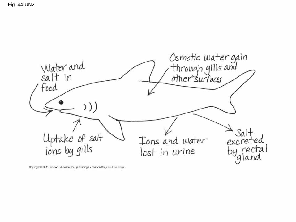
Fig. 44-UN2

Copyright © 2008 Pearson Education Inc., publishing as Pearson Benjamin Cummings
You should now be able to:
1. Distinguish between the following terms:
isoosmotic, hyperosmotic, and hypoosmotic;
osmoregulators and osmoconformers;
stenohaline and euryhaline animals
2. Define osmoregulation, excretion,
anhydrobiosis
3. Compare the osmoregulatory challenges of
freshwater and marine animals
4. Describe some of the factors that affect the
energetic cost of osmoregulation

Copyright © 2008 Pearson Education Inc., publishing as Pearson Benjamin Cummings
5. Describe and compare the protonephridial,
metanephridial, and Malpighian tubule
excretory systems
6. Using a diagram, identify and describe the
function of each region of the nephron
7. Explain how the loop of Henle enhances
water conservation