chapter 31
DESCRIPTION
Chapter 31. Descriptive Terminology. Dental Radiography. Questions What is the difference between a radiograph and an x-ray? What is the difference between radiolucent and radiopaque? What terms may be used to describe radiolucent and radiopaque lesions?. Dental Radiography. - PowerPoint PPT PresentationTRANSCRIPT

Copyright © 2012, 2006, 2000, 1996 by Saunders, an imprint of Elsevier Inc.
Chapter 31Chapter 31
Descriptive TerminologyDescriptive Terminology

Copyright © 2012, 2006, 2000, 1996 by Saunders, an imprint of Elsevier Inc. 2
Dental RadiographyDental Radiography
QuestionsQuestions What is the difference between a radiograph and What is the difference between a radiograph and
an x-ray?an x-ray? What is the difference between radiolucent and What is the difference between radiolucent and
radiopaque?radiopaque? What terms may be used to describe radiolucent What terms may be used to describe radiolucent
and radiopaque lesions?and radiopaque lesions?

Copyright © 2012, 2006, 2000, 1996 by Saunders, an imprint of Elsevier Inc. 3
Dental RadiographyDental Radiography
Chapter 31 Reading Chapter 31 Reading Iannucci & HowertonIannucci & Howerton (pp. 372-382)(pp. 372-382)

Copyright © 2012, 2006, 2000, 1996 by Saunders, an imprint of Elsevier Inc. 4
Dental RadiographyDental Radiography
Chapter 31 OutlineChapter 31 Outline Descriptive TerminologyDescriptive Terminology
Definition and usesDefinition and uses Review of basic termsReview of basic terms

Copyright © 2012, 2006, 2000, 1996 by Saunders, an imprint of Elsevier Inc. 5
What is Descriptive Terminology?What is Descriptive Terminology?
Descriptive terminology is terms used to Descriptive terminology is terms used to describe the appearance, location, and size describe the appearance, location, and size of a lesion.of a lesion. This information should be documented for all This information should be documented for all
lesions viewed on dental images.lesions viewed on dental images.

Copyright © 2012, 2006, 2000, 1996 by Saunders, an imprint of Elsevier Inc. 6
Why Use Descriptive Terminology?Why Use Descriptive Terminology?
Descriptive terminology allows dental Descriptive terminology allows dental professionals to describe and discuss what is professionals to describe and discuss what is seen on a dental image intelligently and to seen on a dental image intelligently and to communicate using a common language.communicate using a common language. It eliminates the chance for miscommunication.It eliminates the chance for miscommunication. It allows for documentation that images were It allows for documentation that images were
reviewed.reviewed.

Copyright © 2012, 2006, 2000, 1996 by Saunders, an imprint of Elsevier Inc. 7
Descriptive Terminology Descriptive Terminology versus Diagnosisversus Diagnosis
Descriptive terminology allows the dental Descriptive terminology allows the dental hygienist to describe what is seen on an hygienist to describe what is seen on an image image without without implying a diagnosis.implying a diagnosis. The patient’s medical and dental history, clinical The patient’s medical and dental history, clinical
findings, signs and symptoms, laboratory tests, findings, signs and symptoms, laboratory tests, and biopsy results are necessary for the dentist to and biopsy results are necessary for the dentist to make a definitive diagnosis.make a definitive diagnosis.

Copyright © 2012, 2006, 2000, 1996 by Saunders, an imprint of Elsevier Inc. 8
Review of Basic TermsReview of Basic Terms
Radiograph/dental image versus x-rayRadiograph/dental image versus x-ray Radiolucent versus radiopaqueRadiolucent versus radiopaque Terms used to describe radiolucent lesionsTerms used to describe radiolucent lesions Terms used to describe radiopaque lesionsTerms used to describe radiopaque lesions

Copyright © 2012, 2006, 2000, 1996 by Saunders, an imprint of Elsevier Inc. 9
Radiograph/Dental Image versus Radiograph/Dental Image versus X-rayX-ray
RadiographRadiograph An image that is produced on photosensitive film An image that is produced on photosensitive film
by exposing the film to x-rays and then processing by exposing the film to x-rays and then processing the film so that a negative is producedthe film so that a negative is produced
Not applicable with digital radiography, in which Not applicable with digital radiography, in which term digital image used insteadterm digital image used instead
X-rayX-ray A beam of energy that has the power to penetrate A beam of energy that has the power to penetrate
substances and to record shadow images on substances and to record shadow images on photographic filmphotographic film

Copyright © 2012, 2006, 2000, 1996 by Saunders, an imprint of Elsevier Inc. 10
Radiolucent versus RadiopaqueRadiolucent versus Radiopaque
Iannucci & HowertonIannucci & Howerton (pp. 373-374) (Figs. 31-1, (pp. 373-374) (Figs. 31-1, 31-2,31-2, 31-331-3))
RadiolucentRadiolucent This that portion of a processed image that is dark or This that portion of a processed image that is dark or
black.black.• Caries appears radiolucent because the area of tooth with Caries appears radiolucent because the area of tooth with
caries is less dense than surrounding structures.caries is less dense than surrounding structures.
RadiopaqueRadiopaque This is that portion of a processed image that This is that portion of a processed image that
appears light or white.appears light or white.• A metallic restoration appears radiopaque because it is very A metallic restoration appears radiopaque because it is very
dense and absorbs the radiation.dense and absorbs the radiation.

Copyright © 2012, 2006, 2000, 1996 by Saunders, an imprint of Elsevier Inc. 11
Terms Used to Describe Terms Used to Describe Radiolucent LesionsRadiolucent Lesions
Appearance Appearance LocationLocation SizeSize

Copyright © 2012, 2006, 2000, 1996 by Saunders, an imprint of Elsevier Inc. 12
AppearanceAppearance
Iannucci & HowertonIannucci & Howerton (pp. 374-375) (Box 31-(pp. 374-375) (Box 31-1)1)
Unilocular radiolucent lesionsUnilocular radiolucent lesions One compartmentOne compartment Tend to be small and nonexpansileTend to be small and nonexpansile Have borders that may appear corticated or Have borders that may appear corticated or
noncorticated on imagenoncorticated on image

Copyright © 2012, 2006, 2000, 1996 by Saunders, an imprint of Elsevier Inc. 13
AppearanceAppearance Iannucci & HowertonIannucci & Howerton (pp. 374-375) (Figs. 31-4, (pp. 374-375) (Figs. 31-4,
31-5)31-5) Unilocular lesion, corticated bordersUnilocular lesion, corticated borders
The lesion exhibits a thin, well-demarcated The lesion exhibits a thin, well-demarcated radiopaque rim of bone at the periphery.radiopaque rim of bone at the periphery.
Usually indicative of a benign, slow-growing process.Usually indicative of a benign, slow-growing process. Unilocular lesion, noncorticated bordersUnilocular lesion, noncorticated borders
The lesion does not exhibit a thin radiopaque rim of The lesion does not exhibit a thin radiopaque rim of bone at the periphery.bone at the periphery.
The periphery appears fuzzy or poorly defined.The periphery appears fuzzy or poorly defined. May represent either a benign or a malignant process.May represent either a benign or a malignant process.
Copyright © 2012, 2006, 2000, 1996 by Saunders, an imprint of Elsevier Inc. 14
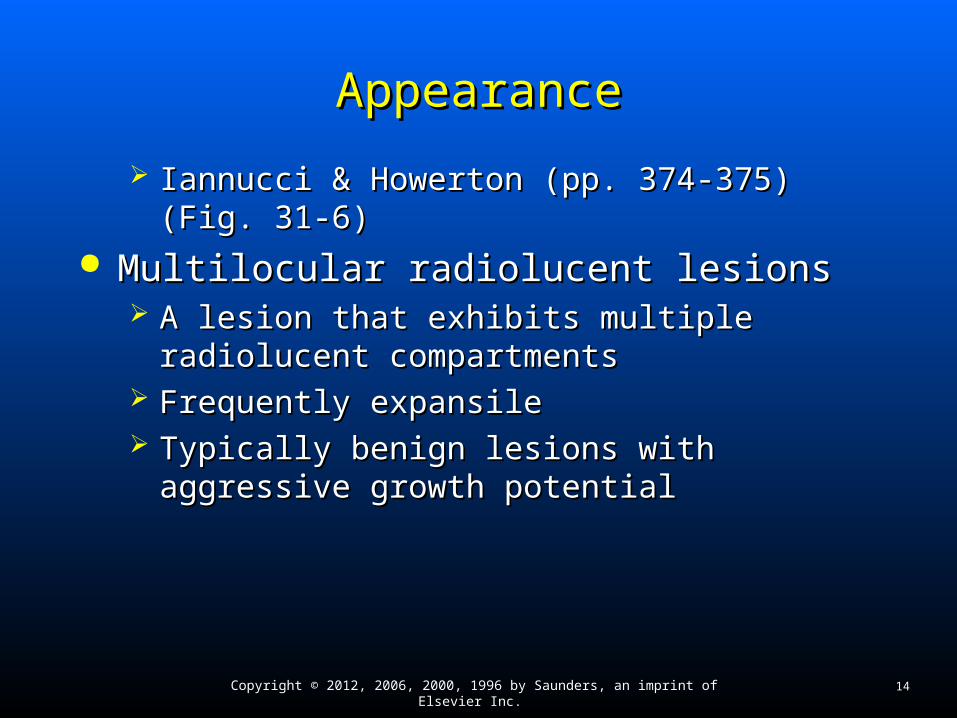
AppearanceAppearance
Iannucci & HowertonIannucci & Howerton (pp. 374-375) (Fig. 31-(pp. 374-375) (Fig. 31-6)6)
Multilocular radiolucent lesionsMultilocular radiolucent lesions A lesion that exhibits multiple radiolucent A lesion that exhibits multiple radiolucent
compartmentscompartments Frequently expansileFrequently expansile Typically benign lesions with aggressive growth Typically benign lesions with aggressive growth
potentialpotential

Copyright © 2012, 2006, 2000, 1996 by Saunders, an imprint of Elsevier Inc. 15
LocationLocation
Iannucci & HowertonIannucci & Howerton (pp. 375-376) (Box 31-(pp. 375-376) (Box 31-2)2)
May appear in a periapical, inter-radicular, May appear in a periapical, inter-radicular, edentulous, or pericoronal location.edentulous, or pericoronal location. May appear as alveolar bone loss.May appear as alveolar bone loss.

Copyright © 2012, 2006, 2000, 1996 by Saunders, an imprint of Elsevier Inc. 16
Periapical LocationPeriapical Location
Iannucci & HowertonIannucci & Howerton (pp. 375-376) (Fig. 31-(pp. 375-376) (Fig. 31-7)7)
A lesion located around the apex of a toothA lesion located around the apex of a tooth ExampleExample
Periapical cyst secondary to pulpal necrosis Periapical cyst secondary to pulpal necrosis

Copyright © 2012, 2006, 2000, 1996 by Saunders, an imprint of Elsevier Inc. 17
Inter-radicular LocationInter-radicular Location
Iannucci & HowertonIannucci & Howerton (p. 376) (Fig. 31-8)(p. 376) (Fig. 31-8) A lesion located between the roots of A lesion located between the roots of
adjacent teethadjacent teeth ExampleExample
Lateral periodontal cystLateral periodontal cyst

Copyright © 2012, 2006, 2000, 1996 by Saunders, an imprint of Elsevier Inc. 18
Edentulous ZoneEdentulous Zone
Iannucci & HowertonIannucci & Howerton (pp. 376-377) (Fig. 31-(pp. 376-377) (Fig. 31-9)9)
A lesion located in an area without teethA lesion located in an area without teeth

Copyright © 2012, 2006, 2000, 1996 by Saunders, an imprint of Elsevier Inc. 19
Pericoronal LocationPericoronal Location
Iannucci & HowertonIannucci & Howerton (p. 377) (Fig. 31-10)(p. 377) (Fig. 31-10) A radiolucent lesion located around the crown A radiolucent lesion located around the crown
of an impacted toothof an impacted tooth ExampleExample
Dentigerous cyst Dentigerous cyst

Copyright © 2012, 2006, 2000, 1996 by Saunders, an imprint of Elsevier Inc. 20
Alveolar Bone LossAlveolar Bone Loss
Iannucci & HowertonIannucci & Howerton (p. 377) (Fig. 31-11)(p. 377) (Fig. 31-11) Loss of bone in the maxilla or mandible that Loss of bone in the maxilla or mandible that
surrounds and supports the teethsurrounds and supports the teeth Appears radiolucentAppears radiolucent

Copyright © 2012, 2006, 2000, 1996 by Saunders, an imprint of Elsevier Inc. 21
SizeSize
Iannucci & HowertonIannucci & Howerton (p. 377) (Fig. 31-12)(p. 377) (Fig. 31-12) Can vary in size from several millimeters in Can vary in size from several millimeters in
diameter to several centimeters in diameterdiameter to several centimeters in diameter Can be measured on an image with a millimeter Can be measured on an image with a millimeter
rulerruler

Copyright © 2012, 2006, 2000, 1996 by Saunders, an imprint of Elsevier Inc. 22
Terms Used to Describe Terms Used to Describe Radiopaque LesionsRadiopaque Lesions
AppearanceAppearance Location Location SizeSize

Copyright © 2012, 2006, 2000, 1996 by Saunders, an imprint of Elsevier Inc. 23
AppearanceAppearance
Iannucci & HowertonIannucci & Howerton (pp. 377-378) (Box 31-(pp. 377-378) (Box 31-3)3)
Can be described as focal opacity, target Can be described as focal opacity, target lesion, multifocal confluent, irregular, ground lesion, multifocal confluent, irregular, ground glass, or mixed lucent-opaque.glass, or mixed lucent-opaque. Radiopaque lesions occur not only in bone but in Radiopaque lesions occur not only in bone but in
soft tissue as well.soft tissue as well.

Copyright © 2012, 2006, 2000, 1996 by Saunders, an imprint of Elsevier Inc. 24
Focal OpacityFocal Opacity
Iannucci & HowertonIannucci & Howerton (pp. 377-378) (Fig. 31-(pp. 377-378) (Fig. 31-13) 13)
A well-defined, localized radiopaque lesion on A well-defined, localized radiopaque lesion on an imagean image
ExampleExample Condensing osteitisCondensing osteitis

Copyright © 2012, 2006, 2000, 1996 by Saunders, an imprint of Elsevier Inc. 25
Target LesionTarget Lesion
Iannucci & HowertonIannucci & Howerton (p. 378) (Fig. 31-14)(p. 378) (Fig. 31-14) A well-defined, localized radiopaque area A well-defined, localized radiopaque area
surrounded by a uniform radiolucent halo surrounded by a uniform radiolucent halo ExampleExample
Benign cementoblastomaBenign cementoblastoma

Copyright © 2012, 2006, 2000, 1996 by Saunders, an imprint of Elsevier Inc. 26
Multifocal ConfluentMultifocal Confluent
Iannucci & HowertonIannucci & Howerton (p. 378) (Fig. 31-15)(p. 378) (Fig. 31-15) Multiple radiopacities that appear to overlap Multiple radiopacities that appear to overlap
or flow togetheror flow together ExampleExample
Osteitis deformans, florid osseous dysplasiaOsteitis deformans, florid osseous dysplasia Multifocal confluent radiopacities involving multiple Multifocal confluent radiopacities involving multiple
quadrants of the jaws usually represent benign quadrants of the jaws usually represent benign fibro-osseous disorders.fibro-osseous disorders.

Copyright © 2012, 2006, 2000, 1996 by Saunders, an imprint of Elsevier Inc. 27
Irregular Ill-DefinedIrregular Ill-Defined
Iannucci & HowertonIannucci & Howerton (pp. 378-379) (Fig. 31-(pp. 378-379) (Fig. 31-16)16)
A radiopacity may exhibit an irregular, poorly A radiopacity may exhibit an irregular, poorly defined pattern.defined pattern. It may represent a malignant condition.It may represent a malignant condition.
ExamplesExamples Osteosarcoma and chondrosarcoma Osteosarcoma and chondrosarcoma

Copyright © 2012, 2006, 2000, 1996 by Saunders, an imprint of Elsevier Inc. 28
Ground GlassGround Glass
Iannucci & HowertonIannucci & Howerton (p. 379) (Fig. 31-17)(p. 379) (Fig. 31-17) A granular or pebbled radiopacity that A granular or pebbled radiopacity that
resembles pulverized glassresembles pulverized glass Often said to resemble the appearance or texture Often said to resemble the appearance or texture
of an orange peelof an orange peel ExamplesExamples
Fibrous dysplasia, osteitis deformans, and Fibrous dysplasia, osteitis deformans, and osteopetrosisosteopetrosis

Copyright © 2012, 2006, 2000, 1996 by Saunders, an imprint of Elsevier Inc. 29
Mixed Lucent-OpaqueMixed Lucent-Opaque
Iannucci & HowertonIannucci & Howerton (p. 379) (Fig. 31-18) (p. 379) (Fig. 31-18) Exhibits both a radiopaque and a radiolucent Exhibits both a radiopaque and a radiolucent
componentcomponent Often represent calcifying tumorsOften represent calcifying tumors
ExampleExample Compound odontomaCompound odontoma

Copyright © 2012, 2006, 2000, 1996 by Saunders, an imprint of Elsevier Inc. 30
Soft Tissue OpacitySoft Tissue Opacity
Iannucci & HowertonIannucci & Howerton (p. 379) (Fig. 31-19)(p. 379) (Fig. 31-19) Appears as a well-defined, radiopaque area Appears as a well-defined, radiopaque area
located in soft tissuelocated in soft tissue ExamplesExamples
SialolithSialolith Calcified lymph nodeCalcified lymph node

Copyright © 2012, 2006, 2000, 1996 by Saunders, an imprint of Elsevier Inc. 31
LocationLocation
Iannucci & HowertonIannucci & Howerton (pp. 379-380) (Box 31-(pp. 379-380) (Box 31-4)4)
Radiopaque lesions may appear in the same Radiopaque lesions may appear in the same places as radiolucent lesions: in a periapical, places as radiolucent lesions: in a periapical, inter-radicular, edentulous, or pericoronal inter-radicular, edentulous, or pericoronal location.location.

Copyright © 2012, 2006, 2000, 1996 by Saunders, an imprint of Elsevier Inc. 32
Periapical LocationPeriapical Location
Iannucci & HowertonIannucci & Howerton (p. 380) (Fig. 31-20)(p. 380) (Fig. 31-20) Periapical refers to a radiopaque lesion Periapical refers to a radiopaque lesion
located around the apex of a tooth.located around the apex of a tooth. ExampleExample
Benign cementoblastomaBenign cementoblastoma

Copyright © 2012, 2006, 2000, 1996 by Saunders, an imprint of Elsevier Inc. 33
Inter-radicular LocationInter-radicular Location
Iannucci & HowertonIannucci & Howerton (p. 380) (Fig. 31-21)(p. 380) (Fig. 31-21) A radiopaque lesion located between the A radiopaque lesion located between the
roots of adjacent teethroots of adjacent teeth ExampleExample
Sclerotic boneSclerotic bone

Copyright © 2012, 2006, 2000, 1996 by Saunders, an imprint of Elsevier Inc. 34
Edentulous ZoneEdentulous Zone
Iannucci & HowertonIannucci & Howerton (p. 380) (Fig. 31-22)(p. 380) (Fig. 31-22) A radiopaque lesion located in an area A radiopaque lesion located in an area
without teethwithout teeth ExampleExample
Complex odontomaComplex odontoma

Copyright © 2012, 2006, 2000, 1996 by Saunders, an imprint of Elsevier Inc. 35
Pericoronal LocationPericoronal Location
Iannucci & HowertonIannucci & Howerton (p. 380) (Fig. 31-23)(p. 380) (Fig. 31-23) A radiopaque lesion located around the A radiopaque lesion located around the
crown of an impacted toothcrown of an impacted tooth ExampleExample
Adenomatoid odontogenic tumor is a mixed Adenomatoid odontogenic tumor is a mixed lucent-opaque lesion. lucent-opaque lesion.

Copyright © 2012, 2006, 2000, 1996 by Saunders, an imprint of Elsevier Inc. 36
SizeSize
Radiopaque lesions can vary from several Radiopaque lesions can vary from several millimeters to several centimeters in diameter.millimeters to several centimeters in diameter. They can be measured on an image with a ruler.They can be measured on an image with a ruler.