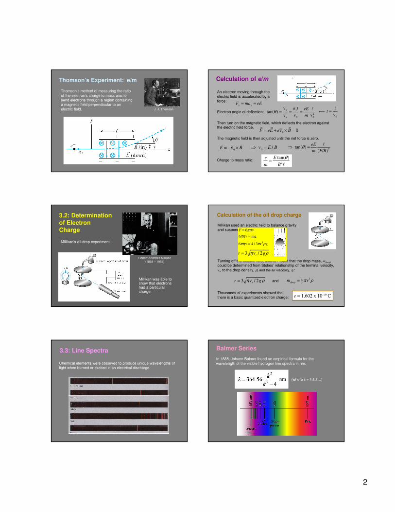
chapter 3 prelude to quantum theory - university...
TRANSCRIPT
1
3.1 Discovery of the X Ray and the Electron
3.2 Determination of Electron Charge3.3 Line Spectra
3.4 Quantization
3.5 Blackbody Radiation3.6 Photoelectric Effect
3.7 X-Ray Production3.8 Compton Effect
3.9 Pair Production and Annihilation
CHAPTER 3Prelude to Quantum TheoryPrelude to Quantum Theory
We have no right to assume that any physical laws exist, or if they have existed up until now, or that they will continue to exist in a similar
manner in the future.
An important scientific innovation rarely makes its way by gradually
winning over and converting its opponents. What does happen is that the opponents gradually die out.
- Max Planck
Max Karl Ernst Ludwig Planck
(1858-1947)
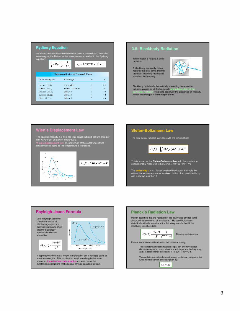
3.1: Discovery of the X-Ray and the Electron
In the 1890s scientists and engineers were familiar with
“cathode rays.” These rays
were generated from one of the metal plates in an
evacuated tube with a large
electric potential across it.
It was surmised that cathode rays had something to do with atoms.
It was known that cathode rays could penetrate matter and were
deflected by magnetic and electric fields.
J. J. Thomson
(1856-1940)
Wilhelm Röntgen
(1845-1923)
Observation of X Rays
Wilhelm Röntgen studied the effects
of cathode rays passing through
various materials. He noticed that a phosphorescent screen near the
tube glowed during some of these experiments. These new rays were
unaffected by magnetic fields and
penetrated materials more than cathode rays.
He called them x rays and deduced
that they were produced by the cathode rays bombarding the glass
walls of his vacuum tube. Wilhelm Röntgen
Röntgen’sX-Ray Tube
Röntgen constructed an x-ray tube by
allowing cathode rays to impact the glass wall of the tube and produced x rays. He
used x rays to make a shadowgram the
bones of a hand on a phosphorescent screen.
Thomson’s Cathode-Ray Experiment
Thomson used an evacuated cathode-ray tube to show that the
cathode rays were negatively charged particles (electrons) by
deflecting them in electric and magnetic fields.
2
Thomson’s method of measuring the ratio
of the electron’s charge to mass was to send electrons through a region containing
a magnetic field perpendicular to an
electric field.
Thomson’s Experiment: e/m
J. J. Thomson
An electron moving through the
electric field is accelerated by a
force:
Electron angle of deflection:
Then turn on the magnetic field, which deflects the electron against the electric field force.
The magnetic field is then adjusted until the net force is zero.
Charge to mass ratio:
Calculation of e/m
y yF ma eE= =
2
0 0
vtan( )
v v v
y y
x
a t eE
mθ = = =
�
0v 0F eE e B= + × =� � ��
0vE B= − ×� ��
0v /E B⇒ =
2
tan( )e E
m B
θ=
�
0vt =�
2tan( )
( / )
eE
m E Bθ⇒ =
�
Millikan’s oil-drop experiment
3.2: Determination
of Electron Charge
Robert Andrews Millikan
(1868 – 1953)
Millikan was able to show that electrons had a particular charge.
Calculation of the oil drop charge
Millikan used an electric field to balance gravity
and suspend a charged oil drop:
e = 1.602 x 10⁻¹⁹ C
y drop
VF eE e m g
d= = = −
343drop
m rπ ρ=
/drop
e m gd V⇒ = −
Thousands of experiments showed that there is a basic quantized electron charge:
Turning off the electric field, Millikan noted that the drop mass, mdrop, could be determined from Stokes’ relationship of the terminal velocity,
vt, to the drop density, ρ, and the air viscosity, η :
3 v / 2tr gη ρ= and
F 6 vπη=
6 v mgπη =
36 v 4 / 3 r gπη π ρ=
3 v / 2tr gη ρ=
Chemical elements were observed to produce unique wavelengths of
light when burned or excited in an electrical discharge.
3.3: Line Spectra Balmer Series
In 1885, Johann Balmer found an empirical formula for the
wavelength of the visible hydrogen line spectra in nm:
nm (where k = 3,4,5…)
3
Rydberg Equation
As more scientists discovered emission lines at infrared and ultraviolet
wavelengths, the Balmer series equation was extended to the Rydberg
equation:
3.5: Blackbody Radiation
When matter is heated, it emits radiation.
A blackbody is a cavity with a material that only emits thermal
radiation. Incoming radiation is
absorbed in the cavity.
Blackbody radiation is theoretically interesting because the radiation properties of the blackbody are independent of the
particular material. Physicists can study the properties of intensity versus wavelength at fixed temperatures.
Wien’s Displacement Law
The spectral intensity I(λ, T) is the total power radiated per unit area per
unit wavelength at a given temperature.
Wien’s displacement law: The maximum of the spectrum shifts to
smaller wavelengths as the temperature is increased.
The total power radiated increases with the temperature:
This is known as the Stefan-Boltzmann law, with the constant σexperimentally measured to be 5.6705 × 10⁻⁸ W / (m2 · K4).
The emissivity є (є = 1 for an idealized blackbody) is simply the
ratio of the emissive power of an object to that of an ideal blackbody and is always less than 1.
Stefan-Boltzmann Law
Rayleigh-Jeans Formula
Lord Rayleigh used the
classical theories of electromagnetism and
thermodynamics to show
that the blackbody spectral distribution
should be:
It approaches the data at longer wavelengths, but it deviates badly at
short wavelengths. This problem for small wavelengths became
known as the ultraviolet catastrophe and was one of the outstanding exceptions that classical physics could not explain.
Planck made two modifications to the classical theory:
The oscillators (of electromagnetic origin) can only have certain
discrete energies, En = nhν, where n is an integer, ν is the frequency, and h is called Planck’s constant: h = 6.6261 × 10⁻³⁴ J·s.
The oscillators can absorb or emit energy in discrete multiples of the
fundamental quantum of energy given by:
∆E = hν
Planck’s radiation law
Planck assumed that the radiation in the cavity was emitted (and
absorbed) by some sort of “oscillators.” He used Boltzman’s
statistical methods to arrive at the following formula that fit the blackbody radiation data.
Planck’s Radiation Law

4
3.6: Photoelectric
Effect
Methods of electron emission:
Thermionic emission: Applying heat allows electrons to gain
enough energy to escape.
Secondary emission: The electron gains enough energy by transfer
from another high-speed particle that strikes the material from outside.
Field emission: A strong external electric field pulls the electron out of
the material.
Photoelectric effect: Incident light (electromagnetic radiation) shining
on the material transfers energy to the electrons, allowing them to
escape. We call the ejected electrons photoelectrons.
Photo-electric Effect Experimental Setup
Photo-electric effect
observationsThe kinetic energy of
the photoelectrons is independent of the
light intensity.
The kinetic energy of the photoelectrons, for
a given emitting
material, depends only on the frequency of
the light.
Classically, the kinetic energy of the
photoelectrons should
increase with the light intensity and not
depend on the
frequency.
Electron
kinetic
energy
Photo-electric effect
observations
There was a threshold
frequency of the light,
below which no photoelectrons were
ejected (related to the
work function φ of the emitter material).
The existence of a threshold frequency is completely inexplicable in classical theory.
Electron
kinetic
energy
Photo-electric effect
observations
When photoelectrons
are produced, their number is proportional
to the intensity of light.
Also, the photoelectrons are emitted almost
instantly following
illumination of the photocathode,
independent of the
intensity of the light.
Classical theory predicted that, for extremely low light intensities, a long
time would elapse before any one
electron could obtain sufficient energy to escape. We observe,
however, that the photoelectrons are
ejected almost immediately.
(number of
electrons)
Einstein suggested that the electro-magnetic radiation field is quantized
into particles called photons. Each photon has the energy quantum:
where ν is the frequency of the light and h is Planck’s constant.
Alternatively,
Einstein’s Theory: Photons
E hν=
E ω= � / 2h π≡�where:

5
Conservation of energy yields:
Einstein’s Theory
In reality, the data were a bit more complex.
Because the electron’s energy can be reduced
by the emitter material, consider vmax (not v):
where φ is the work function of the metal (potential energy to be overcome before an electron could escape).
212
vh mν φ= +
21max2
vh mν φ= +
3.7: X-Ray Production: Theory
An energetic electron passing through matter will
radiate photons and lose kinetic
energy, called bremsstrahlung. Since momentum is conserved, the
nucleus absorbs very little energy, and it can be ignored. The final
energy of the electron is determined
from the conservation of energy to be:
f iE E hν= −
fEiE
hν
X-Ray
Production: Experiment
Current passing through a filament produces copious numbers of electrons by thermionic emission. If one focuses these electrons by
a cathode structure into a beam and accelerates them by potential
differences of thousands of volts until they impinge on a metal anode surface, they produce x rays by bremsstrahlung as they stop
in the anode material.
Inverse Photoelectric Effect
Conservation of energy requires that the electron kinetic energy equal the
maximum photon energy (neglect the
work function because it’s small compared to the electron potential
energy). This yields the Duane-Hunt
limit, first found experimentally. The photon wavelength depends only on
the accelerating voltage and is the
same for all targets.
0 max
min
hceV hν
λ= =
3.8: Compton Effect
When a photon enters matter, it can interact
with one of the electrons. The laws of
conservation of energy and momentum apply, as in any elastic collision between
two particles. The momentum of a particle
moving at the speed of light is:
E h hp
c c
ν
λ= = =
This yields the change in
wavelength of the scattered
photon, known as the Compton effect:
eE
/hc λ
/hc λ′
The electron energy is:
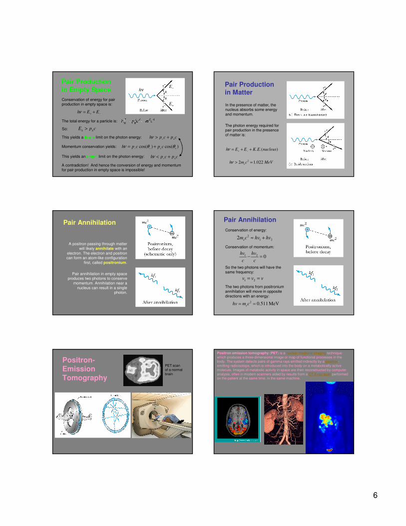
3.9: Pair Production and Annihilation
If a photon can create an electron, it
must also create a positive charge to
balance charge conservation.
In 1932, C. D. Anderson observed a
positively charged electron (e+) in
cosmic radiation. This particle, called a positron, had been predicted to exist
several years earlier by P. A. M. Dirac.
A photon’s energy can
be converted entirely
into an electron and a positron in a process
called pair production:
Paul Dirac
(1902 - 1984)
6
Pair Production
in Empty Space
Conservation of energy for pair production in empty space is:
h E Eν + −= +
cos( ) cos( )h p c p cν θ θ− − + += +
h p c p cν − +< +
This yields a lower limit on the photon energy: h p c p cν − +> +
The total energy for a particle is:
This yields an upper limit on the photon energy:
Momentum conservation yields:
A contradiction! And hence the conversion of energy and momentum for pair production in empty space is impossible!
E p c± ±>So:
hν
E+
E−Pair Production in Matter
In the presence of matter, the nucleus absorbs some energy
and momentum.
The photon energy required for pair production in the presence
of matter is:
. .( )h E E K E nucleusν + −= + +
22 1.022e
h m c MeVν > =
Pair Annihilation
A positron passing through matter
will likely annihilate with an
electron. The electron and positron can form an atom-like configuration
first, called positronium.
Pair annihilation in empty space produces two photons to conserve
momentum. Annihilation near a
nucleus can result in a single photon.
Pair Annihilation
Conservation of energy:
Conservation of momentum:
1 2v v v= =
1 2 0hv hv
c c− =
2
1 22e
m c hv hv≈ +
2 0.511 MeVehv m c= =
So the two photons will have the
same frequency:
The two photons from positronium
annihilation will move in opposite
directions with an energy:
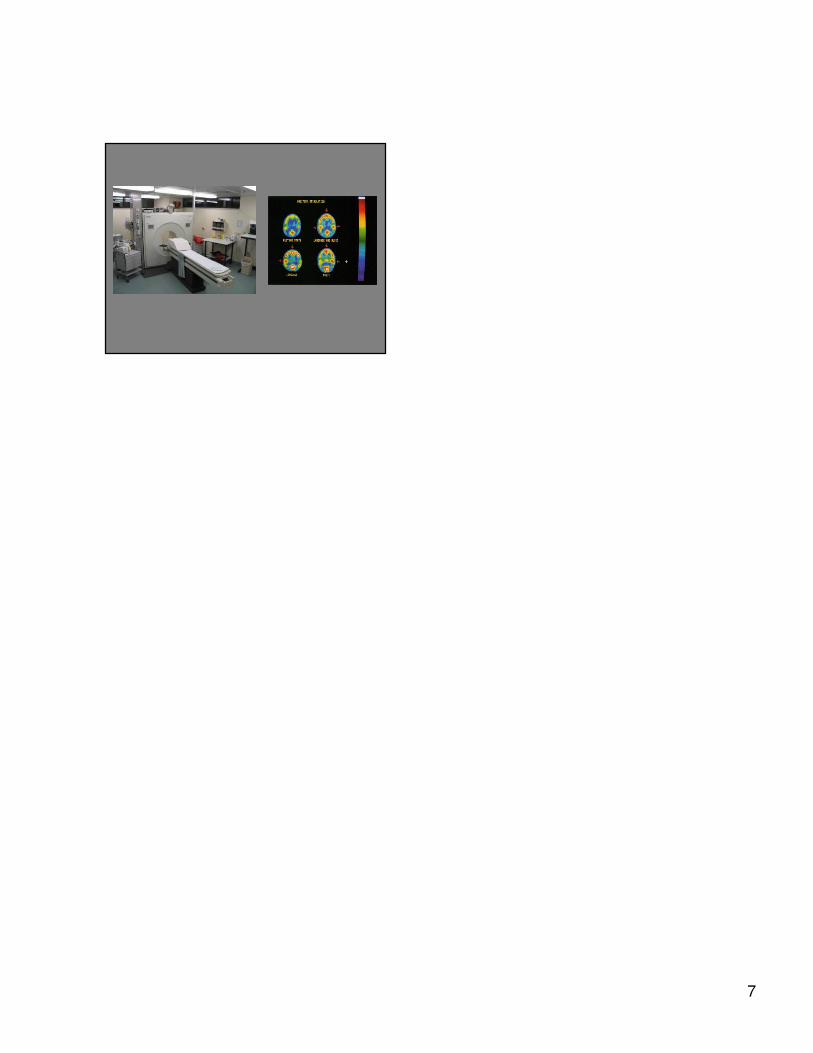
Positron-Emission
Tomography
PET scan
of a normal
brain
Positron emission tomography (PET) is a nuclear medicine imaging technique
which produces a three-dimensional image or map of functional processes in the
body. The system detects pairs of gamma rays emitted indirectly by a positron-
emitting radioisotope, which is introduced into the body on a metabolically active
molecule. Images of metabolic activity in space are then reconstructed by computer
analysis, often in modern scanners aided by results from a CT X-ray scan performed on the patient at the same time, in the same machine.
7