chapter 27 prokaryotes bacteria on the point of a pin
DESCRIPTION
Chapter 27 Prokaryotes Bacteria on the point of a pin. Extreme Thermophiles. The Three Domains of Life. Streptococcus strepto=chain coccus=spherical. Bacilli=rod-shaped. Spirilla=helical includes spirochetes. Largest known prokaryote. Another large prokaryote. paramecium. - PowerPoint PPT PresentationTRANSCRIPT

Chapter 27 Prokaryotes
Bacteria on the point of a pin

Extreme Thermophiles

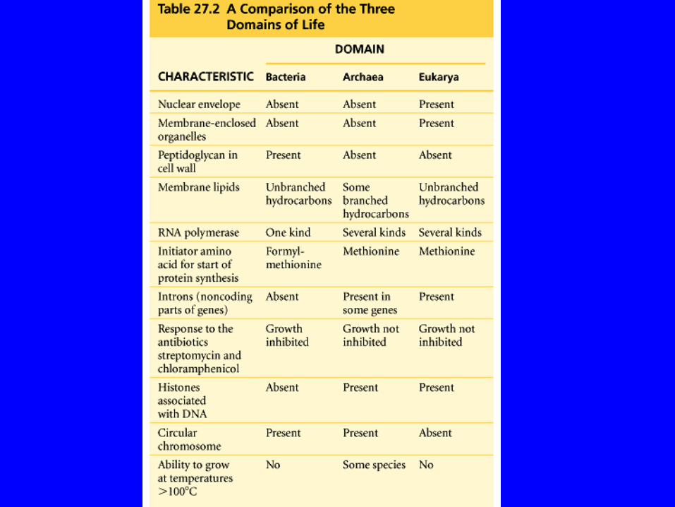
The Three Domains of Life
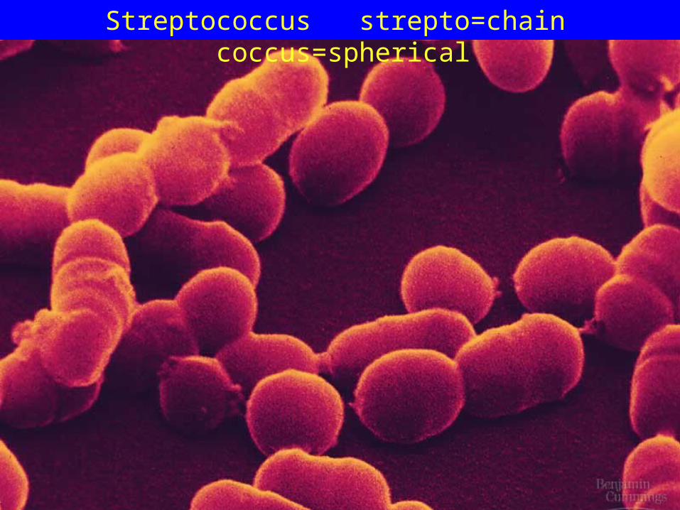
Streptococcus strepto=chain coccus=spherical

Bacilli=rod-shaped
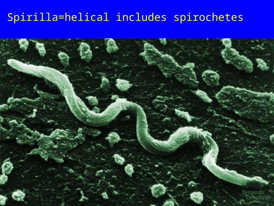
Spirilla=helical includes spirochetes
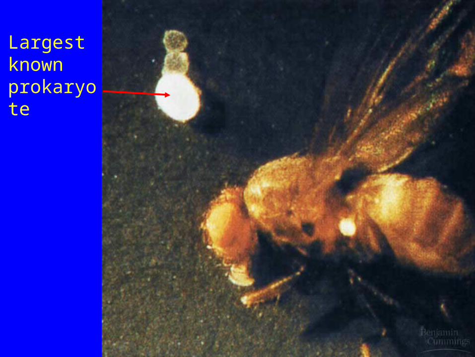
Largest known prokaryote

Another large prokaryote
paramecium
Prokaryotes vary in size from 0.2µ--750µ

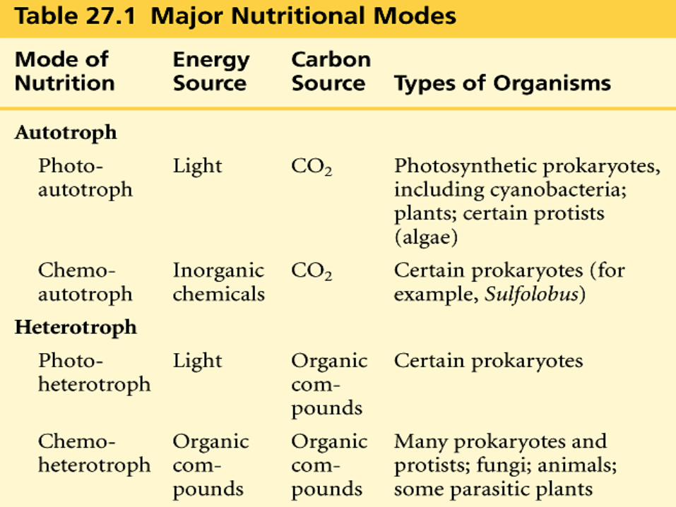
Evolution of Prokaryotic Metabolism1. The Origin of Glycolysis– First prokaryotes 3.5 billion
years ago, probably anaerobic chemoheterotrophs. They absorbed organic compounds and used glycolysis (fermentation) to produce ATP in an atmosphere without oxygen
2. The Origin of Electron Transport Chains and Chemiosmosis– The first proton pumps were probably for pH regulation. Later some bacteria used the oxidation of organic compounds to pump H+’s to save ATP and developed the first Electron Transport Chains. Some got so good at transporting H+’s that they could actually develop a gradient and use the influx to drive the production of ATP.

3. The Origin of Photosynthesis– The first light absorbing pigments probably provided protection by absorbing UV light. Bacteriorhodopsin in extreme halophiles uses light energy to pump H+’s out of the cell and produce a gradient which is then used to produce ATP (Photosystem I) . Photoheterotrophs
4. Cyanobacteria, Photoautotrophs, Splitting H2O and Producing O2– Photosystem II evolved in cyanobacteria and they split water and released free oxygen. The oxygen was toxic to many organisms which became extinct. (First Great Extinction)
5. Origin of Cellular Respiration– Some prokaryotes modified their photosynthetic ETC’s to reduce the level of toxic O2. The
purple non-sulfur bacteria still use their ETC’s for both photosynthesis and respiration. Eventually some bacteria used O2 to pull electrons through proton pumps and aerobic
respiration began. aerobic chemoheterotrophs

Cell WallsAll the proteobacteria and the eubacteria have peptidoglycan cell walls. Archaebacteria have a different type of cell wall. Cell walls protect bacteria from cytolysis in hypotonic solutions but can not protect them from plasmolysis in hypertonic solutions. Mycoplasmas without cell walls are susceptible to both.Penicillin denatures (noncompetitive inhibitor) the enzyme that bacteria use to form their cell walls and leaves them susceptible to cytolysis.

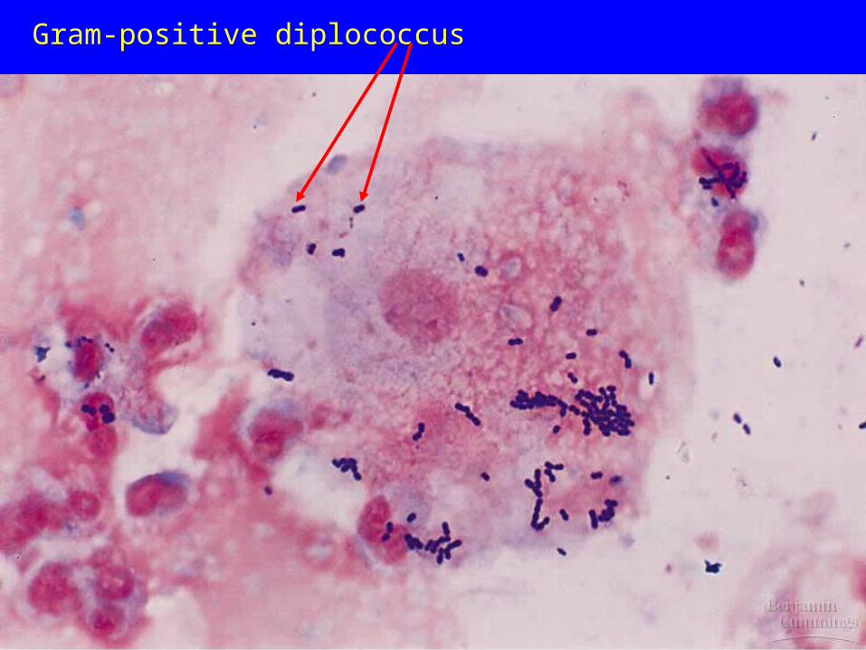
Gram-positive diplococcus

Gram-positive staphlococcus and Gram-negative diplobacillus
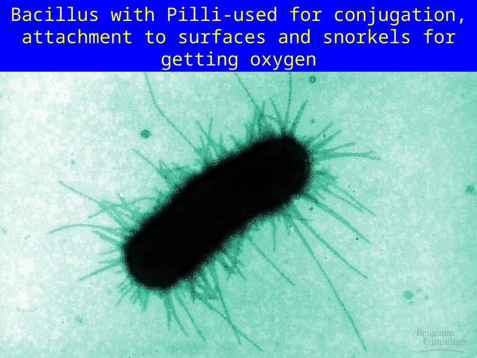
Bacillus with Pilli-used for conjugation, attachment to surfaces and snorkels for getting oxygen

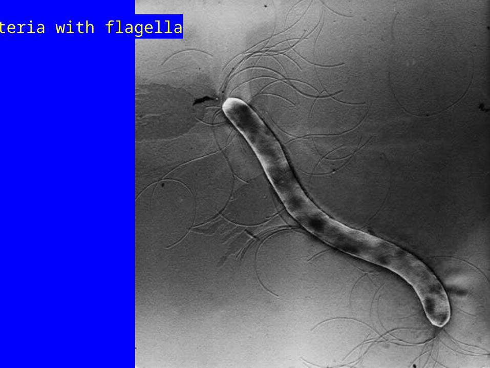
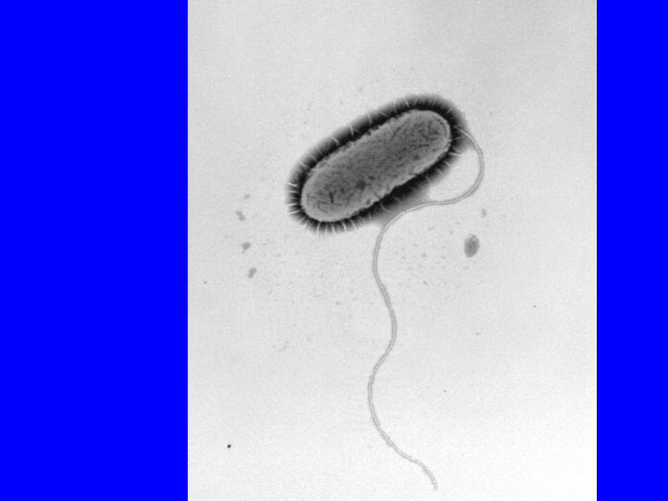
Bacterial flagella rotate rather than bend

Bacteria with flagella

Bacteria with flagella
Bacteria with flagella

Infolding of the plasma membrane give these bacteria respiratory membranes and thylakoid-like membranes
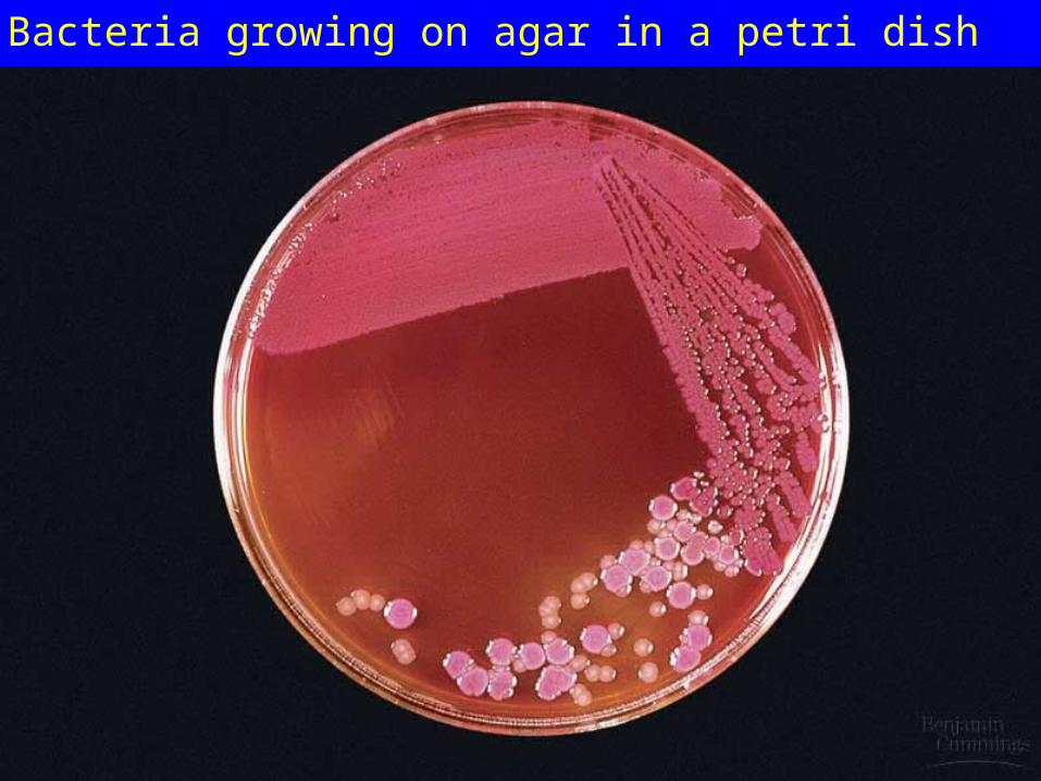
Bacteria growing on agar in a petri dish
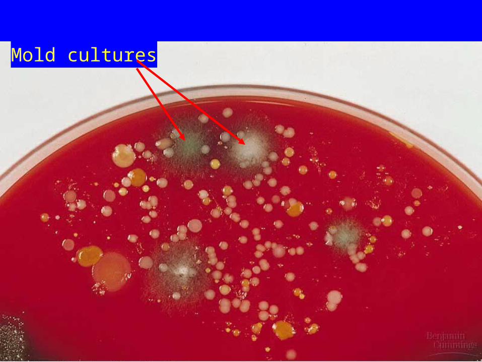
Mold cultures

An anthrax endospore

Endospores


ARCHAEABACTERIA
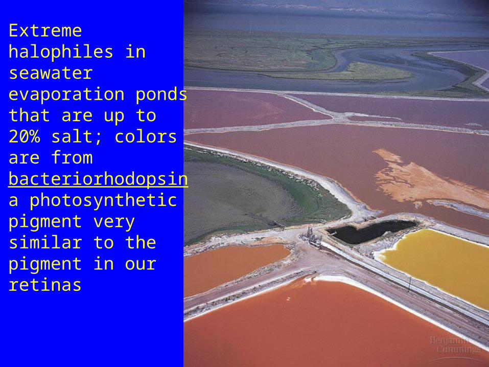
Extreme halophiles in seawater evaporation ponds that are up to 20% salt; colors are from bacteriorhodopsin a photosynthetic pigment very similar to the pigment in our retinas

Hot springs with extreme thermophiles

Hydrogen Sulfide Metabolizing Chemoautotrophic Archaeafound in sulfur springs

Eubacteria

The Proteobacteria are a major group (phylum) of bacteria. They include a wide variety of pathogens, such as Escherichia, Salmonella(rod-shaped Gram-negative enterobacteria that causes typhoid fever and the foodborne illness salmonellosis , Vibrio(motile gram negative curved-rod shaped bacterium with a polar flagellum that causes cholera in humans.) , Helicobacter(stomach ulcers), and many other notable genera.[1] Others are free-living, and include many of the bacteria responsible for nitrogen fixation. The group is defined primarily in terms of ribosomal RNA (rRNA) sequences, and is named for the Greek god Proteus (also the name of a bacterial genus within the Proteobacteria), who could change his shape, because of the great diversity of forms found in this group.
All Proteobacteria are Gram-negative, with an outer membrane mainly composed of lipopolysaccharides. Many move about using flagella, but some are non-motile or rely on bacterial gliding. The last include the myxobacteria, a unique group of bacteria that can aggregate to form multicellular fruiting bodies. There is also a wide variety in the types of metabolism. Most members are facultatively or obligately anaerobic and heterotrophic, but there are numerous exceptions. A variety of genera, which are not closely related to each other, convert energy from light through photosynthesis. These are called purple bacteria, referring to their mostly reddish pigmentation.

Alpha Proteobacteria Alpha Proteobacteria
Rocky Mountain Spotted Fever
Ti plasmid
Symbiosis with Legumes

Alpha Proteobacteria


Helicobacter pylori causes stomach ulcers

The Rickettsia are Gram-negative, obligate intracellular bacteria that infect mammals and arthropods. R. prowazekii is the agent of epidemic typhus. During World War I, approximately 3 million deaths resulted from infection by this bacterium. In World War II, the numbers were similar. This agent is carried by the human louse; therefore, disease is a consequence of overcrowding and poor hygiene. Rocky Mountain spotted fever and Q fever remain relatively common.
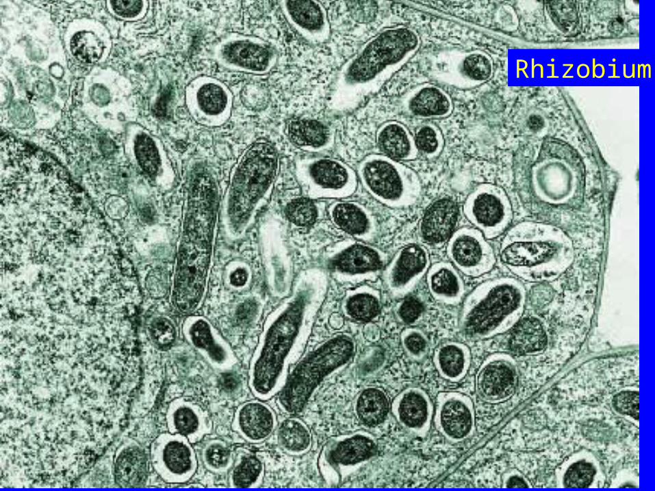
Rhizobium
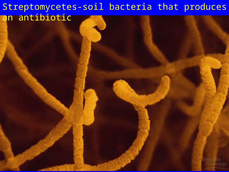
Streptomycetes-soil bacteria that produces an antibiotic

Sulfur bacteria that split H2S in photosynthesis

Cyanobacteria with heterocysts-specialized cells with the enzymes for nitrogen fixation
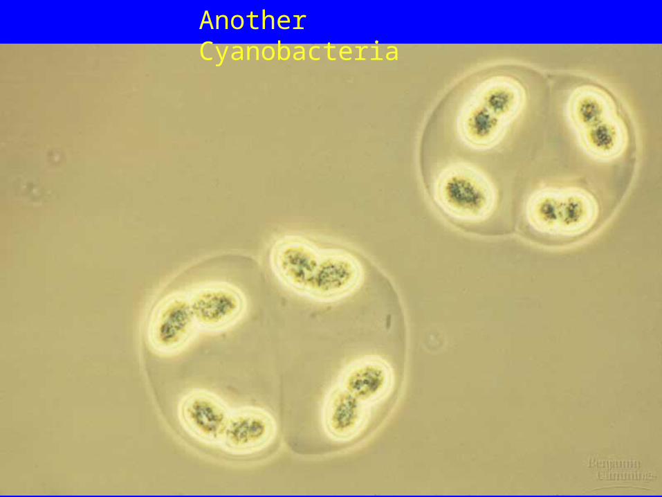
Another Cyanobacteria
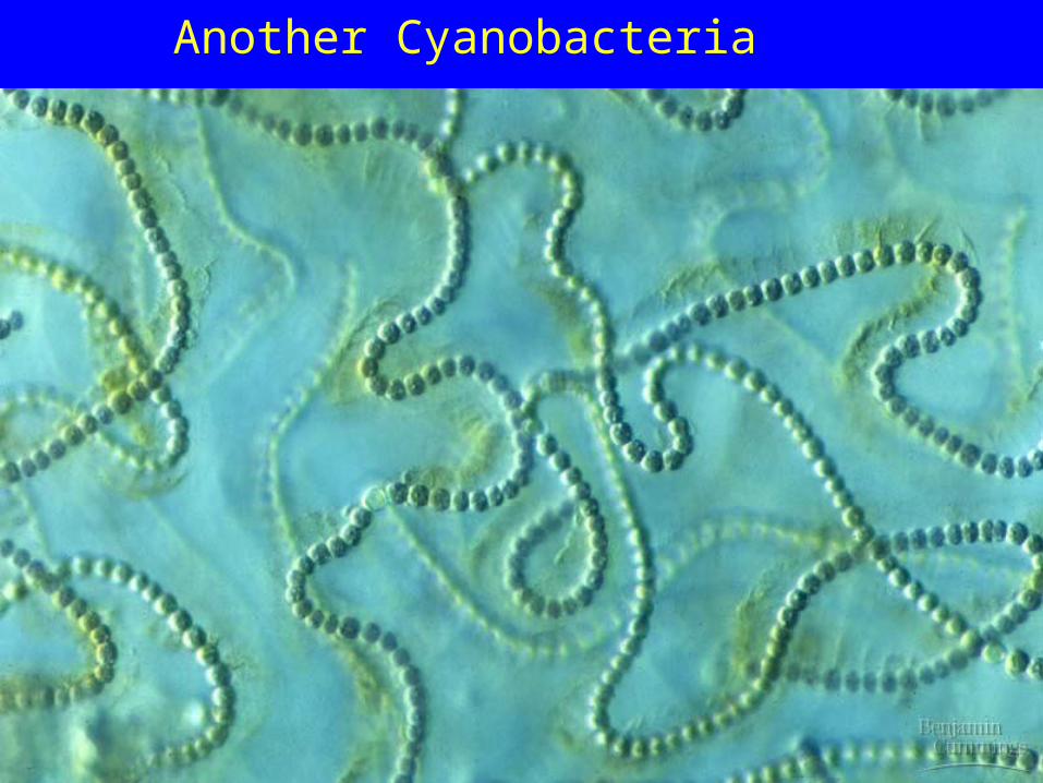
Another Cyanobacteria

Another Cyanobacteria
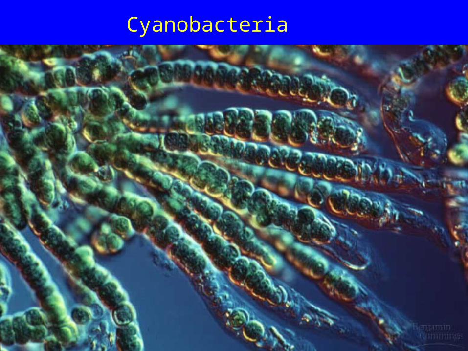
Cyanobacteria

Algae Blooms
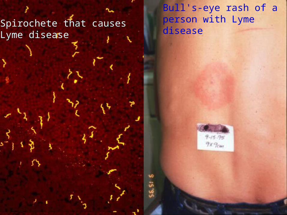
Bull's-eye rash of a person with Lyme diseaseSpirochete that causes Lyme
disease

Bull's-eye rash of a person with Lyme disease
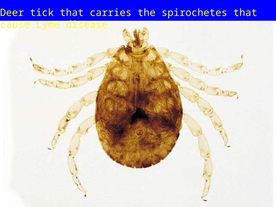
Deer tick that carries the spirochetes that cause Lyme disease
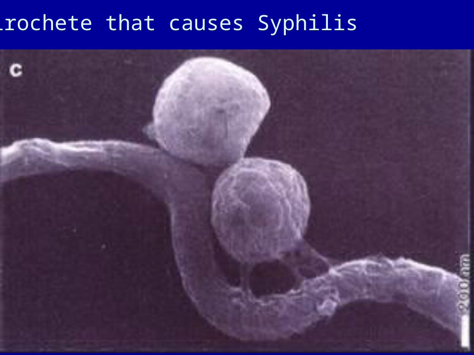
Spirochete that causes Syphilis
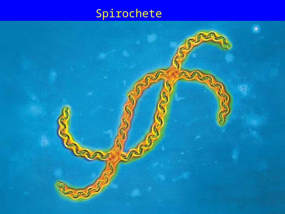
Spirochete
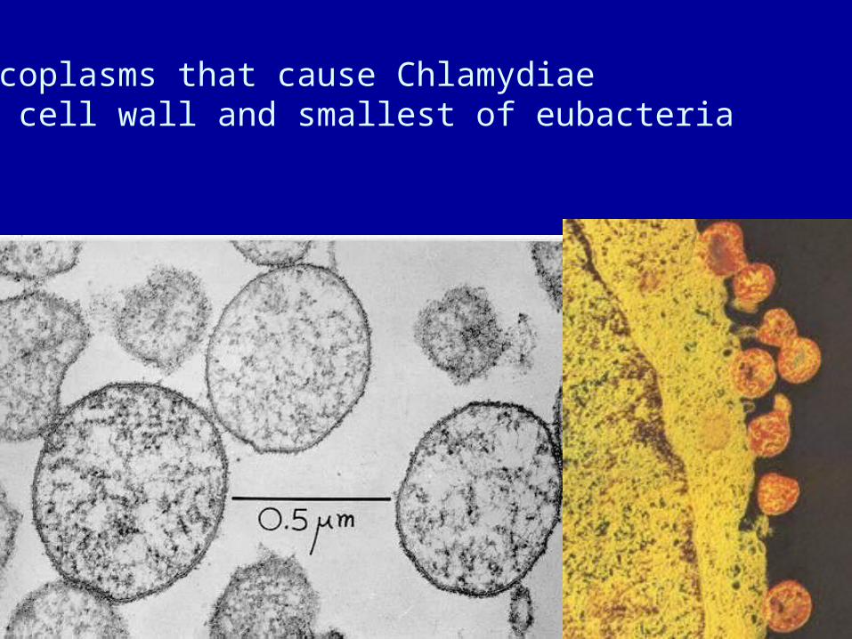
Mycoplasms that cause ChlamydiaeNo cell wall and smallest of eubacteria
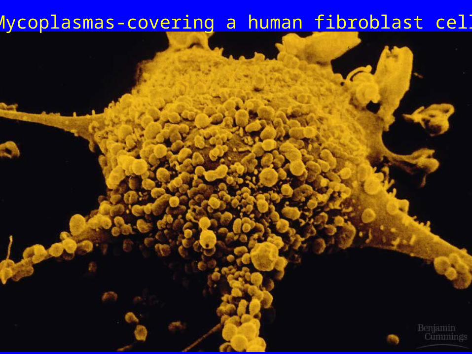
Mycoplasmas-covering a human fibroblast cell
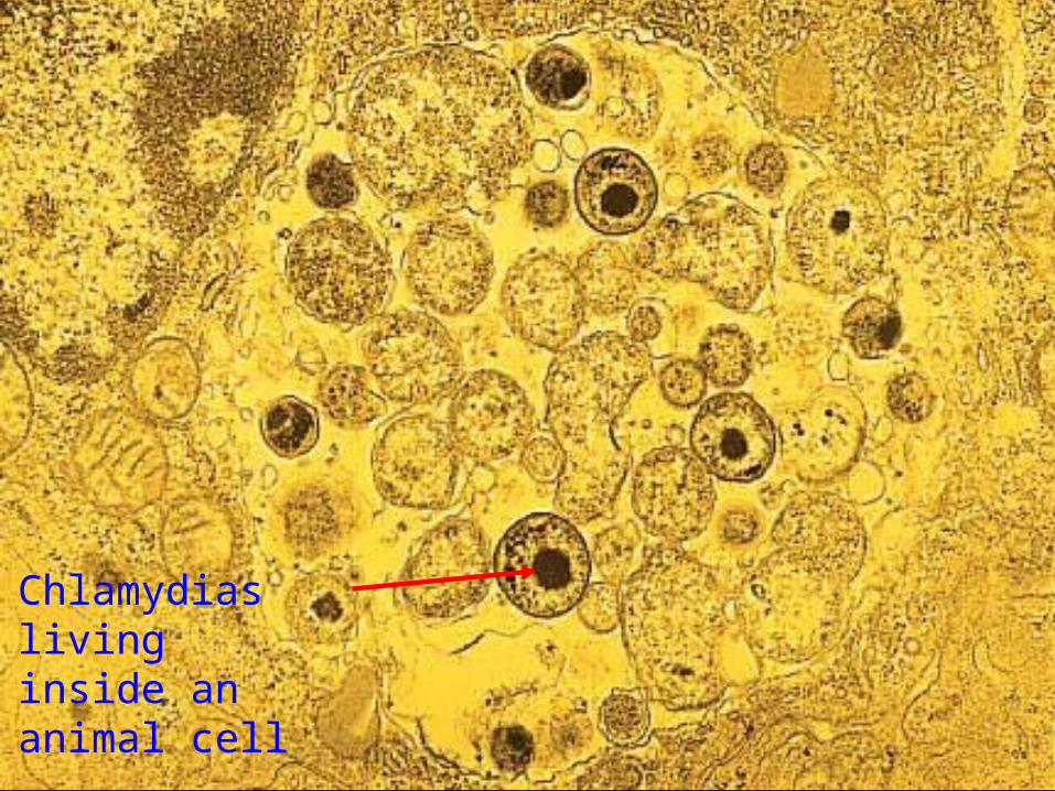
Chlamydias living inside an animal cell
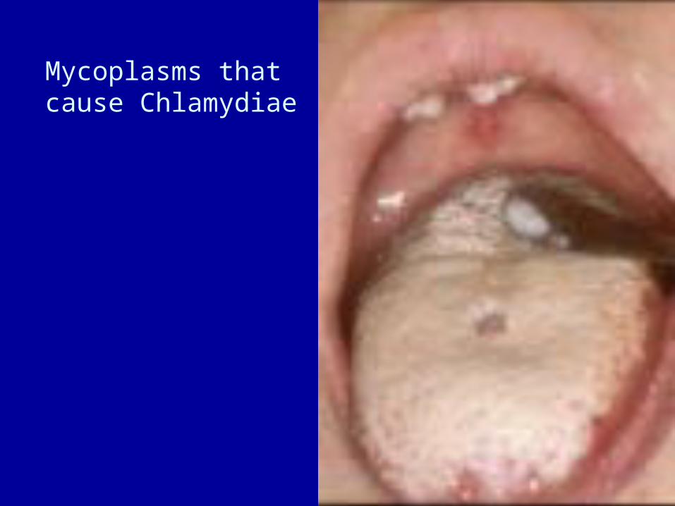
Mycoplasms that cause Chlamydiae

Mutualism of a bioluminescent bacteria in a “headlight fish”

The yellow bacillus is a pathogenic bacteria that causes respiratory infections on the membranes inside the nose.

The blue bacteria on this slide are commensal living on the membranes inside the nose but causing no harm.

Opportunistic infectionKoch’s postulates
Gram-positive actinomycetes causes tuberculosis destroys tissues
Clostridium botulinum releases exotoxins in food it is an obligate anaerobe
Vibrio cholerae releases an exotoxin that causes severe diarrhea
Salmonella typhi endotoxins that cause typhoid fever, another species of Salmonella causes common food poisoning due to endotoxins explains why it takes 12 -48 hours for symptoms to show up

Bioremediation bacteria breakdown
sewage


Spraying fertilizer on oil spills for Bioremediation
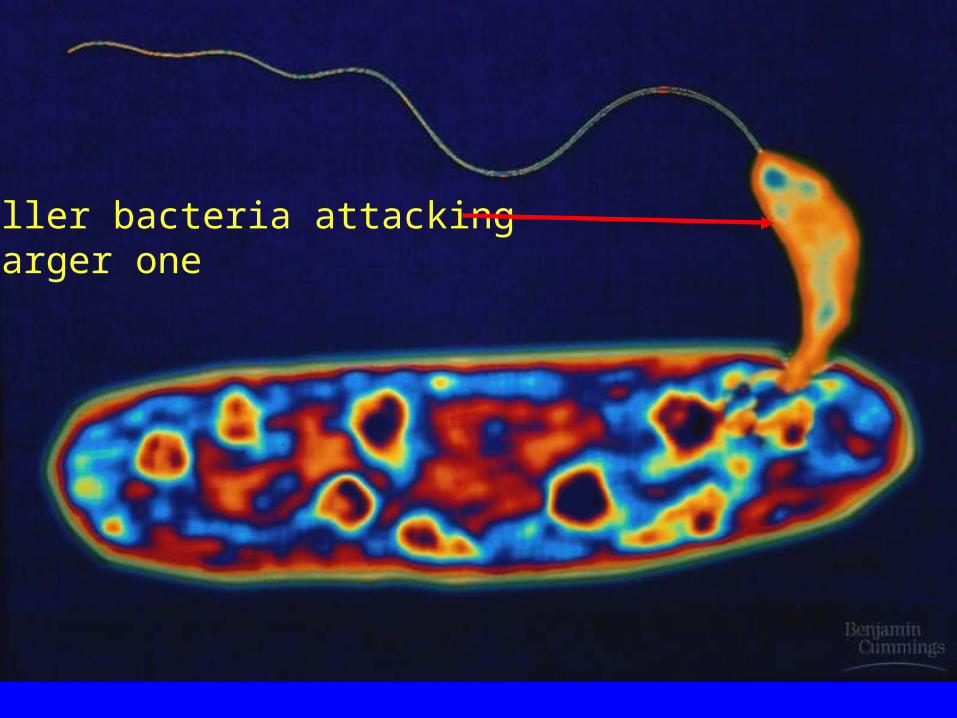
Smaller bacteria attacking a larger one

Cyanobacteria

Conjugation“caught in the
act”