chapter 25 face and neck injuries. introduction (1 of 2) face and neck are vulnerable to injury...
TRANSCRIPT

Chapter 25Chapter 25Chapter 25Chapter 25
Face and Neck Injuries

Introduction (1 of 2)Introduction (1 of 2)
• Face and neck are vulnerable to injury– Relatively unprotected positions on body
– Soft-tissue injuries and fractures are common and vary in severity.
• Some injuries are life-threatening.– Penetrating trauma to the neck may cause
severe bleeding.
– Open injury may result in an air embolism.

Introduction (2 of 2)Introduction (2 of 2)
• With appropriate prehospital and hospital care, a patient with a seemingly devastating injury can have a surprisingly good outcome.

The Head (1 of 2)The Head (1 of 2)
• Cranium– Also referred to as the skull
– Contains the brain
– Most posterior portion is called the occiput.
– Lateral portions on each side are called temples or temporal regions.
– Forehead is called the frontal region.

The Head (2 of 2)The Head (2 of 2)
• Cranium (cont’d)– Anterior to the ear, in the temporal region, you
can feel the pulse of the superficial temporal artery.
The Face (1 of 6)The Face (1 of 6)
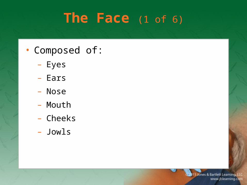
• Composed of:– Eyes
– Ears
– Nose
– Mouth
– Cheeks
– Jowls

The Face (2 of 6)The Face (2 of 6)
• Six major bones include:– Nasal bone
– Two zygomas
– Two maxillae
– Mandible

The Face (3 of 6)The Face (3 of 6)
• The orbit of the eye is composed of:– Lower edge of the frontal bone of the skull
– Zygoma
– Maxilla
– Nasal bone
• Protects the eye from injury
The Face (4 of 6)The Face (4 of 6)
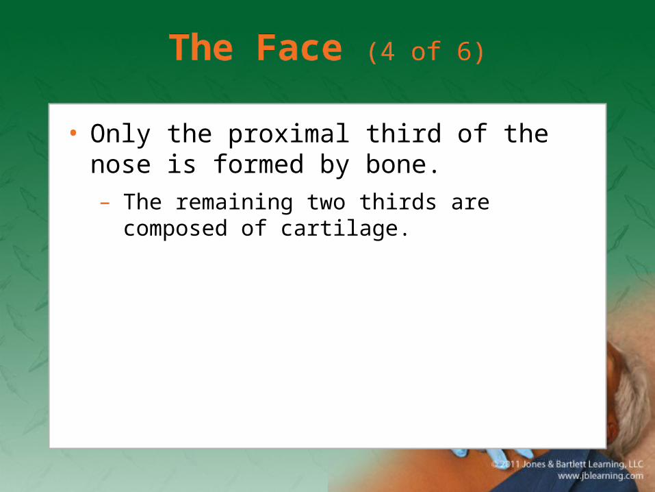
• Only the proximal third of the nose is formed by bone.– The remaining two thirds are composed of
cartilage.
The Face (5 of 6)The Face (5 of 6)
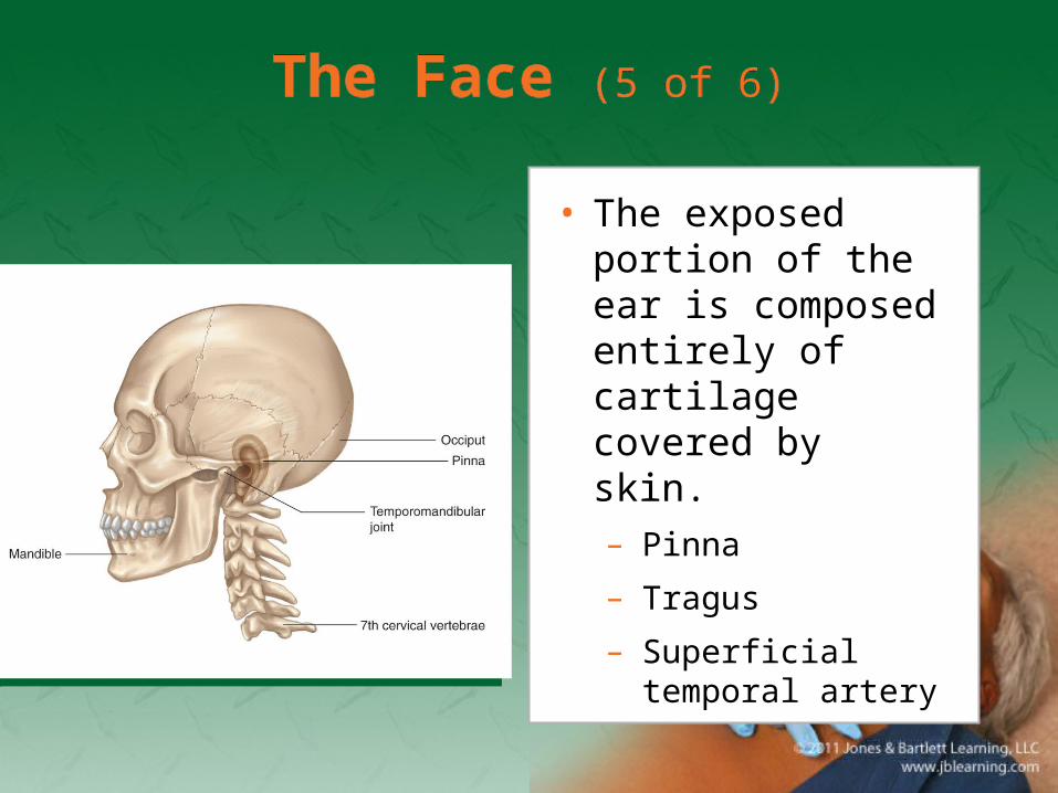
• The exposed portion of the ear is composed entirely of cartilage covered by skin.– Pinna
– Tragus
– Superficial temporal artery
The Face (6 of 6)The Face (6 of 6)
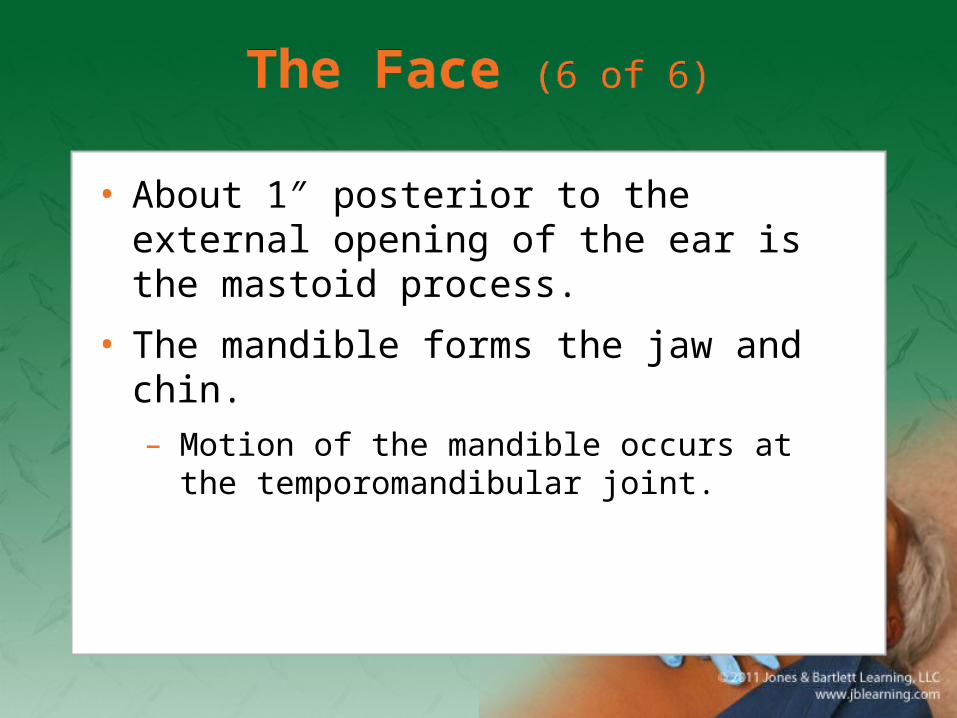
• About 1″ posterior to the external opening of the ear is the mastoid process.
• The mandible forms the jaw and chin.– Motion of the mandible occurs at the
temporomandibular joint.
The Neck (1 of 4)The Neck (1 of 4)
• Contains many important structures
• Supported by the cervical spine
• The upper part of the esophagus and the trachea lie in the midline of the neck.– The carotid arteries are found on either side of
the trachea.

The Neck (2 of 4)The Neck (2 of 4)
• The larynx– Adam’s apple is
located in the center of the neck.
– Other portion of the larynx is the cricoid cartilage.

The Neck (3 of 4)The Neck (3 of 4)
• The larynx (cont’d)– The cricothyroid
membrane lies between the thyroid cartilage and the cricoid cartilage.
– Soft depression in the midline of the neck
The Neck (4 of 4)The Neck (4 of 4)
• The trachea– Below the larynx in the anterior midline of the
neck
– Connects the oropharynx and larynx with the main passages of the lungs
• Sternocleidomastoid muscles– Originate from the mastoid process
– Allow movement of the head
The Eye (1 of 7)The Eye (1 of 7)
• Globe-shaped, approximately 1″ in diameter
• Located within a bony socket in the skull called the orbit– The orbit protects over 80% of the eyeball.

The Eye (2 of 7)The Eye (2 of 7)

The Eye (3 of 7)The Eye (3 of 7)
• Clear, jellylike fluid near the back of the eye is called vitreous humor.– In front of the lens is a fluid called the aqueous
humor, which can leak out in penetrating injuries.

The Eye (4 of 7)The Eye (4 of 7)
• The conjunctiva is a membrane that covers the eye.
• The lacrimal glands produce fluid to keep the eye moist.

The Eye (5 of 7)The Eye (5 of 7)
• The sclera is the white, fibrous tissue that helps maintain the globular shape.
• On the front of the eye, the sclera is replaced by a clear, transparent membrane called the cornea.– Allows light to enter the eye
– The iris is a circular muscle behind the cornea.

The Eye (6 of 7)The Eye (6 of 7)
• The pupil is the opening in the center of the iris.– Allows light to move to the back of the eye
– Anisocoria is a condition in which a person is born with different-sized pupils.
• The lens lies behind the iris.– Focuses images on the retina at the back of the
globe

The Eye (7 of 7)The Eye (7 of 7)
• The retina contains nerve endings.– Respond to light by transmitting nerve impulses
through the optic nerve to the brain
– The retina is nourished by a layer of blood vessels called the choroid.
– Retinal detachment causes blindness.

Injuries of the Face and Neck (1 of 2)
Injuries of the Face and Neck (1 of 2)
• Partial or complete obstruction of the upper airway may be the result.
• Several factors may contribute.– Blood clots from heavy facial bleeding
– Direct injuries to the nose and mouth, larynx, and trachea
– Dislodgment of teeth or dentures in the throat
Injuries of the Face and Neck (2 of 2)
Injuries of the Face and Neck (2 of 2)
• Several factors (cont’d)– Swelling that accompanies direct and indirect
injury
– Airway may be affected when the patient’s head is turned to the side
– Possible injuries to the brain and/or cervical spine
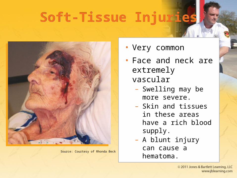
Soft-Tissue InjuriesSoft-Tissue Injuries
• Very common
• Face and neck are extremely vascular– Swelling may be
more severe.– Skin and tissues in
these areas have a rich blood supply.
– A blunt injury can cause a hematoma.Source: Courtesy of Rhonda Beck

Dental Injuries (1 of 2)Dental Injuries (1 of 2)
• Mandible injuries are common.
• Most of these injuries are the result of vehicle collisions and assaults.
• Signs of mandible fractures include:– Misalignment of the teeth
– Numbness of the chin
– An inability to open the mouth

Dental Injuries (2 of 2)Dental Injuries (2 of 2)
• Maxillary fractures are usually found after blunt force high-energy impacts.– Signs of maxillary fractures include:
• Massive facial swelling
• Instability of the facial bones
• Misalignment of teeth
• Fractured and avulsed teeth are common following facial trauma.
Patient AssessmentPatient Assessment
• Patient assessment steps– Scene size-up
– Primary assessment
– History taking
– Secondary assessment
– Reassessment
Scene Size-up (1 of 2)Scene Size-up (1 of 2)
• Scene safety– Observe for hazards and threats.
– Assess for potential violence and environmental hazards.
– Eye protection and face mask are standard.
– Carry several pairs of gloves.
– Determine the number of patients.

Scene Size-up (2 of 2)Scene Size-up (2 of 2)
• Mechanism of injury/nature of illness– Assess the scene.
– Common MOI for face and neck injuries:
• Motor vehicle accidents
• Sports
• Falls
• Penetrating trauma
• Blunt trauma
Primary Assessment (1 of 7)Primary Assessment (1 of 7)
• Focuses on identifying and managing life-threatening concerns
• Perform a rapid scan.
• Form a general impression.– Look for important indicators about the
seriousness of the patient’s condition.
– Injuries may be very obvious, or hidden.

Primary Assessment (2 of 7)Primary Assessment (2 of 7)
• Form a general impression (cont’d).– Control blood loss with direct pressure.
– Consider the need for manual spinal stabilization.
– Check for responsiveness using the AVPU scale.

Primary Assessment (3 of 7)Primary Assessment (3 of 7)
• Airway and breathing– Ensure a clear and patent airway.
– If the patient is unresponsive, consider a properly sized oropharyngeal airway.
– Palpate the chest wall for DCAP-BTLS.
– Face and throat injuries increase the need for airway and breathing maintenance.

Primary Assessment (4 of 7)Primary Assessment (4 of 7)
• Circulation– Quickly assess pulse rate and quality.
– Determine skin condition, color, and temperature.
– Check capillary refill time.
– Significant bleeding is an immediate life threat.

Primary Assessment (5 of 7)Primary Assessment (5 of 7)
• Transport decision– Patients with airway or breathing problems or
with significant bleeding need to be transported immediately.
– A patient with internal bleeding must be transported quickly for treatment by a physician.
Primary Assessment (6 of 7)Primary Assessment (6 of 7)
• Transport decision (cont’d)– Signs of hypoperfusion include:
• Tachycardia
• Tachypnea
• Low blood pressure
• Weak pulse
• Cool, moist, pale skin

Primary Assessment (7 of 7)Primary Assessment (7 of 7)
• Transport decision (cont’d)– Even if the patient has no signs of
hypoperfusion, there is the possibility of eye injuries.
– The patient should be transported rapidly.
– Surgery will need to be accomplished within 30 minutes or permanent blindness may result.

History TakingHistory Taking
• Investigate the chief complaint.– Obtain a medical history.
– Be alert for injury-specific signs and symptoms.
– Be aware of pertinent negatives.
– Gather a SAMPLE history from the patient, or from friends and family.

Secondary Assessment (1 of 4)Secondary Assessment (1 of 4)
• Physical examinations– If multiple systems have been affected, start
with a full-body scan looking for DCAP-BTLS.
– Do not delay transport to complete a thorough physical exam.
– Focus on the isolated injury, the patient’s complaint, and the body region affected.

Secondary Assessment (2 of 4)Secondary Assessment (2 of 4)
• Physical examinations (cont’d)– Ensure that control of bleeding is maintained
and note injury location.
– Inspect the open wound for any foreign matter or impaled object.
– Use both your eyes and your hands.
– Assess all underlying systems.

Secondary Assessment (3 of 4)Secondary Assessment (3 of 4)
• Physical examinations (cont’d)– When evaluating the eyes, start with the outer
aspect and work toward the pupils.
– Look for discoloration, clarity of vision, bleeding, redness, eye symmetry, and pupil size and reaction to light.
– Brain injury, nerve disease, glaucoma, and meningitis are causes of unequal pupils.

Secondary Assessment (4 of 4)Secondary Assessment (4 of 4)
• Vital signs– Assess vital signs to obtain a baseline.
– You must be concerned with visible bleeding and unseen bleeding inside a body cavity.
– With facial and throat injuries, baseline information is very important.
– Use appropriate monitoring devices.

Reassessment (1 of 4)Reassessment (1 of 4)
• Repeat the primary assessment.
• Reassess vital signs and the chief complaint.– Reassess the patient’s condition every
5 minutes.
• Interventions– Provide complete spinal immobilization if
necessary.

Reassessment (2 of 4)Reassessment (2 of 4)
• Interventions (cont’d)– Maintain an open airway, be prepared to
suction, and consider an oropharyngeal airway.
– Whenever you suspect significant bleeding, provide high-flow oxygen.
– Control visible bleeding.

Reassessment (3 of 4)Reassessment (3 of 4)
• Interventions (cont’d)– If the patient has signs of hypoperfusion, treat
aggressively for shock and provide rapid transport.
• Communication and documentation– Include a description of the MOI and the
position in which you found the patient.
– Document the method used to remove the patient from the vehicle.
Reassessment (4 of 4)Reassessment (4 of 4)
• Communication and documentation (cont’d)– Recognize, estimate, and report the amount of
blood loss.
– Inform the hospital about all injuries involving the head and neck.
Emergency Medical Care (1 of 5)Emergency Medical Care (1 of 5)
• Treat soft-tissue injuries to the face and neck the same as soft-tissue injuries elsewhere on the body.– Assess ABCs and life threats first.
– Open and clear the airway.
– Avoid moving the neck in patients with suspected cervical spine injuries.

Emergency Medical Care (2 of 5)Emergency Medical Care (2 of 5)
• Control bleeding by applying direct manual pressure with a dry, sterile dressing.– Use roller gauze, wrapped around the head, to
hold a pressure dressing in place.
– Do not apply excessive pressure if an underlying skull fracture is suspected.
Emergency Medical Care (3 of 5)Emergency Medical Care (3 of 5)
• Apply ice locally to injuries that do not break the skin.
• For soft-tissue injuries around the mouth, check for bleeding inside the mouth.– Broken teeth and tongue lacerations may cause
extensive bleeding and obstruction of the upper airway.

Emergency Medical Care (4 of 5)Emergency Medical Care (4 of 5)
• Physicians can sometimes graft a piece of avulsed skin back into position.– If you find portions of avulsed skin:
• Wrap in a sterile dressing.
• Place in a plastic bag.
• Keep cool, but do not place directly on ice.
• Label and deliver to the emergency department.
Emergency Medical Care (5 of 5)Emergency Medical Care (5 of 5)
• If the avulsed skin is still attached in a loose flap:– Place the flap in position as close to normal as
possible.
– Hold in place with a dry, sterile dressing.

Injuries of the Eyes (1 of 19)Injuries of the Eyes (1 of 19)
• Eye injuries are common, particularly in sports.– Can produce lifelong complications, including
blindness
• In a normal, uninjured eye, the entire circle of the iris is visible.– The pupils are round, usually equal in size, and
react equally to light.

Injuries of the Eyes (2 of 19)Injuries of the Eyes (2 of 19)
• After an injury, pupil reaction or shape and eye movement are disturbed.
• Treatment starts with a thorough exam.– Always use standard precautions.
– Take care not to aggravate any problems.
– Look for abnormalities or conditions that may suggest the nature of the injury.

Injuries of the Eyes (3 of 19)Injuries of the Eyes (3 of 19)
• Foreign objects– Even a small object can cause significant
damage.
– Irrigation with a sterile saline solution will frequently flush away loose particles.
– Use a bulb syringe, or a nasal airway or cannula.

Injuries of the Eyes (4 of 19)Injuries of the Eyes (4 of 19)
• Foreign objects (cont’d)– Always flush from the nose side of the eye
toward the outside to avoid flushing material into the other eye.

Injuries of the Eyes (5 of 19)Injuries of the Eyes (5 of 19)
• Foreign objects (cont’d)– A foreign body will leave a small abrasion on
the surface of the eye.
– Irrigation may not wash out foreign bodies stuck to the cornea or lying under the upper eyelid.
– Follow the steps in Skill Drill 25-1.

Injuries of the Eyes (6 of 19)Injuries of the Eyes (6 of 19)
• Foreign objects (cont’d)– Foreign bodies may be impaled in the eye.
– Bandage the object in place to support it.
– Cover the eye with a moist, sterile dressing.
– Surround the object with a doughnut-shaped collar.
– Follow the steps in Skill Drill 25-2.
Injuries of the Eyes (7 of 19)Injuries of the Eyes (7 of 19)
• Burns of the eye– Stop the burn and prevent further damage.
• Chemical burns– Usually caused by acid or alkaline solutions
– Flush the eye with water or saline.
– Direct the greatest amount of irrigating solution or water into the eye as gently as possible.

Injuries of the Eyes (8 of 19)Injuries of the Eyes (8 of 19)
Injuries of the Eyes (9 of 19)Injuries of the Eyes (9 of 19)
• Chemical burns (cont’d)– You may have to force the lids open.
– Irrigate the eye for at least 5 minutes.
– If the burn was caused by an alkali or a strong acid, irrigate for at least 20 minutes.
– After irrigation, apply a clean, dry dressing to cover the eye, and transport.
Injuries of the Eyes (10 of 19)Injuries of the Eyes (10 of 19)
• Thermal burns– During a fire, the eyes will close to protect from
heat, and the eyelids will burn.
– Transport promptly without further examination.
– Cover both eyes with a sterile dressing moistened with sterile saline.
– Apply eye shields over the dressing.
Injuries of the Eyes (11 of 19)Injuries of the Eyes (11 of 19)
• Light burns– Infrared rays, eclipse light, and laser beams all
can cause significant damage.
– Retinal injuries caused by exposure to light are generally not painful but may result in permanent damage.
– Severe conjunctivitis usually develops with redness, swelling, and excessive tears.

Injuries of the Eyes (12 of 19)Injuries of the Eyes (12 of 19)
• Lacerations– Require very careful repair to restore
appearance and function
– Bleeding may be heavy, but it usually can be controlled with gentle, manual pressure.
– If there is a laceration of the globe itself, apply no pressure to the eye.

Injuries of the Eyes (13 of 19)Injuries of the Eyes (13 of 19)
• Lacerations (cont’d)– Never exert pressure on the injured globe.
– If part of the eyeball is exposed, gently apply a moist, sterile dressing to prevent drying.
– Cover the injured eye with a protective metal eye shield cup, or sterile dressing.

Injuries of the Eyes (14 of 19)Injuries of the Eyes (14 of 19)
• Lacerations (cont’d)– On rare occasions, the eyeball may be
displaced from its socket.
– Do not attempt to reposition it.
– Cover the eye and stabilize it with a moist sterile dressing.
– Cover both eyes to prevent further injury.
– Have the patient lie supine.

Injuries of the Eyes (15 of 19)Injuries of the Eyes (15 of 19)
• Blunt trauma– Injuries range from the ordinary black eye to a
severely damaged globe.
– Hyphema obscures all or part of the iris.
– An orbit fracture is a fracture of the bones that form the eye floor and support the globe.
– Retinal detachment is often seen in sports.

Injuries of the Eyes (16 of 19)Injuries of the Eyes (16 of 19)
• Eye injuries following head injury– The following findings should alert you to the
possibility of a head injury:
• One pupil larger than the other
• Eyes not moving together
• Failure of the eyes to follow your finger
• Bleeding under the conjunctiva
• Protrusion or bulging of the eye

Injuries of the Eyes (17 of 19)Injuries of the Eyes (17 of 19)
• Blast injuries– Signs and symptoms range from severe pain
and loss of vision to foreign bodies within the globe.
– If there is a foreign body, do not remove it.
– If only one eye is injured, follow protocol.
– If the patient has severe swelling, do not force the eyelid open to examine it.
Injuries of the Eyes (18 of 19)Injuries of the Eyes (18 of 19)
• Contact lenses and artificial eyes– Do not attempt to remove contact lenses unless
there is a chemical burn.
– To remove a hard contact lens, use a small suction cup.
– To remove soft contact lenses, place one or two drops of saline in the eye, pinch it between your thumb and index finger, and lift.

Injuries of the Eyes (19 of 19)Injuries of the Eyes (19 of 19)
Hard contact lens
Soft contact lens
Injuries of the Nose (1 of 4)Injuries of the Nose (1 of 4)
• Nosebleeds (epistaxis) are a common problem.– One of the most common causes is digital
trauma.
– Anterior nosebleeds usually originate from the area of the septum and bleed slowly.
– Posterior nosebleeds are usually more severe and often cause blood to drain into the throat.

Injuries of the Nose (2 of 4)Injuries of the Nose (2 of 4)
• The nose often takes the brunt of physical assaults and car crashes.– Blunt injuries may be associated with fractures
and soft-tissue injuries of the face, head injuries, and/or injuries to the cervical spine.
Injuries of the Nose (3 of 4)Injuries of the Nose (3 of 4)
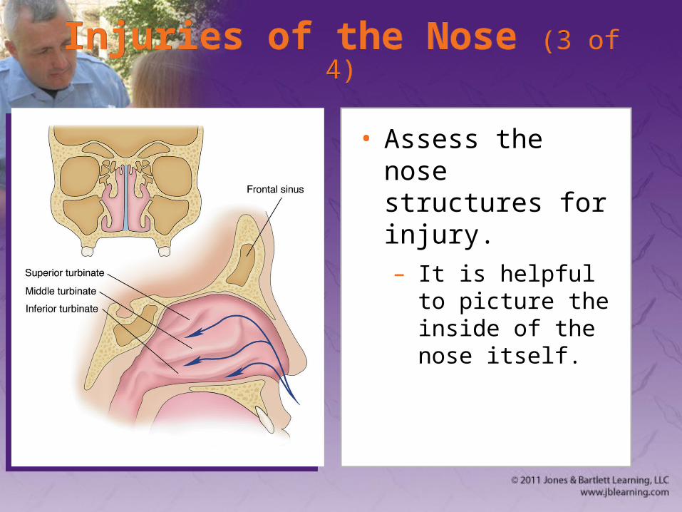
• Assess the nose structures for injury.– It is helpful to
picture the inside of the nose itself.
Injuries of the Nose (4 of 4)Injuries of the Nose (4 of 4)
• Cerebrospinal fluid (CSF) may escape down through the nose following a fracture at the base of the skull.
• Control bleeding by applying a sterile dressing.– See Skill Drill 23-3 from Chapter 23,
“Bleeding.”

Injuries of the Ear (1 of 3)Injuries of the Ear (1 of 3)
• The ear is complex and associated with hearing and balance.
• Divided into three parts:– Outer ear
– Middle ear
– Inner ear

Injuries of the Ear (2 of 3)Injuries of the Ear (2 of 3)

Injuries of the Ear (3 of 3)Injuries of the Ear (3 of 3)
• Ears are often injured, but they do not usually bleed very much.– In case of an ear avulsion, wrap the avulsed
part in a moist, sterile dressing and put it in a plastic bag.
– Children place foreign bodies in the outer ear.
• Clear fluid coming from the ear may indicate a skull fracture.

Facial Fractures (1 of 2)Facial Fractures (1 of 2)
• Typically result from blunt trauma
• Assume a direct blow to the mouth or nose has caused a facial fracture.
• Other clues include:– Bleeding in the mouth
– Inability to swallow or talk
– Absent or loose teeth
– Loose or movable bone fragments

Facial Fractures (2 of 2)Facial Fractures (2 of 2)
• Facial fractures alone are not acute emergencies unless there is serious bleeding.
• Plastic surgeons can repair the damage to the face and mouth if the injuries are treated within 7 to 10 days.
• Swelling can be extreme within the first 24 hours after injury.

Dental Injuries (1 of 2)Dental Injuries (1 of 2)
• Can be traumatic to the patient
• Bleeding will occur whenever a tooth is violently displaced from its socket.– Apply direct pressure to stop the bleeding.
– Perform suctioning if needed.
– Cracked or loose teeth are possible airway obstructions.

Dental Injuries (2 of 2)Dental Injuries (2 of 2)
• Save and transport an avulsed tooth.– Handle it by the crown rather than the root.
– Place the tooth in either cold milk or sterile saline.
– Notify the hospital.
– Reimplantation is recommended within 20 minutes to 1 hour after the trauma.

Injuries of the CheekInjuries of the Cheek
• You may encounter an object impaled in the patient’s cheek.– If you are unable to control the bleeding,
consider removing the object.
– Then provide direct pressure on the inside and outside of the cheek.
– The amount of bandaging should not occlude the mouth.

Injuries of the Neck (1 of 4)Injuries of the Neck (1 of 4)
• The neck contains many structures vulnerable to injury by blunt trauma.– Upper airway
– Esophagus
– Carotid arteries and jugular veins
– Thyroid cartilage (Adam’s apple)
– Cricoid cartilage
– Upper part of the trachea

Injuries of the Neck (2 of 4)Injuries of the Neck (2 of 4)
• Blunt injuries– Any crushing injury of the upper part of the neck
is likely to involve the larynx or trachea.
– Once the cartilages of the upper airway and larynx are fractured, they do not spring back to their normal position.

Injuries of the Neck (3 of 4)Injuries of the Neck (3 of 4)
• Blunt injuries (cont’d)– Can lead to loss of voice, difficulty swallowing,
severe and sometimes fatal airway obstruction, and leakage of air into the soft tissues of the neck
– Subcutaneous emphysema is a characteristic crackling sensation produced by the presence of air.
Injuries of the Neck (4 of 4)Injuries of the Neck (4 of 4)
• Penetrating injuries– Can profuse bleeding from laceration of the
great vessels in the neck
– Injuries to the carotid and jugular veins can cause the body to bleed out (exsanguination).
– Follow the steps in Skill Drill 25-3 to control bleeding from a neck injury.

Laryngeal Injuries (1 of 4)Laryngeal Injuries (1 of 4)
• Blunt force trauma to the larynx can occur when:– Unrestrained driver strikes steering wheel
– Snowmobile rider strikes a clothesline
• The larynx becomes crushed against the cervical spine, resulting in soft-tissue injury, fractures, and/or separation of the fascia.

Laryngeal Injuries (2 of 4)Laryngeal Injuries (2 of 4)
• Penetrating or impaled objects in the larynx should not be removed unless they interfere with CPR.– Stabilize all impaled objects if they are not
obstructing the airway.
– See Skill Drill 24-2 from Chapter 24, “Soft-Tissue Injuries.”
Laryngeal Injuries (3 of 4)Laryngeal Injuries (3 of 4)
• Signs and symptoms of larynx injuries:– Respiratory distress
– Hoarseness
– Pain
– Difficulty swallowing (dysphagia)
– Cyanosis
– Pale skin
– Sputum in the wound
Laryngeal Injuries (4 of 4)Laryngeal Injuries (4 of 4)
• Signs and symptoms (cont’d)– Subcutaneous emphysema
– Bruising on the neck
– Hematoma
– Bleeding
• To manage a laryngeal injury, provide oxygen and ventilation and apply cervical immobilization.

Summary Summary (1 of 10)(1 of 10)Summary Summary (1 of 10)(1 of 10)
• Soft-tissue injuries and fractures of the bones of the face and neck are common and vary in severity.
• In face and neck injuries, your priorities are to prevent further injury to the cervical spine, manage the airway and ventilation of the patient, and control breathing.

Summary Summary (2 of 10)(2 of 10)Summary Summary (2 of 10)(2 of 10)
• Airway compromise may be caused by heavy bleeding into the airway, swelling in and around the structures of the airway located in the face and neck, and injuries to the central nervous system that interfere with normal respiration.

Summary Summary (3 of 10)(3 of 10)Summary Summary (3 of 10)(3 of 10)
• To control heavy bleeding from soft-tissue injuries to the face, use direct pressure with a dry, sterile dressing. If brain tissue is exposed, use a moist, sterile dressing.
Summary Summary (4 of 10)(4 of 10)Summary Summary (4 of 10)(4 of 10)
• Always check for bleeding inside the mouth because this may produce airway obstruction.
• Open the airway using the modified jaw-thrust maneuver (when indicated), and clear the airway in all patients with facial injuries.

Summary Summary (5 of 10)(5 of 10)Summary Summary (5 of 10)(5 of 10)
• Save avulsed pieces of skin and tissue, and transport them with the patient for possible reattachment at the hospital.
• Maintain a high index of suspicion for patients with unequal pupils—this sign may indicate an illness or an injury to the brain.

Summary Summary (6 of 10)(6 of 10)Summary Summary (6 of 10)(6 of 10)
• Foreign bodies on the surface of the eye should be irrigated gently with normal saline solution. Always flush from the region of the eye closest to the nose toward the outside, away from the midline.

Summary Summary (7 of 10)(7 of 10)Summary Summary (7 of 10)(7 of 10)
• If a foreign body is on the underside of the eyelid, remove it gently with a cotton-tipped applicator. Never remove foreign bodies stuck to the cornea.

Summary Summary (8 of 10)(8 of 10)Summary Summary (8 of 10)(8 of 10)
• Chemicals, heat, and light rays can all cause burn injury to the eyes, resulting in permanent damage.
• Be alert to clear fluid draining from the ears or nose. This may indicate a basilar skull fracture.

Summary Summary (9 of 10)(9 of 10)Summary Summary (9 of 10)(9 of 10)
• Blunt and penetrating trauma to the neck can produce life-threatening injuries. Palpate the neck for signs of subcutaneous emphysema. In patients with this sign, complete airway obstruction may develop in minutes.

Summary Summary (10 of 10)(10 of 10)Summary Summary (10 of 10)(10 of 10)
• If bleeding is present from a penetrating injury, direct pressure over the site will usually control most forms of bleeding.
• Be alert to the possibility of an air embolism from an open neck injury. Place an occlusive dressing over the site, and provide direct pressure.