chapter-2 literature reviewshodhganga.inflibnet.ac.in/bitstream/10603/23480/4/04. chapter -...
TRANSCRIPT

18
CHAPTER-2
LITERATURE REVIEW
19
CHAPTER-2
LITERATURE REVIEW
2. General
2.1 Quinone
Quinones are described as a class of cyclic organic compounds
comprising of a six-membered unsaturated ring to which two oxygen atoms are
bonded as carbonyl groups. The name quinone is applied to the whole group, but
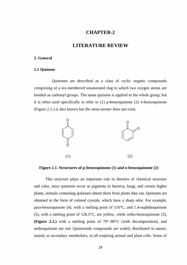
it is often used specifically to refer to (1) p-benzoquinone (2) o-benzoquinone
(Figure 2.1.) is also known but the meta-isomer does not exist.
O
O
O
O
(1) (2)
Figure 2.1. Structures of p-benzoquinone (1) and o-benzoquinone (2)
This structure plays an important role in theories of chemical structure
and color, since quinones occur as pigments in bacteria, fungi, and certain higher
plants; animals containing quinones obtain them from plants they eat. Quinones are
obtained in the form of colored crystals, which have a sharp odor. For example,
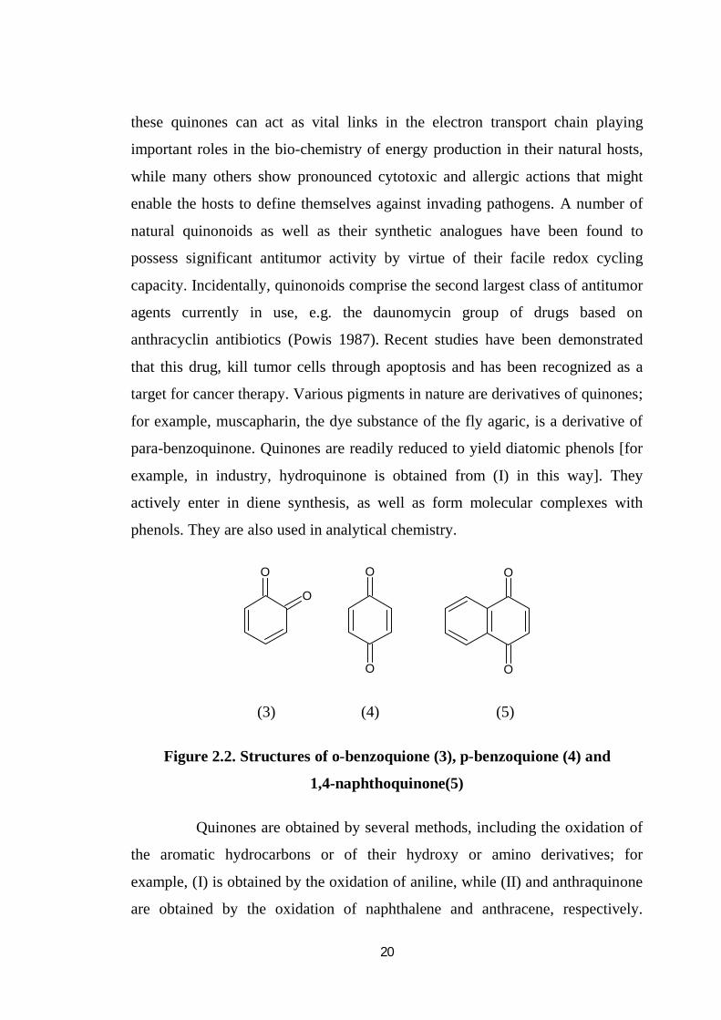
para-benzoquinone (4), with a melting point of 116°C, and 1,4-naphthoquinone
(5), with a melting point of 128.5°C, are yellow, while ortho-benzoquinone (3),
(Figure 2.2.) with a melting point of 70°–80°C (with decomposition), and
anthraquinone are red. Quinonoids compounds are widely distributed in nature,
mainly as secondary metabolites, in all respiring animal and plant cells. Some of
20
these quinones can act as vital links in the electron transport chain playing
important roles in the bio-chemistry of energy production in their natural hosts,
while many others show pronounced cytotoxic and allergic actions that might
enable the hosts to define themselves against invading pathogens. A number of
natural quinonoids as well as their synthetic analogues have been found to
possess significant antitumor activity by virtue of their facile redox cycling
capacity. Incidentally, quinonoids comprise the second largest class of antitumor
agents currently in use, e.g. the daunomycin group of drugs based on
anthracyclin antibiotics (Powis 1987). Recent studies have been demonstrated
that this drug, kill tumor cells through apoptosis and has been recognized as a
target for cancer therapy. Various pigments in nature are derivatives of quinones;
for example, muscapharin, the dye substance of the fly agaric, is a derivative of
para-benzoquinone. Quinones are readily reduced to yield diatomic phenols [for
example, in industry, hydroquinone is obtained from (I) in this way]. They
actively enter in diene synthesis, as well as form molecular complexes with
phenols. They are also used in analytical chemistry.
O
O
O
O
O
O
(3) (4) (5)
Figure 2.2. Structures of o-benzoquione (3), p-benzoquione (4) and
1,4-naphthoquinone(5)
Quinones are obtained by several methods, including the oxidation of
the aromatic hydrocarbons or of their hydroxy or amino derivatives; for
example, (I) is obtained by the oxidation of aniline, while (II) and anthraquinone
are obtained by the oxidation of naphthalene and anthracene, respectively.

21
Quinones and their derivatives are intermediate products in the production of
dyes. These are also used as fungicides, insecticides, and tanning agents.
2-Methyl-1,4-naphthoquinone is a vitamin of the vitamin K group. The K
vitamins (see vitamin K) are naphthoquinones. The term quinone often
specifically denotes para-benzoquinone (C6H4O2), a bright yellow solid with a
sharp odor used in manufacturing dyes and fungicides and in photography.
2.2. Review of previous research on naphthoquinones and bi-
naphthoquinones
2.2.1 Naphthoquinones
Naphtho-1, 4-quinones are widely available in nature, mainly in plants,
fungi and bacteria. These classes of compounds have various properties and
applications. These properties and applications have been extensively reviewed
(Thomson 1971; Patai 1974), they isolated as yellow, orange, red, or purple
solids, and are sparingly soluble in water but readily soluble in most organic
solvents.
2.2.1.1 Naphthoquinones as Privileged Molecules
Naphthoquinones are considered privileged structures in medicinal
chemistry due to their biological activities and structural properties. They are
present in various families of plants and serve as vital links in the electron
transport chains in the metabolic pathway, participating in multiple biological
oxidative processes. The fundamental feature of quinone chemistry is its ease of
reduction and, therefore, its ability to act as an oxidizing or dehydrogenating
agent. This redox property is driven by the formation of a fully aromatic system.
In folk medicine, plants containing naphthoquinones are often employed for the
treatment of various diseases, and several quinonoids isolated from traditional
medicinal plants are being investigated for their anticancer properties.
22
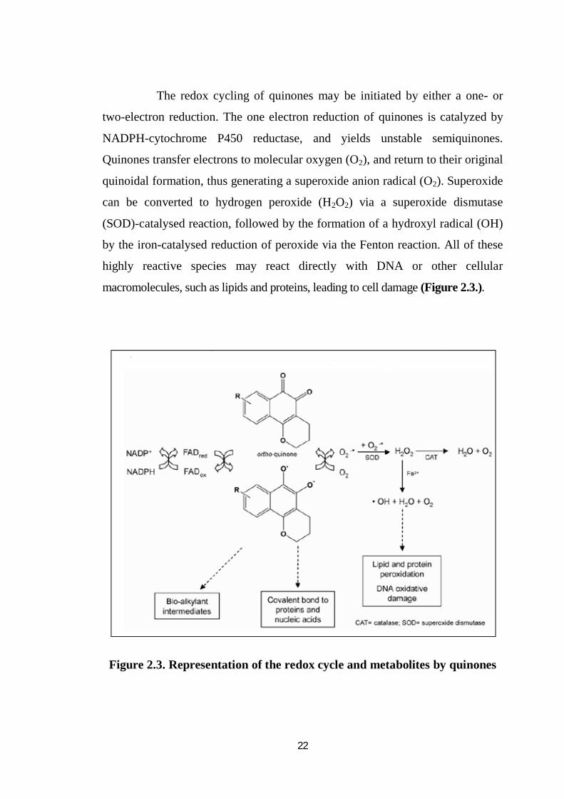
The redox cycling of quinones may be initiated by either a one- or
two-electron reduction. The one electron reduction of quinones is catalyzed by
NADPH-cytochrome P450 reductase, and yields unstable semiquinones.
Quinones transfer electrons to molecular oxygen (O2), and return to their original
quinoidal formation, thus generating a superoxide anion radical (O2). Superoxide
can be converted to hydrogen peroxide (H2O2) via a superoxide dismutase
(SOD)-catalysed reaction, followed by the formation of a hydroxyl radical (OH)
by the iron-catalysed reduction of peroxide via the Fenton reaction. All of these
highly reactive species may react directly with DNA or other cellular
macromolecules, such as lipids and proteins, leading to cell damage (Figure 2.3.).
Figure 2.3. Representation of the redox cycle and metabolites by quinones
23
Owing to their molecular structure and their redox properties, they
exhibit interesting physical properties, as well as a wide range of biological
activities. Extracts from plants containing mixtures of naphtha-1, 4-quinone
derivatives have been used for centuries not only as dyes or ingredients for
cosmetics but also in traditional medicine for the treatment of a great number of
diseases (Thomson 1971). Nowadays, a number of naphtha-1, 4-quonone, such
as phylloquinone (regulation of blood coagulation, bone metabolism and
vascular biology), lawsone (natural dye), naphthazarin (natural dye), atovaquone
(antineumococcal) (Williams and Clark 1998) are used as drugs or ointments
although the exact mode of action of these compounds has not been completely
elucidated, the biological activity is probably due to their redox properties.
2.2.1.2. Anti fungal, antimicrobial and anti-bacterial quinones
2-arylamino-3-chloro-1, 4-naphthoquinone derivatives have been
prepared and studied for their antifungal and antibacterial activities, chloro,
methoxyphenyl and amino derivatives of the compounds were showing potent
antifungal and antibacterial activities (Tandon et al. 2004). Chloro derivative
showed better anti-fungal properties than clinically prevalent anti-fungal drug
Fluconazole (MIC50-2.0 g/mL) against Sporothrix schenckii (MIC50-1.56 g/mL)
potent profile against Candida albicans (MIC50-1.56g/mL),C. neoformans (MIC50-
0.78g/mL) and same anti-fungal activity when compared to Amphotericin-B
against C .neoformans (MIC50-0.78g/mL). Lapacol and its derivatives have two
fold greater activities on Staphylococcus aureus Claudia (Oliveira et al. 2001).
In 2-aryl amino naphthalene derivatives at position three showed more potent
activity compared to position two. Different compounds have been synthesized
for antifungal and antiviral activity. All the compounds having thiol group
showed potent activities. Alpha-amino acid ester, hetero alkyl and aryl
substituted 1,4-naphthoquinone derivatives having antifungal and antibacterial
activities where as amino ester and hetero have potent effect among all for anti-
24
fungal activity (Tandon et al. 2005). Naphtho [2, 3] isoxazole-4,9-dione have
evaluated against ATCC and PYCC strains of candida (Santos et al. 2010). This
system contains electron withdrawing group at position three.
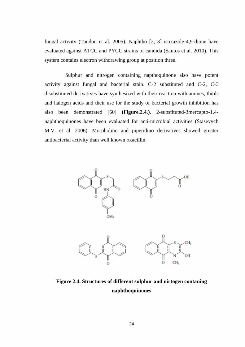
Sulphur and nitrogen containing napthoquinone also have potent
activity against fungal and bacterial stain. C-2 substituted and C-2, C-3
disubstituted derivatives have synthesized with their reaction with amines, thiols
and halogen acids and their use for the study of bacterial growth inhibition has
also been demonstrated [60] (Figure.2.4.). 2-substituted-3mercapto-1,4-
naphthoquinones have been evaluated for anti-microbial activities (Stasevych
M.V. et al. 2006). Morpholino and piperidino derivatives showed greater
antibacterial activity than well known oxacillin.
Figure 2.4. Structures of different sulphur and nirtogen contaning
naphthoquinones
25
2.2.1.3. Naphthoquinone and inflammation
Lapachol, a natural organic compound isolated from the lapachol tree
(Tabebuia avellandedae) identified as naphthoquinone group and is known for its
anti-inflammatory, analgesic and antibiotic properties (Costa et al. 2011). It is also
an anti-tumor agent. Cipura paludosa (Iridaceae) is a plant that forms lapachol
and is distributed in the north region of Brazil. Its bulbs are used in folk
medicine to treat inflammation and pain. It is having four naphthalene
derivatives which have been isolated from the bulbs of the plant. Three of them
were identified as naphthalene derivatives, eleutherine, Iso-eleutherine and
hongkonin. The structure of the fourth was new and elucidated as
11-hydroxyeleutherine (Batista et al. 2011) by NMR. In-vivo effect of two major
compounds eleutherine and iso-eleutherine, was evaluated in carrageenan-
induced hypernociception and inflammation in mice. Eleutherine and iso-
eleutherine (1.04-34.92 mol/kg), dosed i.p. or orally, decreased the carrageenan-
induced paw edema (i.p. - inhibitions of 36 ± 7 % and 58 ± 14 %, resp.;
p.o -inhibitions of 36 ± 7 % and 58 ± 14 %, resp.). Iso-eleutherine, but not
eleutherine, significantly reduced (inhibitions of 39 ± 4 %) the plasma
extravasation induced by intradermal (i.d.) injection of carrageenan. Likewise,
eleutherine and iso-eleutherine (1.04- 34.92 mol/kg,i.p. or p.o.) were also
effective in preventing the carrageenan-induced hypernociceptive response
(i.p.- inhibition of 59 ± 4 % and 63 ± 1 %, resp.; p.o. - inhibitions of 36 ± 7 %
and 58 ± 14 %, resp.).
It was also suggested that the anti-inflammatory and anti-
hypernociceptive effects of eleutherine or iso-eleutherine partly depend on the
interference with the synthesis or activity of mast cell products, kinins, cytokine,
chemokines, prostanoids, or sympathetic amines. Two major compounds of
C. paludosa contain pharmacologically active constituents that possess
antinociceptive and anti-inflammatory activity, justifying, at least in part, its
26
popular therapeutic use for treating conditions associated with pain. Vitamin K3,
which consists of a quinone component, inhibits the activity of human DNA
polymerase (Kazuki et al. 2011). In this study, the inhibitory effects of 1, 4-
quinone derivatives, (1,4-benzoquinone (BQ), 1,4-naphthoquinone (NQ), 9,10-
anthraquinone and 5,12-naphthacenequinone) on the activity of mammalian
polymerase has been shown. BQ and NQ potently inhibited the activity of all the
polymerase species. NQ was a stronger polymerase inhibitor than BQ.
These quinone derivatives could inhibit inflammatory 12-o-
tetradecanoylphorbol -13 acetate (tpa) induced acute responses. BQ and NQ
caused a marked reduction in ion in mouse ear. These anti-inflammatory
responses of NQ were more potent than those of BQ. In conclusion, this study
has identified several quinone derivatives, such as NQ, that are promising anti-
inflammatory candidates (Cherng et al. 1997) .
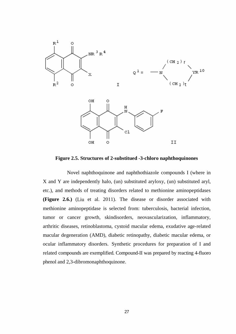
A series of 2-substituted 3-chloro-1, 4-naphthoquinones (Figure 2.5.)
were synthesized, and the antiplatelet, antiinflammatory, and antiallergic
activities of these compounds were evaluated (Tadashi et al. 1996).
The structure-activity relationships in this series were also examined. The title
compounds, I [R1, R2 = H, OH; R3 = acyl, etc.; R4 = H, etc.; or NR3R4 = Q1,
etc.; r, t = 1 - 3; Y = CH, N, etc.; R10 = alkoxyphenyl, etc.; X = halo] are prepared.
The title compd. II in vitro showed IC50 -0.22 µM against neutrophil expression
of CD11b. Naphthoquinone showed potent activities with similar trends in each
of the activities evaluated.
27
Figure 2.5. Structures of 2-substitued -3-chloro naphthoquinones
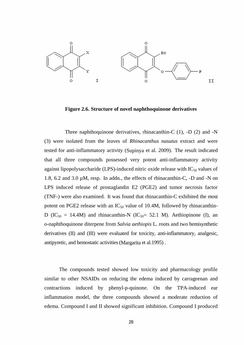
Novel naphthoquinone and naphthothiazole compounds I (where in
X and Y are independently halo, (un) substituted aryloxy, (un) substituted aryl,
etc.), and methods of treating disorders related to methionine aminopeptidases
(Figure 2.6.) (Liu et al. 2011). The disease or disorder associated with
methionine aminopeptidase is selected from: tuberculosis, bacterial infection,
tumor or cancer growth, skindisorders, neovascularization, inflammatory,
arthritic diseases, retinoblastoma, cystoid macular edema, exudative age-related
macular degeneration (AMD), diabetic retinopathy, diabetic macular edema, or
ocular inflammatory disorders. Synthetic procedures for preparation of I and
related compounds are exemplified. Compound-II was prepared by reacting 4-fluoro
phenol and 2,3-dibromonaphthoquinone.
28
Figure 2.6. Structure of novel naphthoquinone derivatives
Three naphthoquinone derivatives, rhinacanthin-C (1), -D (2) and -N
(3) were isolated from the leaves of Rhinacanthus nasutus extract and were
tested for anti-inflammatory activity (Supinya et al. 2009). The result indicated
that all three compounds possessed very potent anti-inflammatory activity
against lipopolysaccharide (LPS)-induced nitric oxide release with IC50 values of
1.8, 6.2 and 3.0 µM, resp. In addn., the effects of rhinacanthin-C, -D and -N on
LPS induced release of prostaglandin E2 (PGE2) and tumor necrosis factor
(TNF-) were also examined. It was found that rhinacanthin-C exhibited the most
potent on PGE2 release with an IC50 value of 10.4M, followed by rhinacanthin-
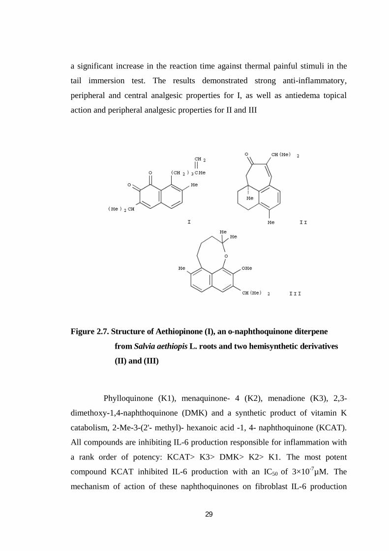
D (IC50 = 14.4M) and rhinacanthin-N (IC50= 52.1 M). Aethiopinone (I), an
o-naphthoquinone diterpene from Salvia aethiopis L. roots and two hemisynthetic
derivatives (II) and (III) were evaluated for toxicity, anti-inflammatory, analgesic,
antipyretic, and hemostatic activities (Margarita et al.1995) .
The compounds tested showed low toxicity and pharmacology profile
similar to other NSAIDs on reducing the edema induced by carrageenan and
contractions induced by phenyl-p-quinone. On the TPA-induced ear
inflammation model, the three compounds showed a moderate reduction of
edema. Compound I and II showed significant inhibition. Compound I produced
29
a significant increase in the reaction time against thermal painful stimuli in the
tail immersion test. The results demonstrated strong anti-inflammatory,
peripheral and central analgesic properties for I, as well as antiedema topical
action and peripheral analgesic properties for II and III
Figure 2.7. Structure of Aethiopinone (I), an o-naphthoquinone diterpene
from Salvia aethiopis L. roots and two hemisynthetic derivatives
(II) and (III)
Phylloquinone (K1), menaquinone- 4 (K2), menadione (K3), 2,3-
dimethoxy-1,4-naphthoquinone (DMK) and a synthetic product of vitamin K
catabolism, 2-Me-3-(2'- methyl)- hexanoic acid -1, 4- naphthoquinone (KCAT).
All compounds are inhibiting IL-6 production responsible for inflammation with
a rank order of potency: KCAT> K3> DMK> K2> K1. The most potent
compound KCAT inhibited IL-6 production with an IC50 of 3×10⁻⁷µM. The
mechanism of action of these naphthoquinones on fibroblast IL-6 production
30
however remains unknown. It was concluded by research that this activity is not
essential for the inhibition of IL-6 production and that activity may be related
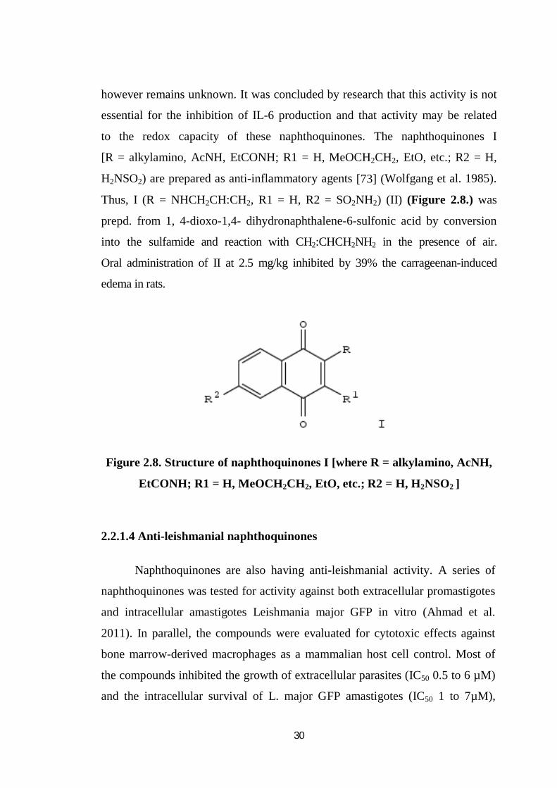
to the redox capacity of these naphthoquinones. The naphthoquinones I
[R = alkylamino, AcNH, EtCONH; R1 = H, MeOCH2CH2, EtO, etc.; R2 = H,
H2NSO2) are prepared as anti-inflammatory agents [73] (Wolfgang et al. 1985).
Thus, I (R = NHCH2CH:CH2, R1 = H, R2 = SO2NH2) (II) (Figure 2.8.) was
prepd. from 1, 4-dioxo-1,4- dihydronaphthalene-6-sulfonic acid by conversion
into the sulfamide and reaction with CH2:CHCH2NH2 in the presence of air.
Oral administration of II at 2.5 mg/kg inhibited by 39% the carrageenan-induced
edema in rats.
Figure 2.8. Structure of naphthoquinones I [where R = alkylamino, AcNH,
EtCONH; R1 = H, MeOCH2CH2, EtO, etc.; R2 = H, H2NSO2 ]
2.2.1.4 Anti-leishmanial naphthoquinones
Naphthoquinones are also having anti-leishmanial activity. A series of
naphthoquinones was tested for activity against both extracellular promastigotes
and intracellular amastigotes Leishmania major GFP in vitro (Ahmad et al.
2011). In parallel, the compounds were evaluated for cytotoxic effects against
bone marrow-derived macrophages as a mammalian host cell control. Most of
the compounds inhibited the growth of extracellular parasites (IC50 0.5 to 6 µM)
and the intracellular survival of L. major GFP amastigotes (IC50 1 to 7µM),
31
when compared with the antileishmanial drug amphotericin B (IC50 of 2.5 and
0.2 µM, resp.). Introduction of a methyl or methoxy group at C-2 of the parent
1, 4-naphthoquinone slightly increased the antileishmanial activity against
clinical relevant amastigotes, while the presence of a hydroxyl function in this
position dramatically reduced the effectiveness. In contrast, hydroxylation at
C-5 and dihydroxy substitution at C-5 and C-8 significantly enhanced the
antiprotozoal activity. Within the series of naphthoquinones tested, the dimeric
mixture of vaforhizin and isovaforhizin showed the highest activity in vitro
against the clinicaly relevant intracellular amastigote with an IC50 of 1.1 µM.
With IC50 values mostly in the range of 1-3 µM, the shikonin/alkannin
derivatives proved to be considerably leishmanicidal. The mode of action
apparently depended on the substitution patterfdn, associated with the
electrophilicity of the naphthoquinone or the efficiency of redox cycling.
Pterocarpanquinones and homologous series of derivatives compounds were
evaluated on breast cancer cell line and parasites Leishmania amazonesis and
Plasmodium falciparum (Silva et al. 2009). 2-phenoxy-1,4-naphthoquinone and
2-phenoxy-1,4-anthraquinone derivatives have inhibitory activity towards
Trypanosoma or leishmania species. Where three of them were active against
Leishmania donovani, Trypanosome cruzi, Trypanosoma brucei rhodesisence
(IC50 = 50 nM, IC50 = 0.28 µM, and IC50 = 1.26 µM). The efficacy of different
formulations of the naphthoquinone buparvaquone and two phosphate prodrugs
against vivo models of both visceral and cutaneous leishmaniasis is described.
Buparvaquone-3-phosphate was shown to be the most effective antileishmanial
(P = 0.0003, 50 mg buparvaquone molar equivalent/kg/day five times), reducing
the liver parasite burden by ~34% when compared with the untreated control.
The introduction of a topical formulation, such as buparvaquone (or its prodrug),
would be a significant advance for the treatment of simple cutaneous lesions.
Lapachol exhibited an anti-amastigote effect. Monomeric and dimeric
naphthoquinones were found active in vitro for treatment of Leishmania
infections using a direct cytotoxicity assay against promastigotes of Leishmania
32
donovani, L. infantum, L. enriettii and L. major.Some naphthoquinones were
active a in the microgram range (EC50 0.9-17.0 µg/mL) (Kayser et al.2000).
The stem barks of P. benensis are employed by the Chimane Indians in
the Bolivian Amazonia as treatment of cutaneous leishmaniasis caused by the
protozoan Leishmania braziliensis (Alain et al. 1992). The chloroform extracts
containing quinones were found to be active against the promastigote forms of
leishmania donovani and the epimastigote forms of Trypanosoma cruzi at
10 µg mL-1. The activity guided fractionation of the extract by chromatography
afforded active compounds. Their structures were elucidated, by spectral and
chemical studies, as known naphthoquinones, plumbagin, 3, 3’-biplumbagin,
8, 8’-biplumbagin, and triterpene, lupeol.
The activity in vitro of each compound was evaluated against 5 strains
of Leishmania (promastigote), 6 strains of T. cruzi (epimastigote) and the
intracellular form (amastigote) of Leishmania amazonensis. The baseline drugs
used were Glucantime and pentamidine (Leishmania spp.), nifurtimox and
benznidazole (T. cruzi). Plumbagin was the most active compound in vitro.
This study has demonstrated that Pera benensis, a medicinal plant used in folk
medicine is an efficient treatment of cutaneous leishmaniasis.
2.2.1.5. Anti-cancer and tumor quinones
The mannich reaction involving lawsone and certain amines with
formaldehyde and acetaldehyde and the condensation product of lawsone with
4-bis (2-chloroethyl) aminobenzaldehyde has been described. Two isomers of
naphthoquinones derivatives 6-(1-azidoalkyl)-DMNQ and 2-(1-azidoalkyl-
DMNQ exhibited higher cytotoxic activity against L1210 mouse leukemia cells
and stronger inhibition of DNA topoisomerase-I (Chae et al.1999). These
molecules contain N- substituted- pyridino [2,3-f] indole-4,9-dione and 6-(α-
diethoxy carbonyl methyl)7-substituted amino quinoline 5,8-dione, which
33
contain the active quinoline 5,8-dione moiety. This moiety have been tested
against SRB (sulphorodamine B) assay against the cancer cell lines of A-549
(human lung cancer), SK-MEL-2 (human melanoma cancer), SK-OV3 (human
ovarian cancer), XF-498 (human brain cancer) and HCT (human colon cancer).
This moiety showed higher activity than cis-platin. Rhinacanthone and 1,2-
pyranonaphthoquinones were synthesized and showed very potent cyto-toxicity
against three cancer cell lines (KB, HeLa and HepG2) with IC50 values of 0.92-
9.63µM (Kongkathip et al. 2003) .
CDC25 dual-specificity phosphatases are essential key regulators of
eukaroytic cell cycle progression and the CDC25A and B isoforms are over-
expressed in different tumors. Polyfluoro derivatives of 1,4-naphthoquinones are
highly potent inhibitors of Cdc25A and Cdc25B phosphatases and growth of
tumor cells and their cytotoxicty in human myeloma, human mammary
adenocarcinoma, mouse fibroblasts and primary mouse fibroblast cells as well as
their mutagenic and antioxidant properties in a Salmonella tester strain were
studied (Brun et al. 2005). The β-lapachone based 1,2,3-triazoles showed the
best cytotoxicity profile and emerge as promising anti-cancer prototypes.
The anti-tubercular activity and cytotoxocity of juglone derivatives were
analyzed with the topological and molecular surface features from a web based
server, MODEL(Molecular Descriptor Lab). Novel compounds derived from
vitamin K3 that inhibit CDC25B activity with IC50 values in the low micromolar
range.Polyamine naphthoquinone conjugates by neuclophilic displacement of
2-methoxy lawsone, 2-methoxy lapachol, 2-methoxynorlapachol with the
polyamine N1- Boc- N5- Bn- spermidine 4. 2-methyl-1,4-naphthoquinone
derivatives especially vitamin K3 .Retardation of cytotoxicty and cell proliferation
by 2-amino alkyl moiety with terminal bromo, chloro, hydroxyl, mercapto groups
were examined on model murine hepatoma cell line-22A. Most active compound
were the hydroxyl and bromo derivatives(Stasiauskaite et al. 2006) .
34
A series of 2-chloro-3-arylsulfanyl-[1, 4] naphthoquinones 2, 3-bis-
arylsulfanyl-[1,4] naphthoquinones and 12H-benzo [b] phenothiazine- 6,11-
diones and their analogs were evaluated for their antiproliferative activity
against human cervical cancer cells (Silva et al. 2011). 1,5-Diazaanthraquinone
derivatives were synthesized employing single and double hetero Diels–Alder
strategies. Their in-vitro antitumor activity was assayed using three cell lines.
Some of these compounds, especially those bearing methyl or ethyl groups at the
C-3,7 positions or chloro at C-4 and methyl at C-7, showed IC50 values in the 10-
8 µM range for human lung carcinoma and human melanoma, which makes them
attractive candidates for further development as anticancer agents.
2.2.2. Bis-naphthoquinones
Bis-naphthoquinones and higher quinone oligomers are a unique group
of natural products, which possess a diverse array of biological activities
(Actinorodins et al. 1996). Their structures are based on two or more quinone
units linked together at the quinine double bond. In almost all cases they possess
an element of symmetry due to their biosynthetic mechanism of origin, which
probably involves oxidative coupling of a common naphthol intermediate in the
key step of the oligomerization process (Laatsh 1994) [85] .One intriguing
member of this class is conocurvone isolated from the Western Australian smoke
bush (Decosterd et al. 1993). Conocurvone (Figure 2.9.) was shown to inhibit
the cytopathogenic effects of HIV-1 in human T-lymphoblastic cells over a
broad concentration range (ID50=0.02 µM; TD50= 50 µM) (Decosterd et al. 1993).
More recently, it was suggested that conocurvone 1 may be a dual inhibitor of
both HIV integrase and HIV mediated cell fusion (Kearney et al. 2001).

35
O
O
O
OO
O
CH3
CH3
CH3
CH3CH3
CH3
O
O
OCH3
CH3CH3
Figure 2.9. Structure of Conocurvone
Over the past decade, extensive efforts have been made resulting in
the discovery of a large number of molecules that can inhibit replication of
HIV(Yang et al. 2001). An essential step in the HIV life cycle is integration of
the viral DNA into the host cell genome. The step is catalyzed by the viral
enzyme, HIV integrase, which is absolutely required for productive infection and
therefore, inhibition of integrase can half the viral life cycle. Integrase catalyses
two separate steps known as 3’-prossessing and DNA strand transfer. In 3’-
prossessing, integrase removes a dinuclotide next to a conserved cytosine-
adenine sequence from each 3’-end of the viral DNA.
Integrase then attaches the processed 3’-end of the viral DNA to the
host cell DNA in the strand transfer reaction. An important result of the
structural and biochemical studies on integrace has been the development of
practical assays used to identify novel HIV integrase inhibitors. These HIV
inhibitors not only represent potential chemotherapeutic lead compounds
(Mazumder et al. 1996), but as a collection, they are also useful in databases for
pharmacophore searching. The most promising inhibitors are proposed to bind to
the active site of the integrase enzyme and chelate important metal cofactors
such as Mn2+ or Mg2+.Sidhu. and Pardhasaradhi. (1967a) (1970) established the
structure of diospyrin, a bisnaphthoquinone with a benzene-quinone linkage .
Bisnaphthoquinone may arise either by the oxidation of a bis-naphthol formed
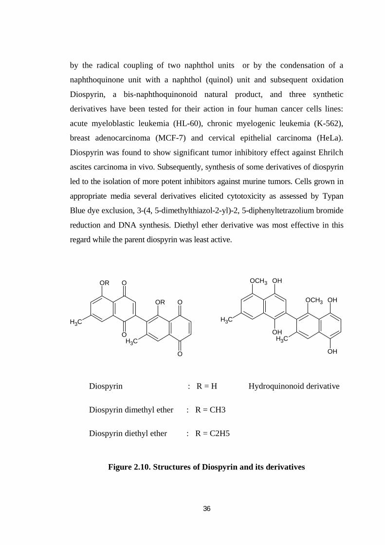
36
by the radical coupling of two naphthol units or by the condensation of a
naphthoquinone unit with a naphthol (quinol) unit and subsequent oxidation
Diospyrin, a bis-naphthoquinonoid natural product, and three synthetic
derivatives have been tested for their action in four human cancer cells lines:
acute myeloblastic leukemia (HL-60), chronic myelogenic leukemia (K-562),
breast adenocarcinoma (MCF-7) and cervical epithelial carcinoma (HeLa).
Diospyrin was found to show significant tumor inhibitory effect against Ehrilch
ascites carcinoma in vivo. Subsequently, synthesis of some derivatives of diospyrin
led to the isolation of more potent inhibitors against murine tumors. Cells grown in
appropriate media several derivatives elicited cytotoxicity as assessed by Typan
Blue dye exclusion, 3-(4, 5-dimethylthiazol-2-yl)-2, 5-diphenyltetrazolium bromide
reduction and DNA synthesis. Diethyl ether derivative was most effective in this
regard while the parent diospyrin was least active.
OR
CH3
O
O
OR
CH3
O
O
OCH3
CH3
OCH3
CH3
OH
OH
OH
OH
Diospyrin : R = H Hydroquinonoid derivative
Diospyrin dimethyl ether : R = CH3
Diospyrin diethyl ether : R = C2H5
Figure 2.10. Structures of Diospyrin and its derivatives
37
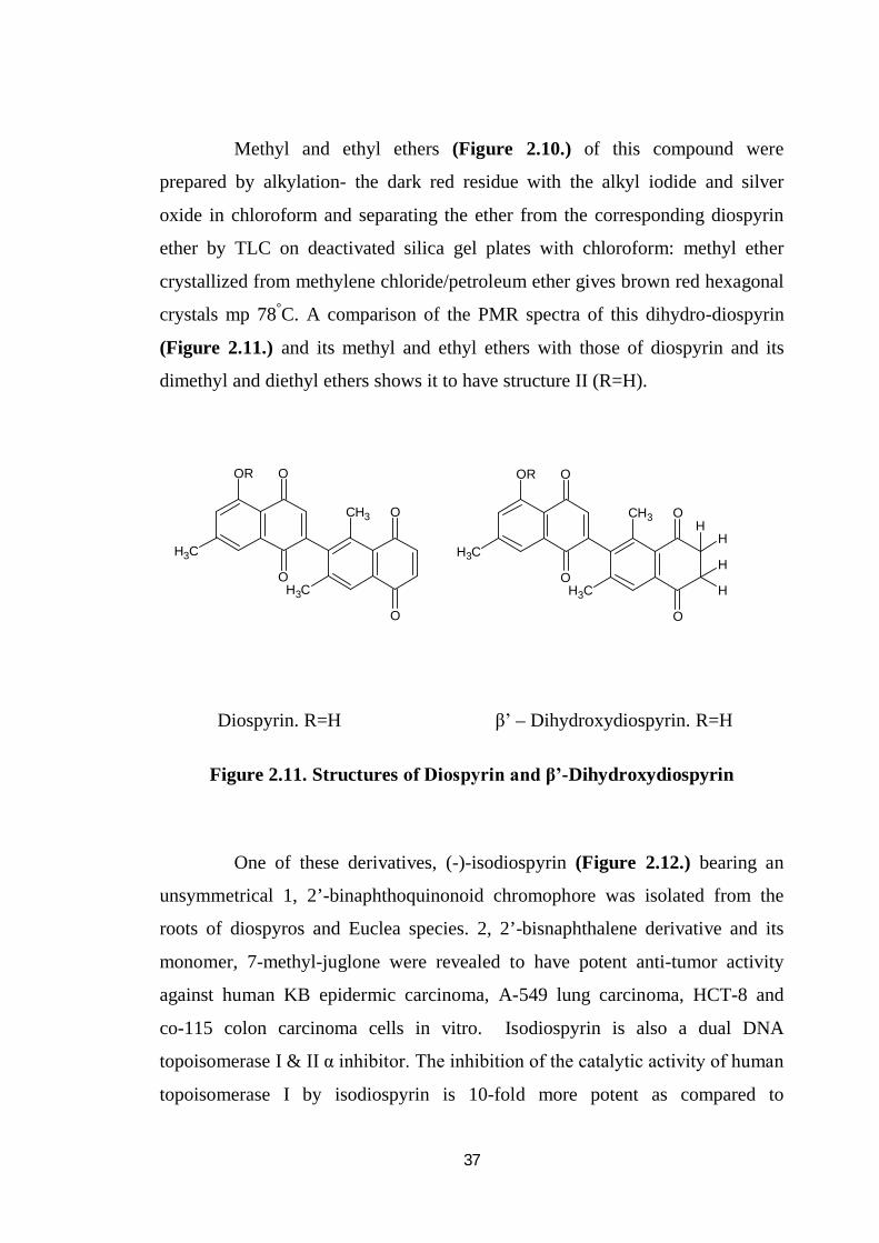
Methyl and ethyl ethers (Figure 2.10.) of this compound were
prepared by alkylation- the dark red residue with the alkyl iodide and silver
oxide in chloroform and separating the ether from the corresponding diospyrin
ether by TLC on deactivated silica gel plates with chloroform: methyl ether
crystallized from methylene chloride/petroleum ether gives brown red hexagonal
crystals mp 78°C. A comparison of the PMR spectra of this dihydro-diospyrin
(Figure 2.11.) and its methyl and ethyl ethers with those of diospyrin and its
dimethyl and diethyl ethers shows it to have structure II (R=H).
OR
CH3
O
O
CH3
CH3
O
O
OR
CH3
O
O
CH3
CH3
O
O
H
H
H
H
Diospyrin. R=H β’ – Dihydroxydiospyrin. R=H
Figure 2.11. Structures of Diospyrin and β’-Dihydroxydiospyrin
One of these derivatives, (-)-isodiospyrin (Figure 2.12.) bearing an
unsymmetrical 1, 2’-binaphthoquinonoid chromophore was isolated from the
roots of diospyros and Euclea species. 2, 2’-bisnaphthalene derivative and its
monomer, 7-methyl-juglone were revealed to have potent anti-tumor activity
against human KB epidermic carcinoma, A-549 lung carcinoma, HCT-8 and
co-115 colon carcinoma cells in vitro. Isodiospyrin is also a dual DNA
topoisomerase I & II α inhibitor. The inhibition of the catalytic activity of human
topoisomerase I by isodiospyrin is 10-fold more potent as compared to

38
camptothecin, a potent anti neoplastic natural product and topoisomerase I
inhibitor.
The isomer, diospyrin was also cytotoxic to several human tumor cell
lines in culture. Ray et al. reported that diospyrin significantly inhibited the
growth of Leishmania donovani promastigotes. This agent also inhibited the
catalytic activity of DNA topoisomerase-I of the parasite and induced DNA
Topoisomerase I-mediated cleavage in vitro, suggesting that the bi-
naphthoquinonoids derivatives exert their inhibitory effect binding to the
enzyme and stabilizing the Topoisomerase-I-DNA cleavable complex. However,
diospyrin did not inhibit topoisomerase-II of L.donovani and required much
concentrations to inhibit calf-thymus topoisomerase-I. Based on the biological
properties of isodiospyrin and diospyrin, they can be exploited for rational drug
design to develop new anticancer agents or drugs human leishmaniasis. Neo-
diospyrin is a structural analogue of diospyrin and isodiospyrin having potent
inhibition against mycobacterium tuberculosis as well.
O
O
O
O
CH3CH3OH
O
OOH
CH3 O
O
OHCH3
Figure 2.12. Structures of Isodiospyrin and Neodiospyrin
Gossypol (Figure 2.13.) was isolated from Gossypium species
(Dechary. and Pradel.1971) and has been studied as a male antifertility agent in
china. The two representative naphthyl –isoquinoline alkaloids, ancistrocladine
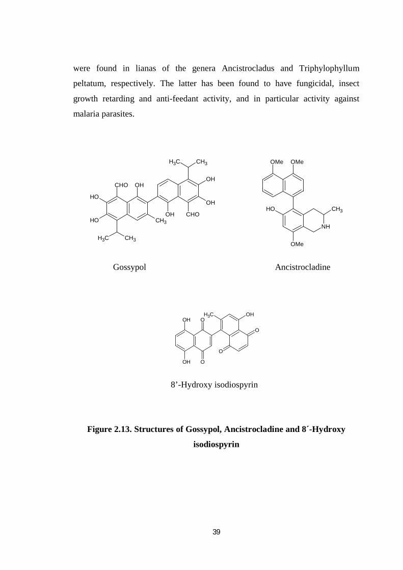
39
were found in lianas of the genera Ancistrocladus and Triphylophyllum
peltatum, respectively. The latter has been found to have fungicidal, insect
growth retarding and anti-feedant activity, and in particular activity against
malaria parasites.
OH
OH
CHOOH
CH3CH3
OH
CH3
OH
OH
CHO
CH3 CH3
NH
OMeOMe
OMe
OH CH3
Gossypol Ancistrocladine
OH
OH
O
O
OHCH3O
O
8’-Hydroxy isodiospyrin
Figure 2.13. Structures of Gossypol, Ancistrocladine and 8´-Hydroxy
isodiospyrin
40
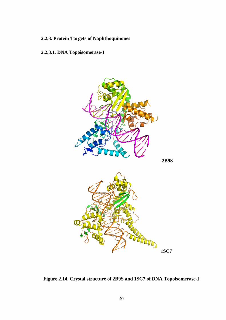
2.2.3. Protein Targets of Naphthoquinones
2.2.3.1. DNA Topoisomerase-I
2B9S
1SC7
Figure 2.14. Crystal structure of 2B9S and 1SC7 of DNA Topoisomerase-I
41
2.2.3.1.1. Function of the enzyme
Releases the supercoiling and torsional tension of DNA introduced
during the DNA replication and transcription by transiently cleaving and
rejoining one strand of the DNA duplex.Introduces a single-strand break via
transesterification at a target site in duplex DNA. The scissile phosphodiester is
attacked by the catalytic tyrosine of the enzyme, resulting in the formation of a
DNA-(3'-phosphotyrosyl)-enzyme intermediate and the expulsion of a 5'-OH
DNA strand. The free DNA strand then undergoes passage around the unbroken
strand thus removing DNA supercoils. Finally, in the relegation step, the DNA
5'-OH attacks the covalent intermediate to expel the active-site tyrosine and
restore the DNA phosphodiester backbone. This enzyme is also known to
regulate the alternative splicing of tissue factor (F3) pre-mRNA in endothelial
cells (D'Arpa.et al. 1988; Interthal et al. 2004; Cushman et al. 2005) .
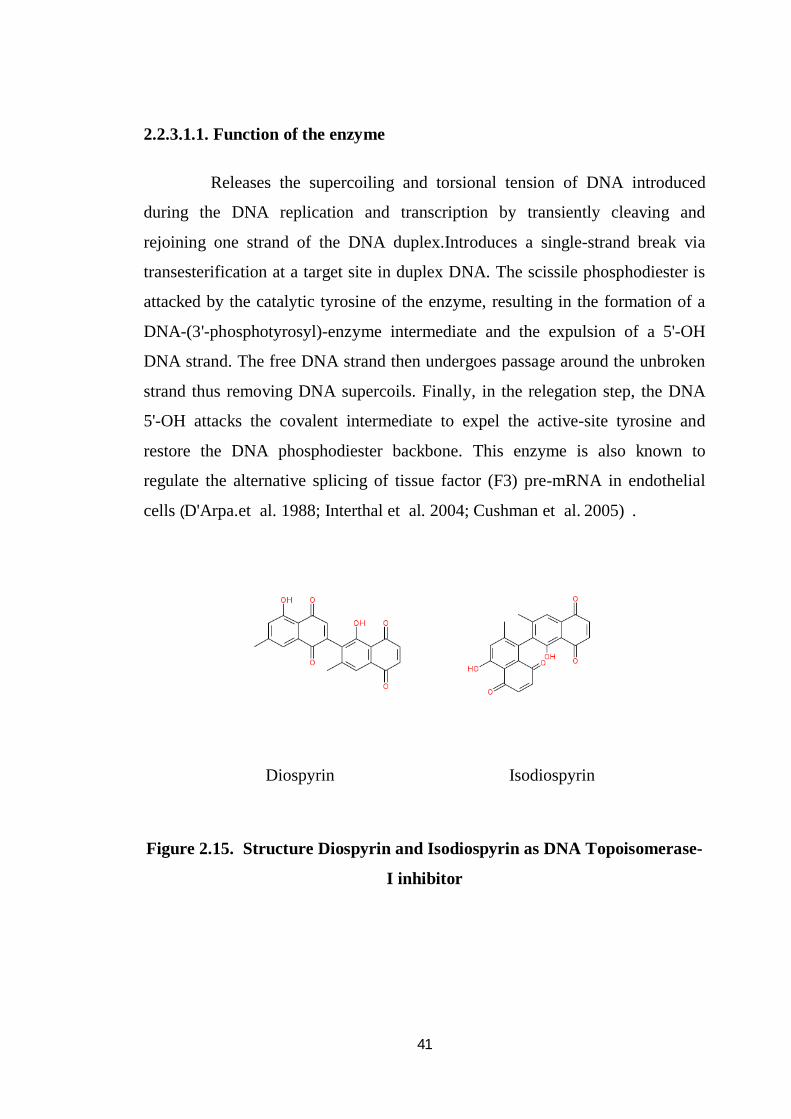
Diospyrin Isodiospyrin
Figure 2.15. Structure Diospyrin and Isodiospyrin as DNA Topoisomerase-
I inhibitor
42
2.2.3.2 Topoisomerase-IIα
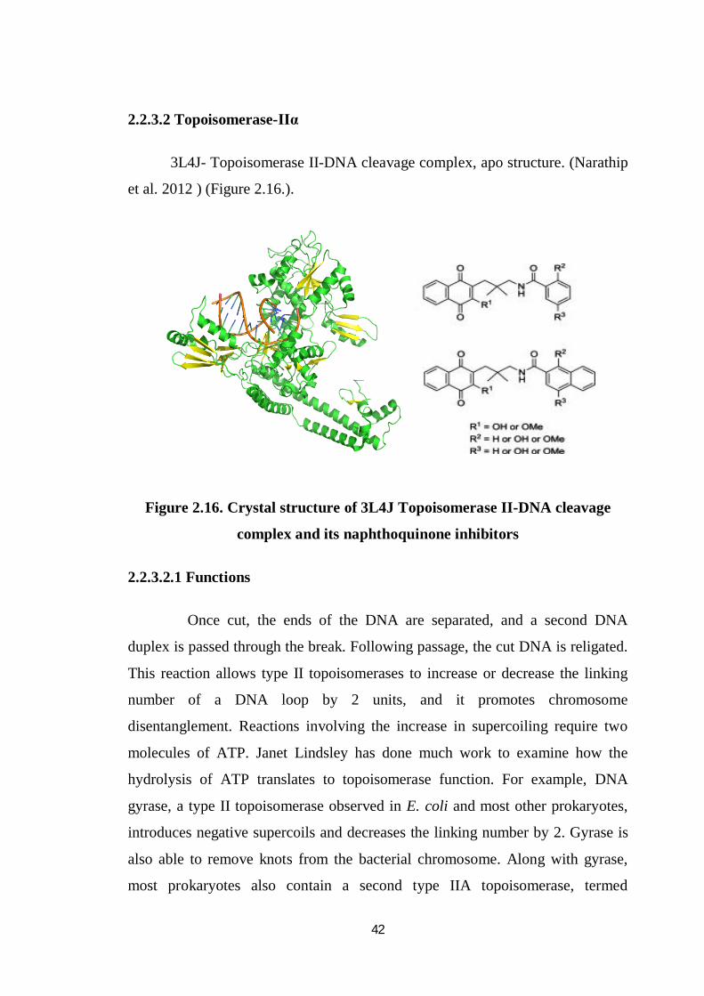
3L4J- Topoisomerase II-DNA cleavage complex, apo structure. (Narathip
et al. 2012 ) (Figure 2.16.).
Figure 2.16. Crystal structure of 3L4J Topoisomerase II-DNA cleavage
complex and its naphthoquinone inhibitors
2.2.3.2.1 Functions
Once cut, the ends of the DNA are separated, and a second DNA
duplex is passed through the break. Following passage, the cut DNA is religated.
This reaction allows type II topoisomerases to increase or decrease the linking
number of a DNA loop by 2 units, and it promotes chromosome
disentanglement. Reactions involving the increase in supercoiling require two
molecules of ATP. Janet Lindsley has done much work to examine how the
hydrolysis of ATP translates to topoisomerase function. For example, DNA
gyrase, a type II topoisomerase observed in E. coli and most other prokaryotes,
introduces negative supercoils and decreases the linking number by 2. Gyrase is
also able to remove knots from the bacterial chromosome. Along with gyrase,
most prokaryotes also contain a second type IIA topoisomerase, termed

43
topoisomerase IV. Gyrase and topoisomerase IV differ by their C-terminal
domains, which are believed to dictate substrate specificity and functionality for
these two enzymes. Footprinting indicates that gyrase, which forms a 140-base-
pair footprint and wraps DNA, allowing it to introduce negative supercoils,
while topoisomerase IV, which forms a 28-base-pair footprint, does not wrap
DNA. Eukaryotic type II topoisomerase cannot introduce supercoils; it can only
relax them. The role of type IIB topoisomerase is less understood. Unlike type II
topoisomerases, it cannot simplify DNA topology, but it shares several structural
features with type IIA topoisomerases..
2.2.3.2.2. Topoisomerases as Drug Targets
Topoisomerases have been the focus for the treatment of certain
diseases. Bacterial gyrase (topoisomerase II) and topoisomerase IV are the
targets of two classes of antibiotic drugs: quinolones and coumarins. These
antibiotics are used to treat an assortment of different diseases, such as
pneumonia, tuberculosis and malaria, by inhibiting DNA replication in the
bacteria responsible.Eukaryotic topoisomerases I and II are the targets of an
increasing number of anti-cancer drugs that act to inhibit these enzymes by
blocking the reaction that reseals the breaks in the DNA. Often the binding of
the drug is reversible, but if a replication fork runs into the blocked
topoisomerase, then a piece of the gapped DNA strand not bound by the
topoisomerase could be released, creating a permanent breakage in the DNA that
leads to cell death. Most of these inhibitors are selective against either
topoisomerase I or II, but some can target both enzymes. Topoisomerase I
inhibitors induce single-strand breaks into DNA, and can work by a variety of
mechanisms. Some drugs, such as camptothecin, inhibit the dissociation of
topoisomerase and DNA, leading to replication-mediated DNA damage, which
can be repaired more efficiently in normal cells than in cancer cells (deficient for
DNA repair). Topoisomerase I inhibitors can also cause gene inactivation
44
through chromatid aberrations. Topoisomerase II inhibitors, such as
anthracyclines, (Figure 2.15.) are amongst the most widely used anti-cancer
agents. These drugs are potent inducers of double strand breaks in DNA, and
can cause arrest in the cell cycle at the G2 stage, the latter occurring by
disrupting the interaction between topoisomerase II and regulators of the cell
cycle, such as Cdc2 (Eisenreich et al.2009). Topoisomerase II inhibitors can
cause a wide range of chromosomal aberrations, and can act by either stabilizing
topoisomerase II-DNA complexes that are easily cleaved, or by interfering with
the catalytic activity of the enzyme, both resulting in double-strand breaks in the
DNA. There are also dual inhibitors that target both topoisomerase I and II,
which increases the potency of the anti-cancer effect. These drugs work by a
variety of means: by recognizing structural motifs present on both enzymes, by
linking separate topoisomerase inhibitors together into a hybrid drug, or by using
inhibitors that bind to and intercalate DNA.
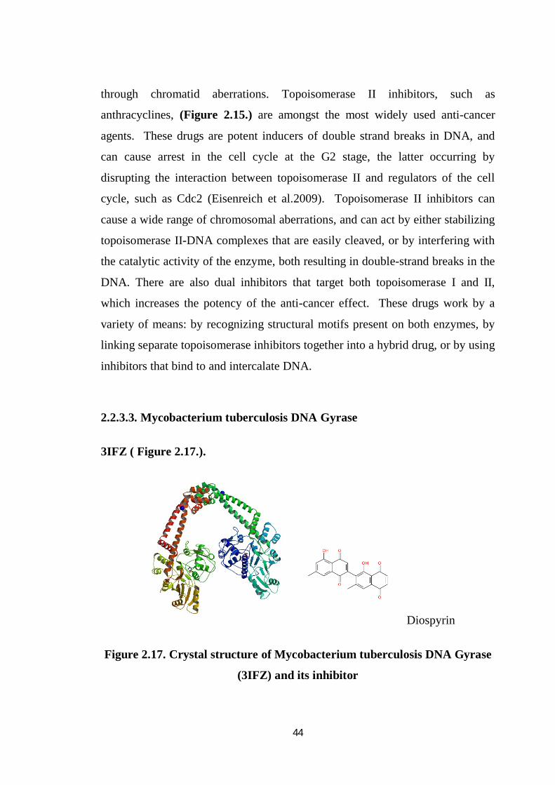
2.2.3.3. Mycobacterium tuberculosis DNA Gyrase
3IFZ ( Figure 2.17.).
Diospyrin
Figure 2.17. Crystal structure of Mycobacterium tuberculosis DNA Gyrase
(3IFZ) and its inhibitor
45
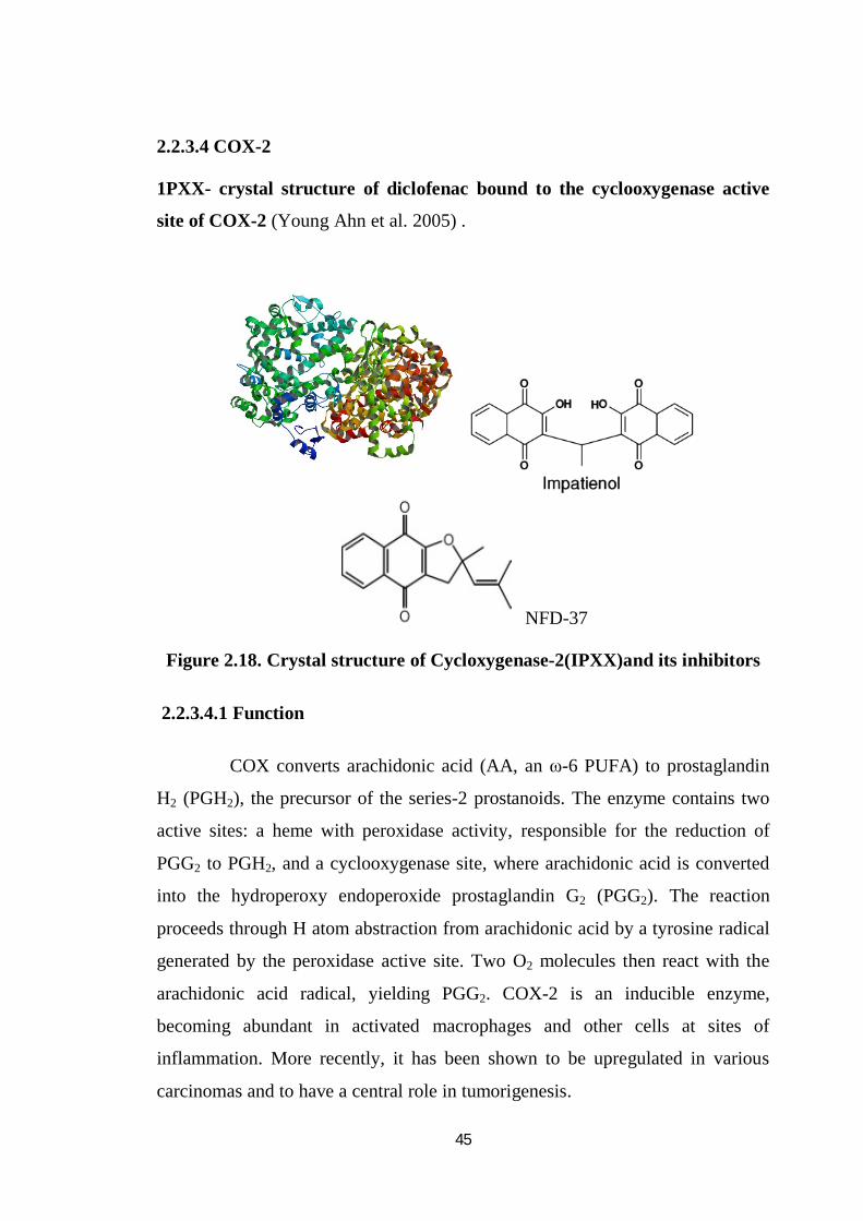
2.2.3.4 COX-2
1PXX- crystal structure of diclofenac bound to the cyclooxygenase active
site of COX-2 (Young Ahn et al. 2005) .
NFD-37
Figure 2.18. Crystal structure of Cycloxygenase-2(IPXX)and its inhibitors
2.2.3.4.1 Function
COX converts arachidonic acid (AA, an ω-6 PUFA) to prostaglandin
H2 (PGH2), the precursor of the series-2 prostanoids. The enzyme contains two
active sites: a heme with peroxidase activity, responsible for the reduction of
PGG2 to PGH2, and a cyclooxygenase site, where arachidonic acid is converted
into the hydroperoxy endoperoxide prostaglandin G2 (PGG2). The reaction
proceeds through H atom abstraction from arachidonic acid by a tyrosine radical
generated by the peroxidase active site. Two O2 molecules then react with the
arachidonic acid radical, yielding PGG2. COX-2 is an inducible enzyme,
becoming abundant in activated macrophages and other cells at sites of
inflammation. More recently, it has been shown to be upregulated in various
carcinomas and to have a central role in tumorigenesis.
46
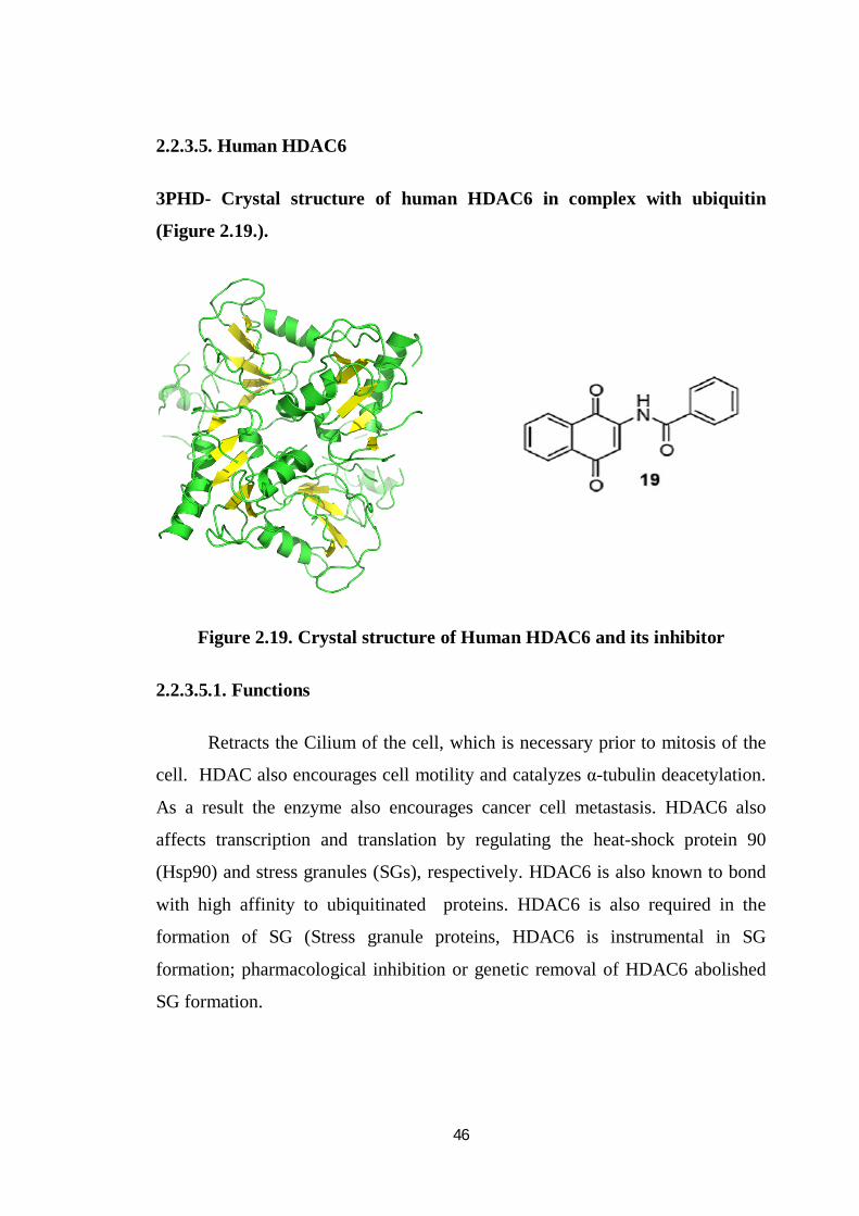
2.2.3.5. Human HDAC6
3PHD- Crystal structure of human HDAC6 in complex with ubiquitin
(Figure 2.19.).
Figure 2.19. Crystal structure of Human HDAC6 and its inhibitor
2.2.3.5.1. Functions
Retracts the Cilium of the cell, which is necessary prior to mitosis of the
cell. HDAC also encourages cell motility and catalyzes α-tubulin deacetylation.
As a result the enzyme also encourages cancer cell metastasis. HDAC6 also
affects transcription and translation by regulating the heat-shock protein 90
(Hsp90) and stress granules (SGs), respectively. HDAC6 is also known to bond
with high affinity to ubiquitinated proteins. HDAC6 is also required in the
formation of SG (Stress granule proteins, HDAC6 is instrumental in SG
formation; pharmacological inhibition or genetic removal of HDAC6 abolished
SG formation.
47
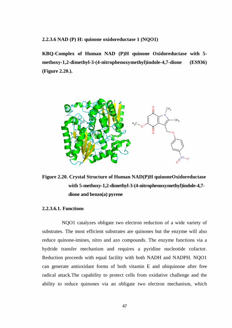
2.2.3.6 NAD (P) H: quinone oxidoreductase 1 (NQO1)
KBQ-Complex of Human NAD (P)H quinone Oxidoreductase with 5-
methoxy-1,2-dimethyl-3-(4-nitrophenoxymethyl)indole-4,7-dione (ES936)
(Figure 2.20.).
Figure 2.20. Crystal Structure of Human NAD(P)H quinoneOxidoreductase
with 5-methoxy-1,2-dimethyl-3-(4-nitrophenoxymethyl)indole-4,7-
dione and benzo(a) pyrene
2.2.3.6.1. Functions
NQO1 catalyzes obligate two electron reduction of a wide variety of
substrates. The most efficient substrates are quinones but the enzyme will also
reduce quinone-imines, nitro and azo compounds. The enzyme functions via a
hydride transfer mechanism and requires a pyridine nucleotide cofactor.
Reduction proceeds with equal facility with both NADH and NADPH. NQO1
can generate antioxidant forms of both vitamin E and ubiquinone after free
radical attack.The capability to protect cells from oxidative challenge and the
ability to reduce quinones via an obligate two electron mechanism, which
48
precludes generation of reactive oxygen radicals, demonstrates that NQO1 is a
chemoprotective enzyme. NQO1 knockout mice demonstrated increased
susceptibility to benzo (a) pyrene and 7, 12-dimethylbenz (a) anthracene induced
skin carcinogenesis. NQO1 has been proposed to stabilize the tumor suppressor
gene p53 and has been shown to interact with p53 in a protein-protein interaction.
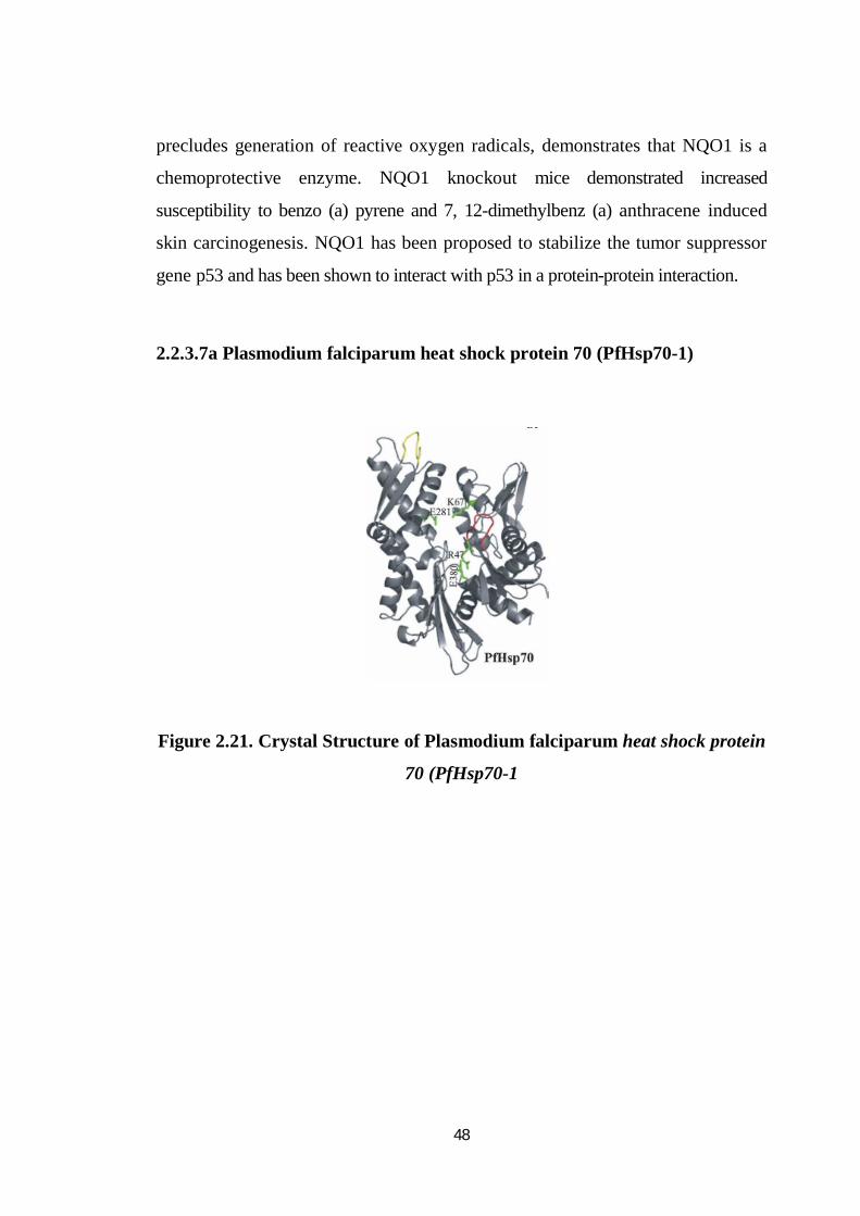
2.2.3.7a Plasmodium falciparum heat shock protein 70 (PfHsp70-1)
Figure 2.21. Crystal Structure of Plasmodium falciparum heat shock protein
70 (PfHsp70-1
49
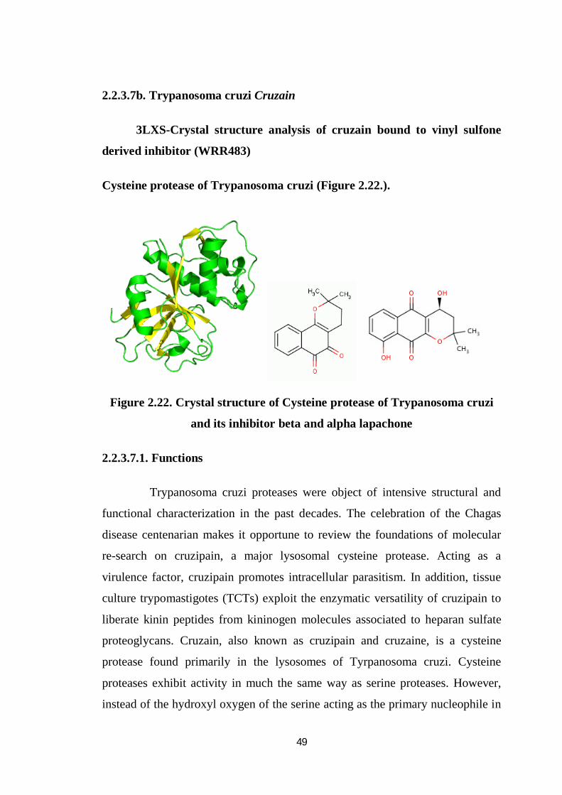
2.2.3.7b. Trypanosoma cruzi Cruzain
3LXS-Crystal structure analysis of cruzain bound to vinyl sulfone
derived inhibitor (WRR483)
Cysteine protease of Trypanosoma cruzi (Figure 2.22.).
Figure 2.22. Crystal structure of Cysteine protease of Trypanosoma cruzi
and its inhibitor beta and alpha lapachone
2.2.3.7.1. Functions
Trypanosoma cruzi proteases were object of intensive structural and
functional characterization in the past decades. The celebration of the Chagas
disease centenarian makes it opportune to review the foundations of molecular
re-search on cruzipain, a major lysosomal cysteine protease. Acting as a
virulence factor, cruzipain promotes intracellular parasitism. In addition, tissue
culture trypomastigotes (TCTs) exploit the enzymatic versatility of cruzipain to
liberate kinin peptides from kininogen molecules associated to heparan sulfate
proteoglycans. Cruzain, also known as cruzipain and cruzaine, is a cysteine
protease found primarily in the lysosomes of Tyrpanosoma cruzi. Cysteine
proteases exhibit activity in much the same way as serine proteases. However,
instead of the hydroxyl oxygen of the serine acting as the primary nucleophile in
50
the mechanism, the thiol sulfur of a cysteine residue is active. Cysteine proteases
act by cleaving the peptide bond between bound amino acid residues. Cruzain’s
specifically binds proteins with either basic or hydrophobic residues
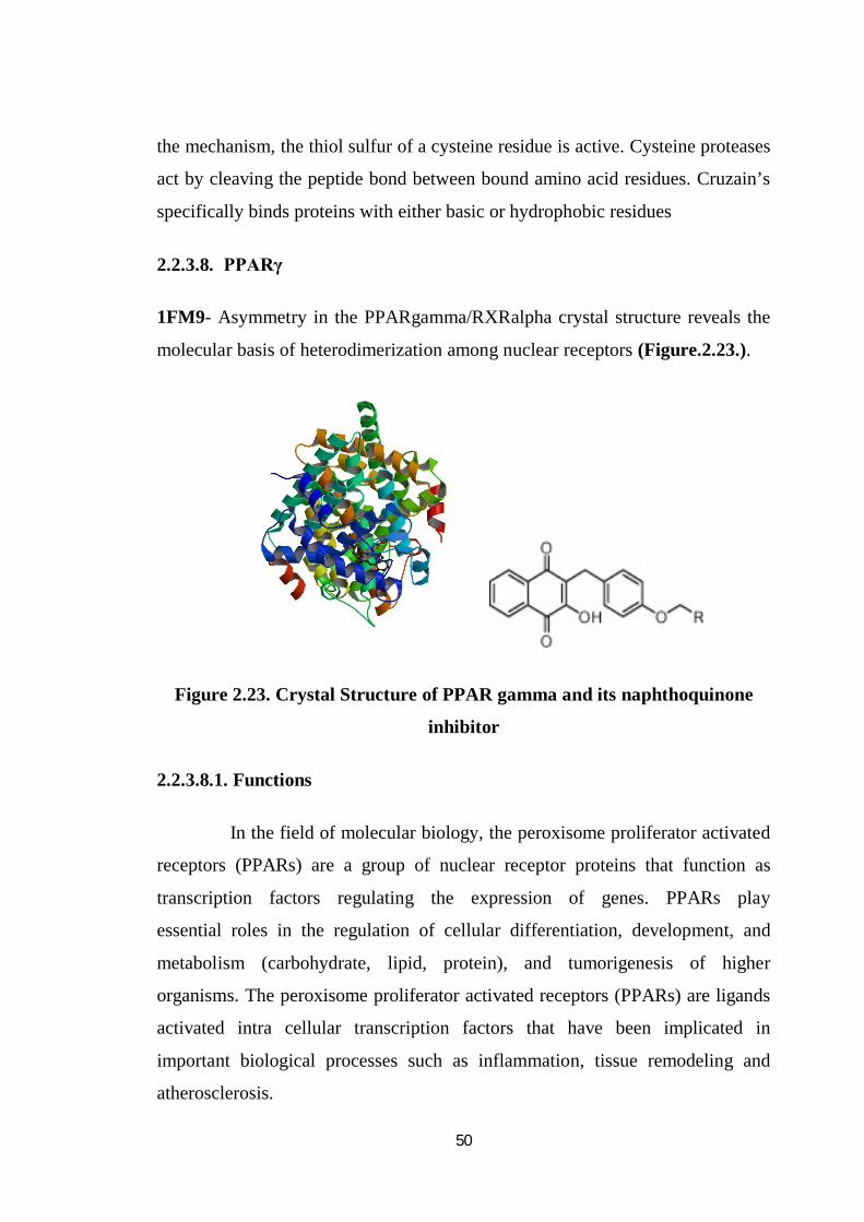
2.2.3.8. PPARγ
1FM9- Asymmetry in the PPARgamma/RXRalpha crystal structure reveals the
molecular basis of heterodimerization among nuclear receptors (Figure.2.23.).
Figure 2.23. Crystal Structure of PPAR gamma and its naphthoquinone
inhibitor
2.2.3.8.1. Functions
In the field of molecular biology, the peroxisome proliferator activated
receptors (PPARs) are a group of nuclear receptor proteins that function as
transcription factors regulating the expression of genes. PPARs play
essential roles in the regulation of cellular differentiation, development, and
metabolism (carbohydrate, lipid, protein), and tumorigenesis of higher
organisms. The peroxisome proliferator activated receptors (PPARs) are ligands
activated intra cellular transcription factors that have been implicated in
important biological processes such as inflammation, tissue remodeling and
atherosclerosis.
51
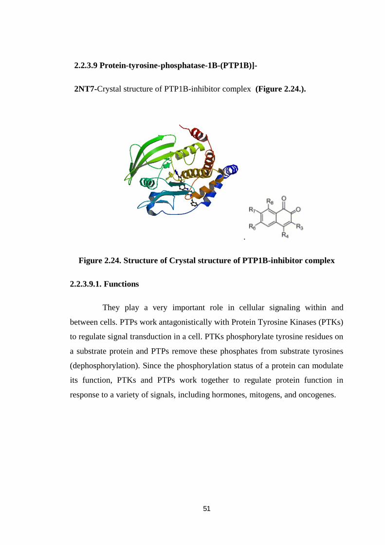
2.2.3.9 Protein-tyrosine-phosphatase-1B-(PTP1B)]-
2NT7-Crystal structure of PTP1B-inhibitor complex (Figure 2.24.).
.
Figure 2.24. Structure of Crystal structure of PTP1B-inhibitor complex
2.2.3.9.1. Functions
They play a very important role in cellular signaling within and
between cells. PTPs work antagonistically with Protein Tyrosine Kinases (PTKs)
to regulate signal transduction in a cell. PTKs phosphorylate tyrosine residues on
a substrate protein and PTPs remove these phosphates from substrate tyrosines
(dephosphorylation). Since the phosphorylation status of a protein can modulate
its function, PTKs and PTPs work together to regulate protein function in
response to a variety of signals, including hormones, mitogens, and oncogenes.
52
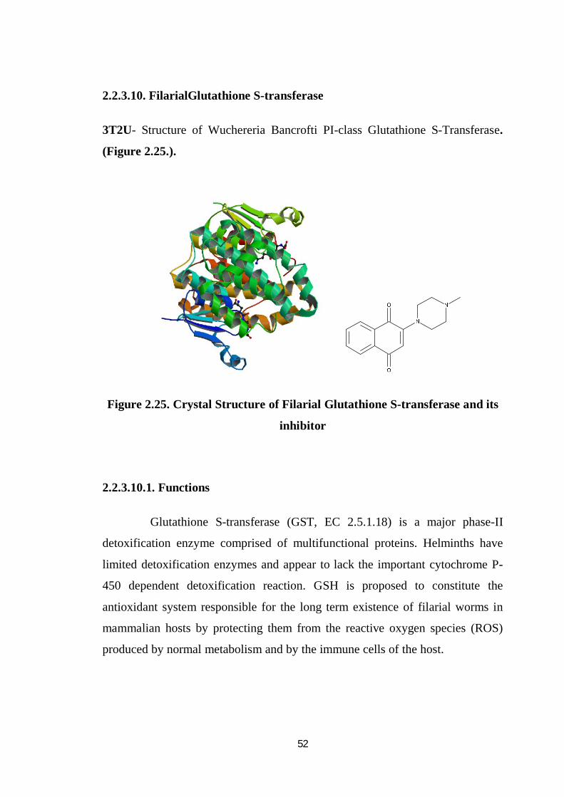
2.2.3.10. FilarialGlutathione S-transferase
3T2U- Structure of Wuchereria Bancrofti PI-class Glutathione S-Transferase.
(Figure 2.25.).
Figure 2.25. Crystal Structure of Filarial Glutathione S-transferase and its
inhibitor
2.2.3.10.1. Functions
Glutathione S-transferase (GST, EC 2.5.1.18) is a major phase-II
detoxification enzyme comprised of multifunctional proteins. Helminths have
limited detoxification enzymes and appear to lack the important cytochrome P-
450 dependent detoxification reaction. GSH is proposed to constitute the
antioxidant system responsible for the long term existence of filarial worms in
mammalian hosts by protecting them from the reactive oxygen species (ROS)
produced by normal metabolism and by the immune cells of the host.
53
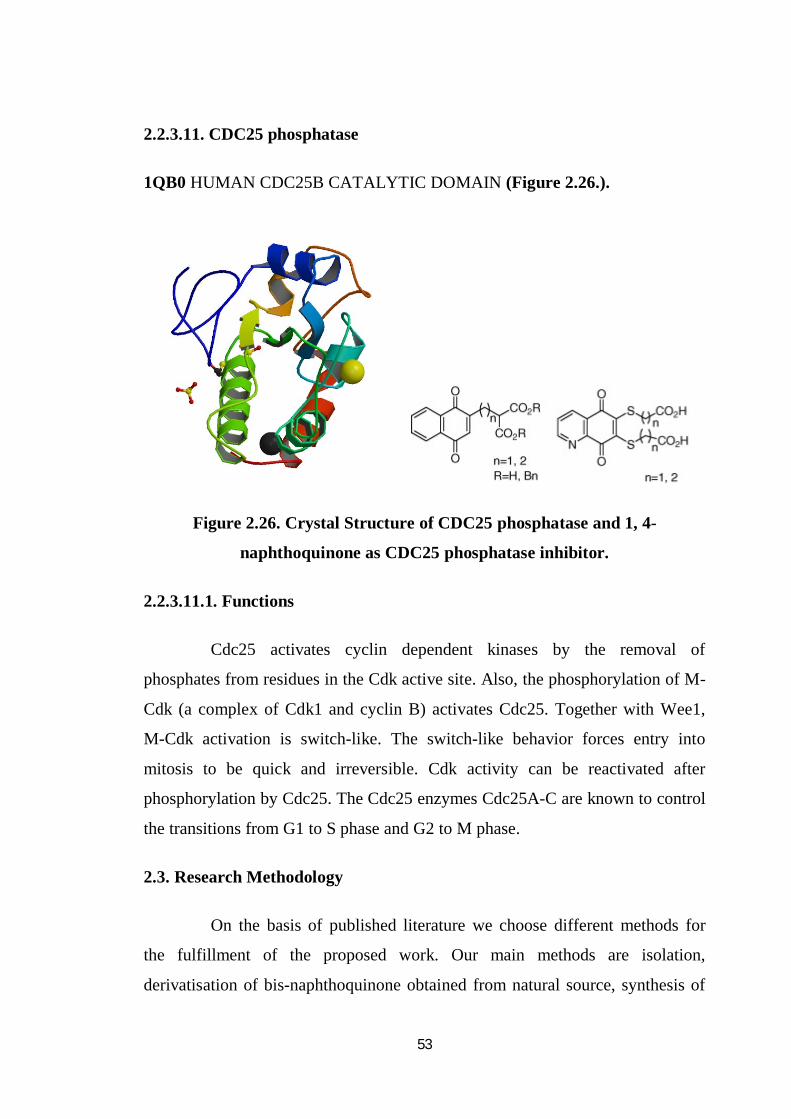
2.2.3.11. CDC25 phosphatase
1QB0 HUMAN CDC25B CATALYTIC DOMAIN (Figure 2.26.).
Figure 2.26. Crystal Structure of CDC25 phosphatase and 1, 4-
naphthoquinone as CDC25 phosphatase inhibitor.
2.2.3.11.1. Functions
Cdc25 activates cyclin dependent kinases by the removal of
phosphates from residues in the Cdk active site. Also, the phosphorylation of M-
Cdk (a complex of Cdk1 and cyclin B) activates Cdc25. Together with Wee1,
M-Cdk activation is switch-like. The switch-like behavior forces entry into
mitosis to be quick and irreversible. Cdk activity can be reactivated after
phosphorylation by Cdc25. The Cdc25 enzymes Cdc25A-C are known to control
the transitions from G1 to S phase and G2 to M phase.
2.3. Research Methodology
On the basis of published literature we choose different methods for
the fulfillment of the proposed work. Our main methods are isolation,
derivatisation of bis-naphthoquinone obtained from natural source, synthesis of
54
naphthoquinone derivatives from synthetic source and biological evaluation of
synthesized/isolated compounds with in-vitro with different important protein
targets and subsequent in-silico bio-molecular interaction studies with the help
of molecular docking.
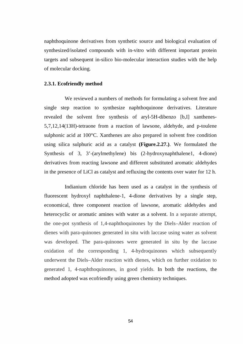
2.3.1. Ecofriendly method
We reviewed a numbers of methods for formulating a solvent free and
single step reaction to synthesize naphthoquinone derivatives. Literature
revealed the solvent free synthesis of aryl-5H-dibenzo [b,I] xanthenes-
5,7,12,14(13H)-tetraone from a reaction of lawsone, aldehyde, and p-toulene
sulphonic acid at 100°C. Xanthenes are also prepared in solvent free condition
using silica sulphuric acid as a catalyst (Figure.2.27.). We formulated the
Synthesis of 3, 3’-(arylmethylene) bis (2-hydroxynaphthalene1, 4-dione)
derivatives from reacting lawsone and different substituted aromatic aldehydes
in the presence of LiCl as catalyst and refluxing the contents over water for 12 h.
Indianium chloride has been used as a catalyst in the synthesis of
fluorescent hydroxyl naphthalene-1, 4-dione derivatives by a single step,
economical, three component reaction of lawsone, aromatic aldehydes and
heterocyclic or aromatic amines with water as a solvent. In a separate attempt,
the one-pot synthesis of 1,4-naphthoquinones by the Diels–Alder reaction of
dienes with para-quinones generated in situ with laccase using water as solvent
was developed. The para-quinones were generated in situ by the laccase
oxidation of the corresponding 1, 4-hydroquinones which subsequently
underwent the Diels–Alder reaction with dienes, which on further oxidation to
generated 1, 4-naphthoquinones, in good yields. In both the reactions, the
method adopted was ecofriendly using green chemistry techniques.
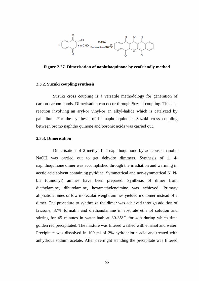
55
Figure 2.27. Dimerisation of naphthoquinone by ecofriendly method
2.3.2. Suzuki coupling synthesis
Suzuki cross coupling is a versatile methodology for generation of
carbon-carbon bonds. Dimerisation can occur through Suzuki coupling. This is a
reaction involving an aryl-or vinyl-or an alkyl-halide which is catalyzed by
palladium. For the synthesis of bis-naphthoquinone, Suzuki cross coupling
between bromo naphtho quinone and boronic acids was carried out.
2.3.3. Dimerisation
Dimerisation of 2-methyl-1, 4-naphthoquinone by aqueous ethanolic
NaOH was carried out to get dehydro dimmers. Synthesis of 1, 4-
naphthoquinone dimer was accomplished through the irradiation and warming in
acetic acid solvent containing pyridine. Symmetrical and non-symmetrical N, N-
bis (quinonyl) amines have been prepared. Synthesis of dimer from
diethylamine, dibutylamine, hexamethyleneimine was achieved. Primary
aliphatic amines or low molecular weight amines yielded monomer instead of a
dimer. The procedure to synthesize the dimer was achieved through addition of
lawsone, 37% formalin and diethanolamine in absolute ethanol solution and
stirring for 45 minutes in water bath at 30-35°C for 4 h during which time
golden red precipitated. The mixture was filtered washed with ethanol and water.
Precipitate was dissolved in 100 ml of 2% hydrochloric acid and treated with
anhydrous sodium acetate. After overnight standing the precipitate was filtered
56
and dried at 50°C. Alternatively, ammonium metavanadate was also used as
catalyst for the dimerisation of lawsone into lawsone dimer in dilute perchloric
acid.
Dimerisation of 1,4-naphthoquinone reaction with lead tetraacetate in
acetic acid gives 2-methyl- 3, 3″ (1, 4-naphthoquinone), 3, 3″ (2- methyl- 3, 3″
(1,4-naphthoquinone) and 2-acetoxy-2’-methyl 3,3″(1,4-naphthoquinone).
Hydrolysis with methanolic and ethanolic sulphuric acid of 2-acetoxy-2’-methyl-
3, 3’ (1, 4-naphthoquione) gives 2-methoxy-2’methyl-3, 3’ (1, 4-
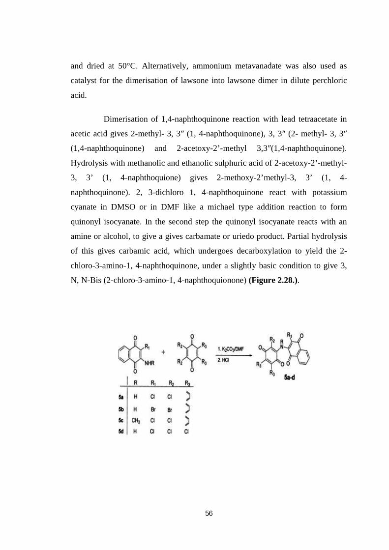
naphthoquinone). 2, 3-dichloro 1, 4-naphthoquinone react with potassium
cyanate in DMSO or in DMF like a michael type addition reaction to form
quinonyl isocyanate. In the second step the quinonyl isocyanate reacts with an
amine or alcohol, to give a gives carbamate or uriedo product. Partial hydrolysis
of this gives carbamic acid, which undergoes decarboxylation to yield the 2-
chloro-3-amino-1, 4-naphthoquinone, under a slightly basic condition to give 3,
N, N-Bis (2-chloro-3-amino-1, 4-naphthoquionone) (Figure 2.28.).
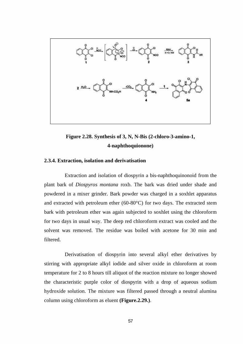
57
Figure 2.28. Synthesis of 3, N, N-Bis (2-chloro-3-amino-1,
4-naphthoquionone)
2.3.4. Extraction, isolation and derivatisation
Extraction and isolation of diospyrin a bis-naphthoquinonoid from the
plant bark of Diospyros montana roxb. The bark was dried under shade and
powdered in a mixer grinder. Bark powder was charged in a soxhlet apparatus
and extracted with petroleum ether (60-80°C) for two days. The extracted stem
bark with petroleum ether was again subjected to soxhlet using the chloroform
for two days in usual way. The deep red chloroform extract was cooled and the
solvent was removed. The residue was boiled with acetone for 30 min and
filtered.
Derivatisation of diospyrin into several alkyl ether derivatives by
stirring with appropriate alkyl iodide and silver oxide in chloroform at room
temperature for 2 to 8 hours till aliquot of the reaction mixture no longer showed
the characteristic purple color of diospyrin with a drop of aqueous sodium
hydroxide solution. The mixture was filtered passed through a neutral alumina
column using chloroform as eluent (Figure.2.29.).
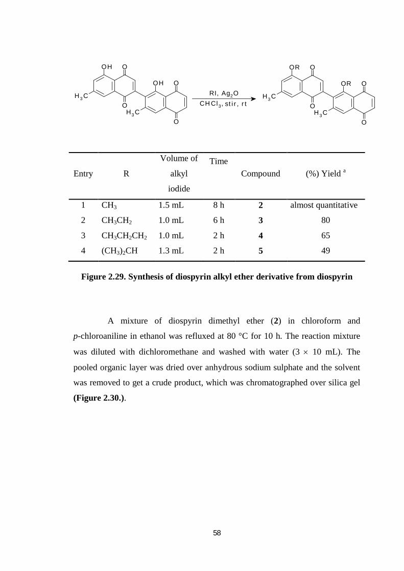
58
OH
CH3
O
O
OH
CH3
O
O
OR
CH3
O
O
OR
CH3
O
O
RI, Ag2OCHCl3, stir, rt
Entry R
Volume of
alkyl
iodide
Time
Compound (%) Yield a
1 CH3 1.5 mL 8 h 2 almost quantitative
2 CH3CH2 1.0 mL 6 h 3 80
3 CH3CH2CH2 1.0 mL 2 h 4 65
4 (CH3)2CH 1.3 mL 2 h 5 49
Figure 2.29. Synthesis of diospyrin alkyl ether derivative from diospyrin
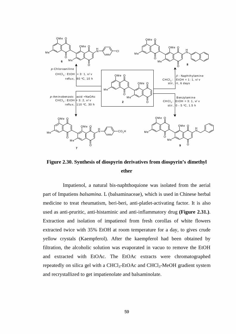
A mixture of diospyrin dimethyl ether (2) in chloroform and
p-chloroaniline in ethanol was refluxed at 80 °C for 10 h. The reaction mixture
was diluted with dichloromethane and washed with water (3 10 mL). The
pooled organic layer was dried over anhydrous sodium sulphate and the solvent
was removed to get a crude product, which was chromatographed over silica gel
(Figure 2.30.).
59
O
O
Me
OMe
O
O
Me
OMe
ClNH
O
O
Me
OMe
O
O
Me
OMe
NH
O
O
Me
OMe
O
O
Me
OMe
NH
O
O
Me
OMe
O
O
Me
OMe
CO2H
O
O
Me
OMe
O
O
Me
OMe
NH
6
7
8
9
2
p-Chloroaniline
CHCl3 : EtOH = 3: 1, v/vreflux, 80 oC, 10 h
reflux, 110 oC, 30 hCHCl3 : EtOH = 3: 2, v/v
p-Aminobenzoic acid +NaOAc
CHCl3 : EtOH = 1: 1, v/vstir, rt, 6 days
- Naphthylamine
CHCl3 : EtOH = 3: 1, v/vstir, 0 - 5 oC, 1.5 h
Benzylamine
Figure 2.30. Synthesis of diospyrin derivatives from diospyrin’s dimethyl
ether
Impatienol, a natural bis-naphthoquione was isolated from the aerial
part of Impatiens balsamina. L (balsaminaceae), which is used in Chinese herbal
medicine to treat rheumatism, beri-beri, anti-platlet-activating factor. It is also
used as anti-pruritic, anti-histaminic and anti-inflammatory drug (Figure 2.31.).
Extraction and isolation of impatienol from fresh corollas of white flowers
extracted twice with 35% EtOH at room temperature for a day, to gives crude
yellow crystals (Kaempferol). After the kaempferol had been obtained by
filtration, the alcoholic solution was evaporated in vacuo to remove the EtOH
and extracted with EtOAc. The EtOAc extracts were chromatographed
repeatedly on silica gel with a CHCl3-EtOAc and CHCl3-MeOH gradient system
and recrystallized to get impatienolate and balsaminolate.
60
Figure 2.31. Structure of Impatienol
2.3.5. Synthesis of naphthoquinones
Synthesis of different heterocyclic naphthoquinone can be done
through cyclo-addition reaction with 1, 4-naphthoquinone, isatin or ninhydrin,
alkene, (Chalcone or β-arylnitrostyrene) with different amino acids. It is a
simple, fast and single pot reaction resulting in good yield. Reaction conditions
are very simple. The purification of the product is also simple method. 2-Amino-
5, 10, 15, 20-tetraphenylporphyrinato) nickel (II) reacts with 1,4-
benzoquinone,1,4-naphthoquinone and 2-hydroxy-1,4-naphthoquinone, in the
presence of a catalytic amount of sulfuric acid, to afford new porphyrin–quinone
dyads and p-extended heterocyclic fused porphyrin derivatives.
Aryliodonium ylides of 2-hydroxy-1, 4-naphthoquinone react with
amines in refluxing dichloromethane to afford good yields of indanedione
2-carboxamides, through a ring-contraction and R,R-dioxoketene formation
reaction. These amides exist in solution in an unusual enol-amide form. In
contrast, the same reactants in a copper-catalyzed reaction afford arylamines and
3-iodo-4-hydroxy-1,2-naphthoquinone.
Addition of tetrahydrofuran to Compound 2 (Figure 2.32.) and LiAlH4
for 30 min and finally oxidation with CAN/CrO3 [added with stirring] for an h at
61
room temperature led to synthesis of novel derivatives of naphthoquinones in
good yields.
Figure 2.32. Preparation of hetero-1, 4-naphthoquinone
2.3.6. Methodology for biology and molecular docking
For biological evaluation standard procedures were adopted.
2.3.6.1 Plasmid relaxation assay
For enzyme assay plasmid relaxation method was selected. It is a well
known method for the enzyme assay especially for DNA topoisomerase-I. Assay
was run using agarose gel electrophoresis. Gel prepared from agarose, distilled
water and TAE. Then agarose was boiled in hot water bath until the solution
becomes clear and cooled to about 50-55˚C. The melted agarose solution was
poured into the casting tray and cooled until it is solid and the gel was loaded in
the electrophoresis chamber with 1X TAE buffer at a constant voltage of 5-6
V/cm for 2-8 hrs. Different concentrations of compounds were added with

62
enzyme and DNA control. DNA was stained by soaking in 0.5 µg/ ml of ethidium
bromide solution, visualized in U.V. transilluminator and photographed.
Assays have differentiated into two methods: 1) Preincubation and 2)
simultaneous. To investigate whether these compounds interact directly with the
enzyme, it was preincubated with these compounds at different concentrations
for 5 min at 37°C before the addition of substrate DNA. In Simultaneous assay
addition of enzyme, compounds and DNA simultaneously. In simultaneous
assay compounds formed DNA-enzyme-compound stable complex.
2.3.6.2. MTT assay
The MTT assay is a colorimetric assay for measuring the activity of
cellular enzymes that reduce the tetrazolium dye, MTT, to its
insoluble formazan, giving a purple color. Other closely related tetrazolium dyes
including XTT, MTS and the WSTs, are used in conjunction with the
intermediate electron acceptor, 1-methoxy PMS. With WST-1, which is cell-
impermeable, reduction occurs outside the cell via plasma membrane electron
transport. These assays measure cellular metabolic activity via NAD (P)
H-dependent cellular oxidoreductase enzymes and may, under defined
conditions, reflect the number of viable cells (cell proliferation). Tetrazolium
dye assays can also be used to measure cytotoxicity (loss of viable cells) or
cytostatic activity (shift from proliferative to resting status) of potential
medicinal agents and toxic materials. MTT Assay usually done under dark area
since MTT reagent is sensitive to light.
2.3.7. Molecular docking
Molecular docking is a method which predicts the preferred orientation
of one molecule to a second when bound to each other to form a stable complex.
Knowledge of the preferred orientation in turn may be used to predict the
63
strength of association or binding affinity between the two molecules using for
example scoring functions.
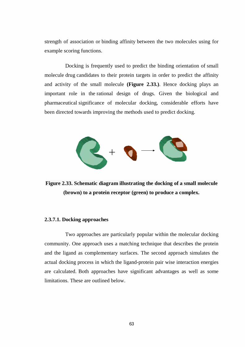
Docking is frequently used to predict the binding orientation of small
molecule drug candidates to their protein targets in order to predict the affinity
and activity of the small molecule (Figure 2.33.). Hence docking plays an
important role in the rational design of drugs. Given the biological and
pharmaceutical significance of molecular docking, considerable efforts have
been directed towards improving the methods used to predict docking.
Figure 2.33. Schematic diagram illustrating the docking of a small molecule
(brown) to a protein receptor (green) to produce a complex.
2.3.7.1. Docking approaches
Two approaches are particularly popular within the molecular docking
community. One approach uses a matching technique that describes the protein
and the ligand as complementary surfaces. The second approach simulates the
actual docking process in which the ligand-protein pair wise interaction energies
are calculated. Both approaches have significant advantages as well as some
limitations. These are outlined below.

64
2.3.7.2 Shape complementarity
Geometric matching shape complementarity methods describe the
protein and ligand as a set of features that make them dockable. These features
may include molecular surface complementary surface descriptors. In this case,
the receptor’s molecular surface is described in terms of its solvent-accessible
surface area and the ligand’s molecular surface is described in terms of its
matching surface description. The complementarity between the two surfaces
amounts to the shape matching description that may help finding the
complementary pose of docking the target and the ligand molecules. Another
approach is to describe the hydrophobic features of the protein using turns in the
main-chain atoms. Yet another approach is to use a Fourier shape descriptor
technique. Whereas the shape complementarity based approaches are typically
fast and robust, they cannot usually model the movements or dynamic changes
in the ligand/ protein conformations accurately, although recent developments
allow these methods to investigate ligand flexibility. Shape complementarity
methods can quickly scan through several thousand ligands in a matter of
seconds and actually figure out whether they can bind at the protein’s active site,
and are usually scalable to even protein-protein interactions. They are also much
more amenable to pharmacophore based approaches, since they use geometric
descriptions of the ligands to find optimal binding.
2.3.7.3 Simulation
The simulation of the docking process as such is a much more
complicated process. In this approach, the protein and the ligand are separated by
some physical distance, and the ligand finds its position into the protein’s active site
after a certain number of “moves” in its conformational space. The moves
incorporate rigid body transformations such as translations and rotations, as well as
internal changes to the ligand’s structure including torsion angle rotations. Each of
these moves in the conformation space of the ligand induces a total energetic cost of
65
the system, and hence after every move the total energy of the system is calculated.
The obvious advantage of the method is that it is more amenable to incorporate
ligand flexibility into its modeling whereas shape complementarity techniques have
to use some ingenious methods to incorporate flexibility in ligands.
Another advantage is that the process is physically closer to what
happens in reality, when the protein and ligand approach each other after
molecular recognition. A clear disadvantage of this technique is that it takes
longer time to evaluate the optimal pose of binding since they have to explore a
rather large energy landscape. However grid-based techniques as well as fast
optimization methods have significantly ameliorated these problems.
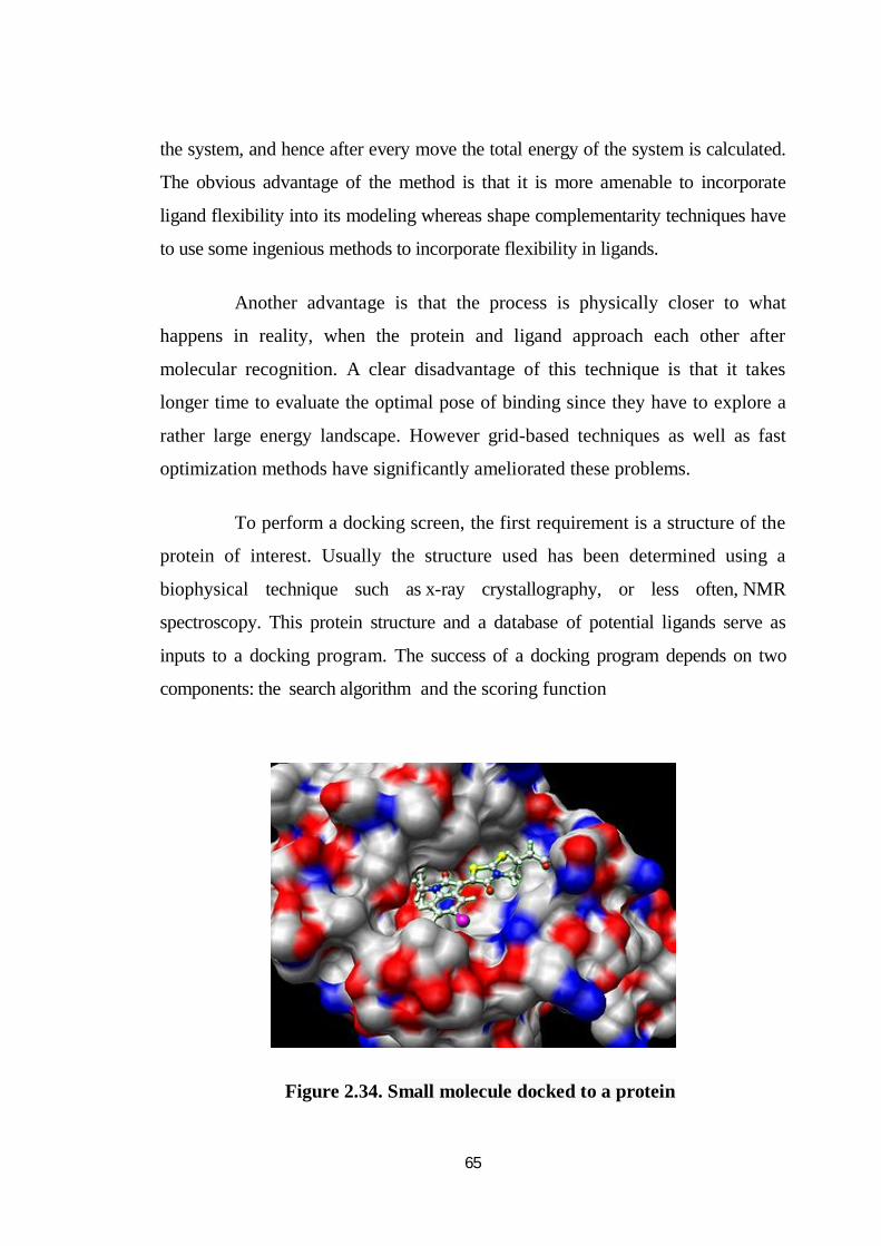
To perform a docking screen, the first requirement is a structure of the
protein of interest. Usually the structure used has been determined using a
biophysical technique such as x-ray crystallography, or less often, NMR
spectroscopy. This protein structure and a database of potential ligands serve as
inputs to a docking program. The success of a docking program depends on two
components: the search algorithm and the scoring function
Figure 2.34. Small molecule docked to a protein
66
2.3.7.4. Search algorithm
The search space in theory consists of all possible orientations
and conformations of the protein paired with the ligand. However in practice
with current computational resources, it is impossible to exhaustively explore the
search space—this would involve enumerating all possible distortions of each
molecule (molecules are dynamic and exist in an ensemble of conformational
states) and all possible rotational and translational orientations of the ligand
relative to the protein at a given level of granularity. Most docking programs in
use account for a flexible ligand, and several attempt to model a flexible protein
receptor. Each "snapshot" of the pair is referred to as a pose.
A variety of conformational search strategies have been applied to the
ligand and to the receptor. These include:
systematic or stochastic torsional searches about rotatable bonds
molecular dynamics simulations
genetic algorithms to "evolve" new low energy conformations
2.3.7.5. Ligand flexibility
Conformations of the ligand may be generated in the absence of the
receptor and subsequently docked or conformations may be generated on-the-fly
in the presence of the receptor binding cavity, or with full rotational flexibility of
every dihedral angle using fragment based docking. Force field energy
evaluation are most often used to select energetically reasonable
conformations, but knowledge-based methods have also been used.
2.3.7.6. Receptor flexibility
Computational capacity has increased dramatically over the last decade
making possible the use of more sophisticated and computationally intensive
67
methods in computer-assisted drug design. However, dealing with receptor
flexibility in docking methodologies is still a thorny issue. The main reason
behind this difficulty is the large number of degrees of freedom that have to be
considered in this kind of calculations. Neglecting it, however, leads to poor
docking results in terms of binding pose prediction. Multiple static structures
experimentally determined for the same protein in different conformations are
often used to emulate receptor flexibility. Alternatively rotamer libraries of
amino acid side chains that surround the binding cavity may be searched to
generate alternate but energetically reasonable protein conformations.
2.3.7.7. Scoring function
The scoring function takes a pose as input and returns a number
indicating the likelihood that the pose represents a favorable binding interaction.
Most scoring functions are physics-based molecular mechanics force fields that
estimate the energy of the pose; a low (negative) energy indicates a stable
system and thus a likely binding interaction. An alternative approach is to derive
a statistical potential for interactions from a large database of protein-ligand
complexes, such as the Protein Data Bank, and evaluate the fit of the pose
according to this inferred potential.
There are a large number of structures from X-ray crystallography for
complexes between proteins and high affinity ligands, but comparatively fewer
for low affinity ligands as the later complexes tend to be less stable and therefore
more difficult to crystallize. Scoring functions trained with this data can dock
high affinity ligands correctly, but they will also give plausible docked
conformations for ligands that do not bind. This gives a large number of false
positive hits, i.e., ligands predicted to bind to the proteins that actually don’t
when placed together in a test tube.
68
One way to reduce the number of false positives is to recalculate the
energy of the top scoring poses using (potentially) more accurate but
computationally more intensive techniques such as Generalized Born or Poisson-
Boltzmann methods.
2.4 Conclusion from the literature
From the literature review we have concluded that green method or
solvent free reaction is good approach for the dimerisation of naphthoquinones.
Now-a-days green methods are the first choice of the chemist due to its benefit
for environment safety, easy to handle, simple, one step method, less use of
hazardous chemicals, less use of equipments, energy and time. Catalyst plays an
important role in the reaction. Some catalyst increase yield, reaction rate and
gives fair product. From the previous literature we came to know about the
different catalyst which have used for the synthesis of bis-naphthoquinone. LiCl,
p-TSA, Indinium Chloride, Triethylamine, silica sulphuric acid, ammonium
meta vandate gives dimer of 1,4-naphthoquinone. In some reactions alkali
(NaOH, KOH) and amines (dibutylamine, piperidine, morpholino, diethylamine)
are also used as a catalyst. Extraction, isolation and derivatisation of bis-
naphthoquinone, diospyrin was carried out from the the bark of Diospyros
montana, a indigenous plant from family Ebenaeceae. This plant is found mainly
in the forests of Bihar, West Bengal, Tamilnadu and other states of India.
Extraction and isolation of diospyrin was carried out through slight
modifications on the reported procedure. The first total synthesis of diospyrin
has been reported through Suzuki coupling method by Kenji Mori research
group. Consecutively, more latest synthesis of novel diospyrin analogues via a
Suzuki cross coupling between bromonaphthoquinones and aryl or naphthyl
boronic acids in presence of tetrakis (triphenylphosphine) palladium (0) as
catalyst were also reported by Ivan R.Green and co-workers. Derivatisation of
diospyrin with alkyl iodide and silver oxide and further derivatisation of alkyl
69
ether derivative by different methods also have been reported. Glucoside
derivatives of diospyrin were also prepared. We have attempted further
derivatisation of diospyrin alkyl ether into novel amino acid ester derivatives
through modification on the published procedure. Further details are provided in
following chapters on experimental section.
Mamegakinone dimethyl ether was prepared by Stille-type coupling
reaction with the bromo-naphthoquinone in presence of bis( triphenylphosphine)
palladium (II) chloride. Dimerisation of Bi-vitamin K3, 3, 3’-bijuglone,in
presence of AgO-40% HNO3. Bi-ramentaceone, a bis-naphthoquinone was
prepared by oxidative coupling of 4-methoxy-1-naphthol compounds using lead
oxide or silver oxide with 65% of HNO3.
In literature review we studied different chemistries and synthetic
procedures on different naphthoquinones and the biological evaluations, mostly
anti-leishmanial, anti-cancer, and anti-bacterial, anti-fungal and anti-tumor
activities of the same. Our two-fold aim was to synthesize novel naphthoquinone
derivatives and subsequently conduct biological screening. For the fulfillment of
this aim we had a thorough study of previous literature for both chemistry and
selection of disease and its protein target against which these novel compounds
were to be tested.
2.5. Conclusion from Biological methods
From the literature we studied different reported activities against
protein targets for naphthoquinone and bis-naphthoquinones. Some targets were
already established for naphthoquinone and bis-naphthoquinones. From review
we found that some naphthoquinones were suitable for leishmanial
Topoisomerase I-DNA cleavage complex, target was for anti-leishmanial studies
and some other for human CDC25, which is a molecular target for anticancer
studies. For docking studies we selected GOLD, GLIDE and Autodock Vina
70
docking software. All the softwares are well known and widely accepted by
industrial as well as academic community. For biological evaluation we
concluded to carry on MTT assay for cytotoxicity.
2.5.1 Significance of MTT assay
Tetrazolium dye reduction is dependent on NAD (P)H-dependent
oxidoreductase enzymes largely in the cytosolic compartment of the
cell.[6][7] Therefore, reduction of MTT and other tetrazolium dyes increases with
cellular metabolic activity due to elevated NAD(P)H flux. Resting cells such as
thymocytes and splenocytes that are viable but metabolically quiet reduce very
little MTT. In contrast, rapidly dividing cells exhibit high rates of MTT
reduction. It is important to keep in mind that assay conditions can alter
metabolic activity and thus tetrazolium dye reduction without affecting cell
viability and that different tetarazolium dyes will give different results
depending on whether they are reduced intracellularly (MTT, MTS) or
extracellularly (WST-1).