chapter 2 › mediafiles › texts › 5 › ... · 17 chapter 2 production of membrane proteins in...
TRANSCRIPT
17
Chapter 2
Production of Membrane Proteins in Escherichia coli and Lactococcus lactis
Eric R. Geertsma and Bert Poolman
Abstract
As the equivalent to gatekeepers of the cell, membrane transport proteins perform a variety of critical functions. Progress on the functional and structural characterization of membrane proteins is slowed due to problems associated with their (heterologous) overexpression. Often, overexpression fails or leads to aggregated material from which the production of functionally refolded protein is challenging. It is still difficult to predict whether a given membrane protein can be overproduced in a functional competent state. As a result, the most straightforward strategy to set up an overexpression system is to screen a mul-titude of conditions, including the comparison of homologues, type and location of (affinity) tags, and distinct expression hosts. Here, we detail methodology to rapidly establish and optimize (membrane) protein expression in Escherichia coli and Lactococcus lactis.
Key words: Arabinose promoter, Escherichia coli, folding indicator, GFP, Lactococcus lactis, mem-brane proteins, nisin-controlled expression, overexpression, PBAD
Although for the elementary steps in the biogenesis of membrane proteins, which are membrane targeting, membrane insertion, and assembly and folding, the crucial components are known (reviewed in (1)), our current understanding of the overall process and the factors involved are far from complete. Predicting whether a given membrane protein can be overproduced in a functional competent state is currently not possible, and bottlenecks have not been identified that could guide a rational troubleshooting strategy should overexpression fail. To date, membrane protein biogenesis has been mostly studied from the perspective of the known components involved in targeting, membrane insertion, and folding. In general, this is done using a limited number of
1. Introduction
I. Mus-Veteau (ed.), heterologous Expression of Membrane Proteins, Methods in Molecular Biology, vol. 601DOI 10.1007/978-1-60761-344-2_2, © Humana Press, a part of Springer Science + Business Media, LLC 2010

18 Geertsma and Poolman
small and simple model proteins, while systematic genomewide studies have indicated that overexpression of complex membrane proteins with multiple transmembrane segments is most challenging (2–4). Only recently efforts have been made to understand the complete process in the context of a cell by determining the physiological responses underlying (un)successful overproduction of (complex) membrane proteins (5,6).
As a consequence, if one wishes to obtain structural and functional information on a certain membrane protein that is not abundant in any natural source, a convenient strategy is to screen a wide range of conditions. Established initial variables involve the screening of homologues (7,8), varying the type and location of affinity tags and domains (9,10), and screening multiple-expression hosts (11,12). In practice, for prokaryotes screening is restricted to several Escherichia coli strains, which offers far less variation than the screening of different species.
The gram-positive bacterium Lactococcus lactis is an attractive alternative to E. coli and yeast-based membrane protein expres-sion systems (13,14). Overexpression of membrane proteins in L. lactis leads mostly to well-folded, membrane-inserted material; inclusion bodies of aggregated material are rare. The lactococcal membrane is easily solubilized with a wide range of detergents. Growth of L. lactis proceeds rapidly at 30°C and does not require aeration. The nisin-inducible controlled expression (NICE) system (15) allows reproducible and modulatable expression from low to high levels. Previous difficulties associated with direct cloning in L. lactis (10) have been overcome by the high-throughput compatible vector backbone exchange (VBEx) procedure (16). As L. lactis is auxotrophic for multiple amino acids, adjustment of the lactococcal chemically defined medium (17) allows incorpora-tion of labeled amino acid analogues into proteins (44, 45). Concerning membrane protein expression, there are five impor-tant differences between E. coli and L. lactis. First, the composi-tion of the machinery involved in membrane protein insertion: L. lactis contains two paralogs of the Oxa/YidC/Alb family of membrane-inserted chaperones (18), whereas E. coli contains one (YidC). Lactococcus lactis does not contain homologues of the Sec translocon accessory proteins SecDF (19) and SecM (20). Finally, substantial differences in structure are observed between lacto-coccal and E. coli homologues of YidC and SecE (20,21). Second, the lipid composition of the membrane is very different: The major lipid component of the E. coli cytoplasmic membrane is phos-phatidylethanolamine (PE), which is absent in L. lactis. Instead, anionic glycolipids and phosphoglycolipids are the major constit-uents in L. lactis (22). The third difference involves codon usage (23); L. lactis DNA is relatively AT rich (64% AT, compared to 50% AT in E. coli). The fourth difference is the absence of disul-fide isomerase homologues in L. lactis (24), albeit that disulfides

19Production of Membrane Proteins in Escherichia coli and Lactococcus lactis
are readily formed either spontaneously or via an alternative route in secreted proteins (25–27). The final difference is that some proteins toxic to one host do not affect the physiology of the other host (C. Mulligan and E.R. Geertsma, March 2007). In addi-tion, although fewer attempts have been made than in E. coli, overexpression of eukaryotic membrane proteins in L. lactis is rela-tively successful (28).
To establish an overexpression system rapidly, several homologues, types and location of affinity tags, and expression hosts need to be screened. Due to the large number of plasmids required for this strategy, it will be advantageous to avoid low-throughput tech-niques and implement efficient cloning procedures such as ligation-independent cloning (LIC) (29). LIC is less restricted in the design of the sequences flanking the genes than other high-throughput cloning techniques, like Gateway (30) or the univector plasmid-fusion system (31). Therefore, the cloning-related sequences attached to the recombinant protein can be minimized, which might be advantageous for structural and functional analysis.
The LIC procedure described here, outlined in Fig. 2.1a, involves linearization of the vector by a unique SwaI site in the middle of the LIC cassette. Using the 3¢ to 5¢ exonuclease activity of T4 DNA polymerase, single-stranded overhangs are generated. By performing the exonuclease treatment in the presence of dCTP (deoxycytidine triphosphate), a nucleoside not present in the 3¢ sequences adjacent to the SwaI site, the removal of bases halts once the first dCMP (deoxycytidine monophosphate) is encountered, thereby creating overhangs of a defined length. Complementary single-stranded overhangs are created in a similar way in the polymerase chain reaction (PCR) product to be cloned (i.e., exonuclease treatment in the presence of dGTP (deoxy-guanosine triphosphate)). On mixing the T4 DNA polymerase-treated vector and PCR product, a stable heteroduplex is formed, and the mixture can be transformed with high efficiency to E. coli.
The efficiencies for direct cloning in L. lactis are low, and LIC is virtually not possible. This bottleneck in cloning has been overcome by the VBEx procedure (16). Using VBEx, the initial LIC step can be performed with high efficiency in E. coli while the final expression vector that is generated can be kept devoid of alien E. coli elements. The VBEx procedure, outlined in Fig. 2.1b, relies on the bisection of a bona fide plasmid of the expression host into two parts, thereby separating the selection marker from the origin of replication. For L. lactis, pNZxLIC, a LIC-compatible derivative of the well-established expression vector pNZ8048 (15) was accordingly bisected. The segment contain-ing the origin of replication was fused to a sequence coding for an erythromycin selection marker, yielding plasmid pERL, which can be maintained in L. lactis. The other segment of pNZxLIC,
1.1. High-Throughput Cloning in E. coli and L. lactis

20 Geertsma and Poolman
containing the chloramphenicol resistance gene and LIC sequence, was fused to the backbone of an E. coli vector (containing an E. coli origin of replication and b-lactamase resistance gene). The resulting plasmid pRExLIC, allows the LIC manipulation to take place in E. coli.
Fig. 2.1. High-throughput cloning in recalcitrant bacteria using LIC and VBEx. (a) Outline of the LIC procedure. Gene X is amplified using primers holding LIC-specific overhangs. The plasmid is linearized by SwaI restriction in the LIC cassette. Single-stranded overhangs on the PCR product and vector are generated using T4 DNA polymerase. The complementary overhangs of PCR product and vector anneal on mixing. The resulting heteroduplex is transformed efficiently to Escherichia coli. (b) Outline of the VBEx strategy. After introduction of two distinct SfiI sites, the Lactococcus lactis expression vector pNZxLIC was bisected. Plasmid pERL received the pSH71 replicon, which was fused to the erythromycin marker. Plasmid pRExLIC received the chloramphenicol marker and LIC sequence, which were fused to the E. coli pBR322 replicon and b-lactamase marker. As these vectors are available, in practice the cloning involves the submission of vector pRExLIC to the LIC procedure (depicted in a). Subsequently, the VBEx procedure is applied: The pNZxLIC vector is restored by mixing pERL and pRExLIC-gene X, digestion with SfiI, ligation, and selection on the ability to replicate in L. lactis (the presence of the pSH71 replicon) in the presence of chloramphenicol. (c) The SfiI sites flanking both segments of the vectors yield different, nonpalindromic overhangs, thereby greatly reducing the number of possible ligation products. (Adapted from (16)).

21Production of Membrane Proteins in Escherichia coli and Lactococcus lactis
Rapid and facile regeneration of the L. lactis expression vector is enabled as the relevant segments of the pRExLIC and pERL vectors are flanked with distinct SfiI sites. DNA cleavage by SfiI generates a 3¢ overhang that can be composed of any combination of three nucleotides. The two SfiI sites used yield different, incom-patible overhangs (Fig. 2.1c). Each segment of the pNZxLIC vector has unique selectable properties: (1) the ability to replicate a plasmid in L. lactis and (2) the resistance to chloramphenicol. Thus, on mixing a pRExLIC derivative and pERL, digestion by SfiI, followed by ligation and transformation to L. lactis in com-bination with selection of clones using chloramphenicol, the pNZxLIC derivative can be exclusively recovered with high effi-ciencies. DNA sequences from virtually all sequenced genomes are compatible with VBEx as SfiI sites are rare (16). Furthermore, genes containing internal SfiI sites are not necessarily excluded as 64 different 3¢ overhangs can be generated after SfiI digestion. Internal SfiI sites with 3¢ overhangs not matching those of the vector will not form a bottleneck in the procedure.
Following initial screening for expression, systematic optimization of the production conditions to increase the amount of well-folded protein inserted into the membrane is often required. As indicated, membrane protein biogenesis requires several additional steps and components beyond translation. Exceeding the capacity of the cell to process the nascent membrane protein correctly may reduce the final yield of well-folded material. This is especially true for E. coli as mem-brane protein production in this host is regularly accompanied by inclusion body formation. Adjustment of the expression rate is most easily done by varying the inducer concentration for well-tunable promoters, such as the E. coli PBAD (32) or the lactococcal PnisA (15). In addition, variation of the growth temperature during induction is an important parameter to affect the expression rate (33,34).
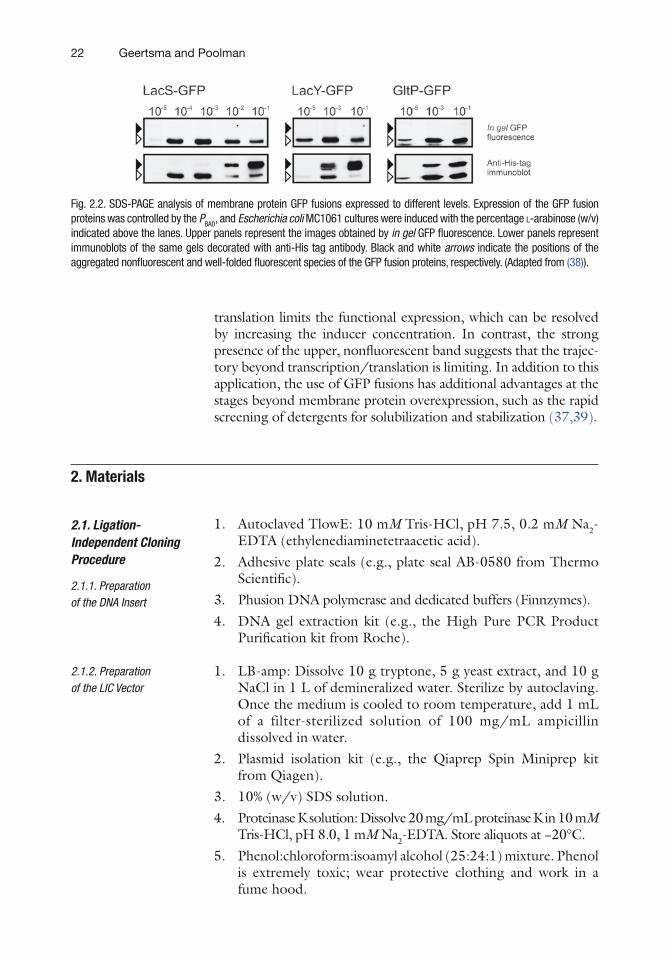
While traditionally optimization of functional expression involved time-consuming steps such as the isolation of membrane vesicles and activity assays, this process has been greatly accelerated by the use of green fluorescent protein (GFP), fused to the C-terminus of the target protein, as folding reporter. Proper matu-ration of GFP in E. coli, leading to a fluorescent species that main-tains its folded state during sodium dodecyl sulfate polyacrylamide gel electrophoresis (SDS-PAGE), depends highly on the correct folding of its fusion partner (35–38). Consequently, only correctly folded target yields a fluorescent fusion protein that has a higher electrophoretic mobility than the misfolded fusion protein. Plain SDS-PAGE and subsequent immunoblotting allow simultaneous quantification of both the well-folded and aggregated protein pro-duced (Fig. 2.2) (38). This facilitates the assignment of bottlenecks limiting the functional overexpression. For instance, the absence of the upper, nonfluorescent band suggests that transcription or
1.2. Optimization of Membrane Protein Overexpression
22 Geertsma and Poolman
translation limits the functional expression, which can be resolved by increasing the inducer concentration. In contrast, the strong presence of the upper, nonfluorescent band suggests that the trajec-tory beyond transcription/translation is limiting. In addition to this application, the use of GFP fusions has additional advantages at the stages beyond membrane protein overexpression, such as the rapid screening of detergents for solubilization and stabilization (37,39).
1. Autoclaved TlowE: 10 mM Tris-HCl, pH 7.5, 0.2 mM Na2-EDTA (ethylenediaminetetraacetic acid).
2. Adhesive plate seals (e.g., plate seal AB-0580 from Thermo Scientific).
3. Phusion DNA polymerase and dedicated buffers (Finnzymes).4. DNA gel extraction kit (e.g., the High Pure PCR Product
Purification kit from Roche).
1. LB-amp: Dissolve 10 g tryptone, 5 g yeast extract, and 10 g NaCl in 1 L of demineralized water. Sterilize by autoclaving. Once the medium is cooled to room temperature, add 1 mL of a filter-sterilized solution of 100 mg/mL ampicillin dissolved in water.
2. Plasmid isolation kit (e.g., the Qiaprep Spin Miniprep kit from Qiagen).
3. 10% (w/v) SDS solution.4. Proteinase K solution: Dissolve 20 mg/mL proteinase K in 10 mM
Tris-HCl, pH 8.0, 1 mM Na2-EDTA. Store aliquots at −20°C.5. Phenol:chloroform:isoamyl alcohol (25:24:1) mixture. Phenol
is extremely toxic; wear protective clothing and work in a fume hood.
2. Materials
2.1. Ligation-Independent Cloning Procedure
2.1.1. Preparation of the DNA Insert
2.1.2. Preparation of the LIC Vector
Fig. 2.2. SDS-PAGE analysis of membrane protein GFP fusions expressed to different levels. Expression of the GFP fusion proteins was controlled by the PBAD, and Escherichia coli MC1061 cultures were induced with the percentage l-arabinose (w/v) indicated above the lanes. Upper panels represent the images obtained by in gel GFP fluorescence. Lower panels represent immunoblots of the same gels decorated with anti-His tag antibody. Black and white arrows indicate the positions of the aggregated nonfluorescent and well-folded fluorescent species of the GFP fusion proteins, respectively. (Adapted from (38)).

23Production of Membrane Proteins in Escherichia coli and Lactococcus lactis
6. Chloroform, analytical grade. Chloroform is irritating and a carcinogen; wear protective clothing and work in a fume hood.
7. 96% EtOH, analytical grade, precooled at −20°C. 8. 3M Na-acetate, pH 5.3; adjust the pH with concentrated
acetic acid. 9. 70% EtOH, analytical grade, precooled at −20°C. 10. SpeedVac (Savant). 11. SwaI and dedicated buffer. 12. Chemically competent E. coli MC1061. Protocols for the
preparation of chemically competent E. coli can be found in (40).
13. LB-amp agar plates: Dissolve 10 g tryptone, 5 g yeast extract, and 10 g NaCl in 1 L of demineralized water and add 15 g agar. Sterilize by autoclaving. Once the medium is cooled to 50–60°C, add 1 mL of a filter-sterilized solution of 100 mg/mL ampicillin dissolved in water; mix well and pour the medium in sterile Petri dishes.
1. T4 DNA polymerase and dedicated buffer. 2. 25 mM dCTP: Dilute a 100 mM dCTP stock in sterile
Milli-Q. Store aliquots at −20°C. 3. 25 mM dGTP: Dilute a 100 mM dGTP stock in sterile
Milli-Q. Store aliquots at −20°C. 4. Heat block.
1. Autoclaved Milli-Q. 2. Sterile toothpicks. 3. 96-well PCR plates. 4. Aluminum sealing films (e.g., PCR-AS-200 from Axygen). 5. Taq DNA polymerase and dedicated buffer. 6. dNTPs: Make a stock solution containing 10 mM of each
nucleotide. Store aliquots at −20°C. 7. Generic primers (see Note 1).
1. GM17-ery: Dissolve 37.25 g of M17 broth (Oxoid) (41) in 1 L of demineralized water. Sterilize by autoclaving. Once the medium is cooled to room temperature, add 25 mL sterile 20% (w/v) glucose and 1 mL of a filter-sterilized solution of 5 mg/mL erythromycin dissolved in water.
2. Resuspension buffer: 10 mM Tris-HCl, pH 8.1, 10 mM Na2-EDTA, 50 mM NaCl, and 20% (w/v) sucrose. Autoclave and store at 4°C. Supplement with 20 mg/mL lysozyme prior to use.
2.1.3. T4 DNA Polymerase Treatment of Vector and Insert
2.1.4. Verifying Clones by Colony PCR
2.2. Vector Backbone Exchange Procedure
2.2.1. Preparation of pERL

24 Geertsma and Poolman
1. SfiI and dedicated buffer.2. 50 mM ATP stock: 50 mM Na2-ATP, 50 mM MgSO4, and 50
mM phosphate buffer, pH 7.0. Adjust the pH to 6.5–7.0 with NaOH. Store aliquots at −20°C.
3. T4 DNA ligase and dedicated buffer.
1. Sterile electroporation cuvets (2 mm gap between the elec-trodes). The cuvets can be reused several times. Clean the cuvets after use with 70% EtOH and wash extensively with Milli-Q. Allow the cuvets to dry and sterilize the cuvets by 20 min expo-sure to an ultraviolet (UV) source. Immediately close the cuvets with the supplied cap.
2. Electroporation device.3. Rescue medium: Mix 1 volume of sterile M17 medium with
1 volume of sterile 1M sucrose, 1% (w/v) glucose, 40 mM MgCl2, 4 mM CaCl2.
4. SGM17-cam agar plates: Mix 37.25 g of M17 broth (Oxoid) and 15 g agar in 750 mL of demineralized water. Sterilize by autoclaving. Once the medium is cooled to about 60°C, add 250 mL sterile 2M sucrose, 25 mL 20% (w/v) glucose, and 1 mL of a filter-sterilized solution of 5 mg/mL chlorampheni-col dissolved in EtOH. Mix well and pour the medium in sterile Petri dishes.
5. Parafilm.
1. Sterile 87% (w/v) glycerol.2. Cryovials.3. TB-amp: Dissolve 24 g tryptone, 48 g yeast extract, and 17.2
mL 87% (w/v) glycerol in 0.9 L of demineralized water and autoclave. In addition, autoclave 100 mL of 170 mM KH2PO4, 720 mM K2HPO4. Mix the solutions prior to use and add 1 mL of 100 mg/mL ampicillin.
4. l-Arabinose: Filter-sterilize a 20% (w/v) l-arabinose stock.5. Screw-cap tubes compatible with the FastPrep-24 (e.g.,
screw-cap microtube 72.693 from Sarstedt).
1. GM17-cam: M17 medium supplemented with 0.5% (w/v) glucose and 5 mg/mL chloramphenicol after sterilization.
2. Nisin-containing supernatant of a L. lactis NZ9700 culture: Use a 1% (v/v) inoculum of a preculture of L. lactis NZ9700 on GM17 to inoculate 200 mL GM17 and incubate overnight at 30°C. Next day, pellet the cells, filtrate the supernatant using a 0.2-mm filter, and store the aliquots at −20°C.
2.2.2. Vector Backbone Exchange
2.2.3. Electrotransformation of L. lactis
2.3. Initial Characterization and Optimization of Protein Production
2.3.1. Cultivation and Induction of MP Overexpression in E. coli
2.3.2. Cultivation and Induction of MP Overexpression in L. lactis

25Production of Membrane Proteins in Escherichia coli and Lactococcus lactis
1. Glass beads with a diameter of about 0.1 mm (Sigma-Aldrich).
2. Disruption buffer: 50 mM phosphate buffer, pH 7.5, 10% (w/v) glycerol, and 1 mM MgSO4. Precool the solution on ice. Add 1 mM PMSF (phenylmethylsulfonyl fluoride) (from a 200 mM stock in EtOH (ethanol)) and 0.1 mg/mL DNase (deoxyribonuclease) prior to use.
3. FastPrep-24 device (MP Biomedicals).4. 5X protein sample buffer: 120 mM Tris-HCl, pH 6.8, 50%
(w/v) glycerol, 100 mM dithiothreitol (DTT), 2% (w/v) SDS, and 0.1% (w/v) bromophenol blue (see Note 2). Store ali-quots at −20°C.
1. SDS-PA gel: For a 10% running gel, mix 3.38 mL 30% acryl-amide/bis solution (cross-linker ratio 37.5:1), 2.54 mL 1.5M Tris-HCl, pH 8.8, 4.15 mL Milli-Q, 100 mL 10% (w/v) SDS, 40 mL 10% (w/v) APS (ammonium persulfate), and 10 mL TEMED. For the stacking gel, mix 0.68 mL 30% acrylam-ide/bis solution (cross-linker ratio 37.5:1), 1.13 mL 0.5M Tris-HCl, pH 6.8, 2.25 mL Milli-Q, 50 mL 10% (w/v) SDS, 30 mL 10% (w/v) APS, and 5 mL TEMED (see Note 2).
2. Precision Plus Protein Dual Color standard (Bio-Rad).3. LAS-3000 imaging system (Fujifilm).4. Electroblotting and immunodetection system with primary
antibody directed against a His-tag.
1. Design two sets of LIC primers for each gene to be cloned. The nLIC and cLIC primer sets will finally produce proteins with an N- or C-terminal affinity tag, respectively. The LIC strategy requires the presence of dedicated 5¢ extensions on the primers (indicated in Table 2.1). In addition, the gene-specific part of the primer should be designed so that it (1) does not contain the endogenous start or stop codon of the target gene; (2) is sufficiently long to anneal in a stable and selective way with the target DNA (in general, 20–25 nucle-otides suffice; see Note 3). The primer should not contain strong secondary structure elements that could interfere with the PCR (see Note 4). Keep the total length of the
2.3.3. Cell Disruption and Sample Preparation
2.3.4. SDS-PAGE, In Gel GFP Fluorescence, and Western Blotting
3. Methods
3.1. Ligation-Independent Cloning Procedure
3.1.1. Preparation of the DNA Insert

26 Geertsma and Poolman
primers below 50 residues to ensure sufficient quality and cost-efficiency.
2. For ordering, select the lowest synthesis scale offered by the supplier and mere desalting as the purification step. For large sets (>48 primers), order the primers in a 96-well plate.
3. On delivery, spin down the material and dissolve the primers to 100 mM in TlowE and make a substock of 5 mM. Use a transparent sticker to seal the plates. The 100 mM stock can be stored for several months at −20°C. Store the 5 mM stock at 4°C. Spin down the material before use.
4. PCR the desired open reading frames using the Phusion DNA polymerase, which combines high fidelity with high production (see Notes 5 and 6). Prepare a 50 mL reaction mix according to the manufacturer’s protocol and add the polymerase just prior to the start of the reaction. Place the sample in a PCR machine preheated to 98°C and start the reaction. A good starting point for a touchdown PCR program (see Note 7) isa. 60 s at 98°C.b. 10 s at 98°C.c. 15 s at 58.5°C (decrease 0.5°C per cycle).d. XX s at 72°C (15–30 s/kb).
Repeat steps 4a–4d 14 times.
e. 10 s at 98°C.f. 15 s at 51°C.g. XX s at 72°C (15–30 s/kb).
Repeat steps 4e–4g 14 times.
h. 300 s at 72°C.i. Forever at 10°C.Once the PCR is finished, add DNA loading buffer to the
sample and analyze all the material by TAE (Tris-acetate-EDTA) gel electrophoresis (see Note 8). Gel purify the band using a
Table 2.1 Primer extensions needed for ligation-independent cloning
Primer type 5¢ Extensions of primers
nLIC forward 5¢ AT GGT GAG AAT TTA TAT TTT CAA GGT
nLIC reverse 5¢ T GGG AGG GTG GGA TTT TCA TTA
cLIC forward 5¢ ATG GGT GGT GGA TTT GCT
cLIC reverse 5¢ TTG GAA GTA TAA ATT TTC

27Production of Membrane Proteins in Escherichia coli and Lactococcus lactis
commercial gel extraction kit according to the manufacturer’s protocol. Determine the DNA concentration spectrophotometri-cally (see Note 9). Store the material at −20°C.
1. Inoculate approximately 20 mL LB-amp with E. coli MC1061 containing one of the pBAD- or pRE-derived LIC vectors (Table 2.2). Cultivate overnight at 37°C with vigorous shaking.
2. Isolate the plasmid using a commercial plasmid isolation kit and elute the plasmid with TlowE. Remove remaining proteins by combining a proteinase K treatment with a phenol:chloroform extraction as described in steps 3–5.
3. Combine several fractions and adjust the volume to 300 mL. Supplement the sample with SDS to a final concentration of 0.5% and add proteinase K to a final concentration of 40 mg/mL. Incubate for 30 min at 60°C to digest remaining proteins.
3.1.2. Preparation of the LIC Vector
Table 2.2 Basic and additional LIC vectors
Vector name Protein sequenceProtein sequence after TEV protease cleavage Expression host
pBADnLIC M-His10-G-TEV site-protein G protein Escherichia coli
pBADcLIC MGGGFA-protein-TEV site-His10
MGGGFA-protein-ENLYFQ
E. coli
pBADcLIC-GFP MGGGFA-protein-TEV site-GFP-His10
MGGGFA-protein-ENLYFQ
E. coli
pBAD-OmpA-nLIC M-ssOmpAa-His10-G-TEV site-protein
G-protein E. coli
pREnLIC M-His10-G-TEV site-protein G-protein Lactococcus lactis
pREcLIC MGGGFA-protein-TEV site-His10
MGGGFA-protein-ENLYFQ
L. lactis
pREcLIC-GFP MGGGFA-protein-TEV site-GFP-His10
MGGGFA-protein-ENLYFQ
L. lactis
pRE-USP45-nLIC M-ssUSP45b-His10-G-TEV site-protein
G-protein L. lactis
assOmpA indicates the signal sequence of the E. coli OmpA protein. The pBAD-OmpA-nLIC vector is used if the N-terminus of the membrane protein of interest is predicted to be on the outside of the cytoplasmic membrane or to replace the signal sequence of the protein of interest with the ssOmpA
bssUSP45 indicates the signal sequence of the L. lactis USP45 protein. The pRE-USP45 -nLIC vector is used if the N-terminus of the membrane protein of interest is predicted to be on the outside of the cytoplasmic membrane or to replace the signal sequence of the protein of interest with the ssUSP4

28 Geertsma and Poolman
4. In a fume hood, add 1 volume of phenol:chloroform and vor-tex shortly. After 2 min, the sample is vortexed again and centrifuged for 2 min at full speed in a table centrifuge. Collect most of the top phase without disturbing the (white) interface and transfer to a new cup. Add 1 volume of chloro-form and mix by vortexing. Centrifuge for 2 min at full speed in a table centrifuge and transfer the top phase without dis-turbing the interface to a new cup.
5. Add 2.5 volume of ice-cold 96% EtOH and 0.1 volume of 3M Na-acetate, pH 5.3. Vortex the sample and precipitate the DNA for 30 min at −80°C. Next, centrifuge the sample at 4°C for 15 min at full speed in a table centrifuge. Remove the supernatant and add 0.9 mL of ice-cold 70% EtOH. Centrifuge 5 min at full speed at room temperature in a table centrifuge. Remove the supernatant and dry the pellets shortly in a SpeedVac. Dissolve the pellet in 20 mL TlowE, determine the DNA concentration, and verify the integrity of the plasmid by restriction analysis.
6. Take 5 mg plasmid and digest the plasmid in a total volume of 40 mL containing 20 U SwaI according to the manufacturer’s protocol (see Note 10). After 3 h, supplement the reaction with 10 U SwaI and extend the reaction for another 3 h. Analyze the full sample on 0.8% (w/v) agarose TAE gel using combs with wide wells. Isolate the linearized vector using a gel purification kit and determine the concentration. Store the material at −20°C.
7. To test the completeness of the digestion, transform 15 ng of the linearized plasmid to chemically competent E. coli MC1061 according to standard procedures. After the trans-formation procedure and 1 h incubation at 37°C with LB medium, sediment the cells by centrifugation (7,600g, 5 min), remove most of the supernatant, and plate the resus-pended material on LB-amp agar plates. After overnight incubation, the colony count should be low (<100 CFU (colony-forming units)).
1. Take 200 ng of the SwaI-digested and gel-purified vector and adjust the volume to 10 mL. Add 3 mL of 5X T4 DNA poly-merase buffer, 1.5 mL of 25 mM dCTP, and 0.5 mL of T4 DNA polymerase (0.5 U) (see Note 11). Mix and incubate for 30 min at room temperature. Immediately heat inactivate the T4 DNA polymerase by incubating for 20 min at 75°C and store the T4 DNA polymerase-treated vector at −20°C.
2. Take an amount of gel-purified insert equimolar to 200 ng vector (see Note 12) and adjust the volume to 10 mL. Add 3 mL of 5X T4 DNA polymerase buffer, 1.5 mL of 25 mM dGTP,
3.1.3. T4 DNA Polymerase Treatment of Vector and Insert

29Production of Membrane Proteins in Escherichia coli and Lactococcus lactis
and 0.5 mL of T4 DNA polymerase (0.5 U) (see Note 11). Mix and incubate for 30 min at room temperature. Immediately heat inactivate the T4 DNA polymerase by incu-bating for 20 min at 75°C and store the T4 DNA polymerase-treated vector at −20°C.
Mix 3 mL of the treated insert with 1 mL of the vector and incubate for 5 min at room temperature. Place the cup on ice and after 5 min add chemically competent E. coli MC1061. Transform the cells using standard procedures. Plate 10% and 90% of the transformed cells on LB-amp (see Note 13). Incubate overnight at 37°C.
1. Divide the bottom plate of an empty LB-amp agar plate in 32 numbered squares with a marker. Fill each well in a 96-well PCR plate with 12.5 mL sterile Milli-Q.
2. Use a sterile toothpick to pick a colony and set a small streak on the agar plate (see Note 14). This plate will be temporarily used for archiving the clones. Subsequently, dip the toothpick in the appropriate well of the 96-well PCR plate and discard it. A set of four clones per construct usually suffices to finally obtain at least three positive clones.
3. Once the clones have been picked, seal the 96-well plate thor-oughly with an aluminum sealing film (see Note 15) and lyse the cells for 15 min at 98°C in a PCR machine.
4. Allow the plate to cool to room temperature and supplement each well with 12.5 mL of a twofold concentrated PCR mix containing double the amount of Taq DNA polymerase, buffer, dNTPs, primers (see Note 1), and other additives as detailed in the manufacturer’s protocol. After sealing the plate with a fresh aluminum sticker, continue the PCR according toa. 30 s at 94°C.b. 30 s at 94°C.c. 60 s at 54°C.d. XX s at 72°C (~45 s/kb).Repeat steps 4a–4d 30 times.e. 300 s at 72°C.f. Forever at 10°C.Add DNA loading buffer to the sample and analyze all the
material by TAE gel electrophoresis. Positive clones of the pBADx-LIC vectors can be used for the expression analysis described in Section 2.3.3. immediately. Positive clones of the pRExLIC vectors first need to be converted into pNZxLIC vectors using the VBEx procedure described next. If positive clones are not sequenced at this stage, continue with three positive clones to spread the risk of PCR errors.
3.1.4. Ligation-Independent Cloning
3.1.5. Verifying Clones by Colony PCR
30 Geertsma and Poolman
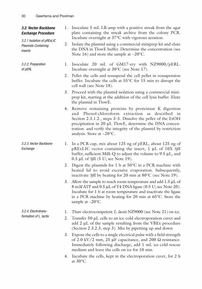
1. Inoculate 5 mL LB-amp with a positive streak from the agar plate containing the streak archive from the colony PCR. Incubate overnight at 37°C with vigorous aeration.
2. Isolate the plasmid using a commercial miniprep kit and elute the DNA in TlowE buffer. Determine the concentration (see Note 16) and store the sample at −20°C.
1. Inoculate 20 mL of GM17-ery with NZ9000/pERL. Incubate overnight at 30°C (see Note 17).
2. Pellet the cells and resuspend the cell pellet in resuspension buffer. Incubate the cells at 55°C for 15 min to disrupt the cell wall (see Note 18).
3. Proceed with the plasmid isolation using a commercial mini-prep kit, starting at the addition of the cell lysis buffer. Elute the plasmid in TlowE.
4. Remove remaining proteins by proteinase K digestion and Phenol:chloroform extraction as described in Section 2.3.1.2., steps 3–5. Dissolve the pellet of the EtOH precipitation in 20 mL TlowE, determine the DNA concen-tration. and verify the integrity of the plasmid by restriction analysis. Store at −20°C.
1. In a PCR cup, mix about 125 ng of pERL, about 125 ng of pRExLIC vector containing the insert, 1 mL of 10X SfiI buffer, sufficient Milli-Q to adjust the volume to 9.5 mL, and 0.5 mL of SfiI (5 U; see Note 19).
2. Digest the plasmids for 1 h at 50°C in a PCR machine with heated lid to avoid excessive evaporation. Subsequently, inactivate SfiI by heating for 20 min at 80°C (see Note 19).
3. Allow the sample to reach room temperature and add 1.5 mL of 8 mM ATP and 0.5 mL of T4 DNA ligase (0.5 U; see Note 20). Incubate for 1 h at room temperature and inactivate the ligase in a PCR machine by heating for 20 min at 65°C. Store the sample at −20°C.
1. Thaw electrocompetent L. lactis NZ9000 (see Note 21) on ice.2. Transfer 50 mL cells to an ice-cold electroporation cuvet and
add 2 mL of the sample resulting from the VBEx procedure (Section 2.3.2.3, step 3). Mix by pipetting up and down.
3. Expose the cells to a single electrical pulse with a field strength of 2.0 kV/2 mm, 25 mF capacitance, and 200 W resistance. Immediately following discharge, add 1 mL ice-cold rescue medium and leave the cells on ice for 10 min.
4. Incubate the cells, kept in the electroporation cuvet, for 2 h at 30°C.
3.2. Vector Backbone Exchange Procedure
3.2.1 Isolation of pRExLIC Plasmids Containing Inserts
3.2.2. Preparation of pERL
3.2.3. Vector Backbone Exchange
3.2.4. Electrotrans-formation of L. lactis

31Production of Membrane Proteins in Escherichia coli and Lactococcus lactis
5. Plate 10% and 90% of the transformed cells on SGM17-cam agar (see Note 13). Seal the plates with Parafilm (see Note 22) and incubate overnight at 30°C.
6. The colonies of L. lactis NZ9000/pNZxLIC holding the insert obtained in this step can be used for the expression analysis described in Section 2.3.3.
1. Inoculate 3 mL LB-amp from a verified streak obtained in Section 2.3.1.5. and incubate overnight at 37°C with vigorous aeration.
2. Mix 770 mL of culture with 230 mL of 87% (w/v) glycerol in a cryovial. Snap-freeze the vial in liquid nitrogen and archive the frozen stock at −80°C.
3. Use 60 mL of the remaining culture to inoculate a culture tube containing 6 mL TB-amp (see Note 23). Incubate the cells at 37°C and 200 rpm and follow the optical density of the culture (see Note 24).
4. At OD660 = ~0.2, place the tubes in a water bath at room tem-perature for 15 min and subsequently continue growth at 25°C.
5. After 30 min, induce the cultures with 60 mL of 5.10−1% (w/v) l-arabinose stock (100-fold concentrated) to obtain a final l-arabinose concentration of 5.10−3% (w/v). Incubate an additional 4 h.
6. Place the tubes in an ice-water bath and determine the OD660 of all cultures. Assuming that OD660 = 1 corresponds to a total protein concentration of 0.3 mg/mL, pellet the amount of culture equivalent to 1 mg protein in a FastPrep-compatible tube. Completely remove the super-natant, snap-freeze the pellet in liquid nitrogen, and store the sample at −20°C.
1. Inoculate 3 mL of GM17-cam with a single colony obtained in Section 2.3.2.4. and incubate overnight at 30°C.
2. Mix 770 mL of culture with 230 mL of 87% (w/v) glycerol in a cryovial. Snap-freeze the vial in liquid nitrogen and archive the frozen stock at −80°C.
3. Use 80 mL of the remaining culture to inoculate a culture tube containing 8 mL of GM17-cam (see Note 23). Incubate the cells at 30°C and follow the optical density of the culture (see Note 24).
4. The nisin A present in the supernatant of an overnight culture of the nisin-producing strain L. lactis NZ9700 is used to induce the cells. Dilute an aliquot of this supernatant 50-fold in GM17-cam to obtain a 100X concentrated stock. At OD660 = ~0.5, induce the cells with 80 mL of the 50-fold diluted
3.3. Initial Characterization and Optimization of Protein Production
3.3.1. Cultivation and Induction of MP Overexpression in E. coli
3.3.2. Cultivation and Induction of MP Overexpression in L. lactis

32 Geertsma and Poolman
supernatant to obtain a final dilution of 5,000-fold. Incubate an additional 2.5 h.
5. Place the tubes in an ice-water bath and determine the OD660 of all cultures. Assuming that OD660 = 1 corresponds to a total protein concentration of 0.2 mg/mL, pellet the amount of culture equivalent to 1 mg protein in a FastPrep-compatible tube. Completely remove the supernatant, snap-freeze the pellet in liquid nitrogen, and store the sample at −20°C.
1. Add about 300 mg of glass beads and 400 mL of ice-cold disruption buffer to the frozen cell pellets.
2. Disrupt the cells by two rounds of bead-beating in the FastPrep-24 device at force 6 for 20 s (for E. coli) or 30 s (for L. lactis). Place the cups in an ice-water bath for 5 min after each round (see Note 25).
3. Allow the glass beads to sediment for about 1 min and mix a 100 mL fraction from the supernatant in a new cup with 25 mL of 5X sample buffer (see Note 2). Do not boil the sample as this will lead to aggregation of the membrane protein and denaturation of GFP fusion proteins. Store the sample at −20°C or proceed immediately with the SDS-PAGE.
1. Load 10 mL of each sample per lane on an SDS-polyacrylamide gel (see Note 2). Include the Precision Plus Protein dual-color prestained molecular weight marker and run the gel at 150 V. Once the dye front has reached the bottom of the gel, disassemble the setup and soak the gel in Milli-Q.
2. For GFP fusion proteins, determine the in gel fluorescence. Place the gel on the black imaging tray of the LAS-3000 imaging system. Adjust the focus of lens to the gel and expose the gel to 460 nm light from the blue LED (light-emitting diode) and use a 515 nm emission filter (see Note 26). The fluorescent bands of the marker at 25 and 75 kDa should be clearly visible.
3. Submit the gel to immunoblotting to detect the His-tagged proteins. Use the LAS-3000 in chemiluminescence mode to measure the signal (see Note 26). The 75 kDa band of the marker should give a clear signal. Once a satisfac-tory exposure has been obtained, without moving the gel, use the top white light mode of the LAS-3000 to obtain an image of the membrane to locate the prestained marker bands.
4. Use the marker bands detected during the in gel fluorescence, chemiluminescence, and the white light image to estimate the intensity and the molecular weight of the detected bands and correlate the different images (see Note 27).
3.3.3. Cell Disruption and Sample Preparation
3.3.4. SDS-PAGE, In Gel GFP Fluorescence, and Western Blotting

33Production of Membrane Proteins in Escherichia coli and Lactococcus lactis
1. Optimize the expression by varying the protein expression rate over a wide range. For the PBAD system, use inducer concentra-tions between 1.10−5% and 1.10−1% (w/v) of l-arabinose; vary the induction temperature between 37°C, 25°C, and 17°C; and induce for 1, 2, 4, and 16 h. Determine the effect on expression using small cultures as described in Sections 2.3.3.1.–2.3.3.4. If possible, make use of the GFP fusion protein to guide the optimization procedure in E. coli. The presence of a strong, nonfluorescent band, indicative for aggregated protein, suggests too high an expression rate; the absence of a nonfluorescent band suggests that the capacity of the target-ing, insertion, and folding pipeline is not yet saturated.
2. For the lactococcal PnisA system, use dilutions of the nisin-containing supernatant ranging from 100,000- to 500-fold; vary the induction temperature between 30°C, 25°C, and 17°C; and induce for 1, 2, 4, and 16 h. Determine the effect on expression using small cultures as described in Sections 2.3.3.1.–2.3.3.4.
1. Use generic primers instead of gene-specific primers. This facilitates the analysis of several genes and provides a positive control for the PCR because for empty vectors a band will be produced as well. Generic primers for the pBADxLIC derivatives are 5¢ctctactgtttctccatacccg and 5¢gctgaaaatct-tctctcatccg. Generic primers for the pRExLIC derivatives are 5¢cgcgagcataataaacggc and 5¢ctgctgctttttggctatcaatc.
2. Deviations in the composition from the here-described buf-fers for sample preparation and gel electrophoresis might result in a decrease in the intensity of the in gel GFP fluorescence.
3. The initial steps of primer design can be easily automated using a spreadsheet program. This will speed up the design process and reduce the number of errors. A spreadsheet that combines the terminal 20 nucleotides of a set of DNA sequences with the nLIC and cLIC extensions to produce a series of initial primers that might require additional corrections can be obtained from the authors’ Web site (http://www.rug.nl/gbb/research/researchgroups/enzymology/index).
4. Removal of secondary structure elements is often best done by making silent mutations in the gene-specific part of the primer while preventing the use of rare codons as much as possible. Do not make any changes in the primer extensions required for LIC.
3.3.5. Expression Optimization
4. Notes

34 Geertsma and Poolman
5. Genomic DNA is easily compromised by repetitive freeze/thawing, vortexing, or vigorous pipetting and thus requires careful handling; storage in sterile tubes at 4°C is preferred.
6. Combine PCRs for targets of similar length and template type (plasmid or genomic) as much as possible instead of performing a PCR for each gene separately. This will speed up the whole process and allows more time for optimizing PCRs of “difficult genes.”
7. Touchdown PCR ensures a more specific product formation (42), and as a range of annealing temperatures is used, it is a good alternative to optimizing the reaction conditions for each target on an individual basis.
8. Should the DNA template that was used for the PCR reaction be a plasmid that confers resistance to b-lactam antibiotics such as ampicillin and carbenicillin, then the PCR mix should be incubated for 30 min at 37°C with 1 mL DpnI prior to the gel electrophoresis to remove the template. As the pBADxLIC and pRExLIC vectors used for the LIC procedure carry the b-lactamase marker, traces of template plasmids that carry the same marker will lead to a high number of background colonies.
9. Should the PCR not yield the desired product in sufficient amounts, consider the use of alternative buffers supplied by the manufacturer, the addition of dimethyl sulfoxide (DMSO), or replacement of the template. More detailed suggestions can be found in the Molecular Cloning protocol series (40).
10. Depending on the producer of SwaI, the suggested incuba-tion temperature is 25°C or 30°C. Condensation at the lid, leading to suboptimal reaction conditions, can be prevented by performing the reaction in an incubator or PCR machine with heated lid.
11. The quality of the dNTP stocks and the T4 DNA polymerase is a critical parameter for the success of the procedure. Prevent repetitive freezing/thawing of the nucleotides by preparing small aliquots and label the aliquots appropriately to prevent mixing up dCTP (used for the vector) with dGTP (used for the insert). Transport the T4 DNA polymerase in a cooling tray and keep the enzyme cold under all conditions.
12. The use of equimolar amounts of vector and insert ensures that both are treated with T4 DNA polymerase to a similar extent. The amount of the purified PCR product needed can be calculated using the following: Amount insert needed (ng) = ((200 ng * Insert size (bp))/Vector size (bp)).
13. Especially for large numbers of samples, spreading the trans-formed cells over the plate is most easily done by making use

35Production of Membrane Proteins in Escherichia coli and Lactococcus lactis
of glass beads (diameter 5 mm). In addition, this prevents cross contamination.
14. Use of toothpicks over an inoculation needle prevents cross contamination when analyzing multiple samples at one time. A cross-locking tweezer facilitates handling the toothpicks for prolonged periods.
15. Take care to properly seal the plate, especially around the sides. Incomplete sealing will lead to evaporation.
16. Especially for large numbers of plasmid samples, determining the DNA concentration can be time consuming. As the VBEx reaction only requires an approximate estimation of the DNA concentration, it suffices to determine the concentration of a small subset (~5 samples) and use the average DNA concentra-tion as representative for this batch.
17. As L. lactis does not require aeration, small cultures are not shaken and can be maintained in closed glass or plastic containers.
18. The extent of the degradation of the cell wall depends on the incubation time and the amount of cells and lysozyme used; insufficient degradation leads to lower plasmid yields. Whether the lysozyme treatment was sufficient can be easily determined by mixing a 10 mL aliquot of the cell suspension with 10 mL of the supplied lysis buffer. Adequate disruption of the cell wall will lead to complete cell lysis within 5 min. If needed, extend the incubation time or use a higher lysozyme concentration.
19. For the overall procedure, it is critical that the SfiI enzyme can be heat inactivated. For unknown reasons, this property seems to depend on the supplier. We have had a good experience with SfiI obtained from Fermentas.
20. For a large number of samples, prepare a premix containing 6 mM ATP, 0.25 U/mL T4 DNA ligase, and 1X T4 DNA ligase buffer. Use ice-cold Milli-Q and prepare the mix just prior to use.
21. Preparation of electrocompetent L. lactis NZ9000 was essen-tially done as described (43), but with minor modifications. Briefly, cells were grown in M17 supplemented with 0.5% glu-cose, 0.5M sucrose, and 2% glycine at 30°C to OD600 = ~0.5. Cells were harvested by centrifugation at 5,000g for 15 min at 4°C. Following washes with 1 volume of ice-cold solution A (0.5M sucrose and 10% glycerol, prepared in Milli-Q), 0.5 volume of solution A supplemented with 50 mM Na-EDTA, pH 7.5, and 0.25 volume of solution A, cells were resuspended in 0.01 volume of solution A. Aliquots of 50 mL were snap-frozen in liquid nitrogen and stored at −80°C until use.
22. Sealing the agar plates with Parafilm is not essential but allows faster appearance of L. lactis colonies.

36 Geertsma and Poolman
23. Preheat the medium without the antibiotics overnight at 37°C or 30°C for E. coli and L. lactis, respectively. This prevents a slow start of the cultivation due to a small temperature shock.
24. When handling a large set of samples, use an additional dedi-cated set of approximately six reference cultures to follow the OD660 and use their average OD660 to time the moment of cooling or induction.
25. Alternative methodologies for cell disruption, such as sonica-tion and vortexing in the presence of glass beads, can be used as well but might be less efficient. This is especially true for L. lactis, whose thicker and stable cell wall makes it more diffi-cult to disrupt than E. coli. French press disruption is compa-rable to cell disruption by a FastPrep but requires larger volumes and is therefore less suited. For L. lactis, treatment with lysozyme to digest the peptidoglycan layer prior to dis-ruption greatly enhances the efficiency. This step is not required for FastPrep-mediated cell disruption.
26. The use of the same tray height for in gel fluorescence and chemiluminescence imaging prevents rescaling of images, thus facilitating the correlation of the images.
27. The transfer efficiencies from the gel to the PVDF (poly-vinylidene difluoride) membrane will differ for different membrane proteins. This complicates selection of promising clones. In contrast, the in gel fluorescence of GFP fusion pro-teins can be compared directly.
Acknowledgments
We thank G.K. Schuurman-Wolters, M.B. Tol, M. Groeneveld, D.J. Slotboom, and S. Henstra for their contribution in estab-lishing the methodologies discussed. This work was funded by the European Membrane Protein Consortium (grant 504601), a FEBS fellowship (to E.R.G.), and the Netherlands Proteomics Centre.
References
1. Driessen AJ, Nouwen N (2008) Protein trans-location across the bacterial cytoplasmic mem-brane. Annu Rev Biochem 77:643–667
2. Korepanova A, Gao FP, Hua Y, Qin H, Nakamoto RK, Cross TA (2005) Cloning and expression of multiple integral membrane proteins from Mycobacterium tuberculosis in Escherichia coli. Protein Sci 14:148–158
3. Dobrovetsky E, Lu ML, Andorn-Broza R, Khutoreskaya G, Bray JE, Savchenko A, Arrowsmith CH, Edwards AM, Koth CM (2005) High-throughput production of prokaryotic membrane proteins. J Struct Funct Genom 6:33–50
4. White MA, Clark KM, Grayhack EJ, Dumont ME (2007) Characteristics affecting expression

37Production of Membrane Proteins in Escherichia coli and Lactococcus lactis
and solubilization of yeast membrane proteins. J Mol Biol 365:621–636
5. Wagner S, Baars L, Ytterberg AJ, Klussmeier A, Wagner CS, Nord O, Nygren PA, van Wijk KJ, de Gier JW (2007) Consequences of membrane protein overexpression in Escherichia coli. Mol Cell Proteom 6:1527–1550
6. Bonander N, Hedfalk K, Larsson C, Mostad P, Chang C, Gustafsson L, Bill RM (2005) Design of improved membrane protein pro-duction experiments: quantitation of the host response. Protein Sci 14:1729–1740
7. Savchenko A, Yee A, Khachatryan A, Skarina T, Evdokimova E, Pavlova M, Semesi A, Northey J, Beasley S, Lan N, Das R, Gerstein M, Arrowmith CH, Edwards AM (2003) Strategies for structural proteomics of prokary-otes: quantifying the advantages of studying orthologous proteins and of using both NMR and X-ray crystallography approaches. Proteins 50:392–399
8. Locher KP, Lee AT, Rees DC (2002) The E. coli BtuCD structure: a framework for ABC transporter architecture and mechanism. Science 296:1091–1098
9. Terpe K (2003) Overview of tag protein fusions: from molecular and biochemical fun-damentals to commercial systems. Appl Microbiol Biotechnol 60:523–533
10. Surade S, Klein M, Stolt-Bergner PC, Muenke C, Roy A, Michel H (2006) Comparative analysis and “expression space” coverage of the production of prokaryotic membrane pro-teins for structural genomics. Protein Sci 15:2178–2189
11. Terpe K (2006) Overview of bacterial expres-sion systems for heterologous protein produc-tion: from molecular and biochemical fundamentals to commercial systems. Appl Microbiol Biotechnol 72:211–222
12. Grisshammer R, Tate CG (1995) Overexpres-sion of integral membrane proteins for structural studies. Q Rev Biophys 28:315–422
13. Kunji ER, Slotboom DJ, Poolman B (2003) Lactococcus lactis as host for overproduction of functional membrane proteins. Biochim Biophys Acta 1610:97–108
14. Quick M, Javitch JA (2007) Monitoring the function of membrane transport proteins in detergent-solubilized form. Proc Natl Acad Sci USA 104:3603–3608
15. de Ruyter PG, Kuipers OP, de Vos WM (1996) Controlled gene expression systems for Lactococcus lactis with the food-grade inducer nisin. Appl Environ Microbiol 62:3662–3667
16. Geertsma ER, Poolman B (2007) High-throughput cloning and expression in recalci-trant bacteria. Nat Methods 4:705–707
17. Poolman B, Konings WN (1988) Relation of growth of Streptococcus lactis and Streptococcus cremoris to amino acid trans-port. J Bacteriol 170:700–707
18. Wegmann U, O’Connell-Motherway M, Zomer A, Buist G, Shearman C, Canchaya C, Ventura M, Goesmann A, Gasson MJ, Kuipers OP, van Sinderen D, Kok J (2007) Complete genome sequence of the prototype lactic acid bacterium Lactococcus lactis subsp. cremoris MG1363. J Bacteriol 189:3256–3270
19. Nouaille S, Morello E, Cortez-Peres N, Le Loir Y, Commissaire J, Gratadoux JJ, Poumerol E, Gruss A, Langella P (2006) Complementation of the Lactococcus lactis secretion machinery with Bacillus subtilis SecDF improves secretion of staphylococcal nuclease. Appl Environ Microbiol 72:2272–2279
20. van der Sluis EO, Driessen AJ (2006) Stepwise evolution of the Sec machinery in Proteobacteria. Trends Microbiol 14:105–108
21. Saaf A, Monne M, de Gier JW, von Heijne G (1998) Membrane topology of the 60-kDa Oxa1p homologue from Escherichia coli. J Biol Chem 273:30415–30418
22. Driessen AJ, Zheng T, In’t Veld G, Op den Kamp JA, Konings WN (1988) Lipid require-ment of the branched-chain amino acid transport system of Streptococcus cremoris. Biochemistry 27:865–872
23. Nakamura Y, Gojobori T, Ikemura T (2000) Codon usage tabulated from international DNA sequence databases: status for the year 2000. Nucleic Acids Res 28:292
24. Kouwen TR, van der Goot A, Dorenbos R, Winter T, Antelmann H, Plaisier MC, Quax WJ, van Dijl JM, Dubois JY (2007) Thiol-disulphide oxidoreductase modules in the low-GC Gram-positive bacteria. Mol Microbiol 64:984–999
25. Steidler L, Hans W, Schotte L, Neirynck S, Obermeier F, Falk W, Fiers W, Remaut E (2000) Treatment of murine colitis by Lactococcus lactis secreting interleukin-10. Science 289:1352–1355
26. Steidler L, Neirynck S, Huyghebaert N, Snoeck V, Vermeire A, Goddeeris B, Cox E, Remon JP, Remaut E (2003) Biological con-tainment of genetically modified Lactococcus lactis for intestinal delivery of human interleu-kin 10. Nat Biotechnol 21:785–789
27. Vandenbroucke K, Hans W, Van Huysse J, Neirynck S, Demetter P, Remaut E, Rottiers P, Steidler L (2004) Active delivery of trefoil factors by genetically modified Lactococcus lactis prevents and heals acute colitis in mice. Gastroenterology 127:502–513
28. Kunji ER, Chan KW, Slotboom DJ, Floyd S, O’Connor R, Monne M (2005) Eukaryotic

38 Geertsma and Poolman
membrane protein overproduction in Lactococcus lactis. Curr Opin Biotechnol 16: 546–551
29. Aslanidis C, de Jong PJ (1990) Ligation-independent cloning of PCR products (LIC-PCR). Nucleic Acids Res 18:6069–6074
30. Walhout AJ, Temple GF, Brasch MA, Hartley JL, Lorson MA, van den Heuvel S, Vidal M (2000) GATEWAY recombinational cloning: application to the cloning of large numbers of open reading frames or ORFeomes. Methods Enzymol 328:575–592
31. Liu Q, Li MZ, Leibham D, Cortez D, Elledge SJ (1998) The univector plasmid-fusion system, a method for rapid construction of recombinant DNA without restriction enzymes. Curr Biol 8:1300–1309
32. Guzman LM, Belin D, Carson MJ, Beckwith J (1995) Tight regulation, modulation, and high-level expression by vectors containing the arabinose PBAD promoter. J Bacteriol 177:4121–4130
33. Wang DN, Safferling M, Lemieux MJ, Griffith H, Chen Y, Li XD (2003) Practical aspects of overexpressing bacterial secondary membrane transporters for structural studies. Biochim Biophys Acta 1610:23–36
34. Chen Y, Song J, Sui SF, Wang DN (2003) DnaK and DnaJ facilitated the folding process and reduced inclusion body formation of mag-nesium transporter CorA overexpressed in Escherichia coli. Protein Exp Purif 32:221–231
35. Waldo GS, Standish BM, Berendzen J, Terwilliger TC (1999) Rapid protein-folding assay using green fluorescent protein. Nat Biotechnol 17:691–695
36. Drew DE, von Heijne G, Nordlund P, de Gier JW (2001) Green fluorescent protein as an indicator to monitor membrane protein over-expression in Escherichia coli. FEBS Lett 507:220–224
37. Drew D, Lerch M, Kunji E, Slotboom DJ, de Gier JW (2006) Optimization of membrane protein overexpression and purification using GFP fusions. Nat Methods 3:303–313
38. Geertsma ER, Groeneveld M, Slotboom DJ, Poolman B (2008) Quality control of overex-pressed membrane proteins. Proc Natl Acad Sci USA 105:5722–5727
39. Kawate T, Gouaux E (2006) Fluorescence-detection size-exclusion chromatography for precrystallization screening of integral mem-brane proteins. Structure 14:673–681
40. Sambrook J, Russell DW (2001) Molecular cloning, a laboratory manual. Cold Spring Harbor Laboratory Press, Cold Spring Harbor, NY
41. Terzaghi BE, Sandine WE (1975) Improved medium for lactic Streptococci and their bac-teriophages. Appl Microbiol 29:807–813
42. Don RH, Cox PT, Wainwright BJ, Baker K, Mattick JS (1991) “Touchdown” PCR to cir-cumvent spurious priming during gene ampli-fication. Nucleic Acids Res 19:4008
43. Holo H, Nes IF (1989) High-frequency transformation, by electroporation, of Lacto coccus lactis subsp. cremoris grown with glycine in osmotically stabilized media. Appl Environ Microbiol 55: 3119–3123
44. Berntsson RP, Alia Oktaviani N, Fusetti F, Thunnissen AM, Poolman B, Slotboom DJ (2009) Selenomethionine incorporation in proteins expressed in Lactococcus lactis. Protein Sci 18(5):1121–1127
45. El Khattabi M, van Roosmalen ML, Jager D, Metselaar H, Permentier H, Leenhouts K, Broos J (2008) Lactococcus lactis as expression host for the biosynthetic incor-poration of tryptophan analogues into recombinant proteins. Biochem J 409(1): 193–198