chapter 16 radiation protection and safety in … · the radiation exposures are therefore divided...
TRANSCRIPT
549
Chapter 16
RADIATION PROTECTION AND SAFETY IN RADIOTHERAPY
P. ORTIZ LÓPEZDivision of Radiation, Transport and Waste Safety,International Atomic Energy Agency,Vienna
G. RAJANMedical Physics and Safety Section,Bhabha Atomic Research Centre,Mumbai, Maharashtra, India
E.B. PODGORSAKDepartment of Medical Physics,McGill University Health Centre,Montreal, Quebec, Canada
16.1. INTRODUCTION
Soon after the discovery of X rays by Roentgen in 1895 and of natural radioactivity by Becquerel in 1896 it became apparent that ionizing radiation was not only useful for the diagnosis and treatment of disease but also harmful to human tissues. It has been recognized since early studies on X rays and radioactive minerals that exposure to high levels of radiation can cause clinical damage to tissues of the human body. In addition, long term epidemiological studies of populations exposed to radiation, especially the survivors of the atomic bombings of Hiroshima and Nagasaki in Japan in 1945, have demonstrated that exposure to radiation also has a potential for delayed effects such as induction of malignancies or damage to genetic material.
Ionizing radiation and radioactive substances are natural and permanent features of the environment, and thus the risks associated with radiation exposure can only be restricted, not eliminated entirely. Additionally, the use of human-made radiation is now widespread. Sources of ionizing radiation are essential to modern health care: disposable medical supplies sterilized by intense radiation have been central to combating disease; radiology and nuclear medicine are a vital diagnostic tool; and radiotherapy is commonly part
CHAPTER 16
550
of the treatment of malignancies. Applications of ionizing radiation are growing in industry, agriculture, medicine and many other fields of industry and research, benefiting humanity. Irradiation is used around the world to preserve foodstuffs and reduce wastage, and sterilization techniques have been used to eradicate disease carrying insects and pests. Industrial radiography is in routine use, for example to examine welds, detect cracks and help prevent failure of engineered structures.
The acceptance by society of the risks associated with radiation is conditional on the benefits to be gained from the use of radiation. Nonetheless, the risks must be restricted and protected against by the application of radiation safety standards. It is therefore essential that activities involving radiation exposure be subject to certain standards of safety in order to protect the individuals who are exposed to radiation, be it occupationally, for medical diagnostic or therapeutic purposes, or as members of the public.
16.2. RADIATION EFFECTS
Exposure to radiation can cause detrimental health effects that fall into one of two categories: deterministic or stochastic.
16.2.1. Deterministic effects
At large doses, radiation effects such as nausea, reddening of the skin or, in severe cases, more acute syndromes are clinically expressed in exposed individuals within a relatively short period of time after the exposure; such effects are called deterministic because they are certain to occur if the dose exceeds a threshold level.
Deterministic effects are the result of various processes, mainly cell death or delayed cell division, caused by exposure to high levels of radiation. If extensive enough, these effects can impair the function of the exposed tissues. The severity of a particular deterministic effect in an exposed individual increases with dose above the threshold for the occurrence of the effect.
16.2.2. Stochastic effects
Radiation exposure can also induce delayed effects such as malignancies, which are expressed after a latency period and may be epidemiologically detectable in a population; this induction is assumed to take place over the entire range of doses, without a threshold level. Hereditary effects due to radiation exposure have been statistically detected in other mammalian
RADIATION PROTECTION AND SAFETY IN RADIOTHERAPY
551
populations and are presumed to occur in human populations also. These epidemiologically detectable effects (malignancies and hereditary effects) are termed stochastic effects because of their random nature.
Stochastic effects may ensue if an irradiated cell is modified rather than killed. Modified cells may, after a prolonged delay, develop into a cancer. The body’s repair mechanisms make this a very improbable outcome at small doses; nevertheless, there is no evidence of a threshold dose below which cancer cannot result. The probability of occurrence of cancer is higher for higher doses, but the severity of any cancer that may result from irradiation is independent of dose. If the cell damaged by radiation exposure is a germ cell whose function is to transmit genetic information to progeny, it is conceivable that hereditary effects of various types may develop in the descendants of the exposed individual. The likelihood of stochastic effects is presumed to be proportional to the dose received, without a dose threshold.
The many aspects of the concept of radiation detriment make it undesirable to select any single quantity to represent it. The concept of detriment as recommended by the ICRP for stochastic effects includes the following quantities: the probability of fatal cancer attributable to radiation exposure; the weighted probability of incurring a non-fatal cancer; the weighted probability of severe hereditary effects; and the length of lifetime lost, if the harm occurs.
16.2.3. Effects on the embryo and foetus
In addition to deterministic and stochastic health effects in adults, other health effects may occur in infants due to exposure of the embryo or foetus to radiation. These effects include a greater likelihood of leukaemia (stochastic effect) and, for exposure above various threshold dose values during certain periods of pregnancy, severe mental retardation and congenital malformations (deterministic effect). For more details on effects on the foetus see ICRP Publication 84.
16.3. INTERNATIONAL CONSENSUS AND RADIATION SAFETY
STANDARDS
Safety standards are based on knowledge of radiation effects and on the principles of protection described below. In this respect, the development of safety standards by the IAEA follows a well established approach. The United Nations Scientific Committee on the Effects of Atomic Radiation (UNSCEAR), a body set up by the United Nations in 1955, compiles, assesses

CHAPTER 16
552
and disseminates information on the health effects of radiation and on levels of radiation exposure due to different sources; this information was taken into account in developing the standards. Following a decision made in 1960, the IAEA safety standards are based on the recommendations of the ICRP, which also take account of the scientific information provided by UNSCEAR.
Purely scientific considerations, however, are only part of the basis for decisions on protection and safety, and the safety standards implicitly encourage decision makers to make value judgements about the relative importance of risks of different kinds and about the balancing of risks and benefits. General acceptance of risk is a matter of consensus, and therefore international safety standards should provide a desirable international consensus for the purpose of protection.
For these reasons, international consensus is basic to the IAEA standards, which are prepared with the wide participation of and approval by its Member States and relevant international organizations. The current version of the safety standard entitled International Basic Safety Standards for Protection against Ionizing Radiation and for the Safety of Radiation Sources (hereinafter referred to as the BSS) was issued in 1996 under the joint sponsorship of the Food and Agriculture Organization of the United Nations, IAEA, International Labour Organisation, OECD Nuclear Energy Agency, Pan American Health Organization and World Health Organization.
The BSS was published as IAEA Safety Series No. 115 and comprises four sections: preamble, principal requirements, appendices and schedules. The purpose of the report is to establish basic requirements for protection against exposure to ionizing radiation and for the safety of radiation sources that may deliver such exposure.
16.4. TYPES OF RADIATION EXPOSURE
Certain industrial or medical practices will result in some radiation exposure with predictable magnitudes, albeit with some degree of uncertainty; such expected exposures are referred to in the BSS as normal exposures.
In addition, scenarios can be envisaged for which there is a potential for exposure, but no certainty that an exposure will in fact occur; such unexpected but feasible exposures are termed potential exposures. Potential exposures can become actual exposures if the unexpected situation does occur, for example as a consequence of equipment failure, design problems or operating errors.
The means specified in the BSS for controlling normal exposures is the restriction of the doses delivered. In the case of exposure of patients, exposures
RADIATION PROTECTION AND SAFETY IN RADIOTHERAPY
553
are controlled through delivering only the doses that are necessary to achieve the diagnostic or therapeutic objective.
The primary means for controlling potential exposures is by optimizing the design of installations, equipment and operating procedures with the following aims:
● To restrict the probability of occurrence of events that could lead to unplanned exposures;
● To restrict the magnitudes of the exposures that could result if such events were to occur.
The radiation exposures covered by the BSS encompass the exposures, both normal and potential, of:
— Workers pursuing their occupations (occupational exposures); — Patients in diagnosis or treatment (medical exposures); — Members of the public.
The radiation exposures are therefore divided into three categories:
(i) Occupational exposure, which is defined as all exposures of workers incurred in the course of their work (with the exception of exposures excluded from the BSS and exposures from practices or sources exempted by the BSS).
(ii) Medical exposure, which is defined as exposure incurred:— By patients as part of their own medical or dental diagnosis or
treatment;— By persons, other than those occupationally exposed, knowingly while
voluntarily helping in the support and comfort of patients; — By volunteers in a programme of biomedical research involving their
exposure.(iii) Public exposure, which is defined as exposure incurred by members of the
public from radiation sources, excluding any occupational or medical exposure and the normal local natural background radiation but including exposure to authorized sources and practices and from intervention situations.
CHAPTER 16
554
16.5. QUANTITIES AND UNITS USED IN RADIATION PROTECTION
16.5.1. Physical quantities
Although most of the requirements of the BSS are qualitative, they also establish quantitative limits and guidance levels. The main physical quantities used in safety standards are the activity and absorbed dose:
● The activity A of an amount of a radionuclide in a particular energy state at a given time is the quotient of dN by dt, where dN is the number of spontaneous nuclear transformations from that energy state in the time interval dt:
(16.1)
where
l is the decay constant of the radioactive nucleus; N is the number of radioactive nuclides (atoms); t1/2 is the half-life of the radioactive nucleus.
The SI unit of activity is 1 s–1 and its name is the becquerel (Bq), representing one nuclear transformation (disintegration or decay) per second (i.e. 1 Bq = 1 s–1). The older unit of activity is the curie (Ci), representing 3.7 × 10¹⁰ s–1 (i.e. 1 Ci = 3.7 × 10¹⁰ Bq). The curie was initially defined as the activity of 1 g of 226Ra; however, refined measurements have shown that the activity of 1 g of 226Ra is 0.988 Ci.
● The absorbed dose D is defined as the quotient of de– by dm, where de– is the mean energy imparted to matter of mass dm:
(16.2)
The SI unit for absorbed dose is 1 J/kg and its name is the gray (Gy). The older unit of dose is the rad, representing 100 erg/g (i.e. 1 Gy = 100 rad).
16.5.2. Radiation protection quantities
The absorbed dose is the basic physical dosimetry quantity, but it is not entirely satisfactory for radiation protection purposes because the effectiveness in damaging human tissue differs for different types of ionizing radiation. In addition to the physical quantities, other dose related quantities have been
A = = =d dN t N t N/ ((ln )/ )/l 2 1 2
Dm
= dde
RADIATION PROTECTION AND SAFETY IN RADIOTHERAPY
555
introduced to account not only for the physical effects but also for the biological effects of radiation upon tissues. These quantities are organ dose, equivalent dose, effective dose, committed dose and collective dose.
16.5.2.1. Organ dose
The organ dose is defined as the mean dose DT in a specified tissue or organ T of the human body, given by:
(16.3)
where
mT is the mass of the organ or tissue under consideration; εT is the total energy imparted by radiation to that tissue or organ.
16.5.2.2. Equivalent dose
The biological detriment (harm) to an organ depends not only on the physical average dose received by the organ but also on the pattern of the dose distribution that results from the radiation type and energy. For the same dose to the organ, a or neutron radiation will cause greater harm compared with grays or electrons. This is because the ionization events produced by a or neutron radiation will be much more closely spaced (densely ionizing radiations) and so there is a higher probability of irreversible damage to the chromosomes and less chance of tissue repair.
Consequently, the organ dose is multiplied by a radiation weighting factor wR to account for the effectiveness of the given radiation in inducing health effects; the resulting quantity is called the equivalent dose HT.
The equivalent dose HT is defined as:
HT = wRDT,R (16.4)
where
DT,R is the absorbed dose delivered by radiation type R averaged over a tissue or organ T;
wR is the radiation weighting factor for radiation type R.
Dm
D mm
m
TT
T
T
d
T
= =Ú1 e
CHAPTER 16
556
For X rays, g rays and electrons wR = 1; for protons wR = 5; for particles wR = 20; and for neutrons wR ranges from 5 to 20, depending on the neutron energy.
The SI unit of equivalent dose is J/kg and its name is the sievert (Sv); the old unit is the rem and the relationship between the two units is 1 Sv = 100 rem; for example, for 1 Gy of photon dose to an organ, the equivalent dose is 1 Sv. However, for the same dose of 20 keV neutrons, the equivalent dose is 10 Sv, since the detriment is ten times larger (i.e. wR = 10 for 20 keV neutrons).
The organ dose DT,R is a measure of the energy absorption per unit mass averaged over the organ, while the equivalent dose HT is a measure of the consequent biological harm (detriment) to the organ or tissue T.
If an organ is irradiated by more than one type of radiation, the equivalent dose is given by the sum:
(16.5)
In earlier ICRP recommendations, weighting factors related to the quality of radiation were applied to the absorbed dose to a point, and the radiation weighted absorbed dose was called the dose equivalent H (not referred to an organ, but to a point).
16.5.2.3. Effective dose
The relationship between the probability of stochastic effects and equivalent dose is also found to depend on the organ or tissue irradiated. This implies that for the same equivalent dose the detriments from the exposure of different organs or tissues are different. To take account of these differences, tissue weighting factors are needed.
Tissue weighting factors wT should represent the relative contribution of an organ or tissue to the total detriment due to the effects resulting from a uniform irradiation of the whole body. For low doses, individual organ or tissue detriments can be treated as additive and the total detriment to the whole body is the summation of individual detriments. The relative contribution to the total detriment is therefore given by the quotient between the individual detriment and the total detriment resulting from a uniform irradiation of the whole body. Since the sum of relative contributions is normalized to unity, the sum SwT = 1.
The effective dose E is defined as the summation of tissue equivalent doses, each multiplied by the appropriate tissue weighting factor wT, to indicate the combination of different doses to several different tissues in a way that correlates well with all stochastic effects combined (ICRP Publication 60):
H w DT R T,R = Â
RADIATION PROTECTION AND SAFETY IN RADIOTHERAPY
557
(16.6)
Tissue weighting factors wT are tabulated in ICRP Publication 60 and in IAEA safety standards. Despite depending on the sex and age of the person, for the purposes of radiation protection the values for tissue weighting factors are taken as constants and are applicable to the average population; for example, wT = 0.20 for gonads, wT = 0.12 for lung or red bone marrow and wT = 0.01 for skin. Thus for the same equivalent dose, the risk of a stochastic effect at low doses is higher for gonads than for the lungs or red bone marrow.
The unit of effective dose is J/kg and its name is the sievert (Sv).A uniform equivalent dose over the whole body gives an effective dose
that is numerically equal to the uniform equivalent dose.The weighing factors wT and wR are mutually independent; that is, the
tissue risk factors wT are independent of radiation type and the radiation weighting factors wR are independent of tissue type, allowing us to write:
(16.7)
When one deals with only one type of radiation in a given situation, the effective dose is given by:
(16.8)
The effective dose is a measure of dose designated to reflect the amount of radiation detriment likely to result from the dose. Effective doses from various radiation types and exposure modes may be compared directly.
Annual dose limits for occupational and public exposure are given in terms of the annual effective dose; in the case of exposure of an organ or of hands or feet they are given in terms of equivalent dose.
The term ‘effective dose’ replaces the term ‘effective dose equivalent’ defined in earlier ICRP reports.
For a well defined geometry of irradiation, the equivalent dose H to individual organs or the effective dose E can be computed for an anthropomorphic phantom that simulates the human body. However, these quantities are not directly measurable, since there are no primary standards established for them.
16.5.2.4. Committed dose
When radionuclides are taken into the body, the resulting dose is received throughout the period of time during which they remain in the body. The total
E w H = T TÂ
E w w D w w D= =Â Â Â ÂTT
RR
T,R RR
TT
T,R
E w D = T T,RÂ
CHAPTER 16
558
dose delivered during this period of time is referred to as the committed dose and is calculated as a specified time integral of the rate of receipt of the dose. Any relevant dose restriction is applied to the committed dose from the intake. The committed dose may refer to the committed effective dose and the committed equivalent dose.
16.5.2.5. Collective dose
The radiation protection quantities discussed above relate to the exposure of an individual. The collective dose relates to exposed groups or populations and is defined as the summation of the products of the mean dose in the various groups of exposed people and the number of individuals in each group. The unit of collective dose is the man-sievert (man-Sv).
16.5.3. Operational quantities
The organ dose DT, equivalent dose H and effective dose E are not directly measurable and there are no laboratory standards to obtain traceable calibrations for the radiation monitors using these quantities. For this reason, the ICRU has defined a set of measurable operational quantities for protection purposes: the ambient dose equivalent, directional dose equivalent and personal dose equivalent; the latter is used for comparing with regulatory requirements such as dose limits.
16.5.3.1. Ambient dose equivalent
The ambient dose equivalent at a point in a radiation field H*(d) is defined as the dose equivalent that would be produced by the corresponding aligned and expanded field in the ICRU sphere at a depth d on the radius opposing the direction of the aligned field. The ICRU sphere is a 30 cm diameter tissue equivalent sphere with a composition of 76.2% oxygen, 11.1% carbon, 10.1% hydrogen and 2.6% nitrogen. A depth d = 10 mm is recommended for strongly penetrating radiation.
16.5.3.2. Directional dose equivalent
The directional dose equivalent at a point in a radiation field H ¢(d, W) is defined as the dose equivalent that would be produced by the corresponding expanded field in the ICRU sphere at depth d on a radius in a specified direction W~. A depth d = 0.07 mm is recommended for weakly penetrating
RADIATION PROTECTION AND SAFETY IN RADIOTHERAPY
559
radiation. Angle W is the angle between the beam direction and the radius of the ICRU sphere on which the depth d is defined.
16.5.3.3. Personal dose equivalent
The personal dose equivalent Hp(d) is defined for both strongly and weakly penetrating radiations as the equivalent dose in soft tissue below a specified point on the body at an appropriate depth d. The relevant depth is generally d = 10 mm for penetrating radiations (photon energies above 15 keV), while depths d = 0.07 mm and d = 3 mm are used for weakly penetrating radiations (photon energies below 15 keV and b radiations) in skin and the eye lens, respectively.
The personal dose equivalent from exposure to penetrating radiationduring the year is the radiation quantity to be compared with the annual dose limits (for effective dose) and to demonstrate compliance with the BSS recommendations as indicated below (see the BSS, Schedule II).
16.6. BASIC FRAMEWORK OF RADIATION PROTECTION
The principles of radiation protection and safety upon which the radiation safety standards are based are those developed by the ICRP. The detailed formulation of these principles can be found in the ICRP publications and they cannot easily be paraphrased without losing their essence. However, a brief, although simplified, summary of the principles is given in this section.
A practice that entails exposure to radiation should only be adopted if it yields sufficient benefit to the exposed individuals or to society to outweigh the radiation detriment it causes or could cause (i.e. the practice must be justified).
Individual doses due to the combination of exposures from all relevant practices should not exceed specified dose limits for occupational and public exposure; dose limits are not applicable to medical exposure.
Radiation sources and installations should be provided with the best available protection and safety measures under the prevailing circumstances, so that the magnitudes and likelihood of exposures and the numbers of individuals exposed be as low as reasonably achievable (ALARA), economic and social factors being taken into account, and the doses they deliver and the risk they entail be constrained (i.e. protection and safety should be optimized):
● In diagnostic medical exposure, optimization of protection is achieved by keeping the exposure of patients to the minimum necessary to achieve the required diagnostic objective;
CHAPTER 16
560
● In therapeutic medical exposure, optimization is achieved by keeping exposure of normal tissue ALARA consistent with delivering the required dose to the planning target volume (PTV) (from the BSS requirements in Appendix II).
As indicated in Section 16.13, pregnant workers shall be protected so as to ensure that the embryo or foetus is afforded the same broad level of protection as required for members of the public.
A safety culture should be inculcated that governs attitudes and behaviour in relation to the protection and safety of all individuals and organizations dealing with sources of radiation; in depth defensive measures should be incorporated into the design and operating procedures for radiation sources to compensate for potential failures in protection or safety measures; and protection and safety should be ensured by sound management and good engineering, quality assurance, training and qualification of personnel, comprehensive safety assessments and attention to lessons learned from experience and research.
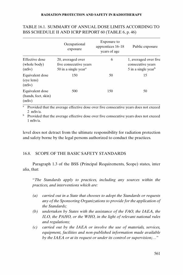
Dose limits do not apply to medical exposure and are not relevant for the control of potential exposures, nor are they relevant for decisions on whether and how to undertake an intervention, but workers undertaking an intervention shall be subject to the relevant requirements of Appendix V of the BSS. Table 16.1 summarizes the values of annual dose limits.
16.7. GOVERNMENTAL REGULATION AND NATIONAL
INFRASTRUCTURE
The BSS place requirements on legal persons authorized to conduct practices that cause radiation exposure or to intervene in order to reduce existing exposures; these legal persons have the primary responsibility for applying the standards. Governments, however, have a responsibility for their enforcement, generally through a system that includes a regulatory authority.
The authorizations of the legal persons to conduct a practice may take the form of a registration or a licence. The difference between a registration and a licence is that the latter requires a more specific safety assessment. The authorized legal persons are therefore called registrants and licensees. In the case of radiotherapy, the authorization usually takes the form of a licence.
In addition, national infrastructures include certain essential services, such as personal dosimetry, and services for calibration and intercomparison of radiation measuring equipment. The provision of such services at the national
RADIATION PROTECTION AND SAFETY IN RADIOTHERAPY
561
level does not detract from the ultimate responsibility for radiation protection and safety borne by the legal persons authorized to conduct the practices.
16.8. SCOPE OF THE BASIC SAFETY STANDARDS
Paragraph 1.3 of the BSS (Principal Requirements, Scope) states, inter alia, that:
“The Standards apply to practices, including any sources within the practices, and interventions which are:
(a) carried out in a State that chooses to adopt the Standards or requests any of the Sponsoring Organizations to provide for the application of the Standards;
(b) undertaken by States with the assistance of the FAO, the IAEA, the ILO, the PAHO, or the WHO, in the light of relevant national rules and regulations;
(c) carried out by the IAEA or involve the use of materials, services, equipment, facilities and non-published information made available by the IAEA or at its request or under its control or supervision;…”
TABLE 16.1. SUMMARY OF ANNUAL DOSE LIMITS ACCORDING TO BSS SCHEDULE II AND ICRP REPORT 60 (TABLE 6, p. 46)
Occupational exposure
Exposure to apprentices 16–18
years of agePublic exposure
Effective dose (whole body) (mSv)
20, averaged over five consecutive years50 in a single yeara
6 1, averaged over fiveconsecutive years5 in a single yearb
Equivalent dose (eye lens) (mSv)
150 50 15
Equivalent dose (hands, feet, skin) (mSv)
500 150 50
a Provided that the average effective dose over five consecutive years does not exceed
2 mSv/a.b Provided that the average effective dose over five consecutive years does not exceed
1 mSv/a.
CHAPTER 16
562
16.9. RESPONSIBILITIES FOR IMPLEMENTATION OF BASIC SAFETY STANDARDS REQUIREMENTS
Paragraph 1.6 of the BSS (Principal Requirements, Responsible Parties) states that:
“The principal parties having the main responsibilities for the application of the Standards shall be:
(a) registrants or licensees; and(b) employers.”
Paragraph 1.7 of the BSS (Principal Requirements, Responsible Parties) states that:
“Other parties shall have subsidiary responsibilities for the application of the Standards. These parties may include, as appropriate:
(a) suppliers;(b) workers;(c) radiation protection officers; (d) medical practitioners;(e) health professionals;(f) qualified experts;(g) Ethical Review Committees; and(h) any other party to whom a principal party has delegated specific
responsibilities.”
Specific responsibilities for medical exposure are given in Section 16.14 of this chapter.
16.10. SAFETY IN THE DESIGN OF RADIATION SOURCES AND
EQUIPMENT
Paragraph II.11 of the BSS (Appendix II, Medical Exposure, Optimization of Protection for Medical Exposures) states, inter alia, that:
“…equipment used in medical exposure shall be so designed that:

RADIATION PROTECTION AND SAFETY IN RADIOTHERAPY
563
(a) failure of a single component of the system be promptly detectable so that any unplanned medical exposure of patients is minimized; and
(b) the incidence of human error in the delivery of unplanned medical exposure be minimized.”
16.10.1. Equipment
Radiation sources, including radioactive material, equipment and acces-sories, should be purchased only from authorized suppliers and should have a valid type test. Procedures for the purchase, installation, acceptance, commis-sioning, use, maintenance and quality control of such material should be developed with the involvement of qualified experts and the quality assurance/radiation protection committee.
Paragraph II.13 of the BSS (Appendix II, Medical Exposure, Optimi-zation of Protection for Medical Exposures) states, inter alia, that:
“Registrants and licensees, in specific co-operation with suppliers, shall ensure that, with regard to equipment consisting of radiation generators and that containing sealed sources used for medical exposures:
(a) whether imported into or manufactured in the country where it is used, the equipment conform to applicable standards of the International Electrotechnical Commission (IEC) and the ISO or to equivalent national standards;
(b) performance specifications and operating and maintenance instructions, including protection and safety instructions, be provided in a major world language understandable to the users and in compliance with the relevant IEC or ISO standards with regard to ‘accompanying documents’, and that this information be translated into local languages when appropriate;
(c) where practicable, the operating terminology (or its abbreviations) and operating values be displayed on operating consoles in a major world language acceptable to the user;…”
Paragraph II.15 of the BSS (Appendix II, Medical Exposure, Optimi-zation of Protection for Medical Exposures) states, inter alia, that:
“Registrants and licensees, in specific co-operation with suppliers, shall ensure that:
CHAPTER 16
564
(a) radiation installations using radioactive sources be fail-safe in the sense that the source will be automatically shielded in the event of an interruption of power and will remain shielded until the beam control mechanism is reactivated from the control panel;
(b) high energy radiotherapy equipment should:(i) have at least two independent ‘fail to safety’ systems for
terminating the irradiation; and(ii) be provided with safety interlocks or other means designed to
prevent the clinical use of the machine in conditions other than those selected at the control panel;…”
The IEC standards applicable to radiotherapy are:
● IEC 601-2-1, for medical electron accelerators;● IEC 60601-2-11, for external beam radiotherapy;● IEC 60601-2-17, for remote afterloading brachytherapy;● IEC 601-2-8, for superficial therapy with X rays; ● IEC 60601-2-29, for therapy simulators;● IEC 62C/62083, for treatment planning systems (TPSs);● IEC 60601-1-4, for computer controlled or programmable medical
systems.
Evidence of compliance with the IEC or equivalent national standards should be demonstrated. For type tests, sufficient evidence of compliance may be provided by the manufacturer’s records with the results of the tests for the relevant equipment type and model. This should be supplemented by acceptance tests for the individual piece of equipment delivered. The relevant safety tests described in the IEC standards should be included in the acceptance protocol and be specified in the purchasing conditions. More detailed guidance is provided in IAEA-TECDOC-1040.
The IEC standards prescribe the tests to be carried out by the manufac-turer for a given type of equipment and for site tests to be carried out at the hospital on every individual piece of equipment. The IEC distinguishes three grades of test:
● Grade A: this grade refers to an analysis of the equipment design related to an IEC safety requirement. It results in a written statement included in the technical description regarding the working principles or construc-tional means by which the IEC requirement is fulfilled.
● Grade B: visual inspection or functional test or measurement. For this test grade the relevant IEC standard specifies a procedure (see, for example,
RADIATION PROTECTION AND SAFETY IN RADIOTHERAPY
565
IEC 60601-2-1). The test should then be performed according to the IEC procedure. Grade B tests may include fault conditions, which are achievable only without interference with the circuitry or construction of the equipment.
● Grade C: functional test or measurement, which may involve interference with circuitry or the construction of the equipment, and should be performed by, or under the direct supervision of, the manufacturer or its agent.
The equipment design should allow interruption of the irradiation from the control panel, and after the interruption resumption of irradiation should only be possible from the control panel. External beam radiotherapy equipment containing radioactive sources and high dose rate (HDR) brachy-therapy equipment should be provided with a device to return sources manually to the shielded position in the event of an emergency. For Gamma Knifes it should be possible to close the shielding door manually.
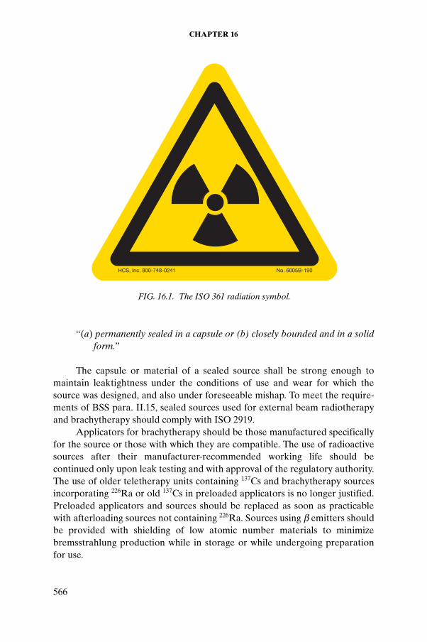
Irradiation heads for external beam radiotherapy, source containers in brachytherapy and other devices containing radioactive sources should have a clear permanent sign indicating the existence of radioactive material (i.e. the ISO 361 symbol).
In addition, when outside the radiotherapy department all devices containing radioactive sources should be labelled with a warning that is recognized as ‘danger’ by any member of the public. The ISO radiation symbol, shown in Fig. 16.1, is not intended to be a warning signal of danger but only of the existence of radioactive material.
Accidents involving members of the public have occurred when the ISO symbol was present but not recognized as indicating danger. This prompted the IAEA to coordinate work on reaching an international agreement for a radiation danger warning sign.
16.10.2. Sealed sources
Paragraph II.15 of the BSS (Appendix II, Medical Exposure, Optimi-zation of Protection for Medical Exposures) states, inter alia, that:
“(e) Radioactive sources for either teletherapy or brachytherapy shall be so constructed that they conform to the definition of a sealed source;…”
A sealed source is defined in the BSS glossary as radioactive material that is:
CHAPTER 16
566
“(a) permanently sealed in a capsule or (b) closely bounded and in a solid form.”
The capsule or material of a sealed source shall be strong enough to maintain leaktightness under the conditions of use and wear for which the source was designed, and also under foreseeable mishap. To meet the require-ments of BSS para. II.15, sealed sources used for external beam radiotherapy and brachytherapy should comply with ISO 2919.
Applicators for brachytherapy should be those manufactured specifically for the source or those with which they are compatible. The use of radioactive sources after their manufacturer-recommended working life should be continued only upon leak testing and with approval of the regulatory authority. The use of older teletherapy units containing 137Cs and brachytherapy sources incorporating 226Ra or old 137Cs in preloaded applicators is no longer justified.Preloaded applicators and sources should be replaced as soon as practicable with afterloading sources not containing 226Ra. Sources using b emitters should be provided with shielding of low atomic number materials to minimize bremsstrahlung production while in storage or while undergoing preparation for use.
HCS, Inc. 800-748-0241 No. 6005B-190
FIG. 16.1. The ISO 361 radiation symbol.
RADIATION PROTECTION AND SAFETY IN RADIOTHERAPY
567
16.10.3. Safety in the design of facilities and ancillary equipment
As a general rule, the design of a radiotherapy facility needs to make provisions for safety systems or devices associated with the equipment and treatment room. This includes electrical wiring related to emergency off switches, safety interlocks and warning signals.
Appropriate methods and data for shielding calculations are presented in ICRP Publication 33 and the NCRP 49 report. An appropriate qualified expert should carry out the overall design of the facility, including shielding calcula-tions. Examples of shielding calculations are given in Section 16.17. Additional information on the design of radiotherapy facilities can be found in IAEA-TECDOC-1040, IEC Report 61859 and a report by the IPEM.
Radiation monitoring equipment should be available on the site in the vicinity of installations using sources of ionizing radiation.
Paragraph II.15 of the BSS (Appendix II, Medical Exposure, Optimi-zation of Protection for Medical Exposures) states, inter alia, that:
“(f) when appropriate, monitoring equipment be installed or be available to give warning of an unusual situation in the use of radiation generators and radionuclide therapy equipment.”
16.10.3.1. Manual brachytherapy
Typical safety features for the storage and preparation of radioactive sealed sources for manual brachytherapy are the following:
● The room should be used only for source storage and preparation by designated and trained personnel.
● The room should be provided with a locked door to control access and maintain source security (see Section 16.12).
● A radiation sign should be posted on the door.● There should be shielded storage (a safe) available for all sources. The
outer surface of the storage shall be made of fireproof materials. The safe should be located near the preparation workbench to reduce the exposure of personnel during the handling and transfer of sources.
● The safe should have compartments for different source activities. Each compartment should be marked so as to permit immediate and easy identification of its contents from the outside with a minimum of exposure.
● The workbench should be provided with an L block shielding with a lead glass viewing window.
CHAPTER 16
568
● The source handling area should be well illuminated and a magnifying glass in a fixed mounting should be available in order to handle sources efficiently and with a minimum of radiation exposure.
● Devices for handling sources, especially forceps, should be available. They should be as long as practicable and compatible with efficient source handling. A device should be provided for threading sources expeditiously, with the fingers protected by distance.
● Sources should be readily identifiable by sight. When radioactive sources of the same appearance but of different activities are used, they should be distinguishable, for example by different coloured threads or beads.
● The working surface for source preparation should be smooth and seamless to avoid loosing small sources such as 192Ir wire fragments.
● The source storage and preparation laboratory should have a sink for cleansing sources, provided with a filter or trap suitable for preventing loss of sources through the drainage system.
● There should be a clear indication of the radiation level in the room. This may be achieved by an area radiation monitor that is visible on entering the room and during any handling of unshielded sources, or a survey meter should be available and in use during source handling.
● Space should be available for secure storage to enable the decay of short half-life sources such as 192Ir.
● Hand carried transport containers must be provided with long handles and the lid of the container must be securely fastened to prevent tipping and dropping of sources during transport. Containers should bear the radiation symbol as well as a warning sign.
● Space should be available for source transport trolleys with source containers.
It is preferable that patient treatment rooms be for individual patients and adjacent to each other. If this is not possible, appropriate shielding between one patient and another is required.
— Shielding should be provided for nurses and visitors of brachytherapy patients, for which movable shields may be used within patient rooms, especially in manual brachytherapy.
— Prior to each treatment, movable shields should be placed close to the patient’s bed in such a way that exposure of the nurses caring for the patient is minimized. This is achieved by anticipating the nurse’s tasks, positions and movements throughout the room.
RADIATION PROTECTION AND SAFETY IN RADIOTHERAPY
569
— The treatment room should contain a shielded storage container (large enough to accept applicators if necessary) and a remote handling tool (forceps) for the event of a dislodged source.
— Sterilization facilities for preloaded applicators, if they are still temporarily used until replacement by remote afterloading applicators, should be available in preparation or treatment rooms to ensure sufficient protection.
— An area monitor should be placed at the treatment room entrance so as to detect when a source or a patient with a source is leaving the room area. In order to ensure that no source remains within the patient, clothes or bed linen, or in the area after treatment, a portable monitor shall be available for monitoring these items.
16.10.3.2. Remote control brachytherapy and external beam radiotherapy
External beam radiotherapy and HDR brachytherapy should be carried out in specially designed treatment rooms within the radiotherapy department, while low dose rate (LDR) remote control brachytherapy can be performed in the ward in the area where manual brachytherapy is performed. The treatment room shielding should be designed in accordance with suitable recommenda-tions (ICRP Publication 33 and the NCRP 49 report). The room should be large enough to accommodate the treatment machine, allowing the full range of motion of the treatment table and patient transport.
With regard to treatment rooms for HDR brachytherapy, IAEA-TECDOC-1040 states the following:
“If the feasibility of sharing a shielded treatment room between an HDR unit and another currently-used treatment machine is considered, it should be carefully evaluated. To avoid scheduling problems considerations should include the anticipated number of HDR procedures as well as the number of external beam treatments. This report recommends against this strategy in most instances.”
Access to the irradiation room shall be furnished with a visible signal indicating whether the radiation source is on or off. A door interlock or other suitable means to prevent unauthorized access should be provided and a power fail safe area radiation monitor should be visible on entering the room. The mechanism should be capable of maintaining irradiation interruption until the door is closed and locked and verification has been made that no person but the patient is inside the room. After an interruption, provided that no
CHAPTER 16
570
operating parameters are changed or reselected, it should be possible to restart the irradiation, but only from the equipment control panel.
One or more emergency off switches should be conveniently placed inside the treatment room to allow interruption of the irradiation from inside the room. The control panel should be installed in such a way that the operator will have a total overview of the access to the irradiation room at all times. Adequate systems, devices or other means should be provided to allow the operator to have a clear and full view of the patient.
The systems for patient observation should be redundant and independent (e.g. closed circuit television or lead glass windows, depending on the type of treatment unit). Oral communication should be possible with the treatment rooms and patients by using an intercom or other communication system. Fire-fighting means should be available in order to preserve the integrity of radioactive sources in the event of a fire. An installed radiation monitor and/or a portable survey instrument should be used to confirm the safe condition of the source.
16.11. SAFETY ASSOCIATED WITH ACCEPTANCE TESTS, COMMISSIONING AND OPERATION
After equipment installation, acceptance tests should be conducted in order to verify that the equipment conforms to the technical specifications given by the manufacturer and to verify compliance with the safety require-ments of the IEC standards. Usually the equipment belongs to the supplier until the acceptance process has been completed. The tests are usually performed by a manufacturer’s representative in the presence of personnel representing the user (a qualified expert in radiotherapy physics), who will decide on acceptance. The first test in the acceptance procedure of a radiation emitting device must be a rigorous area survey of the surroundings of the treatment room that houses the radiation emitting machine.
As discussed in detail in Chapter 10, the tests to be included in the acceptance protocol should be specified in the purchasing conditions and contracts and should clearly establish the responsibility of suppliers for resolving any non-conformity identified during acceptance testing. The grade B and C tests specified in the IEC standard for a particular machine can be used as guidance for preparing the test protocol.
After acceptance and before starting operation, calibration of radiation sources and radiation beams as well as commissioning is performed. These phases are critical to patient safety, as shown in accidental exposures, involving in some instances a large number of patients, in which commissioning tests
RADIATION PROTECTION AND SAFETY IN RADIOTHERAPY
571
were not carried out or were done poorly (see IAEA Safety Reports Series No. 17). During commissioning the qualified expert in radiotherapy physics measures all data required for the clinical use of the machine, including data used in TPSs.
Acceptance tests and commissioning should not be restricted to radiation emitting equipment or sources but should also be conducted for any system that has implications on safety, such as the TPS. Improper commissioning of TPSs has been the cause of several accidental medical overexposures or under-exposures, both detrimental to the treatment outcome.
Quality controls need to be carried out following formally established quality control protocols:
● Periodically under normal operating conditions;● After the source has been installed or replaced;● After repairs or maintenance work carried out on a treatment machine
that have the potential to alter the radiation output.
An independent audit of the calibration of the source should be carried out before starting clinical use of the source. Quality assurance is dealt with in detail in Chapter 10. The BSS requirements on quality assurance for medical exposure are provided in Section 16.14.
Equipment should be operated in accordance with the technical documents, ensuring satisfactory operation at all times in respect of both the tasks to be accomplished and radiation safety. In particular, the manufacturer’s operating manual, and any additional procedures, should be approved in accordance with the quality assurance system (see Section 16.10 for the BSS requirements on equipment) by a national or international body that is responsible for type approval of radiation emitting devices.
Sealed sources should be subject to leak tests prior to the first use and at regular intervals thereafter, in conformity with ISO 9978. Leak tests should be capable of detecting the presence of 0.2 kBq of removable contamination from the sealed source:
— For manual brachytherapy sources the typical method is the direct wet wipe test.
— For external beam radiotherapy and remote control brachytherapy the method to be used is the indirect wipe test of the nearest accessible surface.
— For 226Ra sources immersion or gas emanation tests are adequate; however, 226Ra should be replaced by other radionuclides as soon as practicable.
CHAPTER 16
572
The sterilization process in brachytherapy should be appropriate for preventing damage to sources and applicators that could affect safety.
16.11.1. Safe operation of external beam radiotherapy
Safe operation of external beam treatment units requires procedures for wipe tests, area surveys and interlock checks and procedures for emergencies such as a source that becomes stuck in the on or partially on position. Such procedures require that the necessary equipment be available, calibrated and in working order.
The equipment includes:
● A radiation monitor of the Geiger–Müller (GM) type;● A radiation monitor, ionization chamber type, with scales from micro-
sieverts onwards;● Equipment for wipe tests, such as well counters and multichannel
analysers;● Personal alarm dosimeters, especially for emergency intervention.
The procedures for the use of this equipment should recognize that some instruments will ‘lock up’ in a high radiation field and read erroneously. Hence the procedure should require a three step process:
— Checking the battery; — Checking the monitor response with a check source; — Turning the instrument on and starting to monitor from outside the room
in which the source is located (i.e. from the lower to the higher dose rate areas).
During clinical operation the presence of other staff in the area of the control panel should be limited to the minimum in order to avoid distracting the operator.
16.11.2. Safe operation of brachytherapy
The source strength (usually in terms of the reference air kerma rate) of each brachytherapy source should be determined individually before it is used on a patient (see Chapter 13). The source documentation should be checked carefully. It is essential that the unit of activity used for source calibration be the same as the unit of activity used in the TPS. Some of the accidental exposures in brachytherapy have been caused by errors in the manufacturer’s
RADIATION PROTECTION AND SAFETY IN RADIOTHERAPY
573
specification of the activity of one or several sources and others by the unit of activity used in the hospital being different from the unit stated by the manufacturer (see IAEA Safety Reports Series No. 17 and ICRP Publication 86).
After verification of the source strength, the source or source holder should be marked with unique identifiers (e.g. a pre-established colour) to facilitate visual recognition and to prevent the possibility of confusion among different sources. Containers used for the transport of radioactive sources shall be in conformance with the requirements established in the IAEA Regulations for the Safe Transport of Radioactive Material.
The movements of the sources from the time they have left the safe until their return should be documented and signed by the person responsible for the move (using forms or a logbook). A person should be assigned to be in charge of accountability of the sources. This person should keep a record, with signatures, of the source order and issuance from and return to the safe (see requirements for source security below).
LDR and HDR sources have certain common operating procedures for safe use:
● Source inventories should be performed that show the location and current activity of each source at the facility, with a unique identifier for each source. This may be either a colour coded or a letter–number identifier.
● Sources should never be left on preparation surfaces. They must be in storage, in transit or inside the patient.
● Leak tests (using moist wipes) need to be performed and documented on a periodic basis and should have a sensitivity that is sufficient to detect a very low increase above the background radiation.
● For HDR units wipe tests are only performed on the afterloading drive assembly and transport containers, since the source itself has too high an activity to allow this sort of test.
● Area surveys are to be performed periodically around the source storage facilities for LDR and HDR sources.
● The storage facilities are to be marked to indicate that they contain radioactive material. The person responsible for radiation safety in the event of an emergency should be clearly indicated.
● The storage facilities are to be kept locked at all times.● After every brachytherapy treatment, the patient has to be monitored
with a radiation survey meter so as to ensure that no activity remains in the patient.
CHAPTER 16
574
Specific precautions to be observed during the cutting and handling of 192Ir wires should include ensuring that:
— Appropriate tools and equipment such as forceps, cutting devices, magnifying glasses and good illumination of the work surface are available and used; if 192Ir wires are cut off for immediate use, a container to hold cut lengths should be provided and labelled.
— Radioactive waste is collected and stored in adequate containers.— Surfaces and tools are properly decontaminated.
The following information should be posted for brachytherapy treatments: identification of the patient, sources, date and time of insertion and removal, nursing required, time allowance for nurses and visitors, and concise instructions for unplanned source and applicator removal and for emergency. A patient with a removable source in or upon his or her body should not leave the room unless accompanied by a hospital attendant.
Upon completion of treatment the licensee should ensure that all brachy-therapy sources are removed from the patient, except in the case of permanent implants. The patient should be monitored with a portable detector to ensure that no source remains in or on the patient. Linen, dressings, clothing and equipment should be kept within the room where the removal of sources takes place until all sources are accounted for, and should be monitored with a radiation survey meter. Rubbish bins, soiled dressing bins and laundry baskets coming from a brachytherapy ward or other area where brachytherapy sources are employed should be monitored with a radiation survey meter.
Mobile containers and portable equipment containing radioactive sources should be moved to a storage room or to a secure place when not in use.
16.11.2.1. Safe operation of manual brachytherapy
The following is necessary for the safe operation of manual brachy-therapy:
● The sources should be inspected visually for possible damage after each use by means of magnifying viewers and a leaded viewing window in a shielded work area.
● A diagram should be provided at the source storage safe showing the exact location of each source within the safe; this aids in reducing the time it takes to locate and identify a source.
RADIATION PROTECTION AND SAFETY IN RADIOTHERAPY
575
● The sources should be handled only with long forceps or tongs, never directly with the fingers.
● When transporting the sources a mobile, shielded container is needed and the shortest route possible should be used.
● Sources that come into direct contact with body tissues require cleaning and possible sterilization after each use, which can subject the sources to possible damage from heat, abrasion, chemical attack and mechanical stresses. These sources must therefore be inspected after every use.
● The work surfaces should be easily cleaned and brightly lit to make it easy to find any sources that have been dropped.
● As indicated in Section 16.10, a filter should be used to prevent loss of sources to the drainage system while cleaning in the sink.
16.11.2.2. Safe operation of remote control afterloading brachytherapy
The following is necessary for the safe operation of remote control after-loading brachytherapy:
● Quality control of the afterloader including tests to be performed at the beginning of each treatment day.
● The couplings and transfer tubes need to be checked (for HDR brachy-therapy it has to be done before each treatment) to ensure that there is nothing to prevent source motion.
● Remote afterloading equipment requires specific emergency procedures, which are especially critical in HDR brachytherapy. These procedures are dealt with in Section 16.16.
16.12. SECURITY OF SOURCES
Paragraph 2.34 of the BSS (Requirements for Practices, Technical Requirements) states, inter alia, that:
“Sources shall be kept secure so as to prevent theft or damage and to prevent any unauthorized legal person from carrying out any of the actions specified in the General Obligations for practices of the Standards (see paras 2.7–2.9) by ensuring that:
(a) control of a source not be relinquished without compliance with all relevant requirements specified in the registration or licence and without immediate communication to the Regulatory Authority… of
CHAPTER 16
576
information regarding any decontrolled, lost, stolen or missing source;
(b) a source not be transferred unless the receiver possesses a valid authorization; and
(c) a periodic inventory of movable sources be conducted at appropriate intervals to confirm that they are in their assigned locations and are secure.”
The objective of source security is to ensure continuity in the control and accountability of each source at all times in order to meet the requirement in para. 2.28 of the BSS. Specific provisions shall be made for situations in which loss of control could lead to accidents, such as:
● Storage of sources before installation;● Temporary or permanent cessation in use;● Storage after decommissioning awaiting a decision on source return or
disposal;● Brachytherapy sources remaining in the patient, clothes, bed linen or
treatment area.
To comply with this requirement, the licensee needs to develop procedures to ensure the safe exchange and movement of radioactive sources within the institution and establish controls to prevent theft, loss, unauthorized withdrawal or damage of sources, or entrance of unauthorized personnel to the controlled areas.
The licensee also needs to check the number of sources in a container when removing and when returning the sources and should perform a physical inventory of all sealed sources to confirm that they are present and secure in their assigned locations. The licensee should maintain a source movement log with a record indicating the date of removal, the name of the patient and the return of the source.
Radiotherapy equipment should be equipped with safety systems capable of preventing their use by unauthorized personnel. A key should be required to energize the system, access to which should be restricted to authorized staff. Any loss of a source should be reported immediately to the radiation protection officer, who should report it to the regulatory authority. All linen, dressing, clothing, equipment and refuse containers should be kept within the brachytherapy patient’s room until checks have been performed and documented that sources are not attached to them.
RADIATION PROTECTION AND SAFETY IN RADIOTHERAPY
577
16.13. OCCUPATIONAL EXPOSURE
Detailed requirements for protection against occupational exposure are given in the BSS, while the recommendations on how to meet these require-ments are given in the IAEA Safety Guide on Occupational Radiation Protection, Safety Standards Series No. RS-G-1.1, and the IAEA Safety Guide on Assessment of Occupational Exposure Due to External Sources of Radiation, Safety Standards Series No. RS-G-1.3. A summary of the most relevant issues for radiotherapy is given in this section.
16.13.1. Responsibilities and conditions of service
The parties responsible for protection against occupational exposure are not only the licensees but also the employers. In some cases the employer and the licensee are the same legal person, but in other cases they may be different; for example, the employer of a maintenance engineer may be the maintenance company, while maintenance engineers work in many radiotherapy depart-ments, each one under a different licensee.
16.13.2. Use of dose constraints in radiotherapy
Dose constraints can be used for optimizing protection in the planning stage for each radiation source. Anticipated individual doses should be compared with the appropriate dose constraints and protective measures should be taken to keep doses below the dose constraints. The BSS definition of dose constraint is:
“For occupational exposures, dose constraint is a source related value of individual dose used to limit the range of options considered in the process of optimization.”
Since dose constraints are source related, the source should be specified; for example, when choosing source related dose constraints for the sources involved in a radiotherapy facility, consideration should be given to the fact that medical and paramedical staff may work in more than one hospital and be exposed to sources from two radiotherapy departments (e.g. in one hospital in the morning and in another hospital in the evening).
CHAPTER 16
578
16.13.3. Investigation levels for staff exposure in radiotherapy
Investigation levels are a tool used to provide a ‘warning’ on the need to review procedures and performance, investigate what is not working as expected and take timely corrective action. In radiotherapy, a suitable quantity for use as the investigation level is the monthly effective dose itself, but the dose to the hands can be used as a quantity for the investigation level for staff in manual brachytherapy. In radiotherapy departments in which different staff members are dedicated to specific work or tasks, different investigation levels can be associated with the various tasks.
The following are examples of related tasks and their levels that are rarely exceeded, and therefore could be suitable as investigation levels:
● For staff working only with accelerators or remote control brachytherapy, a monthly investigation level of 0.2 mSv effective dose;
● For staff working with 60Co external beam radiotherapy, brachytherapy nurses and persons inserting and removing manual brachytherapy sources, a monthly investigation level of 0.4 mSv effective dose.
16.13.4. Pregnant workers
Paragraph I.16 of the BSS (Appendix I, Occupational Exposure, Conditions of Service) states that:
“A female worker should, on becoming aware that she is pregnant, notify the employer in order that her working conditions may be modified if necessary.”
Paragraph I.17 of the BSS (Appendix I, Occupational Exposure, Conditions of Service) states that:
“The notification of pregnancy shall not be considered a reason to exclude a female worker from work; however, the employer of a female worker who has notified pregnancy shall adapt the working conditions in respect to occupational exposure so as to ensure that the embryo or foetus is afforded the same broad level of protection as required for members of the public.”
This is especially relevant, for example, in manual brachytherapy, where under normal conditions the dose to the foetus in certain workers may reach
RADIATION PROTECTION AND SAFETY IN RADIOTHERAPY
579
the dose limit for members of the public established in the BSS (see Table 16.1).
16.13.5. Classification of areas
Relevant areas of a practice can be classified as either controlled or supervised, according to the BSS (paras I.21–I.25).
A controlled area is defined as an area in which specific protection measures and safety provisions are needed for controlling normal exposure and for preventing potential exposure. In radiotherapy practice, areas requiring specific protection measures (controlled areas) include:
● All irradiation rooms for external beam radiotherapy; ● Remote afterloading brachytherapy treatment rooms; ● Operating rooms during brachytherapy procedures using real sources;● Brachytherapy patient rooms; ● All radioactive source storage and handling areas.
It is preferable to define controlled areas by physical boundaries such as walls or other physical barriers marked or identified with radiation area signs.
A supervised area is an area that should be kept under review even though specific protection measures and safety provisions are not normally needed. Supervised areas may include areas requiring a regular review of the radiological conditions to determine whether there has been some breakdown of control in the procedures. Supervised areas may involve areas surrounding brachytherapy patient rooms or around radioactive source storage and handling areas.
All areas designated as neither controlled nor supervised areas should be such that persons in them would receive the same level of protection as members of the public.
16.13.6. Local rules and supervision
The rules and procedures listed in Section 16.11 include those needed for occupational protection. Management should make the rules known to those to whom they apply and ensure that they are followed by assigning responsibil-ities for supervision of tasks.
CHAPTER 16
580
16.13.7. Protective equipment and tools
Paragraph I.28 of the BSS (Appendix I, Occupational Exposure, Personal Protective Equipment) states, inter alia, that:
“Employers… and licensees shall ensure that:
(a) workers be provided with suitable and adequate personal protective equipment…”
Protective equipment for radiotherapy is discussed in Section 16.10.
16.13.8. Individual monitoring and exposure assessment
The purpose of monitoring and exposure assessment is to gather and provide information on the actual exposure of workers and to confirm good working practices contributing to reassurance and motivation. The BSS requires individual monitoring for any worker who is normally employed in a controlled area and who may receive a significant occupational exposure.
Those radiotherapy professionals most likely to require individual monitoring are radiation oncologists, qualified experts in radiotherapy physics, radiation protection officers, radiotherapy technologists, source handlers, maintenance staff and any nursing or other staff who must spend time with patients who contain radioactive sources.
Monitoring includes more than just measuring and determining the equivalent dose; it includes interpretation and assessment. Individual external doses can be determined by using individual monitoring devices such as thermoluminescent dosimeters (TLDs) or film badges, which are usually worn on the front of the upper torso (in most radiotherapy procedures the whole body is assumed to be fairly uniformly exposed). When the possibility exists of exposure to the hands, such as in the handling of brachytherapy sources, extremity dosimeters need to be worn (if compatible with clinical practice).
In a radiotherapy department the personal dosimeters should be exchanged at regular intervals not exceeding three months. Moreover, the reports on read dosimeters should become available as soon as possible but no later than within three months after the exchange. Delays in the evaluation of a dosimeter can result in the loss of the stored information.
If an individual’s dosimeter is lost, the licensee shall perform and document an assessment of the dose the individual received and add it to the worker’s dose record. Often the most reliable method for estimating an individual’s dose is to use his or her recent dose history, provided that nothing
RADIATION PROTECTION AND SAFETY IN RADIOTHERAPY
581
unusual occurred in the period. Individual monitoring devices are to be calibrated, and this calibration shall be traceable to a standards dosimetry laboratory.
16.13.9. Monitoring of the workplace
The BSS requires licensees in cooperation with employers to develop programmes for monitoring the workplace (paras I.37–I.40). Initial monitoring is to be conducted immediately after the installation of new radiotherapy equipment and after the replacement of teletherapy sources and remote controlled brachytherapy sources. Initial monitoring should include measure-ments of radiation leakage from equipment within the acceptance tests and area monitoring of occupied space around irradiation rooms.
Monitoring of exposure levels should be conducted through the use of area monitors in teletherapy and HDR treatment rooms. Monitoring of the source storage and handling area is to be conducted with a survey meter immediately following the removal from or return to storage of brachytherapy sources.
Monitoring is to be conducted in association with brachytherapy procedures. Soon after implantation of the sources, a survey should be made of exposure rates in the vicinity of the patient. After removal of the brachy-therapy sources from a patient, a survey is to be performed to confirm removal from the patient and return to shielding of all sources. The transport container should be surveyed before and after brachytherapy procedures.
Monitoring of packages containing radioactive sources upon receipt by the licensee is to be performed. All survey meters used for workplace monitoring must be calibrated, and this calibration shall be traceable to a standards dosimetry laboratory.
16.13.10. Health surveillance
Paragraph I.41 of the BSS (Appendix I, Occupational Exposure, Health Surveillance) states, inter alia, that:
“Employers… and licensees shall make arrangements for appropriate health surveillance in accordance with the rules established by the Regulatory Authority.”
CHAPTER 16
582
The primary purpose of medical surveillance is to assess the initial and continuing fitness of employees for their intended tasks.
Health surveillance programmes shall be based on the general principles of occupational health. No specific health surveillance related to exposure to ionizing radiation is necessary for staff involved in the operation of a radio-therapy practice. Only in the case of overexposed workers at doses much higher than the equivalent dose limits (e.g. 0.2–0.5 Sv or higher) would special investigations involving biological dosimetry and further extended diagnosis and medical treatment be necessary.
Counselling should be available to workers such as women who are or may be pregnant, or are breastfeeding a child, individual workers who have or may have been exposed substantially in excess of the dose limits and workers who may be worried about their radiation exposure. This is particularly necessary for women who are or may be pregnant, such as female technologists working in radiotherapy and nurses working in brachytherapy wards.
16.13.11. Records
Paragraph I.44 of the BSS (Appendix I, Occupational Exposure, Records) states, inter alia, that:
“Employers… and licensees shall maintain exposure records for each worker…”
The exposure records shall include the following:
● Information on the general nature of work involving occupational exposure;
● Information on the doses and data upon which dose assessments have been based;
● When a worker is or has been occupationally exposed while in the employ of more than one employer;
● Information on the dates of employment with each employer and the doses, exposures and intakes in each such employment;
● Records of any doses due to emergency interventions or accidents, which shall be distinguished from doses incurred during normal work.
Employers and licensees shall provide for access by workers to information contained in their own exposure records, and give due care and attention to the maintenance of appropriate confidentiality of records.
RADIATION PROTECTION AND SAFETY IN RADIOTHERAPY
583
16.14. MEDICAL EXPOSURE
The detailed requirements given in Appendix II of the BSS are applicable in particular to radiotherapy sources. In addition, the IAEA Safety Guide on Radiological Protection for Medical Exposure to Ionizing Radiation (Safety Standards Series No. RS-G-1.5) describes strategies to involve organizations outside the regulatory framework, such as professional bodies (those for radiation oncology and medical physics), whose cooperation is essential to ensure compliance with the BSS requirements for medical exposure. Examples that may illustrate this point include acceptance testing processes for radiation equipment, calibration of radiotherapy units and reporting of medical accidental exposure.
Requirements on justification and optimization of protection apply to medical exposure but not to the dose limits. Further, dose constraints do not apply to the exposure of patients as part of their own diagnosis and treatment, but specific dose constraints shall be defined for non-occupational comforters and for medical exposure of individuals exposed for medical research, if these individuals do not benefit directly from the exposure.
16.14.1. Responsibilities for medical exposure
Paragraph II.1 of the BSS (Appendix II, Medical Exposure, Responsibil-ities) states, inter alia, that:
“Registrants and licensees shall ensure that:
(a) no patient be administered a diagnostic or therapeutic medical exposure unless the exposure is prescribed by a medical practitioner;
(b) medical practitioners be assigned the primary task and obligation of ensuring overall patient protection and safety in the prescription of, and during the delivery of, medical exposure;
(c) medical and paramedical personnel be available as needed, and either be health professionals or have appropriate training adequately to discharge assigned tasks in the conduct of the diagnostic or therapeutic procedure that the medical practitioner prescribes;
(d) for therapeutic uses of radiation (including teletherapy and brachytherapy), the calibration, dosimetry and quality assurance requirements of the Standards be conducted by or under the supervision of a qualified expert in radiotherapy physics;…”

CHAPTER 16
584
Furthermore, para. II.1 of the BSS requires that the licensee shall ensure that:
“(f) training criteria be specified or be subject to approval, as appropriate, by the Regulatory Authority in consultation with relevant professional bodies.”
16.14.2. Justification of medical exposure
Paragraph II.4 of the BSS (Appendix II, Medical Exposure, Justification of Medical Exposures) states that:
“Medical exposures should be justified by weighting the diagnostic or therapeutic benefits they produce against the radiation detriment they might cause, taking into account the benefits and risks of available alternative techniques that do not involve medical exposure.”
With respect to medical research, para. II.8 of the BSS states that:
“The exposure of humans for medical research is deemed to be not justified unless it is:
(a) in accordance with the provisions of the Helsinki Declaration and follows the guidelines for its application prepared by Council for International Organizations of Medical Sciences (CIOMS) and WHO; and
(b) subject to the advice of an Ethical Review Committee (or any other institutional body assigned similar functions by national authorities) and to applicable national and local regulations.”
Research on humans in therapeutic procedures should only be performed if there is a direct health benefit to the exposed person.
16.14.3. Optimization of exposure and protection
Paragraph II.18 of the BSS (Appendix II, Medical Exposure, Optimi-zation of Protection for Medical Exposures) states, inter alia, that:
“Registrants and licensees shall ensure that:
RADIATION PROTECTION AND SAFETY IN RADIOTHERAPY
585
(a) exposure of normal tissue during radiotherapy be kept as low as reasonably achievable consistent with delivering the required dose to the planning target volume, and organ shielding be used when feasible and appropriate;
(b) radiotherapeutic procedures causing exposure of the abdomen or pelvis of women who are pregnant or likely to be pregnant be avoided unless there are strong clinical indications;…
(d) any therapeutic procedure for pregnant women be planned to deliver the minimum dose to any embryo or foetus;…”
The optimization of protection in the case of patients is complex and does not necessarily mean the reduction of doses to patients, as priority has to be given to the acquisition of reliable diagnostic information and the achievement of the desired therapeutic effect.
With regard to the exposure of pregnant patients, the ICRP in Publication 84, Pregnancy and Medical Radiation, states:
“Termination of pregnancy is an individual decision affected by many factors. Foetal doses below 100 mGy should not be considered a reason for terminating a pregnancy. At foetal doses above this level, there can be foetal damage, the magnitude and type of which is a function of dose and stage of pregnancy.”
16.14.4. Calibration of radiotherapy sources and machines
Paragraph II.19 of the BSS (Appendix II, Medical Exposure, Optimi-zation of Protection for Medical Exposures) states, inter alia, that:
“Registrants and licensees shall ensure that:
(a) the calibration of sources used for medical exposure be traceable to a Standards dosimetry laboratory;
(b) radiotherapy equipment be calibrated in terms of radiation quality or energy and either absorbed dose or absorbed dose rate at a predefined distance under specified conditions, e.g. following the recommendations given in IAEA Technical Reports Series No. 277;
(c) sealed sources used for brachytherapy be calibrated in terms of activity, reference air kerma rate in air or absorbed dose rate in a specified medium, at a specified distance, for a specified reference date;…
CHAPTER 16
586
(e) the calibrations be carried out at the time of commissioning a unit, after any maintenance procedure that may have an effect on the dosimetry and at intervals approved by the Regulatory Authority.”
At the time of publication of the BSS, the IAEA code of practice based on air kerma in air (IAEA TRS 277) was included in the requirements. More recent protocols (codes of practice) based on standards of absorbed dose to water, such as IAEA TRS 398, were not available at that time. However, it is possible to extend the BSS requirement to the new protocols (see Chapter 9).
Sealed sources used for external beam radiotherapy and brachytherapy need to have a calibration certificate provided by the manufacturer, in accordance with ISO 1677 or its national equivalent standard.
The licensee must implement a protocol for the calibration of radiation sources used for radiotherapy. The regulatory authority should encourage the professional bodies of medical physics to adopt a protocol and require its implementation by the licensees. It is advisable to use international protocols for calibration, as this would avoid confusion and help prevent mistakes. Examples are the calibration procedures described by the IAEA (TRS 277, TRS 381 and TRS 398 for external beams and IAEA-TECDOC-1097 for brachytherapy).
Calibration of new equipment and new radiation sources should be performed independently by at least two different qualified experts in radio-therapy physics and preferably using different dosimetry systems. The results should be compared only after the completion of both measurements.
The licensee should ensure that all teletherapy equipment outputs are compared at least once every two years in a national, regional or international programme for independent dose verification.
One of the simplest mechanisms for independent verifications of external beam calibration or physical dosimetry is participation in the IAEA–WHO thermoluminescence dosimetry postal dose quality audit. The regulatory authority should encourage registrants and licensees to participate in this programme or in similar programmes.
For new brachytherapy sources for which the measurement varies by more than 5% from the manufacturer’s certified activity or air kerma rate in air, the source shall not be used for patient treatment until the difference is investigated further and resolved. The responsibility for the investigation and for further action remains with the licensee, and the investigation is usually performed by a qualified expert in radiotherapy physics, with or without external help.
RADIATION PROTECTION AND SAFETY IN RADIOTHERAPY
587
16.14.5. Clinical dosimetry
Paragraph II.20 of the BSS (Appendix II, Medical Exposure, Optimi-zation of Protection for Medical Exposures) states, inter alia, that:
“Registrants and licensees shall ensure that the following items be determined and documented:…
(b) for each patient treated with external beam radiotherapy equipment, the maximum and minimum absorbed doses to the planning target volume together with the absorbed dose to a relevant point, such as the centre of the planning target volume, plus the dose to other relevant points selected by the medical practitioner prescribing the treatment;
(c) in brachytherapy treatments performed with sealed sources, the absorbed doses at selected relevant points in each patient;…
(e) in all radiotherapeutic treatments, the absorbed doses to relevant organs.”
To meet these requirements (i.e. the items to be determined and the way they are to be determined and documented), a protocol should be used. The ICRU reports are recommended for consultation on such determination and recording.
16.14.6. Quality assurance for medical exposure
Paragraph II.22 of the BSS (Appendix II, Medical Exposure, Optimi-zation of Protection for Medical Exposures) states that:
“Registrants and licensees, in addition to applying the relevant require-ments for quality assurance specified elsewhere in the Standards, shall establish a comprehensive quality assurance programme for medical exposures with the participation of appropriate qualified experts in the relevant fields, such as radiophysics or radiopharmacy, taking into account the principles established by the WHO and the PAHO.”
The regulatory authority should encourage licensees to work with profes-sional associations in the development of such programmes. The licensee should ensure that the programmes are updated on a regular basis. As the development of a national programme may not be feasible in many Member States, a well established and proven international or national programme may
CHAPTER 16
588
be followed (e.g. AAPM TG 40, ESTRO Booklet No. 2, IAEA-TECDOC-1151).
Paragraph II.23 of the BSS (Appendix II, Medical Exposure, Optimi-zation of Protection for Medical Exposures) states that:
“Quality assurance programmes for medical exposures shall include:
(a) measurements of the physical parameters of the radiation generators, imaging devices and irradiation installations at the time of commissioning and periodically thereafter;
(b) verification of the appropriate physical and clinical factors used in patient diagnosis or treatment;
(c) written records of relevant procedures and results; (d) verification of the appropriate calibration and conditions of
operation of dosimetry and monitoring equipment; and(e) as far as possible, regular and independent quality audit reviews of
the quality assurance programme for radiotherapy procedures.”
Following the acceptance of new radiotherapy equipment, sufficient data shall be measured at the commissioning to be used for clinical dosimetry and treatment planning:
● The measured data shall be clearly documented in the workbook;● Before being issued for use in treatment planning, the documentation
shall be independently verified, signed and dated; ● All dosimetry calibrations, clinical dosimetry data and methods of
calculation for therapy equipment are to be reconfirmed at regular intervals.
The measurements and checks carried out for this purpose should be sufficient to detect any significant variations from the data in use. Verification of patient treatment through in vivo dosimetry is advisable under special circumstances. This procedure may not be available in all institutions, but nevertheless it is recommended for incorporation as soon as it becomes feasible.
A routine quality assurance programme is an integral component of modern radiotherapy practice; this programme should include auditing, both internal and external, and continual improvement. These principles need to be linked to the radiation protection programme in order to strengthen safety while at the same time improving quality and efficiency.
RADIATION PROTECTION AND SAFETY IN RADIOTHERAPY
589
Feedback from operational experience and lessons learned from accidents or near misses can help to identify potential problems and correct deficiencies, and should therefore be used systematically as part of the quality assurance programme.
The maintenance of records is an important part of a quality assurance programme. When planning and developing an effective quality assurance programme, licensees need to recognize that it demands a strong managerial commitment and support in the form of training and resources of time, personnel and equipment.
16.14.7. Constraints for comforters and visitors
Dose constraints do not apply to patients. With regard to patients’ comforters and visitors, para. II.27 of the BSS (Appendix II, Medical Exposure, Dose Constraints) recommends the following:
“Registrants and licensees shall constrain any dose to individuals incurred knowingly while voluntarily helping (other than in their occupation) in the care, support or comfort of patients undergoing medical diagnosis or treatment, and to visitors to patients who have received therapeutic amounts of radionuclides or who are being treated with brachytherapy sources, to a level not exceeding that specified in Schedule II, para. II-9.”
Schedule II, para. II-9, of the BSS states, inter alia, that:
“…the dose of any such comforter or visitor of patients shall be constrained so that it is unlikely that his or her dose will exceed 5 mSv during the period of a patient’s diagnostic examination or treatment. The dose to children visiting patients who have ingested radioactive materials should be similarly constrained to less than 1 mSv.”
16.14.8. Discharge of patients
Paragraph II.28 of the BSS (Appendix II, Medical Exposure, Maximum Activity for Patients in Therapy on Discharge from Hospital) states that:
“In order to restrict the exposure of any members of the household of a patient who has undergone a therapeutic procedure with sealed or unsealed radionuclides and members of the public, such a patient shall not be discharged from hospital before the activity of radioactive substances in the body falls below the level specified in Schedule III, Table III-VI. Written
CHAPTER 16
590
instructions to the patient concerning contact with other persons and relevant precautions for radiation protection shall be provided as necessary.”
Table III-VI in the BBS (Schedule III) only includes the value for 131I and sets 1100 MBq as the guidance level for maximum activity for patients in therapy on discharge from hospital. The ICRP has an ongoing task group with the charge of developing guidance for other sources, including those used for permanent implants in brachytherapy, such as 125I and 103Pd.
16.14.9. Investigation of accidental medical exposure
Paragraph II.29 of the BSS (Appendix II, Medical Exposure, Investi-gation of Accidental Medical Exposures) states, inter alia, that:
“Registrants and licensees shall promptly investigate any of the following incidents:
(a) any therapeutic treatment delivered to either the wrong patient or the wrong tissue, or using the wrong pharmaceutical, or with a dose or dose fractionation differing substantially from the values prescribed by the medical practitioner or which may lead to undue acute secondary effects;…
(c) any equipment failure, accident, error, mishap or other unusual occurrence with the potential for causing a patient exposure significantly different from that intended.”
Paragraph II.30 of the BSS (Appendix II, Medical Exposure, Investi-gation of Accidental Medical Exposures) states that:
“Registrants and licensees shall, with respect to any investigation required under para. II.29:
(a) calculate or estimate the doses received and their distribution within the patient;
(b) indicate the corrective measures required to prevent recurrence of such an incident;
(c) implement all the corrective measures that are under their own responsibility;
(d) submit to the Regulatory Authority, as soon as possible after the investigation or as otherwise specified by the Regulatory Authority, a
RADIATION PROTECTION AND SAFETY IN RADIOTHERAPY
591
written report which states the cause of the incident and includes the information specified in (a) to (c), as relevant, and any other information required by the Regulatory Authority; and
(e) inform the patient and his or her doctor about the incident.”
IAEA Safety Reports Series No. 17 and ICRP Publication 86 contain reviews and lessons to be learned from an extensive collection of accidental medical exposures.
16.15. PUBLIC EXPOSURE
16.15.1. Responsibilities
The licensee is responsible for controlling public exposure resulting from a radiotherapy practice. Public exposure is controlled by proper shielding design and, in large part, by ensuring that radiation sources are shielded and secured (e.g. located in a locked area), interlocks are functional and keys to the control panel are secured, to prevent unauthorized access or use. The presence of members of the public in and near the radiotherapy department shall be considered when designing shielding of storage and treatment facilities.
16.15.2. Access control for visitors
The licensee should make arrangements to control access of members of the public to radiotherapy irradiation rooms and to provide adequate information and instruction to these persons before they enter a controlled area so as to ensure appropriate protection (e.g. members of the public shall be accompanied by radiotherapy staff).
16.15.3. Radioactive waste and sources no longer in use
The licensee should notify the regulatory authority and submit a plan for the transfer and disposal of sources if they are no longer in use. The licensee maintains responsibility for the sources until the time of their transfer to another appropriate licensee or to an authorized waste disposal facility; in particular the licensee has to notify the regulatory authority of any intention to transfer or decommission 60Co teletherapy equipment prior to initiating any action. Depleted uranium used as shielding material shall also be treated as radioactive waste; for example, a 60Co teletherapy head may contain depleted uranium and is to be disposed of appropriately.
CHAPTER 16
592
16.15.4. Monitoring of public exposure
Paragraph III.13 of the BSS (Appendix III, Public Exposure, Monitoring of Public Exposure) states, inter alia, that:
“…and licensees shall, if appropriate:
(a) establish and carry out a monitoring programme sufficient to ensure that the requirements of the Standards regarding public exposure to sources of external irradiation be satisfied and to assess such exposure;…
(c) keep appropriate records of the results of the monitoring programmes;…”
The programme for monitoring public exposure from radiotherapy shall include dose assessments in the surroundings of irradiation rooms for external beam radiotherapy, brachytherapy wards, source storage, source preparation rooms and waiting rooms.
16.16. POTENTIAL EXPOSURE AND EMERGENCY PLANS
Requirements on the safety of sources and facilities are set out in Section 16.10. This section focuses on the identification of possible emergency situations or accidents, their prevention, and preparation for and mitigation of them.
16.16.1. Potential exposure and safety assessment
Paragraph IV.3 of the BSS (Appendix IV, Potential Exposure: Safety of Sources, Safety Assessment) states, inter alia, that:
“…licensees shall conduct a safety assessment, either generic or specific for the sources for which they are responsible…”
The assessment is to be provided to the regulatory authority, according to the BSS principal requirements on authorization (paras 2.11–2.13 of the BSS).
Generic safety assessments are suitable for types of equipment with a high degree of uniformity (para. IV.9 of the BSS). As experience in identifying accident scenarios by an individual licensee may be limited, arrangements
RADIATION PROTECTION AND SAFETY IN RADIOTHERAPY
593
between licensees and manufacturers to provide for notification of malfunc-tions and dissemination by feedback to licensees should be encouraged.
16.16.2. Mitigation of consequences: emergency plans
Based on the events identified by the safety assessment, the licensee shall elaborate mitigation measures embodied in a set of emergency procedures. The responsibilities shall be allocated (para. V.2 of the BSS) and the relevant staff shall be trained in the mitigation measures, which shall be periodically rehearsed. The lessons learned from the rehearsals shall be used to review and update the emergency plans. The procedures shall identify the responsibilities of individuals and shall be concise, unambiguous and posted visibly in places where they could be needed.
Only maintenance staff trained and authorized for these tasks should carry out emergency procedures during source change of external beam radio-therapy units and remote control brachytherapy units. If participation of radio-therapy staff is necessary for any of these actions, the scope of this participation shall be restricted to operating the control panel, and responsibilities shall be clearly defined.
For emergency situations there need to be emergency plans that are concise and easily followed, and these should be developed before the startup of a radiation treatment programme. The most frequent types of emergency situation are given below.
16.16.2.1. Lost source
It is critical for this type of event that an up to date inventory exists so that the following can be determined immediately:
● Which sources are missing; ● What is their type and activity; ● Where they were last known to be, and when; ● Who last took possession of the sources.
The area where the sources were last known to be should be closed to entry and exit until a survey has been performed. This search needs to be performed with the most sensitive radiation detection survey meter.
CHAPTER 16
594
16.16.2.2. Stuck source
Emergency procedures need to be short, concise, unambiguous and, if necessary, illustrated with drawings without any explanation text. They need to be suitable for being read at first sight and followed.
● External beam radiotherapy units. Emergency procedures for this event should be posted at the treatment unit. In general, the first step is to use the source driving mechanism to return the source to the shielded position. If this is not immediately successful and there is a patient on the treatment table, the patient should be removed from the area and the area must be secured from further entry. Emphasis should be placed on avoiding exposure of the staff to the primary beam. The radiation protection officer is then notified and takes over control of the situation.
● Remote control brachytherapy units. Emergency plans require an emergency container to be available in the treatment room, as well as an emergency kit containing long handled forceps for manipulation of the source guide tubes and applicators, if the source fails to return to the safe. The emergency container should be placed close to the patient and should be sufficiently large so that it can accept the entire applicator assembly that contains the source and is removed from the patient.
For HDR brachytherapy, the following remark is taken from IAEA-TECDOC-1040:
“High dose rate brachytherapy is potentially a high risk technique and extreme accuracy and care are essential. The short response time required for emergency actions (minutes) imposes the need for the presence of both a physician and physicist trained in emergency procedures during all appli-cations.”
Manufacturers usually provide suggested emergency procedures for an event in which the source fails to return to the safe. They assume that the physical integrity of the applicator is maintained. These procedures are specific to the actual afterloading unit but generally involve the following sequence. Each step assumes that if the previous action fails to lead to recovery, then the following action is required. The generic sequence is as follows:
— Observation at the console of error message and emergency indicators (audible and visible alarms);
— Recovery from the console (e.g. pressing an emergency off button);
RADIATION PROTECTION AND SAFETY IN RADIOTHERAPY
595
— Entry into the room with a portable radiation survey meter (opening the door activates the interlock that retracts the source);
— Monitoring radiation levels in the room; — Recovery from the afterloading unit (by pressing an emergency off
button on the remote afterloading unit); — Manual retraction of the source (using a hand crank); — Patient survey and remote afterloading unit survey (confirming that the
source is in the safe); — Applicator removal and placement in the emergency container; — Patient survey and emergency container survey (to confirm that the
source is not in the patient and is in the emergency container);— Removal of the patient from the vault (with subsequent survey
monitoring).
16.16.2.3. Contamination
There is a very low probability of contamination accidents in radio-therapy departments in which 226Ra and powder form 137Cs sources have been replaced. In the event of a contamination accident it is important that the area be closed to further entry and that all who were in the area remain there to be surveyed and decontaminated, if necessary. If there are windows or other ventilation present, these should be closed. A statement of how to contact the responsible radiation safety individual in the event of an emergency should be available.
16.16.2.4. Off-site accidents
Off-site accidents with major consequences have been caused by loss of security of teletherapy sources not in use. Some of them (in Mexico and Brazil) caused large scale contamination and others external irradiation only (in Thailand and Turkey). Off-site accidents require actions by national and inter-national intervening organizations. Participation of radiotherapy licensees in national emergency plans may be required and should be integrated.
16.16.2.5. Patient accidental exposure
The requirements in the BSS on investigation of accidental medical exposure, including incident reporting and corrective measures to be taken, are referred to in Section 16.14.
CHAPTER 16
596
16.17. GENERAL SHIELDING CALCULATIONS
The three important parameters that influence external radiation exposure are time, distance and shielding:
● The radiation dose received by individuals is proportional to the time they spend in the radiation field.
● The radiation dose generally follows an inverse square law, hence the dose is reduced substantially by increasing the distance from the source of radiation.
● The dose is reduced if shielding attenuates the radiation.
These parameters are involved in shielding design, which basically consists of three steps:
(1) Establishing a design value for the effective dose in the occupied area;(2) Estimating the radiation field in the occupied area if there were no
shielding;(3) Obtaining the attenuation factor that is necessary to reduce the dose
value from the effective dose in (2) to the effective dose in (1).
It is convenient to keep heavily occupied areas as far away as possible from the treatment rooms and to surround these rooms with no occupancy or low occupancy areas (e.g. the roof, with control of access). The treatment room itself should be large enough for easy patient transport in trolleys and also for ease of installation and servicing of the equipment. Design of the room with a maze facility makes a heavy motorized entry door unnecessary for shielding against photons; however, one must still consider shielding against neutrons in a high energy linac facility. With proper design, a maze can make both neutron shielding and a heavy motorized door unnecessary.
Treatment rooms in radiotherapy departments typically fall into one of the following six categories: linac; 60Co teletherapy; orthovoltage X ray; superficial X ray; HDR brachytherapy; and LDR brachytherapy.
Shielding requirements for each of these rooms follow similar rules and conventions; however, each of the rooms introduces a few of its own specific requirements and constraints. If the source room contains only LDR type brachytherapy sources and they are always stored in a locked shielded safe within the room, except while preparing the sources behind a shield, the room may not require special shielding.
A patient room that houses manual afterloading LDR brachytherapy patients may not need shielding if mobile lead shields are used around the
RADIATION PROTECTION AND SAFETY IN RADIOTHERAPY
597
patient’s bed. Installations housing linacs, teletherapy machines, X ray machines or HDR remote afterloading devices all require special shielding to protect the operators, staff, patients and public in adjacent areas.
16.17.1. Step one: Design dose in occupied areas (annual dose and weekly
dose)
The design effective dose rate P (in Sv/a or Sv/week) in a given occupied area (Table 16.2) is derived by constrained optimization (i.e. by selecting a source related dose constraint), with the condition that the individual effective doses from all relevant sources will be well below the prescribed effective dose limits for persons occupying the area to be shielded. However, when using constraints for shielding calculations, consideration should be given to the remark made in ICRP Publication 33 (para. 256) stating that actual dose values to individuals in the occupied areas are to be 1/10 (for equivalent dose) to 1/30 of the effective dose used as the shielding design parameter. This is due to a number of conservative assumptions made in the calculation. Typical conserv-ative assumptions in the calculations are the following:
● The attenuation of the beam by the patient is usually not considered.● The maximum possible leakage radiation is assumed.● The workload, as well as the use and occupancy factors, is overestimated.● An assumption is made that staff are always in the most exposed place of
the occupied area.● For linacs producing X rays and electrons an assumption is made that the
linac always operates in the X ray mode.● For dual energy linacs an assumption is made that the linac always runs in
the higher energy mode.
Since some of these conservative assumptions may be unavoidable to cover uncertainties when using constraints, a critical review of conservative assumptions should be performed so as to achieve a balanced decision and avoid accumulation of over-conservative measures that may go far beyond optimization.
The use of P = 0.01 mSv/week corresponds to an area for permanent occupation by members of the public and may lead to a high value of shielding thickness (of the order of 230–250 cm of regular concrete for 20 MV photons in an area exposed to the primary beam at a distance of about 4 m from the source).
This thickness of regular concrete will be reduced by 50 cm or more if the area is designated as a controlled area and the dose constraint of 0.2 mSv/week

CHAPTER 16
598
is used. This approach would be consistent with keeping the console area away from the public, who may cause distractions or have an effect on other safety aspects. This solution, however, requires individual monitoring of persons who work in or come frequently to the area of the console, but is not necessary for persons who work in the area only occasionally.
The relation between effective dose P and the air kerma in air values for the radiation field is made through the personal dose equivalent Hp(d) for penetrating radiation, where d = 10 mm. An accepted conservative assumption is to make the personal dose equivalent numerically equal to the effective dose P and to the air kerma in air value (Gy/week) of the radiation field.
16.17.2. Step two: Calculation of the radiation field (air kerma in air) in the
occupied area without shielding
The following parameters are used for calculation of the effective dose without shielding:
● Primary radiation, which is the radiation directly emitted from the treatment machine through the collimator opening in the case of external sources and from the radioactive source in the case of brachytherapy.
● Scatter radiation, which is the radiation produced by the scattering of the primary radiation beam from various media struck by the primary beam, such as the patient, collimators, beam shaping accessories and air.
● Leakage radiation, which is the radiation that escapes through the shielded head of the therapy unit (for accelerators leakage radiation only exists while the beam is on; for cobalt units leakage radiation is always present).
TABLE 1. TYPICAL VALUES FOR DESIGN EFFECTIVE DOSE P IN OCCUPIED AREAS ADJACENT TO A RADIOTHERAPY TREATMENTROOM
Annual effective dose (mSv/a)
Weekly effective dose (mSv/week)
Occupational worker 10 0.2
Member of the public 0.5 0.01
RADIATION PROTECTION AND SAFETY IN RADIOTHERAPY
599
● The workload W, which is defined as the machine output per week or per year at a well defined point (usually the machine isocentre in the treatment room) and is expressed in Gy/week or Gy/a, respectively.
16.17.3. Step three: Attenuation by shielding barriers
The primary barrier is the portion of the treatment room walls or ceiling that may be irradiated directly by the primary beam. Therapy machines are usually located on the lowest floor of a building, so shielding of the floor against primary, scattered and leakage radiation is not of concern.
Secondary barriers are all portions of the treatment room walls, floor or ceiling that cannot be irradiated directly by the primary beam. These barriers provide shielding against the scatter and leakage radiation produced in the treatment room.
The use factor U is the fraction of the beam-on time during which the primary beam is directed towards a particular barrier. The following primary beam use factors are usually assumed for external beam machines: U (floor) = 1; U (walls) = 0.25; U (ceiling) = 0.25. For all secondary barriers U is always equal to 1, since secondary radiation is always present when the beam is on.
The occupancy factor T is a factor with which the workload is multiplied to account for the degree of occupancy of the area in question. Typical values are T (offices) = 1; T (corridors) = 0.25; T (waiting rooms) = 0.125.
The half-value layer (HVL) and the tenth-value layer (TVL) are those thicknesses of attenuating material that decrease the photon beam intensities to 50% and 10%, respectively, of the original value (100%).
The barrier transmission factor B provides the fraction of the incident beam air kerma in air transmitted through a given thickness of shielding material. Primary, scatter and leakage barrier transmission factors Bpri, Bscat
and Bleak, respectively, must be calculated, and the required barrier thickness is then determined using published graphs of transmission factors against shielding material thickness for various beam energies and shielding materials.
Shielding materials are materials used in primary and secondary barriers to provide shielding against the primary, scatter and leakage radiation produced in the radiotherapy treatment room. The most common materials used for the shielding of external beam and brachytherapy treatment facilities are: ordinary concrete (density: 2.35 g/cm3), barite concrete (density: up to 3.2 g/cm3), high density concrete (density: up to 5 g/cm3), steel (density: 7.9 g/cm3) and lead (density: 11.3 g/cm3).
To compute the radiation levels beyond the radiation barriers in areas adjacent to treatment rooms, transmission data are required, which take into
CHAPTER 16
600
account not only the primary beam attenuation but also the radiation scattered in the shielding itself (broad beam geometry data).
Narrow beam geometry only includes primary radiation, so that the point of interest does not receive any scattered radiation; for example, to measure the primary beam intensity transmitted through an attenuator, a narrow beam geometry (with suitable collimation and barrier to detector distance) is set up so that the scatter does not reach the detector. Narrow beam geometry is useful to characterize beam quality in the kilovoltage photon range (see Chapter 9).
In broad beam geometry the detector (or the radiation worker) receives not only the transmitted beam but also the scatter from or through the barrier. Sets of broad beam transmission and graphics data for common use in radio-therapy installations are provided in ICRP Publication 33 and the NCRP 49 report.
If KP and KS represent the primary and the scattered air kerma in air ‘seen’ by the detector, respectively, the buildup B for a barrier is given by:
B = (KP + KS)/KP (16.9)
For narrow beam geometry B = 1 and for broad beam geometry B > 1, where B refers to buildup, not to be confused with the transmission factors Bpri, Bscat and Bleak in the text below.
16.18. TYPICAL LINAC INSTALLATION
The main components of a typical linac installation are the treatment room, entrance maze, control room and optional mechanical–electrical room. The maze connects the treatment room with the control room, which houses the operational controls of the linac. The treatment room and maze together are called the linac bunker.
A schematic diagram of a typical installation for an isocentric high energy linac is given in Fig. 16.2. The thickness of the primary and secondary barriers is determined through first determining the transmission factors and then determining the barrier thickness required to achieve the calculated trans-mission.
16.18.1. Workload
Typical linac workload figures vary depending on initial assumptions. An example of slightly conservative assumptions is:
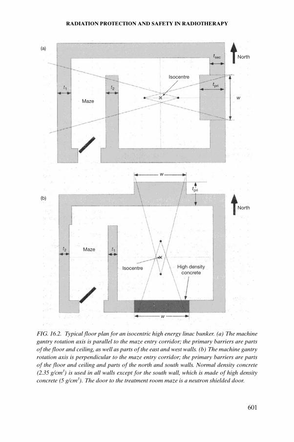
RADIATION PROTECTION AND SAFETY IN RADIOTHERAPY
601
(a)
(b)
North
North
Maze
t2t1
Isocentre
tpri
w
tpri
w
tsec
High density concrete
Isocentre
t2 t1Maze
w
FIG. 16.2. Typical floor plan for an isocentric high energy linac bunker. (a) The machine gantry rotation axis is parallel to the maze entry corridor; the primary barriers are parts of the floor and ceiling, as well as parts of the east and west walls. (b) The machine gantry rotation axis is perpendicular to the maze entry corridor; the primary barriers are parts of the floor and ceiling and parts of the north and south walls. Normal density concrete (2.35 g/cm3) is used in all walls except for the south wall, which is made of high density concrete (5 g/cm3). The door to the treatment room maze is a neutron shielded door.
CHAPTER 16
602
● The clinical workload Wclin for 50 patients per working day, 3.3 Gy delivered dose at the isocentre per patient (pt), five working days per week and 52 working weeks per year:
(16.10)
● The physics workload Wphys includes use of the linac for calibration, quality assurance, phantom measurements, servicing and maintenance. A conservative estimate of this is Wphys = 7100 Gy/a, resulting in the following total linac workload. Total linac workload Wtot at the machine isocentre:
Wtot = Wclin + Wphys = 5 × 10⁴ Gy/a ~ 103 Gy/week (16.11)
In shielding calculations for dual energy linacs a conservative assumption is usually made that the accelerator will operate at the higher energy 100% of the time.
16.18.2. Calculation of the primary barrier transmission factor
The primary barrier transmission factor Bpri is calculated from the following relationship:
(16.12)
where
d0 is the linac source to axis distance (SAD) (usually 1 m); dpri is the distance from the linac target (X ray source) to the point of interest
(usually 0.3 m outside the wall or ceiling to be shielded); W is the total workload of the linac; U is the barrier use factor; T is the occupancy factor at the point of interest.
The primary beam of an external beam unit should only be directed towards primary barriers with sufficient shielding. If part of the primary shielding is incorporated into the equipment with the use of a retractable beam stopper, electrical and/or mechanical interlocks should be provided to prevent the possibility that the radiation beam is directed towards the primary barriers when the beam stopper is not intercepting the beam.
WclinGypt
ptsday
daysweek
weeksyear
Gyyear
= ¥ ¥ ¥ =3 3 50 5 52 42900.
BP d d
WUTpripri 0 =
( / )2
RADIATION PROTECTION AND SAFETY IN RADIOTHERAPY
603
The beam stopper is usually made of lead with a thickness adequate to attenuate the primary radiation beam to 0.1% of its original value (typically 3 TVLs or 10 HVLs, amounting to about 10–15 cm of lead for megavoltage photon beams). Treatment machines equipped with beam stoppers are cumbersome with regard to patient set-up on the treatment machine; however, the beam stoppers minimize the required thickness of the primary barriers and are thus used in installations in which space constraints prevent the use of adequate primary barrier thickness. With the use of beam stoppers, the primary barrier wall thickness becomes close to that required for secondary barriers.
16.18.3. Calculation of the scatter barrier transmission factor
The scatter barrier transmission factor Bscat is determined from:
(16.13)
where
d0 is the SAD of the linac; d1 is the distance from the patient to the point of interest; d2 is the distance from the target to the scattering volume (patient); a is the ratio of the scattered radiation at 1 m from the scattering object
(patient) to the primary radiation at 1 m; F0 is an average field area (400 cm2); F is the actual field size at the position of the patient.
The scattering coefficient a depends on the photon beam energy and scattering angle. Its typical value for 90º scatter is 10–4–10–3. The Compton scattering formula predicts a maximum energy of 0.511 keV (mec
2) for 90º scatter and a maximum energy of 0.255 keV (0.5 mec
2) for 180º scatter.
16.18.4. Calculation of the leakage barrier transmission factor
The leakage barrier transmission factor Bleak is calculated assuming a beam attenuation due to linac head shielding transmission of 0.1%. The energy of the leakage radiation is assumed to be the same as that of the primary radiation. Thus:
BP d d d d F F
aWTscat0 0 0=
( / ) ( / ) ( / )12
22
CHAPTER 16
604
(16.14)
where
d0 is the SAD of the linac; d3 is the distance from the isocentre to the point of interest (average
distance for all possible gantry positions); W is the workload of the linac; T is the occupancy factor.
16.18.5. Determination of barrier thickness
Using broad beam transmission data from ICRP Publication 33 or the NCRP 49 report, one can determine the required primary, leakage and scatter barrier thickness for the calculated transmission factors Bpri, Bleak and Bscat, respectively.
The barrier thickness determined for leakage radiation is usually larger than that determined for scatter radiation. One rule of thumb is that if the leakage barrier thickness exceeds the scatter barrier by more than 3 HVLs (i.e. ~1 TVL), then just the leakage barrier thickness is applied as the required wall thickness. However, if the thicknesses for the two barriers are within 3 HVLs, then one extra HVL is added to the leakage barrier thickness to determine the required secondary barrier thickness to account for the slight increase in effective dose due to the scattered component.
Ordinary concrete is the most common shielding material in megavoltage therapy installations (Table 16.3). Other materials may be used to conserve space, since for megavoltage beams the required primary barrier thickness is inversely proportional to the density of the shielding material (see the Compton effect, Section 1.4.6). It should be kept in mind, however, that
TABLE 16.3. TYPICAL SHIELDING THICKNESS OF ORDINARY CONCRETE TO PROTECT MEMBERS OF THE PUBLIC IN AREAS ADJACENT TO COBALT UNITS OR HIGH ENERGY LINAC BUNKERS
Radiation quality Primary barrier (cm) Secondary barrier (cm)
Co-60 130 65
10–25 MV 240 120
BP d d
WTleak0=
10 33
2( / )
RADIATION PROTECTION AND SAFETY IN RADIOTHERAPY
605
replacing ordinary concrete with other materials will have serious financial implications; for example, high density concrete (5 g/cm3) will cut the barrier thickness roughly to half of that required for ordinary concrete, but on a per volume basis the costs of the shielding material will increase by a factor of 30. The difference is even more pronounced when steel or lead is used for shielding.
16.18.6. Consideration of neutron production in a high energy linac
In high energy (above 10 MV) linac installations, neutrons are produced by X ray–neutron (x, n) and electron–neutron (e, n) reactions. The neutron contamination is produced by high energy photons and electrons incident on the target, primary collimator, beam flattening filter, collimator jaws, beam accessories, air and the patient. The cross-section for a (x, n) reaction is at least an order of magnitude larger than that for an (e, n) reaction; hence neutrons produced by the linac X ray mode are of primary concern. For this reason, the maximum photon energy produced by a linac rather than the maximum electron energy is considered the more significant contributor to the neutron dose.
Neutrons can activate other elements, which remain radioactive and will contribute to the radiation exposure of radiotherapy staff entering the treatment room after a high energy photon beam treatment. The radionuclides from activated components of a linac are generally short lived (of the order of seconds to a few minutes).
A similar problem is posed by the direct activation of elements in (x, n) reactions, such as 15O (half-life 2 minutes) and 13N (half-life 10 minutes). The radioactivity in treatment room air is removed by efficient room ventilation. The ventilation also handles the removal of ozone and noxious gases, in addition to the removal of radioactive gases, through 6–8 air exchanges per hour.
The concrete primary and secondary barriers designed to protect against photon dose are quite adequate to protect against electrons and contamination neutrons. However, neutrons undergoing multiple scattering along the maze can present an unacceptable radiation level in the control area, thus requiring a specially designed door.
16.18.7. Door of a linac room
The door of a high energy linac installation may require shielding against X rays and neutrons scattered through the maze towards the linac control area. High energy neutrons are more of a problem than low energy photons.
CHAPTER 16
606
Neutrons are thermalized and absorbed with a layer of about 12 cm of borated polyethylene in the door, which is followed by about 2.5 cm of lead to absorb the g rays produced by neutron capture reactions in boron nuclei. An alternative to the special neutron shielding door is a double maze design, which avoids the need for a shielded door.
16.18.8. Other considerations
A ‘radiation area’ sign (along with a visible red light) needs to be provided above the door to the treatment room, and preferably also on the control room door, to indicate a beam-on condition. There should be audio communication with the patient and emergency switches inside the room to shut off the radiation in the event of an emergency.
In some cases (e.g. installation of a linac in an existing 60Co room or in a small room) installation of the machine with a beam stopper may be necessary.
The manufacturer often quotes a primary beam stopper that attenuates the primary beam by a factor of 0.001 (about 3 TVLs), reducing the primary barrier thickness requirement by 3 TVLs. If the remaining primary barrier thickness is less than the thickness required for scatter and leakage, the whole wall can be made of a uniform thickness as required for a scattering and leakage barrier.
16.19. SHIELDING DESIGN FOR BRACHYTHERAPY FACILITIES
HDR brachytherapy treatment rooms are designed with similar constraints as are linac and teletherapy rooms. There is, however, one major difference: in brachytherapy rooms all walls are primary barriers, since the source can generally be positioned anywhere in the room and the radiation is emitted isotropically and uncollimated from the source. The attenuation in the patient is not considered in primary barrier transmission calculations. The workload specification is given in terms of air kerma in air per week or year.
The typical workload specification for a remote afterloading HDR 192Ir facility is determined using the following data:
● Maximum source activity: 370 GBq (10 Ci).● Maximum number of patients treated: 10/day.● Number of working days (or treatment days) per week: 5 days/week.● Maximum treatment time: 10 min (for 10 Ci) per patient.● Air kerma rate constant for 192Ir:
RADIATION PROTECTION AND SAFETY IN RADIOTHERAPY
607
111 mGy·m2/(GBq·h) = 4.1 mGy·m2/(mCi·h)
● Workload:
W = 104 × 10 × 5 × 10 × (1/60) × 4.1 mGy·m2/week = 3.4 × 10⁵ mGy·m2/week
Although the treatment time increases due to decay, the product (activity × time) remains the same and hence the example for the computation is made for the maximum treatment time with a 370 GBq (10 Ci) source. All parameters are conservative, introducing a safety factor in the calculated workload. For older HDR units using 60Co a maximum of 20 source pellets may be used, each pellet with a maximum activity of about 1.85 GBq (500 mCi/pellet) for a total source activity of 370 GBq (10 Ci). The maximum treatment time and the number of patients per day, however, remain the same for a 60Co HDR remote afterloading unit as in the HDR 192Ir case.
The workload for planning an HDR 60Co brachytherapy room comes to about 1.1 × 10⁶ mGy·m2/week, using GAKR of 308.5 (mGy·m2)/(GBq·h).
In the case of LDR remote afterloading 137Cs units, two patients may be treated simultaneously with a maximum of 18 sources per patient, the activity of each source being around 1110 GBq (30 mCi). The treatment time for LDR brachytherapy is assumed to be 40 h/week.
BIBLIOGRAPHY
AMERICAN ASSOCIATION OF PHYSICISTS IN MEDICINE, Comprehensive QA for radiation oncology: Report of AAPM Radiation Therapy Committee Task Group 40, Med. Phys. 21 (1994) 581–618.
EUROPEAN SOCIETY FOR THERAPEUTIC RADIOLOGY AND ONCOLOGY, Quality assurance in radiotherapy, Radiother. Oncol. 35 (1995) 61–73.
FOOD AND AGRICULTURE ORGANIZATION OF THE UNITED NATIONS, INTERNATIONAL ATOMIC ENERGY AGENCY, INTERNATIONAL LABOUR ORGANISATION, OECD NUCLEAR ENERGY AGENCY, PAN AMERICAN HEALTH ORGANIZATION, WORLD HEALTH ORGANIZATION, International Basic Safety Standards for Protection against Ionizing Radiation and for the Safety of Radiation Sources, Safety Series No. 115, IAEA, Vienna (1996).
INSTITUTE OF PHYSICS AND ENGINEERING IN MEDICINE, The Design of Radiotherapy Treatment Room Facilities (STEDEFORD, B., MORGAN, H.M., MAYLESS, W.P.M., Eds), IPEM, York, United Kingdom (1997).
CHAPTER 16
608
INTERNATIONAL ATOMIC ENERGY AGENCY (Vienna)
Absorbed Dose Determination in Photon and Electron Beams, 2nd Edition, Technical Reports Series No. 277 (1997).
Method for the Development of Emergency Response Preparedness for Nuclear or Radiological Accidents, IAEA-TECDOC-953 (1997).
The Use of Plane Parallel Chambers in High Energy Electron and Photon Beams, Technical Reports Series No. 381 (1997).
Design and Implementation of a Radiotherapy Programme: Clinical, Medical Physics,Radiation Protection and Safety Aspects, IAEA-TECDOC-1040 (1998).
Assessment of Occupational Exposure Due to External Sources of Radiation, IAEA Safety Standards Series No. RS-G-1.3 (1999).
Calibration of Brachytherapy Sources, IAEA-TECDOC-1079 (1999).
Occupational Radiation Protection, IAEA Safety Standards Series No. RS-G-1.1 (1999).
Absorbed Dose Determination in External Beam Radiotherapy, Technical ReportsSeries No. 398 (2000).
Aspectos Físicos de la Garantía de Calidad: Protocolo de Control de Calidad, IAEA-TECDOC-1151 (2000).
Lessons Learned from Accidental Exposures in Radiotherapy, Safety Reports Series No. 17 (2000).
Regulations for the Safe Transport of Radioactive Material, 1996 Edition (Revised), IAEA Safety Standards Series No. TS-R-1 (ST-1, Rev.) (2000).
Radiological Protection for Medical Exposure to Ionizing Radiation, IAEA Safety Standards Series No. RS-G-1.5 (2002).
INTERNATIONAL COMMISSION ON RADIOLOGICAL PROTECTION, Protection Against Ionizing Radiation from External Sources Used in Medicine, Publication 33, Pergamon Press, Oxford and New York (1982).
— 1990 Recommendations of the International Commission on Radiological Protection, Publication 60, Pergamon Press, Oxford and New York (1991).
— Pregnancy and Medical Radiation, Publication 84, Pergamon Press, Oxford and New York (2000).
— Prevention of Accidents to Patients Undergoing Radiation Therapy, Publication 86, Pergamon Press, Oxford and New York (2002).
RADIATION PROTECTION AND SAFETY IN RADIOTHERAPY
609
INTERNATIONAL ELECTROTECHNICAL COMMISSION (Geneva)
Medical Electrical Equipment — Part 2: Particular Requirements for the Safety of Therapeutic X-ray Generators, IEC 601-2-8 (1987).
General Requirements for Safety. 4. Collateral Standard: Programmable Electrical Medical Systems, IEC 60601-1-4 (1997).
Guidelines for Radiotherapy Treatment Rooms Design, IEC 61859 (1997).
Medical Electrical Equipment — Part 2: Particular Requirements for the Safety of Gamma Beam Therapy Equipment, IEC 60601-2-11 (1997).
Medical Electrical Equipment — Part 2-1: Particular Requirements for the Safety of Electron Accelerators in the Range of 1 MeV to 50 MeV, IEC 60601-2-1 (1998).
Medical Electrical Equipment — Part 2-17: Particular Requirements for the Safety of Remote-controlled Automatically-driven Gamma-ray Afterloading Equipment, IEC 60601-2-17 (1998).
Medical Electrical Equipment — Part 2: Particular Requirements for the Safety of Radiotherapy Simulators, IEC 60601-2-29 (1999).
Medical Electrical Equipment: Requirements for the Safety of Treatment Planning Systems, IEC 62C/62083 (in preparation).
INTERNATIONAL ORGANIZATION FOR STANDARDIZATION, Basic Ionizing Radiation Symbol, ISO 361, ISO, Geneva (1975).
— Radiation Protection — Sealed Radioactive Sources — General Requirements and Classification, ISO 2919, ISO, Geneva (1998).
— Radiation Protection — Sealed Sources — Leakage Test Methods, ISO 9978, ISO, Geneva (1992).
McGINLEY, P.H., Shielding Techniques for Radiation Oncology Facilities, 2nd edn, Medical Physics Publishing, Madison, WI (1998).
NATIONAL COUNCIL ON RADIATION PROTECTION AND MEASURE-MENTS, Protection Against Radiation from Brachytherapy Sources, Rep. 40, NCRP, Bethesda, MD (1972).
— Structural Shielding Design and Evaluation for Medical Use of X Rays and Gamma Rays of Energies up to 10 MeV, Rep. 49, NCRP, Bethesda, MD (1976).
— Radiation Protection Design Guidelines for 0.1 MeV - 100 MeV Particle Accelerator Facilities, Rep. 51, NCRP, Bethesda, MD (1977).