chapter 15:magnetic resonance imaging · 15.2.3 gradient coil design and specifications ... •...
TRANSCRIPT

IAEAInternational Atomic Energy Agency
Slide set of 242 slides based on the chapter authored by
Martin O. Leach
of the IAEA publication (ISBN 978-92-0-131010-1):
Diagnostic Radiology Physics:
A Handbook for Teachers and Students
Objective:
To familiarize the student with practical issues associated
with MRI.
Chapter 15: Magnetic Resonance
Imaging
Slide set prepared
by E. Berry (Leeds, UK and
The Open University in
London)

IAEA Diagnostic Radiology Physics: A Handbook for Teachers and Students – 15. Slide 1 (02/242)
CHAPTER 15 TABLE OF CONTENTS
15.2. Hardware
15.3. Basic image quality issues
15.4. MR Image acquisition and reconstruction
15.5. Artefacts
15.6. Safety and bioeffects

IAEA
15.2 HARDWARE15.2
Diagnostic Radiology Physics: A Handbook for Teachers and Students - 15.2 Slide 1 (03/242)

IAEA
15.2 HARDWARE15.2
Hardware overview
� Several major components
� Components are under control of digital systems that
• provide instructions
• monitor system performance
• acquire and process
• signals used to create images
• or spectroscopic signals reporting on a range of tissue states
� Digital systems are coordinated by one or more computers
• workstations or PCs
• provide interface to the operator
Diagnostic Radiology Physics: A Handbook for Teachers and Students - 15.2 Slide 2 (03/242)

IAEA
15.2 HARDWARE15.2
Computer interface with operator
� Acquisitions
• planned
• executed
� Images
• calculated
• displayed
• stored
� Measurement and analysis software
• for addressing particular clinical questions
Diagnostic Radiology Physics: A Handbook for Teachers and Students - 15.2 Slide 3 (04/242)

IAEA
15.2 HARDWARE15.2
Major components of a clinical magnetic resonance imaging
system utilising a superconducting magnet
Diagnostic Radiology Physics: A Handbook for Teachers and Students - 15.2 Slide 4 (05/242)

IAEA
15.2 HARDWARE15.2.1 THE STATIC MAGNETIC FIELD SUBSYSTEM
Diagnostic Radiology Physics: A Handbook for Teachers and Students - 15.2.1 Slide 1 (07/242)

IAEA
15.2 HARDWARE15.2
Major components
� 15.2.1 The static magnetic field subsystem
� 15.2.2 The radiofrequency (RF) subsystem
� 15.2.3 Gradient coil design and specifications
� 15.2.4 Computer and control systems
� 15.2.5 Common imaging options
Diagnostic Radiology Physics: A Handbook for Teachers and Students - 15.2.1 Slide 2 (06/242)

IAEA
15.2 HARDWARE15.2.1 The static magnetic field subsystem
Requirements for static magnetic field subsystem
� Must provide a highly uniform and stable magnetic field
• Larmor resonance condition must be met over the imaging volume
to within about 1 part per million (ppm)
� Achieve by
• main magnet design
• additional static and electronic shims (adjustments) to correct for
• imperfections in magnet design
• effects of nearby static steel structures
• effects of the patient’s intrinsic magnetic susceptibility
Diagnostic Radiology Physics: A Handbook for Teachers and Students - 15.2.1 Slide 3 (09/242)

IAEA
15.2 HARDWARE15.2.1 The static magnetic field subsystem
Magnet designs and field strengths
� Resistive solenoid, < ∼ 0.5 T
� Superconducting magnet
• clinical use: 0.5 – 3.0 T
• experimental clinical use: 3.0 – 8 T and above
� Electromagnets, 0.1 – 0.2 T
� Permanent magnets
� Geometry relates directly to the intrinsic orientation of the
magnetic field
Diagnostic Radiology Physics: A Handbook for Teachers and Students - 15.2.1 Slide 4 (010/242)

IAEA
15.2 HARDWARE15.2.1 The static magnetic field subsystem
Three different superconducting magnets
� A: Siemens 3T Vario system
� B: Philips 1T Panorama system
� C: General Electric 1.5T Discovery MR450 system.
Diagnostic Radiology Physics: A Handbook for Teachers and Students - 15.2.1 Slide 5 (011/242)

IAEA
15.2 HARDWARE15.2.1 The static magnetic field subsystem
Resistive solenoid
� Simplest design
� Long solenoid generates uniform magnetic field
� Solenoid reduced to typically 4 or 6 coils, separated by
spacers
� Coils may have different diameters
� Uniformity maximised by calculating optimal
• relative current density
• number of windings
• diameter
� Both horizontal and vertical configurations have been built
Diagnostic Radiology Physics: A Handbook for Teachers and Students - 15.2.1 Slide 6 (12/242)

IAEA
Resistive solenoid
Advantages
� Relatively cheap
Disadvantages
� Produce considerable heat
• require water cooling
• resistance changes with
temperature
� Constraints of power supplies
� High current demand
� Maximum field strength 0.5 T
Diagnostic Radiology Physics: A Handbook for Teachers and Students - 15.2.1 Slide 7 (13/242)
15.2 HARDWARE15.2.1 The static magnetic field subsystem

IAEA
15.2 HARDWARE15.2.1 The static magnetic field subsystem
Superconducting magnets
� Design based on similar principles to resistive solenoid
� Usually horizontal bore geometry
� A number of coils in parallel, but wound from
superconducting cable
• niobium/tin encased in copper
• maintained at liquid helium temperatures (4 K, -269 °C)
� Considerably reduced current requirements compared with
resistive solenoid
� Higher field strengths possible 0.5 – 3.0 T or above
Diagnostic Radiology Physics: A Handbook for Teachers and Students - 15.2.1 Slide 8 (14/242)

IAEA
15.2 HARDWARE15.2.1 The static magnetic field subsystem
Cryostat features
� Coils are encased in a vacuum cryostat
• designed to limit heat conductance
• includes a number of thermal layers
• liquid helium surrounded by
• helium vapour barrier
• then vacuum
� Helium vapour is re-liquefied by cryocompressors
• so very little liquid helium is needed during normal operation
� Earlier designs had layers of liquid and gaseous nitrogen
instead
• this needed frequent replenishment to minimise helium boil-off
Diagnostic Radiology Physics: A Handbook for Teachers and Students - 15.2.1 Slide 9 (15/242)

IAEA
15.2 HARDWARE15.2.1 The static magnetic field subsystem
Cryocompressor failure
� May be caused by fault, loss of chilled water, loss of power
� Remaining liquid helium cryogen will boil off
• superconducting windings no longer adequately cooled
• windings become resistive
• windings heat up rapidly due to large current
� Results in a magnet quench
• current is rapidly resistively dissipated as heat
• rapid boil off of any remaining cryogen
� Cryostat is designed to vent resulting release of gas safely
to the outside of the building
Diagnostic Radiology Physics: A Handbook for Teachers and Students - 15.2.1 Slide 10 (16/242)

IAEA
15.2 HARDWARE15.2.1 The static magnetic field subsystem
Shielding
� Up to 3.0 T, system usually self-shielded
� A second, reversed, magnetic field is generated
• cancels much of the field outside the magnet
• retains strong magnetic field in the magnet bore
� Extent of magnetic field considerably reduced
• makes site planning and facility management easier
• reduces area of restricted access
• reduces effects on nearby equipment
Diagnostic Radiology Physics: A Handbook for Teachers and Students - 15.2.1 Slide 11 (17/242)

IAEA
15.2 HARDWARE15.2.1 The static magnetic field subsystem
Variations in geometry
� Interventional systems
• accessible central aperture perpendicular to the magnet axis
� Vertically oriented field
• signal to noise advantages in receiver coils when patient is
horizontal
• as in Philips I T Panorama system
Diagnostic Radiology Physics: A Handbook for Teachers and Students - 15.2.1 Slide 12 (18/242)

IAEA
15.2 HARDWARE15.2.1 The static magnetic field subsystem
Electromagnets
� Incorporate a ferromagnetic core into resistive design
• reduces the electrical current requirement of resistive magnets
• increases stability
• minimises cooling requirements
� Lower field than superconducting, e.g. 0.1 – 0.2 T
� Typically have vertically oriented field between pole faces
� Increased access to patient compared with most
superconducting systems
Diagnostic Radiology Physics: A Handbook for Teachers and Students - 15.2.1 Slide 13 (19/242)

IAEA
15.2 HARDWARE15.2.1 The static magnetic field subsystem
Permanent magnets
� Predominantly have vertical field format
• Also horizontal field system, with patient upright, used for
examining joints
� Field constrained between top and bottom pole faces
• design requires a flux return path
� Advantages
• small stray field
� Disadvantages
• heavy
• cannot be switched off
Diagnostic Radiology Physics: A Handbook for Teachers and Students - 15.2.1 Slide 14 (20/242)

IAEA
15.2 HARDWARE15.2.1 The static magnetic field subsystem
Magnetic field homogeneity
� Typically require at least 1 ppm over field of view (FOV)
• tolerances of magnet design and the environment e.g. structural
steel in building
• adjust magnetic field at installation by shimming
• steel shims
• additional magnetic fields from adjustable currents in shim coils
� Some approaches require about 0.1 ppm over FOV
• fat suppressed imaging and MR spectroscopy
• adjust magnetic field prior to study using shim coils
• compensate for distortions in magnetic field caused by magnetic
susceptibility of the patient
Diagnostic Radiology Physics: A Handbook for Teachers and Students - 15.2.1 Slide 15 (21/242)

IAEA
15.2 HARDWARE15.2.1 The static magnetic field subsystem
Siting issues
� Safety issues – section 15.6
� Effect on magnet homogeneity of
• adjacent steel
• moving steel objects
• nearby current carrying cables
� Adequate structural strength
� Effect of magnet on adjacent equipment
Diagnostic Radiology Physics: A Handbook for Teachers and Students - 15.2.1 Slide 16 (22/242)

IAEA
15.2 HARDWARE15.2.1 The static magnetic field subsystem
Site for static magnetic field - homogeneity
� Contains 0.5 mT field contour within a secure zone with
controlled access
� Magnet homogeneity
• Self shield to minimise effect of structural steel
• but it may need replacing with non ferromagnetic stainless steel
• Seek advice from manufacturer regarding
• Large steel pipes and electrical cables with heavy current load
• Moving steel from vehicles, trains, subway trains
Diagnostic Radiology Physics: A Handbook for Teachers and Students - 15.2.1 Slide 17 (23/242)

IAEA
15.2 HARDWARE15.2.1 The static magnetic field subsystem
Site for static magnetic field – structural and other equipment
� Design must account for structural factors
• floor loading
• equipment access
• avoid sources of vibration
� Other sensitive equipment with potential to be affected
• gamma cameras
• x-ray systems
• radiotherapy linear accelerators
• image intensifiers
• electron microscopes
Diagnostic Radiology Physics: A Handbook for Teachers and Students - 15.2.1 Slide 18 (24/242)

IAEA
15.2 HARDWARE15.2.2 THE RADIOFREQUENCY (RF) SUBSYSTEM
Diagnostic Radiology Physics: A Handbook for Teachers and Students - 15.2.2 Slide 1 (25/242)

IAEA
15.2 HARDWARE15.2
Major components
� 15.2.1 The static magnetic field subsystem
� 15.2.2 The radiofrequency (RF) subsystem
� 15.2.3 Gradient coil design and specifications
� 15.2.4 Computer and control systems
� 15.2.5 Common imaging options
Diagnostic Radiology Physics: A Handbook for Teachers and Students - 15.2.2 Slide 2 (06/242)

IAEA
15.2 HARDWARE15.2.2 The radiofrequency (RF) subsystem
Requirements for the RF subsystem
� Generation of analogue audio frequency pulses
• modulate the RF Larmor frequency
• generated from a digital source and amplified to drive the RF coils
� RF coils transmit and receive RF to and from the patient
� Amplifier may be capable of supplying 15 kW of power
• to drive a coil that can irradiate the whole body
� Weak detected RF signal
• provides information that builds images or spectra
• receive channel(s) must be protected from the transmitted power
• several stages of amplification
Diagnostic Radiology Physics: A Handbook for Teachers and Students - 15.2.2 Slide 3 (27/242)

IAEA
15.2 HARDWARE15.2.2 The radiofrequency (RF) subsystem
Coil designs
� Volume
• transmit and receive
� Surface
• transmit and receive
� Phased array
• receive
Diagnostic Radiology Physics: A Handbook for Teachers and Students - 15.2.2 Slide 4 (28/242)
IAEA
15.2 HARDWARE15.2.2 The radiofrequency (RF) subsystem
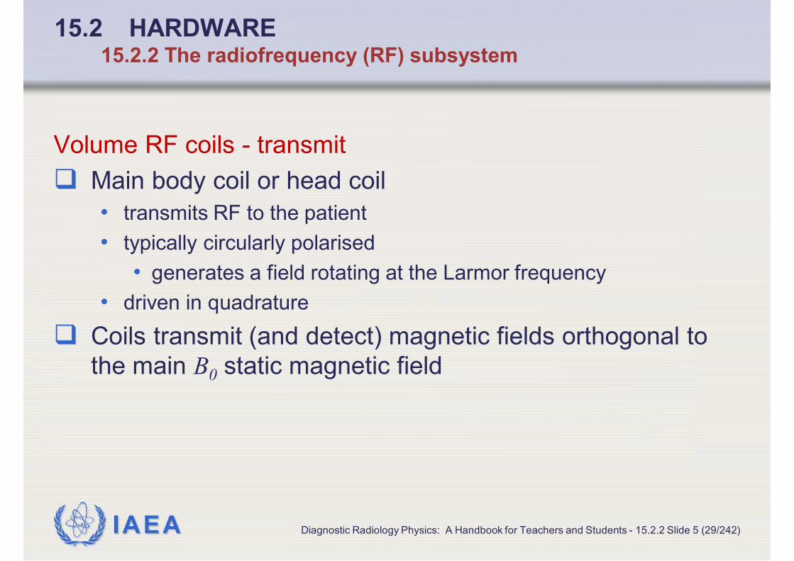
Volume RF coils - transmit
� Main body coil or head coil
• transmits RF to the patient
• typically circularly polarised
• generates a field rotating at the Larmor frequency
• driven in quadrature
� Coils transmit (and detect) magnetic fields orthogonal to
the main B0 static magnetic field
Diagnostic Radiology Physics: A Handbook for Teachers and Students - 15.2.2 Slide 5 (29/242)

IAEA
15.2 HARDWARE15.2.2 The radiofrequency (RF) subsystem
Volume RF coils – Parallel transmit systems
� Overcome the inhomogeneous B1 transmit field that arise
as wavelength of RF approaches dimensions of the body
� In 3 T systems and above
� Array of transmit coils
� Each supplied by separate amplifier
� Allows individual amplitude and phase modulation
Diagnostic Radiology Physics: A Handbook for Teachers and Students - 15.2.2 Slide 6 (30/242)

IAEA
15.2 HARDWARE15.2.2 The radiofrequency (RF) subsystem
Volume RF coils - receive
� Main body coil used as a receiver
• allows imaging of large volumes of the body
• multi-station body imaging
• images of different sections of the body are joined together to
give a whole body image
� Also head coil, knee coil, extremity coil
� Range of designs, including birdcage
• based on two circular loops connected by parallel rods
Diagnostic Radiology Physics: A Handbook for Teachers and Students - 15.2.2 Slide 7 (31/242)

IAEA
15.2 HARDWARE15.2.2 The radiofrequency (RF) subsystem
Surface RF coils - transmit
� Smaller than body coil
� Positioned over volume of interest in the body
� Particularly for multi-nuclear measurements
Surface RF coils – receive
� Provide increased signal to noise from small volumes
� Flexible designs for many parts of the body
Diagnostic Radiology Physics: A Handbook for Teachers and Students - 15.2.2 Slide 8 (32/242)

IAEA
15.2 HARDWARE15.2.2 The radiofrequency (RF) subsystem
Phased array RF coils – receive
� Extension of surface coil approach
� Set of closely positioned separate coils
• each connected to a separate parallel receive channel
• signal is acquired from many surface coils simultaneously
� Improved image quality and faster image acquisition
� Some systems allow body to be covered with such coils
• optimised imaging of many different body parts with no need to
move patient to position additional coils
� Multinuclear measurements
• coils tuned to required frequency
• or may operate several coils at different frequencies
Diagnostic Radiology Physics: A Handbook for Teachers and Students - 15.2.2 Slide 9 (33/242)

IAEA
15.2 HARDWARE15.2.2 The radiofrequency (RF) subsystem
� Top row:
• Siemens 32 channel
head coil
• General Electric torso
array coil
• Philips neck coil
� Bottom row:
• General Electric
cervical, thoracic and
lumbar spine coil
• Four channel flex coils
Diagnostic Radiology Physics: A Handbook for Teachers and Students - 15.2.2 Slide 10 (34/242)

IAEA
15.2 HARDWARE15.2.2 The radiofrequency (RF) subsystem
Siting issues: RF shielding
� RF screened room
� Avoid RF interference to other equipment
• including radio and television reception
� Prevent interference from external sources
• interfering with detection of very weak RF signal
Diagnostic Radiology Physics: A Handbook for Teachers and Students - 15.2.2 Slide 11 (35/242)

IAEA
15.2 HARDWARE15.2.2 The radiofrequency (RF) subsystem
Siting issues: RF screened room
� Made of copper, aluminium or stainless steel
� RF screened windows
� RF doors with knife edge brushes to maintain screen
integrity
� Attached to good earth point
� All services non-conducting, RF filtered and incorporate
non-conducting break
� Wave guide access channels for non-conducting services
Diagnostic Radiology Physics: A Handbook for Teachers and Students - 15.2.2 Slide 12 (36/242)

IAEA
15.2 HARDWARE15.2.3 GRADIENT COIL DESIGN AND SPECIFICATIONS
Diagnostic Radiology Physics: A Handbook for Teachers and Students - 15.2.3 Slide 1 (37/242)

IAEA
15.2 HARDWARE15.2
Major components
� 15.2.1 The static magnetic field subsystem
� 15.2.2 The radiofrequency (RF) subsystem
� 15.2.3 Gradient coil design and specifications
� 15.2.4 Computer and control systems
� 15.2.5 Common imaging options
Diagnostic Radiology Physics: A Handbook for Teachers and Students - 15.2.3 Slide 2 (06/242)

IAEA
15.2 HARDWARE15.2.3 Gradient coil design and specifications
Requirements for gradient coils
� Spatial localisation of image information
• governed by three orthogonal sets of coils
• superimpose a field gradient
• add to, or subtract from, main B0 static magnetic field
� Larmor frequency changes linearly with position in
• x (usually horizontal: orthogonal to B0) direction
• y (usually vertical: orthogonal to B0) direction
• z (along B0) direction
Diagnostic Radiology Physics: A Handbook for Teachers and Students - 15.2.3 Slide 3 (39/242)

IAEA
15.2 HARDWARE15.2.3 Gradient coil design and specifications
Gradient coil design
� For standard superconducting magnet
� For x and y direction
• two pairs of Golay coils
• shaped like a saddle positioned on a cylinder
� For x direction
• pair of coils coaxial with magnet windings
� Mounted on a substantial former placed concentrically
within the room temperature bore of the magnet
Diagnostic Radiology Physics: A Handbook for Teachers and Students - 15.2.3 Slide 4 (40/242)

IAEA
15.2 HARDWARE15.2.3 Gradient coil design and specifications
Gradient coil design
� Capable of rapid switching
� Carry high currents of order of 600 A
• as may generate fields up to about 60 mTm-1
� Require cooling because of high power dissipation
• often water cooled
� Noise and vibration addressed by
• high mass and stiff mountings
• balanced force designs
• mounting within a vacuum jacket
• active noise cancellation
Diagnostic Radiology Physics: A Handbook for Teachers and Students - 15.2.3 Slide 5 (41/242)

IAEA
15.2 HARDWARE15.2.3 Gradient coil design and specifications
Gradient amplitude and slew rate (rise time)
� Large gradients
• allow thin slices and high resolution images
• reduce impact of B0 field inhomogeneities
• can aid diffusion weighted imaging by allowing larger b values with
faster imaging times
� Fast switching
• allows fast imaging sequences
• is defined in terms of slew rate (or rise time)
� Slew rate is the rate of change of the gradient
Diagnostic Radiology Physics: A Handbook for Teachers and Students - 15.2.3 Slide 6 (42/242)

IAEA
15.2 HARDWARE15.2.3 Gradient coil design and specifications
Maximum slew rate (rise time)
� Maximum slew rate is maximum rate of change of gradient
• 200 Tm-1s-1 possible currently
� In practice, maximum used is limited by induction of
currents in body that can lead to nerve stimulation
� For large gradients, slew rate may be slower to ensure
safe operation
� A given machine may offer a choice of gradient modes
Diagnostic Radiology Physics: A Handbook for Teachers and Students - 15.2.3 Slide 7 (43/242)

IAEA
15.2 HARDWARE15.2.3 Gradient coil design and specifications
Eddy current effects
� Calculated gradient pulse waveform
• linear ramp up, maximum value held for a defined period, linear
ramp down
• all within a specified total time
� But, get a non-perfect response arising from
• inductance of gradient coils
• presence of currents in adjacent conducting structures resulting
from the time varying magnetic field generated by the gradient coils
i.e. eddy currents
� Eddy currents can lead to time varying magnetic fields that
distort the image information
Diagnostic Radiology Physics: A Handbook for Teachers and Students - 15.2.3 Slide 8 (44/242)

IAEA
15.2 HARDWARE15.2.3 Gradient coil design and specifications
Compensation for eddy current effects
� Screened gradient designs
• cancel out gradient fields outside magnet bore
• thus reduce generation of eddy currents that would generate
additional magnetic field gradients
� Design of magnet cryostat and heat shields can minimise
generation of eddy currents
� Apply pre-emphasis corrections in gradient circuitry
• impose additional LC terms on the driving waveform to
compensate for eddy current modulation
• performance tuned by manufacturer
Diagnostic Radiology Physics: A Handbook for Teachers and Students - 15.2.3 Slide 9 (45/242)

IAEA
15.2 HARDWARE15.2.4 COMPUTER AND CONTROL SYSTEMS
Diagnostic Radiology Physics: A Handbook for Teachers and Students - 15.2.4 Slide 1 (46/242)

IAEA
15.2 HARDWARE15.2
Major components
� 15.2.1 The static magnetic field subsystem
� 15.2.2 The radiofrequency (RF) subsystem
� 15.2.3 Gradient coil design and specifications
� 15.2.4 Computer and control systems
� 15.2.5 Common imaging options
Diagnostic Radiology Physics: A Handbook for Teachers and Students - 15.2.4 Slide 2 (06/242)

IAEA
15.2 HARDWARE15.2.4 Computer and control systems
Pulse programmer
� Interprets pulse sequences
� Generates and delivers waveforms to different equipment
components
• gradient amplifiers
• RF amplifiers
� Receives and digitises signals at appropriate times
� Access to pulse programmer
• Sometimes user has direct access to generate own sequences
• More usually, user has access using an interface that allows a
limited class of variables to be adjusted for a given sequence
Diagnostic Radiology Physics: A Handbook for Teachers and Students - 15.2.4 Slide 3 (48/242)
IAEA
15.2 HARDWARE15.2.4 Computer and control systems
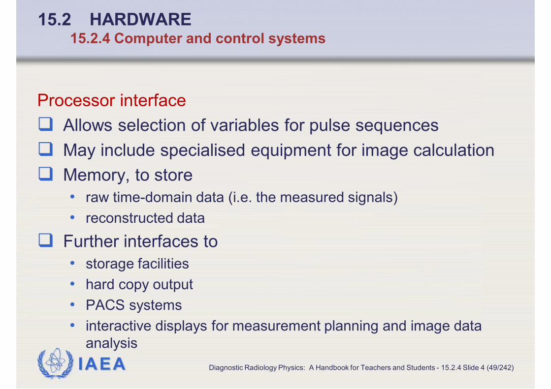
Processor interface
� Allows selection of variables for pulse sequences
� May include specialised equipment for image calculation
� Memory, to store
• raw time-domain data (i.e. the measured signals)
• reconstructed data
� Further interfaces to
• storage facilities
• hard copy output
• PACS systems
• interactive displays for measurement planning and image data
analysis
Diagnostic Radiology Physics: A Handbook for Teachers and Students - 15.2.4 Slide 4 (49/242)

IAEA
15.2 HARDWARE15.2.5 COMMON IMAGING OPTIONS
Diagnostic Radiology Physics: A Handbook for Teachers and Students - 15.2.5 Slide 1 (50/242)

IAEA
15.2 HARDWARE15.2
Major components
� 15.2.1 The static magnetic field subsystem
� 15.2.2 The radiofrequency (RF) subsystem
� 15.2.3 Gradient coil design and specifications
� 15.2.4 Computer and control systems
� 15.2.5 Common imaging options
Diagnostic Radiology Physics: A Handbook for Teachers and Students - 15.2.5 Slide 2 (06/242)

IAEA
15.2 HARDWARE15.2.5 Common imaging options
Choice of equipment for clinical requirements
� MRI systems
• core equipment configurations plus
• packages marketed for clinical applications
� Steps when choosing a system
• Helpful first to develop a clinical specification
• General considerations
• lead to choice of magnet configuration
• Specific equipment considerations
Diagnostic Radiology Physics: A Handbook for Teachers and Students - 15.2.5 Slide 3 (52/242)

IAEA
15.2 HARDWARE15.2.5 Common imaging options
General considerations
� Clinical requirements
� Space
� Budget
� Reliability of power supplies
� Availability of cryogens
� Quality and lifetime cost of engineering support
� Cost of maintenance support
Diagnostic Radiology Physics: A Handbook for Teachers and Students - 15.2.5 Slide 4 (53/242)

IAEA
15.2 HARDWARE15.2.5 Common imaging options
Specific considerations
� Once magnet configuration decided:
� Gradient specification (if an option)
� Clinical packages
� RF coils
Diagnostic Radiology Physics: A Handbook for Teachers and Students - 15.2.5 Slide 5 (54/242)

IAEA
15.3 BASIC IMAGE QUALITY ISSUES15.3
Diagnostic Radiology Physics: A Handbook for Teachers and Students - 15.3 Slide 1 (55/242)

IAEA
15.3 BASIC IMAGE QUALITY ISSUES15.3
Basic image quality issues
Major determinants (there are many other features)
� 15.3.1 B0 field strength, homogeneity and shimming
� 15.3.2 B1 homogeneity and flip angle adjustment
� 15.3.3 Phantoms, equipment assessment and coil
loading
� 15.3.4 Signal-to-noise ratio (SNR) and contrast-to-
noise ratio
� 15.3.5 Spatial resolution
� 15.3.6 Image acquisition time
Diagnostic Radiology Physics: A Handbook for Teachers and Students - 15.3 Slide 2 (56/242)

IAEA
15.3 BASIC IMAGE QUALITY ISSUES15.3.1 B0 FIELD STRENGTH, HOMOGENEITY AND
SHIMMING
Diagnostic Radiology Physics: A Handbook for Teachers and Students - 15.3.1 Slide 1 (57/242)

IAEA
15.3 BASIC IMAGE QUALITY ISSUES15.3
Basic image quality issues
� 15.3.1 B0 field strength, homogeneity and shimming
� 15.3.2 B1 homogeneity and flip angle adjustment
� 15.3.3 Phantoms, equipment assessment and coil
loading
� 15.3.4 Signal-to-noise ratio (SNR) and contrast-to-
noise ratio
� 15.3.5 Spatial resolution
� 15.3.6 Image acquisition time
Diagnostic Radiology Physics: A Handbook for Teachers and Students - 15.3.1 Slide 2 (58/242)

IAEA
15.3 BASIC IMAGE QUALITY ISSUES15.3.1 B0 field strength, homogeneity and shimming
B0 field strength
� Determines signal to noise
� Signal to noise increases approximately linearly with
magnetic field strength
Diagnostic Radiology Physics: A Handbook for Teachers and Students - 15.3.1 Slide 3 (59/242)

IAEA
15.3 BASIC IMAGE QUALITY ISSUES15.3.1 B0 field strength, homogeneity and shimming
B0 homogeneity and shimming
� Fundamental determinant of the quality of an MRI system
� Determined by design of magnet system
� Governed by
• type of magnet
• size of magnet
• larger (longer) the better the homogeneity
• optimisation at installation
• avoid structural steel etc.
• careful shimming at set up (passive and electronic)
• demonstration by engineers that meets specification
Diagnostic Radiology Physics: A Handbook for Teachers and Students - 15.3.1 Slide 4 (60/242)

IAEA
15.3 BASIC IMAGE QUALITY ISSUES15.3.1 B0 field strength, homogeneity and shimming
Changes in B0 homogeneity
� Sudden deterioration could arise from
• changes in environmental steel
• small items of steel that have entered vicinity of magnet and been
attracted to the magnet
� Investigate these potential causes
Diagnostic Radiology Physics: A Handbook for Teachers and Students - 15.3.1 Slide 5 (61/242)

IAEA
15.3 BASIC IMAGE QUALITY ISSUES15.3.1 B0 field strength, homogeneity and shimming
Shimming
� Operator shimming of B0 for some imaging sequences e.g.
• frequency dependent fat suppression
• frequency dependent water excitation
� Performed by adjusting current in a number of room
temperature shim coils incorporated into the system
� Shimming required in spectroscopy to compensate for B0
inhomogeneities arising from susceptibility of patient
Diagnostic Radiology Physics: A Handbook for Teachers and Students - 15.3.1 Slide 6 (62/242)

IAEA
15.3 BASIC IMAGE QUALITY ISSUES15.3.1 B0 field strength, homogeneity and shimming
Other causes of spatial distortion in images
� Gradient non-linearities
� Eddy currents
� Inadequate pre-emphasis correction
Diagnostic Radiology Physics: A Handbook for Teachers and Students - 15.3.1 Slide 7 (63/242)

IAEA
15.3 BASIC IMAGE QUALITY ISSUES15.3.2 B1 HOMOGENEITY AND FLIP ANGLE
ADJUSTMENT
Diagnostic Radiology Physics: A Handbook for Teachers and Students - 15.3.2 Slide 1 (64/242)

IAEA
15.3 BASIC IMAGE QUALITY ISSUES15.3
Basic image quality issues
� 15.3.1 B0 field strength, homogeneity and shimming
� 15.3.2 B1 homogeneity and flip angle adjustment
� 15.3.3 Phantoms, equipment assessment and coil
loading
� 15.3.4 Signal-to-noise ratio (SNR) and contrast-to-
noise ratio
� 15.3.5 Spatial resolution
� 15.3.6 Image acquisition time
Diagnostic Radiology Physics: A Handbook for Teachers and Students - 15.3.2 Slide 2 (65/242)

IAEA
15.3 BASIC IMAGE QUALITY ISSUES15.3.2 B1 homogeneity and flip angle adjustment
B1 homogeneity
� Expectation is that specified flip angle is delivered to
imaging volume uniformly and accurately
� For each new patient MR system performs a calibration
• adjusts voltage delivered to body coil to compensate for
• loading of the coil by the patient (changes coil Q)
• particular coil configurations
• interaction of RF field with subject leading to standing wave and
dielectric effects (at higher fields)
� Calibration allows adjustments for RF pulse lengths, slice
thicknesses and so on
Diagnostic Radiology Physics: A Handbook for Teachers and Students - 15.3.2 Slide 3 (66/242)

IAEA
15.3 BASIC IMAGE QUALITY ISSUES15.3.2 B1 homogeneity and flip angle adjustment
B1 homogeneity in practice
� A number of factors lead to inaccurate pulse angles being
delivered to all or part of the volume
• physical geometry of the transmit coil
• presence of receiver coils within transmit coil
• effects of standing waves or dielectric effects
• imperfect shape of slice select pulses (finite pulse length)
� Conventional diagnostic imaging - useable image
information but
• reduced image contrast and reduced signal to noise ratio
� Quantitative imaging – need to address or compensate in
analysis
Diagnostic Radiology Physics: A Handbook for Teachers and Students - 15.3.2 Slide 4 (67/242)

IAEA
15.3 BASIC IMAGE QUALITY ISSUES15.3.3 PHANTOMS, EQUIPMENT ASSESSMENT AND
COIL LOADING
Diagnostic Radiology Physics: A Handbook for Teachers and Students - 15.3.3 Slide 1 (68/242)
IAEA
15.3 BASIC IMAGE QUALITY ISSUES15.3
Basic image quality issues
� 15.3.1 B0 field strength, homogeneity and shimming
� 15.3.2 B1 homogeneity and flip angle adjustment
� 15.3.3 Phantoms, equipment assessment and coil
loading
� 15.3.4 Signal-to-noise ratio (SNR) and contrast-to-
noise ratio
� 15.3.5 Spatial resolution
� 15.3.6 Image acquisition time
Diagnostic Radiology Physics: A Handbook for Teachers and Students - 15.3.3 Slide 2 (69/242)

IAEA
15.3 BASIC IMAGE QUALITY ISSUES15.3.3 Phantoms, equipment assessment and coil loading
Phantoms
� Manufacturer provides standard phantoms for basic
routine service procedures
• usually simple
• allow comparison with manufacturer’s specification and
maintenance tolerances
• but do not fully evaluate scanner performance
Diagnostic Radiology Physics: A Handbook for Teachers and Students - 15.3.3 Slide 3 (70/242)

IAEA
15.3 BASIC IMAGE QUALITY ISSUES15.3.3 Phantoms, equipment assessment and coil loading
Phantoms for equipment assessment
� More sophisticated phantoms than the manufacturer’s
� Some commercially available
� Use for
• spatial resolution
• T1 and T2 relaxation time measurements
• spatial distortion assessment
• SNR measurements
� Systematic approach valuable
Diagnostic Radiology Physics: A Handbook for Teachers and Students - 15.3.3 Slide 4 (71/242)

IAEA
15.3 BASIC IMAGE QUALITY ISSUES15.3.3 Phantoms, equipment assessment and coil loading
SNR measurements
� Need RF coils to be appropriately loaded
� Achieve by
• including a conducting (ionic) solution in the phantom or
• use a separate loading annulus with phantom
� Must check that results accurate and reflect clinical
situation
� Valuable to make SNR measurement for each imaging coil
• controlled and reproducible conditions
• regularly
• will demonstrate if equipment is operating outside its specification
Diagnostic Radiology Physics: A Handbook for Teachers and Students - 15.3.3 Slide 5 (72/242)

IAEA
15.3 BASIC IMAGE QUALITY ISSUES15.3.3 Phantoms, equipment assessment and coil loading
If local or surface coil is operating outside its specification
� Repeat measurement with body coil
• rule out/establish if fault is with coil or main system
� Call manufacturer or
� Undertake further tests to investigate
• distortion or image artefacts
• phantom with known structures
• ghosting or aliasing
• 3D objects with asymmetries
Diagnostic Radiology Physics: A Handbook for Teachers and Students - 15.3.3 Slide 6 (73/242)

IAEA
15.3 BASIC IMAGE QUALITY ISSUES15.3.4 SIGNAL-TO-NOISE RATIO (SNR) AND CONTRAST-
TO-NOISE RATIO
Diagnostic Radiology Physics: A Handbook for Teachers and Students - 15.3.4 Slide 1 (74/242)

IAEA
15.3 BASIC IMAGE QUALITY ISSUES15.3
Basic image quality issues
� 15.3.1 B0 field strength, homogeneity and shimming
� 15.3.2 B1 homogeneity and flip angle adjustment
� 15.3.3 Phantoms, equipment assessment and coil
loading
� 15.3.4 Signal-to-noise ratio (SNR) and contrast-to-
noise ratio
� 15.3.5 Spatial resolution
� 15.3.6 Image acquisition time
Diagnostic Radiology Physics: A Handbook for Teachers and Students - 15.3.4 Slide 2 (75/242)

IAEA
15.3 BASIC IMAGE QUALITY ISSUES15.3.4 Signal-to-noise ratio (SNR) and contrast-to-noise ratio
Signal-to-noise ratio (SNR)
� Signal is highly dependent on
• object
• imaging sequence
• parameters chosen
� SNR must be defined for specific
• object
• coil
• geometry
• measurement sequence
Diagnostic Radiology Physics: A Handbook for Teachers and Students - 15.3.4 Slide 3 (76/242)

IAEA
15.3 BASIC IMAGE QUALITY ISSUES15.3.4 Signal-to-noise ratio (SNR) and contrast-to-noise ratio
Distribution of noise in MR image
� Should be distributed uniformly throughout the image
• uniform distribution of noise expected from use of Fourier
transform
� May not be uniform if temporal frequency filter applied in
reconstruction or acquisition
• E.g. with parallel processing or array coils, when distribution of
noise may be complex
Diagnostic Radiology Physics: A Handbook for Teachers and Students - 15.3.4 Slide 4 (77/242)

IAEA
15.3 BASIC IMAGE QUALITY ISSUES15.3.4 Signal-to-noise ratio (SNR) and contrast-to-noise ratio
Determination of noise in MR image
� Subtract two identical images
� Not affected by
• non-uniform distribution of noise
• directional propagation of artefacts
� Is appropriate MR noise which has a Rician distribution
Diagnostic Radiology Physics: A Handbook for Teachers and Students - 15.3.4 Slide 5 (78/242)

IAEA
15.3 BASIC IMAGE QUALITY ISSUES15.3.5 SPATIAL RESOLUTION
Diagnostic Radiology Physics: A Handbook for Teachers and Students - 15.3.5 Slide 1 (79/242)

IAEA
15.3 BASIC IMAGE QUALITY ISSUES15.3
Basic image quality issues
� 15.3.1 B0 field strength, homogeneity and shimming
� 15.3.2 B1 homogeneity and flip angle adjustment
� 15.3.3 Phantoms, equipment assessment and coil
loading
� 15.3.4 Signal-to-noise ratio (SNR) and contrast-to-
noise ratio
� 15.3.5 Spatial resolution
� 15.3.6 Image acquisition time
Diagnostic Radiology Physics: A Handbook for Teachers and Students - 15.3.5 Slide 2 (80/242)

IAEA
15.3 BASIC IMAGE QUALITY ISSUES15.3.5 Spatial resolution
Spatial resolution for 2D imaging sequences
Governed by
� In-plane resolution
� Slice width
Diagnostic Radiology Physics: A Handbook for Teachers and Students - 15.3.5 Slide 3 (81/242)

IAEA
15.3 BASIC IMAGE QUALITY ISSUES15.3.5 Spatial resolution
In-plane spatial resolution – theoretical
� In-plane resolution is a function of
• the sampling of the time (k-space) signal in the two orthogonal
directions ∆kx and ∆ky and
• field of view FOVx and FOVy
� See equations in section 14.5.3
Diagnostic Radiology Physics: A Handbook for Teachers and Students - 15.3.5 Slide 4 (82/242)

IAEA
15.3 BASIC IMAGE QUALITY ISSUES15.3.5 Spatial resolution
In-plane spatial resolution – in practice also affected by
� Signal power available at higher k-space frequencies
• depends on order in which k-space data acquired
� Relaxation properties of tissues
� Duration of measurement
Diagnostic Radiology Physics: A Handbook for Teachers and Students - 15.3.5 Slide 5 (83/242)

IAEA
15.3 BASIC IMAGE QUALITY ISSUES15.3.5 Spatial resolution
In-plane spatial resolution – Reconstruction factors
� Blurring of reconstructed images arises from signal
reduction due to
• relaxation or asymmetrical k-space signal
� Interpolation occurs if images reconstructed at higher
resolution than data acquired
• may be greater interpolation in phase encoding than in the read-
out direction, so have asymmetric resolution
� Reduced spatial resolution also arises from
• reduced sampling schemes designed to speed up acquisition
• these also introduce phase errors and increase noise
• spatial filters used in reconstruction
Diagnostic Radiology Physics: A Handbook for Teachers and Students - 15.3.5 Slide 6 (84/242)

IAEA
15.3 BASIC IMAGE QUALITY ISSUES15.3.5 Spatial resolution
In-plane resolution – theoretical
� For 2-D imaging sequences, spatial resolution is governed
by slice width, and by the in-plane resolution, as described
in 14.5.3.
Diagnostic Radiology Physics: A Handbook for Teachers and Students - 15.3.5 Slide 7 (85/242)
IAEA
15.3 BASIC IMAGE QUALITY ISSUES15.3.5 Spatial resolution
Slice width spatial resolution – in practice also affected by
� Slice profile, which depends on
• RF pulse profiles used to excite pulse and (where relevant)
generate echoes
• relaxation properties of tissues
• repetition time (TR)
• non-fully relaxed measurements can result in relative
suppression of signal in the centre of the slice compared with
tissues at the edge, which receive smaller flip angles
• pulse sequence
• sequence parameters
• excitation pulse bandwidth
• gradient strength
Diagnostic Radiology Physics: A Handbook for Teachers and Students - 15.3.5 Slide 8 (86/242)

IAEA
15.3 BASIC IMAGE QUALITY ISSUES15.3.5 Spatial resolution
Slice width spatial resolution – practical points
� Slice profile is best determined experimentally
� Use materials reflecting relaxation times of tissues of
interest
� In 2D imaging, may acquire slices
• contiguous or with inter-slice gaps to reduce overlap of slice
profiles
• serially or in an interleaved fashion
Diagnostic Radiology Physics: A Handbook for Teachers and Students - 15.3.5 Slide 9 (87/242)

IAEA
15.3 BASIC IMAGE QUALITY ISSUES15.3.5 Spatial resolution
Slice width spatial resolution – 3D imaging
� Slice selection is often used to select thick slices
• which are separated into partitions using a phase encoding step
� Towards edges of slice, the slice profile may affect
accuracy of flip angle
• important for quantitative imaging
� Partition thickness defined in same way as in-plane
resolution in the read-out direction
• may be subject to interpolation in the same way
Diagnostic Radiology Physics: A Handbook for Teachers and Students - 15.3.5 Slide 10 (88/242)

IAEA
15.3 BASIC IMAGE QUALITY ISSUES15.3.6 IMAGE ACQUISITION TIME
Diagnostic Radiology Physics: A Handbook for Teachers and Students - 15.3.6 Slide 1 (89/242)

IAEA
15.3 BASIC IMAGE QUALITY ISSUES15.3
Basic image quality issues
� 15.3.1 B0 field strength, homogeneity and shimming
� 15.3.2 B1 homogeneity and flip angle adjustment
� 15.3.3 Phantoms, equipment assessment and coil
loading
� 15.3.4 Signal-to-noise ratio (SNR) and contrast-to-
noise ratio
� 15.3.5 Spatial resolution
� 15.3.6 Image acquisition time
Diagnostic Radiology Physics: A Handbook for Teachers and Students - 15.3.6 Slide 2 (90/242)

IAEA
15.3 BASIC IMAGE QUALITY ISSUES15.3.6 Image acquisition time
Image acquisition time for simple sequences
where TR is the repetition time, NPE is the number of
phase encodes and NEX is the number of signal averages
used for each image
� Multiple slices may be acquired during each TR, where TR
is long, at no time cost
Diagnostic Radiology Physics: A Handbook for Teachers and Students - 15.3.6 Slide 3 (91/242)
NEXNTR PE ××=n timeacquisitio Image

IAEA
15.3 BASIC IMAGE QUALITY ISSUES15.3.6 Image acquisition time
Image acquisition time, for simple sequences at short TR
� For fast sequences, where TR too short for additional
slices to be acquired
where TR is the repetition time, NPE is the number of
phase encodes, NEX is the number of signal averages
used for each image and Nslices is the number of slices
acquired
Diagnostic Radiology Physics: A Handbook for Teachers and Students - 15.3.6 Slide 4 (92/242)
slicesPE NNEXNTR ×××
=n timeacquisitio Image

IAEA
15.3 BASIC IMAGE QUALITY ISSUES15.3.6 Image acquisition time
Image acquisition time, for complex and 3D sequences
� Extended by
• preparation pulses
• inversion or fat suppression pulses
• acquisition of multiple echoes
� Reduced by
• employing different echoes to provide some of the different phase
encodes (as in fast spin-echo)
� For 3D imaging, governed by
• product of number of phase steps in each phase encode direction
• same factors as above will extend or reduce acquisition time
Diagnostic Radiology Physics: A Handbook for Teachers and Students - 15.3.6 Slide 5 (93/242)

IAEA
15.3 BASIC IMAGE QUALITY ISSUES15.3.6 Image acquisition time
Parallel imaging
� Significant increase in imaging speed
� Share information from arrays of coils
• use specialised reconstruction algorithms to combine information
from each coil (or coil element)
• spatial sensitivity of each coil is allowed for
Diagnostic Radiology Physics: A Handbook for Teachers and Students - 15.3.6 Slide 6 (94/242)

IAEA
15.4 MR IMAGE ACQUISITION
AND RECONSTRUCTION15.4
Diagnostic Radiology Physics: A Handbook for Teachers and Students - 15.4 Slide 1 (95/242)

IAEA
15.4 MR IMAGE ACQUISITION AND RECONSTRUCTION15.4
MR Image acquisition and reconstruction
Gradient-echo and spin-echo imaging sequences provide the
core building blocks of the major families of sequences� 15.4.1 Gradient-echo sequence (also 14.5.4)
� 15.4.2 Spin-echo sequence (also 14.5.5)
� 15.4.3 Fast spin-echo sequence
� 15.4.4 Inversion recovery sequences
� 15.4.5 Common sequence options
� 15.4.6 Ultra fast imaging sequences
� 15.4.7 MR angiography sequences
� 15.4.8 Flow measurements
� 15.4.9 Cardiac measurements
� 15.4.10 Diffusion measurements
� 15.4.11 Brain activation measurements
� 15.4.12 Dynamic Contrast Enhanced MRI
� 15.4.12 MR spectroscopy
Diagnostic Radiology Physics: A Handbook for Teachers and Students - 15.4 Slide 2 (96/242)

IAEA
15.4 MR IMAGE ACQUISITION
AND RECONSTRUCTION15.4.1 GRADIENT-ECHO SEQUENCES
Diagnostic Radiology Physics: A Handbook for Teachers and Students - 15.4.1 Slide 1 (97/242)

IAEA
15.4 MR IMAGE ACQUISITION AND RECONSTRUCTION15.4
MR Image acquisition and reconstruction
� 15.4.1 Gradient-echo sequence (also 14.5.4)� 15.4.2 Spin-echo sequence (also 14.5.5)
� 15.4.3 Fast spin-echo sequence
� 15.4.4 Inversion recovery sequences
� 15.4.5 Common sequence options
� 15.4.6 Ultra fast imaging sequences
� 15.4.7 MR angiography sequences
� 15.4.8 Flow measurements
� 15.4.9 Cardiac measurements
� 15.4.10 Diffusion measurements
� 15.4.11 Brain activation measurements
� 15.4.12 Dynamic Contrast Enhanced MRI
� 15.4.12 MR spectroscopy
Diagnostic Radiology Physics: A Handbook for Teachers and Students - 15.4.1 Slide 2 (98/242)

IAEA
15.4 MR IMAGE ACQUISITION AND RECONSTRUCTION15.4.1 Gradient-echo sequences
Gradient-echo sequence
� Small angle slice select pulse (5 - 20°) allows short TR
� Read-out shortened by using larger gradients
� Spoilt gradient echo sequences are widely used
Diagnostic Radiology Physics: A Handbook for Teachers and Students - 15.4.1 Slide 3 (99/242)

IAEA
15.4 MR IMAGE ACQUISITION AND RECONSTRUCTION15.4.1 Gradient-echo sequences
Spoilt gradient-echo sequence
� Any coherent transverse magnetisation destroyed at end
of each acquisition
� Destruction of magnetisation by
• RF spoiling: variations in phase of RF excitation and acquisition
• dephasing gradients: at the end of the acquisition
� So, before each RF pulse, magnetisation entirely in the
longitudinal direction, even for short TR
Diagnostic Radiology Physics: A Handbook for Teachers and Students - 15.4.1 Slide 4 (100/242)

IAEA
15.4 MR IMAGE ACQUISITION AND RECONSTRUCTION15.4.1 Gradient-echo sequences
FLASH sequence
� FLASH (Fast Low Angle SHot) is an example of a spoilt
sequence
� Commonly used for 3D sequences
� Short TR allows rapid imaging (many slices within
reasonable time)
� T2* weighted: useful for imaging joints
Diagnostic Radiology Physics: A Handbook for Teachers and Students - 15.4.1 Slide 5 (101/242)

IAEA
15.4 MR IMAGE ACQUISITION AND RECONSTRUCTION15.4.1 Gradient-echo sequences
Variations on spoilt gradient-echo sequence
� Turbo FLASH (and other similar sequences)
� Preparation pulses used prior to the acquisition to
condition the contrast
• e.g. inversion pulse
� 2D image can then be read out very quickly with very small
flip angles and short TR
Diagnostic Radiology Physics: A Handbook for Teachers and Students - 15.4.1 Slide 6 (102/242)

IAEA
15.4 MR IMAGE ACQUISITION AND RECONSTRUCTION15.4.1 Gradient-echo sequences
Steady state gradient-echo sequences
� Steady state magnetisation maintained
� Achieved by eliminating the spoiling at the end of each
repetition
� Image contrast depends on T2/T1
• exact contrast dependency includes implementation, TE, TR
� e.g. FISP (Fast Imaging with Steady-state Precession)
� Phase encoding is rewound at end of each repetition by
applying reverse of phase encoding gradient pulse
Diagnostic Radiology Physics: A Handbook for Teachers and Students - 15.4.1 Slide 7 (103/242)

IAEA
15.4 MR IMAGE ACQUISITION
AND RECONSTRUCTION15.4.2 SPIN-ECHO SEQUENCE
Diagnostic Radiology Physics: A Handbook for Teachers and Students - 15.4.2 Slide 1 (104/242)

IAEA
15.4 MR IMAGE ACQUISITION AND RECONSTRUCTION15.4
MR Image acquisition and reconstruction� 15.4.1 Gradient-echo sequence (also 14.5.4)
� 15.4.2 Spin-echo sequence (also 14.5.5)� 15.4.3 Fast spin-echo sequence
� 15.4.4 Inversion recovery sequences
� 15.4.5 Common sequence options
� 15.4.6 Ultra fast imaging sequences
� 15.4.7 MR angiography sequences
� 15.4.8 Flow measurements
� 15.4.9 Cardiac measurements
� 15.4.10 Diffusion measurements
� 15.4.11 Brain activation measurements
� 15.4.12 Dynamic Contrast Enhanced MRI
� 15.4.12 MR spectroscopy
Diagnostic Radiology Physics: A Handbook for Teachers and Students - 15.4.2 Slide 2 (105/242)

IAEA
15.4 MR IMAGE ACQUISITION AND RECONSTRUCTION15.4.2 Spin-echo sequence
Spin-echo sequence
� Involves relatively high power deposition because of 90°
and 180° pulses
• Limits TR, number of slices or number of echoes achievable
� Typically
• long TR (∼ 2 s) to allow signal recovery and minimise T1 weighting
• TE 70-120 ms for T2 weighted images
• employ two echoes and can acquire a short TE proton density
weighted image at the same time
� T2 weighting now more commonly obtained using a faster
sequence such as fast spin-echo or RARE (Rapid
Acquisition with Refocused Echoes)
Diagnostic Radiology Physics: A Handbook for Teachers and Students - 15.4.2 Slide 3 (106/242)

IAEA
15.4 MR IMAGE ACQUISITION
AND RECONSTRUCTION15.4.3 FAST SPIN-ECHO SEQUENCE
Diagnostic Radiology Physics: A Handbook for Teachers and Students - 15.4.3 Slide 1 (107/242)

IAEA
15.4 MR IMAGE ACQUISITION AND RECONSTRUCTION15.4
MR Image acquisition and reconstruction� 15.4.1 Gradient-echo sequence (also 14.5.4)
� 15.4.2 Spin-echo sequence (also 14.5.5)
� 15.4.3 Fast spin-echo sequence� 15.4.4 Inversion recovery sequences
� 15.4.5 Common sequence options
� 15.4.6 Ultra fast imaging sequences
� 15.4.7 MR angiography sequences
� 15.4.8 Flow measurements
� 15.4.9 Cardiac measurements
� 15.4.10 Diffusion measurements
� 15.4.11 Brain activation measurements
� 15.4.12 Dynamic Contrast Enhanced MRI
� 15.4.12 MR spectroscopy
Diagnostic Radiology Physics: A Handbook for Teachers and Students - 15.4.3 Slide 2 (108/242)

IAEA
15.4 MR IMAGE ACQUISITION AND RECONSTRUCTION15.4.3 Fast spin-echo sequence
Fast spin-echo sequence
� Multiple spin echoes are applied
� Each receives a separate phase encoding
• phase encoding applied prior to 180° pulse and is reversed
following read out of the echo
• further phase encoding pulse for a different line of k-space prior to
another 180° pulse and is reversed following read out of the echo
• repeated for as many k-space lines as are required
� Each line of k-space has a slightly different echo time
� In extreme case, acquire sufficient echoes for entire
image, each differently phase encoded
Diagnostic Radiology Physics: A Handbook for Teachers and Students - 15.4.3 Slide 3 (109/242)

IAEA
15.4 MR IMAGE ACQUISITION AND RECONSTRUCTION15.4.3 Fast spin-echo sequence
Fast spin-echo sequence
Diagnostic Radiology Physics: A Handbook for Teachers and Students - 15.4.3 Slide 4 (110/242)

IAEA
Fast spin-echo sequence
� Diagram shows first
three 180° refocusing
pulses
� Position in ky direction
depends on
• phase of the 180°
refocusing pulse
• amplitude of phase
encoding gradient
Diagnostic Radiology Physics: A Handbook for Teachers and Students - 15.4.4 Slide 5 (111/242)
15.4 MR IMAGE ACQUISITION AND RECONSTRUCTION15.4.3 Fast spin-echo sequence

IAEA
15.4 MR IMAGE ACQUISITION AND RECONSTRUCTION15.4.3 Fast spin-echo sequence
Fast spin-echo sequence – image quality
� Image quality affected by different echo times for each line
of k-space
� Except for very long T2 materials results is very little signal
in later k-space lines
• reduced spatial resolution
� So, usual to divide k-space sampling across multiple
repetitions of the sequence
• group together bands of k-space with similar echo times
• echoes characterised by a narrow range of echo time
� Now a very common approach for T2 weighted imaging
Diagnostic Radiology Physics: A Handbook for Teachers and Students - 15.4.3 Slide 6 (112/242)

IAEA
15.4 MR IMAGE ACQUISITION
AND RECONSTRUCTION15.4.4 INVERSION RECOVERY SEQUENCES AND
APPLICATIONS
Diagnostic Radiology Physics: A Handbook for Teachers and Students - 15.4.4 Slide 1 (113/242)

IAEA
15.4 MR IMAGE ACQUISITION AND RECONSTRUCTION15.4
MR Image acquisition and reconstruction� 15.4.1 Gradient-echo sequence (also 14.5.4)
� 15.4.2 Spin-echo sequence (also 14.5.5)
� 15.4.3 Fast spin-echo sequence
� 15.4.4 Inversion recovery sequences� 15.4.5 Common sequence options
� 15.4.6 Ultra fast imaging sequences
� 15.4.7 MR angiography sequences
� 15.4.8 Flow measurements
� 15.4.9 Cardiac measurements
� 15.4.10 Diffusion measurements
� 15.4.11 Brain activation measurements
� 15.4.12 Dynamic Contrast Enhanced MRI
� 15.4.12 MR spectroscopy
Diagnostic Radiology Physics: A Handbook for Teachers and Students - 15.4.4 Slide 2 (114/242)

IAEA
15.4 MR IMAGE ACQUISITION AND RECONSTRUCTION15.4.4 Inversion recovery sequences and applications
Inversion recovery imaging
� Apply 180° inversion pulse
� Allow recovery of longitudinal (T1) magnetisation
� Select time point during recovery to optimise contrast
between chosen different tissues
Diagnostic Radiology Physics: A Handbook for Teachers and Students - 15.4.4 Slide 3 (115/242)

IAEA
15.4 MR IMAGE ACQUISITION AND RECONSTRUCTION15.4.4 Inversion recovery sequences and applications
STIR
� Short TI (Inversion Time) Inversion Recovery (STIR)
� Common application is to null signal from fat
� Select image read-out at the time recovery of fat signal is
passing through null point
� Fat nulling technique
• depends on T1 relaxation of fat
• alternative to frequency dependent or selection techniques
� Also applied to null signal from fluid
• allows monitoring of tissue structures that would otherwise be
masked by strong signal from adjacent fluids
Diagnostic Radiology Physics: A Handbook for Teachers and Students - 15.4.4 Slide 4 (116/242)

IAEA
15.4 MR IMAGE ACQUISITION AND RECONSTRUCTION15.4.4 Inversion recovery sequences and applications
STIR imaging read-out by a variety of imaging sequences
� Conventional inversion recovery
• inversion applied for each line in the image
� Very fast gradient-echo or fast spin-echo
• single preparation inversion
� Combination
• apply suitably spaced inversion pulses and acquire several k-
space lines around the inversion time
• e.g. FLAIR (FLuid Attenuated Inversion Recovery), which nulls
CSF fluid for CNS imaging, or fluids elsewhere in body
� Double inversion: allows signal from two tissue groups to
be nulled
Diagnostic Radiology Physics: A Handbook for Teachers and Students - 15.4.4 Slide 5 (117/242)

IAEA
15.4 MR IMAGE ACQUISITION
AND RECONSTRUCTION15.4.5 COMMON SEQUENCE OPTIONS (SPATIAL AND
CHEMICAL SATURATION TECHNIQUES)
Diagnostic Radiology Physics: A Handbook for Teachers and Students - 15.4.5 Slide 1 (118/242)

IAEA
15.4 MR IMAGE ACQUISITION AND RECONSTRUCTION15.4
MR Image acquisition and reconstruction� 15.4.1 Gradient-echo sequence (also 14.5.4)
� 15.4.2 Spin-echo sequence (also 14.5.5)
� 15.4.3 Fast spin-echo sequence
� 15.4.4 Inversion recovery sequences
� 15.4.5 Common sequence options� 15.4.6 Ultra fast imaging sequences
� 15.4.7 MR angiography sequences
� 15.4.8 Flow measurements
� 15.4.9 Cardiac measurements
� 15.4.10 Diffusion measurements
� 15.4.11 Brain activation measurements
� 15.4.12 Dynamic Contrast Enhanced MRI
� 15.4.12 MR spectroscopy
Diagnostic Radiology Physics: A Handbook for Teachers and Students - 15.4.5 Slide 2 (119/242)

IAEA
15.4 MR IMAGE ACQUISITION AND RECONSTRUCTION15.4.5 Common sequence options (spatial and chemical
saturation techniques)
Signal suppression using saturation slices or bands
� Select tissues using broad slice defined by a slice select
pulse
• no read out
• follow by spoiler gradients to null signal in xy plane
� Use to
• reduce field of view
• to null signal, which may cause artefacts, from moving tissue
� To plan timing of such bands, consider
• T1 of tissues concerned
• TR of the sequence
Diagnostic Radiology Physics: A Handbook for Teachers and Students - 15.4.5 Slide 3 (120/242)

IAEA
15.4 MR IMAGE ACQUISITION AND RECONSTRUCTION15.4.5 Common sequence options (spatial and chemical
saturation techniques)
Signal suppression using saturation of chosen tissue
� e.g. CHESS (CHEmically Specific Saturation)
� Frequency selective RF pulse, no slice selection
� Excites fat but not water
� Spoilers dephase fat signal in transverse plane
� Also used in spectroscopy
� Use a tailored range of pulses to better suppress the fat or
water signal
• e.g. WET (Water suppression Enhanced through T1 effects)
Diagnostic Radiology Physics: A Handbook for Teachers and Students - 15.4.5 Slide 4 (121/242)

IAEA
15.4 MR IMAGE ACQUISITION
AND RECONSTRUCTION15.4.6 ULTRAFAST IMAGING SEQUENCES (ECHO
PLANAR IMAGING AND SPIRAL TECHNIQUES)
Diagnostic Radiology Physics: A Handbook for Teachers and Students - 15.4.6 Slide 1 (122/242)

IAEA
15.4 MR IMAGE ACQUISITION AND RECONSTRUCTION15.4
MR Image acquisition and reconstruction� 15.4.1 Gradient-echo sequence (also 14.5.4)
� 15.4.2 Spin-echo sequence (also 14.5.5)
� 15.4.3 Fast spin-echo sequence
� 15.4.4 Inversion recovery sequences
� 15.4.5 Common sequence options
� 15.4.6 Ultra fast imaging sequences� 15.4.7 MR angiography sequences
� 15.4.8 Flow measurements
� 15.4.9 Cardiac measurements
� 15.4.10 Diffusion measurements
� 15.4.11 Brain activation measurements
� 15.4.12 Dynamic Contrast Enhanced MRI
� 15.4.12 MR spectroscopy
Diagnostic Radiology Physics: A Handbook for Teachers and Students - 15.4.6 Slide 2 (123/242)

IAEA
15.4 MR IMAGE ACQUISITION AND RECONSTRUCTION15.4.6 Ultrafast imaging sequences (echo planar imaging and
spiral techniques)
Echo planar imaging (EPI)
� Independent form of single shot imaging
• developed by Peter Mansfield (Nottingham, UK)
• alternative to fast gradient-echo methods
� Slice selective pulse
� Large alternating read-out gradient
• produces string of echoes
� Phase-encoding by a second gradient
• small continual gradient or repeated blips
• origin of k-space adjusted by preparatory phase-encoding offset
pulse
Diagnostic Radiology Physics: A Handbook for Teachers and Students - 15.4.6 Slide 3 (124/242)

IAEA
15.4 MR IMAGE ACQUISITION AND RECONSTRUCTION15.4.6 Ultrafast imaging sequences (echo planar imaging and
spiral techniques)
Blipped echo planar imaging (EPI) sequence
Diagnostic Radiology Physics: A Handbook for Teachers and Students - 15.4.6 Slide 4 (125/242)

IAEA
Blipped echo planar imaging
(EPI) sequence
� Initial phase offset
• dashed line
� Incremented phase for
each line of k-space
• solid lines
Diagnostic Radiology Physics: A Handbook for Teachers and Students - 15.4.6 Slide 5 (126/242)
15.4 MR IMAGE ACQUISITION AND RECONSTRUCTION15.4.6 Ultrafast imaging sequences (echo planar imaging and
spiral techniques)
kx
ky

IAEA
15.4 MR IMAGE ACQUISITION AND RECONSTRUCTION15.4.6 Ultrafast imaging sequences (echo planar imaging and
spiral techniques)
Echo planar imaging (EPI)
� Advanced gradient design required
• but now available on most commercial systems
� Entire image read out in (typically) 50 to 100 ms
� Longer measurement time if build up from interleaved
measurements, this approach can
• reduce T2* weighting
• reduce degree of image distortion
Diagnostic Radiology Physics: A Handbook for Teachers and Students - 15.4.6 Slide 6 (127/242)

IAEA
15.4 MR IMAGE ACQUISITION
AND RECONSTRUCTION15.4.7 MR ANGIOGRAPHY (MRA) SEQUENCES
Diagnostic Radiology Physics: A Handbook for Teachers and Students - 15.4.7 Slide 1 (128/242)

IAEA
15.4 MR IMAGE ACQUISITION AND RECONSTRUCTION15.4
MR Image acquisition and reconstruction� 15.4.1 Gradient-echo sequence (also 14.5.4)
� 15.4.2 Spin-echo sequence (also 14.5.5)
� 15.4.3 Fast spin-echo sequence
� 15.4.4 Inversion recovery sequences
� 15.4.5 Common sequence options
� 15.4.6 Ultra fast imaging sequences
� 15.4.7 MR angiography sequences� 15.4.8 Flow measurements
� 15.4.9 Cardiac measurements
� 15.4.10 Diffusion measurements
� 15.4.11 Brain activation measurements
� 15.4.12 Dynamic Contrast Enhanced MRI
� 15.4.12 MR spectroscopy
Diagnostic Radiology Physics: A Handbook for Teachers and Students - 15.4.7 Slide 2 (129/242)

IAEA
15.4 MR IMAGE ACQUISITION AND RECONSTRUCTION15.4.7 MR angiography (MRA) sequences
MR angiography (MRA) sequences
� Three main approaches used to image and measure
vascular structures
1. Measure direction and velocity of flow from the phase
encoding that occurs when blood flows in presence of a
gradient
2. Obtain high image contrast by exploiting the inflow of
unsaturated blood into a saturated slice
3. Obtain high image contrast and increased signal by using
a bolus of contrast agent
Diagnostic Radiology Physics: A Handbook for Teachers and Students - 15.4.7 Slide 3 (130/242)

IAEA
15.4 MR IMAGE ACQUISITION AND RECONSTRUCTION15.4.7 MR angiography (MRA) sequences
Contrast enhanced MR angiography
� Preferably use blood pool agent
� Contrast agent is bound to a protein such as albumin
which is not rapidly excreted from vascular system
• avoids tissue blush from leakage of smaller molecules into
extracellular space, which can reduce vascular contrast
Diagnostic Radiology Physics: A Handbook for Teachers and Students - 15.4.7 Slide 4 (131/242)

IAEA
15.4 MR IMAGE ACQUISITION
AND RECONSTRUCTION15.4.8 FLOW MEASUREMENTS
Diagnostic Radiology Physics: A Handbook for Teachers and Students - 15.4.8 Slide 1 (132/242)

IAEA
15.4 MR IMAGE ACQUISITION AND RECONSTRUCTION15.4
MR Image acquisition and reconstruction� 15.4.1 Gradient-echo sequence (also 14.5.4)
� 15.4.2 Spin-echo sequence (also 14.5.5)
� 15.4.3 Fast spin-echo sequence
� 15.4.4 Inversion recovery sequences
� 15.4.5 Common sequence options
� 15.4.6 Ultra fast imaging sequences
� 15.4.7 MR angiography sequences
� 15.4.8 Flow measurements� 15.4.9 Cardiac measurements
� 15.4.10 Diffusion measurements
� 15.4.11 Brain activation measurements
� 15.4.12 Dynamic Contrast Enhanced MRI
� 15.4.12 MR spectroscopy
Diagnostic Radiology Physics: A Handbook for Teachers and Students - 15.4.8 Slide 2 (133/242)

IAEA
15.4 MR IMAGE ACQUISITION AND RECONSTRUCTION15.4.8 Flow measurements
Phase contrast MRI
� Measure direction and velocity of bulk flow of blood
� Spins moving along a magnetic field gradient gain (or lose)
phase compared with stationary spins
� Use two sequences
• one with no flow-encoding gradient
• one in which phase gain due to flow is encoded by a bipolar pair of
gradients
� Compare spatial variation in phase
� Phase gain due to flow calculated and presented as image
Diagnostic Radiology Physics: A Handbook for Teachers and Students - 15.4.8 Slide 3 (134/242)

IAEA
15.4 MR IMAGE ACQUISITION AND RECONSTRUCTION15.4.8 Flow measurements
Time of flight methods
� Set up slice along the vessel of interest
� Monitor distance travelled by labelled blood in a given time
• unsaturated
• or tagged e.g. with an inversion pulse
� Also allows measurement of profiles through a particular
vessel
Diagnostic Radiology Physics: A Handbook for Teachers and Students - 15.4.8 Slide 4 (135/242)

IAEA
15.4 MR IMAGE ACQUISITION AND RECONSTRUCTION15.4.8 Flow measurements
Arterial spin labelling
� For measuring tissue perfusion
� Label blood in a slice outside of the tissue of interest
� Observe delivery of the labelled spins in flowing blood to
the tissue of interest
• saturate tissue in region of interest or
• invert the labelled inflowing signal
� To avoid magnetisation transfer effects
• control labelling slice on the opposite side of the sample may also
be acquired
Diagnostic Radiology Physics: A Handbook for Teachers and Students - 15.4.8 Slide 5 (136/242)

IAEA
15.4 MR IMAGE ACQUISITION
AND RECONSTRUCTION15.4.9 CARDIAC MEASUREMENTS
Diagnostic Radiology Physics: A Handbook for Teachers and Students - 15.4.9 Slide 1 (137/242)

IAEA
15.4 MR IMAGE ACQUISITION AND RECONSTRUCTION15.4
MR Image acquisition and reconstruction� 15.4.1 Gradient-echo sequence (also 14.5.4)
� 15.4.2 Spin-echo sequence (also 14.5.5)
� 15.4.3 Fast spin-echo sequence
� 15.4.4 Inversion recovery sequences
� 15.4.5 Common sequence options
� 15.4.6 Ultra fast imaging sequences
� 15.4.7 MR angiography sequences
� 15.4.8 Flow measurements
� 15.4.9 Cardiac measurements� 15.4.10 Diffusion measurements
� 15.4.11 Brain activation measurements
� 15.4.12 Dynamic Contrast Enhanced MRI
� 15.4.12 MR spectroscopy
Diagnostic Radiology Physics: A Handbook for Teachers and Students - 15.4.9 Slide 2 (138/242)

IAEA
15.4 MR IMAGE ACQUISITION AND RECONSTRUCTION15.4.9 Cardiac measurements
Cardiac MR imaging – triggering for anatomical images
� ECG triggered acquisitions
� Navigator triggered acquisitions
• signal profile along a column of tissue measured frequently during
imaging
• dynamics of tissue motion determined
• pulse sequence may be synchronised or adjusted to compensate
for tissue motion
� Obtain excellent anatomical images through the phases of
the cardiac cycle
� Direct visualisation of cardiac wall motion
Diagnostic Radiology Physics: A Handbook for Teachers and Students - 15.4.9 Slide 3 (139/242)

IAEA
15.4 MR IMAGE ACQUISITION AND RECONSTRUCTION15.4.9 Cardiac measurements
Cardiac MR imaging – additional techniques
� Motion encoding by phase techniques
• measurements of tissue motion, bulk flow of blood, cardiac valve
operation
� Tissue tagging
• impose saturation bands on images in one phase of motion
• map subsequent movement of the tagged bands in any direction
� Real time imaging
• capture and follow irregular cardiac motion
� Contrast agents
• evaluate cardiac muscle perfusion and help to identify areas of
cardiac damage
Diagnostic Radiology Physics: A Handbook for Teachers and Students - 15.4.9 Slide 4 (140/242)

IAEA
15.4 MR IMAGE ACQUISITION
AND RECONSTRUCTION15.4.10 DIFFUSION MEASUREMENTS
Diagnostic Radiology Physics: A Handbook for Teachers and Students - 15.4.10 Slide 1 (141/242)

IAEA
15.4 MR IMAGE ACQUISITION AND RECONSTRUCTION15.4
MR Image acquisition and reconstruction� 15.4.1 Gradient-echo sequence (also 14.5.4)
� 15.4.2 Spin-echo sequence (also 14.5.5)
� 15.4.3 Fast spin-echo sequence
� 15.4.4 Inversion recovery sequences
� 15.4.5 Common sequence options
� 15.4.6 Ultra fast imaging sequences
� 15.4.7 MR angiography sequences
� 15.4.8 Flow measurements
� 15.4.9 Cardiac measurements
� 15.4.10 Diffusion measurements� 15.4.11 Brain activation measurements
� 15.4.12 Dynamic Contrast Enhanced MRI
� 15.4.12 MR spectroscopy
Diagnostic Radiology Physics: A Handbook for Teachers and Students - 15.4.10 Slide 2 (142/242)

IAEA
15.4 MR IMAGE ACQUISITION AND RECONSTRUCTION15.4.10 Diffusion measurements
Diffusion
� Random motion of water molecules in unimpeded space in
the body
� Measure motion by
• applying a gradient to cause a phase change
• wait a set time
• apply an opposite gradient to rewind the phase gain
� Molecules that
• have not moved will have no net phase change - no signal loss
• have moved will experience a change in phase proportional to the
distance moved in the direction of the applied gradients - signal
loss
Diagnostic Radiology Physics: A Handbook for Teachers and Students - 15.4.10 Slide 3 (143/242)

IAEA
15.4 MR IMAGE ACQUISITION AND RECONSTRUCTION15.4.10 Diffusion measurements
Loss of signal in diffusion measurements
� Loss of signal dictated by
• strength of gradients
• duration of gradients
• interval for movement to have occurred
� Express in terms of the signal at t=TE
• S(TE)
� compared with the signal at t=0
• S(0)
Diagnostic Radiology Physics: A Handbook for Teachers and Students - 15.4.10 Slide 4 (144/242)

IAEA
15.4 MR IMAGE ACQUISITION AND RECONSTRUCTION15.4.10 Diffusion measurements
where
• γ is the gyromagnetic ratio, G is the applied gradient, D is the
diffusion coefficient of water, and δ and ∆ are shown on the next
slide
� The effects of the pulse sequence parameters are often
incorporated into a term b, known as the b-value, largely
driven by the strength of the gradients and their duration
Diagnostic Radiology Physics: A Handbook for Teachers and Students - 15.4.10 Slide 5 (145/242)
( ) ( ){ }
( )
bDTTE
DGTTE
STES
−−=
−∆−−=
2
222
2 3
0ln
δδγ

IAEA
15.4 MR IMAGE ACQUISITION AND RECONSTRUCTION15.4.10 Diffusion measurements
Diagnostic Radiology Physics: A Handbook for Teachers and Students - 15.4.10 Slide 6 (146/242)
δ
∆

IAEA
15.4 MR IMAGE ACQUISITION AND RECONSTRUCTION15.4.10 Diffusion measurements
Diffusion weighted imaging
� Water molecules in fluid spaces can move freely
• lose signal as a result of diffusion rapidly
� Water molecules in more highly cellular tissues such as
tumours
• can move less freely and lose signal less rapidly
� Approach useful for identifying disseminated cancer
• sensitive to high cellularity lesions and involved lymph nodes
• whole body imaging with good fat suppression required to
maximise lesion contrast
Diagnostic Radiology Physics: A Handbook for Teachers and Students - 15.4.10 Slide 7 (147/242)

IAEA
15.4 MR IMAGE ACQUISITION AND RECONSTRUCTION15.4.10 Diffusion measurements
Apparent Diffusion Coefficient (ADC)
� ADC describes diffusion in a restricted environment
• D in the equation is the diffusion coefficient of free water
� Acquire set of images with
• at least two different b-values
• the same TR and TE
� Compute ADC from measured signals and known b-values
� If acquire a whole series of b-values can identify different
components of signal loss
• one of which is tissue perfusion (IVIM (IntraVoxel Incoherent
Motion) technique)
Diagnostic Radiology Physics: A Handbook for Teachers and Students - 15.4.10 Slide 8 (148/242)

IAEA
15.4 MR IMAGE ACQUISITION AND RECONSTRUCTION15.4.10 Diffusion measurements
Diffusion Tensor Analysis
� Directional properties of diffusion can be exploited
� In diffusion tensor analysis
• diffusion is sensitised in many different directions
• multiple acquisitions required
• in structured tissue can demonstrate orientation and
connectedness of groups of nerve tissues and generate
tractograms of neural interconnections to complement functional
and structural neurological examinations
� Diffusion anisotropy
• simpler measure of directional properties
Diagnostic Radiology Physics: A Handbook for Teachers and Students - 15.4.10 Slide 9 (149/242)

IAEA
15.4 MR IMAGE ACQUISITION
AND RECONSTRUCTION15.4.11 BRAIN ACTIVATION MEASUREMENTS
Diagnostic Radiology Physics: A Handbook for Teachers and Students - 15.4.11 Slide 1 (150/242)

IAEA
15.4 MR IMAGE ACQUISITION AND RECONSTRUCTION15.4
MR Image acquisition and reconstruction� 15.4.1 Gradient-echo sequence (also 14.5.4)
� 15.4.2 Spin-echo sequence (also 14.5.5)
� 15.4.3 Fast spin-echo sequence
� 15.4.4 Inversion recovery sequences
� 15.4.5 Common sequence options
� 15.4.6 Ultra fast imaging sequences
� 15.4.7 MR angiography sequences
� 15.4.8 Flow measurements
� 15.4.9 Cardiac measurements
� 15.4.10 Diffusion measurements
� 15.4.11 Brain activation measurements� 15.4.12 Dynamic Contrast Enhanced MRI
� 15.4.12 MR spectroscopy
Diagnostic Radiology Physics: A Handbook for Teachers and Students - 15.4.11 Slide 2 (151/242)

IAEA
15.4 MR IMAGE ACQUISITION AND RECONSTRUCTION15.4.11 Brain activation measurements
BOLD (Blood Oxygen Level Dependent) measurement
� T2* weighted images of the brain
� Deoxyhaemoglobin is paramagnetic
� Oxyhaemoglobin is diamagnetic
� Areas of the brain with increased function
• utilise more oxygen
• rise in deoxyhaemoglobin
• change in magnetic susceptibility
• reduced signal in T2* weighted images
• also increased perfusion
• affects signal measured
Diagnostic Radiology Physics: A Handbook for Teachers and Students - 15.4.11 Slide 3 (152/242)

IAEA
15.4 MR IMAGE ACQUISITION AND RECONSTRUCTION15.4.11 Brain activation measurements
BOLD (Blood Oxygen Level Dependent) measurement
� Paired images
• acquired with and without a neural stimulus, such as a visual or
mechanical paradigm
� Difference between the two highlights areas of neural
function
� Changes in local blood flow as a result of increased
demand can also result in changes on these images
� Method can be used to map brain functions to localised
regions of the brain
• higher field strengths useful
Diagnostic Radiology Physics: A Handbook for Teachers and Students - 15.4.11 Slide 4 (153/242)

IAEA
15.4 MR IMAGE ACQUISITION
AND RECONSTRUCTION15.4.12 DYNAMIC CONTRAST ENHANCED MRI (DCE-
MRI)
Diagnostic Radiology Physics: A Handbook for Teachers and Students - 15.4.12 Slide 1 (154/242)

IAEA
15.4 MR IMAGE ACQUISITION AND RECONSTRUCTION15.4
MR Image acquisition and reconstruction� 15.4.1 Gradient-echo sequence (also 14.5.4)
� 15.4.2 Spin-echo sequence (also 14.5.5)
� 15.4.3 Fast spin-echo sequence
� 15.4.4 Inversion recovery sequences
� 15.4.5 Common sequence options
� 15.4.6 Ultra fast imaging sequences
� 15.4.7 MR angiography sequences
� 15.4.8 Flow measurements
� 15.4.9 Cardiac measurements
� 15.4.10 Diffusion measurements
� 15.4.11 Brain activation measurements
� 15.4.12 Dynamic Contrast Enhanced MRI� 15.4.12 MR spectroscopy
Diagnostic Radiology Physics: A Handbook for Teachers and Students - 15.4.12 Slide 2 (155/242)

IAEA
15.4 MR IMAGE ACQUISITION AND RECONSTRUCTION15.4.12 Dynamic Contrast Enhanced MRI (DCE-MRI)
Dynamic Contrast Enhanced MRI (DCE-MRI)
� Dynamic behaviour of a contrast agent
• usually a chelate labelled with gadolinium
� Provides information about tissue function that may inform
on pathology, especially in cancer
� Non-dynamic methods look at uptake of contrast and
appearance of lesion
� Extension to dynamic methods
• uptake and washout of the contrast agent
Diagnostic Radiology Physics: A Handbook for Teachers and Students - 15.4.12 Slide 3 (156/242)

IAEA
15.4 MR IMAGE ACQUISITION AND RECONSTRUCTION15.4.12 Dynamic Contrast Enhanced MRI (DCE-MRI)
DCE-MRI and tumours
� Important feature of tumour development is growth of a
highly permeable neovasculature
� Uptake and washout of the contrast agent identify areas of
greatest vascular permeability
� Characteristics of washout curves can aid discrimination
between benign and malignant lesions for diagnosis
Diagnostic Radiology Physics: A Handbook for Teachers and Students - 15.4.12 Slide 4 (157/242)

IAEA
15.4 MR IMAGE ACQUISITION AND RECONSTRUCTION15.4.12 Dynamic Contrast Enhanced MRI (DCE-MRI)
DCE-MRI and pharmacokinetic models
� Quantitative approach
� Calculate concentration of contrast agent
� Fit dynamic change of concentration to a pharmacokinetic
model
� Find values of parameters describing
• the vascular properties of tissue
• the related exchange of contrast agent
Diagnostic Radiology Physics: A Handbook for Teachers and Students - 15.4.12 Slide 5 (158/242)

IAEA
15.4 MR IMAGE ACQUISITION
AND RECONSTRUCTION15.4.13 MR SPECTROSCOPY (MRS) SEQUENCES
Diagnostic Radiology Physics: A Handbook for Teachers and Students - 15.4.13 Slide 1 (159/242)

IAEA
15.4 MR IMAGE ACQUISITION AND RECONSTRUCTION15.4
MR Image acquisition and reconstruction� 15.4.1 Gradient-echo sequence (also 14.5.4)
� 15.4.2 Spin-echo sequence (also 14.5.5)
� 15.4.3 Fast spin-echo sequence
� 15.4.4 Inversion recovery sequences
� 15.4.5 Common sequence options
� 15.4.6 Ultra fast imaging sequences
� 15.4.7 MR angiography sequences
� 15.4.8 Flow measurements
� 15.4.9 Cardiac measurements
� 15.4.10 Diffusion measurements
� 15.4.11 Brain activation measurements
� 15.4.12 Dynamic Contrast Enhanced MRI
� 15.4.13 MR spectroscopy
Diagnostic Radiology Physics: A Handbook for Teachers and Students - 15.4.13 Slide 2 (160/242)

IAEA
15.4 MR IMAGE ACQUISITION AND RECONSTRUCTION15.4.13 MR spectroscopy (MRS) sequences
MR spectroscopy (MRS)
� Allows chemically specific measurements to be made
� Range of nuclei 1H, 19F, 31P, 13C
� Accurately identify chemical shift (or resonance frequency)
of resonant lines
• molecular origin of line can be identified
� 31P spectroscopy
• Energy metabolism in muscle using phosphocreatine and
adenosine triphosphate
• Behaviour of phospholipids in tumours (phosphocholine and
phosphoethanolamine)
Diagnostic Radiology Physics: A Handbook for Teachers and Students - 15.4.13 Slide 3 (161/242)

IAEA
15.4 MR IMAGE ACQUISITION AND RECONSTRUCTION15.4.13 MR spectroscopy (MRS) sequences
1H MR spectroscopy (MRS)
� 1H spectroscopy
• used to require broadband RF capability an additional amplifiers
• now provided on many MRI systems
� Principal signals
• total creatine
• total choline
• n-acetyl aspartate in the brain
• citrate in the prostate
• lipids
• lactate
Diagnostic Radiology Physics: A Handbook for Teachers and Students - 15.4.13 Slide 4 (162/242)

IAEA
15.4 MR IMAGE ACQUISITION AND RECONSTRUCTION15.4.13 MR spectroscopy (MRS) sequences
Requirements for MR spectroscopy (MRS)
� Optimised field homogeneity over the region of interest
• to the order of 0.1 ppm
• automated routines to adjust currents in a number of shim coils
� Suppression of the water signal in 1H spectroscopy
• to avoid saturation of the analogue to digital converter (ADC)
• some systems have sufficient ADC range to avoid need for
suppression
� Localisation of the origin of the signal
• using a small surface receive coil
• using a localisation sequence
Diagnostic Radiology Physics: A Handbook for Teachers and Students - 15.4.13 Slide 5 (163/242)
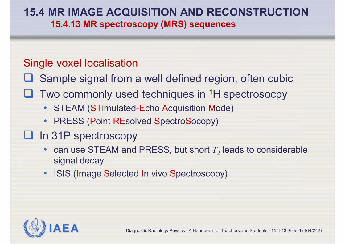
IAEA
15.4 MR IMAGE ACQUISITION AND RECONSTRUCTION15.4.13 MR spectroscopy (MRS) sequences
Single voxel localisation
� Sample signal from a well defined region, often cubic
� Two commonly used techniques in 1H spectrosocpy
• STEAM (STimulated-Echo Acquisition Mode)
• PRESS (Point REsolved SpectroSocopy)
� In 31P spectroscopy
• can use STEAM and PRESS, but short T2 leads to considerable
signal decay
• ISIS (Image Selected In vivo Spectroscopy)
Diagnostic Radiology Physics: A Handbook for Teachers and Students - 15.4.13 Slide 6 (164/242)

IAEA
15.4 MR IMAGE ACQUISITION AND RECONSTRUCTION15.4.13 MR spectroscopy (MRS) sequences
STEAM
� Stimulated echoes utilised
� Three orthogonal slice-selective 90° pulses
• 90° pulses have good slice profile, so get sharp definition of
volume of interest
� Only 50% of available signal is sampled (intrinsic feature)
� Can deliver short echo times
� Magnetisation is stored along the z axis between the last
two pulses, which reduces T2 signal decay
� Can be preceded by water suppression pulses such as
CHESS or WET
Diagnostic Radiology Physics: A Handbook for Teachers and Students - 15.4.13 Slide 7 (165/242)

IAEA
15.4 MR IMAGE ACQUISITION AND RECONSTRUCTION15.4.13 MR spectroscopy (MRS) sequences
PRESS
� One slice-select 90° pulse
� Followed by two orthogonal 180° pulses
� Delivers all of the magnetisation
� Less prone to motion artefacts tha STEAM
� Short echo times achievable with high performance
gradients
� Can be preceded by water suppression pulses such as
CHESS or WET
Diagnostic Radiology Physics: A Handbook for Teachers and Students - 15.4.13 Slide 8 (166/242)

IAEA
15.4 MR IMAGE ACQUISITION AND RECONSTRUCTION15.4.13 MR spectroscopy (MRS) sequences
ISIS for 31P
� Eight separate permutations of three preparation slice-
selective inversions
� Each permutation is followed by a 90° read-out pulse
which generates a free induction decay (FID)
� Combine the eight FIDs
� Localised signal generated from the space representing
the intersection of the planes
� No signal loss from T2 decay
Diagnostic Radiology Physics: A Handbook for Teachers and Students - 15.4.13 Slide 9 (167/242)

IAEA
Pre-treatment (temozolomide)
imagesSTEAM spectra - Serial 1H
spectroscopy
Diagnostic Radiology Physics: A Handbook for Teachers and Students - 15.4.13 Slide 10 (168/242)
15.4 MR IMAGE ACQUISITION AND RECONSTRUCTION15.4.13 MR spectroscopy (MRS) sequences
� Square
shows
position of
voxel for
spectros-
copy
A: Before
treatment
B: 3 months
C: 6 months
D: 9 months
� Cho/Cre ratio
decreased
� NAA peak
increased
From Murphy et al, Br J Cancer 90, 2004

IAEA
15.4 MR IMAGE ACQUISITION AND RECONSTRUCTION15.4.13 MR spectroscopy (MRS) sequences
Spectroscopic imaging
� Single voxel techniques require careful positioning of VOI
prior to acquisition, and must be repeated if further voxels
are required
� Spectroscopic, or chemical shift, imaging techniques
obtain an array of voxels in 2D or 3D
� Signal from many voxels obtained simultaneously
� But
• voxels not as sharply defined as for single voxel spectroscopy
• point spread function is defined by number of sampling points in
each dimension
Diagnostic Radiology Physics: A Handbook for Teachers and Students - 15.4.13 Slide 11 (169/242)

IAEA
15.4 MR IMAGE ACQUISITION AND RECONSTRUCTION15.4.13 MR spectroscopy (MRS) sequences
Spectroscopic imaging
� For 1H (proton) spectroscopy
• select FOV using a stimulated (STEAM) or spin-echo (PRESS)
based technique
• employ phase encoding in 2D or 3D to sample FOV
� For 31P spectroscopy
• preselected VOI less commonly used
• VOI covers entire object space
• or can establish smaller FOV by using saturation slabs surrounding
the desired VOI
Diagnostic Radiology Physics: A Handbook for Teachers and Students - 15.4.13 Slide 12 (170/242)

IAEA
� Acquired on 1.5 T scanner
using 1H decoupled 31P CSI
� Image shows grid used to
acquire the array of spectrum
� Green square shows position
of selected spectrum
31P spectrum
Diagnostic Radiology Physics: A Handbook for Teachers and Students - 15.4.13 Slide 13 (171/242)
15.4 MR IMAGE ACQUISITION AND RECONSTRUCTION15.4.13 MR spectroscopy (MRS) sequences
PC
PCr
NTPs
PDEs
Pi
PE
PE: phosphoethanolamine,PC: phosphocholine, Pi:
inorganic phosphate, PCr: phosphocreatine, NTPs:
nucleotide tri-phosphates

IAEA
15.5 ARTEFACTS15.5 ARTEFACTS
Diagnostic Radiology Physics: A Handbook for Teachers and Students - 15.5 Slide 1 (172/242)

IAEA
15.5 ARTEFACTS15.5 Artefacts
Artefacts
Wide variety of factors can lead to image artefacts in MRI
� 15.5.1 Motion
� 15.5.2 Aliasing or “wrap-around”
� 15.5.3 Metal objects
� 15.5.4 Chemical shift
� 15.5.5 Truncation
� 15.5.6 System-related
Diagnostic Radiology Physics: A Handbook for Teachers and Students - 15.5 Slide 2 (173/242)

IAEA
15.5 ARTEFACTS15.5 Artefacts
Artefacts
� Artefacts generated by
• behaviour of sample
• imperfections in equipment or its operation
• poorly optimised measurement sequences
� Many artefacts can be eliminated or reduced by
• careful attention to quality assurance
• tuning and optimising sequences
� Ameliorate effects of others (intrinsic, patient-related) with
• judicious choice of imaging sequence
• choice of orientation of phase-encode direction
• application of saturation bands
Diagnostic Radiology Physics: A Handbook for Teachers and Students - 15.5 Slide 3 (174/242)

IAEA
15.5 ARTEFACTS15.5.1 MOTION
Diagnostic Radiology Physics: A Handbook for Teachers and Students - 15.5.1 Slide 1 (175/242)

IAEA
15.5 ARTEFACTS15.5 Artefacts
Artefacts
� 15.5.1 Motion
� 15.5.2 Aliasing or “wrap-around”
� 15.5.3 Metal objects
� 15.5.4 Chemical shift
� 15.5.5 Truncation
� 15.5.6 System-related
Diagnostic Radiology Physics: A Handbook for Teachers and Students - 15.5.1 Slide 2 (176/242)

IAEA
15.5 ARTEFACTS15.5.1 Motion
Tissue motion
� Motion during MR acquisition results in additional phase
gains that lead to incorrect phase-encoding
� Results in characteristic artefacts propagated in the phase
encode direction
• ghosted, displaced, lower intensity images of fat in the body
particularly from lower abdomen
• multiple repeat copies of major vessels aligned in the phase
direction with major arteries or veins
Diagnostic Radiology Physics: A Handbook for Teachers and Students - 15.5.1 Slide 3 (177/242)

IAEA
15.5 ARTEFACTS15.5.1 Motion
Reduction or mitigation of tissue motion artefacts
� Ghosted, displaced, lower intensity images of fat in the
body particularly from lower abdomen
• signal averaging
• respiratory gating or navigator triggered acquisitions
• rotation of phase encode direction
• fat suppression
• saturation band placed over source tissue
� Multiple repeat copies of major vessels aligned in the
phase direction with major arteries or veins
• out of plane suppression of inflowing blood
• include motion rephasing gradient lobes in sequences
Diagnostic Radiology Physics: A Handbook for Teachers and Students - 15.5.1 Slide 4 (178/242)

IAEA
15.5 ARTEFACTS15.5.2 ALIASING OR “WRAP-AROUND”
Diagnostic Radiology Physics: A Handbook for Teachers and Students - 15.5.2 Slide 1 (179/242)

IAEA
15.5 ARTEFACTS15.5 Artefacts
Artefacts
� 15.5.1 Motion
� 15.5.2 Aliasing or “wrap-around”
� 15.5.3 Metal objects
� 15.5.4 Chemical shift
� 15.5.5 Truncation
� 15.5.6 System-related
Diagnostic Radiology Physics: A Handbook for Teachers and Students - 15.5.2 Slide 2 (180/242)

IAEA
15.5 ARTEFACTS15.5.2 Aliasing or “wrap-around”
Aliasing or “wrap-around”
� Occurs when sample extends outside the imaging field of
view
� E.g. areas at the side of the body folded into the FOV
� Reconstruction process cannot discriminate between
• tissues positioned within one edge of FOV
• tissues on the other side that are an equal distance outside the
FOV
� Problem increased in areas where gradients are nonlinear,
so several areas have same gradient strength although at
different positions
Diagnostic Radiology Physics: A Handbook for Teachers and Students - 15.5.2 Slide 3 (181/242)

IAEA
15.5 ARTEFACTS15.5.2 Aliasing or “wrap-around”
Reduction or mitigation of aliasing or “wrap-around”
� Alter the phase-encode direction
� Use saturation slabs
� Use RF suppression blankets
Diagnostic Radiology Physics: A Handbook for Teachers and Students - 15.5.2 Slide 4 (182/242)

IAEA
15.5 ARTEFACTS15.5.3 METAL OBJECTS
Diagnostic Radiology Physics: A Handbook for Teachers and Students - 15.5.3 Slide 1 (183/242)

IAEA
15.5 ARTEFACTS15.5 Artefacts
Artefacts
� 15.5.1 Motion
� 15.5.2 Aliasing or “wrap-around”
� 15.5.3 Metal objects
� 15.5.4 Chemical shift
� 15.5.5 Truncation
� 15.5.6 System-related
Diagnostic Radiology Physics: A Handbook for Teachers and Students - 15.5.3 Slide 2 (184/242)

IAEA
15.5 ARTEFACTS15.5.3 Metal objects
Metal objects
� Ferromagnetic material can cause major distortions on the
local magnetic field
� Results in
• displacement of signals in position
• loss of signal
� Minimise by using spin-echo sequences with short TE
� Small effects from other metallic materials
• susceptibility artefacts leading to local distortion and loss of signal
• conduction of currents induced by switched gradients
Diagnostic Radiology Physics: A Handbook for Teachers and Students - 15.5.3 Slide 3 (185/242)

IAEA
15.5 ARTEFACTS15.5.4 CHEMICAL SHIFT
Diagnostic Radiology Physics: A Handbook for Teachers and Students - 15.5.4 Slide 1 (186/242)

IAEA
15.5 ARTEFACTS15.5 Artefacts
Artefacts
� 15.5.1 Motion
� 15.5.2 Aliasing or “wrap-around”
� 15.5.3 Metal objects
� 15.5.4 Chemical shift
� 15.5.5 Truncation
� 15.5.6 System-related
Diagnostic Radiology Physics: A Handbook for Teachers and Students - 15.5.4 Slide 2 (187/242)

IAEA
15.5 ARTEFACTS15.5.4 Chemical shift
Chemical shift
� Water and fat resonate as different frequencies
• difference is 3.4 ppm
� MRI systems adjust their reference frequency to that of
water. So, in slice selection and frequency encoding,
where localisation depends on frequency
• fat signal is spatially displaced from the water signal
• produces a shifted image
• areas of signal void, or brighter signal due to overlap of water and
fat signals in the read-out direction
� Effect greater at higher field where greater separation of
frequencies in Hz
Diagnostic Radiology Physics: A Handbook for Teachers and Students - 15.5.4 Slide 3 (188/242)

IAEA
15.5 ARTEFACTS15.5.4 Chemical shift
Reduction or mitigation of chemical shift artefacts
� Ensure bandwidth per pixel is of order of the frequency
separation between fat and water
• increasing bandwidth per pixel like this can result in increased
noise per pixel compared with a narrower frequency range per
pixel
� Fat suppression
� Water excitation
Diagnostic Radiology Physics: A Handbook for Teachers and Students - 15.5.4 Slide 4 (189/242)

IAEA
15.5 ARTEFACTS15.5.5 TRUNCATION
Diagnostic Radiology Physics: A Handbook for Teachers and Students - 15.5.5 Slide 1 (190/242)

IAEA
15.5 ARTEFACTS15.5 Artefacts
Artefacts
� 15.5.1 Motion
� 15.5.2 Aliasing or “wrap-around”
� 15.5.3 Metal objects
� 15.5.4 Chemical shift
� 15.5.5 Truncation
� 15.5.6 System-related
Diagnostic Radiology Physics: A Handbook for Teachers and Students - 15.5.5 Slide 2 (191/242)

IAEA
15.5 ARTEFACTS15.5.5 Truncation
Truncation
� Signal digitisation involves sampling echo with a
predetermined number of samples (typically 128 or 256)
• each sample takes a specified time
� Evolution of echo, so there may
• be a finite signal at the beginning and end of sampling
• be an asymmetrical echo shape
� Similar effect in phase encoding direction where signal has
not reduced to zero at the maximum gradient values
� Effect is equivalent to multiplying signal with a square
function
Diagnostic Radiology Physics: A Handbook for Teachers and Students - 15.5.5 Slide 3 (192/242)

IAEA
15.5 ARTEFACTS15.5.5 Truncation
Effect of truncation
� Fourier transforms are used to reconstruct images from
the acquired signals
• square function is the Fourier transform pair of a sinc function
• so multiplication by square function for the signal is equivalent of
convolution with a sinc function for the image
� Results in ringing at sharp edges in the image
� Usually seen as parallel bands
• in the frequency or phase encoding direction
• or in both directions
Diagnostic Radiology Physics: A Handbook for Teachers and Students - 15.5.5 Slide 4 (193/242)

IAEA
15.5 ARTEFACTS15.5.6 SYSTEM-RELATED
Diagnostic Radiology Physics: A Handbook for Teachers and Students - 15.5.6 Slide 1 (194/242)

IAEA
15.5 ARTEFACTS15.5 Artefacts
Artefacts
� 15.5.1 Motion
� 15.5.2 Aliasing or “wrap-around”
� 15.5.3 Metal objects
� 15.5.4 Chemical shift
� 15.5.5 Truncation
� 15.5.6 System-related
Diagnostic Radiology Physics: A Handbook for Teachers and Students - 15.5.6 Slide 2 (195/242)

IAEA
15.5 ARTEFACTS15.5.6 System-related
System-related artefacts
� Intrinsic to MR hardware
� May not be adjustable without major hardware changes or
tuning
� Arise from
• Distortions
• RF coil problems
• RF interference
Diagnostic Radiology Physics: A Handbook for Teachers and Students - 15.5.6 Slide 3 (196/242)

IAEA
15.5 ARTEFACTS15.5.6 System-related
Distortions
� Spatial distortions resulting from design of gradient coils
� Difficult to maintain linearity of the gradients towards
edges of FOV
• i.e. where physically close to gradient windings
• particularly relevant for short magnet bore, where gradient coils are
close to edge of FOV
� Reduce distortion by increasing bandwidth per pixel
• but this increase image noise
Diagnostic Radiology Physics: A Handbook for Teachers and Students - 15.5.6 Slide 4 (197/242)

IAEA
15.5 ARTEFACTS15.5.6 System-related
RF coil problems and RF interference
� Transmit RF coil dictates uniformity of B1 irradiation field
and thus consistency of pulse angle over volume of
interest
� Consistency largely governed by design of coil
� But dielectric interactions between RF field and patient
lead to sample dependent variations in B1
• i.e. RF interference
• effects increase with RF frequency (B0 field strength)
• result is flip angle variations across sample
• i.e. variations in excitation and inversion efficiency
Diagnostic Radiology Physics: A Handbook for Teachers and Students - 15.5.6 Slide 5 (198/242)

IAEA
15.5 ARTEFACTS15.5.6 System-related
Reduction or mitigation of RF interference
� Use a circularly polarised transmit coil
� Use adiabatic pulses
• above a certain threshold voltage these are power independent
• but, range of applications of adiabatic pulses very limited
Diagnostic Radiology Physics: A Handbook for Teachers and Students - 15.5.6 Slide 6 (199/242)

IAEA
15.6 SAFETY AND BIOEFFECTS 15.6
Diagnostic Radiology Physics: A Handbook for Teachers and Students - 15.6 Slide 1 (200/242)

IAEA
15.6 SAFETY AND BIOEFFECTS 15.6
Safety and bioeffects
� Safety issues in MR can be divided into acute hazards
(with immediate risk of injury) and biological effects (with
longer term effects)
� Safety concerns arise from four sources:
• 15.6.1 Static field considerations
• projectiles, effects on implants, physiological effects
• 15.6.2 RF field considerations
• tissue heating, specific absorption rate, burn injuries
• 15.6.3 Gradient field considerations
• peripheral nerve stimulation, sound pressure levels
• 15.6.4 Common MR contrast agents
Diagnostic Radiology Physics: A Handbook for Teachers and Students - 15.6 Slide 2 (201/242)

IAEA
15.6 SAFETY AND BIOEFFECTS 15.6
Acute hazards
� Immediate risk of injury from interactions between patient,
staff or extrinsic objects with the fields produced during an
MR examination
� Aspects of most concern and importance in the design
and in day to day operation of MR facilities
Biological effects
� Interactions between MR fields and biological processes
� Could cause longer term effects
Diagnostic Radiology Physics: A Handbook for Teachers and Students - 15.6 Slide 3 (202/242)

IAEA
15.6 SAFETY AND BIOEFFECTS 15.6
Guidance and regulation
� Exposure of patients, staff and general public
� Covered by standards, guidance and regulations
• continually updated
• vary from country to country
� In absence of local guidance prudent to follow
guidance/regulation from elsewhere
Diagnostic Radiology Physics: A Handbook for Teachers and Students - 15.6 Slide 4 (203/242)

IAEA
15.6 SAFETY AND BIOEFFECTS 15.6
Guidance and regulation: examples
� MHRA (UK) publishes guide summarising
recommendations of different bodies and provides
practical advice
• MHRA, Safety guidelines for magnetic resonance imaging
equipment in clinical use Rep. MHRA Device Bulletin DB2007(03)
(2007). www.mhra.gov.uk.
� American College of Radiology guidance on procedures to
ensure patient safety
• AMERICAN COLLEGE OF RADIOLOGY, ACR Guidance
Document for Safe MR Practices (2007).
http://www.acr.org/SecondaryMainMenuCategories/quality_safety/
MRSafety.aspx.
Diagnostic Radiology Physics: A Handbook for Teachers and Students - 15.6 Slide 5 (204/242)

IAEA
15.6 SAFETY AND BIOEFFECTS 15.6
Patient exposures
� Three levels of exposure adopted by IEC, ICNIRP, HPA
� Normal operating mode
• routine scanning of patients
� Controlled mode
• for specific examinations above normal operating mode level
• carried out under medical supervision
• based on clinical decision balancing potential adverse effects
against foreseen benefits
� Experimental operating mode
• carried out at levels above the controlled operating mode
• for which local ethical approval has been obtained
Diagnostic Radiology Physics: A Handbook for Teachers and Students - 15.6 Slide 6 (205/242)

IAEA
15.6 SAFETY AND BIOEFFECTS 15.6.1 STATIC FIELD CONSIDERATIONS
Diagnostic Radiology Physics: A Handbook for Teachers and Students – 15.6.1 Slide 1 (206/242)

IAEA
15.6 SAFETY AND BIOEFFECTS 15.6
� 15.6.1 Static field considerations
• projectiles, effects on implants, physiological effects
� 15.6.2 RF field considerations
• tissue heating, specific absorption rate, burn injuries
� 15.6.3 Gradient field considerations
• peripheral nerve stimulation, sound pressure levels
� 15.6.4 Common MR contrast agents
Diagnostic Radiology Physics: A Handbook for Teachers and Students - 15.6.1 Slide 2 (207/242)

IAEA
15.6 SAFETY AND BIOEFFECTS 15.6.1 Static field considerations
Static field considerations – projectiles and implants
� Static field affects
• ferromagnetic materials
• magnetic storage devices
• magnetically operated switches
• sensitive sound receivers
• sensitive electronic equipment (especially those using accelerated
electrons such as linear accelerators and photomultiplier tubes)
� Two major areas of concern
• Potential for equipment and objects containing ferromagnetic
materials to be attracted into magnet (projectiles)
• Effect on implants that are sensitive to magnetic fields
Diagnostic Radiology Physics: A Handbook for Teachers and Students – 15.6.1 Slide 3 (208/242)

IAEA
15.6 SAFETY AND BIOEFFECTS 15.6.1 Static field considerations
Static field considerations – projectiles
� Equipment and objects to be used close to the magnet
must be tested
� For magnet room or a defined inner controlled zone
• rigorous exclusion of ferromagnetic materials
• access only for
• trained and approved staff
• appropriately screened subjects
Diagnostic Radiology Physics: A Handbook for Teachers and Students – 15.6.1 Slide 4 (209/242)

IAEA
15.6 SAFETY AND BIOEFFECTS 15.6.1 Static field considerations
Static field considerations – implants
� Certain implants may
• be ferromagnetic or become ferromagnetic in the field
• attraction leading to projectile effect
• torque on implanted object leads to force seeking to align object
with the field
• be damaged or malfunction in the field
� Cardiac pacemakers (should not be allowed in field > 0.5
mT), hearing aids and other magnetically controlled or
programmable implants
� Steel clips (some grades of stainless steel can become
magnetic when worked or adjusted)
Diagnostic Radiology Physics: A Handbook for Teachers and Students – 15.6.1 Slide 5 (210/242)

IAEA
15.6 SAFETY AND BIOEFFECTS 15.6.1 Static field considerations
Static field considerations – implants
� Should obtain details of exact make and model of implant
� Check before scanning with
• manufacturer
• Dr F Shellock’s website www.mrisafety.com
Diagnostic Radiology Physics: A Handbook for Teachers and Students – 15.6.1 Slide 6 (211/242)

IAEA
15.6 SAFETY AND BIOEFFECTS 15.6.1 Static field considerations
Static field considerations – physiological effects
� At higher fields (> 2 T) static field can cause a degree of
dizziness or disorientation if the head moves quickly in the
field
� Believed to be due to currents in the vestibular system
interfering with normal sensory perceptions
� At high field
• move slowly in the magnetic field
• avoid abrupt motion of the head
Diagnostic Radiology Physics: A Handbook for Teachers and Students – 15.6.1 Slide 7 (212/242)

IAEA
15.6 SAFETY AND BIOEFFECTS 15.6.1 Static field considerations
Static field considerations – biological effects
� ICNIRP guidance limits patient exposure to 4 T for normal
and to 8 T for controlled operating modes
� At fields used in clinical practice few verified biological
effects
� A moving conductor, such as blood, in a magnetic field can
result in an induced current
� At 8 T the level of currents produced are below levels of
concern
Diagnostic Radiology Physics: A Handbook for Teachers and Students – 15.6.1 Slide 8 (213/242)

IAEA
15.6 SAFETY AND BIOEFFECTS 15.6.1 Static field considerations
Static field considerations – biological effects
� No serious or permanent health effects found from human
exposures up to 8 T
� But there has been only limited scientific investigation
� HEALTH PROTECTION AGENCY, Static Magnetic Fields.
Report of an independent Advisory Group on Non-ionising
Radiation, HPA Rep. Docs HPA, RCE-6 Chilton ( 2008).
http://www.hpa.org.uk/web/HPAwebFile/HPAweb_C/12111
84025757.
Diagnostic Radiology Physics: A Handbook for Teachers and Students – 15.6.1 Slide 9 (214/242)

IAEA
15.6 SAFETY AND BIOEFFECTS 15.6.2 RF FIELD CONSIDERATIONS
Diagnostic Radiology Physics: A Handbook for Teachers and Students - 15.6.2 Slide 1 (215/242)

IAEA
15.6 SAFETY AND BIOEFFECTS 15.6
� 15.6.1 Static field considerations
• projectiles, effects on implants, physiological effects
� 15.6.2 RF field considerations
• tissue heating, specific absorption rate, burn injuries
� 15.6.3 Gradient field considerations
• peripheral nerve stimulation, sound pressure levels
� 15.6.4 Common MR contrast agents
Diagnostic Radiology Physics: A Handbook for Teachers and Students - 15.6.2 Slide 2 (216/242)

IAEA
15.6 SAFETY AND BIOEFFECTS 15.6.2 RF field considerations
RF field considerations
� Tissue heating
� Specific absorption rate (SAR)
• amount of energy deposited by an RF field in a certain mass of
tissue
• units of watts per kilogram (W kg-1)
� Burn injuries
� Non-heat induced long term effects
Diagnostic Radiology Physics: A Handbook for Teachers and Students - 15.6.2 Slide 3 (217/242)

IAEA
15.6 SAFETY AND BIOEFFECTS 15.6.2 RF field considerations
RF field considerations – tissue heating
� Electromagnetic field used to manipulate sample
magnetisation is usually applied by the body coil
• body coil has large volume to enclose body
• driven by high voltages generated by powerful amplifier, typical
output 15 kW
� RF field can give rise to heating in the body
� Equipment designed to limit temperature rise to 0.5 – 1 °C
� Achieved by limiting the specific absorption rate (SAR)
� Tables on next two slides
Diagnostic Radiology Physics: A Handbook for Teachers and Students - 15.6.2 Slide 4 (218/242)

IAEA
Basic restrictions on the maximum temperature for the body
� From these temperature rise limits, specific absorption rates can be
derived. These are usually implemented in the software and hardware
of clinical scanners.
15.6 SAFETY AND BIOEFFECTS 15.6.2 RF field considerations
Diagnostic Radiology Physics: A Handbook for Teachers and Students - 15.6.2 Slide 5 (219/242)
References 1: ICNIRP 2004, 2: IEC 2010, 3: HPA 2008
Operating
modeSpatially localised temperature
limits 1
Body core temperature Local tissue
temperature
Head Trunk ExtremitiesMax. core
temperature rise 1,2
Max. core
temperature 2
Max. local
temperature 2
Normal 38°C 39°C 40°C 0.5°C 39°C 39°C
Controlled 38°C 39°C 40°C 1.0°C 40°C 40°C
Research >38°C >39°C >40°C >1.0°C >40°C >40°C
Research 3 39°C 40°C 41°C 2°C

IAEA
15.6 SAFETY AND BIOEFFECTS 15.6.2 RF field considerations
Diagnostic Radiology Physics: A Handbook for Teachers and Students - 15.6.2 Slide 6 (220/242)
Patient and volunteer SAR limits (W kg-1) for RF field
exposure
Mode ↓Volume transmit coil Local transmit coil
Whole
body
Head Not
heada
Headb Trunk Extremities
Reference 1,2 1 2 1,2 1 2 1 2 1 2
Normal 2 3 3.2 2-10 10 10 10 10 20 20
Controlled 4 3 3.2 4-10 10 20 10 20 20 40
Restricted >4 >3 >3.2 >(4-10) 10 >20 >10 >20 >20 >40
References 1: ICNIRP 2004, 2: IEC 2010

IAEA
15.6 SAFETY AND BIOEFFECTS 15.6.2 RF field considerations
Tissue heating – modes of operation
� Normal operating mode
• exposure to extended volumes of the body should be such as to
avoid a rise of more than 0.5 °C in body temperature of patients
and volunteers, including those compromised with respect to their
thermoregulatory ability
� Controlled operating mode
� Experimental operating mode
Diagnostic Radiology Physics: A Handbook for Teachers and Students - 15.6.2 Slide 7 (221/242)

IAEA
15.6 SAFETY AND BIOEFFECTS 15.6.2 RF field considerations
Tissue heating – modes of operation
� Normal operating mode
� Controlled operating mode
• a relaxation of the basic restrictions on the rise in body
temperature to 1 °C can be envisaged if the patient or volunteer is
monitored medically and with appropriate physiological monitoring
• consider restricting the use of controlled mode for infants, pregnant
women, febrile patients, others with reduced thermoregulatory
ability or with compromised peripheral circulation (HPA 2008)
� Experimental operating mode
Diagnostic Radiology Physics: A Handbook for Teachers and Students - 15.6.2 Slide 8 (222/242)

IAEA
15.6 SAFETY AND BIOEFFECTS 15.6.2 RF field considerations
Tissue heating – modes of operation
� Normal operating mode
� Controlled operating mode
� Experimental operating mode
• requires ethical committee approval
• may result in whole body rise in temperature above 1 °C
• medical, thermal and physiological monitoring required
• absolute limits for core and regional temperatures
• slightly different limits in different publications (see table)
Diagnostic Radiology Physics: A Handbook for Teachers and Students - 15.6.2 Slide 9 (223/242)

IAEA
15.6 SAFETY AND BIOEFFECTS 15.6.2 RF field considerations
RF field considerations – burn injuries
� Receiver coils should be decoupled from the transmit field
to avoid deposition of RF power
� But
• fault conditions in coils
• incorrect set up
• incorrect positioning of connection leads
� Can give rise to
• local coupling
• deposition of excessive local RF power
� And result in heating and RF burns
Diagnostic Radiology Physics: A Handbook for Teachers and Students - 15.6.2 Slide 10 (224/242)

IAEA
15.6 SAFETY AND BIOEFFECTS 15.6.2 RF field considerations
Burn injuries
� RF burns are the most common form of accident in MR
� Operators must keep in communication with patients and
volunteers ready to stop examination if there is any
concern about discomfort
� Follow manufacturer’s guidance on use of conducting
materials in the magnet, such as ECG leads
Diagnostic Radiology Physics: A Handbook for Teachers and Students - 15.6.2 Slide 11(225/242)

IAEA
15.6 SAFETY AND BIOEFFECTS 15.6.2 RF field considerations
RF field considerations – non-heat induced long term effects
� Effects of RF power in producing heat are well established
� However, evidence less clear regarding non-heat induced
long term effects
� No clear evidence of such effects at exposures of up to
4 Wkg-1
• INTERNATIONAL COMMISSION ON NON-IONIZING RADIATION
PROTECTION, Exposure to High Frequency Electromagnetic
Fields, Biological Effects and Health Consequences (100 kHz-300
GHz). Review of the Scientific Evidence and Health
Consequences, Munich (2009).
Diagnostic Radiology Physics: A Handbook for Teachers and Students - 15.6.2 Slide 12 (226/242)

IAEA
15.6 SAFETY AND BIOEFFECTS 15.6.3 GRADIENT FIELD CONSIDERATIONS
Diagnostic Radiology Physics: A Handbook for Teachers and Students - 15.6.3 Slide 1 (227/242)

IAEA
15.6 SAFETY AND BIOEFFECTS 15.6
� 15.6.1 Static field considerations
• projectiles, effects on implants, physiological effects
� 15.6.2 RF field considerations
• tissue heating, specific absorption rate, burn injuries
� 15.6.3 Gradient field considerations
• peripheral nerve stimulation, sound pressure levels
� 15.6.4 Common MR contrast agents
Diagnostic Radiology Physics: A Handbook for Teachers and Students - 15.6.3 Slide 2 (228/242)

IAEA
15.6 SAFETY AND BIOEFFECTS 15.6.3 Gradient field considerations
Gradient field considerations
� Switched gradient fields used for localisation give rise to
two major effects
� Both can produce acute effects
� Induction of currents in conducting materials in the body
• involuntary nerve stimulation
• muscles twitch, or pain at higher levels of stimulation
� Vibration
• depending on pulse sequence can be loud enough to cause
transient loss of hearing, always provide ear protection
� Also biological effects
Diagnostic Radiology Physics: A Handbook for Teachers and Students - 15.6.3 Slide 3 (229/242)

IAEA
15.6 SAFETY AND BIOEFFECTS 15.6.3 Gradient field considerations
Gradient field considerations – nerve stimulation
� Likelihood depends on
• local strength of the gradient
• rate of change of the gradient
• individual's susceptibility to stimulation
� Strength of gradient depends on position in bore of magnet
and design of gradients
• so, hard to predict probability of stimulation even for a given
individual and a particular sequence
Diagnostic Radiology Physics: A Handbook for Teachers and Students - 15.6.3 Slide 4 (230/242)

IAEA
15.6 SAFETY AND BIOEFFECTS 15.6.3 Gradient field considerations
Nerve stimulation – limits
� Gradient levels usually set to avoid stimulation
• based on a threshold for peripheral nerve stimulation (PNS)
defined as the onset of sensation
• next slide
� Mild stimulation can be acceptable
� In principle, much higher gradients can give rise to
sufficiently high cellular potentials to induce cardiac
fibrillation
� Threshold for this is a factor of 10 above that for nerve
stimulation
Diagnostic Radiology Physics: A Handbook for Teachers and Students - 15.6.3 Slide 5 (231/242)

IAEA
15.6 SAFETY AND BIOEFFECTS 15.6.3 Gradient field considerations
Nerve stimulation – modes of operation
� Guidance provided by both IEC and ICNIRP
� Normal operating mode
• the gradient system shall operate at a level that does not exceed
80% of the directly determined PNS, where the threshold PNS is
defined as the onset of sensation
� Controlled operating mode
• the gradient system shall operate at a level that does not exceed
100% of the directly determined mean threshold PNS
� Additionally, IEC has a limit set to prevent cardiac
stimulation
Diagnostic Radiology Physics: A Handbook for Teachers and Students - 15.6.3 Slide 6 (232/242)

IAEA
15.6 SAFETY AND BIOEFFECTS 15.6.3 Gradient field considerations
Gradient field considerations – biological effects
� HPA has summarised literature on effects of low frequency
time varying fields typical of those used in MRI
� No consistent evidence of harm following short term
exposures was found
� Some subtle biological effects were reported
� Subjects with epilepsy, or taking drugs that reduce seizure
activity, may exhibit increased sensitivity to stimulation by
electric fields induced in the cortex and should be imaged
with caution
Diagnostic Radiology Physics: A Handbook for Teachers and Students - 15.6.3 Slide 7 (233/242)

IAEA
15.6 SAFETY AND BIOEFFECTS 15.6.4 COMMON MR CONTRAST AGENTS
Diagnostic Radiology Physics: A Handbook for Teachers and Students - 15.6.4 Slide 1 (234/242)

IAEA
15.6 SAFETY AND BIOEFFECTS 15.6
� 15.6.1 Static field considerations
• projectiles, effects on implants, physiological effects
� 15.6.2 RF field considerations
• tissue heating, specific absorption rate, burn injuries
� 15.6.3 Gradient field considerations
• peripheral nerve stimulation, sound pressure levels
� 15.6.4 Common MR contrast agents
Diagnostic Radiology Physics: A Handbook for Teachers and Students - 15.6.4 Slide 2 (235/242)

IAEA
15.6 SAFETY AND BIOEFFECTS 15.6.4 Common MR contrast agents
Paramagnetic contrast agents
� Paramagnetic agents have unpaired electrons leading to a
strong local magnetic field and a strong net electronic
magnetic moment
� In the raw state, paramagnetic materials are highly
reactive and therefore potentially toxic
� To prevent direct interactions between ions and biological
molecules, the paramagnetic nucleus has to be bound
within a chelate
Diagnostic Radiology Physics: A Handbook for Teachers and Students - 15.6.4 Slide 3 (236/242)

IAEA
15.6 SAFETY AND BIOEFFECTS 15.6.4 Common MR contrast agents
Effects of paramagnetic contrast agents
� Agents relax nearby water molecules, decreasing the
relaxation time
� The T1 relaxation process is the predominant mode of
relaxation
� The degree of relaxation is also affected by ease of access
to water molecules, which is governed by structure of the
molecules
Diagnostic Radiology Physics: A Handbook for Teachers and Students - 15.6.4 Slide 4 (237/242)

IAEA
15.6 SAFETY AND BIOEFFECTS 15.6.4 Common MR contrast agents
Gadolinium
� Gadolinium is the most common contrast agent ion
� Can be bound to a number of different chelates
� Two groups of chelates
• charged and uncharged linear chelates
• charged and uncharged macro cyclic chelates
� A common chelate is DTPA (diethylenetriamine penta-
acetic acid)
� So contrast agent is Gd-DTPA
Diagnostic Radiology Physics: A Handbook for Teachers and Students - 15.6.4 Slide 5 (238/242)

IAEA
15.6 SAFETY AND BIOEFFECTS 15.6.4 Common MR contrast agents
Nephrogenic systemic fibrosis (NSF)
� Rare condition associated with exposure to certain
gadolinium-based contrast agents
� Only in patients with severely impaired renal function,
which leads to prolonged exposure to the agent
� Guidelines (ACR 2007) on use of Gd agents to avoid NSF
• AMERICAN COLLEGE OF RADIOLOGY, ACR Guidance Document
for Safe MR Practices (2007).
http://www.acr.org/SecondaryMainMenuCategories/quality_safety/MR
Safety.aspx.
Diagnostic Radiology Physics: A Handbook for Teachers and Students - 15.6.4 Slide 6 (239/242)

IAEA
15. BIBLIOGRAPHY15.
Diagnostic Radiology Physics: A Handbook for Teachers and Students - 15. Bibliography Slide 1 (240/242)

IAEA Diagnostic Radiology Physics: A Handbook for Teachers and Students - 15. Bibliography Slide 2 (241/242)
15. BIBLIOGRAPHY15.
� BERRY, E., BULPITT, A.J., Fundamentals of MRI, Series in Medical Physics
and Biomedical Engineering, CRC Press (2009).
� BROWN, M.A., SEMELKA, R.C., MRI: Basic principles and applications, John
Wiley, Hoboken NJ (2010).
� DORAN, S., LEACH, M.O., "Spatially localised magnetic resonance", in The
Physics of Medical Imaging, (TAYLOR and FRANCIS, Eds), (2011).
� HAACKE, E.M., BROWN, R.W., THOMPSON, M.R., VENKATESAN, R.,
Magnetic Resonance Imaging: Physical Principles and Sequence Design,
John Wiley and Sons (1999).
� HASHEMI, R.H., BRADLEY, W.G., LISANTI, C.J., MRI: the basics, Lippincott
Williams & Wilkins, Philadelphia (2010).
� INTERNATIONAL COMMISSION ON NON-IONIZING RADIATION
PROTECTION, Medical magnetic resonance (MR) procedures: protection of
patients, Health Phys 87 (2004) 197–216.

IAEA Diagnostic Radiology Physics: A Handbook for Teachers and Students - 15. Bibliography Slide 3 (242/242)
15. BIBLIOGRAPHY15.
� INTERNATIONAL COMMISSION ON NON-IONIZING RADIATION
PROTECTION, Guidelines On Limits Of Exposure To Static Magnetic Fields,
Health Phys 96 4 (2009) 504-514.
� INTERNATIONAL COMMISSION ON NON-IONIZING RADIATION
PROTECTION, Amendment to the ICNIRP “Statement On Medical Magnetic
Resonance (MR) Procedures: Protection Of Patients”, Health Phys 97 3
(2009) 259-261.
� INTERNATIONAL ELECTROTECHNICAL COMMISSION, Medical electrical
equipment – particular requirements for the safety of magnetic resonance
equipment for medical diagnosis, IEC 60601-2-33 Edn 3.0, IEC Geneva
(2010).
� MAMOURIAN, A.C., Practical MR Physics, Oxford, New York (2010).
� WEISHAUPT, D., KOECHLI, V.D., MARINCEK, B., FROEHLICH, J.M., How
does MRI work?: An Introduction to the Physics and Function of Magnetic
Resonance Imaging, Springer, Berlin (2006).