chapter 15 perceptual development - hanoverpsych.hanover.edu/classes/sensation/chapters/chapter...
TRANSCRIPT
Experiencing Sensation and Perception Page 15.1Ch. 15 Perceptual Development
Chapter 15
Perceptual Development
Chapter Outline: I. Introduction II. Changes over Life
A. Infancy i. Research Methods for Infants ii. The sensory experience of the infant
B. Aging and the Loss of Sensory Abilities III. The Biology of Sensory Development
A. Changes in the Brain B. Possible Mechanisms
Experiencing Sensation and Perception Page 15.2Ch. 15 Perceptual Development
Introduction to Perceptual DevelopmentFor most of our lives, our sensory systems see to function pretty much the same way. But those among the readers
who wear glasses probably have some sense that this stability is a bit misleading. Visits to the optometrist or opthalmologist can lead to changes in prescriptions. If the change is large enough, it might be a bit uncomfortable with the new glasses or contacts for awhile. Our sensory systems do indeed change over life, just like the rest of our body. At birth our senses are not completely developed giving us an incomplete view of the world. As we age, our senses will decline leaving us with less of a connection with the world. The trajectory and nature of the development of our senses are important for several reasons. Understanding development helps the medical field intervene in problems to help people of all ages have more normal sensory systems. Understanding development also yields important insight into the basic operations of our sensory systems. Seeing how they change gives researchers peaks into the nature of their general operation. Nature vs. Nurture
In discussions of development, the trite argument over what drives development is usually discussed. Is it nature, or our genetics, or nurture, or experience, that is most important in how our sensory systems operate. Texts usually present the two extreme positions, which I am not sure anyone has ever really held though some get close to one or the other. On one extreme end, there is the genetic position that all that is needed for our sensory systems, or what ever biological or psychological factor is being discussed, is coded in our genes and that is the important factor in our life. Sensory operation is preordained. On the other side is the Tabula Rasa position that we are blank slate at birth and experience writes upon this blank slate to determine who we are going to be. Most texts will then introduce the idea of genes and environment interact to actually determine the outcome. Certainly this interaction position fits all the data and studies that will be discussed here. Still, I would like you consider one more possibility. What if the relationship of genes and environment is more integrated than just simply that of two independent interacting forces, like a push from one side interacting with a push from behind. What if genes and environment have altered together through history and genes and environment work together in common to determine ultimately our development. Genes function in a way that needs the environment to influence how we develop and the environmental influence is built into how genetics develop. In addition, the environment can only have those influences made possible by our genetics. Since organisms alter the environment, it is also possible that our environment is changed along with our genetics as well.
So, we can think of the genetics and environmental effects in different ways: nature vs. nurture, nature plus nurture, nature/nurture together. The different positions are important in how you think about yourself and your relationship to your environment and those around you.
Changes over Life Infancy
When we are born, our sensory systems are not fully functional. But just what our sensory systems can do has been a source of great debate over the years. William James (1890/1952) in his Principles of Psychology thought that infants experiences a “blooming/buzzing confusion.” Not a very attractive view of an infants experience of the world. James thought the infants lacked both the sensory abilities and the mental faculties to process this sensory information. However, viewing infants one becomes aware that they seem to actively, when awake and alter, seek sensory experiences. If these sensory experiences were merely a confusing mess, should not infants seek quiet? Yes, they sleep a lot but they also just stare around actively.
The problem is how to study the sensory abilities of infants. The psychophysical methods discussed in Chapter 2 will not work, at least as described. Infants do not talk and young children lack the ability to talk much. Psychophysics relies on the ability of the participant to verbally respond. While a similar problem occurs in animal research, the infant lacks both motor ability and the patience necessary for even that type of research. Infants have limited motor abilities and usually cannot be run for more than a brief period of time. Without some ability to test the sensory abilities of infants the questions about development seems to be at an impasse.
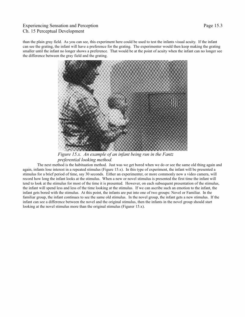
Research Methods for Infants. Fantz (1961) developed the first feasible method for study perception in infants and very young children. The method is called the Fantz preferential looking method. Figurer 15.x shows an example of an infant in this method. The infant in front of a screen where two stimuli are presented. In this example there is a grating of white an black bars on the right and a gray field on the left. Well, if the infant has any preference for one of the stimuli, the infant will start staring at the preferred stimulus. The experimenter is behind the screen looking at the infant. Ideally, the experimenter does not know where the grating or the gray field is. The experimenter then records which direction the infant looks. Since the experimenter does not know which side each of the two stimuli are on, there can be no bias in the experimenter's results. Often a video camera will be used to record the infants looking so that the results can be checked by independent observers to further make the results more objective. It turns out that infants do have strong preferences to what they want to look at. In this experiment, if the infant can see the grating, the infant will prefer that grating much more
Experiencing Sensation and Perception Page 15.3Ch. 15 Perceptual Development
than the plain gray field. As you can see, this experiment here could be used to test the infants visual acuity. If the infant can see the grating, the infant will have a preference for the grating. The experimenter would then keep making the grating smaller until the infant no longer shows a preference. That would be at the point of acuity when the infant can no longer see the difference between the gray field and the grating.
The next method is the habituation method. Just was we get bored when we do or see the same old thing again and again, infants lose interest in a repeated stimulus (Figure 15.x). In this type of experiment, the infant will be presented a stimulus for a brief period of time, say 30 seconds. Either an experimenter, or more commonly now a video camera, will record how long the infant looks at the stimulus. When a new or novel stimulus is presented the first time the infant will tend to look at the stimulus for most of the time it is presented. However, on each subsequent presentation of the stimulus, the infant will spend less and less of the time looking at the stimulus. If we can ascribe such an emotion to the infant, the infant gets bored with the stimulus. At this point, the infants are put into one of two groups: Novel or Familiar. In the familiar group, the infant continues to see the same old stimulus. In the novel group, the infant gets a new stimulus. If the infant can see a difference between the novel and the original stimulus, then the infants in the novel group should start looking at the novel stimulus more than the original stimulus (Figurer 15.x).
Figure 15.x. An example of an infant being run in the Fantz preferential looking method.

Experiencing Sensation and Perception Page 15.4Ch. 15 Perceptual Development
The habituation method would be a good way to study the infant's ability to discriminate between stimuli. For example, if a researcher is interested in how different to colors need to be discriminated by an infant, the researcher can present one color and then for the novel color present colors that are more and more similar until the novel group does not show any increase looking time. At that point, the colors are below the infant's discrimination threshold for color. In fact Bornstein, Kessen and Weiskopf (1976) used the habituation method in infants to discover that they have the same basic color categories (the unique hues of red, green, blue and yellow) as do adults (see Chapter 6). Since our categories of colors are the result of our color opponent cells it seems that infants must have these cells operating at or shortly after birth.
Another way to study infant perception is to know where the infant looks. While the fovea is not completely developed at birth, it is still the area of greatest visual acuity and will be used for visual exploration. Most commonly a video camera will be used to record the direction of the infants gaze. Through processing the images from the camera and calibrating it with the scene, an accurate representation of the direction the infant is looking can be obtained. [OBTAIN LEGAL VIDEO]
Using these methods and others, researchers have discovered much about infant sensory systems. We now have a much more complete view of the sensory experience than William James had. No longer is the infants sensory experience considered to be a “blooming, buzzing, confusion.” Infants do have limitations in their sensory abilities, but there seems to be a nicely evolved match between the infants sensory abilities and their needs. It is to this story, that our attention needs to be directed.
The Senses in Infancy and ChildhoodIn this section, we will examine some of the relevant features of sensory development through childhood.
VisionBasic Visual Abilities. The infant at birth has limited visual abilities with poor acuity (McDonald, et al., 1985),
limited if any accommodation (Banks, 1980), and limited contrast sensitivity across all spatial frequencies (Norcia, Tyler, & Hamer, 1990). Acuity, be several estimates, is about 30 times worse in infants that adults. While adults can resolve objects of 1 arcmin, it takes an object of 30 arcmin for an infant (McDonalt, et al., 1985). Recall that accommodation is the ability to focus the lens between near and far so that the object the person is examining is clear on the retina, at least at the fovea. Infants have little if any ability to adjust their lens. What adjustment they can do seems to be better for near objects than
Figure 15.x. Habituation Method in Infants. The x axis show how many times the stimulus is presented. As can be expected, with repeated presentation, the infant looks less as the stimulus. When a new stimulus is introduced, looking time goes up.
Experiencing Sensation and Perception Page 15.5Ch. 15 Perceptual Development
close objects (Braddick, Atkinson, French & Howland, 1979). There is an interesting correlation associate with the brand new infants limited accommodation ability. Parents hold the infants well within the range that infants accommodate best to (Schoetzau, 1979). While the infant never probably clearly sees the parents face, the parent seems to place their face in an optimal distance for the infant. It is always risky to reach conclusions from correlations because the relationship between variables like infant acuity and parent holding distance could be what they are for many reasons. Still, the correlation is suggestive.
Open Interactive Illustration 15.x, Acuity Development and Vision for a simulated experience of infant vision. When the screen comes up, you will see two copies of the same image. Initially, both images are of a face, not the handsomest face but it will have to do. The copy on the left will never be change and serves a reference to what the face or scene will look like to an adult. The image on the right will be able to be adjusted to simulate how the face or scene might appear to infants at different early ages. Press the Adjust Image button in the upper left corner and you can see an simulation of what this face might look like to a newborn infant shortly after birth. It might take awhile on your computer to complete this adjustment, just wait till the button returns to its normal state from being pressed. When it is done, you should see Birth written below the image. Looking at the image on the right now you can see two major changes to the how the image appears. First, the image is blurry because of the reduction in acuity (Norcia et al., 1990). In addition, the infant is much less able to perceive contrast making the image look more faded and less distinct as well (Banks & Salapatek, 1978). No details are visible and the main features are noticeable only in outline.
While these visual abilities are poor at birth, they develop rapidly. Use the Age in Weeks slider on the right hand side of the screen to change the age of the simulated child's visual system to 4 weeks, not quite one month old. Again, press the Adjust Image button to see the same image now as it might appear to a 4 week wold infant. The acuity and contrast of the image have both increased making the face much more discernible. Details are still not able to be perceived, like the wrinkles around the eyes, at least you can make out many more features such as that there is a mouth. You can continue to increase the simulated age of the infant visual system in this interactivity illustration up to 36 weeks which is the end of the data that I have discovered to make a reliable simulation. If you adjust the simulated age to 36 weeks you will see that the image is still slightly blurry and that contrast is still reduced but the image is much better than even in the earlier months of life. In this illustration, you can also load other images, both other faces and scenes to get some sense of how different images change during the child's development.
So, at birth, the infant has both poor acuity and poor accommodation. Here is a chicken and egg problem. Does the newborn have poor acuity because they have poor accommodation or does the newborn have poor accommodation because their acuity is so poor that accommodation is rendered irrelevant? If accommodation is bad, so will our acuity be. If you wear glasses or contacts with a strong prescription, you already are familiar with the issue whenever you don't have your glasses on or contacts in. For the rest of us, Interactive Illustration 15.x, Retinal Image Quality and Accommodation will help illustrate the problem. This illustration is essentially the same illustration from Chapter 3 when accommodation was first discussed. On this screen you will see a red dot that will serve as the light source and an eye on the right hand side of the screen. Press the Turn Light On button at the bottom of the screen. The red field coming from the light source indicates the spread of light from the light source that will enter the eye. Recall, if the eye focuses that light source well, it should form a point on the retina at the back of the eye. Leave the accommodation off, which will be listed at the top of the screen. Recalling how lenses work, as you move the light back and forth on the screen using the Move Light slider at the bottom of the screen there will be only one distance from the eye that will give a clear image of the light source on the retina. So, if accommodation is poor and focused too close or too far, then most object the infant views will not be clear. So in this case, the lack of accommodation could be driving the lack of acuity.
Conversely, if acuity is bad, what is the good will accommodation do. Reopen Interactive Illustration 15.x, Acuity Development and Vision and then make sure the Age in Weeks slider is at Birth and then press the Adjust Image button. The image, when processed, is really blurry. If acuity is really bad, adjusting accommodation can do nothing to improve the image that the newborn sees because the retina and rest of the visual system is not yet capable of making the image any better. This is an extreme version of what we experience with scotopic (night) vision. Our acuity during rod vision is quite bad, particularly if we are well dark adapted. During the night accommodation is quite sloppy because of this lack of acuity.
Experiments examining our accommodation ability (Banks, 1980, Braddick et al, 1979) find that while accommodation ability is limited at birth, newborns have better accommodation ability than they have acuity. In other words, infants can accommodate some. Not well, but better than predicted by their acuity. As a result, it seems that the lack of acuity is more important to the lack of accommodation and that the develop of the infants ability to accommodate seems to be more determined by the development of acuity than increases in muscular coordination necessary for
Experiencing Sensation and Perception Page 15.6Ch. 15 Perceptual Development
accommodation. It is quite likely that some motor development of accommodation happens, but this development seems to be less important than the development of acuity.
Eye Movements. Since some of what is known about how vision develops in infancy, it is necessary to know something about eye movements during infancy. Recall from Chapter 4 that there were three main times of voluntary eye movements that were discussed. Two of the eye movements discussed were what are known as version eye movements, saccades and smooth pursuit eye movements. In version eye movements, the eyes move together in parallel. Saccades are the very rapid movements that are used to look from object to object or from one part of an object to another. Smooth pursuit eye movements are, like their name implies, an eye movement that tracks objects as they move. The other types of eye movements were vergence eye movements where the eyes rotate oppositely to each other to adjust to see objects at different distances.
At birth, the only voluntary version eye movements that are observed are saccades (Atkinson, Barlow, & Braddick, 1982; Regal, Ashmead, & Salapatek, 1983). The saccades are slow to start, and often more than one is needed to move the eye so that it is looking at the desired object, but they exist (Haith, Hazan & Goodman, 1988). That saccades are the first to be seen in infant is not terribly surprising. Saccades are also the first eye movements to appear in evolution. The simplest animals that have eye movements at all will have saccades and no other eye movements. Tracking of object by smooth pursuit is not apparent at birth. To track an object, the infant will make a series of small saccadic eye movements. It will not be till the infant is six to eight weeks old before smooth pursuit eye movements will appear (Atkinson, et al., 1982).
Open Interactive Illustration 15.x, Eye Movements in Infancy to illustration how eye movements in newborns are different from the same eye movements in adults. This illustration is very similar to the simulation of eye movements that was used in Chapter 4. When this illustration is first opened, the eye movements will simulate those that are seen in infancy. By clicking on each of the five dots, the eyes will saccade to the dot you click on. Since infant saccades are being simulated, when you click on an adjacent do, the saccades might seem to take a while to start but otherwise look normal. However, if you click on some of the dots that have at least one dot in between, then you will see the eye stop briefly on the path to the dot you clicked on. The infant only makes relatively small saccades so it might take two or more saccades to get to the target. Not use the Move Object slider on the left hand side of the screen to move the dots and have the eyes attempt to track the objects. Since, at birth, the infant does not have any smooth pursuit eye movements, you will see jerky eye movements across the screen as the eyes attempt to track the dots. The infant must use saccades to track so most of the time the object being tracked does not fall on the foveas. You can compare the infants eye movements with adults by selected Adult in the Age to Simulate menu in the upper left corner of the screen.
Vergence also has been observed in newborns but the coordination between the eyes is not very good at birth. The coordination of the two eyes improves over the first few months of life (Atkinson, et al., 1982). Of course with poor acuity, some of the problem with vergence seen in very young children might be do to the inability to see well enough to make vergence accurate. Still, some development of coordination has to be related to motor development and other growth factors. As the head grows, which it does, though to a lesser extent than other parts of the body after birth, the amount of vergence needed to align the two images of an object on the eye changes. Thus, vergence must be responsive to the changes in the child's head size as the child grows. As my children grew in their first year, it was enjoyable to observe some of the struggles of their eye movement systems to deal with all the changes happening to the child. Every now and then, an eye would simple wander away, slowly, from the object they were examining. Then, a short moment later, the eye would slowly wander back.
So far the discussion on eye movements, both here and in Chapter 4 have focused on voluntary eye movements. While voluntary eye movements are all well and good, there are a host of reflexive eye movements that we also have. For example, large moving objects and visual scenes can trigger a reflexive eye movement for our eyes to follow the stimulus. Open Interactive Illustration 5.x, Optokinetic Nystagmus to see a small scale version of a stimulus for to study this reflexive eye movement. When you open the illustration you will see a horizontal grating going from one edge of the screen to the other. It the laboratory, this grating would fill the participant's entire visual field. If you put your face close to the center of the screen you will get some idea of what the experience might be like for the participant. Pressing the Start button wills tart the grating moving. You can control the direction of the motion, the speed of motion, and the nature of the grating with other controls on the screen. You might adjust some of them and see if different settings do a better or worse job of stimulating motion in your eyes.
To understand how the optokinetic nystagmus operates in infants, this reflexive eye movement is studied in a particularly way. The participant will sit and stare at a screen covered with, in this case, vertical bars. The bars will then move to one direction or another. The eyes will track the object, and in this reflex at speeds much greater than can be done with smooth pursuit eye movements. The grating keeps going, but the eyes can only rotate so far. To be concrete, say the stimulus is moving to the left of the participant. When the eyes reach the farthest left that they can rotate, they rotate,
Experiencing Sensation and Perception Page 15.7Ch. 15 Perceptual Development
saccade-like, back to the right and then start tracking the grating again. Thus, a repeating pattern of eye movements appears where there is a slow phase where the eyes track the grating and then a fast phase where the eyes move in the opposite direction and reset. Any repetitive pattern of eye movements like this is called a nystagmus. The full name for this pattern of eye movements is called the optokinetic nystagmus. Optokinetic comes from optics, or the study of the bending of light, and kinetic referce to movement. So this name means the repetitive eye movement (nystagmus) that results form a moving (kinetic) stimulus (optics).
Open Interactive Illustration 15.x, Optokinetic Nystagmus Aymmetry to see a simulation of the optokinetic nystagmus. When you open the illustration, you will see two eyes drawn at the bottom of the screen both looking at a grating drawn at the top of the screen. Since you are looking down at the simulated participant from above, the grating is foreshortened and you will see only very small white and black bars. However, the simulated participant will see a grating filling their entire visual world. If you press the start button, the grating will start moving and the eyes, reflexively, will follow as best they can. It might not be perfect, but if the grating is not too fast it will be close. When the eyes reach as far as they can rotate, they will shift back. On the lower right corner of the screen there is a plot of the eye position over time. You will see the tracking of the eyes against the grating as a relatively gradual change in eye position on the graph. The reset of the eyes will show up as a sudden change in eye position. The time when the eyes are tracking is called the slow phase of the nystagmus and the reset is called the fast phase of the nystagmus. You can adjust the speed of the grating with the Grating Speed slider and revers the direction of the grating with the Reverse Direction button.
Developmentally, the optokinetic nystagmus shows an interesting pattern. To see this developmental pattern, the infant is tested with one eye covered and one eye open, monocularly. The two different directions of motion of the stimulus can be tested without a contribution from the stimulation to the other eye. Since our bodies are organized pretty symmetrically around our mid-lines, referring to directions as left and right do not have a lot of meaning. So the directions of motion used are whether the stimulus moves from the side towards the mid-line or from the mid-line to the side. Since the discussion is relative to one eye, recall that the retina is divided into two sections. The half from the fovea to the nose is called the nasal retina and the half from the fovea towards our temples is called the temporal retina. So when the grating moves from the side to the mid-line, it is called temporal-nasal motion. When the grating moves from the mid-line to the side it is called nasal-temporal motion. Awkward, but descriptive terms. At birth and for the first several months, there is a bias in the optokinetic nystagmus favoring temporal-nasal motion (Lewis, Maurer, Chung, Holmes-Shannon, & Van Schaik, 2000; Schor, Narayan, & Westall, 1983). This asymmetry weakens but still last for the first couple of years at least and may even be apparent still in adulthood but at a much weaker level (Posner, 1980).
To get some feeling for this asymmetry in the optokinetic nystagmus in infancy, reopen Interactive Illustration 15.x, Optokinetic Nystagmus Asymmetry if it is not still open. If the illustration is running, press the Stop button to stop the motion of the grating and press the Center Fixation button to center the eyes on the grating. First, to see this asymmetry in the optokinetic nystagmus, only one eye should be tested, click on the Right Eye check box to undo it and remove the right eye from the illustration. This action will simulate covering one of the participant's eyes. When only the left eye is show, the words Nasal and Temporal will show up on the screen with Nasal to the left and Temporal to the right side of the screen below the grating. Recall, Chapter 3, that images on the retina are upside down and backwards, so that images that fall to the left of the left eye actually fall on the nasal part of the retinal. These terms will help keep track of the direction of motion of the grating on the retina of the eye.
First, it might be good just to press the Start button and watch this left eye track the grating. After watching the eye move in this first direction, press the Reverse Direction button to see how the eye tracks the grating when it moves in the opposite direction. The eye should track the grating pretty well in both directions. Now go to the upper left corner and use the Age to Simulate menu to select Infant. Now when the motion is from the temporal side to the nasal side of the retina, the tracking by the eye is pretty good (press Reverse Direction if you need to go get the motion of the grating going from the right to the left on the screen). But if you press Reverse Direction so that the motion is now nasal-temporal, the tracking just does a very poor job of keeping up with the grating. You can see the show tracking of the eyes both by how slow the eyes move and how much faster the grating moves than where the eyes are looking, indicated by the green line, and by the very slow change in eye position shown on the graph on the lower right side of the screen. You can change which eye is being shown and even have both eyes open and see what happens.
What is interesting about this finding is what it implies about the development of the brain. Animals without a cortex, like amphibians, also show this bias in temporal-nasal motion in the optokinetic reflex (Atkinson, et al., 1992). The primary visual center in these animals are the optic tectum, which is equivalent to the superior colliculus in our visual system (Chapter 3). This observation that amphibians have a strong temporal-nasal bias in their optokinetic nystagmus and
Experiencing Sensation and Perception Page 15.8Ch. 15 Perceptual Development
the fact that adult humans have little if any bias suggests that one direction, the temporal-nasal direction, of the optokinetic nystagmus is triggered and/or controlled by stimulation going to the optic tectum/superior colliculus, while the other direction, the nasal-temporal direction, of the optokinetic nystagmus, is controlled by the cortex. Following this logic, it seems that infants must have a more developed superior colliculus at birth than a cortex, at least as those two structures are involved in the control of the optokinetic nystagmus (Johnson, 1990).
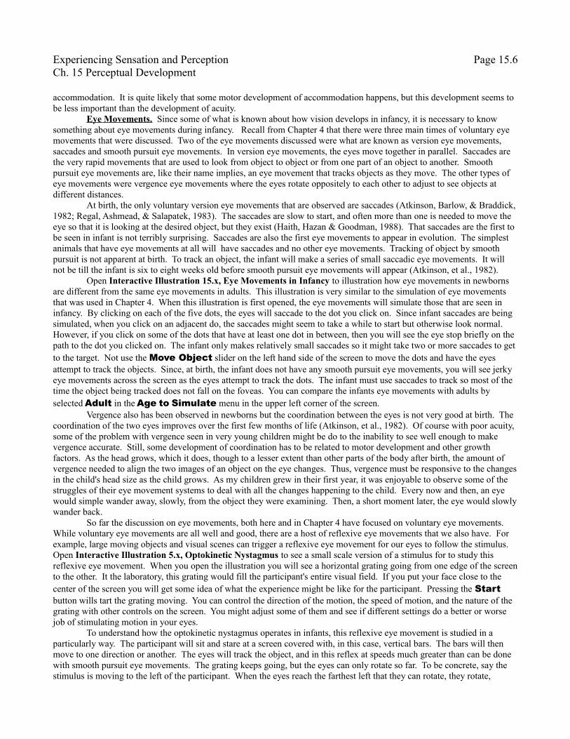
Perceiving Objects. So, the new born infant has poor acuity, contrast sensitivity and does not accommodate well. It does not follow, interestingly enough, that the newborn cannot make anything out about their environment. Fantz (1961) was one of the first researchers that demonstrated that infant do gain some information about objects. Sample results from one of Fantz's (1961) experiments are shown in Figure 15.x. In general the more complex the image, the more it is preferred. In face, the most preferred stimulus is a face. The different objects are all the same size and the same luminance and for the bulls-eye and face, the contrast was the same. So something about the shapes seems important in the infants preferences from even their first days of life.
These results raise some suggestion that there might be something important about faces in infant perceptions. Many studies of adult perception suggest that we process faces in some way that is special. Leo and Simion (2009) did an interesting study that does suggest that this ability to process faces specially appears at or nearly at birth. This study used a classic illusion of face perception the Thatcher illusion named after the former Prime Minister of England Margaret Thatcher whose faces was first used in this illusion (Thompson, 1980). Open Interactive Illustration 15.x, The Thatcher Illusion to see this illusion and help illustrate the experiment by Leo and Simion. When you open the illustration, you will see two copies of the same face but upside down. Look at them for a short period of time noticing any differences. Next use the Rotation Angle slider and rotate the images from upside down to right side up. How do the faces appear now? I feel pretty safe in saying that most of you find the faces far more dissimilar in the upright position than the upside down position. What is different between the faces, as might be apparent, is that for the face on the right, the eyes and mouth have been inverted. When the faces are upside down, this distortion of the face is much less obvious and much less disturbing. Rotate the faces back to upside down to check this out. This illusion does not seem to matter whether the face is right side up or upside down. This illusion shows that our perceptual systems have some expectations about faces. For the present case, they are expected to be right side up. When the face is right side up, deviations within the face that violate that assumption are much more obvious than when the faces are upside down. When the face is upside down, the eyes are still right side up which does not violate our expectations as much. The question here though is do infants start this bias in
Figure 15.x. Percent of time looking at the object as a function of the stimulus type. Solid bars are for 2-6 months and open bars are for < 5 days.
Experiencing Sensation and Perception Page 15.9Ch. 15 Perceptual Development
processing faces. Leo and Simion (2009) used the habituation procedure discussed above. The infants were show a normal face, like the face on the left, repeatedly. Next, the infants were presented the face with the eyes and mouth inverted, like the face on the right. If the infants started looking a lot more, then they could discriminate between the two faces. The two faces were always in the same orientation, either right side up or upside down. The infants showed clear ability to discriminate the normal from the distorted face when right side up but not when the faces were upside down. These results match our experience where we see a much greater difference between the faces when they are right side up than upside down. These results suggest that something about the nature of what a face should look like is already apparent to the newborn child. You can simulate what theses faces might have appeared like to these infants by adjusting the Age in Weeks slider and then pressing the Adjust Image slider. Particularly for ages at birth or the first few weeks, it might take a while for the image to be processed so be patient. You can simulate how the faces looked at different ages and see if you can see a difference, even when the faces are right side up. You can also select different faces with the Image menu.
These results suggest that the infant's perceptual system might, at birth, be developed in a way that allows for selective ability to process biological relevant information to a greater degree than general stimuli. It seems that some of the important characteristics of faces are already present in newborn visual processing. Recall biological perception from Chapter 7. To help you remember this phenomenon, open Interactive Illustration 15.x, Biological Motion. Click on the Move Figure check box to start the motion of the figure and look at the figure on the left. Once the dots start moving it becomes clear that the dots seem to lie on the joints and head of a person apparently walking to the right. (Depending upon the speed of your computer, you might need to adjust the speed of the walking figure with the Speed slider to a nice easy pace, not too fast or slow.) The figure on the right is made up of exactly the same motions as the figure on the left, however, their positions relative to each other are determined randomly. So the motion of the left foot might be up near the top of the head and the motion of the head near where the hips would be. No one reports seeing such motions as being derived from anything let alone from a walking human figure. Now use the Rotation slider and invert these two figures. If you looked initially at these figures upside down, even the Normal Figure one the left would not appear to you to be anything recognizable. Using an experimental setup very similar to Leo and Simion (2009), Bertenthal, Proffitt, and Kramer, (1987) tested whether young infants perceived biological motion. Just as in the infant perception of faces in the Thatcher illusion (Leo & Simion, 2009), the infants could discriminate between the upright motion and the scrambled figure but not the upside down motion and the scrambled figure. From very early in life there seems to be the ability to perceive biologically related stimuli. What these findings suggest is that while perception is limited at birth, it is to some degree biologically tuned to allow the infant to detect biologically important stimuli. Such abilities might be very important in helping the infant respond discriminatively to adult caregivers in a way that might support the growing attachment between the child and caregiver. To let you know that not all is settled on this issue, other researchers observe that learning does take place in our perception of biological motion and they argue that it is not clear that infants show any perception of biologically relevant organizations of movement right at birth (Jastorff, Kourtzi, & Giese, 2006).
The foregoing argument does not imply that infants actually recognize objects in the same way that adults do. Let us look at the ways that infants object perception differs from adult perception of objects in two different ways. First, infants seem to perceive occluded objects differently from adults, at least at first. Given the clutter of our world, it is the rare object that we see completely. Usually some part of the object is covered or occluded from our eyes. Using the Gestalt laws (Chapter 5), we have not problem perceiving the whole object. Second, eye movements on objects reveal that we interact differently with images before us.
Open Interactive Illustration 15.x, Occluded Object Perception in Infancy. This illustration will give sample stimuli used in the experiments with infant about occluded objects. All of these experiments that will be discussed have employed the habituation methodology. On the screen, the left side will always show the stimulus used during the habituation phase. On the right side of the screen, the different stimuli that can be used for the test phase of the experiment can be shown. The stimulus is pretty simple, what looks like a white bar is occluded by a central gray rectangle that covers the middle of the bar. We have no trouble perceiving this stimulus in this way. Gestalt Laws of organization come into play. In particular, the Law of Closure seems relevant here where the mind closes the two ends of the white bar. If you need to review this Gestalt Law, you can open Interactive Illustration 15.x(b), Closure to remind your self about this law. Use the Gap Size slider on the right side of the screen to open gaps in the square and notice how for even fairly large gaps you still see the square as one object and not four disconnected, unaffiliated corners. Kellman and Spelke (1983) presented this stimulus to 3 to 4 month old infant in a habituation experiment. The test stimulus could be one of two other stimuli. Reopen Interactive Illustration 15.x, Occluded Object Perception in Infancy if you have closed it. To see one of the test stimuli, click on the Complete Lines check box in the upper left corner of the screen. The obscuring gray rectangle
Experiencing Sensation and Perception Page 15.10Ch. 15 Perceptual Development
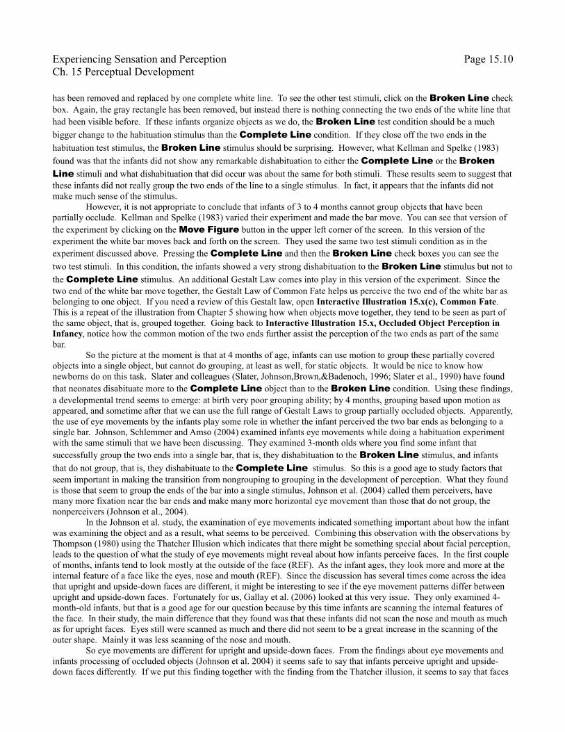
has been removed and replaced by one complete white line. To see the other test stimuli, click on the Broken Line check box. Again, the gray rectangle has been removed, but instead there is nothing connecting the two ends of the white line that had been visible before. If these infants organize objects as we do, the Broken Line test condition should be a much bigger change to the habituation stimulus than the Complete Line condition. If they close off the two ends in the habituation test stimulus, the Broken Line stimulus should be surprising. However, what Kellman and Spelke (1983) found was that the infants did not show any remarkable dishabituation to either the Complete Line or the Broken Line stimuli and what dishabituation that did occur was about the same for both stimuli. These results seem to suggest that these infants did not really group the two ends of the line to a single stimulus. In fact, it appears that the infants did not make much sense of the stimulus.
However, it is not appropriate to conclude that infants of 3 to 4 months cannot group objects that have been partially occlude. Kellman and Spelke (1983) varied their experiment and made the bar move. You can see that version of the experiment by clicking on the Move Figure button in the upper left corner of the screen. In this version of the experiment the white bar moves back and forth on the screen. They used the same two test stimuli condition as in the experiment discussed above. Pressing the Complete Line and then the Broken Line check boxes you can see the two test stimuli. In this condition, the infants showed a very strong dishabituation to the Broken Line stimulus but not to the Complete Line stimulus. An additional Gestalt Law comes into play in this version of the experiment. Since the two end of the white bar move together, the Gestalt Law of Common Fate helps us perceive the two end of the white bar as belonging to one object. If you need a review of this Gestalt law, open Interactive Illustration 15.x(c), Common Fate. This is a repeat of the illustration from Chapter 5 showing how when objects move together, they tend to be seen as part of the same object, that is, grouped together. Going back to Interactive Illustration 15.x, Occluded Object Perception in Infancy, notice how the common motion of the two ends further assist the perception of the two ends as part of the same bar.
So the picture at the moment is that at 4 months of age, infants can use motion to group these partially covered objects into a single object, but cannot do grouping, at least as well, for static objects. It would be nice to know how newborns do on this task. Slater and colleagues (Slater, Johnson,Brown,&Badenoch, 1996; Slater et al., 1990) have found that neonates disabituate more to the Complete Line object than to the Broken Line condition. Using these findings, a developmental trend seems to emerge: at birth very poor grouping ability; by 4 months, grouping based upon motion as appeared, and sometime after that we can use the full range of Gestalt Laws to group partially occluded objects. Apparently, the use of eye movements by the infants play some role in whether the infant perceived the two bar ends as belonging to a single bar. Johnson, Schlemmer and Amso (2004) examined infants eye movements while doing a habituation experiment with the same stimuli that we have been discussing. They examined 3-month olds where you find some infant that successfully group the two ends into a single bar, that is, they dishabituation to the Broken Line stimulus, and infants that do not group, that is, they dishabituate to the Complete Line stimulus. So this is a good age to study factors that seem important in making the transition from nongrouping to grouping in the development of perception. What they found is those that seem to group the ends of the bar into a single stimulus, Johnson et al. (2004) called them perceivers, have many more fixation near the bar ends and make many more horizontal eye movement than those that do not group, the nonperceivers (Johnson et al., 2004).
In the Johnson et al. study, the examination of eye movements indicated something important about how the infant was examining the object and as a result, what seems to be perceived. Combining this observation with the observations by Thompson (1980) using the Thatcher Illusion which indicates that there might be something special about facial perception, leads to the question of what the study of eye movements might reveal about how infants perceive faces. In the first couple of months, infants tend to look mostly at the outside of the face (REF). As the infant ages, they look more and more at the internal feature of a face like the eyes, nose and mouth (REF). Since the discussion has several times come across the idea that upright and upside-down faces are different, it might be interesting to see if the eye movement patterns differ between upright and upside-down faces. Fortunately for us, Gallay et al. (2006) looked at this very issue. They only examined 4-month-old infants, but that is a good age for our question because by this time infants are scanning the internal features of the face. In their study, the main difference that they found was that these infants did not scan the nose and mouth as much as for upright faces. Eyes still were scanned as much and there did not seem to be a great increase in the scanning of the outer shape. Mainly it was less scanning of the nose and mouth.
So eye movements are different for upright and upside-down faces. From the findings about eye movements and infants processing of occluded objects (Johnson et al. 2004) it seems safe to say that infants perceive upright and upside-down faces differently. If we put this finding together with the finding from the Thatcher illusion, it seems to say that faces
Experiencing Sensation and Perception Page 15.11Ch. 15 Perceptual Development
are perceived differently when upright than when upside down. In the upright face, adults and infant move their eyes a great deal from the eyes to the nose/mouth region and back to the eyes (Gallay et al., 2006). When the face is upside down, there is much less movement of the eyes of the infant from the nose/mouth region back and forth to the eyes. While this is an isolated observation, taken with the results from the Thatcher illusion study, it supports the conclusion that upside-down faces are not perceived as much as an integrated whole object in the same way that upright faces are. Again, it seems possible to interpret these findings that there may be something special about faces and that this ability to perceive faces as faces is present at least shortly after birth. Be careful, this conclusion does not mean that experience does not play a role in face perception. It undoubtedly does, but it play a different or less of a role than it might play for objects that are not faces.
AuditionAs in the study of sensory abilities in general, there seems to be far more studies of infant visual development than
development of the other senses. While I have an interest in the visual system, I find this bias both understandable and somewhat unfortunate. Certainly problems in visual development can lead to debilitating issues for the adult, the same can be true for developmental problems in other senses as well. Take for example talking to your friends. If you have some problem in the development of your hearing, will you not then have trouble with this very basic social skill? In this section, a brief look at the normal development of some basic auditory abilities will be presented.
Aging and the Loss of Sensory Abilities
The Biology of Sensory DevelopmentChanges in the Brain
Possible Mechanisms
The complexity of the brain is staggering. A common estimate is that there is over 1 billion neurons in the brain with each neuron making on average 1000 or more synapses (CHECK ON THIS). The number of genes in the human genome is on the order of 10's of thousands. With our current understanding of how the genes work, there is no way that our genetic material can specify all of this complexity of the brain. So the question remains how does the brain develop? At best we can only sketch answers but the answers that are emerging are intriguing. This question is particularly germane to the understanding of the development of sensation and perception as it has been the study of sensory systems that has provided many of the hints about brain development that we have. So in this section, I will discuss some of the brain research that has illuminated how our sensory systems develop and then discuss in general terms some of the ways researchers are working to try to understand how brain development might proceed to give us the sensory systems we have been studying this whole time.
Roger Sperry and Newts. Roger Sperry (1943b, 1944) did a very interesting study of visual development using newts (PICTURE). Newts as amphibians have the ability to regenerate there central nervous system neurons into adulthood. Lucky them. I guess there are some benefits of a simpler nervous system, but I take mine anyway. In this experiment, Roger Sperry took removed the eyes, cutting the optic nerve and then reattached them upside down. Now, news as amphibians do not have a cortex, so their brain visual area is the optic tectum which we call the superior colliculus in mammals. As you recall from Chapter 3, the superior colliculus still serves visual functions in humans, but it is all that the newt has as a brain area processing visual information.
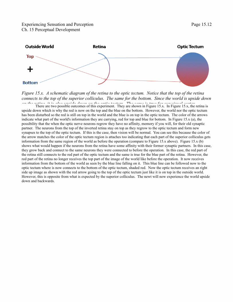
Figure 15.x shows a schematic representation of how the superior retina of the eye connects to the superior colliculus. Recall from Chapter for that the focusing of the lens and pupil causes the image of what we are looking at to be upside down and backwards on the retina. Figure 15.x is two dimensional so only the inversion of up and down is shown. To assist you in following the diagram, the top of the world is red and the bottom of the world is blue. So, on the retina the wold is upside down. Well, the retina connects in a way so that the world is still upside down and backwards on the optic tectum. The top of the retina connects to the top of the optic tectum and the bottom of the retina connects to the bottom of the optic tectum. In Sperry's experiment, the optic nerve, represented by the red and blue arrows from the retina to the optic tectum are cut and the question is what happens when they grow back.
Experiencing Sensation and Perception Page 15.12Ch. 15 Perceptual Development
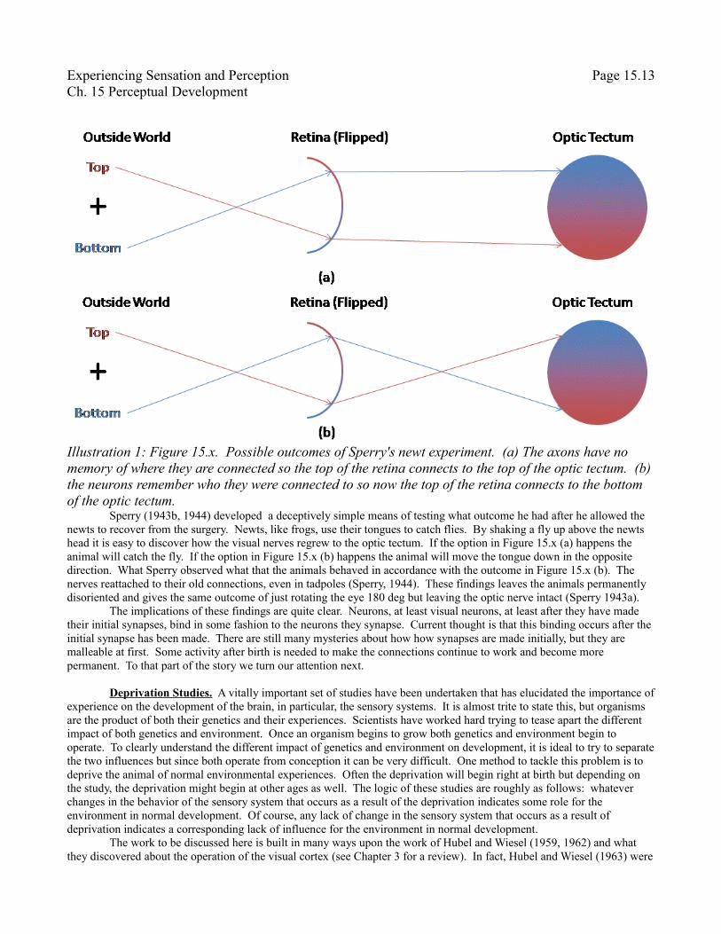
There are two possible outcomes of this experiment. They are shown in Figure 15.x. In Figure 15.x, the retina is upside down which is why the red is now on the top and the blue on the bottom. However, the world nor the optic tectum has been disturbed so the red is still on top in the world and the blue is on top in the optic tectum. The color of the arrows indicate what part of the world's information they are carrying, red for top and blue for bottom. In Figure 15.x (a), the possibility that the when the optic nerve neurons regrow they have no affinity, memory if you will, for their old synaptic partner. The neurons from the top of the inverted retina stay on top as they regrow to the optic tectum and form new synapses to the top of the optic tectum. If this is the case, then vision will be normal. You can see this because the color of the arrow matches the color of the optic tectum region is attaches too indicating that each part of the superior colliculus gets information from the same region of the world as before the operation (compare to Figure 15.x above). Figure 15.x (b) shows what would happen if the neurons from the retina have some affinity with their former synaptic partners. In this case, they grow back and connect to the same neurons they were connected to before the operation. In this case, the red part of the retina still connects to the red part of the optic tectum and the same is true for the blue part of the retina. However, the red part of the retina no longer receives the top part of the image of the world like before the operation. It now receives information from the bottom of the world as seen by the blue line falling on it. This blue line can be followed now to the optic tectum where is now connects to the bottom of the optic tectum, shaded red. Now the optic tectum receives an right side up image as shown with the red arrow going to the top of the optic tectum just like it is on top in the outside world. However, this is opposite from what is expected by the superior colliculus. The newt will now experience the world upside down and backwards.
Figure 15.x. A schematic diagram of the retina to the optic tectum. Notice that the top of the retina connects to the top of the superior colliculus. The same for the bottom. Since the world is upside down on the retina, it is also upside down on the optic tectum. The same is true for our visual cortex.
Experiencing Sensation and Perception Page 15.13Ch. 15 Perceptual Development
Sperry (1943b, 1944) developed a deceptively simple means of testing what outcome he had after he allowed the newts to recover from the surgery. Newts, like frogs, use their tongues to catch flies. By shaking a fly up above the newts head it is easy to discover how the visual nerves regrew to the optic tectum. If the option in Figure 15.x (a) happens the animal will catch the fly. If the option in Figure 15.x (b) happens the animal will move the tongue down in the opposite direction. What Sperry observed what that the animals behaved in accordance with the outcome in Figure 15.x (b). The nerves reattached to their old connections, even in tadpoles (Sperry, 1944). These findings leaves the animals permanently disoriented and gives the same outcome of just rotating the eye 180 deg but leaving the optic nerve intact (Sperry 1943a).
The implications of these findings are quite clear. Neurons, at least visual neurons, at least after they have made their initial synapses, bind in some fashion to the neurons they synapse. Current thought is that this binding occurs after the initial synapse has been made. There are still many mysteries about how how synapses are made initially, but they are malleable at first. Some activity after birth is needed to make the connections continue to work and become more permanent. To that part of the story we turn our attention next.
Deprivation Studies. A vitally important set of studies have been undertaken that has elucidated the importance of experience on the development of the brain, in particular, the sensory systems. It is almost trite to state this, but organisms are the product of both their genetics and their experiences. Scientists have worked hard trying to tease apart the different impact of both genetics and environment. Once an organism begins to grow both genetics and environment begin to operate. To clearly understand the different impact of genetics and environment on development, it is ideal to try to separate the two influences but since both operate from conception it can be very difficult. One method to tackle this problem is to deprive the animal of normal environmental experiences. Often the deprivation will begin right at birth but depending on the study, the deprivation might begin at other ages as well. The logic of these studies are roughly as follows: whatever changes in the behavior of the sensory system that occurs as a result of the deprivation indicates some role for the environment in normal development. Of course, any lack of change in the sensory system that occurs as a result of deprivation indicates a corresponding lack of influence for the environment in normal development.
The work to be discussed here is built in many ways upon the work of Hubel and Wiesel (1959, 1962) and what they discovered about the operation of the visual cortex (see Chapter 3 for a review). In fact, Hubel and Wiesel (1963) were
Illustration 1: Figure 15.x. Possible outcomes of Sperry's newt experiment. (a) The axons have no memory of where they are connected so the top of the retina connects to the top of the optic tectum. (b) the neurons remember who they were connected to so now the top of the retina connects to the bottom of the optic tectum.
Experiencing Sensation and Perception Page 15.14Ch. 15 Perceptual Development
there first to study these issues of development and their Nobel Prize is partly for their work on development. One of the issues that Hubel and Wiesel studied with the deprivation methodology is the effect of eye closure on the sensitivity of cells in the cortex to each eye. Before digging into the studies, a little review seems in order. Recall from Chapter 3 that most cells in V1 or the striate cortex respond more strongly to the information coming from one or the other eye. This tendency of a cell to prefer the signals from one eye or the other is called ocular dominance. Te ocular dominance of the cells are arranged into columns with one column having cells that respond better to signals from the right eye adjacent to cells that respond better to signals from the left eye. These columns are referred to as ocular dominance columns. These columns are organized so that run at essentially a right angle to the columns that are sensitive to different orientations.
So much for review. The question that Hubel and Wiesel address in what role does experience play in the development of these ocular dominance columns. To understand the role of experience they first wanted to know the status of the visual system prior to extensive visual experience. Kittens are idea subjects for these experiments because they are born with their eyes closed. In a small experiment they first determined the type of ocular dominance preference found in the cells of these kittens just as their eyes are opening. What they found is that the pattern of ocular dominance was similar in the kittens and adults (Hubel & Wiesel, 1963). This finding serves as the base line for the deprivation studies.
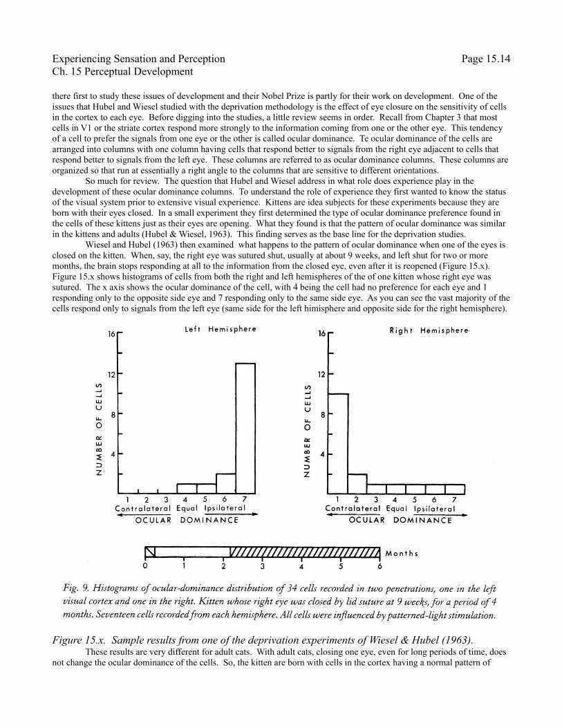
Wiesel and Hubel (1963) then examined what happens to the pattern of ocular dominance when one of the eyes is closed on the kitten. When, say, the right eye was sutured shut, usually at about 9 weeks, and left shut for two or more months, the brain stops responding at all to the information from the closed eye, even after it is reopened (Figure 15.x). Figure 15.x shows histograms of cells from both the right and left hemispheres of the of one kitten whose right eye was sutured. The x axis shows the ocular dominance of the cell, with 4 being the cell had no preference for each eye and 1 responding only to the opposite side eye and 7 responding only to the same side eye. As you can see the vast majority of the cells respond only to signals from the left eye (same side for the left himisphere and opposite side for the right hemisphere).
These results are very different for adult cats. With adult cats, closing one eye, even for long periods of time, does not change the ocular dominance of the cells. So, the kitten are born with cells in the cortex having a normal pattern of
Figure 15.x. Sample results from one of the deprivation experiments of Wiesel & Hubel (1963).
Experiencing Sensation and Perception Page 15.15Ch. 15 Perceptual Development
ocular dominance. However, experience after birth is necessary for these connections to maintain. But after the experience has had a chance to work for awhile, the connections become more stable. These patterns of results suggest that there is a sensitive period for the development of visual connections. There is a period early in life where it is necessary to have a rich experience to help develop and make more permanent the connections that we develop in our brain. Once we reach adulthood changing our visual abilities is much harder.
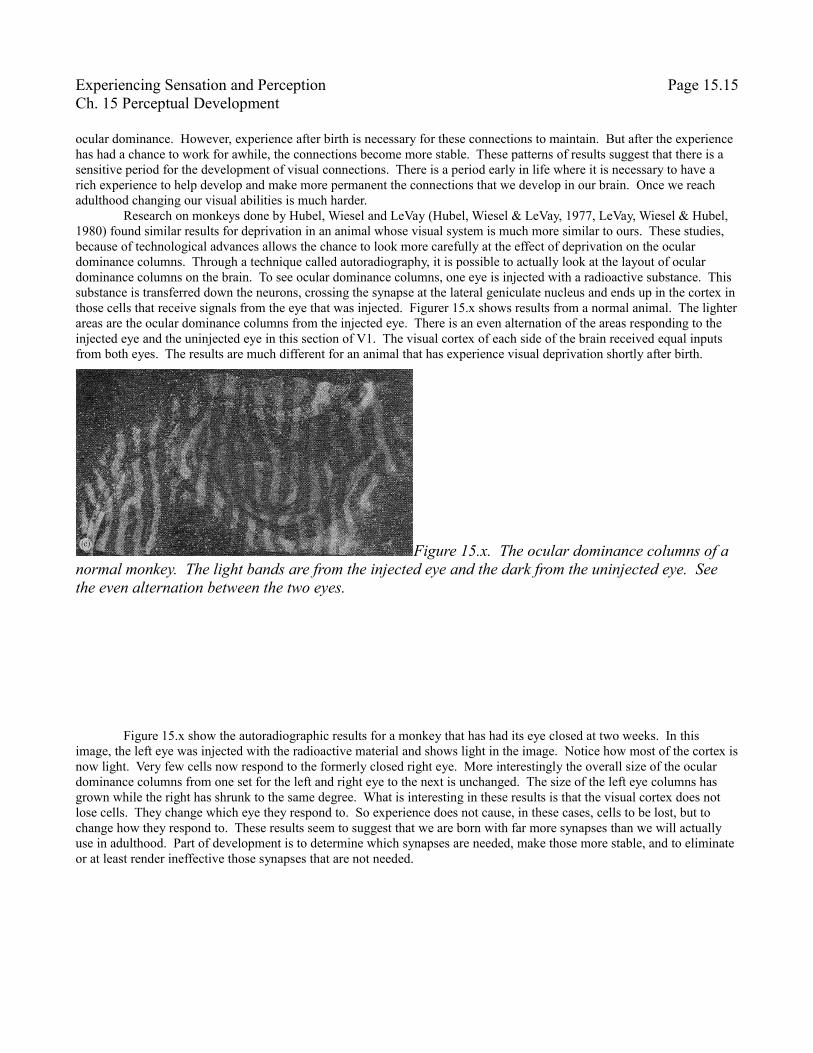
Research on monkeys done by Hubel, Wiesel and LeVay (Hubel, Wiesel & LeVay, 1977, LeVay, Wiesel & Hubel, 1980) found similar results for deprivation in an animal whose visual system is much more similar to ours. These studies, because of technological advances allows the chance to look more carefully at the effect of deprivation on the ocular dominance columns. Through a technique called autoradiography, it is possible to actually look at the layout of ocular dominance columns on the brain. To see ocular dominance columns, one eye is injected with a radioactive substance. This substance is transferred down the neurons, crossing the synapse at the lateral geniculate nucleus and ends up in the cortex in those cells that receive signals from the eye that was injected. Figurer 15.x shows results from a normal animal. The lighter areas are the ocular dominance columns from the injected eye. There is an even alternation of the areas responding to the injected eye and the uninjected eye in this section of V1. The visual cortex of each side of the brain received equal inputs from both eyes. The results are much different for an animal that has experience visual deprivation shortly after birth.
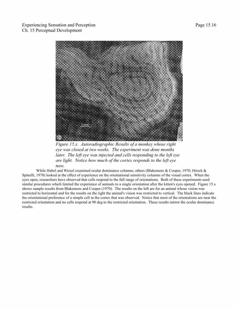
Figure 15.x show the autoradiographic results for a monkey that has had its eye closed at two weeks. In this image, the left eye was injected with the radioactive material and shows light in the image. Notice how most of the cortex is now light. Very few cells now respond to the formerly closed right eye. More interestingly the overall size of the ocular dominance columns from one set for the left and right eye to the next is unchanged. The size of the left eye columns has grown while the right has shrunk to the same degree. What is interesting in these results is that the visual cortex does not lose cells. They change which eye they respond to. So experience does not cause, in these cases, cells to be lost, but to change how they respond to. These results seem to suggest that we are born with far more synapses than we will actually use in adulthood. Part of development is to determine which synapses are needed, make those more stable, and to eliminate or at least render ineffective those synapses that are not needed.
Figure 15.x. The ocular dominance columns of a normal monkey. The light bands are from the injected eye and the dark from the uninjected eye. See the even alternation between the two eyes.
Experiencing Sensation and Perception Page 15.16Ch. 15 Perceptual Development
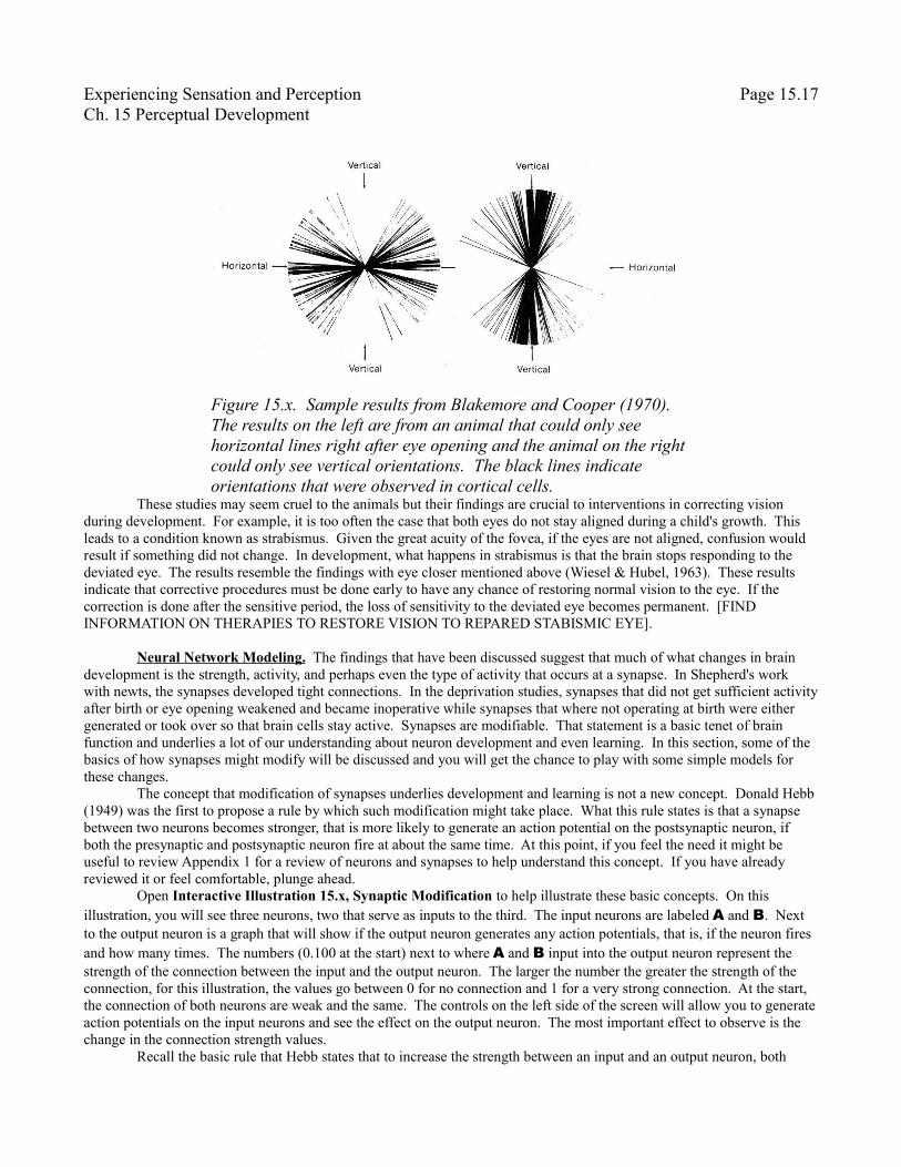
While Hubel and Wiesel examined ocular dominance columns, others (Blakemore & Cooper, 1970; Hirsch & Spinelli, 1970) looked at the effect of experience on the orientational sensitivity columns of the visual cortex. When the eyes open, researchers have observed that cells respond to the full range of orientations. Both of these experiments used similar procedures which limited the experience of animals to a single orientation after the kitten's eyes opened. Figure 15.x shows sample results from Blakemore and Cooper (1970). The results on the left are for an animal whose vision was restricted to horizontal and for the results on the right the animal's vision was restricted to vertical. The black lines indicate the orientational preference of a simple cell in the cortex that was observed. Notice that most of the orientations are near the restricted orientation and no cells respond at 90 deg to the restricted orientation. These results mirror the ocular dominance results.
Figure 15.x. Autoradiographic Results of a monkey whose right eye was closed at two weeks. The experiment was done months later. The left eye was injected and cells responding to the left eye are light. Notice how much of the cortex responds to the left eye now.
Experiencing Sensation and Perception Page 15.17Ch. 15 Perceptual Development
These studies may seem cruel to the animals but their findings are crucial to interventions in correcting vision during development. For example, it is too often the case that both eyes do not stay aligned during a child's growth. This leads to a condition known as strabismus. Given the great acuity of the fovea, if the eyes are not aligned, confusion would result if something did not change. In development, what happens in strabismus is that the brain stops responding to the deviated eye. The results resemble the findings with eye closer mentioned above (Wiesel & Hubel, 1963). These results indicate that corrective procedures must be done early to have any chance of restoring normal vision to the eye. If the correction is done after the sensitive period, the loss of sensitivity to the deviated eye becomes permanent. [FIND INFORMATION ON THERAPIES TO RESTORE VISION TO REPARED STABISMIC EYE].
Neural Network Modeling. The findings that have been discussed suggest that much of what changes in brain development is the strength, activity, and perhaps even the type of activity that occurs at a synapse. In Shepherd's work with newts, the synapses developed tight connections. In the deprivation studies, synapses that did not get sufficient activity after birth or eye opening weakened and became inoperative while synapses that where not operating at birth were either generated or took over so that brain cells stay active. Synapses are modifiable. That statement is a basic tenet of brain function and underlies a lot of our understanding about neuron development and even learning. In this section, some of the basics of how synapses might modify will be discussed and you will get the chance to play with some simple models for these changes.
The concept that modification of synapses underlies development and learning is not a new concept. Donald Hebb (1949) was the first to propose a rule by which such modification might take place. What this rule states is that a synapse between two neurons becomes stronger, that is more likely to generate an action potential on the postsynaptic neuron, if both the presynaptic and postsynaptic neuron fire at about the same time. At this point, if you feel the need it might be useful to review Appendix 1 for a review of neurons and synapses to help understand this concept. If you have already reviewed it or feel comfortable, plunge ahead.
Open Interactive Illustration 15.x, Synaptic Modification to help illustrate these basic concepts. On this illustration, you will see three neurons, two that serve as inputs to the third. The input neurons are labeled A and B. Next to the output neuron is a graph that will show if the output neuron generates any action potentials, that is, if the neuron fires and how many times. The numbers (0.100 at the start) next to where A and B input into the output neuron represent the strength of the connection between the input and the output neuron. The larger the number the greater the strength of the connection, for this illustration, the values go between 0 for no connection and 1 for a very strong connection. At the start, the connection of both neurons are weak and the same. The controls on the left side of the screen will allow you to generate action potentials on the input neurons and see the effect on the output neuron. The most important effect to observe is the change in the connection strength values.
Recall the basic rule that Hebb states that to increase the strength between an input and an output neuron, both
Figure 15.x. Sample results from Blakemore and Cooper (1970). The results on the left are from an animal that could only see horizontal lines right after eye opening and the animal on the right could only see vertical orientations. The black lines indicate orientations that were observed in cortical cells.
Experiencing Sensation and Perception Page 15.18Ch. 15 Perceptual Development
must fire. When the illustration first comes up, it is set up for input neuron A to fire one action potential. Press the Animate button to observe the effect of this setup. When you press the Animate button, an action potential is drawn as it travels down the axon of neuron A. During the travel time, a light blue (cyan) line is drawn on the output neurons graph at the level of the resting potential because nothing yet has reached the output neuron. When the action potential reaches the output neuron, the voltage on the output neuron moves slightly more positive, that is slightly towards the threshold voltage which causes the output neuron to fire. However, voltage on the output neuron does not reach the threshold voltage and no action potential occurs. For this demonstration, the voltage on the output neuron caused by an action potential lasts a pretty long time. That does happen in some neurons but not all. Notice, nothing happens to the strength of either of the connections. No change in how the output neuron responds to either of the input neuron happens.
Now adjust the Number of Action Potentials slider so that it reads 10. Leave only neuron A selected to fire. Now when you press the Animate button again, ten action potentials will be generated by neuron A. Go ahead and run the animation. As each action potential from neuron A reaches the output neuron, the voltage moves a little closer to the threshold through temporal summation. Eventually right at the end of the run, the output neuron fires an action potential seen as large change in the voltage, first positive and then back negative. Look at the connection strength between neuron A and the output neuron. It should be a little bit larger. There was both input and an action potential on the output which are the conditions for strengthening the connection in the Hebb rule. Run the animation several more times without changing the settings. The strength of the connection will continue to grow and it will start to have an impact on how many action potentials from neuron A it will take to fire. First you will see the number of action potentials during the animation grow and it will become apparent that the size of the voltage change caused by an action potential grows. You can reduce the number of action potentials caused by A to get one or more action potentials on the output neuron. However, there is no change in the connection strength for neuron B. The basic rule by Hebb only indicates that there is a change in strength when both neurons fire. In fact if you deselect neuron A to fire and select neuron B to fire, you will see that the strength between neuron B and the output neuron will grow in the exact same way as it did for neuron A.
The basic Hebb rule does demonstrate some features of changing synapses we want to observe. Most important is the fact that in this rule, use is necessary fro strengthening connections. If you think of the newt and deprivation studies, synapses that are used strengthen and will stay strong. Remember that deprivation does not have much impact on the visual system in adults which is what would be expected from a neural connection after use if it followed something like the Hebb rule.
Still, it does not present a complete picture. Let us look at a slight modification to the Hebb rule: a competitive version of the Hebb rule. First, on the illustration, press the Reset button to set the connection strength back to what they were at the beginning. Select the A neuron and deselect the B neuron. Then using the Learning Rule menu select the rule labeled Competitive Hebbian. Keep the Number of Action Potentials slider at 10. When you have it all setup, press the Animate button. As before, one action potential will ultimately be generated by all of the action potentials on neuron A and the connection strength will increase between neuron A and the output neuron. What is new is that because of a modification in the rule, the connection strength between neuron B and the output neuron will decrease. This modification of the Hebb rule is a very simple form of what is called a competitive rule. Increases of strength by one synapse leads to decreases of strength in the other synapse. If you keep running the animation, eventually the connection strength between neuron B and the output neuron will reach 0 (I find this takes about 12 times running the animation). No matter how many times neuron B fires it will never generate any change of voltage on the output neuron, let alone an action potential. Try it.
This outcome much better fits the outcome of the deprivation studies. Let us use the basic ocular dominance findings. They are easier to describe. The output neuron is a neuron in the visual cortex receiving inputs from both eyes. But one eye, say the B eye is closed so that the neuron in the brain does not receive any signals at all, or only a few, from the B eye. All of the signals to the visual cortex cell come from the open eye. The visual cortex cell generates a stronger connection to the open eye and weakens its connection to the closed eye. After the experiment even when the eye is opened, the visual cortex cell will still not respond to the signals from the B, formerly closed, eye. The outcome of this type of synaptic modification looks a lot like the deprivation results which leads researchers to conclude that synapses compete early on in development.
The question remains, how does the cell develop any eye preference at all? That would not happen in this simple competitive case. Hit the Reset button and then select both neurons and press the Animate button. Both neurons
Experiencing Sensation and Perception Page 15.19Ch. 15 Perceptual Development
increase their connection strength. Hit the Reset button again and then got to Learning Rule menu and select the Competitive-Favor A rule. In this case, the initial connection strength is biased in favor of neuron A. If you select both neurons to fire and run the animation several times, you will observe that eventually the output neuron will eventually still have a zero connection strength to neuron B. So, if there is some sort of bias in the initial connection strengths, it is possible to see a way that the normal preferences can arise while still allowing the brain to make use of the cell in unexpected situations, like an eye closer.
It is most unlikely that the actual rules the brain uses to modify neuron connections are as simple as discussed here, these give you some idea about how neurons connections can be modified. The ability of neurons to adjust their connection strength is fundamental to the developmental changes that we have discussed and to the operation of the brain in general. While neurons seem to be able to adjust their connection strength most strongly in when the animal or child it young, the ability remains with us throughout life.
Summary