chapter 14 the arthropods: blueprint for success
TRANSCRIPT

Chapter 14
The Arthropods: Blueprint for Success

Evolutionary Perspective
1. Metamerism modified by tagmatization2. Chitinous exoskeleton3. Paired, jointed appendages4. Ecdysis5. Ventral nervous system6. Coelom reduced to cavity around gonads7. Open circulatory system8. Complete digestive tract9. Metamorphosis often present
Classification and Relationships to other Animals
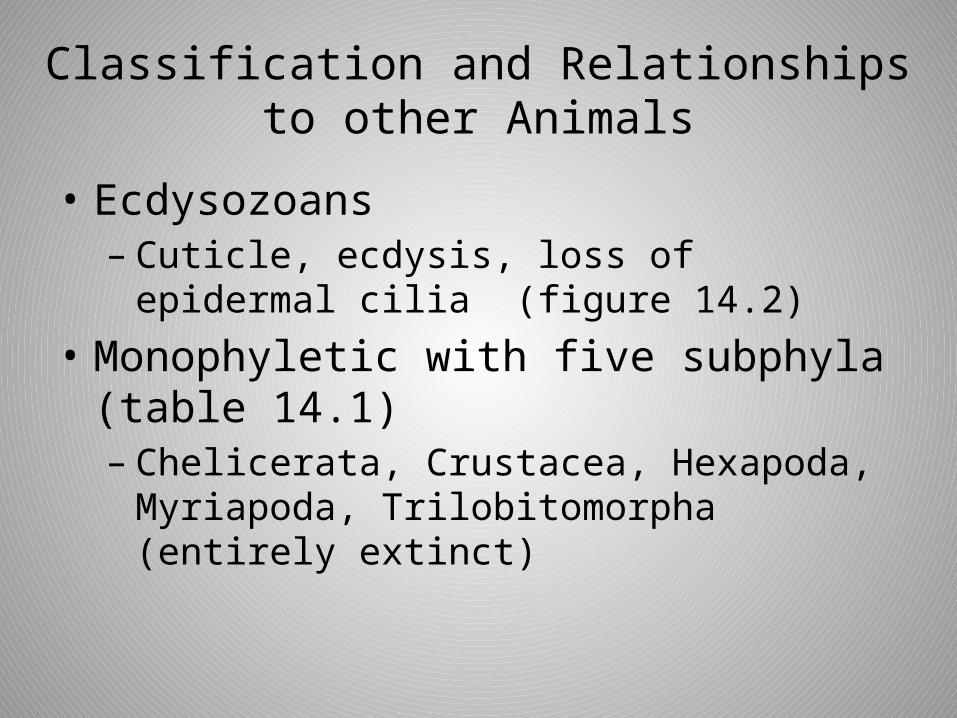
• Ecdysozoans– Cuticle, ecdysis, loss of epidermal cilia
(figure 14.2)
• Monophyletic with five subphyla (table 14.1)– Chelicerata, Crustacea, Hexapoda,
Myriapoda, Trilobitomorpha (entirely extinct)

Figure 14.2 Evolutionary relationships of the arthropods to other animals.
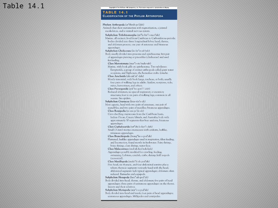
Table 14.1
Metamerism and Tagmatization
• Metamerism evident externally– Segmental body wall– Segmental appendages
• Metamerism reduced internally– No septa– Most organs are not metameric
• Tagmatization obvious– Specializations for feeding, sensory
perception, locomotion, and visceral functions

Learning Outcomes: Section 14.3
• Describe the structure of the arthropod exoskeleton or cuticle.
• Assess the influence the exoskeleton has had on the evolution of the arthropods.
The Exoskeleton
• Exoskeleton or cuticle– External jointed skeleton
• Functions– Structural support– Protection– Prevents water loss– Levers for muscle attachment and movement
• Covers all body surfaces and invaginations
• Secreted by epidermis (hypodermis)

The Exoskeleton
• Epicuticle (figure 14.3)– Lipoprotein– Impermeable to water– Barrier to microorganisms and pesticides
• Procuticle– Chitin
• polysaccharide
– Outer procuticle hardened by sclerotization or deposition of calcium carbonate
– Inner procuticle less hardened and flexible• Articular membranes at joints (figure 14.4)
• Modifications include sensory receptors– Sensilla
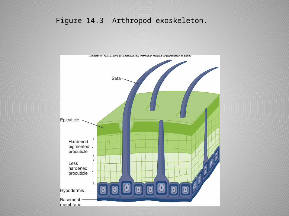
Figure 14.3 Arthropod exoskeleton.

Figure 14.4 Modifications of the exoskeleton.

The Exoskeleton
• Growth accompanied by ecdysis (figure 14.5)1. Enzymes from hypodermal glands begin
digesting old procuticle (a, b).2. New procuticle and epicuticle secreted
(c, d).3. Old exoskeleton splits (e)4. Calcium carbonate deposition and/or
sclerotization hardens new exoskeleton (f).
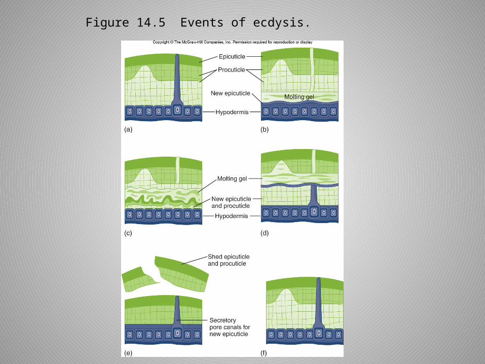
Figure 14.5 Events of ecdysis.
The Hemocoel
• Embryonic blastocoel• Internal cavity for open circulatory
system– Fluids bathe internal organs.– Exchange of nutrients, wastes, and
sometimes gases
• Not a hydrostatic compartment

Metamorphosis
• Radical change in body form and physiology as an immature (larva) becomes an adult.– Reduces competition between adult and
immature stages
Subphylum Trilobitomorpha
• Dominant life form from Cambrian period (600 mya) to Carboniferous period (345 mya)
• Substrate feeders• Three tagmata: head, thorax, and
pygidium• Three longitudinal sections • Biramous appendages

Figure 14.6 Subphylum Trilobitomorpha (Saukia sp).

Subphylum Chelicerata
• Spiders, mites, ticks, horseshoe crabs• Two tagmata
– Prosoma• Eyes• Chelicerae
– Often chelate– Usually feeding appendages
• Pedipalps– Sensory, feeding, locomotion, reproduction
• Walking legs
– Opisthosoma• Digestive, reproductive, excretory, and respiratory
organs

Class Meristomata
• Subclasses– Eurypterida
• Extinct giant water scorpions
(figure 14.7)
Figure 14.7 A eurypterid, Euripterus remipes.
Class Meristomata
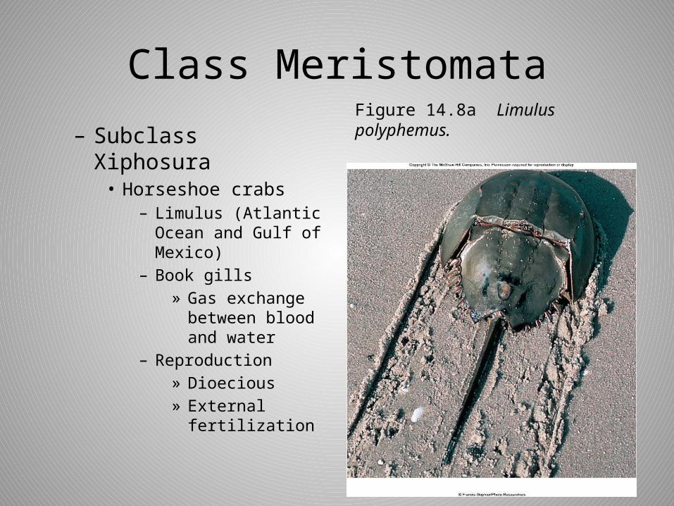
– Subclass Xiphosura• Horseshoe crabs
– Limulus (Atlantic Ocean and Gulf of Mexico)
– Book gills» Gas exchange
between blood and water
– Reproduction» Dioecious» External
fertilization
Figure 14.8a Limulus polyphemus.

Figure 14.8b Ventral view of Limulus.
Class Arachnida
• Spiders, mites, ticks, scorpions• Arose from ancient euryptrids• Very early terrestrial groups– 280-400 mya– Exoskeleton was preadaptation for water
conservation.

Form and Function
• Carnivores– Chelicerae to hold prey or as fangs– Gut
• Foregut – Cuticular – Pumping stomach
• Hindgut– Cuticular – Water reabsorption
• Midgut– Noncuticular– Secretion and absorption

Form and Function
• Excretion– Coxal glands
• Paired sacs bathed in blood of body sinuses• Homologous to nephridia• Excretory pores at base of posterior
appendages
– Malpighian tubules• Blind ending diverticula of gut tract• Empty via digestive tract
– Uric acid

Form and Function
• Gas Exchange– Book lungs• Paired ventral invaginations of body wall• Gas exchange between air and blood across
book lung lamellae
– Tracheae• Branched, chitin-lined tubes• Open at spiracles along abdomen

Figure 14.9 An arachnid book lung.
Form and Function
• Circulation– Open with dorsal contractile vessel– Pumps blood into tissue spaces of
hemocoel– Returns to dorsal vessel via ostia
• Nervous system– Ventral with fusion of ganglia

Form and Function
• Senses– Mechanoreceptors
• Modifications of exoskeleton
• Sensilla respond to displacement.
– Chemical sense• Pores in exoskeleton
– Vision• Eyes detect
movement and changes in light intensity.
Figure 14.10 An arthropod seta (a) and an eye (ocellus) (b).
Form and Function
• Reproduction– Dioecious– Indirect sperm transfer
• Male deposits spermatophores, which are transferred to the female.
– Courtship rituals common– Copulation occurs in spiders via modified
pedipalp of male.
• Development– Direct

Order Scorpionida
• Prosoma– Shieldlike carapace
• Opisthosoma– Preabdomen– Postabdomen (“tail” with sting)
• Courtship prior to mating• Oviparous, ovoviviparous, or
viviparous

Figure 14.11 (a) Hardrurus arizonensis (b) External anatomy.
(a)
(b)
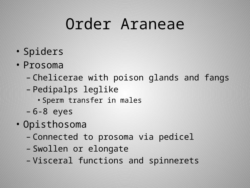
Order Araneae
• Spiders• Prosoma– Chelicerae with poison glands and fangs– Pedipalps leglike
• Sperm transfer in males
– 6-8 eyes
• Opisthosoma– Connected to prosoma via pedicel– Swollen or elongate– Visceral functions and spinnerets

Figure 14.12 External structure of Argiope.
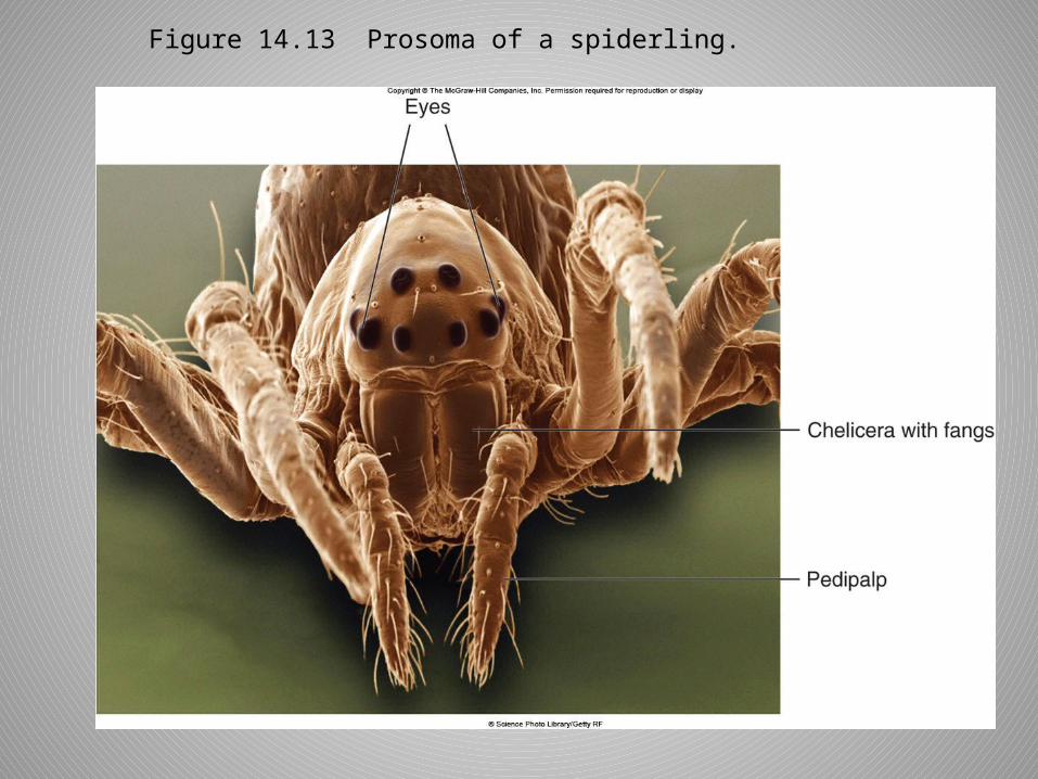
Figure 14.13 Prosoma of a spiderling.

Order Araneae
• Silk– Protein– Repeating sequence of glycine and alanine– Beta sheet– Stored as gel prior to spinning– Chemical modification when forced through
spinnerets
• Webs, line retreats, safety lines, wrapping eggs, dispersal of young (ballooning)

Figure 14.14 Members of the family Araneidae are the orb weavers.

Order Araneae
• Feeding– Insects and other arthropods– Hunt or capture in webs– Paralyze prey• May wrap in silk
– Inject enzymes into prey body wall
• Two spiders are venomous to humans.
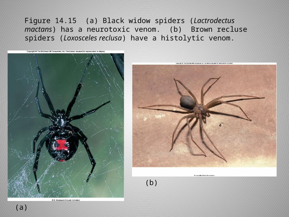
Figure 14.15 (a) Black widow spiders (Lactrodectus mactans) has a neurotoxic venom. (b) Brown recluse spiders (Loxosceles reclusa) have a histolytic venom.
(a)
(b)

Order Araneae
• Reproduction– Complex behaviors• Chemical, tactile, and visual signals
–Male’s pedipalps enlarged into embolus• Male deposits sperm on web and collects
with pedipalps.• Transfers sperm to female during mating
– Female deposits eggs in silk case.• In webbing, a retreat, or carries with her
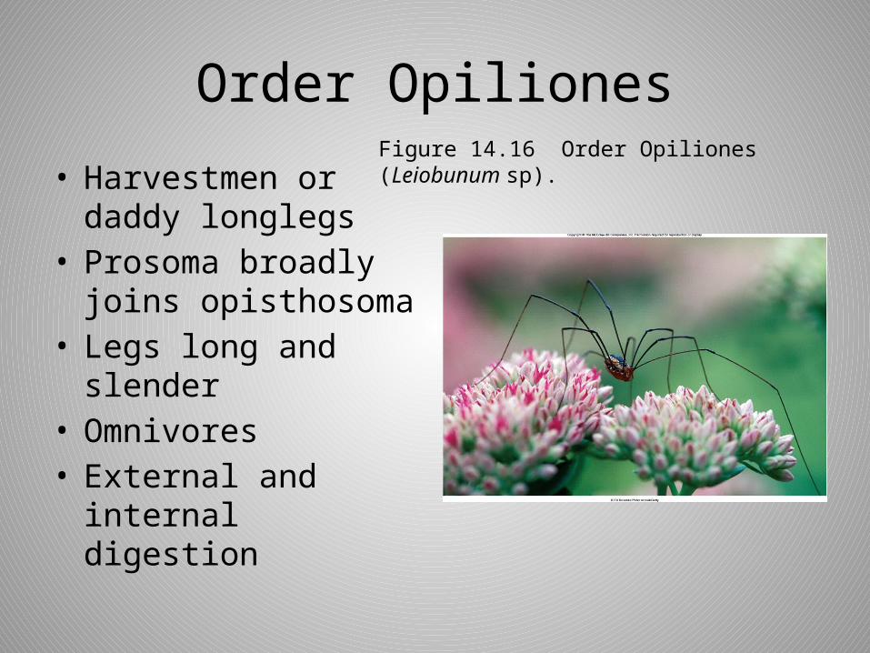
Order Opiliones
• Harvestmen or daddy longlegs
• Prosoma broadly joins opisthosoma
• Legs long and slender
• Omnivores• External and
internal digestion
Figure 14.16 Order Opiliones (Leiobunum sp).

Order Acarina
• Mites– Prosoma and
opisthosoma fused and covered by single carapace
– 1mm or less– Free-living
• Herbivores or scavengers– Many pest species
– Ectoparasites• Chigger (Trombicula)• Follicle mite (Demodex)
Figure 14.17 Dermatophagoides farinae is common in homes and grain storage areas.
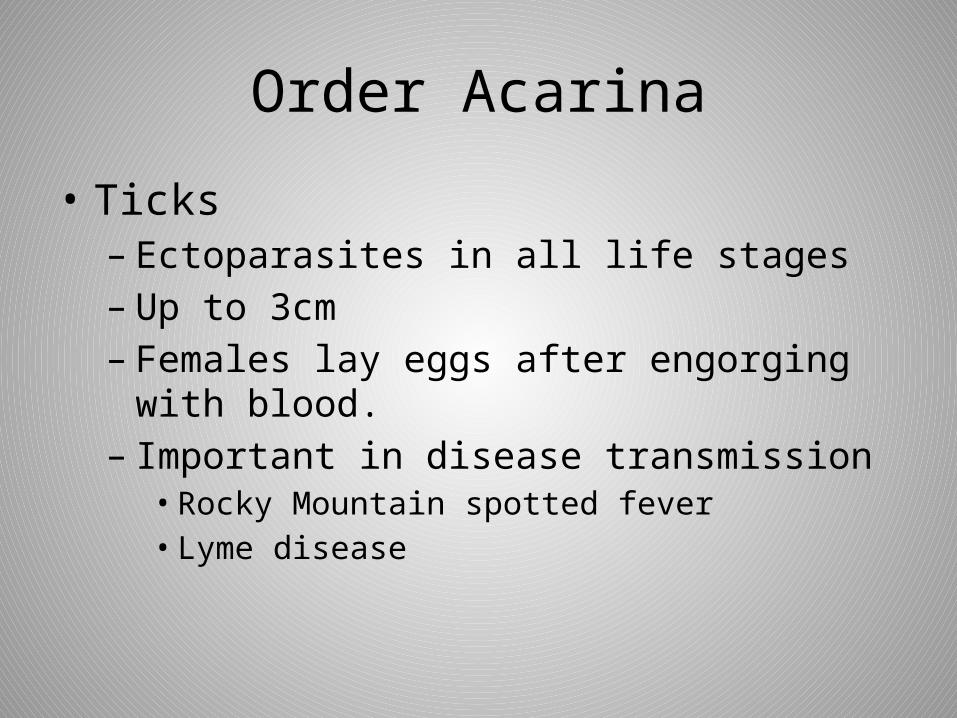
Order Acarina
• Ticks– Ectoparasites in all life stages– Up to 3cm– Females lay eggs after engorging with
blood.– Important in disease transmission• Rocky Mountain spotted fever• Lyme disease
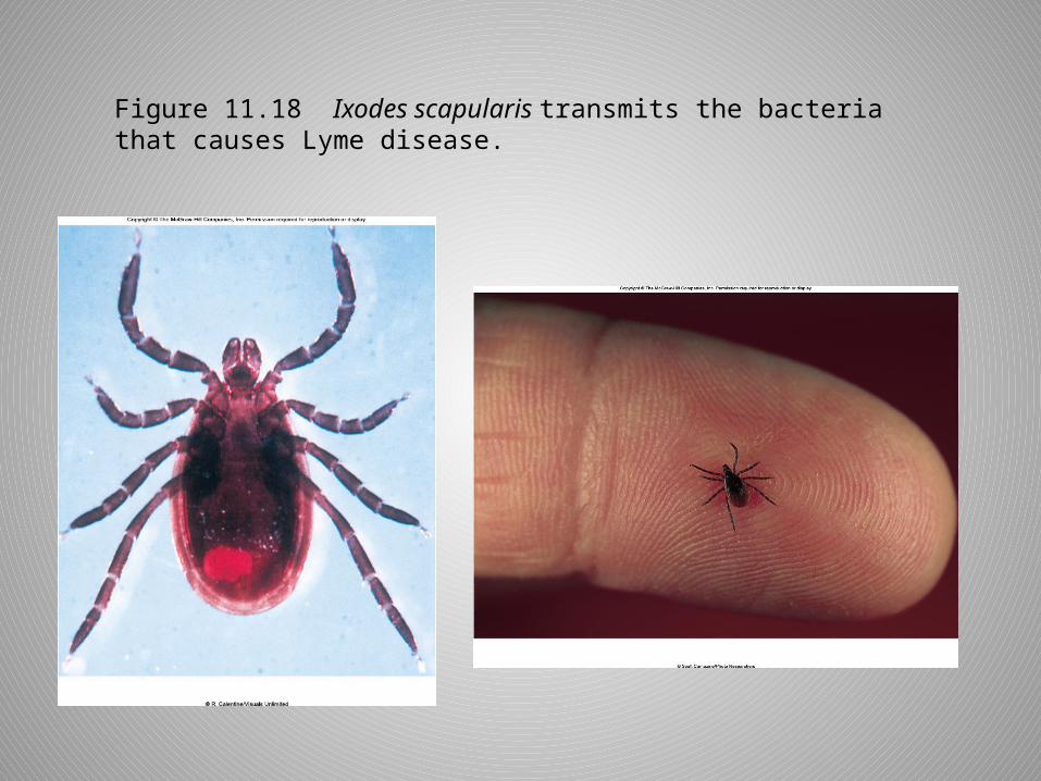
Figure 11.18 Ixodes scapularis transmits the bacteria that causes Lyme disease.
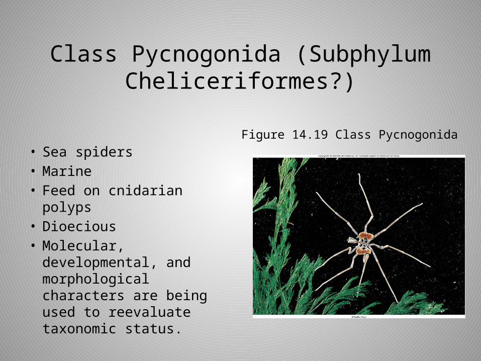
Class Pycnogonida (Subphylum Cheliceriformes?)
• Sea spiders• Marine• Feed on cnidarian polyps• Dioecious• Molecular,
developmental, and morphological characters are being used to reevaluate taxonomic status.
Figure 14.19 Class Pycnogonida
Subphylum Crustacea
• Crayfish, shrimp, lobsters, crabs, copepods cladocerans and others
• Almost all are aquatic– Terrestrial isopods and crabs are
exceptions.
• Two pairs of antennae• Biramous appendages (figure 14.20)
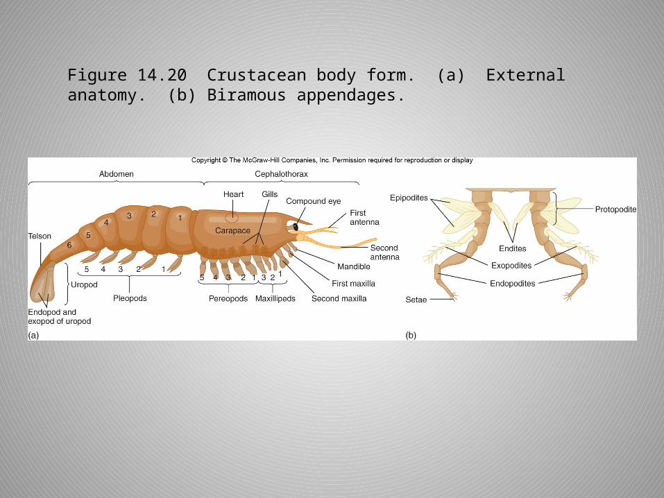
Figure 14.20 Crustacean body form. (a) External anatomy. (b) Biramous appendages.
Class Malacostraca
• Crabs, lobsters, crayfish, shrimp, krill, amphipods, isopods
• Order Decapoda– Largest order– Shrimp, crayfish, lobsters, crabs
Class Malacostraca
• Crayfish external structure– Cephalothorax
• Fusion of head and thorax• Covered dorsally and laterally by carapace• Sensory, feeding, locomotion
– Abdomen• Muscular “tail” in crayfish• Locomotor and visceral functions in others
– Paired appendages• Serially homologous (derived from a common
ancestral pattern)

Figure 14.22 External structure of a male crayfish.
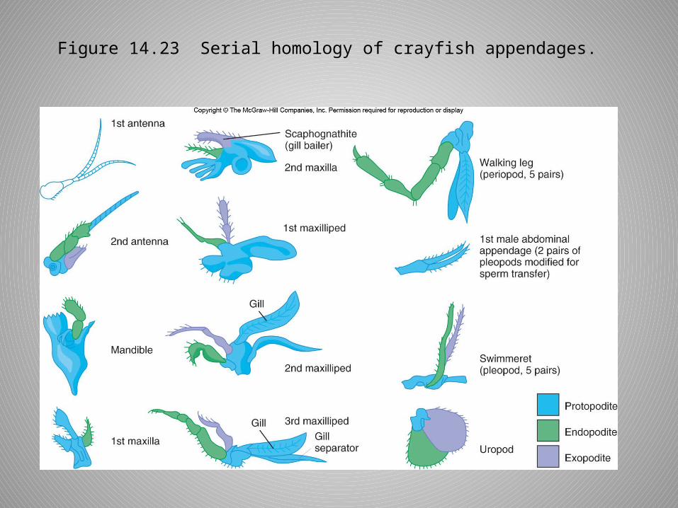
Figure 14.23 Serial homology of crayfish appendages.

Class Malacostraca
• Crayfish internal structure– Digestive system
• Complete with foregut, midgut, and hindgut
– Respiratory system• Gills attach at base of cephalothoracic appendages.• Lie within gill chamber between carapace and lateral
body wall• Second maxilla circulates water.
– Circulation• Open• Dorsal heart and major arteries• Blood enters hemocoel, and gills before returning to
pericardial sinus around heart.

Figure 14.24 Internal structure of a crayfish.

Class Malacostraca
• Ventral nervous system– Cephalization and centralization– Supraesophageal and subesophageal ganglia process
sensory information and control head appendages.– Segmental ganglia
• Sensory structures– Antennae– Compound eyes– Statocysts– Chemoreceptors– Proprioceptors– Tactile setae
Class Malacostraca
• Endocrine system– Ecdysis, sex determination, color change
• X-organs– Neurosecretory tissues in eyestalks– Molt-inhibiting hormone
» Target Y-organ
• Y-organs– Base of maxillae– Releases ecdysone when molt inhibiting hormone is not
present and ecdysis occurs
– Androgenic glands (males)• Promotes development of testes and male
characteristics
Class Malacostraca
• Excretion– Antennal (green) glands in crayfish– Maxillary glands in others– Homologous to coxal glands of arachnids
• Reproduction– Dioecious– Mating after female molts
• Fertilized eggs attach to female’s pleopods• Others have planktonic larvae

Figure 14.25 (a) Nauplius larva of a barnacle. (b) Zoea larvae of a crab.
(a)
(b)
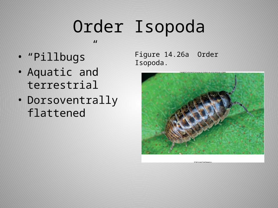
Order Isopoda
• “Pillbugs”• Aquatic and
terrestrial• Dorsoventrally
flattened
Figure 14.26a Order Isopoda.

Order Amphipoda
• Laterally compressed
• Crawl or swim on sides
• Beach-hoppers modified for jumping
Figure 14.26b Order Amphipoda.

Class Branchiopoda
• Fairy shrimp – Temporary ponds
• Brine shrimp– Great Salt Lake
• Cladocera– Freshwater water fleas– Large carapace– Parthenogenesis
common
• Flattened, leaflike appendages
Figure 14.27 Order Cladocera.
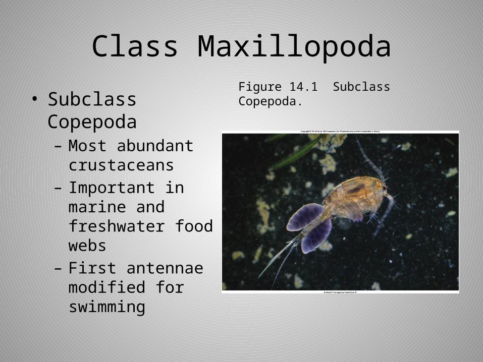
Class Maxillopoda
• Subclass Copepoda– Most abundant
crustaceans– Important in marine
and freshwater food webs
– First antennae modified for swimming
Figure 14.1 Subclass Copepoda.

Class Maxillopoda
• Subclass Thecostracea, Infraclass Cirripedia– Barnacles–Marine–Monoecious• Nauplius and cypris larvae• Cypris larva settles and metamorphoses into
sessile adult.
– Some parasites
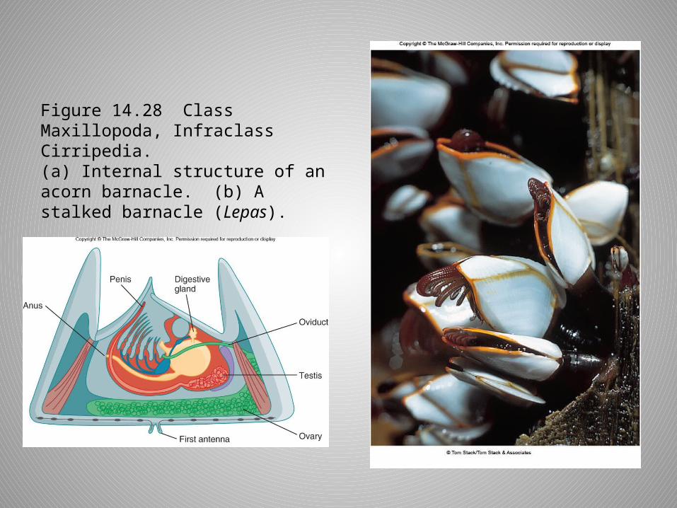
Figure 14.28 Class Maxillopoda, Infraclass Cirripedia. (a) Internal structure of an acorn barnacle. (b) A stalked barnacle (Lepas).

Further Phylogenetic Considerations
• Diverse body forms and lifestyles of Arthropoda arose from single ancestor.
• Crustaceans very successful in aquatic habitats
• Chelicerata– First terrestrial arthropods– Account for evolution of many water
conserving features of the phylum• Exoskeletal, excretory, and respiratory
adaptations