chapter 12 sensory...
TRANSCRIPT
Chapter Concepts12.1 Sensory Receptors and Sensation
• Sensory reception occurs at the senses. Sensation and perception occur in the brain.
• Various sensory receptors detect information in the internal and external environments.
12.2 Photoreception
• The human eye is similar to a camera. It contains a lens that focusses light, a pupil that lets in light, and a dark interior that contains light receptors.
• The retina contains rods and cones. Rods function in dim light and produce black and white images. Cones function in bright light and produce colour images.
12.3 Mechanoreception and Chemoreception
• The outer ear and middle ear transmit the energy of sound waves to the inner ear.
• The inner ear contains mechanoreceptors for hearing and balance.
• Tastes are detected by chemoreceptors in the taste buds of the tongue. Smells are detected by chemoreceptors in the nose.
• The skin contains receptors for light touch, pressure, pain, heat, and cold.
he woman meditating is trying to ignore all sights, sounds, andother sensory input from the outside world. It is a diffi cult task.
Some people prefer the experience of relaxing inside a sensory deprivationtank for an hour or so. Many people fi nd this experience relaxing fora short period of time—usually about an hour or so. If the brain is deprived of sensory information for an extended period of time, however, extreme anxiety, hallucinations, depression, memory loss, and antisocial behaviour can result. People who are in solitary confi nementsometimes experience these effects, as do people who are confi ned to bed in hospital isolation wards. Why does sensory deprivation confusethe brain and upset homeostasis in the body? As you will discover in this chapter, the senses function together to allow the human body to detect and adjust to changes in the body and in the external environment.
12CHAPTER Sensory Reception
404 MHR • Unit 5 The Nervous and Endocrine Systems
1 3296_Chapter_12_Opener.indd 11 3296_Chapter_12_Opener.indd 1 11/4/06 6:22:17 PM11/4/06 6:22:17 PM

Sense It
When one or more of the senses are inhibited, the brain’s perception of the environment can change. Identifying even familiar objects can become diffi cult. In this activity, you will try to identify familiar objects while one or more of your senses is inhibited.
Safety Precautions• Do not bring food meant for consumption into the laboratory.
• Do not taste any sample (you will use only the senses of touch and smell in this exercise).
• Before the exercise begins, alert your teacher to any allergies you have.
Materials• new earplugs • blindfold
• samples of unidentifi ed but familiar objects supplied by your teacher
Procedure 1. Work with a partner. One partner will be the tester, and the other partner will
be the subject.
2. The subject will use earplugs and a blindfold to block the senses of hearing and sight.
3. The tester will acquire samples of the unknowns provided by the teacher.
4. Now, with the subject gently pinching his or her nose shut to block the sense of smell, the tester will provide the fi rst sample for the subject to hold.
5. The subject should use the sense of touch to identify the unknown sample, and then the subject should use the sense of smell if the sample remains unknown. The tester should record how and if the subject is able to identify the sample.
6. Repeat steps 4 and 5 using the other samples. Then switch roles, and repeat the activity with another set of samples.
Analysis 1. Which samples were the most diffi cult to identify? Which samples were the
easiest to identify?
2. Which senses would you normally use to identify the samples?
3. You probably found it easier to identify the samples using a number of senses, rather than only one. Which senses would you use to
a) check if milk is sour
b) remove stones from a bag of dry lentils
c) stand on one leg without falling over
4. Explain why integration is important for interpreting sensory information.
Launch Lab
Chapter 12 Sensory Reception • MHR 405
1 3296_Chapter_12_Opener.indd 21 3296_Chapter_12_Opener.indd 2 11/4/06 6:22:41 PM11/4/06 6:22:41 PM

406 MHR • Unit 5 The Nervous and Endocrine Systems
12.1S E C T I O N
Imagine yourself in the fairgrounds shown in Figure 12.1. What sensory information would your nervous system be gathering? What would you see, hear, and smell? What foods would you taste? How would a light breeze feel on your skin? Your senses of sight, hearing, taste, smell, and touch keep you informed about the world around you, and allow your body to respond to your external environment. Internal sensors, such as those that regulate blood pH, blood pressure, and blood volume, also help your body maintain homeostasis. The senses transmit sensory information, in the form of electrochemical impulses, to the brain. Different forms of energy stimulate the sensory receptors—the nerve endings and cells that detect sensory information. The sensory receptors then initiate neural impulses. Sensation occurs when the neural impulses arrive at the cerebral cortex. For example, your face may detect the warmth of a beam of sunlight. When the brain receives and processes this information, you will feel, or experience, the sensation of warmth on your cheek. The resulting sensation depends on the
area of the brain that has interpreted this information. Neural impulses that begin in the optic nerve are sent to the visual areas of the cerebral cortex, and we see objects (Figure 12.2). Neural impulses that begin in the auditory nerve of the ear are sent to the area of the brain that perceives sound, and we hear sounds. Each person’s unique perception results from how the cerebral cortex interprets the meaning of the sensory information.
Compare sensory reception and sensation. Provide an example of each.
Each type of sensory receptor functions by initiating neural impulses. How is the brain able to convert sensory information into perception?
• • •
• • •
Sensory AdaptationA massive amount of sensory information, coming from many neural pathways, bombards the brain every second. Sometimes, the brain can fi lter out
Section Outcomes
In this section, you will • explain the difference
between sensory reception, sensation, and perception
• describe the process of sensory adaptation
• distinguish among the major sensory receptors in the human body
Key Terms
sensory receptorssensationperceptionsensory adaptationphotoreceptorschemoreceptorsmechanoreceptorsthermoreceptors
Figure 12.1 What information would your senses detect if you were standing in this scene? How would this information help your body maintain homeostasis?
Sensory Receptors and Sensation
2 3296_Chapter_12.indd 12 3296_Chapter_12.indd 1 11/4/06 6:23:00 PM11/4/06 6:23:00 PM

Chapter 12 Sensory Reception • MHR 407
redundant, insignifi cant information. This process is called sensory adaptation. For example, sensory adaptation has occurred when you no longer notice the ticking of a clock or feel the clothes on your skin. When the senses detect a signifi cant change in external or internal conditions, the body readjusts (Figure 12.3). In order to process sensory information quickly, the brain parallels or splits up this input to various areas of the brain—a form of neural multi-tasking. Sometimes the sensory information does not get reintegrated precisely, and what we sense is not necessarily what we perceive. This effect can be demonstrated with optical illusions, which scientists use to try to understand how the brain
perceives sensory information. Use the illusions in Figure 12.4 on page 408 to determine the difference between the sensory information received and what your brain actually perceives.
Describe an example of sensory adaptation, and explain why it occurs.
What happens during integration that causes people to perceive optical illusions?
• • •
• • •
Sensory ReceptorsSensory receptors are specialized cells or neuron endings that detect specifi c stimuli. Human sensory receptors can
Figure 12.2 A PET scan of the human brain showing brain activity in a volunteer at different stages of stimulation. The orange-red colour indicates that the brain is active and perception is taking place. In the last two columns, the visual area at the back of the brain is activated and the perception of sight is occurring. In the first column, the subject’s eyes are closed. Thus neural information is not getting from the senses to the brain, and there is no perception of sight.
Figure 12.3 When you wash your hands in tepid water after being outside in the cold, the water may feel very hot. In contrast, you may perceive the tepid water as chilly after holding a warm cup of tea. You can duplicate this phenomenon by placing your right hand in a cup of warm water and your left hand in a cup of cold water for 1 min. Then put both hands in room-temperature water. What has happened to your perception of temperature, and why?
BiologyFile
FYICan an octopus experience pain and suffering? Most likely, say neuroscientists, which is why these common research animals need to be treated humanely and given behavioural enrichment. Support for this idea has come from experiments conducted by octopus expert Jennifer Mather, professor of psychology at the University of Lethbridge, and Roland Andersen of the Seattle Aquarium. The researchers gave empty plastic pill bottles to eight octopuses, who played with the pill bottles. This was the fi rst time play had been observed in invertebrates.
2 3296_Chapter_12.indd 22 3296_Chapter_12.indd 2 11/4/06 6:23:07 PM11/4/06 6:23:07 PM

408 MHR • Unit 5 The Nervous and Endocrine Systems
be classifi ed into four categories: photoreceptors, chemoreceptors, mechanoreceptors, and thermoreceptors (Table 12.1). Each type of receptor is able to transduce, or convert, one form of energy from a specifi c stimulus into electrochemical energy, which can be processed by the nervous system. Light energy stimulates photoreceptors. Our eyes contain photoreceptors, called rods and cones, that absorb light and allow us to sense different levels of light and shades of colour. Chemoreceptors are stimulated by certain chemicals. The tongue contains taste buds that detect various particles in the food we eat. The nose has olfactory cells that detect odours in the air. Other chemoreceptors detect changes in the internal environment. For example, chemoreceptors in the carotid arteries and aorta detect blood pH. Mechanoreceptors respond to mechanical forces from some form of pressure. For example, hair cells in the inner ear (hearing sensors) are activated when sound waves cause parts of the inner ear to vibrate. Other hair cells in the inner ear (sensors for balance) are stimulated when they bend, thus providing information about body and head position. Proprioceptors in and near the muscles also provide information about body position, as well as movement. There are various mechanoreceptors in the skin, which allow the body to detect light touch, pressure, and even pain. Thermoreceptors in the skin detect heat and cold.
Damage to particular sensory receptors such as photoreceptors can result in loss of the associated sense, even if the rest of the sense organ and nervous system are fully functional. Hundreds of thousands of Canadians have lost their sight due to eye injuries or degenerative eye disorders, such as retinitis pigmentosa and age-related degeneration of the photoreceptors. A newly developed artifi cial eye, however, offers hope to these people. The artifi cial eye includes a digital video camera that is mounted on glasses. The camera captures images and sends them to a small computer on a belt worn by a person who is visually impaired. The images are processed and sent to several electrodes that are implanted in the visual cortex, thus bypassing the damaged light receptors in the eye. The electrodes directly stimulate the brain, producing a pattern of bright
Figure 12.5 The artificial eye sends electrical impulses that directly stimulate the visual cortex of the brain. This man became blind in an accident over 20 years ago. He can now distinguish large letters and several objects, and he has even driven a car around a parking lot.
A Herman’s Grid. When you look at the centre of the large square, the dots in the middle should look white, and the dots you see out of the corners of your eye should look black.
B The Impossible Terrace, by D. MacDonald. Is down up, or is up down?
D Fraser’s Spiral. Try to follow the spiral into the centre.
C Kitaoka’s Rotating Snakes. Look at these circles from off centre. Do you think they are actually spinning?
Figure 12.4 These diagrams produce various optical illusions.
BiologyFile
Web LinkRetinitis pigmentosa includes several inherited diseases that cause the photoreceptor cells (rods and cones) in the retina to degenerate and die. Dr. Paul Schnetkamp at the University of Calgary is one of the scientists studying this disorder, which affects an estimated 50 000 Canadians. What is the current state of understanding and treatment of retinitis pigmentosa?
@wwwwww.albertabiology.ca
2 3296_Chapter_12.indd 32 3296_Chapter_12.indd 3 11/4/06 6:23:37 PM11/4/06 6:23:37 PM

Chapter 12 Sensory Reception • MHR 409
spots that the visual cortex perceives as a crude image. Currently, the resolution of this device is poor—about fi ve pixels per square centimetre—but it allows users to distinguish objects with high contrast. Researchers predict that future developments will improve the device enough to restore vision completely,
Identify fi ve major senses and their corresponding sensory receptors.
List three types of internal sensory receptors and their functions.
• • •
• • •
or perhaps to enhance vision beyond natural human capabilities.
Section 12.1 Summary• The senses are the human brain’s
connection to the outside world. • Sensation is initiated in the senses
through sensory reception, but sensation and perception take place in the brain.
• Everyone’s perception of the world is unique.
• Sensory receptors convert different forms of energy into electrochemical energy, which the nervous system can interpret.
BiologyFile
Web LinkWhat would happen if your optic and auditory nerves were switched? How would you perceive a fl ash of lightening or crash of thunder? Researchers at the University of Waterloo in Ontario, Canada, are studying what happens when senses blend, a condition called synesthesia. Visit the Web Links page to fi nd out what some people with synesthesia experience.
@wwwwww.albertabiology.ca
Table 12.1 Major Sensory Receptors in the Human Body
Category and type of receptor Examples of receptor Stimulus
Photoreceptors
vision rods and cones in the eye visible light
Chemoreceptors
taste taste buds on the tongue food particles in saliva
smell olfactory receptors in the nose odour molecules
internal senses osmoreceptors in the hypothalamus low blood volume
receptors in the carotid artery and aorta blood pH
Mechanoreceptors
touch/pressure/pain receptors in the skin mechanical pressure
hearing hair cells in the inner ear sound waves
balance hair cells in the inner ear fl uid movement
body position proprioceptors in the muscles and tendons, and at the joints
muscle contraction, stretching, and movement
Thermoreceptors
temperature heat and cold receptors in the skin change in radiant energy
1. State the form of energy that stimulates
a) thermoreceptors b) photoreceptors
2. What types of sensory receptors are stimulated in someone who is performing a complicated yoga pose?
3. Crime-scene investigators are often frustrated by different eyewitness accounts of the same crime. With your knowledge of sensation and perception, explain how different people’s perceptions of the same sensory information can be different.
4. When you put on your new winter boots one morning, you cannot help noticing how different they feel from your summer shoes. By the time you reach school, however, you have forgotten about your boots. Why does your perception of the way your boots feel change?
5. Describe, in general terms, what happens in the nervous system when someone receives a large amount of signifi cant sensory information, such as when viewing an optical illusion.
ReviewSection 12.1
2 3296_Chapter_12.indd 42 3296_Chapter_12.indd 4 11/4/06 6:23:46 PM11/4/06 6:23:46 PM

410 MHR • Unit 5 The Nervous and Endocrine Systems
12.2S E C T I O N Photoreception
Section Outcomes
In this section, you will • describe the principal
structures of the human eye and their functions
• observe the principal features of the mammalian eye and perform experiments that demonstrate the functions of the human eye
• describe several eye disorders and treatments
Key Terms
scleracorneachoroidirispupiladaptationretinarodsconesoptic nerveaqueous humourglaucomavitreous humourlensaccommodationcataractsastigmatismmyopiahyperopiafovea centraliscolour blindnessblind spot
Scientists estimate that, in sighted people (people who can see), vision supplies 80 to 90 percent of the important sensory information reaching the brain. The incredible variety of colours and shapes and the range of distance depicted in the landscape shown in Figure 12.6 would be diffi cult to conceive of without the sense of sight. How does the human eye carry out its varied functions? The human eye is essentially a fl uid-fi lled hollow ball, about 2.5 cm in diameter. It focusses incoming light energy on the photoreceptors of the retina. As shown in Figure 12.7, the eye has three layers: external, intermediate, and internal. The external layer of the eye is a white, tough, and fi brous protective layer called the sclera. Light enters the eye through the cornea, the transparent part of the sclera at the front of the eye. The intermediate layer of the eye is called the choroid. The choroid absorbs stray light rays that are not detected by the photoreceptors. As well, the choroid contains blood vessels that nourish the eye. Toward the front, the choroid forms the doughnut-shaped, coloured iris, which contains a central dark pupil. Like
fi ngerprints, the pattern of colour in the iris is unique to each person. Thus, an iris scan could potentially be used for personal identifi cation. The iris allows light to enter the inner eye through the pupil. As shown in Figure 12.8, the iris adjusts the size of the pupil based on the light conditions— a process called adaptation. You can observe adaptation in your own eyes by turning off the lights for 1 min and holding a mirror in front of your face. When you turn the lights back on, you can see your pupils shrink, which allows less light to enter your eyes. Behind the iris, the choroid thickens and forms the ciliary muscle. The ciliary muscle attaches to the lens, which focusses images on the retina. The retina is the internal layer of the eye. It is a thin layer of tissue that contains the photoreceptors—the rods and cones. The rods are sensitive to light intensity (level of brightness), and the cones are sensitive to different colours. The cones are packed most densely at the back of the eye in an area called the fovea centralis. The rods and cones send sensory impulses to the brain via the optic nerve.
Figure 12.6 The human sense of sight can tell us more about the world than any other sense can. How does the human eye detect the colour, contrast, depth, and three-dimensional shapes of this landscape?
BiologyFile
Web LinkSome Canadian companies are using biometrics technology to verify that you are who you say you are. What is biometrics, and what advantages and disadvantages are associated with it?
@wwwwww.albertabiology.ca
2 3296_Chapter_12.indd 52 3296_Chapter_12.indd 5 11/4/06 6:23:48 PM11/4/06 6:23:48 PM

Chapter 12 Sensory Reception • MHR 411
If there were no fl uid inside the eyeball, it would lose its shape, like an empty water balloon. The lens, which is attached to the ciliary muscles by suspensory ligaments, divides the eye into two chambers. The anterior chamber is in front of the lens, and the posterior chamber is behind the lens. In the anterior chamber, a clear, watery fl uid called the aqueous humour maintains the shape of the cornea and provides oxygen and nutrients for the surrounding cells, including those of the lens and cornea. A small amount of aqueous humour is produced every day and drained by small ducts. If these ducts become plugged, pressure can build up in the eye, causing the delicate blood vessels in the eye to rupture. The cells of the eye then
Figure 12.8 Light adaptation in the eye. In bright light, the iris constricts, which shrinks the pupil to let in less light. In dim light, the iris dilates, which widens the pupil and lets in more light.
deteriorate, due to a lack of oxygen and nutrients. This results in glaucoma, which leads to blindness if untreated. The posterior chamber, which is surrounded by the retina, contains a clear, jelly-like fl uid called the vitreous humour. The vitreous humour also helps to maintain the shape of the eyeball, and it supports the surrounding cells. The structures of the human eye, and their functions, are summarized in Table 12.2.
Table 12.2 The Principal Structures of the Human Eye and Their Functions
Structure Function
External layer (Sclera)
sides and back of sclera protects and supports the eyeball
cornea bends light rays into the eye
Intermediate layer (Choroid)
sides and back of choroid absorbs scattered light and contains blood vessels
iris regulates the amount of light that enters the eye
pupil is the opening for light to enter the inner eye
ciliary muscles changes the shape of the lens in order to focus
Internal layer (Retina)
rods photoreceptors that are sensitive to dim light
cones photoreceptors that are sensitive to different wavelengths of light (colour vision)
fovea centralis contains a high density of cones, and provides acute vision
Other
lens focusses light rays onto the fovea centralis
humours support the eyeball, with the pressure of the fl uids they contain
optic nerve transmits sensory information to the brain
Figure 12.7 The anatomy of the human eye
iris pupil sclera
sclera retina
fovea centralis
optic disc (blind spot)
optic nerve
choroid
suspensory ligaments
iris
pupillenscornea
aqueous humour
ciliary muscle
vitreous humour
2 3296_Chapter_12.indd 4112 3296_Chapter_12.indd 411 7/26/07 5:51:37 PM7/26/07 5:51:37 PM

412 MHR • Unit 5 The Nervous and Endocrine Systems
Draw a cross section of an eye in your notebook. Identify the key structures and their functions.
How do the eyes adjust to changing light intensities? Name this process.
How does glaucoma occur, and how can it cause blindness?
• • •
• • •
FocussingHow do eyeglasses and contact lenses help people see? How do the lenses in cameras and microscopes work? If you could closely examine any artifi cial lens,
Figure 12.10 Accommodation. (A) What happens to the shape of the lens when the eye is focussed on a distant object? (B) What happens when the eye is focussed on a nearby object? Note that the ability of the lens to bend light is exaggerated in this diagram. In reality, the cornea also bends light.
you would fi nd that light rays bend as they pass through the lens. In other words, the lens focusses the light in a particular direction. The lens of the eye focusses incoming light in a similar way. Eyeglasses and contact lenses make up for imperfections in the eyes that prevent them from focussing clearly. Figure 12.9 shows what happens when the eye brings an image into focus. Light rays are bent as they pass through the rigid cornea, fl exible lens, and fl uid humours. Notice that, compared with the object being viewed, the image fi xed on the fovea centralis of the retina is smaller, upside down, and reversed from left to right. Because the lens is fl exible, it can change shape. This allows for fi ner focus when viewing objects, whether they are nearby or far away. As shown in Figure 12.10, if an object is far away, the ciliary muscles relax and the suspensory ligaments become taut, causing the lens to fl atten. If an object is nearby, the ciliary muscles contract and the suspensory ligaments relax, causing the lens to become more rounded. Similarly, a nondigital camera focusses by changing the distance between the lens and the fi lm. The ability of the lens to change shape in order to focus images clearly on the retina is a refl ex called accommodation. If you read your textbook up close for an extended period of time, the ongoing contraction of your ciliary muscles will likely cause muscle fatigue, which you will experience as eyestrain.
Conditions Affecting the Cornea and LensAs the lens ages, its protein structure can start to degenerate, making it opaque and preventing light from passing through it. This condition can cause grey-white spots, called cataracts, on the lens. To prevent cataracts from impairing vision, the lens can sometimes be surgically replaced. Inherited conditions that affect the eye’s ability to focus light directly
Figure 12.9 What happens to light rays as they pass through the cornea and lens?
2 3296_Chapter_12.indd 72 3296_Chapter_12.indd 7 11/4/06 6:24:10 PM11/4/06 6:24:10 PM

Chapter 12 Sensory Reception • MHR 413
on the fovea centralis are common. Astigmatism, for example, is due to an uneven curvature of part of the cornea. Because the cornea is asymmetrical, it cannot bend light rays so that they meet at the correct focal point (Figure 12.11). As a result, vision is blurred. When young, most people can clearly see a size 20 letter on an eye chart from a distance of 20 feet (about 6.1 m). This is referred to as 20/20 vision. People who have no diffi culty seeing close objects but cannot see a size 20 letter from 20 feet are nearsighted. They have a condition called myopia. As shown in Figure 12.12, the eyeball of people with myopia is elongated, so the focussed light falls in front of the retina instead of on the photoreceptors. To see distant objects,
nearsighted people can wear concave lenses, which diverge incoming light rays so that the image falls directly on the retina. An alternative to wearing corrective lenses is corrective laser surgery. In this procedure, an ophthalmologist (eye doctor) uses a laser to cut and reshape the cornea so that it will focus light onto the retina. Another common condition is hyperopia. People with this condition are farsighted, which means they can see clearly from 20 feet, but cannot focus on nearby objects because the eyeball is too short. The light rays do not meet before they reach the retina, so the image is focussed behind the retina. Convex lenses can correct hyperopia by bending light rays at a sharper angle.
Light rays from far object
Figure 12.11 Astigmatism and how it can be corrected
A The off-centre bending of light rays by an uneven cornea
B Unevenly ground glasses or contact lenses can correct for astigmatism
C A test for astigmatism. If any of the dark lines in the diagram seem darker than the others, you could have an asymmetrical cornea
A B
C D
Figure 12.12 (A) Image seen by someone who is nearsighted. Which object is most in focus? (B) Myopia. Where does the image fall in the eye? (C) Image seen by someone who is farsighted. (D) Hyperopia. Where does the image fall?
2 3296_Chapter_12.indd 82 3296_Chapter_12.indd 8 11/4/06 6:24:12 PM11/4/06 6:24:12 PM

414 MHR • Unit 5 The Nervous and Endocrine Systems
Describe the functions of the cornea and lens of the eye.
What causes astigmatism, myopia, and hyperopia?
Compare adaptation with accommodation. Give an example of each.
• • •
• • •
The Photoreceptors: The Rods and ConesVision begins as light is focussed on the light-receiving cells, or photoreceptors. The human retina contains about 125 million rods and 6 million cones, which account for at least 70 percent of all sensory receptors in the human body. The rods are extremely sensitive to light. In fact, a rod can be stimulated by a single photon of light. The rods do not enable us to distinguish colours, however, just degrees of black and white. This is why it is diffi cult to detect any colours in a dark room, although you may be able to see the shapes of the furniture. Rods also detect motion and are responsible for peripheral vision. In the human eye, the rods are spread throughout the retina, but are more concentrated in the outside edges. The cones are the colour-detecting sensors of the eye. They are packed most densely at the fovea centralis (see Figure
Figure 12.14 Check your colour vision by studying this colour blindness chart. Can you see the number 8?
12.13) at the back and centre of the retina. You may have noticed that your peripheral vision lacks colour. This is due to the central location of the fovea centralis. The cones require relatively intense light to stimulate them. Thus, the structures of the eye must focus light onto the fovea centralis in order to produce a sharp image. In daylight, you can see best (your eyes produce the sharpest image) if you look at an object straight on. In addition to allowing us to see “in colour,” the cones allow us to perform high-acuity tasks, such as reading. There are three types of cones, and each type absorbs a different wavelength of light. The combination of cones that can detect red, blue, and green wavelengths of light allows us to see a range of colours. Colour blindness is an inherited condition that occurs more frequently in males than in females. (You will discover why in Unit 7.) Colour blindness is actually colour defi ciency, because it is caused by a lack or defi ciency in particular cones, usually red and green cones. Thus, a red-green colour-blind person may fi nd it diffi cult or impossible to distinguish between these colours. If you have a full range of colour vision, you should be able to see a number 8 in Figure 12.14. If you have red-green colour blindness, due to a lack of red cones, you will see a number 3.
Figure 12.13 An electron micrograph of the fovea centralis. The fovea centralis has been called “the most valuable square millimetre in the human body.” This part of the eye is densely packed with cones, which detect colours.
BiologyFile
FYIThere are about 150 000 cones per square millimetre in the fovea centralis of the human retina. In some predatory birds, however, the fovea centralis has over 1 000 000 cones per square millimeter. Thus, a falcon can see a mouse that is 10 cm long from 1.5 km away. The image stays sharp even when the bird dives at over 160 km/h.
Magnification: 26.4 ×
2 3296_Chapter_12.indd 92 3296_Chapter_12.indd 9 11/4/06 6:24:22 PM11/4/06 6:24:22 PM

Chapter 12 Sensory Reception • MHR 415
How do the photoreceptors relay visual information to the brain? Figure 12.15 shows the structure of the rods and cones. The rods contain a light-absorbing pigment called rhodopsin, which is composed of retinal (a vitamin A derivative) and the protein opsin. In the dark, the rods release an inhibitory neurotransmitter that inhibits nearby nerve cells. When the rods absorb light, however, the rhodopsin splits into retinal and opsin. This triggers a chain reaction that stops the release of the inhibitory neurotransmitter, thus allowing transmission of a neural impulse to the optic nerve. A similar process occurs in the cones except the pigment is photopsin, which reacts only to certain wavelengths of light. Figure 12.16 shows the three main layers of neurons in the retina. The layer that is closest to the choroid contains the rods and cones, which synapse with the bipolar cells in the middle layer. When light stimulates the rods and cones, they stop releasing an inhibitory neurotransmitter into the synapse. The bipolar cells then transfer a neural impulse to the ganglion cells, which
are in the layer closest to the vitreous humour. The axons of the ganglion cells form the optic nerve. Optic nerve fi bres that emerge from the back of the eye transmit visual images to the occipetal lobe of the brain.
Figure 12.15 The rods and cones of the retina. When light strikes the cells, molecules in the membranous discs change shape. This triggers a cascade of reactions, which allow a nerve impulse to be sent to the optic nerve.
Figure 12.16 (A) The organization of the principal cells of the retina. Once stimulated, the rods and cones permit a neural impulse to pass through the bipolar cells to the ganglion cells, which form the optic nerve. (B) A light micrograph of the layers of the retina. The outer layer of the retina, called the pigmented layer, does not contain rods or cones. The pigmented layer stores vitamin A and prevents light from scattering in the eye.
A B
2 3296_Chapter_12.indd 102 3296_Chapter_12.indd 10 11/4/06 6:24:31 PM11/4/06 6:24:31 PM

416 MHR • Unit 5 The Nervous and Endocrine Systems
Figure 12.17 Can you detect your blind spot? Hold the textbook about 30 cm from your eyes. Close your right eye and look at the black cross with your left eye. Slowly move your left eye toward this figure. The dot on the left will disappear when its image falls on the blind spot in your left eye.
Compare the locations and functions of the two photoreceptors found in the eye.
What is the direct cause of red-green colour blindness?
How does visual information get from the retina to the brain? List the main steps in this process.
• • •
• • •
Figure 12.18 The neural pathway of the optic nerve from the retina to the occipital lobe. Data from the right half of each retina go to the right side of the occipital lobe, and data from the left half of each retina go to the left side of the occipital lobe. Integration of the image occurs in the brain.
Visual InterpretationFigure 12.16 on the previous page shows where the ganglion cells merge to form the optic nerve. This area, called the blind spot, does not contain photoreceptors and is therefore incapable of detecting light. Use the illustration in Figure 12.17 to fi nd the blind spot in each of your eyes. Usually you do not notice your blind spots because each eye compensates for the visual information that the other eye misses. Before the brain can integrate visual information from both eyes, however, the retina must send information to the optic nerve. From there, the information travels to the thalamus and then to the occipital lobe of the cerebral cortex for interpretation (Figure 12.18). The image is split in the occipital lobe because the left optic tract carries information about the right portion of the visual fi eld, and the right optic tract carries information about the left visual fi eld. The eyes look at an object from different positions, so each neuron carries a slightly different view of the information to the brain. In the cerebrum, the various pieces of visual information are processed and integrated, and the image is perceived right-side up. Because humans (and other primates) have forward-facing eyes, we have what is called binocular vision. This means we use both eyes to look at and collect visual information about an object. This enables the brain to perceive depth and three-dimensional images. The various aspects of sight, such as movement, colour, depth, and shape, appear to be handled simultaneously by different areas of the occipital lobe. This speeds up processing, which is important because the brain must constantly receive visual information in order to see.
What is the blind spot in the eye?
What aspects of vision are integrated in the human brain so that we can see?
• • •
• • •
2 3296_Chapter_12.indd 112 3296_Chapter_12.indd 11 11/4/06 6:24:47 PM11/4/06 6:24:47 PM

Chapter 12 Sensory Reception • MHR 417
Dissection of an Eye
The human eye and cow eye are very similar in structure and function. In this investigation, you will examine diagrams of a dissected cow eye and dissect a real cow eye to gain a better understanding of how the structures of the eye relate to their functions. As an alternative to doing the dissection, you can use only the diagrams or examine an online dissection of a mammalian eye.
Question
How do the structures of a cow eye relate to their functions?
Safety Precautions
• Handle sharp instruments with care.
• Wash your hands well when you are fi nished.
• Disinfect the dissection equipment and area with 10 percent bleach solution when you have fi nished.
Materials
• preserved cow eye • apron
• paper towel • dissecting tray
• safety goggles • dissecting kit
• 10 percent bleach solution
Procedure
1. Put on safety goggles, gloves, and an apron. Rinse off the cow eye, and place it on the dissecting tray.
2. Examine and locate the fat, muscles, and optic nerve on the back of the eye.
3. Remove as much of the outer fat as possible. Then use the scalpel to cut the eye in half. Cut in a circle, about halfway between the cornea and the optic nerve, as shown in the diagram. Do not put too much pressure
on the eye; apply light slicing cuts until you penetrate the eye.
4. Use the diagrams and Figures 12.7 and 12.16, to help you identify the structures of the eye. Examine the posterior (rear) chamber of the eye. Notice the clear, jelly-like vitreous humour, which fi lls this chamber. Identify the tough, white sclera (external layer), the black choroid (intermediate layer), and the thin retina (internal layer). Follow the converging blood vessels to locate the area where the retina is attached to the back of the eyeball at the blind spot. Identify the fovea centralis in the middle of the retina.
5. Examine the anterior (front) chamber of the eye. Identify the large, central lens, as well as the ciliary muscle and suspensory ligaments attached to it. The aqueous humour is the fl uid in the anterior chamber. Identify the iris and the pupil. Identify the cornea.
6. If the lens is not too cloudy, hold it over some typed letters on a scrap piece of paper. What do you observe?
7. Dispose of the cow eye and clean your dissecting tray and work area as instructed by your teacher.
Analysis
1. Sketch and label the structures of the eye.
2. Create a two-column table in your notebook. Use the following headings: Eye structure, Eye function. Record the structures you labelled in your diagram and their functions.
3. On your diagram, indicate where you would fi nd the rods and cones. What is the function of each structure?
Conclusion
4. Summarize how the structures of the eye direct light onto the fovea centralis.
12.AI N V E S T I G A T I O N T a r g e t S k i l l s
Observing and describing the major structures of the mammalian eye
2 3296_Chapter_12.indd 122 3296_Chapter_12.indd 12 11/4/06 6:24:50 PM11/4/06 6:24:50 PM

418 MHR • Unit 5 The Nervous and Endocrine Systems
1. The sclera, choroid, and retina are the three main layers of the eye. Identify the structures associated with each.
2. Suppose that someone you think you recognize is walking toward you. Explain what happens to the lenses of your eyes as this person approaches. What is this refl ex called?
3. How do the structures of the eye focus light on the retina? What happens if the light is focussed behind or in front of the retina?
4. Can a person who has a functioning optic nerve still be blind? Explain your answer.
5. As people age, the lenses become less elastic. How would this affect the accommodation refl ex? Why might older
people often need bifocals (a combination of convex and concave lenses) to correct their vision?
6. Use word processing software to make a three-column table. List the following eye conditions in the fi rst column: glaucoma, cataract, astigmatism, nearsightedness (myopia), farsightedness (hyperopia), macular degeneration. In the other two columns, describe the specifi c problem that causes each condition and the method of treatment. ICTICT
7. A signifi cant portion of the human brain is devoted to processing visual information. What does this suggest about how humans are adapted to perceive their surroundings?
ReviewSection 12.2
Preventing Vision LossYou have already read about two of the most frequent causes of vision loss: glaucoma and cataracts. The leading cause of vision loss involves disorders of the retina. There are different kinds of retinal disorders. In diabetic retinopathy, for example, capillaries to the retina burst, spilling blood into the vitreous fl uid between the lens and the retina. Careful regulation of blood glucose levels can help guard against this disorder. Changes in the consistency of the vitreous fl uid may also cause retinal detachment—a condition in which the retina becomes separated from the choroid vessels that supply it with nutrients and oxygen. These changes may be brought on by an infl ammatory disorder, advanced diabetes, or an injury or trauma to the eye. Retinal detachment is a medical emergency, because it can cause permanent vision loss. If detected early, the condition may be corrected surgically. Macular degeneration occurs when the cones are destroyed due to thickened choroid vessels that no longer function as they should. The result is a blurring of, or the development of, a blind spot in central vision. A principal factor in developing macular degeneration is age. The condition is the main cause of severe loss of vision in people of age 60 or older.
Cigarette smoking, obesity, and exposure to sunlight are other factors implicated in the condition. There is no cure. However, behaviours that reduce the risk of developing the condition and preventing further vision loss include exercise, not smoking, and a diet rich in green leafy vegetables and fi sh.
Section 12.2 Summary• The cornea, lens, and humours focus
light on the retina. • The lens is fl exible. Its ability to change
shape in order to focus on near or far objects is called accommodation.
• The ability to focus is impaired if the cornea is uneven (causes astigmatism), or if the eyeball is too long (myopia) or too short (hyperopia).
• Each eye has a blind spot, where the optic nerve connects to the eyeball, but each eye compensates for what the other cannot see. Binocular vision also allows us to see in three dimensions.
• The retina contains the photoreceptor cells. The rods function in dim light and produce black and white images. The cones function in bright light and produce colour images.
• Once stimulated, the rods and cones send a neural message to the occipital lobe of the brain, which processes and integrates the information, and then perceives it as an image.
2 3296_Chapter_12.indd 132 3296_Chapter_12.indd 13 11/4/06 6:24:52 PM11/4/06 6:24:52 PM

Chapter 12 Sensory Reception • MHR 419
Many animals exhibit senses that are beyond human capabilities. Elephants can hear sounds that are too low for us to detect, penguins can see into the ultraviolet range, and many snakes have sensors for infrared energy. Birds, whales, and insects migrate thousands of kilometres using a sense of navigation that humans have yet to understand. Nevertheless, human senses go well beyond vision (Figure 12.19). They include the senses of hearing, taste, touch, and smell, as well as internal senses, such as the sense of balance. This section explores the structures and functions of these vital senses.
Hearing and BalanceHumans spend considerable time talking and listening, and so, when we communicate, hearing is often our most important sense. Our hearing can also warn us of danger. When listening to a favourite tune, it can trigger a strong emotional response. In addition, sense receptors in the muscles and ears provide constant information about the body’s orientation in space. This sensory input gives us our sixth sense—our sense of
balance, which allows us to move, sit, and stand without falling. The specialized sensory receptor cells for both hearing and balance are mechanoreceptors.
Capturing Sound
Sound causes particles around the source to vibrate and move. The auditory system (sense of hearing) detects these movements as small fl uctuations in air pressure, called sound waves. You can visualize sound waves as ripples, like the ripples of water that move out from a stone when it is thrown into a pond. At room temperature in air, sound travels at about 1217 km/h. Mechanoreceptors in the inner ear convert the energy of sound waves into the electrochemical energy that the brain perceives as sound. Three major divisions of the ear—the outer ear, middle ear, and inner ear—collect and direct auditory information to the hearing receptors (Figure 12.20 on page 420). The outer ear consists of the pinna and auditory canal. The pinna is the outside fl ap of the ear. It is made of skin and cartilage, shaped in a way that enhances sound vibrations and focusses them into the ear. The auditory canal
Figure 12.19 These blind Paralympic athletes are playing goalball, a sport developed in the 1940s specifically for people who are blind or vision-impaired.
Section Outcomes
In this section, you will • describe how the
structures of the human ear support the functions of hearing and balance
• explain how humans sense their environment through taste, smell, and touch
• design and plan experiments that demonstrate the structures and sensory functions of the human ear, nose, tongue, and skin
• explain how small doses of neurotoxins can be used as painkillers
Key Terms
sound wavesouter earpinnaauditory canalmiddle eartympanumossiclesoval windowEustachian tubeinner earcochleaorgan of Cortibasilar membranehair cellstectorial membranesemicircular canalsrotational equilibriumgravitational equilibriumutriclesacculeotolithsproprioceptorstaste budsolfactory cellsolfactory bulb
Mechanoreception and Chemoreception12.3
S E C T I O N
2 3296_Chapter_12.indd 142 3296_Chapter_12.indd 14 11/4/06 6:24:53 PM11/4/06 6:24:53 PM
420 MHR • Unit 5 The Nervous and Endocrine Systems
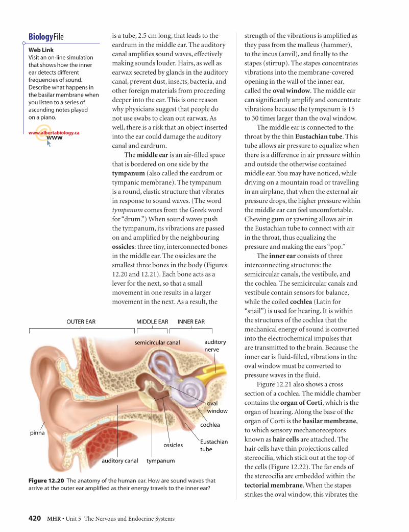
is a tube, 2.5 cm long, that leads to the eardrum in the middle ear. The auditory canal amplifi es sound waves, effectively making sounds louder. Hairs, as well as earwax secreted by glands in the auditory canal, prevent dust, insects, bacteria, and other foreign materials from proceeding deeper into the ear. This is one reason why physicians suggest that people do not use swabs to clean out earwax. As well, there is a risk that an object inserted into the ear could damage the auditory canal and eardrum. The middle ear is an air-fi lled space that is bordered on one side by the tympanum (also called the eardrum or tympanic membrane). The tympanum is a round, elastic structure that vibrates in response to sound waves. (The word tympanum comes from the Greek word for “drum.”) When sound waves push the tympanum, its vibrations are passed on and amplifi ed by the neighbouring ossicles: three tiny, interconnected bones in the middle ear. The ossicles are the smallest three bones in the body (Figures 12.20 and 12.21). Each bone acts as a lever for the next, so that a small movement in one results in a larger movement in the next. As a result, the
Figure 12.20 The anatomy of the human ear. How are sound waves that arrive at the outer ear amplified as their energy travels to the inner ear?
strength of the vibrations is amplifi ed as they pass from the malleus (hammer), to the incus (anvil), and fi nally to the stapes (stirrup). The stapes concentrates vibrations into the membrane-covered opening in the wall of the inner ear, called the oval window. The middle ear can signifi cantly amplify and concentrate vibrations because the tympanum is 15 to 30 times larger than the oval window. The middle ear is connected to the throat by the thin Eustachian tube. This tube allows air pressure to equalize when there is a difference in air pressure within and outside the otherwise contained middle ear. You may have noticed, while driving on a mountain road or travelling in an airplane, that when the external air pressure drops, the higher pressure within the middle ear can feel uncomfortable. Chewing gum or yawning allows air in the Eustachian tube to connect with air in the throat, thus equalizing the pressure and making the ears “pop.” The inner ear consists of three interconnecting structures: the semicircular canals, the vestibule, and the cochlea. The semicircular canals and vestibule contain sensors for balance, while the coiled cochlea (Latin for “snail”) is used for hearing. It is within the structures of the cochlea that the mechanical energy of sound is converted into the electrochemical impulses that are transmitted to the brain. Because the inner ear is fl uid-fi lled, vibrations in the oval window must be converted to pressure waves in the fl uid. Figure 12.21 also shows a cross section of a cochlea. The middle chamber contains the organ of Corti, which is the organ of hearing. Along the base of the organ of Corti is the basilar membrane, to which sensory mechanoreceptors known as hair cells are attached. The hair cells have thin projections called stereocilia, which stick out at the top of the cells (Figure 12.22). The far ends of the stereocilia are embedded within the tectorial membrane. When the stapes strikes the oval window, this vibrates the
BiologyFile
Web LinkVisit an on-line simulation that shows how the inner ear detects different frequencies of sound. Describe what happens in the basilar membrane when you listen to a series of ascending notes played on a piano.
@wwwwww.albertabiology.ca
2 3296_Chapter_12.indd 152 3296_Chapter_12.indd 15 11/4/06 6:24:58 PM11/4/06 6:24:58 PM

Chapter 12 Sensory Reception • MHR 421
window and creates pressure waves in the fl uid of the cochlea. The pressure waves make the basilar membrane move up and down, which causes the stereocilia of the hair cells to bend against the tectorial membrane. The hair cells, which synapse with the nerve fi bres of the auditory nerve, sense the bending of the stereocilia and relay this message to the nerves. The nerves then send an impulse to the brain.
List, in order, the structures of the ear that a sound wave encounters, starting with the outer ear.
What happens to the energy of sound waves, which travel through air, after it reaches the tympanum?
What is the role of the Eustachian tube?
• • •
• • •
Frequencies of Sound
The hair cells of the organ of Corti are able to distinguish both the frequency (pitch) and amplitude (intensity) of sound waves. Frequency is the number of waves that pass through a specifi c point every second. It is measured in hertz (Hz). The frequency of speech usually ranges from 100 to 4000 Hz, although humans can hear sounds that are between 20 and 20 000 Hz. If you could hear below 20 Hz, you would hear the blood moving through your ears! Different areas of the organ of Corti are sensitive to different frequencies. High frequencies, such as the sound of a whistle, most strongly stimulate the hair cells that are closest to the oval window. Low frequencies, such as a low note played by a tuba, most strongly stimulate the hair cells that are farthest from the oval window. In the next investigation, you will test your ability to detect sounds of different frequencies.
Figure 12.21 The structure of the cochlea. Notice how membranes separate its three fluid-filled chambers, the upper chamber, cochlear duct, and tympanic chamber. Since fluids cannot be compressed, the round window bulges out as pressure waves move through the inside of the cochlea.
Figure 12.22 Electron micrographs of hair cells and stereocilia from the organ of Corti. What causes the stereocilia to move?
hair cell
stereocilia
Magnification: 2196 ×Magnification: 836 ×
2 3296_Chapter_12.indd 162 3296_Chapter_12.indd 16 11/4/06 6:25:00 PM11/4/06 6:25:00 PM
422 MHR • Unit 5 The Nervous and Endocrine Systems
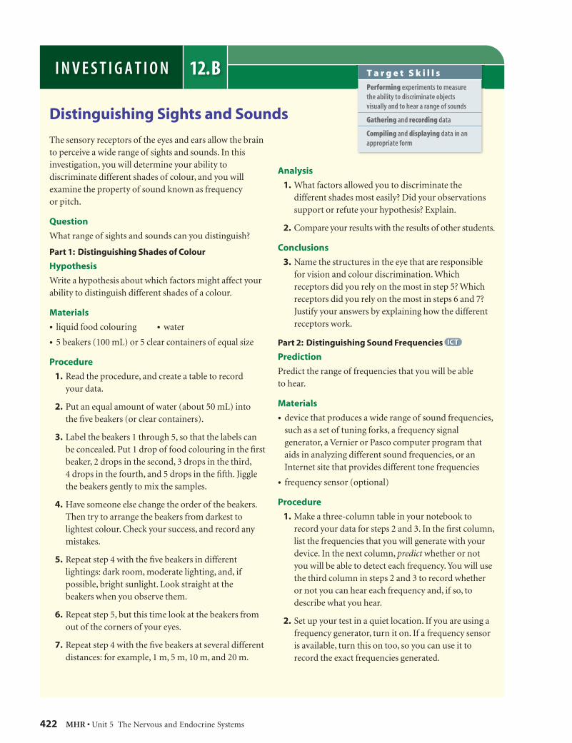
Distinguishing Sights and Sounds
The sensory receptors of the eyes and ears allow the brain to perceive a wide range of sights and sounds. In this investigation, you will determine your ability to discriminate different shades of colour, and you will examine the property of sound known as frequency or pitch.
Question
What range of sights and sounds can you distinguish?
Part 1: Distinguishing Shades of Colour
Hypothesis
Write a hypothesis about which factors might affect your ability to distinguish different shades of a colour.
Materials
• liquid food colouring • water
• 5 beakers (100 mL) or 5 clear containers of equal size
Procedure
1. Read the procedure, and create a table to record your data.
2. Put an equal amount of water (about 50 mL) into the fi ve beakers (or clear containers).
3. Label the beakers 1 through 5, so that the labels can be concealed. Put 1 drop of food colouring in the fi rst beaker, 2 drops in the second, 3 drops in the third, 4 drops in the fourth, and 5 drops in the fi fth. Jiggle the beakers gently to mix the samples.
4. Have someone else change the order of the beakers. Then try to arrange the beakers from darkest to lightest colour. Check your success, and record any mistakes.
5. Repeat step 4 with the fi ve beakers in different lightings: dark room, moderate lighting, and, if possible, bright sunlight. Look straight at the beakers when you observe them.
6. Repeat step 5, but this time look at the beakers from out of the corners of your eyes.
7. Repeat step 4 with the fi ve beakers at several different distances: for example, 1 m, 5 m, 10 m, and 20 m.
Analysis
1. What factors allowed you to discriminate the different shades most easily? Did your observations support or refute your hypothesis? Explain.
2. Compare your results with the results of other students.
Conclusions
3. Name the structures in the eye that are responsible for vision and colour discrimination. Which receptors did you rely on the most in step 5? Which receptors did you rely on the most in steps 6 and 7? Justify your answers by explaining how the different receptors work.
Part 2: Distinguishing Sound Frequencies ICTICT
Prediction
Predict the range of frequencies that you will be able to hear.
Materials
• device that produces a wide range of sound frequencies, such as a set of tuning forks, a frequency signal generator, a Vernier or Pasco computer program that aids in analyzing different sound frequencies, or an Internet site that provides different tone frequencies
• frequency sensor (optional)
Procedure
1. Make a three-column table in your notebook to record your data for steps 2 and 3. In the fi rst column, list the frequencies that you will generate with your device. In the next column, predict whether or not you will be able to detect each frequency. You will use the third column in steps 2 and 3 to record whether or not you can hear each frequency and, if so, to describe what you hear.
2. Set up your test in a quiet location. If you are using a frequency generator, turn it on. If a frequency sensor is available, turn this on too, so you can use it to record the exact frequencies generated.
12.BI N V E S T I G A T I O N T a r g e t S k i l l s
Performing experiments to measure the ability to discriminate objects visually and to hear a range of sounds
Gathering and recording data
Compiling and displaying data in an appropriate form
2 3296_Chapter_12.indd 172 3296_Chapter_12.indd 17 11/4/06 6:25:14 PM11/4/06 6:25:14 PM

Chapter 12 Sensory Reception • MHR 423
Hearing Loss
Hearing loss generally results from nerve damage (damage to the hair cells, called nerve deafness) or damage to the sound-conduction system of the outer or middle ear (called conduction deafness). Birth defects, ear infections, noise, and aging are common causes of hearing loss.
A
B
Figure 12.23 Scanning electron micrographs of stereocilia from a mouse ear. (A) Healthy stereocilia. (B) Stereocilia damaged by brief exposure to a 115 dB noise.
The amplitude of a sound wave is experienced as the intensity or volume of a sound. The louder the noise is, the more pressure that the fl uid in the cochlea puts on the hair cells of the basilar membrane.The stereocilia of the hair cells are very delicate, however. Repeated or sustained exposure to loud noise destroys the stereocilia, and the resulting damage is permanent (Figure 12.23). Noise is measured in decibels (dB), and any noise over 80 dB can damage the hair cells. In today’s society, people are commonly exposed to a variety of loud and potentially damaging noises (Table 12.3).
Table 12.3 Noises That Affect Hearing
Type of noise Sound level (dB) Effect
jet engine or rock concert
over 125 Noise is beyond the threshold of pain. There is a high potential for hearing loss.
boom box, chain saw, or snowmobile
100–125 Regular exposure for short periods of time may cause permanent hearing loss.
farm tractor, lawn mower, or motorcycle
90–100 15 min of exposure may cause hearing loss.
food blender or average city traffi c
80–90 Continuous daily exposure for longer than 8 h can cause damage.
Source: United States National Institute of Deafness and Other Communication Disorders, September 2005.
3. Starting with the lowest frequency, check your ability to hear the sound generated. Then switch to higher frequencies in 1000 Hz increments, until you reach the highest frequency that can be generated. At each increment, fi ll in your data table.
4. Close your eyes, and have a partner generate a 4000 Hz sound at different locations around your head. Indicate to your partner where the sound is coming from. After the test, check with your partner to see if you were correct. Note the locations that you identifi ed correctly.
Analysis
1. Name the specifi c structures of the inner ear that allow us to discriminate different frequencies. How did these structures function to allow you to hear sounds of different frequencies?
2. What range of frequencies were you able to hear? Was your prediction correct?
3. Compare your results with other students’ results or with data supplied by your teacher. Are there people who can hear frequencies that you cannot, or vice versa? Suggest a reason for this.
Conclusions
4. If you cannot hear certain frequencies within the range of 20 to 20 000 Hz, suggest a reason why. Explain specifi cally what damage might have occurred in your ears.
5. In step 5, why was it easier to locate the source of a sound when the sound was directly in front of you or behind you, and more diffi cult to locate the source when the sound came from either side of you?
Extension
6. Research different causes of hearing loss. Contact an audiologist (hearing specialist), and arrange for a visit to learn how hearing tests are performed.
2 3296_Chapter_12.indd 182 3296_Chapter_12.indd 18 11/4/06 6:25:15 PM11/4/06 6:25:15 PM
424 MHR • Unit 5 The Nervous and Endocrine Systems
While hearing aids to amplify sounds can often help people with conduction deafness, nerve deafness is more diffi cult to treat. In some cases, a device can be implanted in the ear to pick up sounds and directly relay signals to the auditory nerve. As well, researchers are exploring techniques to regenerate damaged or lost hair cells. One technique is to use a virus to insert a gene into the inner ear cells. The gene causes these cells to “sprout” new hair cells. So far, the technique has worked in guinea pigs, but scientists are not yet sure if it will work as a treatment for people with nerve deafness.
State the parts of the ear that have been damaged in someone with
a) nerve deafness
b) conduction deafness
• • •
• • •
The Perception of Sound
Sensory neurons in the ear send information through the auditory nerve to the brain stem, thalamus, and ultimately the temporal lobes of the cerebrum for processing. Depending on which sensory neurons are stimulated, the brain can perceive the frequency and amplitude of the sound. Recent research suggests that the source of the sound determines the specifi c neurons that are stimulated in the temporal lobes. Therefore, the brain can also perceive the location where the sound came from. Next time you hear a sound in front or behind you, imagine a cluster of neurons being stimulated in the corresponding area of your temporal lobes!
How is the brain able to perceive sounds of higher or lower frequencies and higher or lower amplitudes?
• • •
• • •
Balance and Coordination
Three major structures in the inner ear—the semicircular canals, utricle, and saccule—help us stand upright and move without losing our balance (Figure 12.24(A)). Thus, these structures function in our sense of equilibrium. The semicircular canals contain mechanoreceptors that detect head and body rotation (rotational equilibrium). The semicircular canals are three fl uid-fi lled loops, arranged in three different planes—one for each dimension of space. The base of each semicircular canal ends in a bulge. Inside each bulge, the stereocilia of the hair cells stick into a jelly-like covering called a cupula. When the head rotates, the fl uid inside the semicircular canals moves and bends the stereocilia, causing the hair cells to send rotational information to the brain (Figure 12.24(B)). On a fast-spinning midway ride, for example, the rapid circular motion causes the fl uid within the semicircular canals to rotate and send information confi rming this to the brain. When the ride stops, however, the fl uid is still moving. Why might the moving fl uid make someone feel dizzy or nauseous? The balance required while moving the head forward and backward is called gravitational equilibrium. Gravitational equilibrium depends on the utricle and the saccule, which together make up the fl uid-fi lled vestibule of the inner ear (Figure 12.24(A)). Both of these structures contain calcium carbonate granules, called otoliths. The otoliths lie in a cupula over a layer of hair cells. When the head dips forward or back, gravity pulls on the otoliths. This puts pressure on some of the hair cells, causing them to send a neural impulse to the brain, indicating the position of the head (Figure 12.24(C)). Proprioceptors are another type of mechanoreceptor involved in coordination. Proprioceptors are found in muscles, tendons, and joints throughout the body, and they send information about body position to the brain. For
BiologyFile
FYIMany animals can hear frequencies of sound that are outside the human range of hearing. Whales communicate regularly in frequencies below 20 Hz. Bats and dogs can detect frequencies well above 20 000 Hz. This explains why humans cannot hear dog whistles.
BiologyFile
Web LinkDescribe how a hearing aid and a cochlear implant work, and compare how sounds are perceived with each type of technology.
@wwwwww.albertabiology.ca
2 3296_Chapter_12.indd 192 3296_Chapter_12.indd 19 11/4/06 6:25:22 PM11/4/06 6:25:22 PM

Chapter 12 Sensory Reception • MHR 425
example, proprioceptors give the brain enough information for you to get dressed in the dark—although they do not ensure that you will put on matching socks!
Explain how the structures of the inner ear allow for rotational equilibrium.
Explain how the structures of the inner ear allow for gravitational equilibrium.
How does the brain perceive that the body is lying down?
• • •
• • •
TasteThe tongue contains chemoreceptors that allow us to taste substances entering the mouth. The ability to distinguish different tastes probably developed as an adaptation. Animals that avoided harmful substances and instead ate foods that were good for the body survived and reproduced. Poisonous plants, for example, often contain bitter-tasting molecules made of alkaloid compounds. Most scientists recognize four basic tastes: sour, sweet, salty, and bitter. When we eat, saliva dissolves some of our food. Specifi c molecules dissolved in the saliva
Figure 12.24 (A) The organs of balance: the semicircular canals, utricle, and saccule. Each semicircular canal ends in a bulge called an ampulla. (B) Rotational equilibrium. Rotating fluid bends the stereocilia in the cupula, and the hair cells send a message through the vestibular nerve to the brain. (C) Gravitational equilibrium. The hair cells of the utricle and saccule bend in response to head position.
2 3296_Chapter_12.indd 202 3296_Chapter_12.indd 20 11/4/06 6:25:24 PM11/4/06 6:25:24 PM

426 MHR • Unit 5 The Nervous and Endocrine Systems
are detected by the taste buds: the sensory receptors in the bumps (papillae) on the tongue (Figure 12.25). Specifi c taste cells within the taste buds detect molecules from one of the four basic tastes. Impulses from the taste buds travel to areas of the brain stem, to the thalamus, and then to the gustatory centre of the parietal lobe, which is responsible for the perception of taste. The combination of taste information sent from different areas of the tongue, as well as from sensory neurons in the nose, allows us to perceive fl avours. The salivary glands are connected to the brain stem, which is why they are stimulated whenever we taste, smell, or think about something delicious.
Describe how taste buds detect taste.
• • •
• • •
SmellScientists estimate that the human sense of smell can distinguish over 10 000 different odours. They think that each of these odours is produced from particles that fi t, much like a lock and key, into specifi c chemoreceptors, called olfactory cells, lining the upper nasal cavity (Figure 12.26). When the particles bind to the olfactory cells, ion channels in the cell membrane open. This generates an action potential in the olfactory cells, which are directly linked to the olfactory
bulb of the brain. From there, the impulse is sent to the emotional centres of the brain (the limbic system) and the frontal lobe, where the perception of odour occurs. Have you noticed that particular odours can instantly conjure up scenes and emotions from the past? Perfume experts create fragrances to evoke certain memories and emotions. The sense of smell is closely linked to the sense of taste. In fact, someone who is born without a functional olfactory system has no concept of taste, despite having functional taste buds. As much as 80 to 90 percent of what we perceive as taste is actually due to the sense of smell. This is why everything tastes so bland when you have a cold. Molecules from food travel through the nose and the passages in the throat. There, they trigger the chemoreceptors which, in turn, trigger the olfactory sensory neurons. The sense of smell is what lets you experience the complex fl avours that you associate with your favourite foods. Many animals, including humans, release substances, called pheromones, that aid in the recognition and attraction of a mate, sometimes over long distances. These hormone-like chemicals are detected in the nose by a structure called the vomeronasal organ. Recently, scientists determined that the human nose also contains a vomeronasal organ, although people cannot consciously smell pheromones.
Figure 12.25 (A) Papillae on the tongue contain taste buds that are sensitive to salty, sweet, sour, and bitter molecules. (B) Electron micrograph of many papillae. (C) Electron micrograph of one papilla. (D) Taste cells within the taste buds. The taste buds depolarize in response to particular tastes and generate an action potential that sends a neural impulse to the brain.
BiologyFile
FYIThere is evidence for a fi fth basic taste, called umami. Umami is described as a savoury taste, characteristic of monosodium glutamate (MSG), and detectable in some meats and cheeses.
2 3296_Chapter_12.indd 212 3296_Chapter_12.indd 21 11/4/06 6:25:27 PM11/4/06 6:25:27 PM

Chapter 12 Sensory Reception • MHR 427
How do the olfactory cells detect odours?
Why might a particular scent evoke a strong emotional response in a person?
• • •
• • •
Touch Unlike the mechanoreceptors associated with balance, hearing, taste, and smell, the mechanoreceptors associated with the sense of touch are located all over the body. The skin contains more than four million sensory receptors, but, as you learned in Chapter 11, they are not evenly distributed. Many of them are concentrated in the genitals, fi ngers, tongue, and lips. Different receptors in the skin are sensitive to different stimuli, such as light touch, pressure, pain, and high and low temperatures. These receptors gather information and transmit it back through sensory neurons to the brain and spinal cord for processing and a possible reaction (Figure 12.27 on page 429). Pain is a complicated sense that occurs when specialized sensors or nerve endings in the skin are activated by
mechanical pressure or chemical signals. If tissue is damaged, for example, nerve cells called nociceptors release chemicals that trigger pain receptors to send impulses to the brain. Painkillers, such as ibuprofen and Aspirin™, block the release of these chemicals. Everyone is familiar with the sensation of pain. How we experience pain and the effects of different painkillers is highly subjective, however. How do our senses, such as the ability to feel pain, contribute to homeostasis? Consider this question as you complete Investigation 12.C.
List the different types of stimuli that sensory receptors in skin can detect.
Where are the greatest concentrations of touch receptors in the body?
• • •
• • •
Sensation and HomeostasisThe senses allow us to navigate and experience the world around us. The senses relay information to the nervous system that allows the body to maintain homeostasis. Seeing the bright morning
Figure 12.26 (A) The human olfactory system. Trace how the smell of this rose is detected and then perceived in (B). The cilia of each olfactory cell can bind to only one type of odour molecule (represented here with colour).
BiologyFile
Try ThisBend a paper clip into a U-shape with the two ends about 2 mm apart. Close your eyes, and gently push down on the palm of your hand. Then try this on your shoulder. In which location could you distinguish the prongs as separate? What do your results suggest about the number of sensory receptors in the palm of your hand, relative to your shoulder?
A B
2 3296_Chapter_12.indd 222 3296_Chapter_12.indd 22 11/4/06 6:25:30 PM11/4/06 6:25:30 PM

428 MHR • Unit 5 The Nervous and Endocrine Systems
Feel, Taste, or Smell: Design Your Own Investigation
How can you determine the range and types of information that each of your senses can detect? Your group will explore one of the topics described below by designing, planning, and conducting an investigation. You will need to show clearly how a specifi c sense (touch, taste, or smell) can distinguish numerous sensations.
Question
How can you design an investigation to show how a particular sense, or a combination of senses, can distinguish various sensations?
Safety Precautions
• Do not bring food meant for consumption into the laboratory.
• Do not eat or drink anything in the laboratory.
Topic 1: Feel Those Sensations
Design an investigation to distinguish the different sensory receptors found in the skin, including the receptors for touch, pressure, pain, heat, and cold.
Suggested Materials
• 500 mL beaker of hot water (60 °C)
• 500 mL beaker of ice water (0–2 °C)
• non-permanent pen for marking gridlines on the body
• alcohol thermometer • fi nishing nails
Topic 2: Tantalize Those Taste Buds
Design an investigation to distinguish the four basic tastes (sweet, salty, sour, and bitter) using the tongue. Also investigate the relationship between smell and taste. Note: You must conduct this investigation at home or in the school cafeteria, not in the laboratory.
Suggested Materials
• salty water • lemon juice
• sugary water or candy • garbage bin
• onion juice or tonic water • blindfold
• clean toothpicks or cotton swabs
Topic 3: Expose Your Nose
Design an investigation to determine the ability of the olfactory receptors to distinguish various smells.
Suggested Materials
• ginger • peppermint • vinegar
• lemon • pine needles • perfume
• menthol • vanilla • blindfold
Experimental Plan
1. As a group, record the question(s) that you plan to investigate.
2. Write a hypothesis related to your experimental question(s).
3. Using the suggested materials as a starting point, develop a procedure to investigate your topic. List the manipulated, responding, and controlled variables. Note what you can use as a control test or trial (point of reference for your experimental trials).
4. Decide how your group will make the appropriate measurements, how many samples you will use, and whether you will pool your data with other groups’ data. Design a table to record your data.
5. After obtaining approval from your teacher for your experimental design, conduct your investigation.
Data and Observations
Record your data. When you have completed your investigation, present your experimental design and results to the rest of the class. Include a diagram that shows the neural pathway from the sensory receptors to the area of the brain where perception occurs.
Analysis
1. Describe any unexpected results. Hypothesize if and how your results would have been different if you had tested a combination of senses.
2. How could you improve your experimental design?
Conclusion
3. How are the sensory receptors organized so that we can distinguish different strengths and types of touch, taste, and smell?
12.CI N V E S T I G A T I O N T a r g e t S k i l l s
Asking questions about observed relationships
Designing an experiment to investigate various sensory receptors
Analyzing data to show interrelationships between the different senses
2 3296_Chapter_12.indd 232 3296_Chapter_12.indd 23 11/4/06 6:25:34 PM11/4/06 6:25:34 PM

Chapter 12 Sensory Reception • MHR 429
sunlight, for example, tells the body that it is time to wake up. Internal sensors help to prevent you from slipping in the shower. In Chapter 13, you will explore how hormones—chemical messengers in the body—trigger responses to sensations, as well as changes in the body that we do not consciously feel.
Section 12.3 Summary• The mechanoreceptors for hearing and
balance are located in the inner ear. • The cochlea, semicircular canals,
utricle, and saccule all contain hair cells that react to movement.
• The hair cells synapse with nerve fi bres, which transmit the sensory information to the nerves. The nerves then send an impulse to the brain.
• Proprioceptors in the muscles, joints, and tendons also inform the brain about the position of body parts.
• Sensory receptors in the tongue (taste buds), nose (olfactory cells), and skin (temperature, pressure, and pain receptors) provide additional information to the brain.
Figure 12.27 Various types of sensory neurons found in the skin
1. Explain how the structures of the ear amplify sounds.
2. How do the inner ear and brain distinguish the high shriek of a sea gull from the low sound made by a drum? What area of the brain perceives these sounds?
3. Based on your understanding of the inner ear, explain why someone might feel unwell after riding in a fast elevator in a tall building.
4. Suppose that you have been swimming along the bottom of the deep end of a pool. When you surface, you experience discomfort in your ears. Explain why plugging your nose while gently exhaling may help to alleviate the discomfort.
5. Suppose that your aunt works long hours at a newspaper printing press, where she is constantly exposed to sounds over 97 dB. What precautions, if any, would you recommend to her? Explain your answer in terms of the cellular structures of the inner ear.
ReviewSection 12.3
2 3296_Chapter_12.indd 242 3296_Chapter_12.indd 24 11/4/06 6:25:35 PM11/4/06 6:25:35 PM

Connections Nature of ScienceNature of Science
430 MHR • Unit 5 The Nervous and Endocrine Systems
Poisonous Frogs and PainThe golden poison arrow frog (Phyllobates terribilis) secretes a powerful neurotoxin called batrachotoxin. For centuries, aboriginal peoples of the Columbian rainforests have used batrachotoxin in traditional spiritual rituals and in healing and hunting practices. Before a hunt, arrows are dipped in a blend made from the frog’s secretions. When the arrow penetrates the prey’s skin, the animal dies swiftly. Batrachotoxin affects the victim’s neurons by preventing sodium gates in the neural membrane from closing. The resulting constant stimulation leads to a depletion of neurotransmitters and irreversible depolarization of nerves and muscle cells. Muscles throughout the body, most signifi cantly the heart and diaphragm, remain contracted, which causes rapid paralysis and death. Researchers are now studying the neurotoxin for its use as a targeted pain medication. Severe pain is commonly treated with morphine, which suppresses the whole central nervous system as it blocks the transmission of pain signals to the brain from the receptors. Morphine also interferes with other parts of the nervous system, causing nausea, sedation, and confusion. In addition, patients can build up a tolerance for the drug that makes progressively higher doses necessary in order for it to work. Very small doses of neurotoxins such as batrachotoxin could, on the other hand, be used to stop impulse transmission in the cell membranes of the pain receptors, without the side effects associated with morphine. Scientists are searching the world’s jungles and oceans to fi nd organisms, including plants, snails, fi sh, bacteria, and amphibians, which contain similar neurotoxins.
Biodiversity: A Medicine ChestSubstances with medicinal potential come from some unlikely sources. For example, coral reefs are home to over 500 species of cone snail (Conus), each of which produces a version of the deadly venom called conotoxin, which the snails use to immobilize prey. Conotoxin contains a component that causes muscle paralysis, plus a pain-reducing component, which blocks the acetylcholine receptors on pain transmission neurons. It could potentially be used to control nerve pain caused by surgery, diabetes, cancer, or AIDS. Puffer fi sh, or fugu (Takifugu), is a popular delicacy in Japan, despite containing the deadly neurotoxin tetrodotoxin. Ingesting even a milligram of tetrodotoxin can be fatal. The toxin appears to work by blocking sodium ion channels in the neurons, which can lead to respiratory paralysis. A Vancouver, British Columbia company produces a purifi ed form of tetrodotoxin called Tectin™, which has shown promise as a pain reliever for some people undergoing cancer treatment. Tetrodotoxin is 3000 times stronger than morphine but in low doses appears to safely inhibit the pain neurons.
• • • 1. Why might some organisms have evolved to contain
or secrete potent neurotoxins?
2. The bacterium Clostridium botulinum produces botulinum toxin, which can cause fatal food poisoning. Botulinum toxin inhibits the release of acetylcholine, resulting in paralysis. (A by-product, Botox®, is used to paralyze facial muscles and temporarily smooth wrinkles.) Knowing that one of the causes of migraine headaches is the constriction of arteries that bring blood to the brain, why might low doses of botulinum toxin be effective for controlling migraines?
3. What are the advantages of targeted painkillers over those with more general effects?
Pain Relievers or Deadly Neurotoxins?Pain receptors called nociceptors are free nerve endings found throughout the skin and internal organs. When tissue injury or chemical messengers stimulate the nociceptors, they transmit information to the brain and we feel pain. Pain may be unpleasant, but it usually serves as a warning to the brain to take action to prevent further injury. However, for millions of Canadians who suffer from the chronic pain of headaches, back pain, muscular-skeletal disorders, fi bromyalgia, cancer, or AIDS, pain can be debilitating. To help those with chronic pain, researchers are looking to natural—but deadly—neurotoxins for targeted pain relief. Phyllobates terribilis
Takifugu
2 3296_Chapter_12.indd 252 3296_Chapter_12.indd 25 11/4/06 6:25:37 PM11/4/06 6:25:37 PM
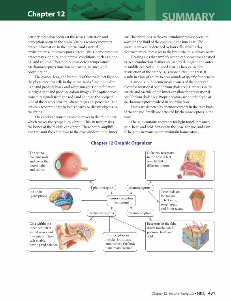
Chapter 12 Sensory Reception • MHR 431
Chapter 12
Sensory reception occurs at the senses. Sensation and perception occur in the brain. Various sensory receptors detect information in the internal and external environments. Photoreceptors detect light. Chemoreceptors detect tastes, odours, and internal conditions, such as blood pH and volume. Thermoreceptors detect temperature. Mechanoreceptors function in hearing, balance, and coordination. The cornea, lens, and humours of the eye direct light on the photoreceptor cells in the retina. Rods function in dim light and produce black and white images. Cones function in bright light and produce colour images. The optic nerve transmits signals from the rods and cones to the occipetal lobe of the cerebral cortex, where images are perceived. The lens can accommodate to focus nearby or distant objects on the retina. The outer ear transmits sound waves to the middle ear, which makes the tympanum vibrate. This, in turn, makes the bones of the middle ear vibrate. These bones amplify and transmit the vibrations to the oval window in the inner
ear. The vibrations in the oval window produce pressure waves in the fl uid of the cochlea in the inner ear. The pressure waves are detected by hair cells, which relay electrochemical messages to the brain via the auditory nerve. Hearing aids that amplify sound can sometimes be used to treat conduction deafness caused by damage to the outer or middle ear. Noise-induced hearing loss, caused by destruction of the hair cells, is more diffi cult to treat. It results in a loss of ability to hear sounds of specifi c frequencies. Hair cells in the semicircular canals of the inner ear allow for rotational equilibrium (balance). Hair cells in the utricle and saccule of the inner ear allow for gravitational equilibrium (balance). Proprioceptors are another type of mechanoreceptor involved in coordination. Tastes are detected by chemoreceptors in the taste buds of the tongue. Smells are detected by chemoreceptors in the nose. The skin contains receptors for light touch, pressure, pain, heat, and cold. Sensors in the nose, tongue, and skin all help the nervous system maintain homeostasis.
Chapter 12 Graphic Organizer
2 3296_Chapter_12.indd 262 3296_Chapter_12.indd 26 11/4/06 6:25:45 PM11/4/06 6:25:45 PM

Chapter 12
432 MHR • Unit 5 The Nervous and Endocrine Systems
Understanding Concepts 1. List the major structures in the pathway of neural
conduction from the organ of smell to the appropriate area of the brain.
2. Draw a table in your notebook with the following headings: Photoreceptors, Mechanoreceptors, Chemoreceptors, Thermoreceptors. For each type of receptor, list what it detects and provide an example of a human sense organ that contains this receptor.
3. Copy the diagram of the eye into your notebook. Label the visible structures, and indicate their functions. Also indicate the locations and functions of the rods and cones.
4. Describe how the touch receptors transduce energy into a form that the nervous system can use.
5. People who have lost a limb in a war or accident often report feeling sensations and even extreme pain in the limb that is no longer there. The cause of this phenomenon, called phantom limb syndrome, is unclear. Why is it possible for someone to perceive a sensation despite missing the associated sense receptors?
6. Explain how the inner ear and brain distinguish sounds of different frequencies and different volumes.
7. Once sensory neurons for a specifi c odour are stimulated, insensitivity to that odour rapidly occurs both in the sensory neurons of the nose and in the brain. What is this phenomenon called? Why would it be an adaptive advantage in certain circumstances?
8. Explain what happens to the pupils of your eyes if you walk into a dark movie theater after a stroll outside on a sunny day. What is this refl ex called?
9. Which kind of photoreceptor would you expect to be most numerous in the retinas of an animal that hunts at night? Explain your reasoning.
10. When you close one eye, you may notice a blind spot in your fi eld of vision. What is the blind spot? Why do you not see it all the time?
11. Use word processing or graphics software to make a fl owchart mapping the events that occur from the time the light of a full Moon reaches your retina to the time your brain perceives it. ICTICT
12. Studies show that some people who play action video games can process visual information faster and pick out more objects in a scene compared to people with little or no experience playing video games. Which area(s) of the brain must have been affected in the video game players?
13. Which receptors does your body rely on when you are standing still for a photo? Explain where these receptors are located, how they are stimulated, and how they send information to the brain.
14. What happens in your eyes when you focus on the words on this page? What is the process called? Use graphics software to sketch what happens to the lens of the eye when you focus on the words, and then when you look out the window at a bird in a tree. ICTICT
15. Using an example, compare rotational equilibrium with gravitational equilibrium. Identify the specifi c structures involved.
16. The early stages of glaucoma tend to affect the peripheral vision. Why might someone with early stage glaucoma want to avoid driving at night?
17. Which senses might be affected by an injury to one of the temporal lobes of the brain?
18. Use word processing software to make a fl owchart tracing the pathway of a sound wave as it travels through the outer, middle, and inner ear. Indicate where the auditory information is sent to the brain. Also indicate the principal structures involved and their functions, and the specifi c area of the brain where the perception of sound occurs. ICTICT
19. A heavy machinery operator is tested for hearing loss. It turns out that the operator has trouble hearing some frequencies of sound, but not others. Explain why.
Applying Concepts 20. List some possible effects of sensory deprivation on
homeostasis. Suggest a hypothesis to explain why these effects occur.
21. A severe blow to a particular area of the head can cause blindness. Describe how you would design a safety helmet to protect a cyclist from damage to the visual centres of the brain.
3 3296_Chapter_12_Review.indd 13 3296_Chapter_12_Review.indd 1 11/4/06 6:25:50 PM11/4/06 6:25:50 PM
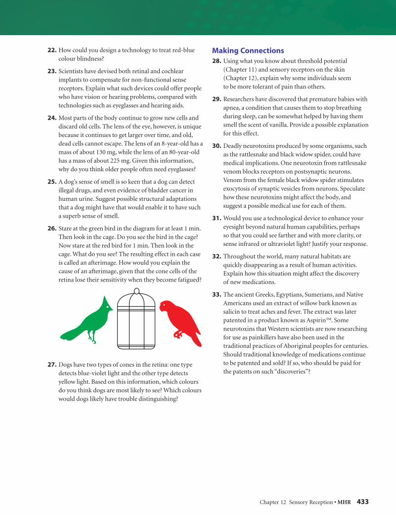
Chapter 12 Sensory Reception • MHR 433
22. How could you design a technology to treat red-blue colour blindness?
23. Scientists have devised both retinal and cochlear implants to compensate for non-functional sense receptors. Explain what such devices could offer people who have vision or hearing problems, compared with technologies such as eyeglasses and hearing aids.
24. Most parts of the body continue to grow new cells and discard old cells. The lens of the eye, however, is unique because it continues to get larger over time, and old, dead cells cannot escape. The lens of an 8-year-old has a mass of about 130 mg, while the lens of an 80-year-old has a mass of about 225 mg. Given this information, why do you think older people often need eyeglasses?
25. A dog’s sense of smell is so keen that a dog can detect illegal drugs, and even evidence of bladder cancer in human urine. Suggest possible structural adaptations that a dog might have that would enable it to have such a superb sense of smell.
26. Stare at the green bird in the diagram for at least 1 min. Then look in the cage. Do you see the bird in the cage? Now stare at the red bird for 1 min. Then look in the cage. What do you see? The resulting effect in each case is called an afterimage. How would you explain the cause of an afterimage, given that the cone cells of the retina lose their sensitivity when they become fatigued?
27. Dogs have two types of cones in the retina: one type detects blue-violet light and the other type detects yellow light. Based on this information, which colours do you think dogs are most likely to see? Which colours would dogs likely have trouble distinguishing?
Making Connections 28. Using what you know about threshold potential
(Chapter 11) and sensory receptors on the skin (Chapter 12), explain why some individuals seem to be more tolerant of pain than others.
29. Researchers have discovered that premature babies with apnea, a condition that causes them to stop breathing during sleep, can be somewhat helped by having them smell the scent of vanilla. Provide a possible explanation for this effect.
30. Deadly neurotoxins produced by some organisms, such as the rattlesnake and black widow spider, could have medical implications. One neurotoxin from rattlesnake venom blocks receptors on postsynaptic neurons. Venom from the female black widow spider stimulates exocytosis of synaptic vesicles from neurons. Speculate how these neurotoxins might affect the body, and suggest a possible medical use for each of them.
31. Would you use a technological device to enhance your eyesight beyond natural human capabilities, perhaps so that you could see farther and with more clarity, or sense infrared or ultraviolet light? Justify your response.
32. Throughout the world, many natural habitats are quickly disappearing as a result of human activities. Explain how this situation might affect the discovery of new medications.
33. The ancient Greeks, Egyptians, Sumerians, and Native Americans used an extract of willow bark known as salicin to treat aches and fever. The extract was later patented in a product known as Aspirin™. Some neurotoxins that Western scientists are now researching for use as painkillers have also been used in the traditional practices of Aboriginal peoples for centuries. Should traditional knowledge of medications continue to be patented and sold? If so, who should be paid for the patents on such “discoveries”?
3 3296_Chapter_12_Review.indd 23 3296_Chapter_12_Review.indd 2 11/4/06 6:25:52 PM11/4/06 6:25:52 PM