chapter 11d2jw81rkebrcvk.cloudfront.net/.../ppt_lecture_notes/chapter_011.pdf · chapter 11 general...
TRANSCRIPT

9/10/2012
1
1
Chapter 11
General Principles of
Pathophysiology
2
Lesson 11.1
Cellular Environment,
Water and Electrolyte Balance
3
Copyright © 2013 by Jones & Bartlett Learning, LLC, an Ascend Learning Company

9/10/2012
2
Learning Objectives
• Describe the normal characteristics of the cellular environment and the key homeostatic mechanisms that strive to maintain an optimal fluid and electrolyte balance.
• Outline pathophysiological alterations in water and electrolyte balance and list their effects on body functions.
• Describe the treatment of patients with particular fluid or electrolyte imbalances.
4
Cells
• Basic unit of higher life forms
• Components
– Cell membrane
• Holds cell together
• Separates internal cellular environment from external
– Enzymes help biochemical processes
– Internal membranes to encapsulate chemicals
– Genetic material for replication
5
Cells
• Form four basic types of tissue
– Epithelial tissue
– Connective tissue
– Muscle tissue
– Nervous tissue
6
Copyright © 2013 by Jones & Bartlett Learning, LLC, an Ascend Learning Company

9/10/2012
3
Cellular Environment
• Human body cells live in a fluid environment, consists mainly of water
– Body water essential
• Medium in which all metabolic reactions occur
• Body’s health depends on precise regulation of volume and composition of this fluid
– Body has two fluid compartments
• Intracellular fluid (ICF)
• Extracellular fluid (ECF)
7
8
Intracellular Fluid and Extracellular Fluid
• Intracellular fluid (ICF)
– Found in all body cells
– 40% of body weight
• Extracellular fluid (ECF)
– Fluid found outside of cells
– 20% of total body weight
– Blood plasma composes about 1/3
9
Copyright © 2013 by Jones & Bartlett Learning, LLC, an Ascend Learning Company
9/10/2012
4
Interstitial Fluid
• Cellular fluid between cells and outside vascular bed
• Includes cerebrospinal and intraocular fluid
• Accounts for 15 to 16% of total body weight
10
Aging and Fluid Distribution
• Body water accounts for 50 to 60% of the total weight in adults
– With age, distribution and amount decrease to about 45 to 55%
• Increases risk of dehydration, electrolyte abnormalities
11
Based on the causes of dehydration and your knowledge of anatomy and physiology, what two age groups do you think are at highest risk for
dehydration?
12
Copyright © 2013 by Jones & Bartlett Learning, LLC, an Ascend Learning Company

9/10/2012
5
Water Movement Between ICF and ECF
• Body fluids constantly move from one compartment to another
– Remains about the same in healthy people
• To keep volume stable
– Osmosis
– Diffusion
– Mediated transport mechanism
13
Osmosis
• For healthy body, molecules must be able to move within cell/across cell membrane
• Semipermeable membranes
– Separate fluid compartments
– Allow fluid to pass freely
– Regulate flow of solutes on the basis of size, shape, electrical charge
• Maintain homeostasis
– Channels within permit solute passage
14
Osmosis
• Diffusion or spreading of water molecules across semipermeable membrane from lower solute concentration to higher solute concentration
• Separates two solutions of different concentrations by blocking transport of salts or other solutes
15
Copyright © 2013 by Jones & Bartlett Learning, LLC, an Ascend Learning Company

9/10/2012
6
16
Osmosis
• Osmotic pressure
– Pressure that prevents the flow of fluid across a semipermeable membrane
– Pressure to maintain equilibrium depends on
• Number and molecular weight of particles on each side of the cell membrane
• Membrane permeability to these particles
17
Solutions
• Hypertonic solution
– When a living cell is placed in solution with a higher solute concentration, lower water concentration than that inside the cell
– When a cell is in solution, the osmotic pressure exerted produces net movement of water out of the cell
– Causes cell to dehydrate, shrink, possibly die
18
Copyright © 2013 by Jones & Bartlett Learning, LLC, an Ascend Learning Company

9/10/2012
7
Solutions
• Hypotonic solution
– When a living cell is placed in solution with a lower solute concentration, higher water concentration than that inside the cell
– Osmotic pressure draws water from the solution into the cell
• Net movement of water into the cell
• Can swell, possibly burst
19
What happens to a raisin when it is placed in a cup of water for an hour? Why does this change occur? Is the
water hypotonic, hypertonic, or isotonic relative to the inside of the raisin? Does
a concentration gradient exist?
20
Solutions
• Isotonic solution
– When a cell is placed in solution with the same solute and water concentration as the solution inside the cell
• No net movement of water molecules
21
Copyright © 2013 by Jones & Bartlett Learning, LLC, an Ascend Learning Company

9/10/2012
8
22
Diffusion
• Result of constant motion of all atoms, molecules, or ions in a solution
• Passive process
– Molecules or ions move from an area of higher concentration to an area of lower concentration
– Area of high concentration has more solute particles than area of low concentration
23
Diffusion
• Passive process
– More solute particles move from higher concentration to lower one
– Once at equilibrium, movement of solutes in one direction is balanced by equal movement in opposite direction
24
Copyright © 2013 by Jones & Bartlett Learning, LLC, an Ascend Learning Company

9/10/2012
9
25
Diffusion
• Concentration gradient– When the concentration of the solute is greater at one point in the solvent than at another point
• Solutes diffuse down their concentration gradients from high to low concentration until equilibrium is achieved
• Some nutrients enter and some waste products leave the cell by diffusion
– Maintenance of proper intracellular concentrations of certain substances depends on this process
26
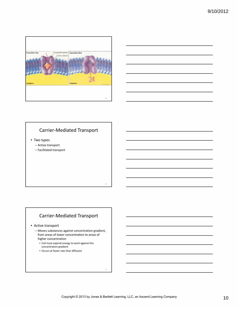
Mediated Transport Mechanism
• Required to move large, water‐soluble molecules, electrically charged molecules across cell membranes– Some vital molecules (glucose) cannot enter by diffusion
– Some products (proteins) cannot exit by diffusion
• Use carrier molecules– Proteins combine with solute molecules on one side of the membrane
– Change shape, pass through the membrane, release solute molecule on other side
27
Copyright © 2013 by Jones & Bartlett Learning, LLC, an Ascend Learning Company
9/10/2012
10
28
Carrier‐Mediated Transport
• Two types
– Active transport
– Facilitated transport
29
Carrier‐Mediated Transport
• Active transport
– Moves substances against concentration gradient, from areas of lower concentration to areas of higher concentration
• Cell must expend energy to work against this concentration gradient
• Occurs at faster rate than diffusion
30
Copyright © 2013 by Jones & Bartlett Learning, LLC, an Ascend Learning Company

9/10/2012
11
Carrier‐Mediated Transport
• Facilitated diffusion
– Moves substances into/out of cells from area of higher concentration to area of lower concentration
• Direction of movement is with concentration gradient
• Occurs more quickly than in normal diffusion
• Facilitated diffusion does not require cell to expend energy
• Moving force is downhill concentration gradient
31
What is the connection between necessary cellular processes and
patient care?
32
Water Movement Between Plasma and IF
• Fluid is transferred between circulating blood and interstitial fluid as a result of pressure changes
– Occur at arterial and venous ends of the capillary
• Human body has about 10 billion capillaries
• Few of the body’s functional cells are farther than 5/1000 inch (20 to 30 microns) from one another
33
Copyright © 2013 by Jones & Bartlett Learning, LLC, an Ascend Learning Company
9/10/2012
12
Anatomy of Capillary Network
• Thin‐walled tube of endothelial cells
• No elastic, connective tissue, smooth muscle that would impede transfer of water, solutes
• Blood enters from arterioles, flows through capillary network into venules
– Capillary ends closest to arterioles are arteriolar capillaries
– Ends closest to venules are venous capillaries
34
Anatomy of Capillary Network
• Nutrient, metabolic end product exchange takes place at the capillary level
• Arterioles give rise to capillaries, metarterioles
35
Anatomy of Capillary Network
• Most tissues have distinct types
– True capillaries
– Thoroughfare channels
• From metarteriole, blood may flow into thoroughfare channel that connects arterioles and venules directly, bypassing true capillaries
– Blood flow through thoroughfare channel is constant
– From channel, fluid exits/reenters network of true capillaries
36
Copyright © 2013 by Jones & Bartlett Learning, LLC, an Ascend Learning Company

9/10/2012
13
Anatomy of Capillary Network
• Sphincters
– Capillary: small cuffs of smooth muscle that encircle proximal and distal capillary portions
– Precapillary: arterial end sphincter
– Postcapillary: venous end sphincter
– Control blood flow, open, close capillary entrance, exit
– True capillary blood flow is not uniform, depends on contractile state, sphincter presence
37
Anatomy of Capillary Network
• Nutritional flow
– Blood flow through capillaries that provides exchange of gases, solutes between blood, tissue
• Nonnutritional, shunt flow
– Blood bypasses capillaries traveling from arteriole to venous side of circulation
38
AV Shunts
• True arteriovenous anastomoses (AV shunts)
– Occur naturally in sole of foot, palm of hand, terminal phalanges, nail bed
– Regulate body temperature
– Some evidence suggests presence upstream from capillary sphincters
39
Copyright © 2013 by Jones & Bartlett Learning, LLC, an Ascend Learning Company

9/10/2012
14
Capillary Network
• Sympathetic fibers innervate all blood vessels, except
– Capillaries
– Capillary sphincters
– Most metarterioles
40
Capillary Network
• Sympathetic innervation includes vasoconstrictor, vasodilator, vasomotor fibers
– Vasoconstrictor fibers most important in regulating blood flow
– Normal circulation with adequate arterial BP, arterioles are open, AV shunts closed, 20% capillaries open at any given time
41
42
Copyright © 2013 by Jones & Bartlett Learning, LLC, an Ascend Learning Company

9/10/2012
15
Capillary Network
• Diffusion across capillary wall
– Tissue cells do not exchange material directly with blood
– Interstitial fluid acts as “middle man”
– Nutrients must diffuse across capillary wall into interstitial fluid to enter cell
– Metabolic end products (CO2, lactic acid) must cross membrane into interstitial fluid to diffuse into plasma
43
Capillary Network
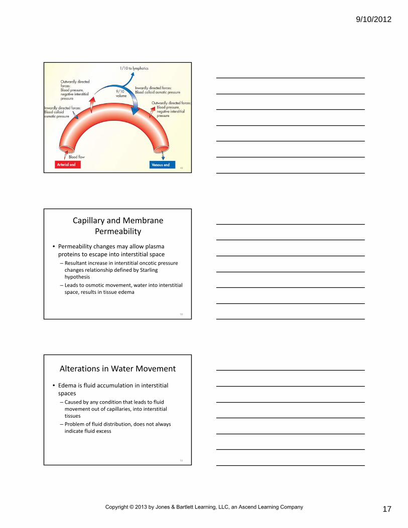
• Diffusion across capillary wall– At capillary arteriole end, the forces moving fluid out of capillary are greater than the forces attracting fluid into it
– At venous end, forces are reversed, more fluid is attracted into capillary
– Hydrostatic and osmotic pressure forces responsible for fluid movement
– Hydrostatic pressure, created with each heart beat, forces water out arterial end
44
Capillary Network
• Diffusion across capillary wall
– Blood colloid osmotic pressure or oncotic pressure
• When osmotic pressure results from presence of plasma proteins, mostly albumin, too large to pass through capillary wall
– At venous end
• Hydrostatic pressure is lower
• Protein concentration increases slightly, occurs from fluid movement out arteriolar end
45
Copyright © 2013 by Jones & Bartlett Learning, LLC, an Ascend Learning Company

9/10/2012
16
Capillary Network
• Diffusion across capillary wall
– Result is greater plasma protein concentration, greater colloid osmotic pressure
– Nearly all fluid that leaves capillary arteriolar end reenters venous end
– Remaining fluid enters lymphatic capillaries, eventually returned to general circulation
– Net filtration, fluid movement back and forth across capillary wall
46
Capillary Network
• Diffusion across capillary wall
– Starling hypothesis
• Net filtration = forces favoring filtration – forces opposing filtration
• Forces favoring filtration include capillary hydrostatic pressure, interstitial oncotic pressure
• Forces opposing filtration are plasma oncotic pressure, interstitial hydrostatic pressure
47
Capillary Network
• Diffusion across capillary wall
– Starling hypothesis
• Fluid also exchanged across wall as a result of cyclic dilation, constriction of precapillary sphincter
• When sphincter dilates, pressures rise in the capillary, which forces fluid into interstitial spaces
• When precapillary sphincter constricts, pressure drops, fluid moves into capillary
48
Copyright © 2013 by Jones & Bartlett Learning, LLC, an Ascend Learning Company
9/10/2012
17
49
Capillary and Membrane Permeability
• Permeability changes may allow plasma proteins to escape into interstitial space
– Resultant increase in interstitial oncotic pressure changes relationship defined by Starling hypothesis
– Leads to osmotic movement, water into interstitial space, results in tissue edema
50
Alterations in Water Movement
• Edema is fluid accumulation in interstitial spaces
– Caused by any condition that leads to fluid movement out of capillaries, into interstitial tissues
– Problem of fluid distribution, does not always indicate fluid excess
51
Copyright © 2013 by Jones & Bartlett Learning, LLC, an Ascend Learning Company

9/10/2012
18
Pathophysiology of Edema
• Factors of normal fluid flow through interstitial spaces
– Capillary hydrostatic pressure filters from blood through capillary wall
– Oncotic pressure exerted by proteins in blood plasma, attracts fluid from interstitial space back into vascular compartment
52
Pathophysiology of Edema
• Factors of normal fluid flow through interstitial spaces – Permeability of capillaries, determines how easily fluid can pass through capillary wall
– Presence of open lymphatic channels, which collect some fluid forced out of capillaries by hydrostatic pressure of blood, return fluid to circulation
• When any factors are disturbed, changes in water movement can develop
53
Pathophysiology of Edema
• Mechanisms most often responsible for edema
– Increase in hydrostatic pressure
– Decrease in plasma oncotic pressure
– Increase in capillary permeability
– Lymphatic obstruction
54
Copyright © 2013 by Jones & Bartlett Learning, LLC, an Ascend Learning Company

9/10/2012
19
Pathophysiology of Edema
• Increased capillary hydrostatic pressure
– Caused by venous obstruction or sodium and water retention
– With venous obstruction, hydrostatic pressure of fluid in capillaries can become great enough to cause fluid to escape into interstitial spaces
55
Pathophysiology of Edema
• Increased capillary hydrostatic pressure
– Conditions that can lead to venous obstruction, edema
• Thrombophlebitis (blood clot formation, inflammation in vein)
• Chronic venous disease
• Hepatic obstruction (hepatic veins or common bile duct blockage)
• Tight clothing around extremity
• Prolonged standing
56
Pathophysiology of Edema
• Increased capillary hydrostatic pressure
– Sodium, water retention can cause increase in circulating fluid volume, edema
– Conditions associated with sodium, water retention
• Congestive heart failure
• Renal failure
57
Copyright © 2013 by Jones & Bartlett Learning, LLC, an Ascend Learning Company

9/10/2012
20
Pathophysiology of Edema
• Decreased plasma oncotic pressure
– Decreased plasma albumin leads to decreased plasma oncotic pressure
• Result: fluid moves into interstitial space
• Most often results from liver disease, protein malnutrition
58
Pathophysiology of Edema
• Increased capillary permeability
– Result: greater than normal fluid filtration into interstitial space
– Associated with allergic reactions
– Linked to inflammation and immune response triggered by trauma
• Burns, crushing injuries
• Proteins escape from vascular bed
• Capillary oncotic pressure decreases
• Fluid oncotic pressure increases
• Result: edema59
Pathophysiology of Edema
• Lymphatic obstruction
– Proteins, fluid accumulate in interstitial space when lymphatic channels are blocked by infection, surgically removed
– Obstruction blocks normal pathway by which fluid is returned from interstitial space into circulation
60
Copyright © 2013 by Jones & Bartlett Learning, LLC, an Ascend Learning Company

9/10/2012
21
Pathophysiology of Edema
• Lymphatic obstruction
– Leads to edema in region normally drained by lymphatic channels
– Conditions that can cause obstruction
• Certain malignancies
• Parasitic infections
• Surgical removal of lymphatics
61
Pathophysiology of Edema
• Clinical manifestations of edema
– Localized
• Limited to injury site or organ system
• Sprained ankle
• Cerebral edema
• Pulmonary edema
• Can be life threatening
62
Pathophysiology of Edema
• Clinical manifestations of edema
– Generalized
• More widespread
• Obvious in dependent body parts
• First noted in legs and ankles when standing/sitting, sacrum and buttocks when lying down
• Causes weight gain, swelling, puffiness
• Linked to other symptoms caused by underlying illness
63
Copyright © 2013 by Jones & Bartlett Learning, LLC, an Ascend Learning Company

9/10/2012
22
Why does the RICE (rest, ice, compression, elevation) treatment for swelling from a sprained ankle
decrease tissue edema?
64
In septic shock, toxins affect the cell membrane permeability, allowing fluids to leak out of the blood
vessels more freely. How could that affect cardiac output?
65
Pathophysiology of Edema
• Clinical manifestations of edema
– Generalized
• In industrialized countries, most often caused by heart, kidney, liver disease
• In developing countries, most common cause is malnutrition and parasitic disease
• When tissue is compressed, fluid is pushed aside, leaving indentation that gradually refills, pitting edema
• Ascites: fluid accumulation in peritoneal cavity
66
Copyright © 2013 by Jones & Bartlett Learning, LLC, an Ascend Learning Company

9/10/2012
23
Pathophysiology of Edema
• Water follows osmotic gradient established by changes in sodium concentration
– Sodium and water balance are closely related
67
Water Balance
• Regulated by antidiuretic hormone (ADH)– ADH secretion, thirst perception help regulate
• ADH release triggered by increase in plasma osmolality, decrease in circulating blood volume, and decline in venous and arterial pressure– Increase in plasma osmolality stimulates hypothalamic neurons, osmoreceptors, causes thirst, increases ADH release from posterior pituitary gland
68
Water Balance
• ADH release response, water is reabsorbed into plasma from distal renal tubules, collecting ducts of kidneys– Reduces water amount lost in urine
– Water reabsorbed, plasma osmolality deceases, returning to normal
– Volume‐sensitive receptors and pressure‐sensitive receptors also stimulate release
• Vomiting, diarrhea, excessive sweating cause fluid depletion
69
Copyright © 2013 by Jones & Bartlett Learning, LLC, an Ascend Learning Company

9/10/2012
24
Sodium and Chloride Balance
• Sodium is major ECF cation
• Sodium balance regulated by aldosterone
• Regulates osmotic forces with chloride, bicarbonate, hence water balance
70
Sodium and Chloride Balance
• Chloride is major ECF anion, provides electroneutrality in relation to sodium
• Increases or decreases in chloride occur in proportion to changes in sodium
• Aldosterone secretion is triggered by decrease in sodium levels, increase in potassium levels
– Causes distal kidney tubules to increase sodium reabsorption, potassium secretion
71
Sodium and Chloride Balance
• Renin enzyme is secreted by kidney
– Occurs when circulating blood volume is reduced, sodium–water balance is disrupted
– Stimulates formation of angiotensin I, changed to angiotensin II
72
Copyright © 2013 by Jones & Bartlett Learning, LLC, an Ascend Learning Company

9/10/2012
25
Sodium and Chloride Balance
• Renin enzyme is secreted by kidney
– Angiotensin II
• Potent vasoconstrictor
• Stimulates ADH secretion
• Results in reabsorption of sodium and water and increase in systemic BP
– Renin‐angiotensin‐aldosterone system
• Mechanism regulating sodium and water
73
74
Sodium and Chloride Balance
• Natriuretic hormone
– Helps regulate sodium
– Promotes secretion of sodium in urine
– Decreases tubular sodium reabsorption
– Subsequent sodium, water loss
• Atrial natriuretic factor
– Substance released from arterial heart cells
– Helps control sodium, water balance
– Promotes renal elimination of sodium75
Copyright © 2013 by Jones & Bartlett Learning, LLC, an Ascend Learning Company

9/10/2012
26
Sodium, Chloride, Water Balance Alterations
• Homeostatic mechanisms maintain constant balance between intake and water excretion
• Water gained each day equals water lost
• Ways body gains water
– Person drinks fluids, eats moist foods
– Water formed through oxidation of hydrogen in food during metabolic process
76
• Body loses water through
– Kidneys as urine
– Bowel as feces
– Skin as perspiration
– Exhaled air as vapor
– Tears and saliva
• Abnormal states of body‐fluid balance
– Dehydration: water lost exceeds water gained
– Overhydration: water gained exceeds water lost
Sodium, Chloride, Water Balance Alterations
77
Dehydration
• Isotonic dehydration
– Excessive loss of sodium and water in equal amounts
• Hypernatremic dehydration
– More water loss than sodium
• Hyponatremic dehydration
– More sodium loss than water
78
Copyright © 2013 by Jones & Bartlett Learning, LLC, an Ascend Learning Company

9/10/2012
27
Dehydration
• Isotonic dehydration
– Possible causes
• Severe, long‐term diarrhea
• Systemic infection
• Intestinal obstruction
79
Dehydration
• Isotonic dehydration
– Signs, symptoms
• Dry skin, mucous membranes
• Poor skin turgor
• Longitudinal wrinkles, tongue furrows
• Oliguria, decreased urine output
• Anuria, essentially no urine output
• Acute weight loss
• Depressed, sunken fontanelles in infants
80
Dehydration
• Isotonic dehydration
– Treatment
• Administer intravenous infusion of isotonic solution
• Solute concentration equal that of blood, 0.9% sodium chloride, normal saline typically used
81
Copyright © 2013 by Jones & Bartlett Learning, LLC, an Ascend Learning Company

9/10/2012
28
Why do mucous membranes become dry in patients who are dehydrated?
82
Dehydration
• Hypernatremic dehydration
– Possible causes
• Excessive use/misuse of diuretics
• Continued intake of sodium in absence of water consumption
• Excessive water loss, little sodium loss
• Profuse, watery diarrhea
83
Dehydration
• Hypernatremic dehydration
– Signs, symptoms
• Dry, sticky mucous membranes
• Flushed, doughy skin
• Intense thirst
• Oliguria, anuria
• Increased body temperature
• Altered mental status
84
Copyright © 2013 by Jones & Bartlett Learning, LLC, an Ascend Learning Company

9/10/2012
29
Dehydration
• Hypernatremic dehydration
– Treatment
– Administer volume replacement
– Begins with isotonic fluids, patient is often salt and water depleted, with water supply more depleted
– Isotonic fluids relatively hypotonic with these patients
85
Dehydration
• Hyponatremic dehydration
– Possible causes
• Diuretic use
• Excessive perspiration, heat‐related illness
• Salt‐losing renal disorders
• Increased water intake
86
Dehydration
• Hyponatremic dehydration
– Signs, symptoms
• Abdominal, muscle cramps
• Seizures
• Rapid, thready pulse
• Diaphoresis, profuse sweating
• Cyanosis
87
Copyright © 2013 by Jones & Bartlett Learning, LLC, an Ascend Learning Company

9/10/2012
30
Dehydration
• Hyponatremic dehydration
– Treatment
• IV fluid replacement
• Normal saline, lactated Ringer’s solution
• Occasionally, hypertonic saline in seizures
88
How might dehydration present in patients of various ages?
89
Overhydration
• Increase in body water, results in decrease of solute concentration
• May result from parenteral administration of excessive fluids, impaired cardiac, renal function, some endocrine dysfunctions
90
Copyright © 2013 by Jones & Bartlett Learning, LLC, an Ascend Learning Company

9/10/2012
31
How can you predict what types of patients will be at risk for overhydration?
91
Overhydration
• Signs, symptoms
– Shortness of breath
– Puffy eyelids
– Edema
– Polyuria, large urine volumes
– Moist crackles, pulmonary examination
– Acute weight gain
92
Overhydration
• Treatment
– Depends on cause
– Water restriction main treatment for excessive water administration, certain endocrine problems
– Diuretic for cardiac and renal impairment
– Saline for profound hyponatremia associated with overhydration
93
Copyright © 2013 by Jones & Bartlett Learning, LLC, an Ascend Learning Company

9/10/2012
32
Electrolyte Imbalances
• Potassium
– Major positively charged ICF ion
– Narrow range, allows normal function of nerves, cardiac system, skeletal muscle
– Obligate potassium losses
• Losses that cannot be avoided
• Minimal loss
• Replenished through diet
• Excess excreted by kidneys
94
Electrolyte Imbalances
• Potassium
– Key role in muscle contraction, enzyme action, nerve impulses, cell membrane function
– Imbalances interfere with neuromuscular function, dysrhythmias
95
Electrolyte Imbalances
• Hypokalemia
– Abnormally low potassium in blood
– Causes
• Reduced dietary intake
• Poor potassium absorption
• Increased GI losses
• Vomiting
• Diarrhea
• Renal disease
• Infusion solutions low in potassium
• Some medications, most commonly diuretics, steroids
96
Copyright © 2013 by Jones & Bartlett Learning, LLC, an Ascend Learning Company

9/10/2012
33
What common illness mimics many of the signs and symptoms of fluid
and electrolyte imbalance?
97
Electrolyte Imbalances
• Hypokalemia
– Signs, symptoms• Malaise
• Skeletal muscle weakness
• Cardiac dysrhythmias
• Decreased reflexes
• Weak pulse
• Faint, distant heart sounds
• Hospital treatment: intravenous, oral administration
• Shallow respiration
• Low blood pressure
• Anorexia
• Vomiting
• Gaseous distention
• Excessive thirst, rare
98
Electrolyte Imbalances
• Hyperkalemia
– Abnormally high potassium in blood
– Causes
• Acute, chronic renal failure
• Crush injuries
• Severe infections
• Conditions in which large amounts of potassium are released
• Excessive use of potassium salts
• Shift of potassium from cells into ECF
99
Copyright © 2013 by Jones & Bartlett Learning, LLC, an Ascend Learning Company

9/10/2012
34
Electrolyte Imbalances
• Hyperkalemia
– Signs, symptoms
• Cardiac conduction disturbances
• Irritability
• Abdominal distention
• Nausea
• Diarrhea
• Oliguria
• Weakness, early
• Paralysis, late
100
Electrolyte Imbalances
• Hyperkalemia
– Hospital treatment• Restriction of potassium
• Giving cation exchange resin, orally or nasogastric tube
• Severe cases: hemodialysis
• Emergency: calcium intravenously
• Administration of glucose, insulin, lowers serum potassium, forces intracellulary with glucose
• Sodium bicarbonate causes potassium to shift back into cells
• High‐dose nebulized albuterol lowers potassium, stimulates insulin release, which stimulates sodium‐potassium pump, which shifts potassium into cells
101
Electrolyte Imbalances
• Calcium
– Bivalent cation (two positive charges)
– Essential for body functions
• Neuromuscular transmission
• Cell membrane permeability
• Hormone secretion
• Growth and ossification of bones
• Muscle contraction: smooth, cardiac, skeletal
– Balanced diet, sufficient for body needs
– Excreted through urine, feces, perspiration102
Copyright © 2013 by Jones & Bartlett Learning, LLC, an Ascend Learning Company

9/10/2012
35
Electrolyte Imbalances
• Hypocalcemia
– Abnormally low calcium in blood
– Causes
• Endocrine dysfunction, mostly underactive parathyroid gland
• Renal insufficiency
• Decreased calcium intake, malabsorption
• Toxic shock syndrome
• Deficiency, malabsorption, inability to activate vitamin D, responsible for calcium absorption
103
Electrolyte Imbalances
• Hypocalcemia
– Signs, symptoms• Paresthesia, numbness, tingling
• Tetany, muscle twitching
• Abdominal cramps
• Muscle cramps
• Neural excitability
• Personality changes
• Abnormal behavior
• Convulsions
• Heart failure
104
Electrolyte Imbalances
• Hypocalcemia
– Hospital treatment
• IV administration, calcium ions
• Calcium salt, vitamin D orally
105
Copyright © 2013 by Jones & Bartlett Learning, LLC, an Ascend Learning Company

9/10/2012
36
Electrolyte Imbalances
• Hypercalcemia
– Abnormally high calcium in blood
– Causes
• Various tumors
• Parathyroid overactivity
• Thyroid dysfunction
• Diuretic therapy
• Some cancers
• Excessive vitamin D
106
Electrolyte Imbalances
• Hypercalcemia
– Can be deposited in various body tissues, organ systems
• Gastrointestinal system
• Central nervous system
• Renal system
• Neuromuscular system
• Cardiovascular system
107
Electrolyte Imbalances
• Hypercalcemia
– Signs, symptoms
• Hypotonicity of muscles, decreased muscle tone, tension
• Renal stones
• Altered mental status, seizures, coma
• Deep bone pain
• Cardiac dysrhythmias
108
Copyright © 2013 by Jones & Bartlett Learning, LLC, an Ascend Learning Company
9/10/2012
37
Electrolyte Imbalances
• Hypercalcemia– Treatment
• Control underlying disease
• Hydration
• Drug therapy
– Hospital treatment• Forced diuresis, normal saline, furosemide
• Calcium‐lowering drugs, thyrocalcitonin steroids, plicamycin
• Hemodialysis for heart failure, renal insufficiency
109
Electrolyte Imbalances
• Magnesium– Bivalent cation
– Activates enzymes
– Distributed throughout body• 50% insoluble state in bone
• 45% intracellular cation
• 5% extracellular solution
– Excreted by kidneys
– Physiological effects on nervous system resemble effects of calcium
110
Electrolyte Imbalances
• Hypomagnesemia
– Abnormally low magnesium in blood
– Causes
• Alcoholism
• Diabetes
• Malabsorption
• Starvation
• Diarrhea
• Diuresis
• Diseases that cause hypocalcemia, hypokalemia
• Increased irritability of CNS
111
Copyright © 2013 by Jones & Bartlett Learning, LLC, an Ascend Learning Company

9/10/2012
38
Electrolyte Imbalances
• Hypomagnesemia
– Signs, symptoms
• Tremors
• Nausea, vomiting
• Diarrhea
• Hyperactive deep reflexes
• Confusion, hallucinations
• Seizures, myoclonus, muscle spasms
• Cardiac dysrhythmias, can lead to cardiac arrest
112
Electrolyte Imbalances
• Hypomagnesemia
– Treatment for significant, symptomatic
• IV magnesium solution
• Magnesium sulfate for torsades de pointes
113
Electrolyte Imbalances
• Hypermagnesemia
– Abnormally high magnesium in blood
– Mainly patients with chronic renal insufficiency
– Large amounts of magnesium‐containing compounds
• Cathartics, magnesium citrate, sulfate
• Antacids, magnesium hydroxide
114
Copyright © 2013 by Jones & Bartlett Learning, LLC, an Ascend Learning Company
9/10/2012
39
Electrolyte Imbalances
• Hypermagnesemia– Can cause
• CNS depression
• Profound muscular weakness
• Areflexia, absence of reflexes
• Cardiac rhythm disturbances, may lead to death
– Signs, symptoms• Sedation
• Confusion
• Muscle weakness
• Respiratory paralysis
115
Electrolyte Imbalances
• Hypermagnesemia
– Treatment
• Hemodialysis, returns blood levels to normal in about 4 hours
• Calcium salts, parenterally, antagonist to magnesium
• IV glucose, insulin, drives magnesium back into cells, used in emergencies, respiratory depression, cardiac conduction defects
116
Lesson 11.2
Acid‐Base Balance
117
Copyright © 2013 by Jones & Bartlett Learning, LLC, an Ascend Learning Company

9/10/2012
40
Learning Objectives
• Describe the mechanisms in the body that maintain normal acid‐base balance.
• Outline pathophysiological alterations in acid‐base balance.
• Describe the management of a patient with an acid‐base imbalance.
118
Acid‐Base Balance
• Acids produced by normal metabolism
– Respiratory acids, culminating in CO2
– Nonrespiratory (metabolic) acids
• Bases return body’s plasma to normal in metabolic disturbances
119
Acid‐Base Balance
• Balance between acid and bases must be kept in narrow range for physiological functioning
• Main regulators are lungs and kidneys
– Lungs secrete respiratory acids
– Kidneys secrete metabolic acids
120
Copyright © 2013 by Jones & Bartlett Learning, LLC, an Ascend Learning Company

9/10/2012
41
Acid‐Base Balance
• pH
– Hydrogen ions are positively charged protons
– Hydrogen ions that lose charge marked H+
– Hydrogen ions that gain charge marked H‐
– Acids release or donate hydrogen ions
– Bases receive or absorb hydrogen ions, neutralize positively charged ions
– Hydrogen ion concentration expressed as pH, potential for hydrogen
121
Acid‐Base Balance
• pH
– Small change in pH very important
– Acid, base strength changes 10 times with each unit change of pH
– 7.4 to 7.1 doubles hydrogen ion concentration
– pH neutral, 6.8 to 7, when equal numbers are positive, negative ions are present
– Solution increases acidity as pH decreases
– Increases alkalinity, basicity, as pH rises
122
123
Copyright © 2013 by Jones & Bartlett Learning, LLC, an Ascend Learning Company
9/10/2012
42
Acid‐Base Balance
• Buffer systems
– Stimulated by changes in pH
– Require normal organ function to maintain acid‐base balance
– Carbonic acid‐bicarbonate buffering• Bicarbonate, CO2, and carbonic acid are always present in dynamic balance in blood
• Bicarbonate (HCO3) arises from transport of CO2 in blood
• Carbonic anhydrase enzyme dissolves CO2 in water of blood, reacts with water in red blood cells, forms carbonic acid (H2CO3)
124
Acid‐Base Balance
• Buffer systems
– Carbonic acid‐bicarbonate buffering• Carbonic acid breaks down into hydrogen, bicarbonate ions
• Because of effects of carbonic acid and sodium bicarbonate, buffering must occur through lungs, kidneys
• pH can be moved up or down
• By renal system that excretes or retains sodium bicarbonate
• By respiratory system that excretes or retains carbonic acid or CO2
• By both systems acting together
125
Acid‐Base Balance
• Buffer systems
– Carbonic acid–bicarbonate buffering
• At physiological pH of 7.4, normal ratio of carbonic acid to bicarbonate is 1:20, CO2 + H2O↔H2CO3↔H + HCO3‐
• Bicarbonate may link up with cation, form base bicarbonate (NaHCO3)
• Ratio of carbonic acid to base bicarbonate determines pH
• 1 milliequivalent (mEq) of carbonic acid for each 20 mEq base bicarbonate in ECF, pH stays in normal range
126
Copyright © 2013 by Jones & Bartlett Learning, LLC, an Ascend Learning Company

9/10/2012
43
Acid‐Base Balance
• Buffer systems
– Carbonic acid–bicarbonate buffering
• Mechanism triggered immediately by pH changes
• Respiratory rate helps maintain balance
• Most important buffering system in ECF, buffer up to 90% hydrogen ion in ECF, little effect on cells
127
128
Acid‐Base Balance
• Protein buffering
– Intracellular and extracellular proteins have negative charges, both serve as buffers for changes in pH
– Most proteins are inside cells
• Protein buffering is mainly an intracellular buffer system
– Hemoglobin (Hb) is excellent intracellular buffer, binds with hydrogen ions and CO2
– Hb binds with CO2 and hydrogen after O2 is released in peripheral tissues
129
Copyright © 2013 by Jones & Bartlett Learning, LLC, an Ascend Learning Company

9/10/2012
44
Acid‐Base Balance
• Protein buffering
– As blood reaches lungs, actions reverse, Hb binds with O2, releasing CO2 and hydrogen ions
– Hydrogen ions released combine with bicarbonate ions, forming carbonic acid
– Carbonic acid breaks down into CO2 and water
– Lungs exhale CO2
– Respirations help maintain pH
130
Acid‐Base Balance
• Protein buffering
– Respiratory centers more responsive to pH changes than changes in O2 level of tissues
– CO2 in blood controls rate of breathing in healthy individuals
– Within minutes of decrease in pH, alveolar ventilation increases effort to lower CO2 concentration
131
Acid‐Base Balance
• Renal buffering
– Kidneys help maintain acid‐base balance through• Recovery of bicarbonate, filtered into tubules
• Excretion of hydrogen ions against gradient to acidify urine
• Excretion of ammonium ions (NH4), each carries hydrogen ion with it
– Renal system makes up for acid‐base imbalances slowly compared with protein, bicarbonate buffer systems
– Kidneys can take several hours, days to restore pH normal range
132
Copyright © 2013 by Jones & Bartlett Learning, LLC, an Ascend Learning Company

9/10/2012
45
Acid‐Base Imbalance
• Maintained through respiratory and metabolic element
• Acidosis: any condition that increases carbonic acid or decreases base bicarbonate
• Alkalosis: any condition that increases base bicarbonate or decreases carbonic acid
133
Acid‐Base Imbalance
• Acidosis makes pH more acidic
• Alkalosis makes pH less acidic
• Possible to have both disorders simultaneously
– Respiratory acidosis, metabolic alkalosis
– One usually dominates, other attempts to compensate
134
Acid‐Base Imbalance
• Acidosis
– Acid accumulation and resulting acidosis, pH < 7.35, cause pH more acidic than normal 7.4
– Respiratory acidosis
• Caused by CO2 retention
• Leads to increase partial pressure of CO2 (Pco2)
• Caused by imbalance in production of CO2 and its elimination through alveolar ventilation
• ↓Respira on =↑CO2 + H2O↑H2CO3↑H++HCO3‐
135
Copyright © 2013 by Jones & Bartlett Learning, LLC, an Ascend Learning Company

9/10/2012
46
Think about the last time you ran so fast you had a muscle cramp. What acid‐base changes were going on inside your body? How did your body compensate for those
changes?136
137
Acid‐Base Imbalance
• Respiratory acidosis
– Alveolar ventilation reductions occur
• Respiratory depression
• Respiratory arrest
• Cardiac arrest
• Neuromuscular impairment
• Medications, sedatives, hypnotics
• Chest wall injury
• Pulmonary disorders
138
Copyright © 2013 by Jones & Bartlett Learning, LLC, an Ascend Learning Company

9/10/2012
47
Acid‐Base Imbalance
• Respiratory acidosis– When the respiratory system cannot continue as compensatory mechanism to correct acidosis, renal system must conserve bicarbonate, excrete more hydrogen ions to bring pH into normal range
– Kidneys take time to restore pH
– Treat by improving ventilation quickly to eliminate CO2
• Assist ventilations to decrease Pco2, supplemental O2 to correct accompanying hypoxemia
139
Acid‐Base Imbalance
• Metabolic acidosis
– Buildup of acid or loss of base
– When excess acid is produced, spills into ECF, consumes some bicarbonate buffers
– Result: acid increase, available base decrease
– ↑H++HCO3‐ →↑H2CO3→H2O+↑CO2
– Increase in available hydrogen ions forces reaction to right, decreases amount of base bicarbonate
140
What kind of acid‐base imbalance exists in a patient you have just
defibrillated and resuscitated from cardiac arrest? How are you going to
correct that imbalance?
141
Copyright © 2013 by Jones & Bartlett Learning, LLC, an Ascend Learning Company

9/10/2012
48
142
Acid‐Base Imbalance
• Metabolic acidosis
– Healthy respiratory system instantly makes up for acidosis, increases rate, depth of breathing to reduce CO2
– As CO2 falls, carbonic acid concentration falls, moves pH toward normal
– Kidneys also excrete more hydrogen ion to equilibrate excess acid in ECF
143
Acid‐Base Imbalance
• Metabolic acidosis
– Most common forms encountered in prehospital setting
• Lactic acidosis
• Diabetic ketoacidosis
• Acidosis caused by renal failure
• Acidosis caused by ingestion of toxins, poisons
144
Copyright © 2013 by Jones & Bartlett Learning, LLC, an Ascend Learning Company

9/10/2012
49
Acid‐Base Imbalance
• Lactic acidosis
– Made when large number of cells are inadequately perfused
– Results in shift from aerobic to anaerobic metabolism
– End product of anaerobic metabolism is lactic acid• Releases hydrogen ions, becomes lactate, creates systemic acidosis
– Normally liver changes lactate back into glucose or lactate oxidized to CO2 and water
– When lactic acid is produced faster than it is metabolized, lactic acidosis occurs
145
Acid‐Base Imbalance
• Lactic acidosis
– Most common causes
• Extreme exertional states, seizures
• Ischemia large muscles, organs
• Circulatory failure
• Shock
– Specific complications
• Decreased force of cardiac contraction
• Decreased peripheral response to catecholamines
• Hypotension and shock
• Cardiac muscle that is refractory to defibrillation146
Acid‐Base Imbalance
• Lactic acidosis
– Treatment
• Reestablishing tissue perfusion, cardiac output, which allows liver to regenerate bicarbonate by metabolizing lactate to CO2 and water
• Hyperventilation induces respiratory alkalosis
• Vigorous rehydration to support circulation
• IV sodium bicarbonate for immediate compensation, cardiac arrest
• Often depends on identification and correction of underlying cause
147
Copyright © 2013 by Jones & Bartlett Learning, LLC, an Ascend Learning Company

9/10/2012
50
How do you think the body will compensate for lactic acidosis?
148
Acid‐Base Imbalance
• Diabetic ketoacidosis
– Usually complication of diabetes mellitus, alcoholics (alcoholic ketoacidosis)
– Results from inadequate insulin, when insulin need increases
– Insulin required for cells to absorb glucose
149
Acid‐Base Imbalance
• Diabetic ketoacidosis
– With impaired glucose utilization, fatty acids are metabolized, producing ketone bodies, releasing hydrogen ions
– Large amounts of ketone bodies exceed ability of body’s buffering system to compensate
• Results in acidosis and decreased pH
– Prehospital care: administration of normal saline for volume repletion
150
Copyright © 2013 by Jones & Bartlett Learning, LLC, an Ascend Learning Company

9/10/2012
51
Acid‐Base Imbalance
• Acidosis caused by renal failure
– Kidneys help maintain acid‐base balance
– Renal failure affects compensatory mechanisms of kidneys to varying degrees
– Moderate, severe renal failure has mild, moderate acidosis
– Acidosis results because failing kidneys are unable to excrete acid waste products efficiently
– Waste products are result of normal metabolic processes
151
Acid‐Base Imbalance
• Acidosis caused by ingestion of toxins
– Toxins can cause metabolic acidosis
• Ethylene glycol
• Methanol
• Salicylate, aspirin component
– Toxins produce toxic metabolites, result in acid‐base disorders
• Disorders characterized by metabolic acidosis, compensatory respiratory alkalosis
152
Acid‐Base Imbalance
• Acidosis caused by ingestion of toxins
– Treatment
• Gastrointestinal evacuation
• Hemodialysis
• Diuresis
• Hydration, promote excretion
• Specific antagonistic, antidotal therapy
153
Copyright © 2013 by Jones & Bartlett Learning, LLC, an Ascend Learning Company

9/10/2012
52
Acid‐Base Imbalance
• Alkalosis– pH greater than 7.45 causes blood, body fluids to be less acidic compared to normal pH 7.4
– Respiratory alkalosis• May be caused by hyperventilation, which decreases Pco2
• Hyperventilation common in acutely ill
• Sepsis
• Peritonitis
• Shock
• Respiratory ailments
154
155
Acid‐Base Imbalance
• Alkalosis
– Respiratory alkalosis• ↑Respira on = ↓CO2 + H2O→↓H2CO3 →↓H++HCO3‐
• When carbonic acid is lacking because of excessive elimination of CO2, blood pH rises, kidneys must excrete bicarbonate ions and retain hydrogen ions in an effort to return pH to normal
– Treatment• Correct underlying cause of hyperventilation
• Low‐concentration O2
• Provide calming measures to assist in slow, controlled breathing
156
Copyright © 2013 by Jones & Bartlett Learning, LLC, an Ascend Learning Company

9/10/2012
53
Acid‐Base Imbalance
• Metabolic alkalosis
– Rare
– Factors
• Loss of hydrogen ions (from stomach), vomiting, gastric suction, increased renal excretion
• Ingestion of large amounts of absorbable base sodium bicarbonate (baking soda), calcium carbonate (Tums, antacids)
• Excessive IV alkali
• Chronic use of diuretics results in volume depletion
157
158
Acid‐Base Imbalance
• Metabolic alkalosis
– Sodium chloride and potassium loss causes relative increase in bicarbonate, kidneys defend volume depletion, increase reabsorption of sodium, H2O
– When sodium is reabsorbed, potassium or hydrogen ions must be excreted, maintains electrical neutrality
– Excretion of hydrogen ions can lead to net increase in bicarbonate
• Leads to metabolic alkalosis
159
Copyright © 2013 by Jones & Bartlett Learning, LLC, an Ascend Learning Company

9/10/2012
54
Acid‐Base Imbalance
• Metabolic alkalosis
– ↓H++HCO3‐ → ↓H2CO3‐ → H2O+↓CO2
– Respiratory system compensates, retains CO2, limited by development of hypoxemia
– Hypoventilation causes rise in Pco2, decrease in partial pressure O2 (Po2), stimulates respiration
160
Acid‐Base Imbalance
• Metabolic alkalosis
– Treatment
• Correct underlying condition
• Correct volume depletion with isotonic solutions
• Hypokalemia corrected with potassium replacement
161
Acid‐Base Imbalance
• Mixed acid‐base disturbances
– Causes
• Include various forms of shock
• Simultaneous respiratory and metabolic alterations commonly seen
• Develop because pathophysiological changes occur in respiratory, metabolic components of acid‐base system
162
Copyright © 2013 by Jones & Bartlett Learning, LLC, an Ascend Learning Company
9/10/2012
55
Acid‐Base Imbalance
• Mixed acid‐base disturbances
– Examples
• Combined respiratory and metabolic acidosis
• Metabolic acidosis and respiratory alkalosis
• Respiratory acidosis and metabolic alkalosis
• Combined respiratory and metabolic alkalosis
163
Acid‐Base Imbalance
• Emergency care primary points
– Acid‐base balance components: respiratory (CO2) factor, nonrespiratory (metabolic) factor
– Respiratory acidosis
• Caused by increase in CO2 level of blood, body fluids as result of inadequate breathing
• Treatment: improve ventilation to lower CO2 level
– Respiratory alkalosis results from hyperventilation
164
Acid‐Base Imbalance
• Emergency care primary points
– Metabolic acidosis
• Caused by anaerobic metabolism and lactic acidosis
• Treatment: neutralize acid by reestablishing tissue perfusion and cardiac output
– Metabolic alkalosis is rare
– Two simultaneous acid‐base disturbances
• One dominates, the other attempts compensation
165
Copyright © 2013 by Jones & Bartlett Learning, LLC, an Ascend Learning Company

9/10/2012
56
Acid‐Base Imbalance
• Emergency care primary points
– pH
• Product of both respiratory and metabolic components
• Neutral pH 6.8 to 7
• Normal blood pH = 7
• Decreased pH indicates increase in acidity, increase in pH indicates decrease in acidity
166
Lesson 11.3
Cell Alterations
167
Learning Objectives
• Describe the changes in cells and tissues that occur with cellular adaptation, injury, neoplasia, aging, or death.
• Outline the effects of cellular injury on local and systemic body functions.
168
Copyright © 2013 by Jones & Bartlett Learning, LLC, an Ascend Learning Company

9/10/2012
57
Cellular Injury and Diseases
• Changes in cells and tissue structure can result from
– Cellular adaptation
– Injury
– Neoplasia
– Aging
– Death
169
Cellular Adaptation
• Environment adaptation
– Escape, protect from injury
– Adapted cell neither normal nor injured
– Common
– Central part response changes physiological condition
170
Cellular Adaptation
• Environment adaptation– Do so to escape and protect themselves from injury
– Allows cell to function more efficiently in many cases• Can be difficult to distinguish between pathological response and extreme adaptation to changing conditions
– Most significant adaptive changes• Atrophy (decrease in size)
• Hypertrophy (increase in size)
• Hyperplasia (excessive increase in number of cells)
• Metaplasia (change one cell type to another, tolerates adverse conditions better, conversion form not normal for cell)
• Dysplasia (abnormal changes in mature cells)
171
Copyright © 2013 by Jones & Bartlett Learning, LLC, an Ascend Learning Company

9/10/2012
58
What would happen to muscle strength if there were hyperplasia,
hypertrophy, or atrophy of muscle cells?
172
173
Cellular Adaptation
• Atrophy
– Decrease size, adversely affects function
– Can affect any organ
• Skeletal muscle
• Heart
• Secondary sex organs
• Brain
174
Copyright © 2013 by Jones & Bartlett Learning, LLC, an Ascend Learning Company

9/10/2012
59
Cellular Adaptation
• Atrophy– Decrease in cellular size that adversely affects cell function
– Causes• Decreased use
• Chronic inflammation
• Poor nutrition
• Starvation
• Inadequate hormonal, nervous stimulation
• Reduced blood supply
• May be reversed when normal function is restored
175
Cellular Adaptation
• Hypertrophy
– Increase in cell size
– Increase in size of affected organ
– Results when cells are required to do more work for task
176
Cellular Adaptation
• Hyperplasia
– Excessive increase in number of cells
– Results in increased size of tissue or organ
– Response to increased demand
– May be pathological event
– May be normal adaptive mechanism that allows certain organs to regenerate
– Hyperplasia and hypertrophy often occur together
177
Copyright © 2013 by Jones & Bartlett Learning, LLC, an Ascend Learning Company
9/10/2012
60
Cellular Adaptation
• Hyperplasia
– Compensatory hyperplasia, normal adaptive mechanism, allows certain organs to regenerate
• Callus formation
• Increased formation of red blood cells, occurs at high altitudes
– Pathological hyperplasia
• Endometrial hyperplasia, can cause excessive menstrual bleeding
178
Cellular Adaptation
• Metaplasia
– Change into form that is not normal for cell
– Reversible replacement to normal tissue cells by other cells better able to tolerate poor environmental conditions
179
Cellular Adaptation
• Dysplasia– Development of abnormal changes in mature cells
– Vary in size, shape, color, relationship to one another
– Frequently precancerous, found in cells near cancerous cells
– Occur often in epithelial tissue
– Often result from chronic irritation, inflammation
– Not considered true cellular adaptation but atypical hyperplasia
180
Copyright © 2013 by Jones & Bartlett Learning, LLC, an Ascend Learning Company
9/10/2012
61
Cellular Injury
• Injury occurs when cell is unable to maintain homeostasis as a result of
– Hypoxic injury
– Chemical injury
– Infectious injury, bacteria viruses
– Immunological, inflammatory injury
– Genetic factors
– Nutritional imbalances
– Physical agents
181
Cellular Injury
• Hypoxic injury
– Most common cause of cell damage
– Causes
• Decrease amount of O2 in air
• Loss of hemoglobin, altered hemoglobin function
• Decrease in number of red blood cells
• Diseases of respiratory or cardiovascular system
• External compression
• Poisoning
• Loss of cytochromes
• Atherosclerosis
• Thrombosis
182
Cellular Injury
• Hypoxic injury
– Prolonged ischemia leads to infarction or cell death
– Atherosclerosis and thrombosis are leading causes of myocardial infarction and stroke
183
Copyright © 2013 by Jones & Bartlett Learning, LLC, an Ascend Learning Company

9/10/2012
62
Cellular Injury
• Chemical injury
– Many chemical agents can damage cells
• Heavy metals, lead
• Carbon monoxide
• Ethanol
• Drugs
• Complex toxins
• Some injure directly
• Others, when metabolized, produce toxin that affects cells
184
Cellular Injury
• Begins with biochemical interaction– Between toxic substance and integral part of cell’s structure
• Some drugs, toxins affect cellular membrane, can damage plasma membrane
• Can lead to increased permeability, cellular swelling, and irreversible cellular injury
• CO2 mainly affects cytochrome system found in mitochondria
• Leads to a halt in oxidative metabolism
• Other toxins affect genetic material
185
Infectious Injury
• Virulence of microorganisms (bacteria, viruses) depends on their ability to survive and reproduce in human body
• Disease‐producing potential of microorganisms depends on ability to
– Invade, destroy cells
– Overcome organism’s defense system
– Produce toxins
– Produce hypersensitivity reactions
186
Copyright © 2013 by Jones & Bartlett Learning, LLC, an Ascend Learning Company

9/10/2012
63
Infectious Injury
• Bacteria
– Survival and growth determined by success of body’s defenses, ability to resist
– Many survive, multiply, produce toxins, can injure/destroy cells and tissues
– Toxin forms• Exotoxins: secreted, excreted by living organism
• Endotoxins: contained in cell walls
– Bacteria make exotoxins when they have been identified by virus‐like particles called bacteriophages
187
Infectious Injury
• Exotoxins
– Made when identified by virus‐like particles called bacteriophages
– Particles carry genetic material needed to make toxin
– Produced by microorganism, excreted into medium surrounding it
– Highly specific effects produced by exotoxin released as metabolic products during bacterial growth
– Streptococci and Clostridium botulinum produce exotoxins
188
Infectious Injury
• Endotoxins
– Complex molecules
– Contained in cell walls
– Released during antibiotic treatments, when cell walls disintegrate
– Gonococci, meningococci produce endotoxins
– Do not stimulate production of strong antibodies, vaccine development for endotoxin‐bearing bacteria not possible
– Body uses group proteins to complement system to fight bacteria
189
Copyright © 2013 by Jones & Bartlett Learning, LLC, an Ascend Learning Company

9/10/2012
64
Infectious Injury
• Endotoxins
– Coat bacteria, help kill microorganisms directly, help destroy by assisting bacteria taken up by neutrophils and macrophages
– Reticuloendothelial system works with lymphatic system, disposes of debris produced by immune system’s attack on invading organisms
– Also called pyrogenic bacteria• Activate inflammatory process
• Produce fever directly through release of cell membrane toxins, white blood cells also released from bone marrow
190
Infectious Injury
• Endotoxins
– Inflammation also increases capillary permeability, allows substances that destroy bacteria to migrate from capillaries to infection site
– Fever caused by release of endogenous pyrogens by macrophages, circulating white blood cells attracted to injury site
191
192
Copyright © 2013 by Jones & Bartlett Learning, LLC, an Ascend Learning Company

9/10/2012
65
Infectious Injury
• Hypersensitivity reaction
– Life‐threatening pathogenic mechanism of bacterial toxins
• Few toxins capable of producing this reaction
• Immunological response occurs with first exposure to toxin
193
Infectious Injury
• Next exposure, hypersensitivity develops
– Result is inflammatory response
– Extreme response can kill person instead of bacteria
– Complement system activates blood clotting, causes white blood cells to clump, block blood vessels
– Overactivation net effect of complement system by endotoxins is blockage of small blood vessels in lungs and formation of tiny small artery blood clots elsewhere
– Complement system normally acts as an efficient defense against toxins without causing damage
194
Infectious Injury
• Viruses
– Cause many diseases
• Common cold
• Influenza
• Chickenpox
• Smallpox
• Hepatitis
• Herpes
• AIDS
195
Copyright © 2013 by Jones & Bartlett Learning, LLC, an Ascend Learning Company

9/10/2012
66
Infectious Injury
• Viruses– Intracellular parasites, work differently from bacteria
– Lack machinery for rapid growth, multiplication
– Reproduce by infecting host tissue’s living cells
– Consist of protein coat that encloses core nucleic acid
– No organelles, no metabolism
– Do not produce endotoxins or exotoxins
– Need nucleic acid, DNA, or RNA for replication
196
Infectious Injury
• Viruses– Cells engulf virus particles with cell membrane
• Once inside cell, virus loses capsid, begins to replicate viral nucleic acids
• Can cause cell to burst or replicate without destruction
• Capsid enables virus particle to resist phagocytosis, even though it often triggers strong immune response
• Can cause permanent, lethal injury in hosts, immunosuppressed or not
• Rabies, smallpox, influenza, highly infectious viral diseases, high rates of illness, death
197
Infectious Injury
• Viruses
– Viral infections easier to prevent than treat
• Vaccines proven best guard
• Usually cause active illness
• Signs, symptoms based on type, location of cells infected
• Influenza causes respiratory illness
• Enteroviruses cause nervous system disease
• Hepatitis causes liver disease
198
Copyright © 2013 by Jones & Bartlett Learning, LLC, an Ascend Learning Company
9/10/2012
67
Immunological, Inflammatory Injury
• Cellular membranes damaged by direct contact with cellular, chemical components of immune, inflammatory responses– Phagocytic cells
• Monocytes
• Neutrophils
• Macrophages
– Antibodies
– Lymphokines
– Complements
– Proteases
199
• If cell membrane is injured, transport mechanism begins to fail, intracellular water increases
– Causes cell to swell, may rupture
Immunological, Inflammatory Injury
200
Injurious Genetic Factors
• Genetic disease results from chromosomal abnormality, defective gene
– May be inherited
– Spontaneous mutations, Down syndrome
• Some can alter cell’s structure, function
– Cause changes in structural, metabolic component of specific target cells
– Huntington disease
– Muscular dystrophy
201
Copyright © 2013 by Jones & Bartlett Learning, LLC, an Ascend Learning Company

9/10/2012
68
• Cells need adequate amounts of essential nutrients to function normally
– If not gained through diet, pathophysiological effects on cells can occur
– Excessive nutrient amounts have damaging effects
• Protein‐calorie malnutrition
• Obesity
• Hyperglycemia
• Scurvy
• Rickets
Injurious Nutritional Imbalances
202
Injurious Physical Agents
• Temperature extremes
– Hypothermic and hyperthermic injury
• Atmospheric pressure changes
– Blast injury
– Decompression sickness
• Ionizing radiation
– Radiation injury
203
Injurious Physical Agents
• Nonionizing radiation
– Radio waves
– Microwaves
• Illumination
– Light injury
– Vision injury
– Skin cancer
• Mechanical stresses
– Noise‐induced hearing loss
– Overuse syndromes
204
Copyright © 2013 by Jones & Bartlett Learning, LLC, an Ascend Learning Company

9/10/2012
69
Manifestations of Cellular Injuries
• Morphological abnormalities
– Injured cell shows abnormalities in form and structure
– Examples: cellular swelling, fatty change
– Indicated by local and systemic signs
205
Manifestations of Cellular Injuries• Cellular manifestations
– Injured cells accumulate substances• Fluids
• Electrolytes
• Triglycerides, lipids
• Glucose
• Calcium
• Uric acid
• Protein
• Melanin
• Bilirubin
206
Manifestations of Cellular Injuries
• Cellular manifestations
– Injured cells may be unable to get rid of excessive water, sodium, calcium
• Leads to increased injury
• If accumulation continues, permanent damage occurs
– Macrophages ingest debris from injured cells
• Some circulate throughout body
• Some fixed in tissues (liver, spleen)
207
Copyright © 2013 by Jones & Bartlett Learning, LLC, an Ascend Learning Company

9/10/2012
70
Phagocytes
• Migrate to injured tissue– Engulf dying cells and abnormal extracellular substances
– Affected tissue swells
– Phagocytosis by fixed macrophages of reticuloendothelial system causes enlargement of the liver or spleen
– Seen with diseases associated with abnormal accumulation of various metabolic products (amyloidosis) or abnormal cells (hemolytic disease)
208
Cellular Manifestations
• Cellular swelling
– Swelling in injured cells results from membrane changes that allow potassium to leak rapidly out of cell and sodium and water to enter cell
– Increase in intracellular sodium increases osmotic pressure
• Draws more water into cell
• If swelling affects all cells in an organ, the organ increases in weight and becomes distended
• Cellular swelling usually is reversible
209
Cellular Manifestations
• Fatty change
– Occurs when enzyme systems that metabolize fat are impaired or overwhelmed
• Lipids accumulate inside cell
• Common in liver cells because cells are actively involved in fat metabolism
• Hepatic metabolism and secretion lipids are crucial to proper body function
• Deficiencies lead to major pathological changes
• Alcohol abuse is common cause, precursor cirrhosis
210
Copyright © 2013 by Jones & Bartlett Learning, LLC, an Ascend Learning Company
9/10/2012
71
Cellular Manifestations
• Systemic manifestations– Fever
– Malaise
– Loss of well‐being
– Appetite change
– Altered heart rate
– Abnormal rise in white blood cells
– Pain
– Testing of ECF may reveal presence of cellular enzymes released by injured cells, tissue
211
Cellular Death and Necrosis
• Cell dies from irreparable damage
• Structural changes begin in nucleus, cytoplasm
• Lysosome begins membrane breakdown
– Releases lysosomal enzymes, begins to digest cell
– Nucleus shrinks, dissolves, breaks into fragments
212
Cellular Death and Necrosis
• Necrosis
– Cell and tissue death caused by injury, disease, autolysis
– Different types occur in different organs, tissues
• May indicate cellular injury cause
– Changes take several hours to develop
– Recognized on histological examination by structure and staining characteristics
213
Copyright © 2013 by Jones & Bartlett Learning, LLC, an Ascend Learning Company

9/10/2012
72
Lesson 11.4
Hypoperfusion, Self‐Defense,
Immunity, and Inflammation
214
Learning Objectives
• Describe changes in body functions that can occur as a result of genetic and familial disease factors.
• Outline the causes, adverse systemic effects, and compensatory mechanisms associated with hypoperfusion.
• Describe the ways in which the inflammatory and immune mechanisms respond to cellular injury or antigenic stimulation.
215
Learning Objectives
• Explain how changes in immune status and the presence of inflammation can adversely affect body functions.
• Describe the impact of stress on the body’s response to illness or injury.
• Describe factors that influence disease.
216
Copyright © 2013 by Jones & Bartlett Learning, LLC, an Ascend Learning Company

9/10/2012
73
Hypoperfusion
• Used to describe decreased circulation of blood and nutrients to tissues and organs
– If prolonged, can result in permanent cellular dysfunction and death
• Can be caused by medical and traumatic conditions
217
Hypoperfusion
• Result of decreased cardiac output
– If prolonged
• Leads to shock (a continued state of hypoperfusion)
• Multiple organ dysfunction syndrome
• Other disease states associated with impaired cellular metabolism
218
Decreased Cardiac Output
• Total amount of blood pumped by ventricles each minute– Usually expressed in liters/minute (L/min)
• Cardiac output is crucial determinant of organ perfusion– Depends on several factors
• Strength of contraction
• Rate of contraction
• Amount of available blood returning through veins (venous return) to ventricle (preload)
219
Copyright © 2013 by Jones & Bartlett Learning, LLC, an Ascend Learning Company

9/10/2012
74
Compensatory Mechanisms
• Body uses to manage BP and cardiac output
• Negative feedback mechanisms
– Any mechanism that tends to balance change in system
– Crucial to process of maintaining cardiac output and tissue perfusion
• Include baroreceptor reflexes
• Chemoreceptor reflexes
• CNS ischemic response
• Hormonal mechanisms
• Reabsorption of tissue fluids
• Splenic discharge of stored blood (minimal in humans)
220
Baroreceptor Reflexes
• Pressure‐sensitive nerve endings found in heart and great vessels
• Keep BP and cardiac output within normal range
• Normal BP produces constant, low‐level stimulation of baroreceptors– When BP moves out of normal range, either up or down, stimulation of baroreceptors increases
• Baroreceptor reflexes then act to correct condition
221
Baroreceptor Reflexes
• If arterial BP increases, act to lower BP
• If arterial BP decreases, act to increase BP
• When baroreceptor stimulation ceases because of fall in arterial pressure, negative feedback mechanism evokes several cardiovascular responses
222
Copyright © 2013 by Jones & Bartlett Learning, LLC, an Ascend Learning Company

9/10/2012
75
Baroreceptor Reflexes
• Increase in sympathetic impulses results in increased peripheral vascular resistance (PVR)
– Results in increase in heart rate and stroke volume
– Sympathetic responses also cause generalized arteriolar vasoconstriction
• Reduces size of vascular compartment
• As veins constrict, blood is shifted into central circulation
• This, with constriction of blood vessels in skin, muscles, and viscera, helps maintain perfusion of central organs
• Vasoconstriction in these peripheral vascular beds results in characteristic pale, cool skin seen in patients suffering from hypovolemic shock
223
224
225
Copyright © 2013 by Jones & Bartlett Learning, LLC, an Ascend Learning Company
9/10/2012
76
Chemoreceptor Reflexes
• When low arterial pressure leads to hypoxemia, acidosis, or both, peripheral chemoreceptor cells are stimulated– Found in carotid and aortic bodies
– Chemoreceptor cells have a vast blood supply
– When Po2 or pH decreases, stimulate vasomotor center of medulla
– Rate and depth of ventilation are increased• Helps to eliminate excess CO2, helps to maintain acid‐base balance
226
Chemoreceptor Reflexes
• More involved in regulation of respiration than in cardiovascular rate and rhythm or BP
– During profound hypotension or acidosis, will produce vasoconstriction
• Results in enhanced peripheral vasoconstriction, which is initiated by baroreceptors
227
CNS Ischemic Response
• Reduced blood flow to vasomotor center medulla can cause ischemia
• When occurs, vasomotor center neurons are excited, raises arterial BP
• Degree of sympathetic vasoconstriction can be intense, elevates arterial pressure for 10 minutes, sometimes more than 200 mmHg
228
Copyright © 2013 by Jones & Bartlett Learning, LLC, an Ascend Learning Company

9/10/2012
77
CNS Ischemic Response
• If ischemia lasts longer than a few minutes, vagal centers are activated, results in vasodilation in periphery and bradycardia
• Functions only in emergency situations
• Not active unless arterial blood pressure is less than 50 mmHg
229
Hormonal Mechanisms
• Help control arterial pressure through negative feedback
– Adrenal medullary
– Renin‐angiotensin‐aldosterone
– Vasopressin mechanisms
230
Adrenal Medullary Mechanism
• Stimulation increases when sympathetic stimulation of heart, blood vessels increases
• Hormones secreted are epinephrine, norepinephrine
– Similar effect as those produced by sympathetic nervous system
• Result: heart rate, stroke volume, vasoconstriction increase
231
Copyright © 2013 by Jones & Bartlett Learning, LLC, an Ascend Learning Company

9/10/2012
78
Renin‐Angiotensin‐Aldosterone Mechanism
• Renin
– Enzyme released by kidneys into circulatory system
– Changes plasma protein angiotensinogen structure, produces angiotensin I
• Angiotensin‐converting enzyme converts angiotensin I, creating angiotensin II, active angiotensin
• Angiotensin II causes vasoconstriction of arterioles, lesser degree in veins
232
Renin‐Angiotensin‐Aldosterone Mechanism
• Vasoconstriction results in increased peripheral vascular resistance, increased venous return to heart, increased BP
• Angiotensin II stimulates aldosterone release
• Aldosterone acts on kidneys to conserve sodium, water
233
Renin‐Angiotensin‐Aldosterone Mechanism
• Mechanism is important regulatory loop
– Increases BP in circulatory shock
• Takes 20 minutes to become effective in hypovolemia caused by hemorrhagic shock
• Remains active about one hour
234
Copyright © 2013 by Jones & Bartlett Learning, LLC, an Ascend Learning Company
9/10/2012
79
Vasopressin Mechanism
• When BP drops
– Concentration of plasma solutes increases
– Hypothalamic neurons stimulated
• Causes anterior pituitary to increase secretion of vasopressin, ADH
• ADH acts directly on blood vessels, causes vasoconstriction within minutes after rapid BP fall
• ADH reduces urine production rate, enhances reabsorption of water, helps maintain blood volume, BP
235
Tissue Fluids Reabsorption
• Arterial hypotension, arteriolar constriction, reduced venous pressure during hypovolemia lower BP in capillaries
– Promotes reabsorption of interstitial fluids into vascular compartment
– Large amounts of fluid may be drawn into circulation during hemorrhage
236
Splenic Discharge Blood
• Some blood circulating through spleen continues through microcirculation
– Stored in venous sinuses, more than 300 mL blood
– Sudden BP reductions cause sympathetic nervous system to stimulate constriction of sinuses
– Constriction expels as much as 200 mL blood into venous circulation, helps restore blood volume, pressure circulation
237
Copyright © 2013 by Jones & Bartlett Learning, LLC, an Ascend Learning Company

9/10/2012
80
Types of Shock
• Classified according to primary cause
– Two or more can be combined
• Hypovolemic shock
– Causes
• Hemorrhage
• Severe dehydration
– Circulating volume is lost
238
Types of Shock
• Cardiogenic shock
– Heart’s pumping action cannot deliver adequate circulation for tissue perfusion
• Neurogenic shock
– Spinal cord injury accompanied by loss of sympathetic vasomotor tone
239
Types of Shock
• Anaphylactic shock
– Body exposed to substance that produces severe allergic reaction
• Septic shock
– Results from serious systemic bacterial infection
240
Copyright © 2013 by Jones & Bartlett Learning, LLC, an Ascend Learning Company
9/10/2012
81
Multiple Organ Dysfunction Syndrome (MODS)
• Progressive failure of two or more organ systems
– Occurs after severe illness, injury
– Sepsis, septic shock are common causes
241
Multiple Organ Dysfunction Syndrome (MODS)
• Pathophysiology
– Any process that triggers body’s inflammatory response may initiate
– Begins with vascular endothelial damage caused by endotoxins, inflammatory mediators into circulation
242
Multiple Organ Dysfunction Syndrome (MODS)
• Pathophysiology
– Damaged vascular endothelium becomes permeable
• Contributes to hypotension, hypoperfusion
– Mediator release activates major plasma enzyme cascades
• Complement
• Coagulation
• Kallikrein/kinin
243
Copyright © 2013 by Jones & Bartlett Learning, LLC, an Ascend Learning Company
9/10/2012
82
Multiple Organ Dysfunction Syndrome (MODS)
• Pathophysiology– Plasma protein cascade systems responsible for mediating inflammatory response
– Each system consists of a series of inactive enzymes• Converted into active enzymes
• Initiates cascade in which substrate of activated enzyme is next component of system
– Complement • Activates phagocytes
• Induces further inflammation
• Damage to endothelium causes uncontrolled coagulation• Results: formation of microvascular thrombi, tissue ischemia
244
Multiple Organ Dysfunction Syndrome (MODS)
• Pathophysiology
– Kallikrein/kinin system activation • Releases bradykinin, which contributes to low systemic vascular resistance
– Overall effect is hyperinflammatory and hypercoagulable state
• Leads to edema
• Cardiovascular instability
• Clotting abnormalities
– Inflammatory processes alter normal pathways of systemic blood flow, blood flow in individual organs
245
Multiple Organ Dysfunction Syndrome (MODS)
• Pathophysiology
– Result is hyperdynamic circulation
• Cardiovascular system responds to decrease of PVR by elevation in cardiac output above normal
• Also increase in amount of blood returning to heart through veins
• Blood is shunted past some regional capillary beds
246
Copyright © 2013 by Jones & Bartlett Learning, LLC, an Ascend Learning Company

9/10/2012
83
Multiple Organ Dysfunction Syndrome (MODS)
• Pathophysiology
– Result is hyperdynamic circulation
• Changes in capillary permeability allow formation of interstitial edema
• Results: delivery of O2 to tissues decreased
• Capillaries become blocked by tiny blood clots and clumps of inflammatory cells
• Resultant ischemia contributes to MODS
247
Multiple Organ Dysfunction Syndrome (MODS)
• Pathophysiology
– Hormonal responses that help conserve volume in shock cause body to enter hypermetabolic state
• Alters carbohydrate, fate, lipid metabolism to meet increased energy demand
– Sympathetic drive and hyperdynamic circulation place great demands on heart
• Result: depletion of O2, fuel supplies
248
Multiple Organ Dysfunction Syndrome (MODS)
• Pathophysiology
– Decrease in O2 delivery to cells, hypermetabolism, and associated myocardial depression
• Create imbalance in O2 supply and demand
• Tissue hypoxia with acidosis
• Impaired cellular function
• Multiple organ failure begins
– No specific therapy for MODS, early detection is critical
249
Copyright © 2013 by Jones & Bartlett Learning, LLC, an Ascend Learning Company

9/10/2012
84
250
• Energy required for nearly all activities that support life
– Active transport pumps in cell membrane use large portion of energy
• Use energy to maintain normal fluid, electrolyte composition inside cell
– Adenosine triphosphate (ATP) and high‐energy phosphate molecules provide fuel for energy‐related functions of cell
Cellular Metabolism Impairment
251
• Most cellular metabolism is aerobic metabolism
– Anaerobic metabolism occurs when need for energy outstrips O2 supply
• Can supply only small fraction of energy produced by aerobic metabolism
• Cannot meet body’s energy needs alone
Cellular Metabolism Impairment
252
Copyright © 2013 by Jones & Bartlett Learning, LLC, an Ascend Learning Company

9/10/2012
85
• Glucose
– Key fuel for production of energy
– Only fuel that can be used anaerobically with cellular hypoxia
Cellular Metabolism Impairment
253
• Glucose
– Metabolized to lactate and pyruvate
• Produces net sum of two ATP molecules
– If O2 is present, pyruvate enters Krebs cycle
Cellular Metabolism Impairment
254
• Krebs cycle
– Sequence of reactions that breaks down molecule of pyruvic acid into molecules of CO2 and H2O
– 18 times more efficient in producing ATP than is glycolysis
– Cannot in absence of O2
– Anaerobic production of ATP is inefficient
– With anaerobic metabolism, glycolysis rate must be greatly increased to meet energy demands
• Leads to increase in production of lactic acid
• Results in metabolic acidosis
Cellular Metabolism Impairment
255
Copyright © 2013 by Jones & Bartlett Learning, LLC, an Ascend Learning Company

9/10/2012
86
256
• Accumulation of tissue metabolites stimulates vasodilation
– Vasodilation opposes hormonally regulated constriction of precapillary sphincters
• Reduces body’s ability to continue vital tissue perfusion by maintaining proper vascular compartment size
• Increases capillary hydrostatic pressure, results in fluid loss from vascular space into interstitial space
Cellular Metabolism Impairment
257
• Insufficient energy production of anaerobic metabolism affects cell’s ability to maintain normal sodium‐potassium differential across membrane– Potassium leaks out
– Sodium leaks into cell
– Creates swelling and decreased transmembranepotential
– Energy production is further impaired, cells irreversibly damaged
Cellular Metabolism Impairment
258
Copyright © 2013 by Jones & Bartlett Learning, LLC, an Ascend Learning Company
9/10/2012
87
Self‐Defense Mechanisms
• External barriers are first lines of defense against illness, injury
– Skin
– Mucous membranes of digestive, respiratory, genitourinary tracts
• Form barrier between internal organs and environment
• When breached, chemicals, foreign bodies, microorganisms enter cells, tissues
• Second, third lines of defense activated
• Inflammatory, immune response
259
Inflammatory Response
• Inflammation
– Local reaction to cellular injury
• Response may be triggered by physical, thermal, chemical damage or microbial infection
– When microbial invasion occurs, defense activated
• Prevents further invasion of pathogen, isolates, destroys, neutralizes microorganism
– Response is protective, considered beneficial
• If response is sustained or directed toward host’s own antigens, healthy tissue may be destroyed
260
Inflammatory Response Stages
• Inflammatory response divided into three separate stages
– Cellular response to injury
– Vascular response to injury
– Phagocytosis
261
Copyright © 2013 by Jones & Bartlett Learning, LLC, an Ascend Learning Company

9/10/2012
88
Inflammatory Response Stages
• Cellular injury response
– Metabolic changes occur
– Effect of injury
• Damage to cell’s aerobic metabolism, ATP‐generating process
• Decrease in energy reserves
• With depleted energy sources, sodium‐potassium pump is no longer efficient
262
Inflammatory Response Stages
• Cellular injury response• Effect of injury
– Cell swells, sodium ions accumulate
– Organelles swell, increasing acidosis, further impairment of enzyme function, deterioration of cell membrane
– Hydrolytic enzyme release by lysosomes contributes to further cellular destruction, autolysis
– Inflammatory response stimulated as cellular contents dissolve
263
Inflammatory Response Stages
• Vascular injury response
– Localized hyperemia develops as surrounding arterioles, venules, capillaries dilate
– Increase in filtration pressure and capillary permeability causes fluid leak from vessels into interstitial space, creates edema
– Leukocytes collect along vascular endothelium
– Result of release of chemotactic factors, leukocytes migrate to injured tissue
264
Copyright © 2013 by Jones & Bartlett Learning, LLC, an Ascend Learning Company

9/10/2012
89
What pathophysiological inflammatory response causes each of the following signs or symptoms: heat, redness, pain, and swelling?
265
Inflammatory Response Stages
• Phagocytosis
– Leukocytes engulf, digest, destroy pathogens
– Macrophages clear dead cells, other debris
266
Inflammatory Response Stages
• Phagocytosis
– Intracellular phagocytosis
– Ingestion of bacteria and dead cell fragments
• Occurs at site of tissue invasion
• May extend into general circulation if there is systemic infection
– Stimulates release of chemicals that induce lysis of leukocytes
• Leukocytes combine with dead organisms, proteins, fluid to form inflammatory exudate
• Exudate is byproduct of inflammatory process associated with bacterial infection
267
Copyright © 2013 by Jones & Bartlett Learning, LLC, an Ascend Learning Company

9/10/2012
90
Inflammatory Response
• Mast cells
– Specialized cells, throughout connective tissues
– Cytoplasm is granule filled containing vasoactive amines, chemotactic factors
– Discharge granules for injured tissues
– Degranulation is stimulated by physical injury, chemical agents, hypersensitivity reactions, activity of complement components
268
269
Inflammatory Response
• Acute inflammation: both local and systemic effects
– Local responses
• Vascular changes
• Exudate formation
– Systemic responses
• Fever
• Leukocytosis
• Increase in circulating plasma proteins
270
Copyright © 2013 by Jones & Bartlett Learning, LLC, an Ascend Learning Company

9/10/2012
91
Inflammatory Response
• Acute inflammation: both local and systemic effects
– Localized inflammation signs
• Heat
• Redness
• Tenderness
• Swelling
• Pain
271
272
Inflammatory Response
• Chronic inflammation responses
– Lasts 2 or more weeks
– Result from persistent acute inflammatory response
• Bacterial contamination, foreign body
• Persistent infection
• Continued antigen exposure
273
Copyright © 2013 by Jones & Bartlett Learning, LLC, an Ascend Learning Company

9/10/2012
92
Inflammatory Response
• Chronic inflammation responses
– If severe, prolonged inflammatory process, body attempts to repair, replace damaged tissue
• Body produces connective tissue fibers, new blood vessels
• If large area, scar tissue forms
274
Immune Response
• Skin
– Inflammatory responses are first to defend body from injurious agents
– Responds to every agent using identical nonspecific mechanism
– Immune response is specific to each individual pathogen
275
Immune Response
• Immunity may be natural, present at birth, acquired– Acquired immunity develops through exposure to specific antigenic agent, pathogen, vaccination induced
• Humoral immunity: production of antibodies that combine with and eliminate foreign material
• Cell‐mediated immunity: formation of group of lymphocytes that attack, destroy foreign material, best defense against viruses, fungi, parasites, some bacteria, rejects transplanted organs
276
Copyright © 2013 by Jones & Bartlett Learning, LLC, an Ascend Learning Company

9/10/2012
93
Immune Response
• Induction of immune response
– Antigen
• Substance that reacts with preformed components of immune system
• May react with lymphocytes and antibodies
• May be molecule or molecular complex
– Immunogen
• Specific antigen type
• Can bring about/induce antibody formation
277
Immune Response
• Induction of immune response
– To be immunogenic, antigenic molecule must be
• Sufficiently foreign to host
• Sufficiently large
• Sufficiently complex
• Present in sufficient amounts
278
Immune Response
• Induction of immune response
– Triggered after foreign materials cleared from inflammation area
• After phagocytes digest pathogens, antigenic material appears on their surface
• Antigen recognized by lymphocyte receptors as foreign
• Chain of events destroys, neutralizes antigen
279
Copyright © 2013 by Jones & Bartlett Learning, LLC, an Ascend Learning Company

9/10/2012
94
Immune Response
• Triggered after foreign materials cleared from inflammation area
– Involves primary changes that occur among lymphocytes
• Some mature into plasma cells (B lymphocyte derived), produce antibody
• Others mature into sensitized lymphocytes, T lymphocytes, capable of interacting with foreign antigen to neutralize, destroy it
280
Immune Response
• 1900s, researchers discover blood had individual variations
– Donor’s blood was separated into plasma and red blood cell components and mixed with separated blood samples from another donor
– When combined with foreign plasma, red cells either clumped together (agglutinated) or did not
• Two distinct agglutinins were responsible for clumping
• Four types of human blood identified
281
Immune Response
• Blood group antigens
– Type A blood
• Anti‐B antibodies in plasma
• Clumps type B blood
– Type B blood
• Anti‐A antibodies
• Clumps type A blood
282
Copyright © 2013 by Jones & Bartlett Learning, LLC, an Ascend Learning Company

9/10/2012
95
Immune Response
• Blood group antigens
– Type AB blood
• Neither antibody
• Can get any blood type, universal, recipient
– Type O blood
• Both anti‐A, anti‐A antibodies, no antigens
• Can give to any blood type, universal donor
283
284
What is your blood type?
285
Copyright © 2013 by Jones & Bartlett Learning, LLC, an Ascend Learning Company

9/10/2012
96
Immune Response
• Rh factor– Acronym taken from rhesus monkey, species used in research
– Monkey blood injected into rabbit, rabbit immune system developed antibodies
– Rabbit plasma mixed with human red blood cells, human cells usually clumped, Rh‐positive
– 85% of Americans have Rh‐positive blood
– Rh+, Rh‐ blood incompatibilities can cause harmful immune response
286
Immune Response
• Rh factor
– General population percentages ABO, Rh blood groups
• O positive 38.4%
• O negative 7.7%
• A positive 32.3%
• A negative 6.5%
• B positive 9.4%
• B negative 1.7%
• AB positive 3.2%
• AB negative 0.7%
287
• Immune responses may be inappropriate
• Hypersensitivity
– Altered immunological reactivity to antigen
– Results in pathological immune response upon reexposure
• Allergy
• Autoimmunity
• Isoimmunity
Immunity and Inflammation Variances
288
Copyright © 2013 by Jones & Bartlett Learning, LLC, an Ascend Learning Company

9/10/2012
97
• Hypersensitivity
– Allergy
• Exaggerated immune response provoked by environmental allergens
• Most common
Immunity and Inflammation Variances
289
Immunity and Inflammation Variances
– Hypersensitivity
• Autoimmunity– Immune response against host’s own cells, self‐antigens
• Isoimmunity
– Immune response directed against beneficial foreign tissues
– Blood transfusions
– Transplanted organs
290
Immunity and Inflammation Variances
• Hypersensitivity
– Autoimmunity, isoimmunity responsible for some diseases• Graves disease
• Rheumatoid arthritis
• Myasthenia gravis
• Immune thrombocytopenic purpura
• Isoimmune neutropenia
• Systemic lupus erythematosus (SLE)
• Rh, ABO isoimmunization
• Multiple sclerosis (MS)
291
Copyright © 2013 by Jones & Bartlett Learning, LLC, an Ascend Learning Company

9/10/2012
98
Immunity and Inflammation Variances
• Mechanisms of hypersensitivity
– Reactions immediate or delayed
– Immediate hypersensitivity
• Antibodies present in serum, trigger antigen‐antibody reaction upon reexposure
• Mild reactions include itching, hives
• Severe reactions include life‐threatening respiratory distress, anaphylaxis
292
Immunity and Inflammation Variances
• Delayed hypersensitivity
– Reactions produce cell‐mediated immunity
– Body develops hypersensitivity after exposure to foreign antigen from bacteria, parasites, other microorganisms
– Reaction takes several hours, 1 to 2 days to appear, maximum severity several days later
• Example: response against grafted tissue
• Poison ivy more common example
293
Immunity and Inflammation Variances
• IgE reactions
– Antibodies, immunoglobulins (Ig) produced by plasma cells in response to antigenic stimulation
– Five distinct classes produced in humans
– IgE accounts for less than 1% of antibodies in normal serum, responsible for immediate (type I) hypersensitivity reactions, mediated through IgE, which is bound to mast cells, basophils
294
Copyright © 2013 by Jones & Bartlett Learning, LLC, an Ascend Learning Company

9/10/2012
99
Immunity and Inflammation Variances
• IgE reactions
– Antigen reacts with IgE molecule bound to mast cell, circulating basophil, cells release host chemical mediators into extracellular space
– Target organs and reaction manifestations vary
• Hives to hay fever to asthma to life‐threatening anaphylaxis
295
Immunity and Inflammation Deficiencies
• Failure of self‐defense mechanisms to function at normal capacity
– Deficiency source may be congenital or acquired
– Acquired immune deficiencies may be caused by infections• HIV
• Cancer, particular leukemias
• Immunosuppressive drugs
• Aging
– Deficiency usually disruption in function of lymphocytes, neutrophil dysfunction
296
Acquired Deficiencies
• More common than congenital forms
• Classifications
– Nutritional deficiencies• Calorie, protein intake
– Iatrogenic deficiencies• Some from medical treatment
– Deficiencies by trauma
• Bacterial infection, burns
– Deficiencies by stress• Depressed immune function
– AIDS297
Copyright © 2013 by Jones & Bartlett Learning, LLC, an Ascend Learning Company

9/10/2012
100
Think about a time when you or someone close to you became ill because of an acquired immune
deficiency. What kind of deficiency caused it? Was it preventable?
298
Stress and Disease
• Emotional, psychological stress can result in physical illness
– Produce disturbances in cognition, emotion, behavior
– Psychoneuroimmunology, study of three‐way interaction of emotional state, CNS, body’s defense against external infection and abnormal cell division
299
300
Copyright © 2013 by Jones & Bartlett Learning, LLC, an Ascend Learning Company

9/10/2012
101
Neuroendocrine Regulation of Stress
• Stress causes adrenal glands to release catecholamines, epinephrine, norepinephrine, dopamine into bloodstream
– Hypothalamus stimulates pituitary gland to release ADH, prolactin growth hormone, adrenocorticotropic hormone (ACTH)
– ACTH stimulates cortex of adrenal gland to release cortisol
301
Catecholamines
• Stimulate two major classes of receptors
– Alpha‐adrenergic receptors
• Alpha‐1 receptors: postsynaptic, located on effector organs, stimulate contraction of smooth muscle
• Alpha‐2 receptors: located on presynaptic nerve endings, stimulation serves as negative feedback mechanism, inhibits further release of norepinephrine
302
Catecholamines
• Stimulate two major classes of receptors
– Beta‐adrenergic receptors
• Beta‐1 receptors: located mainly in the heart
• Beta‐2 receptors: located primarily in bronchiolar, arterial smooth muscle
• Beta receptors stimulate the heart, dilate bronchioles, blood vessels in skeletal muscle, brain, heart, aid glycogenolysis
• Epinephrine excites alpha receptors
303
Copyright © 2013 by Jones & Bartlett Learning, LLC, an Ascend Learning Company

9/10/2012
102
Cortisol
• Circulates in plasma
• Mobilizes substances needed for cellular metabolism
• Main metabolic effect is stimulation of gluconeogenesis
• Enhances elevation of blood glucose
– Reduces glucose utilization
304
Cortisol
• Immunosuppressant, reduces reproduction of lymphocytes, particularly among T lymphocytes
– Leads to decrease of cellular immunity
• Reduces macrophage migration into inflamed area
• Reduces phagocytosis by stabilizing lysosomal membranes
305
Cortisol
• Decrease in immune cell activity may be beneficial
– Prevents immune‐mediated tissue damage
• Factors determine whether effects are adaptive or destructive
– Stress event type
– Stress exposure length
306
Copyright © 2013 by Jones & Bartlett Learning, LLC, an Ascend Learning Company

9/10/2012
103
Immune System Role
• Believed immune, nervous, endocrine systems communicate through complex pathways, affected factors involved in stress reaction
307
Interrelationship of Stress, Coping, Illness
• Stress damage determined by nature, intensity, duration of stressors
– Also affected by way person perceives stressors, coping
• Spotting signs of symptoms of stress is crucial to good health
308
Interrelationship of Stress, Coping, Illness
• Stress reduction techniques help prevent harmful physiological, psychological illness from arising
– Meditation
– Exercise
– Imagery
309
Copyright © 2013 by Jones & Bartlett Learning, LLC, an Ascend Learning Company

9/10/2012
104
Genetics, Familial Diseases
• Born with genetic predisposition to development of certain diseases– Genetics of some diseases well understood (hemophilia, sickle cell anemia)
– Patients either• Have no genetic predisposition
• Are carriers of disease
• Have disease
– Other disease processes genetically linked but strongly associated with environmental factors
310
Factors Causing Disease
• Genetic and environmental; have strong interaction
– Genes cannot exert effects without environment to operate
– Factors act differently on different people
– Conversely, environment may be the same, people have unique genetic makeups
311
Factors Causing Disease
• Genetic factors
– Heredity, laws of chance, probability
– Each pair of chromosomes randomly sorted when packaged into eggs, sperm
– More than 100,000 genes involved in genetic makeup
– Genetic diseases arise because of individual genetic changes, abnormalities involving entire chromosome
312
Copyright © 2013 by Jones & Bartlett Learning, LLC, an Ascend Learning Company

9/10/2012
105
Factors Causing Disease
• Genetic factors
– Entire chromosomal abnormalities lead to diseases
• Down syndrome
• Turner syndrome
– More often, single chromosome gene is passed on, resulting in abnormal protein
• Sickle cell anemia
• Hemophilia
313
Factors Causing Disease
• Genetic factors
– Polygenic: conditions involving more than one gene, may have strong inherited component
• Coronary artery disease
• Hypertension
• Cancer
314
Factors Causing Disease
• Environmental factors
– Common chronic diseases result in mismatch between genetic and environmental factors
– Important environmental factors
• Microorganisms, immunological exposure
• Personal habits, lifestyle
• Chemical substances
• Physical environment
• Psychosocial environment
315
Copyright © 2013 by Jones & Bartlett Learning, LLC, an Ascend Learning Company

9/10/2012
106
Factors Causing Disease
• Environmental factors
– Disease prevention goal is to find genetic, environmental influences leading to major diseases, help susceptible people
– Change environmental factors, lessen risk
316
Think about how many risk factors you have for heart disease. Which of these factors are genetic, and which could you eliminate by modifying your habits or environment?
317
Factors Causing Disease
• Age, gender
– Play role in incidence of hereditary disease
– Especially true for diseases not caused by single genetic defect
– In polygenic disorders, combined effects of genes and environment play a role over time
• Age‐related metabolism changes
• Heart disease, hypertension, cancer seen more over age 40
318
Copyright © 2013 by Jones & Bartlett Learning, LLC, an Ascend Learning Company

9/10/2012
107
Factors Causing Disease
• Age, gender
– Gender associated to sex‐specific diseases arising from hormonal, anatomical differences
• Breast cancer, women
• Testicular cancer, men
– Lifestyle, environmental differences also play a role
• Differences may be responsible for predisposition to some diseases
• Higher rate of lung cancer, coronary artery disease in male smokers
319
Disease Rates
• Commonly used statistics assess society disease impact
– Incidence rate
• Number of new cases detected during given time period per number of people surveyed, usually 1 year
– Prevalence rate
• Number of people living with disease per number of people surveyed
– Mortality rate
• Number of people who died from disease during given time period per number of people surveyed, usually 1 year
320
Disease Rates
• Risk factor analysis
– Causal, noncausal risk factors
– Causal risk factors
• Removal, elimination of risk factors delays or prevents disease
– Noncausal risk factors
• Help predict chances of developing disease
• No direct effect on underlying cause
321
Copyright © 2013 by Jones & Bartlett Learning, LLC, an Ascend Learning Company

9/10/2012
108
322
Familial Disease Tendency
• In some cases, family members are more prone to some diseases than the general population
– Risk factors: genetic or shared environmental factors
• Heart disease, pulmonary disease from smoking, dietary fat
323
Familial Disease Tendency
• Aging, age‐related disorders– Advanced age is risk factor for many diseases
• Heart attack
• Stroke
• Cancer
– Represents cumulative effects of genetics, environmental factors
– Disorders such as dental cavities, strep throat, more common in younger ages
– Degenerative disorders, arthritis, more common in older ages
324
Copyright © 2013 by Jones & Bartlett Learning, LLC, an Ascend Learning Company

9/10/2012
109
Familial Disease Tendency
• Common familial diseases, associated risk factors
– High‐risk individuals, take steps to avoid familial diseases
• Coronary heart disease
• Colorectal cancer
325
Summary
• Two facts illustrate importance of body water
– Is medium in which all metabolic reactions occur
– Precise regulation of volume and composition of body fluids is essential to health
• Water follows osmotic gradients established by changes in sodium concentrations
• Sodium and water balance are closely related
326
Summary
• Two abnormal states of body‐fluid balance can occur
– If water gained exceeds water lost, a state of water excess, or overhydration, exists
– If water lost exceeds water gained, a state of water deficit, or dehydration, exists
327
Copyright © 2013 by Jones & Bartlett Learning, LLC, an Ascend Learning Company

9/10/2012
110
Summary
• Disturbances in the balance of electrolytes (other than sodium) may occur
– These electrolytes include potassium, calcium, and magnesium
– Imbalances can interfere with neuromuscular function
• May cause cardiac rhythm disturbances
328
Summary
• Treatment of isotonic dehydration may include volume replacement with isotonic or occasionally hypotonic solutions
– Treatment of hypotonic dehydration may involve IV replacement with normal saline or lactated Ringer’s solution
– Occasionally, hypertonic saline (e.g., in seizures caused by hyponatremia) is used
– Interventions for overhydration depend on cause
• May include water restriction, administration of a diuretic, or, if hyponatremia is present, administration of saline
329
Summary
• In‐hospital treatment of hypokalemia involves IV or oral potassium replacement
• Management of hyperkalemia may involve potassium restriction, enteral administration of a cation exchange resin, or intravenous administration of glucose and insulin, sodium bicarbonate, or calcium
330
Copyright © 2013 by Jones & Bartlett Learning, LLC, an Ascend Learning Company

9/10/2012
111
Summary
• Treatment of hypocalcemia involves IV administration of calcium ions
• Management of hypercalcemia may include controlling the underlying disease, hydration, and, occasionally, drug therapy such as with furosemide and other calcium‐lowering drugs
331
Summary
• Hypomagnesemia typically is corrected by administration of IV magnesium sulfate
• Most effective treatment for hypermagnesemia is hemodialysis
– Calcium salts that antagonize magnesium may also be given
332
Summary
• Healthy body is sensitive to changes in concentration of hydrogen ions (pH)
– Tries to maintain pH of extracellular fluid at 7.4
• Accomplished through three interrelated compensatory mechanisms: carbonic acid–bicarbonate buffering, protein buffering, and renal buffering
333
Copyright © 2013 by Jones & Bartlett Learning, LLC, an Ascend Learning Company

9/10/2012
112
Summary
• Metabolic acidosis occurs when amount of acid generated exceeds body’s buffering capacity– Four most common forms of metabolic acidosis encountered in the prehospital setting are lactic acidosis, diabetic ketoacidosis, acidosis resulting from renal failure, and acidosis caused by ingestion of toxins
– Treatment for metabolic acidosis is aimed at correcting underlying cause
334
Summary
• Loss of hydrogen is initial cause of metabolic alkalosis
– May be caused by vomiting (hydrochloric acid loss), gastric suction, or increased renal excretion of hydrogen ion in the urine
– Treatment is directed at correcting underlying condition
– Volume depletion, if present, should be corrected with isotonic solutions
335
Summary
• Respiratory acidosis is caused by retention of carbon dioxide
– Leads to increase in Pco2– Condition usually is caused by an imbalance in production of carbon dioxide and its elimination through alveolar ventilation
– Treatment involves improving ventilation quickly to eliminate carbon dioxide
336
Copyright © 2013 by Jones & Bartlett Learning, LLC, an Ascend Learning Company
9/10/2012
113
Summary
• Hyperventilation may produce respiratory alkalosis by decreasing Pco2– Treatment is directed at correcting underlying cause of hyperventilation
– Initial approach is to place patient on low‐concentration oxygen
• Another is to provide calming measures to assist patient with slow, controlled breathing
337
Summary
• An understanding of the processes of disease is crucial– Requires knowledge of structural and functional reactions of cells and tissues to injurious agents
– Changes in cells and tissues can be caused by adaptation, injury, neoplasia, aging, or death
• An injured cell may have an abnormal physical shape or size– Cell injury has both cellular and systemic indications
338
Summary
• Certain factors cause disease
– These factors may be classified as genetic or environmental
• Strong interaction occurs between the two
339
Copyright © 2013 by Jones & Bartlett Learning, LLC, an Ascend Learning Company

9/10/2012
114
Summary
• The term hypoperfusion is used to describe inadequate tissue circulation– May result from decreased cardiac output
– Decreased cardiac output can lead to shock, multiple organ dysfunction syndrome, and other disease states associated with impaired cellular metabolism
– Negative feedback mechanisms important in maintaining cardiac output and tissue perfusion are baroreceptor reflexes, chemoreceptor reflexes, the central nervous system ischemia response, hormonal mechanisms, reabsorption of tissue fluids, and splenic discharge of stored blood
340
Summary
• External barriers are body’s first line of defense against illness and injury
– These barriers include skin and mucous membranes of the digestive, respiratory, and gastrointestinal tracts
• When barriers are breached, chemicals, foreign bodies, or microorganisms are allowed to penetrate cells and tissues
• Then the second and third lines of defense are activated
• These are inflammatory response and immune response
• Both external barriers and inflammatory response respond to all organisms using the identical nonspecific mechanism
• Immune response is specific to individual pathogens
341
Summary
• Immune responses usually are protective
– Help to protect body from harmful microorganisms and other injurious agents
– At times responses may be inappropriate
• May even have undesirable effects
• Examples of inappropriate responses include hypersensitivity and immunity or inflammation deficiencies
342
Copyright © 2013 by Jones & Bartlett Learning, LLC, an Ascend Learning Company

9/10/2012
115
Summary
• Many immune‐related conditions and diseases are associated with stress
– Exact mechanisms causing these illnesses have not yet been clearly defined
• Believed that the immune, nervous, and endocrine systems communicate through complex pathways and that they may be affected by factions involved in the stress reaction
343
Summary
• Factors that cause disease are complex
– May involve genetic or environmental factors or a combination of both
– Age and gender also influence illness
344
Questions?
345
Copyright © 2013 by Jones & Bartlett Learning, LLC, an Ascend Learning Company