chapter 10: roles of phosphorylation in dna replication (pdf)
TRANSCRIPT

10 Roles of Phosphorylation in DNA Replication
Klaus Weisshart‘ and Ellen Fanning’. ‘Institute for Biochemistry 81375 Munich, Germany
2Department of Molecular Biology Vanderbilt University Nashville, Tennesee 37235
Phosphorylation of proteins by protein kinases and dephosphorylation by protein phosphatases represents one of the most common, versatile, and perhaps confusing regulatory mechanisms in eukaryotic cells. Many, per- haps most, proteins in the eukaryotic nucleus are phosphoproteins, among them the proteins involved in DNA replication and its control. The importance of protein phosphorylation as a regulatory mechanism lies in its ready response to intracellular or extracellular signaling, its re- versibility, and its ability to act as a measuring device to translate gradual changes into a molecular switch thrown at a threshold level of phosphorylation of a key target protein. The net level of protein phosphorylation is determined by the balance between the activities of protein kinases and those of protein phosphatases (for review, see Cohen 1989; Hubbard and Cohen 1993). The activities of kinases and phosphatases themselves are regulated by their own phosphorylation state. To complicate matters further, the number of known protein kinases, phosphatases, and regulatory subunits is growing rapidly, and their specificity is not always predictable from the primary sequence of the substrate protein (see Moreno and Nurse 1990; Cegielska et al. 1994a). Moreover, the effect of phosphorylation on the activity of a given target protein usually depends on the exact sites that are modified and unmodified. Thus, to understand how protein phosphorylation regu- lates the activity of replication proteins, one must know which sites in a target protein are phosphorylated, how physiological signals affect phosphorylation at each site, and how this phosphorylation influences the protein’s activity. Knowledge of protein kinases or phosphatases that act at each phosphorylation site is also useful in elucidating the role of protein phosphorylation in regulating the target protein.
DNA Replication in Eukawotic Cells 0 1996 Cold Spring Harbor Laboratory Press 0-87969-459-9/96 $5 + .OO 295

296 K. Weisshart and E. Fanning
The role of protein phosphorylation in control of the timing of DNA replication in the cell cycle has been studied extensively (for review, see Norbury and Nurse 1992; Coverley and Laskey 1994), and these phosphorylation events, as well as the cyclins, cyclin-dependent kinases, and their effects on substrates such as the retinoblastoma tumor suppres- sor protein are discussed by Nasmyth (this volume). Here we discuss specific examples of DNA replication proteins whose regulation by phosphorylation has been described in detail, summarize our current un- derstanding of the regulation of several cellular replication proteins by phosphorylation, and, finally, speculate how phosphorylation could con- trol the activity of some DNA replication proteins in the cell cycle.
PROTEINS THAT RECOGNIZE AND ACTIVATE REPLICATION ORIGINS
Simian Virus 40 Large Tumor Antigen
The replication of SV40 DNA is a useful model for eukaryotic DNA replication (see Brush and Kelly; Hassell and Brinton; both this volume). SV40 DNA is double-stranded, circular, and supercoiled. In infected monkey cells and in the virus particle, viral DNA is complexed with cel- lular histones, forming nucleosomes that resemble those in cellular chromatin (for review, see Fanning and Knippers 1992). A set of ten hu- man replication proteins that is sufficient to replicate SV40 DNA in vitro has been purified and characterized (Waga et al. 1994; see Brush and Kelly; Hassell and Brinton; both this volume). Many of these mam- malian proteins are functionally conserved in yeast, and the genes encod- ing many of them have been shown to be essential for viability in yeast (for discussion of these proteins, see Stillman; Wang; Coen; Hubscher et al.; Borowiec; Nash and Lindahl; all this volume), providing strong sup- port for the validity of the SV40 model system and arguing that mam- malian chromosomes are probably replicated by the same set of cellular proteins. Like mammalian chromosomal DNA, SV40 DNA replicates in the nucleus during the S phase in the cell cycle. It is not clear how SV40 DNA replication is limited to the S phase, but it is evident that cell-cycle- dependent protein kinases, phosphatases, and their regulatory subunits play an important role. However, unlike mammalian chromosomes, SV40 chromatin undergoes multiple rounds of replication during S phase, indicating that SV40 somehow circumvents the cellular mecha- nisms that limit chromosomal replication to a single round per S phase. Thus, viral replication is subject to some of the cellular controls on DNA replication, but not all of them.

Phosphorylation of Replication Proteins 297
The only viral protein needed for SV40 DNA replication in vitro and in cultured monkey cells is the large tumor antigen (T antigen). T antigen is a multifunctional phosphoprotein whose intrinsic biochemical ac- tivities include sequence-specific binding to the SV40 origin of DNA replication, ATP binding and hydrolysis, and 3 ' -5 ' DNA helicase ac- tivity (for review, see Fanning 1992; Fanning and Knippers 1992). T antigen resides primarily in the nucleus of infected cells, where it serves to stimulate the cell to progress to the S phase prior to viral DNA replica- tion and controls expression of viral and cellular genes and virion as- sembly, in addition to participating directly in viral DNA replication.
A basic understanding of the process of SV40 DNA replication and the action of the proteins involved has been developed through work per- formed using cell-free systems (for review, see Kelly 1988; Stillman 1989; Borowiec et al. 1990; Hunvitz et al. 1990). In the current model, SV40 T antigen binds specifically to sequences in duplex origin DNA and assembles in the presence of ATP into a double hexamer on the origin, causing local distortion or melting in the origin DNA sequences flanking the T-antigen-binding site (see Borowiec, this volume). In the next step of the reaction, the DNA helicase activity of the double hexamers, driven by ATP hydrolysis, bidirectionally unwinds the two parental strands by reeling the DNA through the complex (Dean et al. 1992; Wessel et al. 1992). The single-stranded regions are stabilized by the single-stranded DNA-binding protein RP-A, and the enzyme responsible for initiation of synthesis, DNA polymerase-a:primase (pol- cprimase), joins the protein-DNA complex. The assembly of this com- plex is directed by specific protein-protein interactions that occur among T antigen, RP-A, and pol-a:primase (Dornreiter et al. 1990, 1992, 1993; Melendy and Stillman 1993; Murakami and Hunvitz 1993; for review, see Diffley 1992). After synthesis of the first primer and Okazaki frag- ment, replication factor C (RF-C) displaces pol-mprimase from the primer-template junction and recruits proliferating cell nuclear antigen (PCNA). The RF-C/PCNA complex then targets DNA polymerase-6 to the junction, where it takes over leading-strand DNA synthesis (Lee et al. 1991a; Tsurimoto and Stillman 1991a, b, c). On the lagging strand, pol- a:primase initiates a new RNA primer, extends it into a short DNA primer, dissociates and then repeats this process to generate the next DNA primer (see Salas et al., this volume). To extend the DNA primers on the lagging strand into Okazaki fragments, polymerase 6, again with the help of RF-C and PCNA, binds to the termini and extends the frag- ments until it encounters the previous Okazaki fragment. RNase H, to- gether with maturation factor I (MFI), removes the RNA primer, and
298 K. Weisshart and E. Fanning
ligase I joins the two fragments (Turchi et al. 1994; Waga and Stillman1994; Waga et al. 1994; see Brush and Kelly, this volume). Topoiso-merases I and II release the torsional stress caused by the progression ofthe replication fork, and topoisomerase II also functions in the resolutionof catenated replication products (Yang et al. 1987; Ishimi et al. 1988).
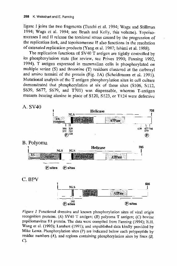
The replication functions of SV40 T antigen are tightly controlled byits phosphorylation state (for review, see Prives 1990; Fanning 1992,1994). T antigen expressed in mammalian cells is phosphorylated onmultiple serine (S) and threonine (T) residues clustered at the carboxyland amino termini of the protein (Fig. 1A) (Scheidtmann et al. 1991).Mutational analysis of the T antigen phosphorylation sites in cell culturedemonstrated that phosphorylation at six of these sites (S106, S112,S639, S677, S679, and T701) was dispensable, whereas T-antigenmutants bearing alanine in place of S120, S123, or T124 were defective
A. SV40NLS"
Helicase 708
ATPase
106 120112 123
^124(E)1
B. Polyoma1
NLS NLSHelicase
639 677701679
785
T/tcommon ATPase
©sites ) sites
C. BPVNLS
Oriliimliii!! ATPase
605I
Figure 1 Functional domains and known phosphorylation sites of viral originrecognition proteins. (A) SV40 T antigen; (B) polyoma T antigen; (C) bovinepapillomavirus El protein. The data were compiled from Fanning (1994); E.H.Wang et al. (1993); Lambert (1991); and unpublished data kindly provided byMike Lentz. Phosphorylation sites (P) are indicated below each polypeptide byresidue numbers (A), and regions containing phosphorylation sites by lines (B,

Phosphorylation of Replication Proteins 299
in viral replication in vivo (Schneider and Fanning 1988), suggesting that modification of these sites is required for SV40 DNA replication in monkey cells.
The role of phosphorylation of T antigen at these three sites in the control of viral DNA replication has been further investigated in cell-free reactions using purified proteins. These studies confirm the requirement for phosphorylation of T124 that was observed in vivo but suggest a quite different role for modification of S120 and S123, namely, that it in- hibits viral DNA replication in vitro. For example, the bulk of the T antigen isolated from SV40-infected mammalian cells is hyper- phosphorylated and inactive in SV40 DNA replication in vitro (Fig. 2) (Virshup et al. 1989). T antigen expressed in bacteria is not modified at these sites and is also unable to initiate SV40 DNA replication in vitro (Fig. 2) (McVey et al. 1989). However, T antigen expressed in recom- binant baculovirus systems has an intermediate level of phosphorylation, with T124 modified in nearly all molecules, but with most serine sites remaining unmodified (Hoss et al. 1990); this T antigen is highly active in SV40 replication in vitro (Fig. 2).
In agreement with these findings, treatment of T antigen with protein kinases and phosphatases that alter the phosphorylation state of T124, S120, or ,3123 activates or inhibits its replication activity in vitro. Phosphorylation of T124 by cdc2-cyclin B activated the replication ac- tivity of unphosphorylated bacterially expressed T antigen (McVey et al. 1989). The positive effect of phosphorylation of T124 on the replication activity of T antigen was reversed by treating it with isoforms of protein phosphatase PP2A that specifically targeted T124, i.e., PP2A-D and PP2A-T55 (Cegielska et al. 1994b). Treatment of hyperphosphorylated T antigen from mammalian cells with the catalytic subunit of PP2A (C36, PP2Ac) or with an isoform of PP2A (PP2A-T72) stimulated SV40 replication by dephosphorylating S120 and S123 (Virshup et al. 1992; Cegielska et al. 1994b). More recently, an isoform of casein kinase I was shown to phosphorylate these serines in vitro (Cegielska and Virshup 1993; Cegielska et al. 1994a). Kinase treatment of hypophosphorylated replication-active T antigen isolated from baculovirus-infected insect cells led to inactivation of SV40 replication activity. Taken together, these results indicate that SV40 DNA replication in vitro is regulated positively and negatively by the phosphorylation state of T antigen (Fig.
The biochemical mechanism by which phosphorylation of T antigen regulated its replication activity was elucidated by comparing the ac- tivities of baculovirus-expressed wild-type and mutant T antigens.
2).

300 K. Weisshart and E. Fanning
p34cdc2 kinase Casein kinase I + PP2A-D, -T55
- PP2A-C36, -T72
INACTIVE ACTIVE INACTIVE
SOWE: E.wli SD &La
Figure 2 Phosphorylation controls SV40 T-antigen replication activity in vitro. Unphosphorylated T antigen synthesized in bacteria is inactive in replication un- less it is first phosphorylated on Thr-124. Hypophosphorylated T antigen ex- pressed in insect cells using baculovirus vectors is active, whereas hyper- phosphorylated T antigen expressed in mammalian cells is inactive. Protein kinases and phosphatases (Cegielska et al. 1994a,b) that act on the crucial Thr- 124, Ser-120, and Ser-123 sites are indicated. (Reprinted, with permission, from Fanning 1994.)
Phosphorylation at T124 is necessary for the bidirectional unwinding of duplex SV40 origin DNA by T antigen in the initiation reaction (McVey et al. 1993; Moarefi et al. 1993b). In contrast, other enzymatic activities like hexamer assembly, unidirectional DNA helicase, DNA binding, and origin DNA distortion were not markedly influenced by phosphorylation of T124. The specific effect of phosphorylation on bidirectional unwind- ing appears to result from its ability to stabilize hexamer-hexamer con- tacts that are necessary for bidirectional, but not unidirectional, DNA un- winding (K. Weisshart and E. Fanning, unpubl.; T. Kelly, pers. comm.). Phosphorylation of T124 also results in cooperative assembly of the two hexamers on the SV40 origin (K. Weisshart and E. Fanning, unpubl.; T. Kelly, pers. comm.).
Interestingly, phosphorylation of T antigen on the inhibitory serine residues also decreased the cooperative assembly of T antigen double hexamers on SV40 origin DNA, blocking bidirectional origin-unwinding activity and viral DNA replication in vitro (Virshup et al. 1992; Cegielska and Virshup 1993; Cegielska et al. 1994a). Thus, T antigen un- phosphorylated on T124 or phosphorylated on S120 and S123 could potentially bind to the origin as a double hexamer and distort it locally without initiating bidirectional unwinding and replication until it became phosphorylated on T124 and dephosphorylated on S120 or 123, thereby facilitating interactions between the two hexamers that are required for bidirectional unwinding (Fig. 3). We suggest that the interactions be- tween the hexamers are dynamic, undergoing cycles of association and dissociation as unwinding proceeds (Fig. 3). Although this mechanism

Phospholylation of Replication Proteins 301
Double hexamer formation
Casein kinase I PPZA-D, -T55 It CyclinA-cdkZ
PPZA-C36, -TI2
Cooperative double hexamer formation & DNA distortion
ATP hydrolysis 1 Local unwinding
Extensive unwinding
Figure 3 The phosphorylation state of SV40 T antigen governs hexamer- hexamer interactions in bidirectional origin unwinding. Prior to viral DNA replication in infected mammalian cells, T antigen accumulates primarily in hy- perphosphorylated forms that can assemble as a double hexamer on the SV40 origin, but the hexamers cannot interact properly with each other to unwind the origin and permit initiation. Replication-active hexamers can be formed upon dephosphorylation of Ser-120 or Ser-123, or by phosphorylation of newly synthesized unphosphorylated T antigen on Thr-124. When a sufficient con- centration of this hypophosphorylated form of T antigen is reached in the in- fected cell, ATP hydrolysis and dynamic hexamer-hexamer interactions lead to bidirectional unwinding at the origin and initiation of replication. Subsequent phosphorylation of Ser-120 and Ser-123 or dephosphorylation of Thr-124 would block reinitiation of replication. (Reprinted, with permission, from Fanning 1994.)

302 K. Weissharl and E. Fanning
remains speculative, the data demonstrate clearly that the phosphoryla- tion state of T antigen controls SV40 DNA replication both negatively and positively at the level of unwinding of origin DNA. It will be inter- esting to see whether phosphorylation of cellular origin recognition or unwinding proteins regulates their function in a similar manner.
The significance of T antigen phosphorylation in viral DNA replica- tion in vivo remains for the most part unresolved. Although it is well es- tablished that phosphorylation of T124 is essential for SV40 DNA replication in vivo (Schneider and Fanning 1988), this modification may play additional roles in regulation of replication. One attractive model proposes that phosphorylation of T antigen at T124 could link the replication activity of T antigen to the cell cycle (McVey et al. 1989, 1993). T antigen is associated with cdk2-cyclin A in the cell (Adam- czewski et al. 1993) and is a much better substrate for this kinase than for cdc2-cyclin B in vitro (C. Voitenleitner et al., unpubl.). cdk2-cyclin A is activated at the GI-S transition and is found associated with replicating SV40 DNA (Fotedar and Roberts 1991). Together these data suggest a role for cdk2-cyclin A as the best candidate for phosphorylation of T124 in vivo and support the notion that this modification event could link SV40 DNA replication to S phase. However, the phosphate turnover on this site is relatively slow in SV40-infected cells, and most T-antigen molecules carry the modification (Scheidtmann 1986; for review, see Fanning and Knippers 1992). Furthermore, there is no direct evidence to suggest that modification of T124 in SV40-infected or -transformed cells is cell-cycle-dependent. Thus, it remains an open question whether the cell-cycle-dependence of SV40 DNA replication is controlled by this phosphorylation event.
The physiological role of phosphorylation of T-antigen residues S120 and S123 in vivo is even more puzzling. Whereas phosphorylation of S120 and S123 inhibits viral DNA replication in vitro, phosphorylation at these sites is required in vivo (Schneider and Fanning 1988). Reexamination of the kinetics of SV40 DNA replication in cultured cells with the S120A and S123A mutants yielded preliminary evidence that the mutant T antigens did begin viral DNA replication, but only a small amount of DNA accumulated (Moarefi et al. 1993a). To resolve the ap- parent discrepancy between the in vivo and in vitro results, it has been suggested that phosphorylation of these serine residues is necessary for regulatory functions not detectable in the in vitro replication system (Fanning 1994). Support for this hypothesis comes from the finding that phosphate turnover on these serines is rapid (Scheidtmann 1986), so that the balance between phosphorylation and dephosphorylation on these

Phosphorylation of Replication Proteins 303
two serines could determine the fraction of T antigen active in initiation of DNA replication (Fanning 1992). Phosphorylation at these serines may be required in the termination of replication, or it could influence the association of viral DNA with replication foci (Hozak and Cook 1994). These or other mechanisms might reduce the ability of T antigen to sustain multiple rounds of viral DNA replication in vivo, resulting in the defect observed with the S120 and S123 mutants in cell culture but not in vitro (Schneider and Fanning 1988; Moarefi et al. 1993a). Further work is required to resolve this paradox.
The physiological relevance of PP2A and casein kinase I action on T antigen in vivo also remains obscure. Casein kinase I is ubiquitously ex- pressed and not regulated in the cell cycle. In addition, PP2A is found primarily in the cytoplasm rather than in the nucleus. However, one can- not exclude the possibility that the different isoforms of kinase and phosphatase that affect T antigen have different subcellular localizations. Indeed, there is evidence that the activity of PP2A on T antigen is restricted to S phase (Ludlow 1992). Other protein phosphatases that act at the GI-S border may also target T antigen, such as a mammalian homolog of SIT4 from budding yeast and Drosophilu (Fernandez- Sarabia et al. 1992; Mann et al. 1993) or a mammalian homolog of the yeast cdc25A phosphatase (Hoffmann et al. 1994; Jinno et al. 1994).
Polyomavirus Large Tumor Antigen
The initiator protein for polyomavirus (PyV) replication is the polyoma large T antigen, Py T, which interacts with specific sequences within the PyV origin of replication (Cowie and Kamen 1984; Hassell and Brinton, this volume). Like its SV40 counterpart, Py T antigen unwinds duplex DNA (Wang and Prives 1991) using its ATPasehelicase function (Seki et al. 1990). Py T antigen is a phosphoprotein with two clusters of phos- phorylation sites located at the amino terminus (Fig. 1B) (Hassauer et al. 1986; Bockus and Schaffhausen 1987). No assessment of the importance of single phosphorylation sites has been undertaken, but treatment with calf intestinal phosphatase had differential effects on Py T antigen, depending on the degree of phosphate removal (E.H. Wang et al. 1993). Treatment with limiting amounts of phosphatase stimulated origin bind- ing, whereas incubation with high concentrations of phosphatase had the opposite effect. These data are reminiscent of early results obtained with partially dephosphorylated SV40 T antigen (Grhser et al. 1987) and sug- gest that the replication activity of PyV T antigen might be regulated through differential phosphorylation. It will be interesting to test whether

304 K. Weisshatt and E. Fanning
phosphorylation of Py T regulates initiation of viral DNA replication by controlling the ability of Py T to catalyze bidirectional unwinding of the origin DNA as in the SV40 system.
Papillomavirus E l Protein
Two bovine papillomavirus (BPV)-encoded proteins are necessary and sufficient for BPV DNA replication in vivo, the 68-kD E l protein and the 48-kD E2 protein (for review, see Lambert 1991; Ustav and Stenlund 1991; Ustav et al. 1993; Stenlund, this volume). The E2 protein can be dispensable in vitro when high levels of E l are used (Seo et al. 1993b; Spalholz et al. 1993). All other replication proteins are supplied by the host cell. E l , like SV40 and Py T antigens, is a phosphoprotein that pos- sesses several activities necessary for replication function (Ustav and Stenlund 1991; Seo et al. 1993a; Thorner et al. 1993; Yang et al. 1993; Park et al. 1994; Bonne-Andrea et al. 1995): origin binding activity, polymerase-a binding, ATPase activity, and 3 ' -5 ' helicase activity (Fig. 1C). E2 protein is a transcriptional activator (Spalholz et al. 1993) and also a phosphoprotein (Bream et al. 1993) that stimulates the replica- tion activity of E l protein in vitro, at least in part by binding to it (Seo et al. 1993b; Spalholz et al. 1993; Benson and Howley 1995). It has been proposed that the role of E2 is to tether E l more tightly to the origin and to recruit cellular replication factors, e.g., RP-A and pol-a:primase (Li and Botchan 1993), to the BPV origin.
The facts that BPV DNA is maintained as an episome at a constant copy number in latently transformed cells (Law et al. 1981) and that it replicates during S phase along with the host chromosomal DNA (Gilbert and Cohen 1987) might suggest that the activities of E l and E2 are regu- lated in the cell cycle. Since both proteins are phosphoproteins, this regulation could depend on phosphorylation events. The E l residue T102 near the nuclear localization signal is a substrate in vitro for cdc2-cyclin kinase (Fig. 1C). However, replacing that threonine by isoleucine showed no observable effect on either the nuclear localization of E l or its efficiency in DNA replication (Lentz et al. 1993). Thus, the functional role, if any, of phosphorylation of the BPV replication proteins remains unknown.
Yeast Origin Recognition Complex
The most likely candidate for the replication initiator in yeast is the origin recognition complex (ORC) (for review, see Bell and Stillman 1992; Diffley 1992; Huberman 1992; Li and Alberts 1992; see Newlon;

Phosphorylation of Replication Proteins 305
Nasmyth; Stillman; all this volume). It consists of six polypeptides with molecular masses of 50, 53, 56, 62, 72, and 120 kD, respectively, that have been termed ORC-1 to ORC-6 according to the decrease in molecular weight. The ORC complex binds specifically to a bipartite site in yeast origins of replication in an ATP-dependent manner (Rao and Stillman 1995; Rowley et al. 1995). Mutations in the gene coding for ORC-2 have shown that ORC is necessary for the initiation of DNA replication and transcriptional silencing at the mating-type locus (Bell et al. 1992; Foss et al. 1993; Micklem et al. 1993; for review, see Newlon 1993; Huberman 1994; Kelly et al. 1994). ORC-6 has been shown to be important for the initiation of DNA replication (Li and Herskowitz 1993).
Evidence has been obtained by in vivo footprinting that ORC is bound to ARS-1 sequences in yeast cells in the same manner as it is in vitro (Diffley and Cocker 1992). Interestingly, ORC remains bound to the origin throughout the cell cycle. These data suggested that ORC could repress replication except in S phase, when it could be activated for replication. ORC-2 and ORC-6 contain multiple potential phosphorylation sites for cyclin-dependent kinases and other kinases (Bell et al. 1992; Li and Herskowitz 1993; Micklem et al. 1993), so that phosphorylation by cell-cycle-regulated kinases may influence ORC ac- tivity. However, mutations in the potential sites of phosphorylation by cyclin-dependent kinases have not yet revealed any loss of replication function (J. Diffley, pers. comm.). Interestingly, DBF4, the CDC7 protein kinase regulatory subunit, interacts with the yeast replication origin (Dowel1 et al. 1994), and activation of CDC7 by DBF4 at the G1/S-phase transition is important for the initiation of S phase (Jackson et al. 1993; for review, see Newlon 1993; Nasmyth, this volume). Thus, the interaction of DBF4 with ORC-2 may target the CDC7 catalytic sub- unit to the ORC complex, enabling it to phosphorylate the ORC-2 polypeptide (for review, see Barinaga 1994). However, the functional ef- fects of this phosphorylation have not yet been described.
Although the footprint of ARS sequences remains detectable through- out the cell cycle, there is an increase in the protected area during G1 phase, which is indicative of prereplicative complexes binding in the vi- cinity of ORC (Diffley et al. 1994). The enlarged footprint may arise through the association of additional proteins with the ORC complex. There is evidence that ORC-6 interacts with CDC6 and CDC46/MCM5 proteins (Li and Herskowitz 1993), which are good candidates for com- ponents of the prereplicative complex. CDC6 of budding yeast and its counterpart Cdcl8 from fission yeast are important for entry into S phase

306 K. Weisshart and E. Fanning
(Kelly et al. 1993). The CDC6 gene product shows ATPase activity, but no detectable DNA unwinding activity (Zwerschke et al. 1994). CDC46 also acts at an early stage in DNA replication. The subcellular localiza- tion of CDC46 and other members of the MCM 2-3-5 family is regulated in the cell cycle (Hennesey et al. 1991; Y. Chen et al. 1992; Yan et al. 1993; for review, see Tye 1994). These proteins are nuclear only be- tween M and S phase, consistent with the notion that they could partici- pate in the GI-specific enlargement of the footprint on the origin of replication, but no data are available on this point. One might expect that cell-cycle-dependent phosphorylation events would regulate the func- tions of the MCM proteins, but again, mutations in the potential sites of phosphorylation of MCM proteins by cyclin-dependent kinases have not affected their function (B. Tye, pers. comm.) (but see section below). It has been speculated based on their amino acid sequences that the MCM proteins may function as ATP-dependent helicases (Koonin 1993).
Xenopus and Mammalian Initiation Proteins
The MCM proteins are found not only in yeast, but also in frogs, mice, and humans (for review, see Tye 1994). Their importance in control of DNA replication is underscored by their recent identification (Chong et al. 1995; Kubota et al. 1995; Madine et al. 1995) as components of a replication licensing factor that was postulated by Blow and Laskey (1988) to limit eukaryotic chromosomal DNA replication to one round per cell cycle (for review, see Coverley and Laskey 1994). Taken togeth- er, the data strongly support the notion that a complex of different MCM proteins is required for chromatin replication in the Xenopus in vitro sys- tem, that their presence in the nucleus before replication is necessary, and that upon replication, they are lost or become easily extractable from the nucleus. Interestingly, replication licensing factor activity is blocked by treatment of Xenopus extracts with protein kinase inhibitors, suggest- ing that phosphorylation is directly or indirectly involved in their func- tion (for review, see Coverley and Laskey 1994; Tye 1994).
A mammalian homolog of MCM3, the P1 protein, was found associ- ated with DNA pol-ccprimase (Thommes et al. 1992b). The nuclear localization of P1 is regulated during the cell cycle like that of its yeast and frog counterparts (Kimura et al. 1994). Nuclear P1 appears to exist in distinct subpopulations: one that is hyperphosphorylated and loosely bound in the nucleus, and another that is underphosphorylated and tightly bound in the nucleus. In early S phase, the tightly bound form of P1 was predominant, but as S phase progressed, the loosely bound P1 gradually

Phosphorylation of Replication Proteins 307
increased and became the major form by the end of S phase (Kimura et al. 1994). This behavior would be consistent with a role for P1 in prereplicative chromatin complexes and disassembly of these complexes after cell-cycle-dependent phosphorylation,
OTHER PROTEINS THAT ARE REQUIRED FOR DNA REPLICATION
Pol-a:Primase
Pol-a:primase is composed of four subunits with molecular masses of 180, 68, 58, and 48 kD (p180, p68, p58, and p48) (Thommes and Hubscher 1990; Wang 1991 and this volume). All the cDNAs encoding the human, mouse, and yeast subunits have been cloned (Lucchini et al. 1987; Wong et al. 1988; Collins et al. 1993; Miyazawa et al. 1993; Stadlbauer et al. 1994). The structure and function of pol-a:primase are highly conserved in eukaryotic organisms (Wang 1991). Pol-a:primase is an essential component of the cellular replication machinery (Pizzagalli et al. 1988; Francesconi et al. 1991), where it functions both in initiation at origins of replication and in the synthesis of Okazaki fragments on the lagging strand of the replication fork (Focher et al. 1988; Prelich and Stillman 1988; Thommes and Hiibscher 1990; Wang 1991). Pol- mprimase also initiates the leading strand at the origin of SV40 replica- tion (Tsurimoto et al. 1990). The p180 subunit harbors the DNA polymerase activity (Pizzagalli et al. 1988; Wong et al. 1988; Nasheuer et al. 1991), the p68 subunit is thought to play a role in the initiation pro- cess (Collins et al. 1993; Foiani et al. 1994), and the p58 and p48 sub- units comprise the primase activity (Santocanale et al. 1993).
During activation of quiescent mammalian cells, the expression of all subunits is up-regulated. However, in actively cycling cells, gene expres- sion is constitutive and mRNA and protein levels are only marginally en- hanced prior to S phase (Wahl et al. 1988; Miyazawa et al. 1993). Thus, if pol-a:primase function is regulated during the cell cycle, it is likely to involve a posttranslational mechanism. Indeed, both the p180 and the p68 subunits are phosphorylated on serine and threonine residues in a cell-cycle-dependent manner (Wong et al. 1986). The p180 subunit is phosphorylated throughout the cell cycle but becomes hyperphosphory- lated in the G2-M transition, whereas the p68 subunit is phosphorylated beginning in S and more heavily at the G2-M transition (Nasheuer et al. 1991).
Phosphorylation of DNA polymerase-a:primase at different points in the cell cycle has also been studied in budding yeast (Foiani et al. 1995). Both the p180 and p86 (B) subunits are phosphorylated in yeast, but only

308 K. Weisshart and E. Fanning
the B subunit showed a reduced electrophoretic migration upon modifi- cation. The kinetics of phosphorylation during the cell cycle revealed that newly synthesized B subunit occurred in an underphosphorylated form during S phase and became modified-during G2. Maternal B sub- unit was stable and became phosphorylated in early S phase. Both popu- lations of B subunit were dephosphorylated during exit from mitosis (Foiani et al. 1995). The observation that the B subunit is required very early in S phase for initiation, but has no effect on the enzymatic func- tions of yeast pol-a:primase, suggested that phosphorylation may regu- late the activity of the B subunit in initiation of replication (Fig. 4) (Foiani et al. 1995).
The functional relevance of phosphorylation of pol-a:primase has been investigated in vitro. Treatment of pol-a:primase with casein kinase I1 in vitro phosphorylated the p180 and p58 subunits, but had no in- fluence on either polymerase or primase activity (Podust et al. 1990). Some of the in vivo cell-cycle-dependent phosphorylation sites on the p180 and p68 subunits of mammalian pol-a:primase were shown to be phosphorylated by kinases in vitro, implying these kinases as possible regulators. However, these kinases also had little effect on the enzymatic activities of pol-a:primase; only a slight reduction in affinity for single-stranded DNA was found (Nasheuer et al. 1991).
The functional effect of phosphorylation of pol-a:primase by cyclin- dependent kinases has recently been reexamined using purified proteins. Pol-a:primase expressed in the baculovirus system was modified on the p180 and p68 subunits by cyclin E-, A-, and B-dependent kinases (C. Voitenleitner et al., unpubl.). All of these kinases utilized pol-a:primase approximately equally well as a substrate. Phosphorylation of the en- zyme did not affect its polymerase activity, in agreement with published data, although it stimulated primase activity severalfold. In contrast, the ability of pol-a:primase to initiate SV40 DNA replication in vitro in a reaction performed with purified proteins was markedly inhibited after phosphorylation by cyclin-A-dependent kinases. Treatment with cyclin- B-dependent kinase reduced initiation activity more modestly. Cyclin-E- dependent kinase caused no reduction in initiation activity; indeed, a small stimulation was observed in some experiments (C. Voitenleitner et al., unpubl.).
These results raise the question whether phosphorylation of pol- a:primase may influence its interaction with other initiation proteins. Both the p180 and the p68 subunits interact with SV40 T antigen (Dorn- reiter et al. 1990, 1992, 1993; Collins and Kelly 1991; Collins et al. 1993; Murakami and Hurwitz 1993), and these interactions play a sub-

Phosphorylation of Replication Proteins 309
DNA pola-primase
Figure 4 Model for regulation of origin-specific initiation activity of pol- a:primase by cell-cycle-dependent phosphorylation. Hypophosphorylated pol- a:primase that appears in late M or early G , (Foiani et al. 1995) is suggested to assemble in a prereplicative complex together with cellular initiation proteins on the cognate origin of DNA replication (Diffley et al. 1994). The complex would remain dormant until activation at the G,/S border, an event that probably re- quires cyclin-E-dependent kinase directly or indirectly (Knoblich et a1.1994; Jackson et al. 1995; Sauer et al. 1995). Upon initiation, phosphorylation of pol- a:primase is postulated to inhibit its ability to initiate new rounds of replication at an origin. Perhaps phosphorylation of pol-a:primase at the origin is even in- volved in the transition from initiation to elongation. If cyclin A-dependent kinases, which accumulate as the cell progresses from S through M phase, were responsible for these modifications, the fraction of initiation-active pol- a:primase would be predicted to decline from S to M phase without a con- comitant reduction in lagging-strand DNA replication activity. A similar mecha- nism may regulate the SV40 initiation activity of pol-a:primase in virus-infected cells, thereby facilitating assembly of new virus particles in late S phase and G,.
stantial role in the initiation of SV40 replication. The ability of phosphorylated pol-a:primase to undergo these interactions has not yet been assayed directly. Intriguingly, however, only a subpopulation of mouse pol-a:primase was associated with Py T antigen, and only this population was able to stimulate viral DNA replication (Moses and

310 K. Weisshart and E. Fanning
Prives 1994). It will be interesting to investigate the phosphorylation state of these different fractions of pol-a:primase.
Taken together, the results of these studies in different systems with different sets of reagents argue that cell-cycle-dependent phosphorylation of pol-a:primase regulates its function in imtiation of DNA replication. Figure 4 depicts a speculative model for this regulation. Since the model makes several predictions that can be tested experimentally, it may be useful in gaining a better understanding of the role of protein phos- phorylation in replication.
Replication Protein A
Replication protein A (RP-A), also known as replication factor A (RF-A) and human single-stranded DNA-binding protein (HSSB), was identified as an essential factor for SV40 DNA replication in vitro (Wobbe et al. 1987; Fairman and Stillman 1988; Wold and Kelly 1988). It is the major single-stranded DNA-binding protein in mammalian cells (Seroussi and Lavi 1993) and is highly conserved in all eukaryotes (Wold and Kelly 1988; Brill and Stillman 1989; Mitsis et al. 1993). RP-A is a heterotrimeric protein with subunits of 14, 32, and 70 kD (p14, p32, and p70) (Fairman and Stillman 1988; Wold and Kelly 1988). The cDNAs encoding all three subunits have been cloned from human (Erdile et al. 1990, 1991; Umbricht et al. 1993) and yeast (Heyer et al. 1990; Brill and Stillman 1991) and are essential for cell viability in yeast (Heyer et al. 1990; Brill and Stillman 1991; Longhese et al. 1994).
Human RP-A binds tightly to single-stranded DNA (Wobbe et al. 1987; Fairman and Stillman 1988; Wold and Kelly 1988) with low cooperativity (Kim and Wold 1995) in two distinct complexes, one oc- cupying 8-10 nucleotides, the other 20-30 nucleotides (Blackwell and Borowiec 1994; Kim et al. 1994). Binding affinity to RNA and double- stranded DNA is much lower (Brill and Stillman 1989; Wold et al. 1989; Kim et al. 1992). RP-A has been shown to be associated with an intrinsic DNA-unwinding activity (Georgaki et al. 1992). The nucleic acid- binding properties of the homolog from Drosophila melanogaster (dRPA) were reported to be quite similar to those of human RP-A (Mitsis et al. 1993). However, RP-A from Saccharomyces cerevisiae (yRPA) was found to be quite different, binding to a site of about 90 nucleotides with high cooperativity (Alani et al. 1992). Yeast RP-A, however, cannot substitute for its human counterpart in SV40 DNA replication in vitro (Brill and Stillman 1991).

Phosphorylation of Replication Proteins 31 1
The p70 subunit of RP-A contains the intrinsic DNA-binding activity (Wold et al. 1989; Kenny et al. 1990). The p32 and p14 subunits form a stable subcomplex (Henricksen et al. 1994; Stigger et al. 1994) that is important for proper formation of the hetqrotrimeric complex (Henrick- sen et al. 1994). The biological functions of the 32-kD and 14-kD sub- units are not known. However, monoclonal antibodies directed against the 70-kD7 32-kD7 or 14-kD subunits block SV40 DNA replication, sug- gesting a role for all three subunits in this process (Erdile et al. 1990; Kenny et al. 1990; Umbricht et al. 1993).
RP-A has multiple functions in the replication process (Kenny et al. 1989). During bidirectional unwinding of the SV40 origin of replication by T antigen, it stabilizes the created single-stranded regions. Moreover, it is essential during primosome assembly (Melendy and Stillman 1993) and primer synthesis on the SV40 origin (Schneider et al. 1994), reflect- ing the specific protein-protein interactions that occur between RP-A, T antigen, and pol-a:primase (Dornreiter et al. 1992, 1993; Nasheuer et al. 1992; Murakami and Hurwitz 1993; K. Weisshart and E. Fanning, un- publ.). RP-A also plays a role in the elongation reaction, where it has been shown to stimulate DNA polymerases a (Kenny et al. 1989; Erdile et al. 1991; Brown et al. 1992), 6 (Kenny et al. 1989; Tsurimoto and Stillman 1989; Lee et al. 1991a), and E (Lee et al. 1991b). In addition, it increases the fidelity of DNA synthesis by pol-a:primase (Carty et al. 1992; Roberts et al. 1993; Suzuki et al. 1994) and stimulates copurifying helicases (Seo et al. 1991; Thommes et al. 1992a; Georgaki et al. 1994).
Besides its essential functions in DNA replication, RP-A is also re- quired for DN-A repair and recombination in vivo (Longhese et al. 1994) and nucleotide excision repair of UV-damaged DNA in vitro (Coverley et al. 1991). It also stimulates in vitro strand-exchange proteins involved in homologous recombination (Heyer et al. 1990; Moore et al. 1991; Alani et al. 1992). RP-A has also been shown to interact with the acidic domains of transcription factors VP16, GAL4, and p53 (Dutta et al. 1993; He et al. 1993; Li and Botchan 1993). The significance of these in- teractions is still unknown. On the one hand, these factors stimulated BPV replication (Li and Botchan 1993); on the other hand, p53 inhibited SV40 replication (Dutta et al. 1993). It was found recently that yRP-A binds specifically to sequence elements that are involved in the regula- tion of transcription (Luche et al. 1993; Singh and Samson 1995), so that the interaction of RP-A with transcription factors could play a role in transcriptional regulation.
Several posttranslational modifications of the RP-A subunits have been observed. Both the p70 and p32 subunits are pol-ADP-ribosylated

312 K. Weisshart and E. Fanning
(Eki and Hurwitz 1991) and phosphorylated (Din et al. 1990; Fang and Newport 1993). The abundance of RP-A remains constant throughout the cell cycle, but the p32 subunit is phosphorylated in a cell-cycle-specific manner on multiple serines (Din et al. 1990). In cycling cells, p32 be- comes extensively phosphorylated at the 6,-S transition and remains phosphorylated until G2-M, when dephosphorylation occurs (Din et al. 1990). The hyperphosphorylated form of p32 shows decreased mobility during gel electrophoresis and runs as a 36-kD species.
On the basis of amino acid sequence, human RP-A p32 has two potential sites for cyclin-dependent kinases, five for DNA-PK, and five for casein kinase 11. There is good evidence that cyclin-dependent kinases and DNA-PK phosphorylate RP-A p32 in vitro and in vivo. In vitro, cdc2-cyclin B kinase phosphorylates p32 at Ser-23 and Ser-29, sites that are also phosphorylated in vivo (Dutta and Stillman 1992), con- comitant with a modest stimulation of SV40 DNA replication and origin unwinding in G, extracts. The same sites were also targeted specifically within the replication initiation complex by cdk2-cyclin A kinase (Fotedar and Roberts 1991, 1992; Dutta and Stillman 1992; Elledge et al. 1992; Pan et al. 1993a). However, mutant RP-A lacking the two cyclin- dependent kinase sites in the p32 subunit supported SV40 DNA replica- tion in vitro as well as wild-type RP-A, demonstrating that phosphoryla- tion of RP-A by cyclin-dependent kinases is dispensable in this system (Henricksen and Wold 1994). Consistent with these results, phosphory- lated and unphosphorylated RP-A were equally active in SV40 DNA replication in vitro (Pan et al. 1995).
Under the conditions of the in vitro SV40 replication system, un- derphosphorylated RP-A becomes hyperphosphorylated (Fotedar and Roberts 1992; Henricksen and Wold 1994). The kinase involved appears to be DNA-PK, but cyclin A-cdk2 may also play a role. RP-A p32 is pre- dominantly phosphorylated when bound to DNA, and the kinase that phosphorylates it is itself bound to single-stranded DNA (Fotedar and Roberts 1992). The p32 subunit in RP-A from cycling embryonic Xenopus extracts is also phosphorylated in S phase only in nuclear- localized RP-A bound to single-stranded DNA (Fang and Newport 1993). Hyperphosphorylation of RP-A p32 by DNA-PK in a cyclin-A- activated G, extract was reported to require prephosphorylation by cyclin A-cdk2 (Pan et al. 1994). However, mutant RP-A lacking the two poten- tial cyclin-dependent kinase sites in p32 still becomes hyperphosphory- lated (Henricksen and Wold 1994; Lee and Kim 1995). Moreover, replication-competent extracts depleted of cyclin-dependent kinases still hyperphosphorylate DNA-bound RP-A in vitro (Dutta and Stillman

Phosphorylation of Replication Proteins 31 3
1992), whereas extracts depleted of DNA-PK do not (Brush et al. 1994). Human RP-A is also found hyperphosphorylated in GI cells that have
been exposed to ionizing radiation (Liu and Weaver 1993) or UV light (Carty et al. 1994). A good candidate for the kinase that phosphorylates RP-A during the damage response is the DNA-PK (Lees-Miller and Anderson 1991), a kinase that is known to be involved in DNA repair and is stimulated by double-strand breaks (for review, see Gottlieb and Jackson 1994). Purified DNA-PK phosphorylates RP-A p32 in vitro (Brush et al. 1994; Pan et al. 1994), and cells that are deficient in DNA- PK activity fail to hyperphosphorylate RP-A after irradiation (Boubnov and Weaver 1995). Hyperphosphorylated RP-A in extracts from ir- radiated cells appeared to be inactive in SV40 DNA replication in vitro, and additional RP-A purified from unirradiated cells relieved that block (Carty et al. 1994), suggesting that phosphorylation on the potential DNA-PK sites might activate RP-A for its tasks in repair while blocking its function in the replication process. However, other workers have found that phosphorylation of RP-A by DNA-PK did not affect its ac- tivity in SV40 DNA replication in vitro (Brush et al. 1994).
Based on the evidence from in vitro SV40 DNA replication, phos- phorylation of RP-A appears to have little or no functional importance. However, it may play a role in vivo, such as correct localization to replication centers (for review, see Hozak and Cook 1994). Chromatin replication in Xenopus egg extracts is dependent on RP-A (Adachi and Laemmli 1994). Before being assembled into replication centers, RP-A is targeted to the prereplicative sites that form after mitosis and prior to ini- tiation (Adachi and Laemmli 1994). This association is loose due to the absence of single-stranded DNA and is likely to be mediated by protein- protein interactions. Assembly of RP-A into prereplicative complexes does not occur in the presence of active cyclin B-cdc2 kinase, suggesting that only the underphosphorylated form of RP-A found after mitosis can be targeted to the complexes. Active cyclin A-cdk2 kinase did not facili- tate or inhibit the assembly of prereplicative complexes (Adachi and Laemmli 1994). However, it colocalized with RP-A, and presumably many other replication proteins, to replication foci (Adachi and Laemmli 1992, 1994; Cardoso et al. 1993), suggesting a possible function in this process.
In summary, it seems likely that phosphorylation of RP-A regulates its activity in vivo, but the mechanism of this regulation remains poorly understood. Much of the confusion may stem from the multiple functions of RP-A in the cell, but it should be noted that the assay system used most frequently to assess the effects of phosphorylation, in vitro SV40
314 K. Weisshart and E. Fanning
DNA replication, probably does not reveal all of the relevant steps in in vivo regulation of DNA replication.
DNA Polymerases 6 and E
Polymerases 6 and/or E are thought to play a pivotal role in leading- strand DNA synthesis as well as lagging-strand synthesis (Burgers 1991; Nethanel et al. 1992; Podust and Hubscher 1993; see Wang, this volume). Polymerase-6 is a two-subunit complex with polypeptides of approximately 124 kD and 55 kD (Chung et al. 1991) whose activity and processivity are stimulated by complexing with PCNA (Bauer and Burgers 1988). Polymerase-& consists of a catalytic subunit of 258 kD (Kesti et al. 1993) that copurifies with a smaller 55-kD polypeptide in human (Syvaoja and Linn 1989; Lee et al 1991b) and with polypeptides of 80, 34, 30, and 29 kD in yeast extracts (Hamatake et al. 1990). RF-C and PCNA also function to stimulate polymerase-e (Burgers 1991; Lee et al. 1991b). Besides their involvement in DNA replication, both polymer- ase-6 and polymerase-e are implicated in DNA repair (Z. Wang et al. 1993; Blank et al. 1994; Zeng et al. 1994b; Budd and Campbell 1995).
Polymerase-6 mRNA and protein expression is somewhat increased at the GI-S border, but more significantly, the protein is most actively phosphorylated during S phase (Zeng et al. 1994a). It will be important to assess the enzymatic activities of hyper- and hypophosphorylated polymerase-& and to look for differences in its interactions with other replication proteins like PCNA (Bauer and Burgers 1988) or RP-A (Longhese et al. 1994).
Polymerase-& mRNA expression and protein levels are strongly de- pendent on cell proliferation (Kesti et al. 1993; Tuusa et al. 1995), but their abundance fluctuated less dramatically in actively cycling cells. The phosphorylation state of polymerase-e has not been investigated.
PCNA and RF-C
PCNA is a phosphoprotein and its phosphorylation state appears to vary in a cell cycle-dependent fashion (Prosperi et al. 1993, 1994). Inter- estingly, highly phosphorylated PCNA associated with insoluble nuclear structures primarily during S phase, whereas soluble PCNA was found in early GI and G2/M in a weakly phosphorylated form. Expression of PCNA appears to be independent of the cell cycle in actively growing cells. On the other hand, its expression declines in quiescent cells and is strongly induced by mitogens (Mathews et al. 1984). It is also subject to

Phosphorylation of Replication Proteins 315
regulation by binding to cdk-cyclin kinases and their inhibitors (for review, see Heichman and Roberts 1994; Pines 1994).
RF-C is a multisubunit protein consisting of five polypeptides (p36.5, p37, p38, p40, and p140; also designated RFC1-5) (Lee et al. 1991b; Tsurimoto and Stillman 1991~). The mammalian and yeast genes encod- ing the RF-C subunits have been cloned (M. Chen et al. 1992a,b; Bunz et al. 1993; Pan et al. 1993b; Li and Burgers 1994a,b; Luckow et al. 1994; Noskov et al. 1994; Cullman et al. 1995; Hiibscher et al., this volume). The p140 subunit has an ATP-binding domain and binds unspecifically to DNA. Its transcripts are most abundant in strongly proliferative tissues but are also relatively high in brain (Luckow et al. 1994). The p40 polypeptide has an ATP-binding motif and shows ATPase activity. It seems to interact with PCNA, polymerase-6, and the p37 subunit of RF- C (Pan et al. 1993b). The steady-state levels of the mRNA fluctuate only slightly during the cell cycle (Noskov et al. 1994). The p38 subunit binds ATP, and its ATPase activity is stimulated by single-stranded DNA (Li and Burgers 1994a). The p37 subunit shows specific affinity to polymerase-& and binds specifically to primer-template termini (Pan et al. 1993b). The p37 mRNA levels do not change significantly during the cell cycle (Li and Burgers 1994b). Since RNA levels of two of the sub- units are constant during the cell cycle, protein modifications like phosphorylation might contribute to the regulation of RF-C function. A thorough investigation of RF-C expression and modification, however, must await the availability of suitable immunological reagents.
Topoisomerases
Topoisomerases are involved in the maintenance of chromatin and DNA structure (see Hangaard-Andersen et al., this volume). Topoisomerase I relaxes supercoiled DNA, whereas topoisomerase I1 catalyzes decatena- tion and unknotting of topologically linked DNA circles, as well as the relaxation of supercoiled DNA (Wang 1985). Both topoisomerases have been shown to be phosphorylated.
Dephosphorylation of serinehhreonine residues in topoisomerase I isolated from different sources reduced or abolished its enzymatic ac- tivity (Durban et al. 1983; Kaiserman et al. 1988; Pommier et al. 1990; Tournier et al. 1992). Phosphorylation of the protein on tyrosine decreased activity as well (Tse-Dinh et al. 1984). Conversely, phosphorylation of topoisomerase I by casein kinase I1 and related kinases (Durban et al. 1983; Kaiserman et al. 1988) or protein kinase C (Pommier et al. 1990; Samuels and Shimizu 1992) stimulated activity.

316 K. Weisshart and E. Fanning
The decatenation activity of topoisomerase I1 was shown to be stimu- lated by phosphorylation by casein kinase 11, protein kinase C, cdc2- cyclin kinases, and Ca++/calmodulin-dependent protein kinase (Sahyoun et al. 1986; Rottman et al. 1987; Ackerman et a]. 1988; Cardenas and Gasser 1993; for review, see Poljak and Kas 1995). The major in vivo kinase acting on topoisomerase I1 in S. cerevisiae has been identified as casein kinase I1 (Cardenas et al. 1992). Topoisomerase I1 is hyper- phosphorylated during G,/M phase (Cardenas et al. 1992), consistent with the finding that this phosphorylation is required for activity of topoisomerase I1 during chromosome segregation. The stimulation of decatenation induced by phosphorylation was found to be at least partly due to an increase in the DNA-binding affinity of topoisomerase I1 (Dang et al. 1994) and an increased rate of ATP hydrolysis (Corbett et al. 1993).
SUMMARY AND OUTLOOK
Our understanding of the role of protein phosphorylation in regulation of DNA replication remains fragmentary. All of the proteins that recognize and activate eukaryotic origins of replication are phosphorylated, but with the exception of SV40 T antigen, there is no clear picture of how these modifications affect the function of the proteins. Similarly, the key replication enzymes in eukaryotic cells are phosphoproteins, but the functional importance of phosphorylation is established only for topoiso- merase 11, and in preliminary fashion for pol-a:primase. The lack of data to support a role for phosphorylation in regulation of DNA replication is not due to lack of effort. The mutational analysis of potential phosphory- lation sites that was successful in investigating SV40 T antigen has now been extended to many of the cellular origin recognition proteins and several replication enzymes, including pol-a:primase and RP-A, without conspicuous success. Does this mean that these phosphorylation events are fortuitous? The cell-cycle-dependence of phosphorylation of replica- tion proteins, the correlation between phosphorylation of MCM proteins with changes in their subcellular localization, and the importance of the cyclin-dependent kinases, other cell-cycle-dependent kinases, and the DNA-dependent protein kinase suggest that phosphorylation of most replication proteins is probably physiologically relevant. How then can one demonstrate this importance more directly? It should be noted that the current understanding of phosphorylation in control of SV40 T antigen’s replication activity was not achieved solely through rnuta- genesis. Not only were the in vivo phosphorylation sites mapped, but

Phosphorylation of Replication Proteins 317
several overexpression systems and a wide variety of biochemical assays were available in many laboratories to probe the functions of T antigen and its mutants before and after treatment with purified kinases and phosphatases over the course of the past decade. As outlined in the intro- duction, such a broad approach will likely be required to understand the roles of phosphorylation in the control of DNA replication.
ACKNOWLEDGMENTS
We thank Andreas Zeitvogel and Achim Dickmanns for assistance with the figures; Me1 DePamphilis, Vladimir Podust, and Christoph Rehfuess for helpful criticism of the manuscript; and M. Lentz, M. Foiani, C. Leh- ner, T. Kelly, B. Tye, and J. Diffley for communication of results before publication. Work in the authors’ laboratories was supported by the Deutsche Forschungsgemeinschaft, Fonds der Chemischen Industrie, BMFT-Genzentrum, the European Community (CHRX-CT93-0248 DG12), and the National Institutes of Health (1 R 0 1 GM-52948-01).
REFERENCES
Ackerman, P., C.V. Glover, and N. Osherhoff. 1988. Phosphorylation of DNA topoisomerase in vivo and in total homogenates of Drosophilu Kc cells. J. Biol. Chem.
Adachi Y. and U. Laemmli. 1992. Identification of nuclear pre-replication centers poised for DNA synthesis in Xenopus egg extracts: lmmunolocalization study of replication protein A. J. Cell Biol. 119: 1-15.
-. 1994. Study of the cell cycle dependent assembly of DNA pre-replication cen- ters in Xenopus egg extracts. E M B O J . 13: 4153-4164.
Adamczewski, J.P., J.V. Cannon, and T. Hunt. 1993. Simian virus 40 large T-antigen as- sociates with cyclin A and ~ 3 3 ~ ~ ~ ~ . J. Virol. 67: 6551-6557.
Alani, E., R. Thresher, J.D. Griffith, and R.D. Kolodner. 1992. DNA binding properties of yRP-A and interactions with yeast and E. coli strand-exchange pr0teins.J. Mol. B i d .
Barinaga, M. 1994. Yeast enzyme finds fame in link to DNA replication. Science 265:
Bauer, G.A. and P.M.J. Burgers. 1988. The yeast analog of mammalian cyclin/prolifer- ating cell nuclear antigen interacts with mammalian DNA polymerase 6. Proc. Nutl. Acad. Sci. 85: 7506-7510.
Bell, S.P. and B. Stillman. 1992. ATP-dependent recognition of eukaryotic origins of DNA replication by a multiprotein complex. Nature 357: 128-134.
Bell, S.P., R. Kobayashi, and B. Stillman. 1993. Yeast origin recognition complex func- tions in transcription silencing and DNA replication. Science 262: 1844-1849.
Benson, J.D. and P.M. Howley. 1995. Amino-terminal domains of the bovine papil-
263: 12653-12660.
227: 54-71.
1175-1176.

318 K. Weisshart and E. Fanning
lomavirus type 1 E l and E2 proteins participate in complex formation. J. Virol. 69:
Blackwell, L.J. and J.A. Borowiec. 1994. Human replication protein A binds single- stranded DNA in two distinct complexes. Mol. Cell. Biol. 14: 3993-4001.
Blank, A., B. Kim, and L.A. Loeb. 1994. Polymerase 6 is required for base excision repair of DNA methylation damage in Saccharomyces cerevisiae. Proc. Natl. Acad. Sci. 91: 9047-9051.
Blow, J.J. and R.A. Laskey. 1988. A role for the nuclear envelope in controlling DNA replication within the cell cycle. Nature 332: 546-548.
Bockus, B.J. and B. Schaffhausen. 1987. Localization of the phosphorylations of polyomavirus large T antigen. J. Virol. 61: 1155-1163.
Bonne-Andrea, C., S. Santuccii, P. Clertant, and F. Tillier. 1995. Bovine papillomavirus E l protein binds specifically DNA polymerase a but not replication protein A. J. Virol.
Borowiec, J.A., F.B. Dean, P.A. Bullock, and J. Hurwitz. 1990. Binding and unwind- ing-How T antigen engages the SV40 origin of DNA replication. Cell 60: 181-184.
Boubnov, N.V. and D.T. Weaver. 1995. scid cells are deficient in Ku and replication protein A phosphorylation by the DNA-dependent protein kinase. Mol. Cell. Biol. 15:
Bream, G.L., C.-A. Ohmstede, and W.C. Phelps. 1993. Characterization of human papil- lomavirus type 11 E l and E2 proteins expressed in insect cells. J. Virol. 67:
Brill, S.J. and B. Stillman. 1989. Replication factor-A from Saccharomyces cerevisiae functions in the unwinding of SV40 origin of replication. Nature 342: 92-95.
-. 1991. Replication factor-A from Saccharomyces cerevisiae is encoded by three essential genes coordinately expressed at S-phase. Genes Dev. 5: 1589-1600.
Brown, G.W., T.E. Melendy, and D.S. Ray. 1992. Conservation of structure and function of DNA replication protein A in the trypanosomatid Crithidia fasciculata. Proc. Natl. Acad. Sci. 89: 10227-10231.
Brush, G.S., C.W. Anderson, and T.J. Kelly. 1994. The DNA-activated protein kinase is required for the phosphorylation of replication protein A during simian virus 40 DNA replication. Proc. Natl. Acad. Sci. 91: 12520-12524.
Budd, M.E. and J.L. Campbell. 1995. DNA polymerases required for repair of UV- induced damage in Saccharomyces cerevisiae. Mol. Cell. Biol. 15: 2173-2179.
Bunz, F., K. Kobayashi, and B. Stillman. 1993. cDNAs encoding the large subunit of hu- man replication factor C. Proc. Natl. Acad. Sci. 90: 11014-11018.
Burgers, P. 1991. Saccharomyces cerevisiae replication factor C. 11. Formation and ac- tivity of complexes with the proliferating cell nuclear antigen and with DNA polymerases 6 and E. J. Biol. Chem. 266: 22698-22706.
Cardenas, M.E. and S.M. Gasser. 1993. Casein kinase I1 copurifies with yeast topoisomerase I1 and reactivates the dephosphorylated enzyme. J. Cell Sci. 104: 533-543.
Cardenas, M.E., Q. Dang, C.V.C. Glover, and S.M. Gasser. 1992. Casein kinase I1 phosphorylates the eukaryote-specific C-terminal domain of topoisomerase I1 in vivo.
Cardoso, M.C., H. Leonhardt, and B. Nadal-Ginard. 1993. Reversal of terminal differen- tiation and control of DNA replication: Cyclin A and cdk2 specifically localize at sub- nuclear sites of DNA replication. Cell 74: 979-992.
4364-4372.
69: 2341-2350.
5700-5706.
2655-2663.
EMBO J. 11: 1785-1795.

Phosphorylation of Replication Proteins 319
Carty, M.P., A.S. Levine, and K. Dixon. 1992. HeLa cell single-stranded DNA-binding protein increases the accuracy of DNA synthesis by DNA polymerase a in vitro. Mutat. Res. 274: 29-43.
Carty, M.P., M. Zernik-Kobak, S. McGrath, and K. Dixon. 1994. UV light-induced DNA synthesis arrest in HeLa cells is associated with changes in phosphorylation of human single-stranded DNA-binding protein. EMBO J. 13: 21 14-2123.
Cegielska, A. and D.M. Virshup. 1993. Control of simian virus 40 replication by the HeLa nuclear kinase casein kinase I. Mol. Cell. Biol. 13: 1202-1211.
Cegielska, A., I. Moarefi, E. Fanning, and D.M. Virshup. 1994a. T-antigen kinase in- hibits simian virus 40 DNA replication by phosphorylation of intact T antigen on serines 120 and 123. J. Virol. 68: 269-275.
Cegielska, A., S. Shaffer, R. Derua, J. Goris, and D.M. Virshup. 1994b. Different oligomeric forms of protein phosphatase 2A activate and inhibit simian virus 40 DNA replication. Mol. Cell. Biol. 14: 4616-4623.
Chen, M., Z.-Q. Pan, and J. Hurwitz. 1992a. Sequence and expression in Escherichia coli of the 40-kDa subunit of activator 1 (replication factor C) of HeLa cells. Proc. Natl. Acad. Sci. 89: 2516-2552.
-. 1992b. Studies of the cloned 37-kDa subunit of activator 1 (replication factor C) of HeLa cells. Proc. Natl. Acad. Sci. 89: 521 1-5215.
Chen, Y., K. Hennessy, D. Botstein, and B.-K. Tye. 1992. CDC46/MCM5, a yeast protein whose subcellular localization is cell-cycle regulated, is involved in DNA replication at autonomously replicating sequences. Proc. Natl. Acad. Sci. 89:
Chong, J.P.J., H.M. Mahbubani, C.-Y. Khoo, and J.J. Blow. 1995. Purification of an MCM-containing complex as a component of the DNA replication licensing system. Nature 375: 418-421.
Chung, D.W., J. Zhang, C.-K. Tan, E.W. Davie, A.G. So, and K.M. Downey. 1991. Pri- mary structure of the catalytic subunit of human DNA polymerase 6 and chromosomal location of the gene. Proc. Natl. Acad. Sci. 88: 11 197-11201.
Cohen, P. 1989. The structure and regulation of protein phosphatases. Annu. Rev. Biochem. 58: 453-508.
Collins, K.L. and T.J. Kelly. 1991. The effects of T antigen and replication protein A on the initiation of DNA synthesis by polymerase a:primase. Mol. Cell. Biol. 11:
Collins, K.L., A.A.R. Russo, B.Y. Tseng, and T.J. Kelly. 1993. The role of the 70 kDa subunit of human DNA polymerase a in DNA replication. EMBO J. 12: 4555-4566.
Corbett, A.H., A.W. Fernald, and N. Osterhoff. 1993. Protein kinase C modulates the catalytic activity of topoisomerase I1 by enhancing the rate of ATP hydrolysis: Evi- dence for a common mechanism of regulation by phosphorylation. Biochemistry 32:
Coverley, D. and R.A. Laskey. 1994. Regulation of eukaryotic DNA replication. Annu. Rev. Biochem. 63: 745-776.
Coverley, D., M.K. Kenny, M. Munn, W.D. Rupp, D.P. Lane, and R.D. Wood. 1991. Re- quirement for the replication protein SSB in human excision repair. Nature 349: 538- 541.
Cowie, A. and R. Kamen. 1984. Multiple binding sites for polyomavirus large T antigen within regulatory sequences of pol yomavirus DNA. J. Virol. 52: 750-760.
Cullman, G., K. Fien, R. Kobayashi, and B. Stillman. 1995. Characterization of the five
10459-10463.
2108-21 15.
2090-2097.
320 K. Weisshart and E. Fanning
replication factor C genes of Saccharomyces cerevisiae. Mol. Cell. Biol. 15:
Dang, Q., G.-C. Alghisi, and S.M. Gasser. 1994. Phosphorylation of the C-terminal domain of yeast topoisomerase 11 by casein kinase I1 affects DNA-protein interaction. J. Mol. Biol. 243: 10-24.
Dean, F.B., J.A. Borowiec, T. Eki, and J. Hurwitz. 1992. The simian virus 40 T antigen double hexamer assembles around the DNA at the replication origin. J. Biol. Chem.
Diffley, J.F.X. 1992. Early events in eukaryotic DNA replication. Trends Cell Biol. 2:
Diffley, J.F.X. and J.H. Cocker. 1992. Protein-DNA interactions at a yeast replication origin. Nature 357: 169-172.
Diffley, J.F.X., J.H. Cocker, J. Dowell, and A. Rowley. 1994. Two steps in the assembly of complexes at yeast replication origins in vivo. Cell 78: 303-316.
Din, S.U., S.J. Brill, M.P. Fairman, and B. Stillman. 1990. Cell cycle regulated phosphorylation of DNA replication factor A from human and yeast cells. Genes Dev.
Dornreiter, I., W.C. Copeland, and T.S.F. Wang. 1993. Initiation of simian virus 40 DNA replication requires the interaction of a specific domain of human DNA polymerase a with large T antigen. Mol. Cell. Biol. 13: 809-820.
Dornreiter, I., A. Hoss, A.K. Arthur, and E. Fanning. 1990. SV40 T antigen binds directly to the catalytic subunit of DNA polymerase a. EMBO J . 9: 3329-3336.
Dornreiter, I., L.F. Erdile, U. Gilbert, D. von Winkler, T.J. Kelly, and E. Fanning. 1992. Interaction of DNA polymerase a-primase with replication protein A and SV40 T antigen. EMBO J . 11: 769-776.
Dowell, S.J., P. Romanowski, and J.F.X. Diffley. 1994. Interaction of Dbf4, the Cdc7 protein kinase regulatory subunit, with yeast replication origins in vivo. Science 265:
Durban, E., J.S. Mills, D. Roll, and H. Busch. 1983. Phosphorylation of purified Novikoff hepatoma topoisomerase I. Biochem. Biophys. Res. Commun. 111: 897-905.
Dutta, A. and B. Stillman. 1992. cdc2 family kinases phosphorylate a human cell DNA replication factor, RPA, and activate DNA replication. EMBO J. 11: 2189-2199.
Dutta, A., J.M. Ruppert, J.C. Aster, and E. Winchester. 1993. Inhibition of DNA replica- tion by p53. Nature 365: 79-82.
Eki, T. and J. Hurwitz. 1991. Influence of poly (ADP-ribose) polymerase on the en- zymatic synthesis of SV40 DNA. J. Biol. Chem. 266: 3087-3100.
Elledge, S.J., R. Richman, F.L. Hall, R.T. Williams, N. Lodgson, and J.W. Harper. 1992. CDK2 encodes a 33-kDa cyclin A-associated protein kinase and is expressed before CDC2 in the cell cycle. Proc. Natl. Acad. Sci. 89: 2907-2911.
Erdile, L.F., M.S. Wold, and T.J. Kelly. 1990. The primary structure of the 32-kDa sub- unit of human replication protein A. J. Biol. Chem. 265: 3177-3182.
Erdile, L.F., W.-D. Heyer, R.D. Kolodner, and T.J. Kelly. 1991. Characterization of a cDNA encoding the 70-kDa single-stranded DNA-binding subunit of human replication protein A and its role in DNA replication. J. Biol. Chem. 266: 12090-12098.
Fairman M.P. and B. Stillman. 1988. Cellular factors required for multiple stages of SV40 DNA replication in vitro. EMBO J. 7: 1211-1218.
Fang, F. and J.W. Newport. 1993. Distinct roles of cdk2 and cdc2 in RP-A phosphoryla- tion during the cell-cycle. J. Cell Sci. 106: 983-994.
4661-4671.
267: 14129-14137.
298-303.
4: 968-977.
1243-1246.

Phosphorylation of Replication Proteins 321
Fanning, E. 1992. Simian virus 40 large T antigen: The puzzle, the pieces, and the emerg- ing picture. J. Virol. 66: 1289-1 293.
-. 1994. Control of SV40 DNA replication by protein phosphorylation: A model for cellular DNA replication. Trends Cell. Biol. 4: 250-255.
Fanning, E. and R. Knippers. 1992. Structure and function of simian virus 40 large T antigen. Annu. Rev. Biochem. 61: 55-85.
Fernandez-Sarabia, M.J., A. Sutton, T. Zhong, and K.T. Arndt. 1992. SIT4 protein phosphatase is required for the normal accumulation of SW14, CLNI, CLN2, and HCS26 RNAs during late G1. Genes Dev. 2: 2417-2428.
Focher, F., E. Ferrari, S. Spadari, and U. Hiibscher. 1988. Do DNA polymerases 6 and a act coordinately as leading and lagging strand replicases. FEBS Lett. 229: 6-10.
Foiani, M., G. Liberi, G. Lucchini, and P. Plevani. 1995. Cell cycle-dependent phosphorylation and dephosphorylation of the yeast DNA polymerase a-primase B subunit. Mol. Cell. Biol. 15: 883-891.
Foiani, M., F. Marini, D. Gamba, G. Lucchini, and P. Plevani. 1994. The B subunit of the DNA polymerase a-primase complex in Saccharomyces cerevisiae executes an essen- tial function at the initial stage of DNA replication. Mol. Cell. Biol. 14: 923-933.
Foss, M., F.J. McNally, P. Laurenson, and J. Rine. 1993. Origin recognition complex (ORC) in transcriptional silencing and DNA replication in S. cerevisiae. Science 262:
Fotedar, R. and J.M. Roberts. 1991. Association of p34cdc2 with replicating DNA. Cold Spring Harbor Symp. Quant. Biol. 56: 325-333.
. 1992. Cell cycle regulated phosphorylation of RPA-32 occurs within the replica- tion initiation complex. EMBO J. 11: 2177-2187.
Francesconi, S., M.P. Longhese, A. Piseri, C. Santocanale, G. Lucchini, and P. Plevani. 1991. Mutations in the conserved yeast DNA primase domains impair DNA replication in vivo. Proc. Natl, Acad. Sci. 88: 3877-3881.
Georgaki, A., B. Strack, V. Podust, and U. Hiibscher. 1992. DNA unwinding activity of replication protein A. FEBS Lett. 308: 240-244.
Georgaki, A., N. Tuteja, B. Sturzenegger, and U. Hiibscher. 1994. Calf thymus DNA helicase F, a replication protein A copurifying enzyme. Nucleic Acid Res. 22:
Gilbert, D.M. and S.N. Cohen. 1987. Bovine papillomavirus plasmids replicate randomly in mouse fibroblasts throughout S-phase of the cell cycle. Cell 50: 59-68.
Gottlieb, T.M. and S.P. Jackson. 1994. Protein kinases and DNA damage. Trends Biochem. Sci. 19: 500-503.
Grasser, F.A., K. Mann, and G. Walter. 1987. Removal of serine phosphates from simian virus 40 T antigen increases its ability to stimulate DNA replication in vitro but has no effect on ATPase and DNA binding.J. Virol. 61: 3373-3380.
Hamatake, R.K., H. Hasegawa, A.B. Clark, K. Bebenek, T. Kunkel, and A. Sugino. 1990. Purification and characterization of DNA polymerase 11 from the yeast Saccharomyces cerevisiae. J. Biol. Chem 265: 4072-4083.
Hassauer, M., K.H. Scheidtman, and G. Walter. 1996. Mapping of phosphorylation sites in polyomavirus large T antigen. J. Virol. 58: 805-816.
He, Z., B.T. Brinton, J. Greenblatt, J.A. Hassell, and C.J. Ingles. 1993. The transactivator proteins VP16 and GAL4 bind replication factor A. Cell 73: 1223-1232.
Heichman, K.A. and J.M. Roberts. 1994. Rules to replicate by. Cell 79: 557-562. Hennesey, K., C.D. Clarek, and D. Botstein. 1991. Subcellular localisation of yeast CDC
1838-1 844.
1128-1 134.

322 K. Weisshart and E. Fanning
46 varies with the cell cycle. Genes Dev. 4: 2252-2263. Henricksen, L.A. and M.S. Wold. 1994. Replication protein A mutants lacking
phosphorylation sites for p34cdc2 kinase support DNA replication. J. Biol. Chem. 269:
Henricksen, L.A., C.B. Umbricht, and M.S. Wold. 1994. Recombinant replication protein A: Expression, complex formation, and functional characterization. J. Biol. Chem. 269:
Heyer, W.-D., M.R.S. Rao, L.F. Erdile, T.J. Kelly, and R.D. Kolodner. 1990. An essen- tial Saccharomyces cerevisiae single-stranded DNA binding protein is homologous to the large subunit of RP-A. EMBO J. 9: 2321-2329.
Hoffmann, I., G. Draetta, and E. Karsenti. 1994. Activation of the phosphatase activity of human cdc25A by a cdk2-cyclin E dependent phosphorylation at the Gl/S transition.
Hoss, A., I. Moarefi, K.-H. Scheidtmann, L.J. Cisek, J.L. Corden, I. Dornreiter, A.K. Arthur, and E. Fanning. 1990. Altered phosphorylation pattern of simian virus 40 T antigen expressed in insect cells by using a baculovirus vector. J. Virol. 64:
24203-24208.
11121-11 132.
EMBO J . 13: 4302-4310.
4799-4807. Hozak, P. and P.R. Cook. 1994. Replication factories. Trends Cell Biol. 4: 48-52. Hubbard, M.J. and P. Cohen. 1993. On target with a new mechanism for the regulation of
Huberman, J.A. 1992. Quest's end and question's begin. Curr. Biol. 2: 351-352. -. 1994. A tale of two functions. Nature 367: 20-21. Hurwitz, J., F.B. Dean, A.D. Kwong, and S.H. Lee. 1990. The in vitro replication of
DNA containing the SV40 origin. J. Biol. Chem. 265: 18043-18046. Ishimi, Y., A. Claude, P. Bullock, and J. Hurwitz. 1988. Complete enzymatic synthesis of
DNA containing the SV40 origin of replication. J. Biol. Chem. 263: 19723-19733. Jackson, A.L., P.M.B. Pahl, K. Harrison, J. Rosamond, and R.A. Sclafani. 1993. Cell
cycle regulation of the yeast Cdc7 protein kinase by association with Dbf4 protein. Mol. Cell. Biol. 13: 2899-2908.
Jackson, P.K., S. Chevalier, M. Philippe, and M.W. Kirschner. 1995. Early events in DNA require cyclin E and are blocked by ~ 2 1 ~ ' ~ ' . J. Cell Biol. 130: 755-769.
Jinno, S., K. Suto, A. Nagata, M. Igarashi, Y. Kanaoka, H. Nojima, and H. Okayama. 1994. Cdc25A is a novel phosphatase functioning early in the cell cycle. EMBO J . 13:
Kaiserman, H.B., T.S. Ingebritsen, and R.M. Benbow. 1988. Regulation of Xenopus laevis DNA topoisomerase I activity by phosphorylation in vitro. Biochemistry 27:
protein phosphorylation. Trends Biochem Sci. 18: 172-177.
1549- 1556.
3216-3222. Kelly, T.J. 1988. SV40 DNA replication. J. Biol. Chem. 263: 17889-17892. Kelly, T.J., P.V. Jallepalli, and R.K. Clyne. 1994. Silence of the ORCs. Curr. Biol. 4:
Kelly, T.J., G.S. Martin, S.L. Forsburg, R.J. Stephen, A. Russo, and P. Nurse. 1993. The fission yeast c d c W gene product couples S phase to START and mitosis. Cell 74:
Kenny, M.K., S.-H. Lee, and J. Hurwitz. 1989. Multiple functions of human single- stranded-DNA binding protein in simian virus 40 DNA replication: Single-strand stabi- lization and stimulation of DNA polymerases a and 6. Proc. Natl. Acad. Sci. 86:
Kenny, M., U. Schlegel, H. Furneaux, and J. Hurwitz. 1990. The role of human single-
238-241.
371-382.
9757-9761.

Phosphorylation of Replication Proteins 323
stranded DNA binding protein and its individual subunits in simian virus 40 DNA replication. J. Biol. Chem. 265: 7693-7700.
Kesti, T., H. Frantti, and J.E. Syvaoja. 1993. Molecular cloning of the cDNA for the catalytic subunit of human DNA polymerase E.J. Biol. Chem. 268: 10238-10245.
Kim, C. and M.S. Wold. 1995. Recombinant human replication protein A binds to polynucleotides with low cooperativity. Biochemistry 34: 2058-2064.
Kim, C., B.F. Paulus, and M.S. Wold. 1994. Interactions of human replication protein A with oligonucleotides. Biochemistry 33: 141 97-14206.
Kim, C., R.O. Snyder, and M.S. Wold. 1992. Binding properties of replication protein A from human and yeast cells. Mol. Cell. Biol. 12: 3050-3059.
Kimura, H., N. Nozaki, and K. Sugimoto. 1994. DNA polymerase a associated protein P1, a murine homolog of yeast MCM3, changes its intranuclear distribution during the DNA synthetic period. EMBOJ. 13: 4311-4320.
Knoblich, J.A., K. Sauer, L. Jones, H. Richardson, R. Saint, and C.F. Lehner. 1994. Cyclin E controls S phase progression and its down-regulation during Drosophila em- bryogenesis is required for the arrest of cell proliferation. Cell 77: 107-120.
Koonin, E.V. 1993. A common set of conserved motifs in a vast variety of putative nucleic acid-dependent ATPases including MCM proteins involved in the initiation of eukaryotic DNA replication. Nucleic Acid. Res. 21: 2541-2547.
Kubota, Y., S. Mimura, S . 4 . Nishimoto, H. Takisawa, and H. Nojima. 1995. Identifica- tion of the yeast MCM3-related protein as a component of Xenopus DNA replication licensing factor. Cell 81: 601-609.
Lambert, P.F. 1991. Papillomavirus DNA replication. J. Virol. 65: 341-342. Law, M.-F., D.R. Lowy, I. Dvoretzky, and P.M. Howley. 1981. Mouse cell transformed
by bovine papillomavirus contain only extrachromosomal viral DNA sequences. Proc. Natl. Acad. Sci. 78: 2727-2731.
Lee, S.-H. and D.K. Kim. 1995. The role of the 34-kDa subunit of human replication protein A in simian virus 40 DNA replication in vitro. J. Biol. Chem. 270:
Lee, S.-H., A.D. Kwong, Z.Q. Pan, and J. Hurwitz. 1991a. Studies on the activator 1 protein complex, an accessory factor for proliferating cell nuclear antigen-dependent DNA polymerase 6. J. Biol. Chem. 266: 594-602.
Lee, S.-H., Z.-Q. Pan, A.D. Kwong, P.M.J. Burgers, and J. Hurwitz. 1991b. Synthesis of DNA by DNA polymerase E in vitro. J . Biol. Chem. 266: 22707-22717.
Lees-Miller, S.P. and C.W. Anderson. 1991. The DNA-activated protein kinase DNA- PK: A potential coordinator of nuclear events. Cancer Cells 3: 341-346.
Lentz, M.R., D. Pak, I. Mohr, and M.R. Botchan. 1993. The E l replication protein of bovine papillomavirus type 1 contains an extended nuclear localization signal that in- cludes a p34cdc2 phosphorylation site. J. Virol. 67: 1414-1423.
12801-12807.
Li, J.J. and B.M. Alberts. 1992. Eukaryotic initiation rites. Nature 357: 114-115. Li, J.J. and I. Herskowitz. 1993. Isolation of ORC6, a component of the yeast origin
recognition complex by a one-hybrid system. Science 262: 1870-1874. Li, R. and M.R. Botchan. 1993. The acidic transcriptional activation domains of VP16
and p53 bind the cellular replication protein A and stimulate in vitro BPV-1 DNA replication. Cell 73: 1207-1221.
Li, X. and P.M.J. Burgers. 1994a. Molecular cloning and expression of the Saccharomyces cerevisiae RFC3 gene, an essential component of replication factor C. Proc. Natl. Acad. Sci. 91: 868-872.
324 K. Weisshart and E. Fanning
-. 1994b. Cloning and characterization of the essential Saccharomyces cerevisiae RFC4 gene encoding the 37-kDa subunit of replication factor C. J. Biol. Chem. 269:
Liu, V.F. and D.T. Weaver. 1993. The ionizing-radiation induced replication protein A phosphorylation response differs between ataxia telangiectasia and normal human cells. Mol. Cell. Biol. 13: 7222-723 1.
Longhese, M.P., P. Plevani, and G. Lucchini. 1994. Replication factor A is required in vivo for DNA replication, repair and recombination. Mol. Cell. Biol. 14: 7884-7890.
Lucchini, G., S. Francesconi, M. Foiani, G. Badaracco, and P. Plevani. 1987. Yeast DNA polymerase-primase complex: Cloning of PRIZ, a single essential gene related to DNA primase activity. EMBO J. 6: 737-742.
Luche, R.M., W.C. Smart, T. Marion, M. Tillman, R.A. Sumrada, and T.G. Cooper. 1993. Saccharomyces cerevisiae BUF protein binds to sequences participating in DNA replication in addition to those mediating transcriptional repressing (URS1) and activa- tion. Mol. Cell. Biol. 13: 5749-5761.
Luckow, B., F. Bunz, B. Stillman, P. Lichter, and G. Schiitz. 1994. Cloning, expression and chromosomal localization of the 140-kDa subunit of replication factor C from mice and humans. Mol. Cell. Biol. 14: 1626-1634.
Ludlow, J.W. 1992. Selective ability of S-phase cell extracts to dephosphorylate large T antigen in vitro. Oncogene 7 : 1011-1014.
Madine, M.A., C.-Y. Khoo, A.D. Mills, and R.A. Laskey. 1995. MCM3 complex re- quired for cell cycle regulation of DNA replication in vertebrate cells. Nature 375:
Mann, D.J., V. Dombradi, and P.-T.W. Cohen. 1993. Drosophila protein phosphatase V functionally complements a SIT4 mutant in Saccharomyces cerevisiae and its amino- terminal region can confer this complementation to a heterdogous phosphatase catalytic domain. EMBO J. 12: 4833-4842.
Mathews, M.B., R.M. Bernstein, B.R. Franza, and J.T. Garrels. 1984. Identity of the proliferating cell nuclear antigen and cyclin. Nature 309: 374-376.
McVey, D., L. Brizuela, I. Mohr, D.R. Marshak, Y. Gluzman, and D. Beach. 1989. Phosphorylation of large tumor antigen by cdc2 stimulates SV40 DNA replication. Na- ture 341: 503-507.
McVey, D., S. Ray, Y. Gluzman, L. Berger, A.G. Wildeman, D.J. Marshak, and P. Tegtmeyer. 1993. Cdc2 phosphorylation of threonine 124 activates the origin- unwinding functions of simian virus 40 T antigen. J. Virol. 67: 5206-5215.
Melendy, T. and B. Stillman. 1993. An interaction between replication protein A and SV40 T antigen appears essential for primosome assembly during SV40 DNA replica- tion. J. Biol. Chem. 268: 3389-3395.
Micklem, G., A. Rowley, J. Harwood, K. Nasmyth, and J.F.X. Diffley. 1993. Yeast origin recognition complex is involved in DNA replication and transcriptional silenc- ing. Natwure 366.83 -89.
Mitsis, P.G., S.C. Kowalczykowski, and I.R. Lehman. 1993. A single-stranded DNA binding protein from Drosophila melanogaster: Characterization of the heterotrimeric protein and its interaction with single-stranded DNA. Biochemistry 32: 5257-5266.
Miyazawa, H., M. Izumi, S. Tada, R. Takada, M. Masutani, M. Ui, and F. Hanaoka. 1993. Molecular cloning of the cDNAs for the four subunits of mouse DNA polymerase a-primase complex and their gene expression during cell proliferation and the cell cycle. J. Biol. Chem. 268: 8111-8122.
21880-21 884.
421-424.

Phosphorylation of Replication Proteins 325
Moarefi, I., C. Schneider, K. van Zee, A. Hoss, A.K. Arthur, and E. Fanning. 1993a. Con- trol of SV40 DNA replication by protein phosphorylation. In DNA replication and the cell cycle (ed. E. Fanning et al.), pp. 157-169. Springer Verlag, Berlin.
Moarefi, I., D. Small, I. Gilbert, M. Hoffner, S.K. Randall, C. Schneider, A.A.R. Russo, U. Ramsperger, A.K. Arthur, H. Stahl, T.J. Kelly, and E. Fanning. 1993b. Mutation of cyclin-dependent kinase phosphorylation site in simian virus 40 (SV40) large T antigen specifically blocks SV40 origin DNA unwinding. J. Virol. 67: 4992-5002.
Moore, S.P., L. Erdile, T. Kelly, and R. Fishel. 1991. The human homologous pairing protein HPP-1 is specifically stimulated by the cognate single-stranded binding protein hRP-A. Proc. Natl. Acad. Sci. 88: 9067-9071.
Moreno, S. and P. Nurse. 1990. Substrates for ~ 3 4 ~ ~ ~ ~ : In vivo veritas? Cell 61: 549-551. Moses, K. and C. Prives. 1994. A unique subpopulation of murine DNA polymerase
a/primase specifically interacts with polyomavirus T antigen and stimulates DNA replication. Mol. Cell. Biol. 14: 2767-2776.
Murakami, Y. and J. Hurwitz. 1993. DNA polymerase a stimulates the ATP-dependent binding of simian virus tumor T antigen to the SV40 origin of replication. J. Biol. Chem. 268: 11018-11027.
Nasheuer, H.-P., A. Moore, A.F. Wahl, and T.S.-F. Wang. 1991. Cell cycle-dependent phosphorylation of human DNA polymerase a. J. Biol. Chem. 266: 7893-7903.
Nasheuer, H.-P., D. von Winkler, C. Schneider, 1. Dornreiter, 1. Gilbert, and E. Fanning. 1992. Purification and functional characterization of bovine RP-A in an in vitro replica- tion system. Chromosoma 102: S52-S59.
Nethanel, T., T. Zlotkin, and G. Kaufmann. 1992. Assembly of simian virus 40 Okazaki pieces from DNA primers is reversibly arrested by ATP depletion. J. Virol. 66:
Newlon, C.S. 1993. Two jobs for the origin replication complex. Science 262:
Norbury, C. and P. Nurse. 1992. Animal cell cycles and their control. Annu. Rev. Biochem. 61: 441-470.
Noskov, V., S. Maki, Y. Kawasaki, S.-H. Leem, B . 4 Ono, H. Araki, Y. Pavlov, and A. Sugino. 1994. The RFC2 gene encoding a subunit of replication factor C of Saccharomyces cerevisiae. Nucleic Acids Res. 22: 1527-1535.
Pan, Z.-Q., A. Amin, and J. Hurwitz. 1993a. Characterization of the in vitro reconstituted cyclin A or B1 dependent cdk2 and cdc2 kinase activities. J. Biol. Chem. 268:
Pan, Z.-Q., M. Chen, and J. Hurwitz. 1993b. The subunits of activator 1 (replication fac- tor C) carry out multiple functions for proliferating-cell nuclear antigen-dependent DNA synthesis. Proc. Natl. Acad. Sci. 90: 6-10.
Pan, Z.-Q., A.A. Amin, E. Gibbs, H. Niu, and J. Hurwitz. 1994. Phosphorylation of p34 subunit of human single-stranded-DNA-binding protein in cyclin A-activated G, ex- tracts is catalyzed by cdk-cyclin A complex and DNA-dependent protein kinase. Proc. Natl. Acad. Sci. 91: 8343-8347.
Pan, Z.-Q., C.-H. Park, A.A. Amin, J. Hurwitz, and A. Sancar. 1995. Phosphorylated and unphosphorylated forms of human single-stranded DNA-bindng protein are equally ac- tive in simian virus 40 DNA replication and in nucleotide excision repair. Proc. Natl. Acad. Sci. 92: 4636-4640.
Park, P., W. Copeland, L. Yang, T. Wang, M.R. Botchan. and I.J. Mohr. 1994. The cel- lular DNA polymerase a-primase is required for papillomavirus DNA replication and
6634-6640.
1830-1831.
20443-20451.
326 K. Weisshaft and E. Fanning
associates with the viral E l helicase. Proc. Natl. Acad. Sci. 91: 8700-8704. Pines, J. 1994. p21 inhibits cyclin shock. Nature 369: 520-521. Pizzagalli, A., P. Valsasnini, P. Plevan, and G. Lucchini. 1988. DNA polymerase I gene
of Saccharomyces cerevisiae: Nucleotide sequence, mapping of temperature sensitive mutation, and protein homology with other DNA polymerases. Proc. Natl. Acad. Sci.
Podust, V. and U. Hiibscher. 1993. Lagging strand DNA synthesis by calf thymus DNA polymerases a, p, 6, and E in the presence of auxiliary proteins. Nucleic Acids Res. 21:
Podust, V., G. Bialek, H. Sternbach, and F. Grosse. 1990. Casein kinase I1 phosphory- lates DNA-polymerase-a-DNA-primase without affecting its basic enzymatic properties. Eur. J. Biochem. 193: 189-193.
Poljak, L. and E. Kas. 1995. Resolving the role of topoisomerase I1 in chromatin structure and function. Trends Cell Biol. 5: 348-354.
Pommier, Y., D. Kerrigan, K.J.D. Hartman, and R.I. Glatzer. 1990. Phosphorylation of mammalian DNA topoisomerase I and activation by protein kinase C. J. Biol. Chem.
Prelich, G. and B. Stillman. 1988. Coordinated leading and lagging strand synthesis duing SV40 DNA replication in vitro requires PCNA. Cell 53: 117-126.
Prives, C. 1990. The replication functions of SV40 T antigen are regulated by phosphorylation. Cell 61: 735-738.
Prosperi, E., A.I. Scovassi, L.A. Stivala, and L. Bianchi. 1994. Proliferating cell nuclear antigen bound to DNA synthesis sites: Phosphorylation and association with cyclin D1 and cyclin A. Exp. Cell Res. 215: 257-262.
Prosperi, E., L.A. Stivala, E. Sala, LA. Scovassi, and L. Bianchi. 1993. Proliferating cell nuclear antigen complex formation induced by ultraviolet irradiation in human quies- cent fibroblasts as detected by immunostaining and flow cytometry. Exp. Cell Res. 205:
Rao, H. and B. Stillman. 1995. The origin recognition complex interacts with a bipartite DNA binding site within yeast replicators. Proc. Natl. Acud. Sci. 92: 2224-2228.
Roberts, J.D., D. Nguyen, and T.A. Kunkel. 1993. Frameshift fidelity during replication of double-stranded DNA in HeLa cell extracts. Biochemistry 32: 4083-4089.
Rottman, M., H.C. Schroder, M. Gramzow, K. Reinneisen, B. Kurelec, A. Dorn, U. Friese, and W.E. Muller. 1987. Specific phosphorylation of proteins in pore complex- laminae from sponge Geodia cydonium by the homologous aggregation factor and phorbol ester. Role of protein kinase C in phosphorylation of DNA topoisomerase 11.
Rowley, A., J.H. Cocker, J. Harwood, and J.F.X. Diffley. 1995. Initiation complex as- sembly at budding yeast replication origins begins with the recognition of a bipartite se- quence by limiting amounts of the initiator, ORC. EMBO J. 14: 2631-2641.
Sahyoun, N., M. Wolf, J. Besterman, T. Hsieh, M. Sander, H. LeVine, K.J. Chang, and P. Cuatrecasas. 1986. Protein kinase C phosphorylates topoisomerase 11: Topoisomerase activation and its possible role in phorbol ester-induced differentiation of HL-60 cells. Proc. Natl. Acad. Sci. 83: 1603-1607.
Samuels, D.S. and N. Shimizu. 1992. DNA topoisomerase I phosphorylation in mouse fibroblasts treated with 12-0-tetradecanoylphorbol-13-acetate in vitro by protein kinase C.J. Biol. Chem. 267: 11156-11162.
Santocanale, C., M. Foiani, G. Lucchini, and P. Plevani. 1993. The isolated 48,000-dalton
85: 3772-3776.
841-846.
265: 9418-9422.
320-325.
EMBO J . 6: 3939-3944.

Phosphorylation of Replication Proteins 327
subunit of yeast DNA primase is sufficient for RNA primer synthesis. J. Biol. Chem.
Sauer, K., J.A. Knoblich, H. Richardson, and C.F. Lehner. 1995. Distinct modes of cyclin E/cdc2c kinase regulation and S-phase control in mitotic and endoreduplication cycles of Drosophila embryogenesis. Genes Dev. 9: 1327-1339.
Scheidtmann, K.-H. 1986. Phosphorylation of simian virus 40 large T antigen: Cytoplas- mic and nuclear phosphorylation sites differ in their metabolic stability. Virology 150:
Scheidtmann, K.-H., M. Buck, J. Schneider, D. Kalderon, E. Fanning, and A.E. Smith. 1991. Biochemical characterization of phosphorylation site mutants of simian virus 40 large T antigen: Evidence for interactions between amino- and carboxy-terminal domains. J. Virol. 65: 1479-1490.
Schneider, C., K. Weisshart, L.A. Guarino, I. Dornreiter, and E. Fanning. 1994. Species- specific functional interactions of DNA polymerase a-primase with simian virus 40 (SV 40) T antigen require SV 40 origin DNA. Mol. Cell. Biol. 14: 3176-3185.
Schneider, J. and E. Fanning. 1988. Mutations in the phosphorylation sites of simian virus 40 (SV40) T antigen alter its origin DNA-binding specificity for site I or 11 and affect SV40 DNA replication activity. J. Virol. 62: 1598-1605.
Seki, M., T. Enomoto, T. Eki, A. Miyahima, Y. Murakami, R. Hanaoka, and M. Ui. 1990. DNA helicase and nucleotide 5 ' triphosphate activities of polyoma virus large tumor antigen. Biochemistry 29: 1003-1009.
Seo, Y.-S., S.-H. Lee, and J. Hurwitz. 1991. Isolation of a DNA helicase from HeLa cells requiring the multisubunit human single-stranded DNA-binding protein for activity. J. Biol. Chem. 266: 13161-13170.
Seo, Y.-S., F. Miiller, M. Lusky, and J. Hurwitz. 1993a. Bovine papillomavirus (BPV)- encoded E l protein contains multiple activities required for BPV DNA replication. Proc. Natl. Acad. Sci. 90: 702-706.
Seo, Y.-S., F. Miiller, M. Lusky, E. Gibbs, H.-Y. Kim, B. Phillips, and J. Hurwitz. 1993b. Bovine papillomavirus (BPV)-encoded E2 protein enhances binding of E l protein to the BPV replication origin. Proc. Natl. Acad. Sci. 90: 2865-2869.
Seroussi, E. and S. Lavi. 1993. Replication protein A is the major single-stranded DNA binding protein detected in mammalian cell extracts by gel retardation assays and UV- crosslinking of long and short single-stranded DNA molecules. J. Biol. Chem. 268:
Singh, K.K. and L. Samson. 1995. Replication protein A binds to regulatory elements in yeast DNA repair and DNA metabolism genes. Proc. Natl. Acad. Sci. 92: 4907-4911.
Spalholz, B.A., A.A. McBride, T. Sarafi, and J. Quintero. 1993. Binding of bovine papil- lomavirus El to the origin is not sufficient for DNA replication. Virology 193: 201-212.
Stadlbauer, F., A. Brueckner, C. Rehfuess, C. Eckerskorn, F. Lottspeich, V. Forster, B.Y. Tseng, and H.-P. Nasheuer. 1994. DNA replication in vitro by recombinant DNA- polymerase a-primase. Eur. J. Biochem. 222: 781-793.
Stigger, E., F.B. Dean, J. Hurwitz, and S.-H. Lee. 1994. Reconstitution of functional single-stranded DNA-binding protein from individual subunits expressed by recom- binant baculoviruses. Proc. Natl. Acad. Sci. 91: 579-583.
Stillman, B. 1989. Initiation of eukaryotic DNA replication in vitro. Annu. Rev. Cell Biol.
Suzuki, M., S . Izuta, and S. Yoshida. 1994. DNA polymerase a overcomes an error-
268: 1343-1348.
85-95.
7147-7154.
5: 197-245.

328 K. Weisshart and E. Fanning
prone pause site in the presence of replication protein-A. J. Biol. Chem. 269:
Syvaoja, J. and S. Linn. 1989. Characterization of a large form of DNA polymerase 6 from HeLa cells that is insensitive to proliferating cell nuclear antigen. J. Biol. Chem.
Thommes P. and Hiibscher, U. 1990. Eukaryotic DNA replication: Enzymes and proteins acting at the fork. Eur. J. Biochem. 194: 699-712.
Thommes, P., E. Ferrari, R. Jessberger, and U. Hiibscher. 1992a. Four different DNA helicases from calf thymus. J. Biol. Chem. 267: 6063-6073.
Thommes, P., R. Fett, B. Schray, R. Burkhart, M. Barnes, C. Kennedy, N.C. Brown, and R. Knippers. 1992b. Properties of the nuclear P1 protein, a mammalian homolog of the yeast Mcm3 replication protein. Nucleic Acids Res. 20: 1069-1074.
Thorner, L.K., D.A. Lim, and M.R. Botchan. 1993. DNA-binding domain of papil- lomavirus type 1 E l helicase: Structural and functional aspects. J. Virol. 67:
Tournier, M.-F., J. Sobczak, B. Nechaud, and M. Duguet. 1992. Comparison of biochemical properties of DNA-topoisomerase I from normal and proliferating liver. Eur. J. Biochem. 210: 359-364.
Tse-Dinh, W.-C., T.W. Wong, and A.R. Goldberg. 1984. Virus and cell-encoded protein tyrosine kinases inactivate DNA topoisomerases in vitro. Nature 312: 785-786.
Tsurimoto, T. and B. Stillman. 1989. Multiple replication factors augment DNA synthesis by two eukaryotic DNA polymerases, a and 6. EMBO J. 8: 3883-3889.
-. 1991a. Replication factors required for SV40 DNA replication in vitro. I. DNA structure specific recognition of a primer-template junction by eukaryotic DNA polymerases and their accessory proteins. J. Biol. Chem. 266: 1950-1960.
. 1991b. Replication factors required for SV40 DNA replication in vitro. 11. Switching of DNA polymerase a and 6 during initiation of leading and lagging strand synthesis. J. Biol. Chem. 266: 1961-1968.
-. 1991c. Purification of cellular replication factor, RF-C, that is required for coor- dinated leading and lagging strands during simian virus 40 DNA replication in vitro. Mol. Cell. Biol. 9: 609-619.
Tsurimoto, T., T. Melendy, and B. Stillman. 1990. Sequential initiation of lagging and leading strand synthesis by two different polymerase complexes at the SV40 DNA replication origin. Nature 346: 534-539.
Turchi, J.J., L. Huang, R.S. Murante, Y. Kim, and R.A. Bambara. 1994. Enzymatic com- pletion of mammalian lagging-strand DNA replication. Proc. Natl. Acad. Sci. 91:
Tuusa, J., L. Uitto, and J.E. Syvaoja. 1995. Human DNA polymerase E is expressed dur- ing cell proliferation in a manner characteristic of replicative DNA polymerases. Nucleic Acids Res. 23: 2178-2183.
Tye, B.-K. 1994. The MCM2-3-5 proteins: Are they replication licensing factors? Trends Cell Biol. 4: 160-166.
Umbricht, C.B., L.F. Erdile, E.W. Jabs, and T.J. Kelly. 1993. Cloning, overexpression, and genomic mapping of the 14-kDa subunit of replication protein A. J. Biol. Chem.
Ustav, E. and A. Stenlund. 1991. Transient replication of BPV-1 requires two viral polypeptides encoded by the El and E2 open reading frames. EMBO J. 10: 449-457.
10225-10228.
264: 2489-2497.
6000-60 14.
9803-9807.
268: 6131-6138.

Phosphorylation of Replication Proteins 329
Ustav, E., M. Ustav, P. Szymanski, and A. Stenlund. 1993. The bovine papillomavirus origin of replication requires a binding site for the E2 transcriptional activator. Proc. Natl, Acad. Sci. 90: 898-902.
Virshup, D.M., M.G. Kaufmann, and T.J. Kelly. 1989. Activation of SV40 DNA replica- tion in vitro by cellular protein phosphatase 2A. EMEOJ. 8: 3891-3898.
Virshup, D.M., A.A.R. Russo, and T.J. Kelly. 1992. Mechanism of activation of simian virus 40 DNA replication by protein phosphatase 2A. Mol. Cell. Biol. 12: 4883-4895.
Waga, S. and B. Stillman. 1994. Anatomy of a DNA replication fork revealed by reconstitution of SV40 DNA replication in vitro. Nature 369: 207-212.
Waga, S., G. Bauer, and B. Stillman. 1994. Reconstitution of complete SV40 DNA replication with purified DNA replication factors. J. Biol. Chem. 269: 10923-10934.
Wahl, A.F., A.M. Geis, B.H. Spain, S.W. Wong, D. Korn, and T.S.-F. Wang. 1988. Gene expression of human DNA polymerase a during cell proliferation and the cell cycle. Mol. Cell. Biol. 8: 5016-5025.
Wang, E.H. and C. Prives. 1991. DNA helicase and duplex DNA unwinding activities of polyoma and SV40 large T antigen display similarities and differences. J. Biol. Chem.
Wang, E.H., S . Bhattacharyya, and C. Prives. 1993. The replication functions of polyomavirus large tumor antigen are regulated by phosphorylation. J. Virol. 67:
266: 12668-12675.
6788-6796. Wang, J.C. 1985. DNA topoisomerases. Annu. Rev. Biochem. 54: 665-697. Wang, T.S.-F. 1991. Eukaryotic DNA polymerases. Annu. Rev. Biochem. 60: 513-552. Wang, Z., X. Wu, and E.C. Friedberg. 1993. DNA repair synthesis during base excision
repair in vitro is catalyzed by polymerase E and is influenced by polymerase a and 6 in Saccharomyces cerevisiae. Mol. Cell. Biol. 13: 105 1-1058.
Wessel, R., J. Schweizer, and H. Stahl. 1992. Simian virus 40 T-antigen DNA helicase is a hexamer which forms a binary complex during bidirectional unwinding from the viral origin of DNA replication. J. Virol. 66: 804-815.
Wobbe, C.R., L. Weissbach, J.A. Borowiec, F.B. Dean, Y. Murakami, P. Bullock, and J. Hurwitz. 1987. Replication of simian virus 40 origin-containing DNA with purified components. Proc. Natl. Acad. Sci. 84: 1834-1838.
Wold, M.S. and T. Kelly. 1988. Purification and characterization of replication protein A, a cellular protein required for in vitro replication of simian virus 40 DNA. Proc. Natl. Acad. Sci. 85: 2523-2527.
Wold, M.S., D.H. Weinberg, D.M. Virshup, J.J. Li, and T.J. Kelly. 1989. Identification of cellular proteins required for simian virus 40 DNA replication. J. Biol. Chem. 264:
Wong, S.W., L.R. Paborski, P.A. Fisher, T.S.-F. Wang, and D. Korn. 1986. Structural and enzymological characterization of immunoaffinity-purified DNA polymerase a- primase complex from KB cells. J. Biol. Chem. 261: 7958-7968.
Wong, S.W., A.F. Wahl, P.-M. Yuan, N. Arai, B.E. Pearson, K.4. Arai, D. Korn, M.W. Hunkapiller, and T.S-F. Wang. 1988. Human DNA polymerase a gene expression is cell proliferation dependent and its primary structure is similar to both prokaryotic and eukaryotic replicative DNA polymerases. EMBO J. 7: 37-47.
Yan, H., A.M. Merchant, and B.K. Tye. 1993. Cell cycle-regulated nuclear localization of MCM2 and MCM3, which are required for the initiation of DNA synthesis at chromosomal replication origins in yeast. Genes Dev. 7 : 2149-2160.
2801-2809.

330 K. Weisshart and E. Fanning
Yang, L., M.S. Wold, J.J. Li, T.J. Kelly, and L.F. Liu. 1987. Roles of DNA topoisomerases in simian virus 40 DNA replication in vitro. Proc. Nutl. Acad. Sci. 84:
Yang, L., I. Mohr, E. Fouts, D.A. Lim, M. Nohaile, and M. Botchan. 1993. The El protein of bovine papillomavirus 1 is an ATP-dependent DNA helicase. Proc. Nutl. Acad. Sci. 90: 5086-5090.
Zeng, X.-R., H. Hao, Y. Jiang, and M.Y.W.T. Lee. 1994a. Regulation of human polymerase 6 during the cell cycle. J . Biol. Chem. 269: 24027-24033.
Zeng, X.-R., Y. Jiang, S.J. Zhang, H. Hao, and M.Y.W.T. Lee. 1994b. Polymerase 6 is involved in the cellular response to UV damage in human cells. J. Biol. Chem. 269:
Zwerschke, W., H.-W. Rottjakob, and H. Kiintzel. 1994. The Succharomyces cerevisiae CDC6 gene is transcribed at late mitosis and encodes an ATP/GTPase controlling S phase initiation. J. Biol. Chem. 269: 23351-23356.
950-954.
1348-1351.