chapter 10 hematology and blood biochemistry
TRANSCRIPT

CHAPTER 10
HEMATOLOGY AND BLOOD BIOCHEMISTRY
Introduction
Hematological and blood biochemical studies have been performed in
most North American wild ungulates (Barrett & Chalmers 1977). These
studies have aided in understanding population processes, physiological
conditions and ecological relationships in natural populations
(Franzmann 1972; Seal et al. 1975). Since blood values may vary with
race nutrition, age, sex, stress and disease (Dimopoullos 1963),
investigators have been able to examine various relationships between an
animal's physiological condition and habitat factors (Franzman 1972).
Numerous studies on blood values have been reported for domestic
pigs (Dunne & Leman 1975), but very few exist for wild and feral
animals. Hie importance of blood analyses in suid population studies
has been shown by four previous works. Williamson and Pelton (1975f
1976) observed that hematological and serological parameters in the
free-roaming wild boar could be used as baseline information to compare
blood values of the ancestor with its domesticant. Kostelecka-Myrcha
(1974) used cellular hematological data to explain the elimination of
roan mutants from normal boar populations. Mclntosh and Pointon (1981)
observed that blood values in an insular population of feral pigs
reflected habitat characteristics and adaptation by the animals. Singer
and Ackerman (1981) observed that some blood values were correlates of
boar conditions during seasons of good or poor acorn production.

None of these previous studies was designed to monitor the health
of pigs in their natural environments. Knowledge of the health of
animals would have been useful in the management of animal populations
since the state of health of the animals invariably affects the
decision-making process by dictating the methods, relative urgency and
direction of a management program.
The objectives of this study were: 1) to obtain complete baseline
hematological and biochemical profiles of the Kipahulu Valley for future
comparative studies, 2) to determine the influence of disease or organ
dysfunction in the overall health of the population, and 3) to determine
any physiological adjustments shown by the pigs to the rain forest
habitat.
Materials and Methods
From July 1979 to December 1980, 31 animals (17 females and 14
males) were trapped and each bled once for a blood sample. Three of the
females were in the last trimester, three in the second trimester of
pregnancy, six were lactating and the reproductive status of the other
females was unknown. Eighty-four percent (26) of the study animals were
captured on the upper plateau; the altitudinal range for all animals
extended from 610 to 1370m.
All blood-sampled animals were derived from the trap line. Traps
were usually baited with corn and hapuu (starchy core of tree ferns,
Cibotium sp.) which are rich sources of carbohydrates. Captured animals
were manually restrained in the lateral recumbent position, their sexes

recorded and their ages estimated using tooth eruption and replacement
sequence (Matschke 1967). Ages of bled animals ranged from 5 to 36
months. Animals younger than 10 months were considered to be subadults;
those older than 10 months were considered to be adults (Williamson &
Pelton 1975). Most blood values in pigs are believed to have stabilized
by the age of 10 months.
Animals were bled by venipuncture of the anterior vena cava (Carle
& Dewhirst 1942; Pond & Houpt 1978), using a 7on, 18 guage multidraw
hypodermic needle fitted to a Vacuumtainer-Leur adapter and evacuated
tubes (Bectonf Dickenson & Co.f New Jersey). Blood was collected into
one 3ml Vacuumtainer tube containing ethylenediaminetetracetic acid
(EDTA) as the anti-coagulant and into four 10ml Vacuumtainer tubes
containing no anti-coagulant. Two peripheral blood smear slides for
differential white cell counts and the study of cellular morphology were
prepared following the instructions in the 3M 1978 Specimen Preparation
Guide. Blood collection tubes were temporarily stored in a thermos
bottle until clotting was complete.
The elapsed time between bleeding and serum separation from the
blood clot varied from 4-8 hours, depending on the order the animals
were bled. Blood samples for biochemical determinations were
centrifuged. Three ml whole blood, three ml serum in serum shipping
vials containing 3M stabilizers for glucose and enzymes, and two
peripheral blood smear slides were then airmailed to Automated
Analytical Laboratory Servicesf Ventura, California. Laboratory methods
of blood analyses are shown in Table 33. This service has
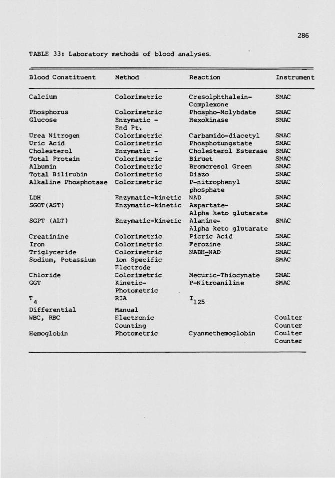
TABLE 33: Laboratory methods of blood analyses.
Blood Constituent
Calcium
PhosphorusGlucose
Urea NitrogenUric AcidCholesterolTotal ProteinAlbuminTotal BilirubinAlkaline Phosphotase
LDHSCOT (AST)
SGPT (ALT)
CreatinineIronTriglycerideSodium, Potassium
ChlorideGGT
T4DifferentialWBC, RBC
Hemoglobin
Method
Colorimetric
Color imetricEnzymatic -End Pt.ColorimetricColorimetricEnzymatic -ColorimetricColorimetricColorimetricColorimetric
Enzymatic -kineticEnzymatic-kinetic
Enzymatic-kinetic
ColorimetricColorimetricC olor imetr icIon SpecificElectrodeColorimetricKinetic-PhotometricRIA
ManualElectronicCountingPhotometric
Reaction
Cresolphthalein-ComplexonePhospho-MolybdateHexokinase
Carbamido-diacetylPhosphotungstateCholesterol EsteraseBirxietBromcresol GreenDiazoP-nitrophenylphosphateNADAspartate-Alpha keto glutarateAlan in e-Alpha keto glutaratePicric AcidFerozineNADH^NAD
Mecuric-ThiocynateP-Nitroaniline
?ia
Cyanmethemoglobin
Instrument
SMAC
SMACSMAC
SMACSMACSMACSMACSMACSMACSMAC
SMACSMAC
SMAC
SMACSMACSMACSMAC
SMACSMAC
CoulterCounterCoulterCounter
satisfactorily analyzed similar specimens from other free-ranging
ungulates (Matula & Lindzey 1976). Serum samples were also shipped to
the State Department of Agriculture/ Honolulu/ to assay for Leptospira
and Brucella antibody titers.
Statistical analysis of the data included calculating the means,
standard deviations and 95% confidence levels for each variable. Tlie
t-test compared age and sex related differences, and also compared
selected mean values in the study population with those in other
populations. Statistical analyses assumed that blood values were
normally distributed. The Schalm at al. (1975) method of determining
the absolute number of each leukocyte cell type from its relative
distribution in the peripheral blood smear, given the total leukocyte
count, was used to evaluate abnormal leukocyte distribution.
Results
(a) Blood chemistries
Baseline data for 30 biochemical parameters are statistically
described in Tables 34 and 35. Several parameters showed a wide range.
These were most pronounced for glucose, cholesterol, blood urea nitrogen
(BUN)rcreatinine ratio, iron, triglycerides and total lipids. Many
serum enzyme levels such as those for AKP, SCOT, SGPT and GGPT, were
also highly variable. Among the cations, calcium was the least
variable.
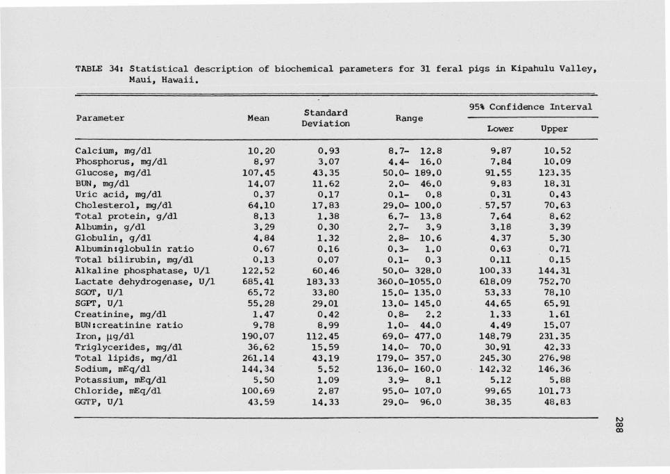
TABLE 34: Statistical description of biochemical parameters for 31 feral pigs in Kipahulu Valley,Maui, Hawaii.
Parameter
Calcium, mg/dlPhosphorus, mg/dlGlucose, mg/dlBUN, mg/dlUric acid, mg/dlCholesterol, mg/dlTotal protein, g/dlAlbumin, g/dlGlobulin, g/dlAlbumins globulin ratioTotal bilirubin, mg/dlAlkaline phosphatase, U/lLactate dehydrogenase, U/lSCOT, U/lSGPT, U/lCreatinine, mg/dlBUN;creatinine ratioIron, (ig/dlTriglycerides, mg/dlTotal lipids, mg/dlSodium, mEq/dlPotassium, mEq/dlChloride, mEq/dlGGTP, U/l
Mean
10.208.97
107.4514.070.3764.108.133.294.840.670.13
122.52685.4165.7255.281.479.78
190.0736.62261.14144,345.50
100.6943.59
StandardDeviation
0.933.0743.3511.620.1717.831.380.301.320.160.0760.46183.3333.8029.010.428.99
112.4515.5943.195.521.092.87
14.33
Range
8.7- 12.84.4- 16.050.0- 189.02.0- 46.00.1- 0.829.0- 100.06.7- 13.82.7- 3.92.8- 10.60.3- 1.00.1- 0.350.0- 328.0360.0-1055.015.0- 135.013.0- 145.00.8- 2.21.0- 44.069.0- 477.0i4.o- 7o.o179.0- 357.0136.0- 160.03.9- 8.195.0- 107.029.0- 96.0
95% Confidence Interval
Lower
9.877.8491.559.830.31
. 57.577.643.184.370.630.11
100.33618.0953.3344.651.334.49
148.7930.91245.30142.325.1299.6538.35
Upper
10.5210.09123.3518.310.4370.638.623.395.300.710.15
144.31752.7078.io65.911.6115.07231.3542.33276.98146.365.88
101.7348.83

TABLE 35: Statistical description of hematological parameters for feral pigs in Kipahulu Valley,Maui, Hawaii.
Parameter
Erythrocytic seriesErythrocytes, 10 /mm3
Hemaglobin, g/dlHematocrit, %MCV, ̂MCH, ̂ gMCHC, %
Leukocytic seriesLeukocytes, 10*Vmm3
Neutrophils, %Segmentals, %Bands, %
Lymphoc yt e s , %Monocytes, %Eosinophils, %Basophils, %
N
252525252525
2626262626262626
Mean
7.0614.7350.4972.0420.9329.03
26.7756.2153.502.7131.503.088.330.92
Range
5.42- 8.4810.14-16.9035.60-62.1064.00-89.0018.80-25.5025.20-32.90
13.40-39.2012.00-90.0012.00-80.000.00-12.009.00-69.000.00- 8.000.00-22.000000- 3.00
StandardDeviation
0.941.475.796.961.681.53
7.1420.1317.884.3316.351.896.861.11
95% Confidence Interval
Lower
6.6814.1348.1769.1720.2428.73
23.8848.0346.270.9524.882.315.570.48
Upper
7.4315.3252.8674.9021.6129.64
29.6564.3860.734.4638.113.8411.091.35

Thyroid hormones T and T as measured by radioimmunoassay (RIA),
averaged 34.35% and 3.36ud/dl (Tables 36, 37). T3 uptake was more
variable than T4-RIA; the mean T_ index was 1.05.
(b) Cellular hematology
Hematologic parameters showed high variability as indicated by the
wide ranges and relatively large standard deviations. In the
erythrocytic series, hematocrit and mean corpuscular volume (MCV) were
most variable. It should be noted that an index of anemia, the mean
corpuscular hemoglobin concentration (MCHC), was only 29.03 whereas a
value of 30 or greater in all mammals is considered normal. Erythrocyte
counts, which averaged 7.6 x 10⁶/mm3 weref howeverf least variable. In
the leukocytic series, total leukocyte counts averaged 26.77 x 10 /mm ;
the highest count was 39.20 x 10³/mm and the peripheral blood smear
differential data indicated that these feral pigs had neutrophilia.
Neutrophils dominated over other cell types in the leukocytic series;
the mean neutrophil:lymphocyte ratio was 1.78:1. Lymphocytosis (>13fOOO
cells/dl) was, however, observed in 9.6% (3) of blood-sampled animals.
The most extreme lymphocyte:neutrophil ratio was 5.75:1. The
neutrophils were principally mature segmented cells which averaged
56.20%; the highest value was 80%. Band neutrophils averaged 2.71% of
total leukocyte counts; the highest value was 12%. Basophils were least
represented, and averaged only 0.92%. Eosinophil counts ranged from
0-22%f with a mean of 8.33%. Eosinophilia (>72/000 cells/dl) was

TABLE 36: Serum T , T values by radioiinmunoassay and free thyroxine,
T index for 27 feral pigs in Kipahulu Valley, Maui, Hawaii.
StandardVariable Mean _ , .. .. RangeDeviation
T4-RIA, fig/dl 3.36 1.37 1.1-7.5
T UPTK, % 34.35 4.83 27.7-44.7
T? Index 1.05 0.40 0,4- 2.1
95% Confidence Interval
Lower Upper
2.85 3.86
32.43 36.26
0.89 1.20
TABLE 37: Normal Serum T , T and T values in domestic pigs.
Variable
T4-RIA, [ig/dl
T UPTK, %
T Index
N
10
52
52
52
Mean /Range
3(142
3732
10
.32
.70
.70
.10
.10
.60
.74
.69
±_
±±
±±
±±
0400
12
00
.80
.68)
.45*
.42t
.18*,20t
.18*
.18t
Reference
Reap et ,al (1978)
KallfelzKallfelz
KallflezKallfelz
KallfelzKallfelz
andand
andand
andand
EraliErali
EraliErali
EraliErali
(1973)*(1973)*
(1973)*(1973)*
(1973)*(1973)*
* young adult, N = 5t mature adult, N = 2t T by resin sponge uptake of labelled 3,5,3* - Triiodothyronine; Tby competitive protein binding.

observed in 33.30% of the animals. A high eosinophil count was
occasionally associated with a lower serum iron concentration.
(c) Age and sex variation
No hematological or biochemical parameters showed any significant
sex or age-specific variation. Several biochemical parameters show
significant difference between sexes (Table 38) than they did between
subadult and adult age classes (Table 39).
(d) Abnormal erythrocyte morphology
Abnormal morphological variations in erythrocyte shape, size and
color were identified from peripheral cells in blood smears.
Poikilocytosis, the deviation of erythrocytes from their normal shape/
was seen in two animals and took the form of burrs. Hypochromasia and
polychromasia were also observed, with the latter condition occuring
more frequently. Anisocytosis occurred in 45% (14) of the animals where
the aberrant cell types were microcytes. Marked microcytosis was
observed in two animals. Serum iron concentration in these markedly
microcytic animals was at the lowest level recorded (77.00mcg/dl) as
opposed to the sample mean of 190.07mcg/dl. Markedly microcytic animals
were also hypochromic. Nucleated red blood cells (NRBC) present at
INRBC/100WBC and 2NRBC/100WBC were observed in the peripheral blood
smears of two animals. Except for polychromasia, none of these
abnormalities have been reported from free-ranging pigs studied
elsewhere.

TABLE 38: Serological parameters of feral pigs that show differences between sexes.
"D -*-*>- ~1Tr\^4~ S^1S-C-f GiL cu\\Q. UGi o
Uric acid, mg/dlTotal proteins, g/dlAlbumin, g/dlGlobulin, g/dlAlbumin : Globulin ratioTotal bilirubin, mg/dlCreatinine, mg/dlPotassium, mEq/dlT -RIA, mcg/dl
Mean ±
Males (N=14)
0.32 ± 0.148.47 ± 1.763.30 ± 0.345.17 ± 1.720.64 ± 0.150.14 ± 0.081.53 ± 0.335.75 ± 1.273.04 ± 0.91
S.D.
Females (N=17)
0.40 ± 0.18 26.7.85 ± 0.85 2.3.26 ± 0.26 3.4.58 ± 0.78 2.0.69 ± 0.14 18.0.12 ± 0.05 40.I.421± 0.47 4.5.30 ± 0.86 3.3.61 ± 1.60 2.
t
67, P< 0.001167, P< 0.0548, P< 0.0173, P<0.0552, P< 0.00100, P<0.00196, P< 0.00106, P< 0.0145, P<0.05
* Subadult and adult age classedivisiona are after Williamson and Pelton (1976).
Total proteins, g/dlGlobulin, g/dlAlbumin ; Globulin ratioTriglycerides , mg/dlTotal lipids, mg/dl
Mean ± S.D., , . , _ . _.„.. +-
Subadults*(N=13)
7.26 + 0.595.47 ± 112400.76 ± 0.14
42.25 ± 15.74281.37 ± 38.29
Adults (N=18)
8.83 ± 1.424.06 ± 0.610.58 ± 0.10
19.69 ± 12.23236.23 ± 35.08
3.75, P< 0.0013.39, P<0.014.18, P <0.0012.39, P<0.053.35, P<0.01
TABLE 39s Serological parameters of feral pigs that show significant differences between age classes.

Discussion
Stress and its effects on blood biochemical henatologic parameters
are problems inherent in all studies using free ranging animals.
Animals generally exhibit a normal physiological stress response when
captured, handled and bled. Hence, the interpretation of blood
chemistries and hematology can be spurious if these variations are not
distinguished from those produced by pathological factors. Results in
this study are interpreted by discussing major sources of variations and
the relative importance of each in this study.
(a) Sources of variation
Variations in blood parameters are generally produced by the
following factors: 1) stress associated with capture, immobilization or
handling (Franzmann & Thome 1970; Williamson & Pel ton 1975, 1976;
Matula & Lindzey 1976; Barrett & Chalmers 1977); 2) age, sex and
reproductive condition (Coles 1980); 3) blood collection techniques,
storage and analytical methods (Wintrobe 1967; Kaneko 1980); 4)
nutritional status of animals (Doxey 1971; Singer & Ackerman 1981); 5)
breed-types (Payne et al. 1974; Pond & Houpt 1978); 6) environment
(Coles 1980); and 7) disease (Dunne & Leman 1975). Two other sources of
variation identified in this study were hemolysis and bacterial
contamination in several serum samples.
Stress factors probably accounted for some of the variability in
serum biochemical (Tables 34, 36) and hematological (Table 35) values.
During restraintf feral pigs were very active, visibly excited and made

repeated attempts to escape by charging their snouts into the sides of
the trap to the point of self injury. In fact, bleeding of the nasal
plate was sometimes observed. Increased excitability and forced
activity are known to elevate serum enzymes, glucose and cholesterol in
wild ungulates (Barrett & Chalmers 1977). Handling stress, therefore,
may probably have elevated SGOTf SGPT, GGTP, LDH, AKP cholesterol and
glucose levels. LDH measured in this study as high as 1055U/1;
transaminases increased to 145U/1 (Table 34). Glucose averaged 107.45
plus or minus 43.35mg/dl; the highest concentration was 189mg/dl.
Nutritional status and dietary composition of the animal's last
meal could also affect several blood biochemical values. When acorns
were abundant, the serum levels of total proteinsf albumin, cholesterol
and the A:G ratio in the wild boar in Great Smoky Mountains National
Park (GSMNP) was significantly higher than during acorn failure (Singer
& Ackerman 1981). The levels for glucose and total lipids would be
affected by diets, too. Animals in this study, trapped with baits rich
in carbohydrates, were bled the morning after this meal and, hence, gave
normal or high glucose levels in general. Depressed glucose level
(50mg/dl) in some animals was probably due to factors other than the
nutritional status of the animals. Bacterial growth was reported in
some serum samples with low glucose levels. Although the presence of
erythrocytes could also drastically lower glucose levels, depressed
levels were most likely produced principally by the bacterial growth
rather than by the length of time serum and clots remained in the
samples (Green 1980 Ventura, California - pers. comm.).

Pain during venipuncture could alter certain blood parameters (Pond
& Houpt 1978). Other workers have bled pigs from the tarsal veins
(Mclntosh & Pointon 1981), jugular, cephalic, auricular, orbital, and
coccygeal veins or even by tail amputation (Pond & Houpt 1978).
Sampling blood from the anterior vena cava did not appear to produce
discomfort to the animal as much as restraint itself did. Heart
puncture was initially used in this study to collect blood from the
first five animals, but none of these blood samples could be used
because of severe hemolysis. Blood drawn from the anterior vena cava is
less prone to hemolysis (Carle & Dewhirst 1942) than from the ear vein
or other bleeding sites (Hoerlein et al. 1951). However/ slight
hemolysis occurred, nevertheless, in four samples. In addition, serum
was routinely separated from the blood clot only after 4-8 hours.
Additionally, the erythrocytes of pigs are very fragile and prone to
hemolysis (Hoerlein et al. 1951). Agitation from backpacking,
transportation and heat probably brought about the hemolysis. Serum
potassium, inorganic phosphorus and LDH were slightly elevated in these
hemolysed samples.
Age and sex related differences in serum biochemical and
hematological parameters have been evaluated in domestic pigs. Total
proteins, globulin, A:G ratio, calcium, AKP and inorganic phosphorus
concentrations were greater in the serum of male than female pigs/
(Tumbleson et al. 1969). In this study, no hematological value showed
age or sex differences. Ihis result is consistent with boar studies in
Tennessee (Williamson & Pelton 1976) and GSMNP (Singer & Ackerman 1981).

Total proteins and the A:G ratio were higher in adults than in
subadults. The boars in Tennessee showed age related differences for
total proteins, albuminf sodium and chloride concentrations (Williamson
& Pelton 1975). Age differences may be due to differences in the
nutritional state or in the activity of the hematopoietic systems
between subadult and adult pigs. In contrast to the boar in Tennessee,
feral pigs in this study showed several sex related differences in serum
biochemical values. These differences may be due to reproductive status
condition of the sows, lactation stress and diet.
(b) Comparison between domestic, feral and wild populations
Serum biochemical values compared favorably with those in other
populations (Table 40). Differences in some biochemical values were
probably due to dietary differences and stress factors. Phosphorus
levels fell within the normal range of values in domestic pigs but were
higher than in Kangaroo Island (K.I.) strain feral pigs. LDH and
transaminases were much higher than the normal range for domestic pigs
and K.I. feral pigs. These elevated values were probably produced by
the stress factors already discussed. Cation concentrations fell within
the normal range of domestic pigs, and compared well with those in other
populations. Cholesterol was much lower than in K.I. feral pigs; this
was probably due to the grower ration on which K.I. feral pigs were
maintained. The A:G ratio was higher than in domestic pigs but lower
than in all other normal populations. Variations in albumin and
globulin concentrations are normally produced by dietary protein
deficiency, nutritional imbalances and vitamin deficiencies (Pond &

TABLE 40:Comparison of selected biochemical parameters (means and/or range) for feral, wildand domestic pigs.
* Pen-reared.+ Converted to International Units from King-Armstrong Unit by multiplying conventional
unit by a factor of 7.10.t Feral history of 180 years on Kangaroo Island, Australia, but blood-sampled animals are
derived from a piggery.£ SI Unit converted into conventional unit using conversion factors in Kaneko (1980), p,787.
Kaneko (1980)Pond and Houpt (1978)Cornelius et ^1 (1959)CysewsJci .et .al (1968)Ostadius et ail (1959)Witzel et al (1967)Zimmerman et al (1965)
a Doxey (1960)' Kelly (1974)10 Baetz and Mengeling (1971)n Melby and Altman (1974)12 Medway (1969)u Tumbleson (1969)
Population
NAge (months)
Calcium (mg/dl)
Phosphorus (mg/dl)Glucose (mg/dl)BUN (mg/dl)Cholesterol (mg/dl)
Total SerumProtein (g/dl)
A:GTotal Bilirubin
(mg/dl)AKP (U/l)
LDH (U/l)SCOT (U/l)
SGPT (U/l)
Creatinine (mg/dl)Iron (jig/dl)Total Lipids (mg/dl)Sodium (mEq/1)
Potassium (mEq/1)Chloride (mEq/1)
Reference
Feralpigs
315-36
10.20
8.97107.4514.0764.10
8.10
0.670.13
122.52
685.4165.72
55.28
1.47190.07261.14144.34
5.50100 . 69
THISSTUDY
Europeanwild boar
331.5-26.
107.2910.52
7.14
0.90
142.00
6,7897.10
Williamson& Pelton1975. 1976
Europeanwild boar*
371.5-26
90.7914.06
8.99
0,41
141.33
8,67100.18
Williamson& Pelton1975. 1976
Feralpigst/*
193-24
11.60
7.12124.30
15.41104.4
1.270.15
109.00
456.00
1.22
146.0±3.3
5.4±0.9101.9+3.1
Mclntosh& Pointon1981
Domestic pigs
3-36
7.1-11.6; 9. 0-12. 59;(10.2-11.9)10
5.3-9.61; 4. 6-10. 29
85. 0-150. Ol; 118. 03
10.0-30.0lj 8. 0-24. 0*36, 0-54. tf; 57. 0-160. 02;
64. 0-104. 0**7.9-8.9'; 6.31 6.9U
0.37-0.511
0-0.6; 0.4-1. if; 0.2s;0-0. 49
118. 0-395, 01; 71. 0-110. Off;97.9 (61.7-14 2f
499. 01; (380.0-634.0)32, 0-84. Ol; 14.8±6, T3; 13.9C;14.0±7.55; 11. 0-13. 06
45.015 (31.0-58.0); 25.0±77;X2.9±3.83; 10.0±2.75
1.0-2.71; 1.0-2.73
91.0-199.0'; 121,0±33.0U
272. 0-447. tf135. 0-150. Ol; 110. 0-154. (f;146. 013
4.4-6.71; 3.5-5.5°; 4.713
94. 0-106. 01; 88. 0-115. rfj104, <f
See below

Houpt 1978). Infection could have elevated globulin and depressed
albumin levels, thus altering the A:G ratio (Payne 1976). In pigsf
chronic anteric disease, including chronic iteitis (Nielson 1966), may
produce excessive plasma albumin loss and hence yield an A:G ratio with
a value of less than one. It was unlikely that A:G variations were due
to protein deficiency (see below).
(c) Physiologic health: Total leukocyte count and differential
cell data
Leukocyte count in healthy domestic pigs is very variable (Pond &
Houpt 1978). The total leukocyte count in the study animals was
considerably higher than the normal range of values for domestic pigs
(Table 41). Mean leukocyte count was significantly higher than in the
free-roaming [P(t > 9.68) < 0.001; df=62] and pen-reared boar [P(t >
3.68) < 0.001; df=66] in Tennessee, the boar in GSMNP during the
abundance of acorn [P(t > 8.11) < 0.001; df=72] and in K.I. strain feral
pigs [P(t > 9.07) < 0.001; df=48] in Australia. Additionally, this
study population was leukocytic (>22,OOQ cells/yl). Leukocytosis does
not characterize the K.I. feral animals or GSMNPf Polish and Tennessee
wild populations.
Sometimes the health of animals may be assessed solely from total
leukocyte count, but differential cell types are often more revealing.
Since any interpretation or conclusion on population health from
differential cell data hinges on the accuracy of total leukocyte count,
it is necessary to first consider the nonpathologic factors in this

TABLE 41: Comparison of leukocytic variables (mean and/or range) for feral, wild and domestic pigs.
Population N ^months
Feral pigs 31 5-36(Hawaii)
European wild boar 33 1.5-26(Tennessee)
European wild boar* 37 1.5-26(Tennessee)
European wild boar 6 2.1(Poland)
European wild boart 11 2.4(Poland)
Feral pigs* 19 3-24(Australia)
Domestic pigs 5-36
WBC103/mm3
26.77
12.24
18.82
15,35
14.48
13.80
15-20'
18. OO8
(10-23)16.00
(11-22)
(10-20)6
15. 5010
(10-21)
Bands SEGS Neutrophils% % %
2.71 53.50 56.21
54.78
37.38
74.00
60.40
0-2; 37.00J 58. OO1
(28-47)l.O9 40-60' 39. OO6
(0-4) (30.0-50.0)32. OO8
37.00*(28.0-47.0)
39. OO10
(30.3-47.7)
* Pen-rearedt Roan individualst Feral history of 180 years on Kangaroo Island, Australia,
but blood- sampled animals are derived from a piggery.
Lymphocytes Monocytes Eosinophils Basophils
31.29 3.22 8.33 2.00 THIS STUDY
40.81 2.95 1.52 0.07 A
55.70 3.22 3.62 0.05 A
25.20 0.80 0 B
38.60 0.70 0.30 B
C
36.801 1.901 2. 30' 0.301 See below
53. OO3 5.003 3.00J 1.206
(29.0-62.0) (2.0- 5.0) (1.0-11.0) (0.0-4.0)52. OO6 3.30b 4.506 0;
(40.0-60.0) (1.0-10.0) (1.0-10.0)53. OO9 2-8' 1-5' 0.508
(39.0-62.0)30-50' 5.009 3.80 0.50*
(2.0-10.0) (0.0-2.0)59.00a 5.00* 3.509
(0.5-11.0)52.10'° 3.3010 4.5010 1.2010
(43.1-62.9) (2.4- 4.1) (2.1- 6.9) (0.6-1.8)
Key to ReferenceiA Williamson and Pelton (1975, 1976)B Kostelecka- Myrcha (1974)C Mclntosh and Pointon (1981)

TABLE 41(contd.): Comparison of leukocytic variables (mean and/or range)for feral, wild and domestic pigs.
Key to notations;
Wirth (1950)Giltner (1907), N = 24, Age. * 4 monthsMedway (1961)Miller (1961), Age = 4 monthsPond and Houpt (1978)Scarborough (1931), for adult pigsKelly (1974)Doxey (1971), for conventional and miniature pigsSchalm (1965)Mitruka and Rawsley (1977), for male animals, mini pigs 70-100kg.

study and their importance in affecting leukocyte counts. In the
present evaluation, leukocytosis in the study animals was considered to
be a clinical reflection of the health of the pigs, rather than due
primarily to physiological stress causes. Comparison of leukocyte
counts with other populations is considered valid because stress factors
in this and other studies were similar. Feral pigs were trapped,
handled and bled with methods similar to those of Williamson and Pelton
(1975, 1976) in Tennessee. Sample sizes and ages of animals in their
boar studies were comparable to those of this study. Effects of
handling stress on leukocyte counts may be assumed to be a common
denominator to this and the Tennessee studies. However, the GSMNP boar
studies included animals bled, after being shot, by heart puncture
(Singer & Ackerman 1981). In that study the total leukocyte counts were
lower than those for the feral pigs in this present study. Pregnancy
may elevate leukocyte counts, but while this physiologic response is an
important source of variation in cows and dogs, it is less important in
pigs (Coles 1980). Leukocytes also increase in numbers an hour after
feeding. Stage of digestion was not considered a significant factor
influencing leukocyte counts in this study because pigs ate the
food-baits soon after capture, and more than an hour would have elapsed
between the last feed and blood sampling.
Pathologic factors that produce leukocytosis are disease, infection
and parasitism (Dunne & Leman 1975; Coles 1980)f and microbial milieu in
the animalfs home range. When these factors are reduced or absent,
leukocyte counts can be expected to be low. Disease-free feral pigs

derived from a stock experimentally treated with streptomycin,
penicillin and an antihelminthic drug, had low (13.80 x 10 /mm ) total
leukocyte count (Mclntosh & Pointon 1981). The microbial milieu in the
rain forest habitat could have caused leukocytosis. Restricted home
ranges of feral pigs have two consequences: 1) the frequency of animal
encounters, and 2) the intensity of home range use per unit area of home
range size, are greater in smaller home ranges. These factors
presumably facilitate the localization of pathogens and their
transmission among animals. Leukocytosis in the study animals may, in
part, be a reflection of the population response to these phenomena.
Supportive evidence may be drawn from two other studies. Leukocyte
counts tend to increase when large groups of pigs are housed together;
but pigs kept in minimal disease herds have lower total leukocyte counts
than pigs kept under ordinary commercial conditions (McTaggart &
Rowntree 1969). Wild boar that were pen-reared had higher total
leukocyte counts than free-roaming animals (Table 41). Williamson and
Pelton (1976) attributed the higher counts in pen-reared boars to their
confinement in a small area, which increases the tendency of pathogens
becoming concentrated in one area and disseminating to all confined
animals.
Differential cell counts support this tentative conclusion that
leukocytosis in the study animals was probably pathological rather than
physiological. Leukocytosis was observed to be in consequence to
increases in granulocytes (Table 41). This blood profile is in contrast
with that in domestic pigs where leukocytes are represented by more

agranulocytes (lymphocytes) rather than granulocytes. Neutrophil count
56.21% (12-90) was in excess of 10,000 cells/yl; the study animals were
therefore neutrophilic. Neutrophilia is caused by bacterial, fungal or
viral infections and intoxications. Fungal infection appears to be
unimportant because monocytosis was not evident in the blood profile.
Neutrophilia did not characterize the Tennessee populations (contra
Williamson & Pelton 1976) or Kangaroo Island feral pigs, but occurred in
the Polish boar; the absolute neutrophil cell density in the Polish boar
wasf however, less than in the feral pigs in this study. The study
population was, therefore, in clinically poorer health than animals in
other populations. Neutrophils were predominantly segmented cells;
these differential data are indicative of a disease factor. Pathologic
leukocytosis is generally associated with an increase in segmented
neutrophils (Coles 1980). Band neutrophils are very rare in domestic
pigs and extremely rare in the wild boar (Williamson & Pelton 1976), but
were high in the study animals. Band neutrophils are released into the
blood circulation from the bone marrow only when animals respond to some
disease factor (Coles 1980).
Although the study animals were neutrophilic, lymphocytosis was
observed in only three animals; increase in lymphocytes normally occurs
during the recovery stages of certain infections (Coles 1980).
Eosinophils exceeded 2000 cells/ul; the study animals were therefore
eosinophilic. Absolute eosinophil cell counts were higher than in
domestic pigs and the Tennessee populations. Pigs with high eosinophil
counts were occasionally associated with low serum iron, aid in four

autopsied animals, heavy infestations of Stephanurus dentatus were
found. Among the three stress factors (nutrition, parasitism and
handling) that commonly elevate neutrophiliaf parasitic infections
invariably produce eosinophilia (Doxey 1971; Coles 1980) as do allergic
or anaphylactic reactions. Eosinophilia in the study animals was most
likely due to tissue invasive parasites, the important nematodes being
Stephanurus dentatus, Metastrongylus elor*gatus and Ascaris lumbcicoides.
Abnormal erythrocytic forms seen in the stained blood smears may
have resulted from either increased erythrogenesis or irregularities in
erythrocyte maturation. Polychromatic erythrocytes appear to be common
in domestic pigs (Wisecup & Crouch 1962) and the wild boar (Williamson &
Pelton 1976). Microcytes are indicative of iron deficiency (Doxey
1971). Markedly microcytic erythrocytes which were also hypochromatic
are manifestations of microcytic anemias. This, however, does riot
appear to be the case. Although serum iron concentrations in these
animals were very low (77mcg/dl), hemoglobin, MCVf MCH and MCHC
concentrations were not depressed below the normal range of values in
domestic pigs (Table 42). Dietary iron is probably abundant in the
forest soils although it often is in a form not utilized by plants. T3
thyroxine and T -RIA levels were normal (Table 36, 37) suggesting no
iron deficiencies in the animals. However, depressed levels of iron may
have been produced by the heavy lice infestation and especially by the
heavy intestinal parasite loads observed at necropsy. Other factors
known to affect iron metabolism and produce microcytic anemias are
copper deficiency, molybdenum and bracken fern poisoning (Coles 1980).

TABLE 42: Comparison of erythrocytic variables (mean and/or range) for feral, wild and domestic pigs.
Population N ^ ,months
Feral pigs 31 5-36(Hawaii)
European wild boar 33 1,5-26(Tennessee)
European wild boar* 37 1.5-26(Tennessee)
European wild boar 6 2.1(Poland)
European wild boart 4 2.4(Poland)
Feral pigs* 19 3-24(Australia)
Domestic pigs 5-36
Platelets FU3C103/mm3 106/mm3
360.33 7.06
323.17 7.26
418.03 7.72
6.76
6.33
8.80
200-500? 5.0-8.01
400. OO8 6.807
(250-700) (5.0-8.5)300.00'° 7.20"
(232-368) (6.0-9.0)7.09'°
(5.5-8.7)
HGB
g/dl
14.73
14.82
15.22
16.00
17.50
16.30
13. OO2
14.00*(11.0-17.0)
13.00(10.0-16.0)
10.11'( 8.3-12.7)
13.00'°(12.5-13.5)
HCt MCV% H
50.49 72.04
39.02 54.48
42.16 55.03
45.90 69.06
48.00 77.06
47.00 53.60
42. OO2 60. 054
(32.0-50.0)42. OO8 63. OO9
(37.0-50.0) (50-68)43.20 60. OO10
(42.4-44.2) (58-62)39. 607
(32.2-46.3)
MCH MCHC
MM9 %
20.93 29.03
20.81 27.20
19.92 27.65
24.00 35.04
28.15 36.77
34.60
19. 904 27-405
20. OO9 32. OO9
(17-23) (30-34)18. 30'° 30. 1010
(18-19) (26-34)
References
THIS STUDY
Williamson andPelton (1975, 1976)
Williamson andPelton (1975, 1976)
Kostelecka-Myrcha(1974)
Kostelecka-Myrcha(1974)
Mclntosh andPointon (1981)
See below
* Pen-rearedt Roan individualst Feral history of 180 years on Kangaroo Island, Australia, but blood-sampled animals are derived from a piggery.

Nucleated erythrocyte (NRBC) are usually found only in bone marrow in
healthy adult animals and appear in the peripheral blood only in
diseased animals in response to anemias in remission, tumorsf or toxic
compounds from bacteria or ingested food (Schalm et al. 1975).
(d) Protein status
Several blood chemical values have been used as condition
indicators and for evaluation of protein status in wild ungulates. BUN
increases with increased intake of dietary protein (Coles 1980). It is
less influenced by handling stress (Seal et al. 1972), Total proteins
are directly influenced by nutritional levels (Dimopoullos 1963)f
although this chemical value may be relatively insensitive as a
condition correlate in sane ungulates (Seal et al. 1975). When examined
together, albumin, hemoglobin and BUN become a more valid diagnostic
test for protein status; depressed levels of all three parameters are
usually indicative of a protein deficiency (Payne 1981). Levels of
these three proteins in the Kipahulu Valley population compare favorably
with the normal range of values (Kaneko 1980:792-797) in domestic pigs.
BUN was higher than in the boar in GSMNP (BUN = 11 +, 3mg/dl) (Singer &
Ackerman 1981) and the Tennessee populations, but was lower than the
K.I. strain feral pigs (Table 40); the higher BUN in K.I. pigs was due
to the relatively rich dietary protein in growers ration on which the
experimental K.I. pigs were maintained (Mclntosh & Pointon 1981). BUN
in this study was, however, very variable. Catabolic breakdown of
tissues could increase BUN (Medway et al. 1969). Very low BUN was
recorded from lactating sows. Lactation may be viewed as a drain on the

sow's proteins, and hence the lower BUN. Total serum protein was higher
than in the boar in GSMNP during abundant acorn (7.0 HH 0.3g/dl) (Singer
& Ackerman 1981) and free-roaming boar in Tennessee; but lower than
pen-reared boar fed with corn (Table 40). These comparisons indicate an
adequate nitrogen intake and a normal total protein status of the feral
pigs in the rain forest habitats,
(e) Kidneyf liver and thyroid functions
BUN and cceatinirie may increase to very high levels following
kidney damage (Ntedway et al. 1969). None of these indices, however,
were abnormal in the feral pigs in this study, even though abscesses
were noted in some kidneys at necropsy from parasitism by j5. dentatus.
Liver function is normally assessed from specific serum enzyme levels
and other specific biochemical tests. Bilirubin levels will increase in
obstructive liver diseases including parasitic obstruction of bile ducts
or starvation (Cornelius 1980). Bilirubin levels were normal in the
pigs in this study; values obtained compared favorably with normal
values in other domestic pigs. Serum enzymes were, however/
considerably elevated. Elevation of SCOT in pigs have been reported for
bile duct obstruction, aflatoxicosis (Cysewski et al. 1968), hepatic
necrosis and diseases of the cardiac and skeletal system (Cornelius et
al. 1959). Ostadius et al. (1959) found that SOOT rose to 87 U/l and
SGPT to 45 U/l following liver dystrophy. Although serum enzymes were
considerably elevated, a conclusive statement on dysfunction of any one
organ seems unwise. SOOT does not occur in especially high
concentrations in pig liver and is found in other major tissues. Hencef

elevated SGOT need not necessarily imply liver disease. Despite their
non-tissue specificity, SGOT, SGPT, AKP and LDH have been found to be
useful in diagnosing liver dysfunction in dogs and cats, but the use of
these enzymes in assessing liver function in pigs has to be done with
caution (Cornelius 1980). While these elevated enzyme levels in the
study animals may also be indicative of some nematode-induced liver
dysfunction, other extrinsic factors already discussed are also equally
likely contributors to these elevated values.
T3 thyroxine and free thyroxine T4-RIA are two principal thyroid
hormone assays for assessing thyroid functions (Reap et al. 1978; Kaneko
1980). Both thyroid hormone levels compare favorably with the normal
range of values found in domestic pigsf suggesting an adequate
thyrometabolic status in the study animals. Caution should be used in
comparing thyroxine values (Tables 36, 37) because thyroid
determinations are method specific.
Blood profiles indicated that nitrogen intake and the protein
status of the feral pigs were adequate. Neutrophilic leukocytosis
characterized the feral pigs in this study population, but not in other
boar and feral populations. It is suggested that although condition
indicators (BUN, total proteins, albumin, hemaglobin) of feral pigs were
relatively good, the clinical health of the study population is poorer
than pigs in other free-ranging populations. Leukocytosis was probably
in response to some disease factor, tissue invasive parasites or
microbial milieu in home ranges.