changes in oxidative stress intensity in blood of tumor...
TRANSCRIPT

ISSN 2409-4943. Ukr. Biochem. J., 2016, Vol. 88, N 4
29
UDC 546.719:54.024:577.151:616-006.699
Changes in oxidative stress intensity in bloodof tumor-bearing rats following different modes
of administration of rhenium-platinum system
K. L. ShameLaShVILI1, N. I. ShtemeNKo2, І. V. Leus3,S. o. BaBIy4, o. V. ShtemeNKo5
1Se "Dnipropetrovsk medical academy" of health ministry of Ukraine;2Palladin Institute of Biochemistry, National academy of Sciences of Ukraine, Kyiv;
3University of oklahoma, oklahoma-city, USa;4Institute of Gastroenterology, National academy of medical Sciences
of Ukraine, Dnipropetrovsk;5Ukrainian State University of Chemical technology, Dnipropetrovsk;
e-mail: [email protected]
effects of the different modes of administration of dichlorotetra-μ-isobutyratodirhenium(ІІІ) – І – (in water solution, liposomes, nanoliposomes and together with cisplatin – in the rhenium-platinum system) on the intensity of lipid peroxidation (LP) in blood plasma and the activity of the erythrocyte antioxidant enzymes were investigated on the model of tumor growth. a decrease in the concentration of tBa-active substances caused by dirhenium compounds was shown to be independent of the administration mode and the extent of the tumor growth inhibition. I was four-times more effective in inhibition of the LP burst than any known antioxidant. I induced the increasing activity of erythrocyte superoxide dismutase and decreasing activity of catalase. In vitro experiments with native superoxide dismutase, the interaction of І with following activation of the active center of the enzyme was confirmed and the superoxide dismutase activity of І was shown, that may contribute to the enhancement of the enzyme activity in vivo. The cluster rhenium compounds may be promising nontoxic potent antioxidants capable of deactivating superoxide radicals.
K e y w o r d s: lipid peroxidation, oxidative stress, Guerin’s carcinoma, catalase, superoxide dismutase, cisplatin.
I n our previous work it was shown that clus-ter rhenium(III) compounds with organic li-gands exhibit pronounced anticancer and
antiradical activities, particularly in combination with platinum compounds (Re-Pt system) [1, 2]. The most studied among them is dichlorotetra-μ-isobutyratodirhenium(ІІІ) Re2(i-C3H7COO)4Cl2 – (I), which both individually and as a part of the Rhe-nium-Platinum system has anticancer, hepato- and nephroprotective activity. Compound i has several structural features (Fig. 1), which might be respon-sible for the above indicated properties: a) binuclear clustering of rhenium atoms; as the main component of the molecule is characterized by low toxicity; b) quadrupole bond, which is responsible for anti-radical and antioxidant properties; c) chlorine atoms , which are able to interact with polar molecules; g) branched alkyl groups and their symmetrical ar-rangement that provide hydrophobic interactions.
Fig. 1. Dichlorotetra-μ-isobutyratodirhenium(ІІІ) – (I)
One of the directions of further studies of the compound I and other cluster rhenium compounds is to find the optimal modes of administration of i alone or in the Re-Pt system, at which both an effec-tive inhibition of tumor growth and suppression of the intensity of the free radical burst that accompany tumor growth can be achieved.
doi: http://dx.doi.org/10.15407/ubj88.04.029

30
ISSN 2409-4943. Ukr. Biochem. J., 2016, Vol. 88, N 4
Experiments in vitro showed that the rhenium compounds with isobutyrate ligands, which are di-carboxylate structural type, in cis- and trans-config-urations interact with erythrocyte SOD and change protein conformation. It was found that cis-dicarbo-xylate dirhenium(III) exhibits superoxide dismutase activity that is not inherent in trans-dicarboxylat-dirhenium(III) [3]. Therefore, the study of the in-fluence of rhenium compound with isobutyrate li-gands of tetracarboxylate type on SOD molecule in experiments in vivo and in vitro is a consequential continuation of previous studies that may highlight the problem of direct interaction of i with the SOD molecule.
Thus, the aim of this work was to study the ef-fect of i at various modes of administration on basic parameters of oxidative stress in blood of rats with Guerin’s carcinoma and on the activity of native SOD in vitro.
materials and methods
In our research we studied cluster compound dichlorotetra-μ-isobutyratodirhenium(ІІІ) Re2(i-C3H7COO)4Cl2 – (i) (Fig. 1) synthesized in the Ukrainian State University of Chemical Technology [1]. Liposome [i]lip, nanoliposome [i]nl forms of i and solid nanoparticles loaded with i and the Re-Pt system with various ratios of the components inside lipid capsule [i+cPt (4:1)] lip, [i+cPt (4:1)] nl, [i+cPt (4: 1)] np, [i+cPt (4:2)] nl, [i+cPt (4:2)] were prepared according to [2].
Wistar rats weighing 100-150 g maintained in standard vivarium conditions were used in the experi ments. Rats without tumor were control group. All animal experiments were performed in accordan ce with the rules of the European Conven-tion for the Protection of Vertebrate Animals used for Experimental and Other Scientific Purposes (Strasbourg, 1986). Tumor growth was modeled by transplantation of 20% suspension of Guerin’s car-cinoma cells (T8) in solution to healthy rats (group T8) according to [2]. The compounds of rhenium and platinum were administered by various modes:
mode 1 – administration of the compound i alone. The compound i in solution (sln), in the form of normal liposome (lip) 5-10 µm, in the form of nanoliposoms (nl) and solid nanoparticles (np) 10-100 nm (group T8+[i] sln, T8+[i] lip, T8+[i] nl, T8+[i] np, respectively) were administered intraperi-toneally ten times at a dose of 7 µmol/kg for 21 days with an interval of one day starting from the 3rd day
after cancer cells transplantation. Administration of i was also conducted per os ten times at the same dose in the form of solid nanoparticles 10-100 nm starting from the 3rd day after cancer cells transplan-tation (group T8+[i] np (per os));
mode 2 – administration of the Re-Pt system components, wherein cisplatin was administered once in aqueous solution, and rhenium compound was administered ten times in liposome form or in the form of nanoparticles with a final molar ratio of rhenium compound:platinum 4:1. A single intraperi-toneal injection of cisplatin solution (cPt) at a dose of 1.75 μmol/kg was performed on the 9th day after tu-mor transplantation, and intraperitoneal administra-tion of i was conducted at the same doses and modes (as described above for mode 1), groups: T8+cPt+[i] lip, T8+cPt+[i] nl, T8+cPt+[i] np, T8+cPt+[i] np (per os);
mode 3 – combined use of the Re-Pt system in the form of mixed nanoliposomes and nanopar-ticles, where both cytostatics were inside lipid na-nocapsules. Intraperitoneal administration of na-noliposomes and nanoparticles 100-150 nm loaded with i and cPt in molar ratios of 4:1 and 4:2 was con-ducted starting from the 3rd day after tumor trans-plantation in the amount of 7 μmol/kg of rhenium compound and the corresponding amount of cispla-tin; groups: T8+[i+cPt (4:1)] nl, T8+[i+cPt(4:1)] np, T8+[i+cPt(4:2)] nl, T8+[i+cPt(4:1)] np (per os).
Animals were decapitated under chloroform anesthesia on the 21st day. Tumor was removed and weighed. Content of the compounds that react with thiobarbituric acid (TBA-active compounds) in blood plasma, the activities of superoxide dismutase (SOD), catalase (CAT) and glutathione peroxidase (GP) in erythrocyte hemolysates were examined using methods [4-7]. The antioxidant effectiveness (E%) was calculated as a ratio of the concentration of TBA-active compounds in blood in the experiment with administration of antioxidant to the concentra-tion of TBA-active compounds upon the pathology in the experiments without antioxidant administra-tion.
In vitro studies were performed using Cu,Zn-SOD from bovine erythrocytes (Sigma-Aldrich, USA), 0.056 mg/ml. Fluorescence was measured in phosphate buffer pH 7.4 using spectrofluorime-ter Shimadzu RF-5301 PC (Japan), slit width EX: 10.0 nm and EM: 10.0 nm, excitation wavelength 290 nm [8]. The effect of the compound i in a molar ratio of 1(SOD):10(i) was studied immediately after mixing. SOD activity in the presence of i and SOD-

31
like activity of i in vitro were determined on the xanthine-xanthine oxidase system expenditure using 19160 SOD Determination Kit (Sigma-Aldrich Che-mie GmbH). Measurements were carried out every minute during a period of 5 min after the addition of xanthine oxidase solution at 25 °C using spectropho-tometer Shimadzu UV-1601 UV-visible at 440 nm. We registered the changes in absorbance (A·10-2) as the differences between the absorbance every minute and the absorbance of the mixture at the beginning of the experiment, i.e., absorbance values growth. All measurements were carried out in triplicate and presented as means with standard errors. The re-sults were statistically processed using the Student’s t-test. Differences in values were considered statisti-cally significant at P ≤ 0.05.
results and discussion
The chosen model of tumor growth – Guerins carcinoma (T8) – is characterized by intense growth
and reaches a weight of 50-65 g (which is 20-40% of rat body weight) by the 21st day after transplantation of cancer cells (Table 1).
In this group we observed a significant increase (more than 6-fold) in the concentration of TBA-active compounds, which indicates intensification of the lipid peroxidation (LP) process, inherent in “radical burst” in this model. Upon the action of cPt (group cPt) the tumor weight by the 21st day was 3.5-fold less than that in the group T8. The intensity of LP in the blood plasma of these animals was also reduced almost 2-fold due to inhibition of the tumor growth.
When compound i was administered intraperi-toneally and per os by mode 1, the tumor weight was reduced to 30-50%, i.e. much less than upon adminis tration of cPt. However, inhibition of the LP intensity was more pronounced than in the group upon administration of cPt (in some experimental groups was almost twice more).
t a b l e 1. tumor weight, tumor inhibition index and concentration of tBa-active compounds in the plasma of the experimental animals, n = 8-10
# P < 0.05 relative to the control group; * P < 0.05; ** P < 0.01; *** P < 0.001 relative to the group T8; ¥ P < 0.05 relative to the group T8 + [i] np
Experimental group Tumor weight, g Inhibition, % of Т8 TBA content, mol/lControl – – 7,69 ± 0,38T8 63.27 ± 3.16 – 52.88 ± 2.64#
cPt 17.72 ± 0.89** 71.99 ± 3.60 21.16 ± 1.06#,*Mode 1
Т8 + [І] sln 50.34 ± 3.08*,¥ 20.44 ± 1.02* 19.48 ± 0.99*,#
Т8 + [І] lip 46.33 ± 2.31*,¥ 26.77 ± 1.34* 17.54 ± 0.88*,#
Т8 + [І] nl 45.64 ± 2.15* 27.86 ± 1.39* 19.50 ± 0.90*,#
T8 + [І] np 34.68 ± 2.46* 45.18 ± 2.26* 12.44 ± 0.60**,#
T8+ І np (per os) 29.80 ± 1.49* 52.90 ± 2.65* 11.54 ± 0.58**,#
Mode 2Т8+ cPt +[І] lip 0.28 ± 0.01*** 99.56 ± 4.98*** 5.12 ± 0.26 **T8+cPt+[І] nl 1.98 ± 0.11*** 96.87 ± 4.84*** 11.90 ± 0.62 **,#
T8+cPt+[І] np 0.93 ± 0.05*** 98.53 ± 4.93*** 8.65 ± 0.43 **T8+cPt+[І] np (per os) 0.21 ± 0.01*** 99.67 ± 4.98*** 12.02 ± 0.64 **,#
Mode 3T8+[І+cPt(4:1)] nl 1.38 ± 0.07*** 97.82 ± 4.89*** 7.53 ± 0.38 **T8+[І+cPt(4:1)] np 2.39 ± 0.12*** 96.22 ± 4.81*** 12.72 ± 0.64 **,#
T8+[І+cPt(4:1)] np (per os) 27.31 ± 1.37** 56.84 ± 2.84** 31.09 ± 1.55 *,#
T8+[І+cPt(4:2)] nl 18.13 ± 0.90** 71.35 ± 3.57** 10.90 ± 0.55 **,#
T8+[І +cPt(4:2)] np 30.03 ± 1.51* 52.54 ± 2.63* 26.44 ± 1.22 *,#
K. L. shamelashvili, N. I. shtemenko, І. V. Leus et al.

32
ISSN 2409-4943. Ukr. Biochem. J., 2016, Vol. 88, N 4
It should be noted that various modes of adminis tration of equal amounts of i differ in their effectiveness in tumor growth inhibition: the adminis tration of liposomal forms or solution re-sulted in less tumor inhibition compared to adminis-tration of solid nanoparticles loaded with i. These findings point out an importance of further research using solid nanoparticles loaded with anticancer agents. It is known that this drug formulation is suitab le for storage and administration of medicines [9]. It should also be noted that the liposome size does not affect the parameters of efficiency of tu-mor growth inhibition and oxidative stress in these experiments, when compared groups T8+[i]l and T8+[i]nl. Thus, it was shown that the compound i administered alone to tumor-bearing rats in various forms exhibited moderate antitumor activity and ability to inhibit oxidative stress, regardless of the form of administration. So, the use of solid nano-particles in therapy is promising in a search for new classes of antitumor agents.
Administration of the components of Re-Pt system by mode 2 demonstrates efficient, almost complete inhibition of the tumor growth. The tumor growth in the most of experimental animals was sig-nificantly inhibited regardless of the modes of the system components administration. In our previous works, effectiveness of the Re-Pt system as anti-tumor agent was shown on many cluster rhenium compounds, and, in our opinion, it might be caused, by a synergistic or additive mechanism of interac-tion between the platinum, rhenium compounds and DNA. It should be noted that administration of the components of the Re-Pt system by mode 2 led not only to a significant inhibition of tumor growth but also to a significant reduction (normalization) in con-centrations of the LP products.
The obtained results for Re-Pt system effi-ciency became an impetus for development of new nanoforms, namely nanoliposomes and solid nano-particles, which would contain both cytostatics - cPt and i inside lipid nanocapsule. Such mixed and solid nanoparticles were synthesized and characterized [2]. Administration of the combined Re-Pt system in mixed nanoliposomes and nanoparticles to the ex-perimental animals by the mode 3 was also effective, in terms of both antitumor and antioxidant activities, except for experiments, where the amount of cispla-tin within the nanocapsules was increased and where the mixed nanoparticles were administered per os. In our view, the possibility to change the content of
cytostatics inside nanocapsules is a significant step toward nanotechnology and combined therapy in the cancer treatment.
It is known that the concentration of the TBA-active compounds in the blood plasma is an impor-tant biochemical marker that reflects the intensity of the whole body LP - a process that is a consequence of membrane lipid oxidation by reactive oxygen species, excessive formation of which occurs under many pathological conditions [10, 11]. Administra-tion of the antioxidants significantly reduces the oxi-dative stress intensity in the organisms [12].
If application of known antioxidants lead to a maximum 1.5-2-fold reduction in the blood LP in-tensity at various pathologies, the efficiency of the compound i in LP inhibition is much higher (4-fold) than the efficiency of known antioxidants. The mechanism of action of known antioxidants lies in direct interaction with radicals via conjugated π-bonds (e.g., vitamins A, C, E) to form stable par-ticles, which inhibit radical chain reaction. Quadru-pole bond of cluster direnium fragment easily binds the electrons of free radicals on low-energy δ-anti-bonding molecular orbital. Known organometallic compounds also exhibit antioxidant effects, however, these effects are realized via π-unsaturated ligands, of e.g. the polyphenol or flavonoid nature [14]. The rhenium compounds with quadrupole bond represent a new class of highly effective δ-antioxidants that, along with their non-toxicity, makes them promising drugs.
Red blood cells, or erythrocytes, are the most numerous and specialized cells in the organisms. The main function of red blood cells is to transport oxygen and carbon dioxide. During erythropoiesis, erythrocytes lose nucleus, ribosomes, mitochondria, and thus the ability to cell division, protein synthe-sis and oxidative processes inherent in mitochondria [15]. Due to their physiological role, the red blood cells are permanently affected by endogenous oxida-tive stress: the auto-oxidation of oxyhemoglobin to methemoglobin generates superoxide radical (O2
•-), which is the main source of free radicals in erythro-cyte. Oxidative stress, which occurs at pathologies, can damage erythrocytes as well as other biological molecules of the organism [10]. Normal erythrocytes have a reducing activity, which is 250-fold greater than their oxidation potential [13]. However, when the intensity of oxidative stress is increased, eryth-rocyte antioxidant system lacks defense, and the cell becomes the storage and carrier of extracellu-

33
lar superoxide anion-radical and hydrogen peroxide, which can be transported through the anion channel, or freely penetrate the membrane, respectively.
Erythrocyte antioxidant system mainly in-volves activities of: Cu,Zn-SOD, which maintains the concentration of superoxide anion at 10-13 mol/l; CAT, which decomposes hydrogen peroxide to water and oxygen (activities of these enzymes in erythro-cyte much higher than in the other body tissues ex-cept for liver); glutathione peroxidase (GP), which catalyzes the same reaction as CAT. Changes in the activity of these enzymes lead to an imbalance of redox status of cells and consequently of the whole organism. Thus, a study of enzymes of erythrocyte antioxidant system upon various pathologies gives an insight into the redox status of the whole organ-ism. The role of erythrocyte SOD and CAT as an-tioxidant enzymes maintaining redox status of the whole body is confirmed by the fact that administra-tion of these proteins as drugs leads to antioxidant protection of the whole body [11]. For example, ad-ministration of the liposomes loaded with SOD and CAT to rats with oxygen intoxication (100% oxygen) significantly increased their life span [16]. Discove-ry of the antioxidant, anti-inflammatory, anti-car-cinogenic effect of these proteins has triggered an intensive search for their low molecular biomimetics based on organometallic compounds [17]. Namely these organometallic compounds are the basis for the creation of such mimetics since SOD and CAT are metalloproteids.
Numerous studies on the activity of erythro-cyte antioxidant enzymes upon various pathologies, summarized in the review [10], have indicated that the high intensity of oxidative stress in the body leads to a decrease in the activity of erythrocyte SOD and CAT, as well as to the imbalance in ac-tion of antioxidant enzymes. It has been found that high concentrations of the free radical compounds inactivate SOD, CAT and GP. For example, in the SOD molecule under the influence of significant concentrations of hydroxyl radical, histidine 118 is oxidized to oxo-2 histidine in the active site that, in turn, leads to complete enzyme inactivation [18].
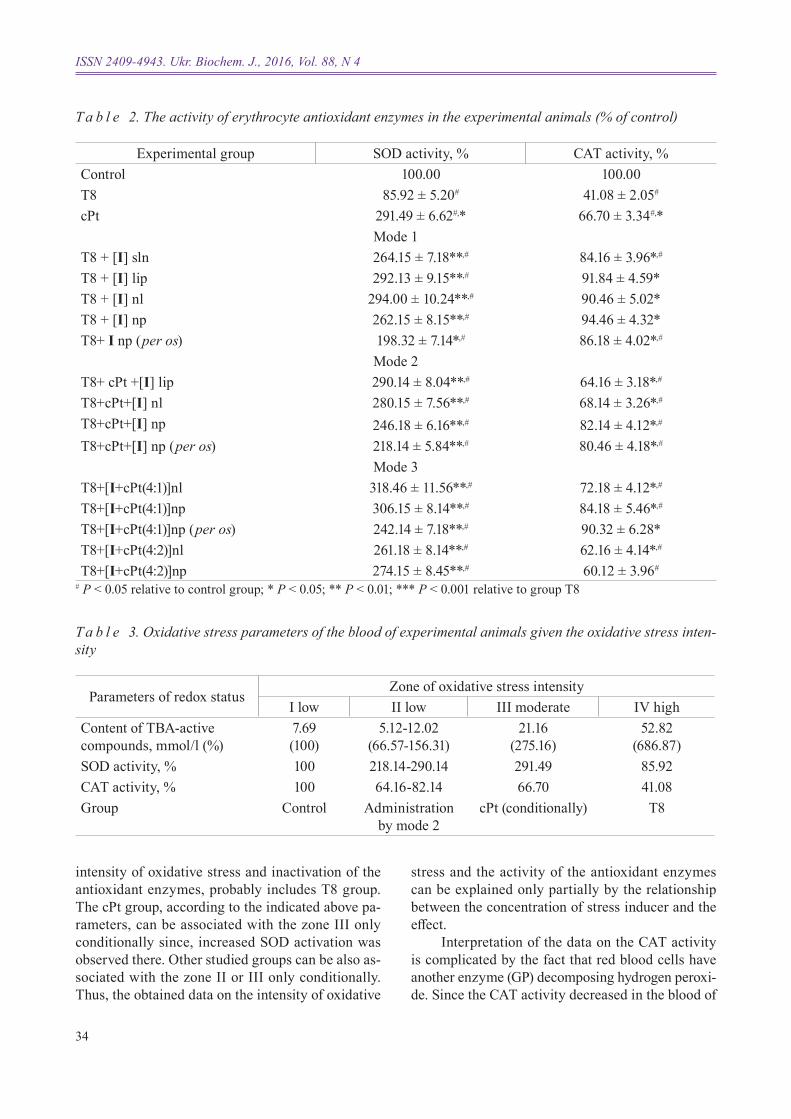
In our experiments, development of oxidative stress (group T8) resulted in a decrease in the eryth-rocytic SOD activity (by 14%) and CAT activity (by 59%) (Table 2) compared with the control.
Administration of cisplatin (group cPt) led to a significant increase in the SOD activity (about 3-fold) and a decrease in the CAT activity (by a fac-
tor of 1.5) compared to control, and an increase in the SOD and CAT activities compared to the group T8. Administration of i led to increasing of the SOD activity compared to the control group (2-3.1-fold), compared to the tumor-carrier group (2.1-3.3-fold), and the cisplatin group (by 15-27% for groups T8+[i+cPt(4:1)]nl, T8+[i+cPt(4:1)]np). Administra-tion of i increased the CAT activity 1.5-2.3-fold compared to the group T8 and max. 1.5-fold com-pared to the cisplatin group, although did not reach to the control values.
The data on the different effects of Re and Pt compounds on the SOD and CAT activities can be explained by their different effects on erythropoiesis. In particular, during carcinogenesis, inhibition of the Nrf-2 (eritroid derivative nuclear factor) that regu-lates the expression of antioxidant enzymes has been shown [11]; rhenium cluster compounds support the erythropoiesis process upon the neoplasm develop-ment [20]; cisplatin suppresses erythropoiesis; the red blood cells, short-lived but with high amount of SOD, are formed.
On the other hand, the organometallic com-pounds may directly interact with erythrocyte anti-oxidant enzymes. However, it is known that cispla-tin, at direct interaction, inactivates SOD and CAT enzymes by binding thiol residues of cysteine [20]. At the same time, direct activation of the rhenium compounds is possible [3].
Given the calculations based on our data and carried out according to [21], we can say that parame-ters of the control group correspond to zone i of oxi-dative stress intensity [22]. Parameters of the groups, in which the studied compounds were administered by mode 2, correspond to zone II. Parame ters of the groups cPt and T8 correspond to zones III and IV, respectively (Table 3).
According to this classification, there are four zones in hormetic (from the Greek hormesis - rapid motion) oxidative stress curve, which differ in the stress intensity and response of enzyme systems to an increase in the concentration of inducers of free radical burst. The first zone, to which, perhaps, the control group refers, is characterized by basal oxida-tive stress.
The second zone is characterized by higher in-tensity of oxidative stress and activation, or mode-rate inactivation of the antioxidant enzymes. The groups, in which the Re-Pt system was adminis-tered by mode 2, may be associated with this zone. The fourth zone, which is characterized by a high
K. L. shamelashvili, N. I. shtemenko, І. V. Leus et al.
34
ISSN 2409-4943. Ukr. Biochem. J., 2016, Vol. 88, N 4
T a b l e 2. The activity of erythrocyte antioxidant enzymes in the experimental animals (% of control)
# P < 0.05 relative to control group; * P < 0.05; ** P < 0.01; *** P < 0.001 relative to group T8
Experimental group SOD activity, % CAT activity, %Control 100.00 100.00T8 85.92 ± 5.20# 41.08 ± 2.05#
cPt 291.49 ± 6.62#,* 66.70 ± 3.34#,*Mode 1
Т8 + [І] sln 264.15 ± 7.18**,# 84.16 ± 3.96*,#
Т8 + [І] lip 292.13 ± 9.15**,# 91.84 ± 4.59*Т8 + [І] nl 294.00 ± 10.24**,# 90.46 ± 5.02*T8 + [І] np 262.15 ± 8.15**,# 94.46 ± 4.32*T8+ І np (per os) 198.32 ± 7.14*,# 86.18 ± 4.02*,#
Mode 2Т8+ cPt +[І] lip 290.14 ± 8.04**,# 64.16 ± 3.18*,#
T8+cPt+[І] nl 280.15 ± 7.56**,# 68.14 ± 3.26*,#
T8+cPt+[І] np 246.18 ± 6.16**,# 82.14 ± 4.12*,#
T8+cPt+[І] np (per os) 218.14 ± 5.84**,# 80.46 ± 4.18*,#
Mode 3T8+[І+cPt(4:1)]nl 318.46 ± 11.56**,# 72.18 ± 4.12*,#
T8+[І+cPt(4:1)]np 306.15 ± 8.14**,# 84.18 ± 5.46*,#
T8+[І+cPt(4:1)]np (per os) 242.14 ± 7.18**,# 90.32 ± 6.28*T8+[І+cPt(4:2)]nl 261.18 ± 8.14**,# 62.16 ± 4.14*,#
T8+[І+cPt(4:2)]np 274.15 ± 8.45**,# 60.12 ± 3.96#
t a b l e 3. oxidative stress parameters of the blood of experimental animals given the oxidative stress inten-sity
Parameters of redox statusZone of oxidative stress intensity
І low ІІ low ІІІ moderate ІV highContent of TBA-active compounds, mmol/l (%)
7.69(100)
5.12-12.02 (66.57-156.31)
21.16(275.16)
52.82(686.87)
SOD activity, % 100 218.14-290.14 291.49 85.92CAT activity, % 100 64.16-82.14 66.70 41.08Group Control Administration
by mode 2сPt (conditionally) T8
intensity of oxidative stress and inactivation of the antioxi dant enzymes, probably includes T8 group. The cPt group, according to the indicated above pa-rameters, can be associated with the zone III only conditiona lly since, increased SOD activation was observed there. Other studied groups can be also as-sociated with the zone II or III only conditionally. Thus, the obtained data on the intensity of oxidative
stress and the activity of the antioxidant enzymes can be explained only partially by the relationship between the concentration of stress inducer and the effect.
Interpretation of the data on the CAT activity is complicated by the fact that red blood cells have another enzyme (GP) decomposing hydrogen peroxi-de. Since the CAT activity decreased in the blood of

35
rats of all experimental groups compared with the control, additional analysis of the GP activity was carried out in some groups (Fig. 2).
During the growth of transplanted Guerin’s carcinoma, GP activity reduced by 50% compared to control (Fig. 2). Causes of a decrease in this enzyme activity is probably the same as for other antioxidant enzymes. Administration of cPt did not affect the en-zyme activity compared to the tumor-bearing rats, despite a significant antitumor effect of cPt. Since, GP is glutathione-dependent enzyme, it should be noted that cPt directly interacts with glutathione [23] and binds specifically with selen and thiol groups at the GP active center [13] that results in the enzyme inactivation. Probably, that is why the level of the GP activity did not increase, although a significant inhibition of tumor growth was observed.
Upon administration of the Re-Pt system (modes 2 and 3) an increase in the enzyme activi-ty by 22-77% compared with the group T8 was observed. Thus, normalizing of the redox-status of erythrocytes under the action of i can be explained by both SOD activation that led to reduction of the superoxide anion concentration and GP activation that led to reduction of the hydrogen peroxide con-centration. Activation of the erythrocyte antioxidant enzymes and decrease in the ROS concentration caused lowering of the LP intensity in the cell and the whole organism.
Activation of the erythrocyte SOD upon ad-ministration of certain rhenium compounds, we explained by their SOD-like activity, or by possible interaction with the SOD molecule and following
by changes in apoenzyme conformation and a cor-responding increase in the reaction rate in the active site of the enzyme [3]. The possibility to apply the same explanation for the compound i was the next stage of this study.
The addition of i to the SOD solution resul-ted in a change in tyrosine fluorescence intensity (Fig. 3).
We chose fluorescence of tyrosine for estab-lishing the possible interaction between i and SOD, since only this amino acid residue is a fluorophore, and the SOD molecule does not contain tryptophan [24]. During the reaction of SOD with i in the ratio of 1:10 we observed the main peak with λmax = 321 nm
Fig. 2. erythrocyte glutathione peroxidase activity in some groups of the experimental animals: 1 – control; 2 – T8; 3 – cPt; 4 – T8+cPt+[I]nl; 5 – T8+[I+cPt(4:1)]nl. # P < 0.05 relative to control, * P < 0.05 relative to group t8
Fig. 3. Fluorescence spectrum for sOD ( ) and SoD with I ( ) in a molar ratio of 1 : 10
300 350 400 450
300
250
0
200
150
100
50
F
λ, nm
GP
activ
ity, %
1 2 3 4 5
120
100
80
60
40
20
0
Experimental group
K. L. shamelashvili, N. I. shtemenko, І. V. Leus et al.

36
ISSN 2409-4943. Ukr. Biochem. J., 2016, Vol. 88, N 4
T a b l e 4. Changes in absorption (A·10-2) of the reaction mixture upon the addition of sOD, sOD with I in ratio of 1 : 10 in xanthine-xanthineoxidase system (M ± m; n = 3)
Time, min SOD SOD+ І І1 4.34 ± 0.25 1.42 ± 0.23 10.62 ± 2.222 10.42 ± 0.61 9.17 ± 0.49 9.43 ± 1.943 16.40 ± 0.93 17.83 ± 1.45 8.15 ± 2.134 22.21 ± 1.86 24.62 ± 1.83 14.66 ± 3.365 27.77 ± 2.48 30.36 ± 4.14 26.92 ± 5.12
(blue shift compared to the native protein) with the longwave shoulder λ = 360 nm. This indicates that compound i directly interact with the protein mole-cule. Similar changes in the SOD fluorescence were observed when direnium dicarboxylates were added [3]. It allows us to state that interaction with SOD oc-curs by means of the structural element which exists in all of these compounds namely, quadrupole bond Re-Re, and the shift of the fluorescence peak cor-responds to a change in polarity around the chromo-phore molecule (in this case, Tyr-108). Fluorescence quenching was 11.4%, which is much less than that for the cis-dicarboxylates.
We also studied the effect of the compound i on the activity of SOD as well as the SOD-like ac-tivity of I in the xanthine-xanthineoxidase system (Table 4).
It is known that in such experiments the inten-sity of the superoxide dismutase reaction is inversely proportional to the rate of the absorption change. The addition of SOD+i mixture into xanthine-xanthineoxidase system did not lead to significant changes in the enzyme activity compared to native SOD, except a decrease (by 67.28%) within 1st min of the experiment, indicating the short-term SOD activation in the presence of i. Previously, it was shown that rhenium cis-diisobutyrate had a similar effect [3]. So, we concluded that the binding of i to the protein occurs a significant distance from the ac-tive site, and the presence of i in long-last experi-ment did not significantly affect the SOD activity.
The study of SOD-like activity of I showed that this compound exhibited pronounced activity in the re-action with superoxide ion starting from the 2nd min of the experiment compared to the native enzyme and dichlorotetra-μ-isobutyratodirhenium(ІІІ) [3]. Absorption of i on the 3rd min of the experiments was 2-fold lower than that of native enzyme indica-ting significant SOD-like activity. SOD-like activi ty of i at the end of the experiments was close to the activity of native SOD. Similar SOD-like activi ty was shown for copper compounds [25]. Thus, a sig-nificant increase in the SOD activity in the blood of tumor-bearing rats upon administration of i can be partially explained by SOD-like activity of this compound and activation of the SOD molecule upon its interaction with i.
Thus, regulation of oxidative stress in blood of tumor-bearing rats by claster rhenium compounds occurs mainly owing to a unique quadrupole bond capable of quenching of the free radical reaction, the positive effect on erythropoiesis, the direct interac-tion with antioxidant enzymes and their intrinsic SOD-like activity. It should be noted that a search for low-molecular SOD-mimetics among the rhenium compounds is a promising direction in researches.
acknowledgementAuthors express their deep gratitude to PhD
D. E. Kitova for the preparation and analysis of li-posomes and nanoparticles. We are also grateful for the partial financial support of COST Action CM1105.

37
ЗмІни ІнтенсивностІ оксидативного стресу кровІ щурІв-пухлиноносІїв За введення системи ренІй-платина рІЗними способами
К. Л. Шамелашвілі1, Н. І. Штеменко2, І. В. Леус3, С. О. Бабій4, О. В. Штеменко5
1ДУ «Дніпропетровська медична академія» МОЗ України;
2Інститут біохімії ім. О. В. Палладіна НАН України, Київ;
3Університет Оклахоми, Оклахома-сіті, США;4ДУ «Інститут гастроентерології НАМН
України», Дніпропетровськ; 5Український державний хіміко-технологічний
університет, Дніпропетровськ;e-mail: [email protected]
Досліджено вплив способу введення дихлоротетра-μ-ізобутиратодиренію(ІІІ) – І – (у водному розчині, ліпосомах, наноліпосомах, твердих наночастинках і разом з цисплатином – у системі реній-платина) на інтенсивність про-цесу пероксидного окислення ліпідів (ПОЛ) плазми крові та активність ензимів антиокси-дантного захисту еритроцитів на моделі пух-линного росту. Встановлено зменшення вмісту ТБК-активних продуктів плазми за введен ня І щурам-пухлиноносіям незалежно від спосо-бу та інтенсивності гальмування пухлини. По-казано ефективність зниження (у чотири рази) гасіння ПОЛ сполукою І, що значно перевищує ефективність відомих антиоксидантів. Виявле-но збільшення активності супероксиддисмутази та зменшення активності каталази еритроцитів за введення І різними способами у порівнянні з контролем. В експериментах in vitro з нативною супероксиддисмутазою доведено факт взаємодії ензиму з І із наступною активацією активно-го центру та виявлено супероксиддисмутазну активність І, що може бути певним внеском у підвищення активності ензиму in vivo. Показа-но перспективність використання кластерних сполук ренію в медицині як нетоксичних ефек-тивних антиоксидантів, здатних до дезактивації супероксидного радикала.
К л ю ч о в і с л о в а: пероксидне окислення ліпідів, оксидативний стрес, кластерна сполука ренію, карцинома Герена, каталаза, супероксид-дисмутаза, цисплатин.
иЗменение интенсивности оксидативного стресса крови крыс-опухоленосителей при введении системы рений-платина раЗными способами
К. Л. Шамелашвили1, Н. И. Штеменко2, И. В. Леус3, С. О. Бабий4, А. В. Штеменко5
1ГУ «Днепропетровская медицинская академия» МЗ Украины;
Институт биохимии им. А. В. Палладина НАН Украины, Киев;
3Университет Оклахомы, Оклахома-сити, США;4ГУ «Институт гастроэнтерологии НАМН Украины, Днепропетровск;
5Украинский государственный химико-технологический университет, Днепропетровск;
e-mail:[email protected]
Исследовано влияние способа введения дихлоротетра-μ-изобутиратодирения(ІІІ) – І – (в водном растворе, липосомах, нанолипосомах, твердых наночастицах и вместе с цисплатином – в системе рений-платина) на интенсивность про-цесса пероксидного окисления липидов (ПОЛ) плазмы крови и активность энзимов антиокси-дантной защиты эритроцитов на модели опухо-левого роста. Показано снижение содержания ТБК-активных продуктов в плазме при введе-нии І опухоленосителям независимо от способа и интенсивности торможения роста опухоли. Продемонстрирована эффективность (четырех-кратное снижение) гашения ПОЛ веществом І, что значительно превышает эффективность из-вестных антиоксидантов. Выявлено увеличение активности эритроцитарной супероксиддисму-тазы и снижение активности эритроцитарной каталазы при введении І разными способами по сравнению с контролем. В экспериментах in vitro с нативной супероксиддисмутазой доказа-но взаимодействие энзимов с І с последующей активацией активного центра и обнаружена су-пероксиддисмутазная активность І, что может быть определенным вкладом в повышение ак-тивности энзима in vivo. Показанa перспектив-ность использования кластерных соединений рения как нетоксичных эффективных антиок-сидантов, способных дезактивировать суперок-сидный радикал.
K. L. shamelashvili, N. I. shtemenko, І. V. Leus et al.

38
ISSN 2409-4943. Ukr. Biochem. J., 2016, Vol. 88, N 4
К л ю ч е в ы е с л о в а: пероксидное окис-ление липидов, оксидативный стресс, кластер-ное соединение рения, карцинома Герена, ката-лаза, супероксиддисмутаза, цисплатин.
references
1. Shtemenko N, Collery P, Shtemenko A. Dichlorotetra-mu-Isobutyratodirhenium(III): enhancement of cisplatin action and RBC-stabilizing properties. anticancer Res. 2007; 27(4B): 2487-2492.
2. Li Z, Shtemenko NI, Yegorova DY, Babiy SO, Brown AJ, Yang T, Shtemenko AV, Dunbar KR. Liposomes loaded with a dirhenium compound and cisplatin: preparation, properties and improved in vivo anticancer activity. J Liposome Res. 2015; 25(1): 78-87.
3. Leus IV, Shamelashvili KL, Skoryk OD, Tretyak SIu, Golichenko OA, Shtemenko OV, Shtemenko NI. Antioxidant and antitumor activity of dirhenium dicarboxylates in animals with Guerin carcinoma. Ukr Biokhim Zhurn. 2012; 84(3): 72-81. (In Ukrainian).
4. Andreeva LI, Kozhemiakin LA, Kishkun AA. Modification of the method of determining lipid peroxidation in a test using thiobarbituric acid. Lab Delo. 1988; (11): 41-43. (In Russian).
5. Kostiuk VA, Potapovich AI, Kovaleva ZhV. A simple and sensitive method of determination of superoxide dismutase activity based on the reaction of quercetin oxidation. Vopr med Khim. 1990; 36(2): 88-91. (In Russian).
6. Koroliuk MA, Ivanova LI, Mayorova IG, Tokarev VE. A method of determining catalase activity. Lab Delo. 1988; (1):16-19. (In Russian).
7. Yablonsky SV, Filinska AM. Evaluation of hepatotoxicity of novel maleimide derivative with cytostatic activity and its effect on peroxidation process and antioxidant system in the liver. Ukr Biokhim Zhurn. 2009; 81(5): 83-92. (In Ukrainian).
8. Leus IV, Klenina IO, Zablotska KA, Golichenko OA, Shtemenko OV, Shtemenko NI. Interaction of serum albumins with cluster rhenium compounds of cis- and trans-configuration. Biopolym Cell. 2011; 27(6): 465-471.
9. Seeta Rama Raju G, Benton L, Pavitra E, Yu JS. Multifunctional nanoparticles: recent progress in cancer therapeutics. Chem Commun (Camb). 2015; 51(68): 13248-13259.
10. Nikoliс-Kokiс A, Blagojeviс D, Spasiс M. Complexity of free radical Metabolism in human Erythrocytes. J med Biochem. 2010; 29(3): 189-195.
11. Hybertson BM, Gao B, Bose SK, McCord JM. Oxidative stress in health and disease: the therapeutic potential of Nrf2 activation. mol aspects med. 2011; 32(4-6): 234-246.
12. Ratnam DV, Ankola DD, Bhardwaj V, Sahana DK, Kumar MN. Role of antioxidants in prophylaxis and therapy: A pharmaceutical perspective. J Control Release. 2006; 113(3): 189-207.
13. Ognjanović BI, Pavlović SZ, Maletić SD, Zikić RV, Stajn AS, Radojicić RM, Saicić ZS, Petrović VM. Protective influence of vitamin E on antioxidant defense system in the blood of rats treated with cadmium. Physiol Res. 2003; 52(5): 563-570.
14. Jungwirth U, Kowol CR, Keppler BK, Hartinger CG, Berger W, Heffeter P. Anticancer activity of metal complexes: involvement of redox processes. antioxid Redox Signal. 2011; 15(4): 1085-1127.
15. Spasojevic I. Electron paramagnetic resonance – A powerful tool of medical biochemistry in discovering mechanisms of disease and treatment prospects. J med Biochem. 2010; 29(3): 175-188.
16. Turrens JF, Crapo JD, Freeman BA. Protection against oxygen toxicity by intravenous injection of liposome-entrapped catalase and superoxide dismutase. J Clin Invest. 1984; 73(1): 87-95.
17. Muscoli C, Cuzzocrea S, Riley DP, Zweier JL, Thiemermann C, Wang ZQ, Salvemini D. On the selectivity of superoxide dismutase mimetics and its importance in pharmacological studies. Br J Pharmacol. 2003; 140(3): 445-460.
18.Uchida K, Kawakishi S. Identification of oxidized histidine generated at the active site of Cu,Zn-superoxide dismutase exposed to H2O2. Selective generation of 2-oxo-histidine at the histidine 118. J Biol Chem. 1994; 269(4): 2405-2410.
19. Voronkova YS, Babiy SO, Ivans'ka LV, Shtemenko OV, Shtemenko NI. Antioxidant properties of cluster rhenium compounds and their effect on erythropoiesis of rats with guerin carcinoma. Ukr Biochem J. 2015; 87(1): 99-108. (In Ukrainian).
20. Weidt SK, Mackay CL, Langridge-Smith PR, Sadler PJ. Platination of superoxide dismutase

39
with cisplatin: tracking the ammonia ligands using Fourier transform ion cyclotron resonance mass spectrometry (FT-ICR MS). Chem Commun (Camb). 2007; (17): 1719-1721.
21. Lushchak VI. Classification of oxidative stress based on its intensity. eXCLI J. 2014; 13: 922-937.
22. Lushchak VI. Dissection of the hormetic curve: analysis of components and mechanisms. Dose Response. 2014; 12(3): 466-479.
23. Khynriam D, Prasad SB. Changes in endogenous tissue glutathione level in relation to murine ascites tumor growth and the anticancer activity of cisplatin. Braz J Med Biol Res. 2003; 36(1): 53-63.
24. Ferreira ST, Stella L, Gratton E. Conformational dynamics of bovine Cu, Zn superoxide dismutase revealed by time-resolved fluorescence spectroscopy of the single tyrosine residue. Biophys J. 1994; 66(4): 1185-1196.
25. González-Alvarez M, Alzuet G, Borrás J, Castillo Agudo L, Montejo-Bernardo JM, García-Granda S. Development of novel copper(II) complexes of benzothiazole- N-sulfonamides as protective agents against superoxide anion. Crystal structures of [Cu( N-2-(4-methylbenzothiazole)benzenesulfonamidate)(2)(py)(2)] and [Cu( N-2-(6-nitrobenzothiazole)naphthalenesulfonamidate)(2)(py)(2)]. J Biol Inorg Chem. 2003; 8(1-2): 112-120.
Received 14.12.2015
K. L. shamelashvili, N. I. shtemenko, І. V. Leus et al.