changes in adolescent intervertebral discs, end … · invaluable expertise in reading the mri...
TRANSCRIPT
CHANGES IN ADOLESCENT INTERVERTEBRAL DISCS, END PLATES AND
BONE MARROW OF LUMBAR SPINE IN IDIOPATHIC THORACIC SCOLIOSIS
AN MRI BASED STUDY
BY: DR AHMAD FAUZEY KASSIM
DISSERTATION SUBMITTED IN PARTIAL FULFILLMENT FOR THE DEGREE
OF MASTER OF MEDICINE (ORTHOPAEDICS)
UNIVERSITI SAINS MALAYSIA
MAY 2010

CHANGES IN ADOLESCENT INTERVERTEBRAL DISCS, END PLATES AND
BONE MARROW OF LUMBAR SPINE IN IDIOPATHIC THORACIC SCOLIOSIS
AN MRI BASED STUDY
Dr Ahmad Fauzey Kassim M. Med(Ortho)
Department of Orthopaedic
School of Medical Sciences, Universiti Sains Malaysia
Health Campus, 16150 Kelantan , Malaysia
Introduction : Intervertebral disc degeneration is known to occur as early as the first decade
in normal individuals. In adolescent idiopathic thoracic scoliosis, the spinal curvature is
thought to impart mechanical stresses on the lumbar spine. This in theory would lead to early
degenerative discs changes, Modic changes and Schmorl’s nodes as part of the whole
degenerative process. We have developed the study with the aim to identify and grade lumbar
intervertebral discs, end plates, bone marrow changes and Schmorl’s nodes in adolescent
idiopathic thoracic scoliosis patients.
Objectives : The aims of this study were to identify and grade the discs degeneration, Modic
changes (vertebral end plates signal), bone marrow changes and Schmorl nodes of the lumbar
spine below the thoracic scoliosis curve in idiopathic adolescent thoracic scoliosis. We also
aimed to determine the association between thoracic spine curvature and discs degeneration,
Modic changes, bone marrow changes and Schmorl’s nodes.
Patients and methods : The study was conducted as cross sectional study involving adolescent
idiopathic scoliosis patients treated in Universiti Sains Malaysia Hospital, Kubang Kerian,
Kelantan. The period of study was 6 months from November 2009 till April 2010.
Results: The majority of discs changes in our study fell into grade II of Pfirrmann
classification ranging from 50% – 95 %. Grade III accounted for the second biggest group
ranging from 5%-42.5% while grade IV made up the remaining group ranging from 2.5%-
7.5%. Interestingly, none of the discs were graded as Pfirrmann 1 indicating all discs below
the thoracic curve displays some degree of degenerative changes. There were also no
Pfirrmann grade 5 discs. Most changes occurs at L2/3 and L3/4 lumbar discs levels and
confined to grade 2 and 3 of Pfirrmann classification. In our study, the Modic changes is seen
in 6 (15%) out of 40 patients. We also found that type 1 Modic changes (66.7%) is more
common in adolescent idiopathic scoliosis. In our study, only 5% of the lumbar discs were
found to have Schmorls nodes. We also found out that there were no association between
thoracic curve and Pfirrman disc changes, Modic changes and Schmorl’s nodes.
Conclusion: The intervertebral discs of lumbar region in adolescent idiopathic thoracic
scoliosis patients did show evidence of degenerative changes. This is demonstrated by
absence of grade Pfirrmann 1(normal) discs. Modic changes(15%) and Schmorl’s nodes(5%)
are not commonly found in the lumbar discs of idiopathic thoracic scoliosis. The severity of
thoracic spinal curvature was not proven to affect grades(Pfirrmann) of degenerative disc
changes, Modic changes and Schmorls nodes. (all p value >0.05
Dr Abdul Halim Yusof : Supervisor

Dr Rohsila Muhammad : Co researcher
i
Acknowledgement
I would like to thank everybody who have been of great help and support to me in the completion of this research and dissertation.
First and foremost, (دمحلاهلل ) "All praise is due to Allah, The Almighty".
Special mention has to be given to my supervisor who have supported me with his encouragement and knowledge. To Dr Abdul Halim Yusof , I thank you.
A very special thank you to Dr Rohsila Muhammad(radiologist), my co researcher for her invaluable expertise in reading the MRI films.
Also to all my teachers, PM Dr Wan Faisham Nu’man Wan Ismail, PM Dr Mohd Imran Yusof, PM Dr Abdul Razak Sulaiman, and fellow lecturers who stimulated my interest in knowledge and self improvisation.
And Dr Sarimah, who made statistic easier to swallow.
Last but not least, my beloved family, my deepest gratitude and love.
Thank you all.

1
Table of content
Acknowledgement i
Abstrak ( Bahasa Malaysia ) 3
Abstract ( English ) 5
1. Introduction 7
2. Literature review
2.1 Anatomy of the disc and disc degeneration
2.1.1 Anatomy 11
2.1.2 Changes with age 12
2.1.3 Pathophysiology of disc degeneration 12
2.2 Pathogenesis of adolescent idiopathic scoliosis 13
2.3 Pfirrmann classification for disc degeneration 20
2.4 Modic changes ( Vertebral end plate signal ) 25
2.5 Schmorl nodes and bone marrow changes 28
3. Objectives
3.1 General 30
3.2 Specific 30
3.3 Hypothesis 30
3.4 Rationale of study 31
3.5 Research question 31
4. Methodology 32
5. Results 39

2
6. Discussion 52
7. Conclusion 58
8. Recommendation, Limitation and Ethical Issues 59
9. Reference 61
10. Appendix
10.1 Study Flow Chart 67
10.2 Ethical Approval
10.3 Approval letter
3
Abstrak
Cakera tulang belakang mula mengalami proses ‘degenerasi’ seawal dekad yang pertama
lagi. Bagi pesakit skoliosis remaja, tulang belakang bahagian toraks yang bengkok
memberikan tekanan yang tinggi kepada cakera di bahagian ‘lumbar’. Secara teori, ini
akan mengakibatkan perubahan lebih awal kepada proses ‘degenerasi’ cakera, perubahan
Modic dan nodul Schmorl. Sebelum ujian imbasan gelombang magnet (MRI) dicipta,
tiada kaedah dapat digunakan untuk mengkaji cakera tulang belakang, sum-sum tulang
dan nodul Schmorl’s. Pemeriksaan ini kini dapat dijalankan dengan tepat dan terperinci
dengan bantuan MRI.
Kajian secara merentas kumpulan ini bertujuan mengkaji cakera tulang belakang,
perubahan ‘Modic’,‘Schmorl’s nodes’ dan keadaan sum-sum tulang belakang pada
pesakit skoliosis toraks remaja. Imej MRI dari 40 orang pesakit merangkumi 200 cakera
tulang belakang telah dipilih untuk kajian sepanjang tempoh 6 bulan. Purata umur pesakit
ialah 15.5 ± 2.7 tahun ( julat dari 10 hingga 20). Purata sudut Cobb’s ialah 47.63° ± 14.1°
(julat dari 20° hingga 80°). Pesakit adalah mereka yang menerima rawatan di klinik
tulang belakang, Jabatan Ortopedik, Hospital Universiti Sains Malaysia dan telah dirujuk
untuk menjalani pemeriksaan MRI. Imej MRI telah diperolehi dari pengkalan data
radiologi yang terdapat di universiti ini dan telah dianalisa oleh seorang pakar radiologi.
Sudut Cobb’s pada bahagian toraks telah dikira menggunakan gambar radiologi tulang
belakang pesakit. Maklumat mengenai jantina dan umur pesakit telah diperolehi dari
rekod perubatan pesakit. Keputusan kajian menunjukkan semua cakera tulang belakang
pada bahagian ‘lumbar’ mengalami proses degenerasi dan kebanyakannya di tahap gred 2
dan 3 pada sistem klasifikasi ‘Pfirrmann’. Tiada cakera yang digredkan sebagai normal

4
(Pfirrmann 1). Perubahan ‘Modic’(15%) dan nodul ‘Schmorl’s(5%) jarang dapat dilihat
pada bahagian ‘lumbar’. Tahap bengkok tulang belakang juga tidak dapat dibuktikan
mempengaruhi tahap ‘degenerasi’ cakera tulang belakang, perubahan ‘Modic’ dan nodul
‘Schmorl’s’.( nilai p >0.05)
Kesimpulan dapat dibuat bahawa semua cakera tulang belakang bahagian ‘lumbar’ pada
pesakit skoliosis remaja bahagian toraks mengalami proses ‘degenerasi’. Tahap
‘degenerasi’ cakera tulang belakang juga tidak mempunyai kaitan yang jelas dengan
tahap bengkok tulang belakang toraks.
Kata kunci: skoliosis remaja; cakera tulang belakang; perubahan ‘Modic’; ‘Schmorl’s
nodes’; tulang belakang toraks bengkok

5
Abstract.
Intervertebral disc degeneration is known to occur as early as the first decade in normal
individuals. In adolescent idiopathic thoracic scoliosis, the spinal curvature is thought to
impart mechanical stresses on the lumbar spine. This in theory would lead to early
degenerative discs changes, Modic changes and Schmorl’s nodes as part of the whole
degenerative process. Before the advent of magnetic resonance imaging (MRI), proper
assessment of the discs, end plates, vertebral bone marrow changes and Schmorl’s nodes
were not possible. With the help of MRI, a more detailed and accurate assessment of the
intervertebral disc and vertebral bone marrow is now possible.
This was a cross sectional study with the aim to identify and grade lumbar intervertebral
discs, end plates, bone marrow changes and Schmorl’s nodes in adolescent idiopathic
thoracic scoliosis patients. Lumbar MRI films from 40 patients totaling 200 discs were
recruited into this study during 6 month period. The patients’ ages were 15.5 ± 2.7 years
old ( range 10 to 20). The Cobb’s angles average were 47.63° ± 14.1° (range 20° to 80°).
The patients were those attending Spine clinic, Hospital Universiti Sains Malaysia and
diagnosed with idiopathic thoracic scoliosis. MRI were ordered for them mainly due to
complaints of back pain, neurological deficit and others. MRI films were obtained from
the online database system and analyzed by a radiologist, looking for the changes
mentioned before. Thoracic scoliosis Cobb’s angles were measured from patient’s
AP(anteroposterior) radiological film of the spine. Demographic data regarding sex and
age were obtained from the patients’ medical records. Our results showed that in
adolescent idiopathic thoracic scoliosis patients, all lumbar discs were affected by
degenerative changes mainly grade 2 and 3 in Pfirrmann classification system. No disc

6
was graded as normal (Pfirrmann 1). Modic changes(15%) and Schmorl’s nodes(5%) are
not commonly found in the lumbar discs of idiopathic thoracic scoliosis. The severity of
thoracic spinal curvature was not proven to affect grades (Pfirrmann) of degenerative disc
changes, Modic changes and Schmorls nodes. (all p value >0.05)
We concluded that lumbar discs in adolescent idiopathic scoliosis with a single thoracic
curve are all affected by degenerative changes. The thoracic curvature was not proven to
have a direct effect on the severity of disc changes, Modic changes and Schmorl’s nodes.
Keyword: idiopathic scoliosis; intervertebral discs; Modic changes; Schmorl’s nodes;
thoracic curvature
7
CHAPTER 1.
1.0 Introduction
Adolescent idiopathic scoliosis (AIS) is a structural, lateral, rotated curvature of
the spine with changes in sagittal profile that arises in otherwise healthy children at or
around puberty. The diagnosis is one of exclusion, and is made only when other causes of
scoliosis, such as vertebral malformation, neuromuscular disorder, and syndromic
disorders, have been ruled out. Patients are generally screened with Adams’ forward
bending test to rule out functional scoliosis. In the frontal plane the normal load-bearing
spine is straight. Scoliosis is defined as a deviation from the midline in a frontal plane. A
small deviation < 10° is sometimes called spinal asymmetry, whereas “true” scoliosis has
a deviation of ≥10 °. This deviation is accompanied by a rotation that is maximum at the
apex of the curve.
Scoliosis curves can range from mild to severe type of deformity. The effect of
this deformity on the individual is variable, causing cosmetic abnormalities and hence
psychological effects in many patients and, at its most severe, life-threatening respiratory
compromise due to thoracic cage deformity. Loads acting on scoliotic spines are thought
to be asymmetric and involved in progression of the scoliotic deformity. Abnormal
loading patterns theoretically can lead to changes in bone and disc cell activity and hence
to vertebral body and disc wedging. Meir et al proceeded to obtain quantitative
measurements of the intradiscal stress environment in scoliotic intervertebral discs and
determine if loads acting across the scoliotic spine are asymmetric ( Meir & Fairbank et
al 2007). They performed in vivo measurements of stresses across the intervertebral disc
in patients with scoliosis, both parallel (termed horizontal) and perpendicular (termed

8
vertical) to the end plate, using a side mounted pressure transducer (stress profilometry).
Results were compared with similar stress profiles measured during surgery across 10
discs of 4 spines with no lateral curvature and with data from the literature. They have
shown that profiles across scoliotic discs were very different from those of normal,
young, healthy discs of equivalent age previously presented in the literature. Hydrostatic
pressure regions were only seen in 14/25 discs, extended only over a short distance. Non-
scoliotic discs of equivalent age would be expected to show large centrally placed
hydrostatic nuclear regions in all discs. Mean pressures were significantly greater (0.25
MPa) than those measured in other anaesthetised patients (<0.07 MPa). A stress peak was
seen in the concave annulus in 13/25 discs. Stresses in the concave annulus were greater
than in the convex annulus indicating asymmetric loading in these anaesthetised,
recumbent patients. From these findings, they concluded that intradiscal pressures and
stresses in scoliotic discs are abnormal, asymmetrical and high in magnitude even in the
absence of significant applied muscle loading.
In another study by Buttermann & Beaubien et al, they performed a
biomechanical human cadaveric study comparing straight and simulated scoliotic spines
with healthy and degenerated L4/L5 lumbar discs ( Buttermann & Beaubien et al 2008).
Their objective was to determine the conditions of discs under various spinal alignments
by measuring the coronal intradiscal pressure profiles. Intradiscal pressure profiles for the
L4/5 disc and resultant moments were obtained under axial follower loads up to 1500 N.
They found that scoliosis significantly increased coronal moments (P < 0.003). Disc
pressures increased linearly with greater applied loads for all specimens. Healthy L4/5
discs exhibited uniform pressure profiles with normal spinal alignment and minimal
9
effect with simulated scoliosis. For degenerated discs, there was a relative pressure
profile depression in the nucleus relative to the anulus region. Furthermore, with spinal
malalignment due to scoliotic curvature, there was disc pressure profile asymmetry. The
ratio of maximum intradiscal pressure at the concavity relative to the convexity was 1.1
(range, 1.0-1.2) for healthy discs and 3.6 (range, 2.2-4.4) for degenerated discs in the
scoliotic specimens (P = 0.008). The conclusion made was that disc pressure profilometry
below long spinal constructs found asymmetric loading with the greatest loads at the
concave inner anulus, especially in the presence of disc degeneration and scoliosis. For
the degenerated cases, there was substantial disc pressure profile asymmetry despite only
mildly severe scoliotic curvatures. These results suggest a compounding effect of
asymmetric loading and progression of disc degeneration.
The possible mechanical and biological consequences of high or asymmetrical
pressures and stresses in the spine can be divided into affects on the vertebral body and
end plate or on the disc itself. The growth plate of the vertebral body has been shown to
be sensitive to mechanical influences, with sustained loading causing reduction in the
size of hypertrophic chondrocytes and reduction in the number of proliferating cells
(Stokes & Mente et al 2002). The composition of the disc matrix and hence its material
properties are also directly affected by hydrostatic pressure changes and changes in
osmotic pressure and fluid movement acting directly on the intervertebral disc
cells.(Handa & Ishihara et al 1997). Finally, in addition to these biological effects,
abnormal loading can cause shape changes due to creep of the viscoelastic disc with fluid
flow occurring both within and from the disc to surrounding structures. This fluid
movement can cause further secondary effects such as nucleus depressurization and

10
compressive loading of the annulus with subsequent deleterious effects to these
structures.
Classically, majority of AIS patients do not present with back pain but increasing
number of studies have shown this to be not necessarily true. A study by Ramirez et al.
showed a 23% prevalence of pain in patients with a presumed diagnosis of idiopathic
scoliosis (Ramirez et al., 1997). He performed a retrospective study of 2442 patients who
had idiopathic scoliosis to determine the prevalence of back pain and its association with
an underlying pathological condition. He found a significant association between back
pain and an age of more than fifteen years, skeletal maturity (a Risser sign of 2 or more),
post-menarchal status, and a history of injury. There was no association with gender,
family history of scoliosis, limb-length discrepancy, magnitude or type of curve, or spinal
However, in a study by Buttermann, G.R. et al, they found that degenerated discs
themselves did not contribute to presence of low back pain. They actually associated
presence of end plates changes and Schmorl’s nodes with low back pain in scoliosis.
(Buttermann and Mullin et al., 2008)
alignment.
Previously, magnetic resonance imaging (MRI) was used to assess soft tissues of
the spine and previous MRI studies of scoliosis patients have assessed abnormalities of
the spinal canal or cord (Davids & Chamberlin et al 2004). The MRI is also useful for
assessment of degenerative changes in the disc, such as loss of hydration leading to
degeneration, Schmorl’s nodes, and inflammatory endplate changes. However, studies
using MRI in scoliosis patients are very difficult to find in the literature. This study has
been put forward in an attempt to shed some light in this aspect.
11
CHAPTER 2 2. Literature review 2.1 Intervertebral disc anatomy and degeneration
2.1.1 Anatomy
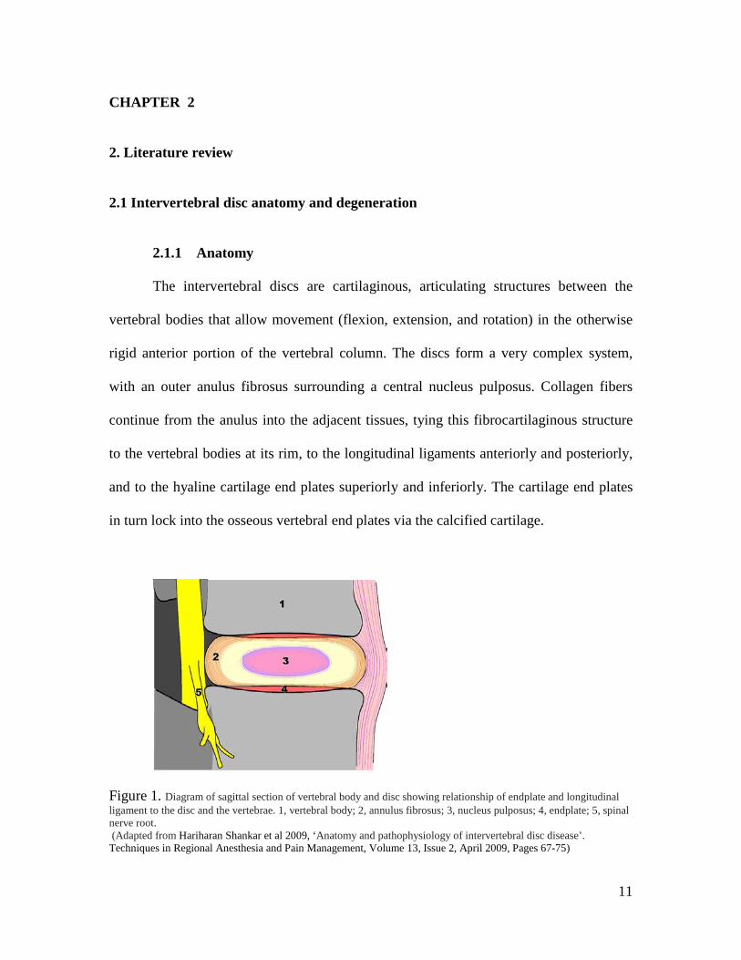
The intervertebral discs are cartilaginous, articulating structures between the
vertebral bodies that allow movement (flexion, extension, and rotation) in the otherwise
rigid anterior portion of the vertebral column. The discs form a very complex system,
with an outer anulus fibrosus surrounding a central nucleus pulposus. Collagen fibers
continue from the anulus into the adjacent tissues, tying this fibrocartilaginous structure
to the vertebral bodies at its rim, to the longitudinal ligaments anteriorly and posteriorly,
and to the hyaline cartilage end plates superiorly and inferiorly. The cartilage end plates
in turn lock into the osseous vertebral end plates via the calcified cartilage.
Figure 1. Diagram of sagittal section of vertebral body and disc showing relationship of endplate and longitudinal ligament to the disc and the vertebrae. 1, vertebral body; 2, annulus fibrosus; 3, nucleus pulposus; 4, endplate; 5, spinal nerve root. (Adapted from Hariharan Shankar et al 2009, ‘Anatomy and pathophysiology of intervertebral disc disease’. Techniques in Regional Anesthesia and Pain Management, Volume 13, Issue 2, April 2009, Pages 67-75)

12
2.1.2 Changes with age
A previous study has reported as early as two years of age, mild microscopic
degenerative changes are seen, including decay and/or proliferation of nucleus pulposus
cells, alterations in cell density and matrix degeneration in the cartilage end plate. This is
thought to coincide with regression of blood vessels in the anulus, cartilage, and osseous
end plates (Boos et al., 2002). It has also been observed in this study that the disc
undergoes degenerative changes earlier in life than other tissues. It is likely that the rapid
reduction in nutrient supply contributes to this early degeneration.
2.1.3 Pathophysiology of disc degeneration
The degenerative process begins in the nucleus pulposus (NP) with cell loss and matrix
alteration. Progression of disease causes the outer annulus fibrosus (AF) to lose its
normal lamellar arrangement and compromises the mechanical strength of the disc. As
the disc fails, tissue fissures and clefts progress from the inner AF outward and contribute
to the loss of mechanical integrity. These changes increase the mechanical forces
transferred to the surrounding vertebral end plates and cause microfractures and marginal
osteophyte formation (Berlemann et al., 1998). Cytokines produced within the disc
stimulate ingrowth of nerve and vascular elements that may play a role in the etiology of
spinal pain. Although this sequence happens during life in essentially all humans, there
are significant variations in the degree of symptoms noted by different people ( Freemont
et al., 2002 ). There is also little known about correlation between the degree of
degenerative changes noted on imaging studies and the presence of symptoms of spinal
pain.

13
2.2 Pathogenesis of adolescent idiopathic scoliosis
Despite extensive studies, the understanding of etiology in AIS remains a mystery. The
identification of etiological factors will depend on continued research in possible areas
listed below. A better understanding of this disorder will enable the clinician to better
predict prognosis and to aid in the development of more effective treatment modalities.
The areas of studies on suggested etiological factors :
a) Genetic factors
The role of hereditary or genetic factors in the development of idiopathic scoliosis
has been widely accepted as one of the possibilities. Clinical observations as well as
population studies have documented scoliosis within families, with the prevalence higher
among relatives than within the general population ( Filho and Thompson, 1971). Studies
on scoliosis twins have further supported the genetic basis of AIS. A twin study
compared the clinical similarity of monozygotic (MZ), or identical, twins who share
100% of their genes, to that of dizygotic (DZ,) or fraternal, twins who share only 50% of
their genes. A concordance for idiopathic scoliosis was 92.3% in monozygotic twins and
62.5% in dizygotic twins. 70% of the monozygotic twins also had similar back shape
(Inoue et al., 1998 ). A meta-analysis by Kesling et al on these clinical twin studies
revealed 73% MZ concordances compared to 36% DZ concordances. Interestingly, when
curve measurements were compared between monozygous twins, the correlation

14
coefficient was significantly greater than the same correlation measurement between
dizygous twins (P < 0.0002). (Kesling and Reinker et al 1997)
A more recent report utilizing the Danish Twin Registry found 25% concordance
in monozygotic twins (6 of 44 concordant) compared to 0% concordance (0 of 91) in
dizygotic twins, with an overall prevalence of approximately 1%. The lower
concordances in both groups as compared with prior results may be explained by
differences in methodology, i.e., ascertainment in clinics vs. registry and screening by
examination vs. questionnaire. Nevertheless, the overall trend obtained for all studies
suggests strong genetic effects in AIS. A second important feature of AIS revealed by
these studies was that monozygotic twins shared disease less than 100% of the time,
reflecting the complexity of disease and suggesting the involvement of unknown factors
in disease susceptibility. (Andersen and Thomsen et al 2007)
Despite documentation of the familial nature of this condition, the mode of
inheritance has been debated. Studies based on a wide variety of populations have
suggested an autosomal dominant, X-linked, or
multifactorial inheritance pattern although
so far, a conclusion has not been made.
b) Role of melatonin
In 1983, Dubousset et al. found that scoliosis routinely developed in
pinealectomized chickens and attributed this effect to decreased melatonin production
(Dubousset et. al., 1983). Dubousset and Machida went on to measure the levels of
melatonin in thirty adolescents with idiopathic scoliosis and in fifteen age-matched

15
controls. The patients had severe scoliosis ranging from 57 to 75 degrees. The curves
were divided into those that had progressed more than 10 degrees in the preceding year
and those that had not. Patients with progressive scoliosis had a 35 percent decrease in
melatonin levels throughout the night compared with those with stable scoliosis or the
control subjects. (Machida and Dubousset et al 1996)
The diurnal variation in melatonin levels seems to be important in determining
development of idiopathic scoliosis. However, scoliosis is not observed when this rhythm is
obliterated in several diseases. Moreover, patients with idiopathic scoliosis do not normally
present with sleep difficulties or immune function, which might be expected with a substantial
decrease in melatonin. On this basis, it is more likely that melatonin plays a secondary role in the
development of idiopathic scoliosis.
c) Role of connective tissue
Collagen and elastic fibers are principal elements in the supporting structures of
the spinal column. As scoliosis is characteristic of many connective-tissue disorders, such
as Marfan syndrome, the hypothesis that the connective tissue defect is the causative
factor of idiopathic scoliosis
The morphology and composition of the intervertebral disc and cartilage end-plate
were studied in patients with idiopathic or congenital scoliosis by
is quite an acceptable opinion.
Roberts S et al. He
compared the findings to those obtained from autopsy as controls. The proteoglycan and
water contents were reduced in both structures in specimens from scoliotic patients,
particularly toward the concavity of the curve. The distribution of some collagen types
also differed in tissue from scoliotic patients and autopsy tissue. Calcification of the
16
cartilage end-plate, and sometimes of the adjacent disc, occurred in all but three scoliotic
patients, whereas there was minimal calcification in the autopsy specimens. They
concluded that these changes are probably a secondary response to altered loading in the
scoliotic patients. (Roberts et al., 1990)
The second major component of the extracellular matrix, elastic fiber has also
been studied in individuals with idiopathic scoliosis. Elastic fibers are made up of two
components: elastin core and microfibrils mainly consisting of fibrillin. A study was
performed on the elastic fiber system of the ligamentum flavum in twenty-three patients
who had scoliosis and in five age-matched individuals who did not. Fresh-frozen
histological specimens of ligamentum flavum removed at the time of an operation were
examined by Verhoeff staining for elastic fibers and by immunohistochemical staining
with use of a monoclonal antibody to fibrillin (Hadley-Miller et al., 1994). They found
elastic fiber abnormalities in the spinal ligaments in majority of patients with idiopathic
scoliosis compared with normal individuals.
There is still divided opinion regarding the changes observed within the
connective
tissues of individuals with idiopathic scoliosis. The possibility of them to be
the consequence of scoliosis rather than the causative factor is still being debated.
However, most researchers concede that abnormalities reported in majority of individuals
affected with idiopathic scoliosis are probably secondary to the structural forces
of the
scoliotic deformity itself. (Harrington, 1977)
17
d) Skeletal muscle abnormalities
Paraspinous muscles abnormality have been thought to be the cause of idiopathic
scoliosis for many years. Two types of muscle fibers in paravertebral muscles of patients
with adolescent idiopathic scoliosis have been described namely type-I (slow-twitch) and
type-II (fast-twitch) fibers. It was found out that the number of type-II fibers was
decreased in scoliotic patients, suggesting a myopathic process. A normal distribution of
type-I and type-II fibers on the convexity of the curve but a lower frequency of type-I
fibers on the concavity was found. ( Bylund et al., 1987).
Other studies have reported findings of marked decrease in muscle spindles in
the
paraspinous muscles, increased calcium content due to generalized membrane defect and
higher muscle protein synthesis on the convexity than on the concavity in patients( Slager
and Hsu, 1986, Yarom et al., 1978). However, no definite conclusions can be reached
with regard to
involvement of skeletal muscle abnormalities as one of the etiology.
e) Thrombocyte abnormalities (role of calmodulin)
Calmodulin, a calcium-binding receptor protein, is a critical mediator of cellular
calcium function and regulates many important enzymatic systems. Calmodulin regulates
the contractile properties of muscles and platelets by interacting with actin and myosin
and regulating calcium movement from the sarcoplasmic reticulum. Increased calmodulin
levels in platelets have been shown to influence the progression in adolescent idiopathic
scoliosis ( Kindsfater et al., 1994 ). He studied on seventeen patients who had idiopathic
scoliosis of varying severity and patterns and a control group consisting of ten age and
sex-matched subjects. Level of platelet calmodulin in the patients who had a progressive

18
curve (more than 10 degrees of progression in the previous twelve months) (3.83
nanograms per microgram of protein) was significantly higher than the level in the
patients who had a stable curve (less than 5 degrees of progression in the previous twelve
months) (0.60 nanogram per microgram of protein) (p < 0.01). However, the levels in the
stable group and the control group (0.69 nanogram per microgram of protein) were
similar. He concluded that level of platelet calmodulin appeared to be an independent and
possibly more acute predictor of progression of the curve.
f) Neurological mechanisms
Over the last twenty years, advanced neurological investigations have been used
to compare idiopathic scoliosis patients with controls and to compare patients who have
curves progression with those who have do not. However, the results have been
inconsistent ( Yekutiel et al., 1981). No proven neurological tests either for diagnosing
idiopathic scoliosis
The advent of magnetic resonance imaging has led to renewed
or for predicting its progression have so far been established.
interest in
abnormal neuroanatomy linked to scoliosis. MRI was performed in 31 scoliosis patients
with onset between the ages of four and 12 years. In eight patients (26%) there was a
significant neuroanatomical abnormality; there were six cases of Chiari-1 malformation
associated with a syrinx, one isolated Chiari-1 malformation and one astrocytoma of the
cervical spine. Four of these patients had left-sided curves. There were no clinical
features which could reliably identify those patients with abnormalities on MRI. The
unilateral absence of abdominal reflexes was found to be non-specific (1 of 8 of patients
with neuroanatomical abnormalities (12.5%) versus 2 of 23 with normal scans (8.7%). It

19
is not yet known whether this is secondary to the syrinx formation as the first sign of
syringomyelia or whether this asymmetry might reflect a more proximal hindbrain or
midbrain lesion. Alternatively, the Chiari malformation and the syrinx could be the result
of traction on the medulla distally through the foramen magnum.

20
2.3 Pfirrmann classification
Magnetic resonance imaging (MRI) is one of the most important method for the clinical
assessment of intervertebral disc pathology. The signal characteristics of the disc in T2-
weighted MRIs have been shown to reflect changes caused by aging or degeneration.
(Pearce et al., 1991). The signal intensity of the disc in relation to chemical composition
and histologic changes has also been studied. The brightness of the nucleus has been
shown to correlate directly with the proteoglycan concentration, but not with the water or
collagen content. ( Tertti et al., 1991 ).
Few attempts have been made to classify the level of degenerative changes in the
intervertebral discs. Most previous classification systems and reliability studies of lumbar
disc abnormalities on MRI have focused on the posterior aspect of the disc,
distinguishing among bulging, protrusion, and extrusion. Studies focusing on the MRI
characteristics of the disc structure are rare. The ideal classification system for disc
degeneration should be quantitative, permits region-specific evaluation within the disc
sub-structures, avoids observer bias, can detect early subtle changes, and correlates with
clinical symptoms.
Among the classification systems suggested is the classification developed by Pfirrmann
which is based on the preliminary work by Pearce.. A grade between I and V is assigned
based on signal intensity and structural morphology of the disc. (Pfirrmann et al., 2001).
The classification is as illustrated in Table 1. In this study, three observers with different
levels of experience analyzed spinal MRIs (i.e., an orthopedic surgeon, a fellowship-
trained musculoskeletal radiologist, and a musculoskeletal senior staff radiologist) and
graded each of the 300 lumbar intervertebral discs on the T2-weighted sagittal images.
21
The appearance of MRI images for each grade is illustrated in Figure 2. Intraobserver
agreement was excellent for all three readers, with kappa values ranging from 0.84 to
0.90 while interobserver agreement ranged from substantial to excellent, with kappa
values ranging from 0.69 to 0.81. This is in accordance with Landis and Koch who rated
the agreement as follows: kappa 0 to 0.2 indicated slight agreement, 0.21 to 0.4 fair
agreement, 0.41 to 0.60 moderate agreement, 0.61 to 0.8 substantial agreement, and 0.81
upward excellent agreement. (Landis and Koch, 1977)
Pfirrmann also developed an algorithm to facilitate the process of grading the discs. This
algorithm is shown in Figure 3. The authors also suggested adding the Modic
classification (Types 1–3) to their grading system for further specification of a
degenerated intervertebral disc in the case of concomitant end plate change. (Pfirrman et
al., 2001). In order to achieve an objective assessment of the intervertebral discs in
adolescent scoliosis patients, we have chosen this classification to help identify the
degree of degeneration in the lumbar spine.
22
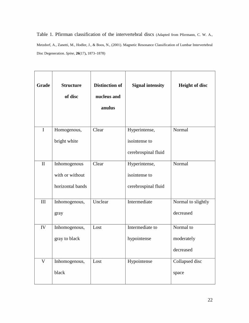
Table 1. Pfirrman classification of the intervertebral discs (Adapted from Pfirrmann, C. W. A.,
Metzdorf, A., Zanetti, M., Hodler, J., & Boos, N., (2001). Magnetic Resonance Classification of Lumbar Intervertebral
Disc Degeneration. Spine, 26(17), 1873–1878)
Grade
Structure
of disc
Distinction of
nucleus and
anulus
Signal intensity
Height of disc
I Homogenous,
bright white
Clear Hyperintense,
isointense to
cerebrospinal fluid
Normal
II Inhomogenous
with or without
horizontal bands
Clear Hyperintense,
isointense to
cerebrospinal fluid
Normal
III Inhomogenous,
gray
Unclear Intermediate Normal to slightly
decreased
IV Inhomogenous,
gray to black
Lost Intermediate to
hypointense
Normal to
moderately
decreased
V Inhomogenous,
black
Lost Hypointense Collapsed disc
space
23
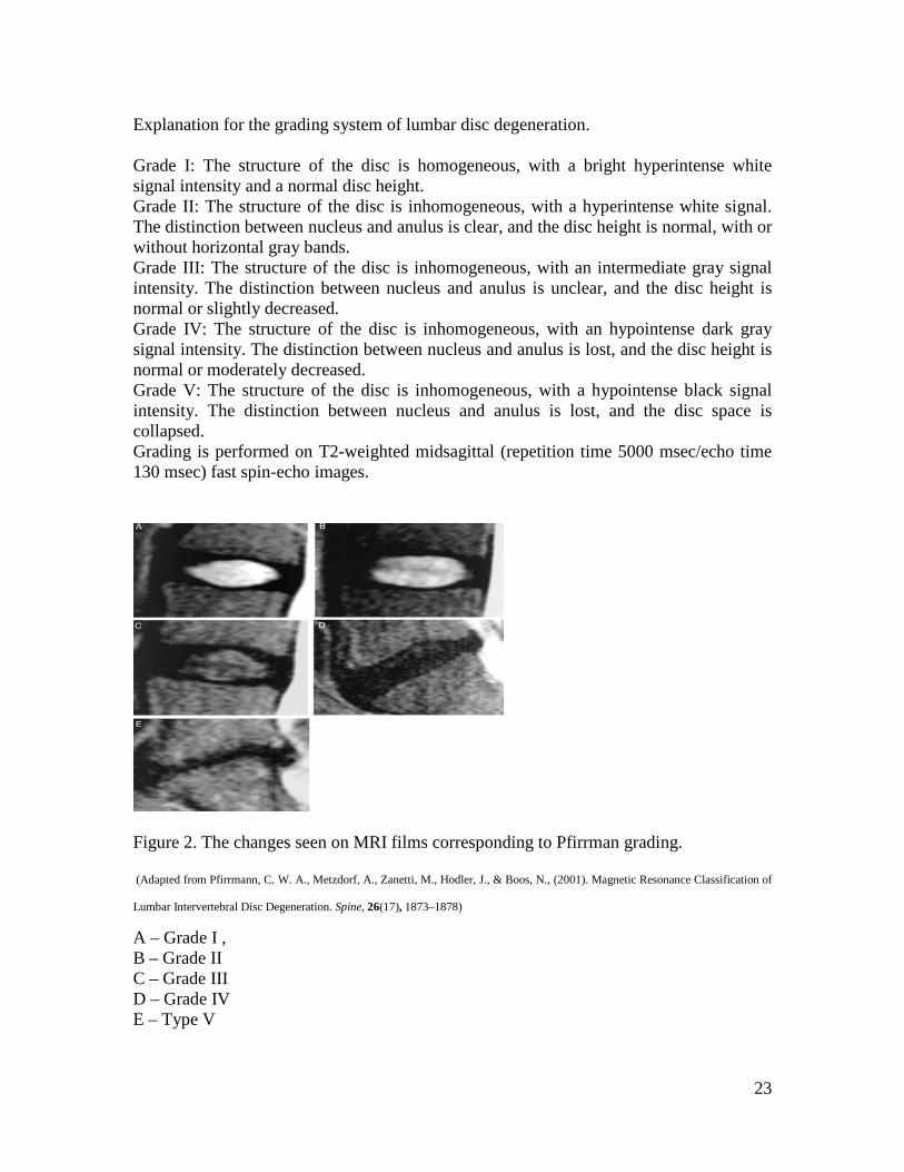
Explanation for the grading system of lumbar disc degeneration. Grade I: The structure of the disc is homogeneous, with a bright hyperintense white signal intensity and a normal disc height. Grade II: The structure of the disc is inhomogeneous, with a hyperintense white signal. The distinction between nucleus and anulus is clear, and the disc height is normal, with or without horizontal gray bands. Grade III: The structure of the disc is inhomogeneous, with an intermediate gray signal intensity. The distinction between nucleus and anulus is unclear, and the disc height is normal or slightly decreased. Grade IV: The structure of the disc is inhomogeneous, with an hypointense dark gray signal intensity. The distinction between nucleus and anulus is lost, and the disc height is normal or moderately decreased. Grade V: The structure of the disc is inhomogeneous, with a hypointense black signal intensity. The distinction between nucleus and anulus is lost, and the disc space is collapsed. Grading is performed on T2-weighted midsagittal (repetition time 5000 msec/echo time 130 msec) fast spin-echo images.
Figure 2. The changes seen on MRI films corresponding to Pfirrman grading.
(Adapted from Pfirrmann, C. W. A., Metzdorf, A., Zanetti, M., Hodler, J., & Boos, N., (2001). Magnetic Resonance Classification of
Lumbar Intervertebral Disc Degeneration. Spine, 26(17), 1873–1878)
A – Grade I , B – Grade II C – Grade III D – Grade IV E – Type V

24
Figure 3. Algorithm suggested by Pfirrman to facilitate the grading of discs. (Adapted from
Pfirrmann, C. W. A., Metzdorf, A., Zanetti, M., Hodler, J., & Boos, N., (2001). Magnetic Resonance Classification of
Lumbar Intervertebral Disc Degeneration. Spine, 26(17), 1873–1878)