change of mammographic density predicts the risk of contralateral breast cancer - a case-control...
TRANSCRIPT

RESEARCH ARTICLE Open Access
Change of mammographic density predicts therisk of contralateral breast cancer - acase-control studyMaria EC Sandberg1*, Jingmei Li1,2, Per Hall1, Mikael Hartman1,3,4, Isabel dos-Santos-Silva5, Keith Humphreys1 andKamila Czene1
Abstract
Introduction: Mammographic density is a strong risk factor for breast cancer, but it is unknown whether density atfirst breast cancer diagnosis and changes during follow-up influences risk of non-simultaneous contralateral breastcancer (CBC).
Methods: We collected mammograms for CBC-patients (cases, N = 211) and unilateral breast cancer patients(controls, N = 211), individually matched on age and calendar period of first breast cancer diagnosis, type ofadjuvant therapy and length of follow-up (mean follow-up time: 8.25 years). The odds of CBC as a function ofchanges of density during follow-up were investigated using conditional logistic regression, adjusting for non-dense area at diagnosis.
Results: Patients who experienced ≥10% absolute decrease in percent density had a 55% decreased odds of CBC(OR = 0.45 95% CI: 0.24 to 0.84) relative to patients who had little or no change in density from baseline to firstfollow-up mammogram (mean = 1.6 (SD = 0.6) years after diagnosis), whereas among those who experienced anabsolute increase in percent density we could not detect any effect on the odds of CBC (OR = 0.83 95% CI: 0.24 to2.87).
Conclusion: Decrease of mammographic density within the first two years after first diagnosis is associated with asignificantly reduced risk of CBC, this potential new risk predictor can thus contribute to decision-making in follow-up strategies and treatment.
Keywords: Contralateral breast cancer, mammographic density, risk, breast density, epidemiology
IntroductionMammographic density is one of the strongest risk fac-tors for breast cancer; a meta-analysis of 14,000 casesand 226,000 non-cases showed that the women with>75% mammographic density have almost five times therisk of breast cancer compared to women in the lowestdensity group (<5%) [1]. Mammographic density hasalso been shown to be important for breast cancerrecurrence [2] and survival [3]. Several hormonal factorsaffect mammographic density and changes of densityhave been shown to be associated with pharmacological
therapies, such as hormone replacement therapy [4] andtamoxifen [5].Despite the well-known and strong association between
mammographic density and unilateral breast cancer, theeffect of mammographic density on the risk of a secondprimary breast cancer in the opposite breast, contralat-eral breast cancer (CBC), has to our knowledge not beeninvestigated before. Breast cancer patients have approxi-mately double the risk of CBC, compared to healthywomen’s risk of breast cancer [6] and this increased riskdoes not seem to decline with time after first diagnosis[7-9]. This translates into 10 to 15% of all breast cancerpatients being diagnosed with CBC within 20 years ofinitial diagnosis [10,11]. When investigating hormonalrisk factors for unilateral breast cancer no association
* Correspondence: [email protected] of Medical Epidemiology and Biostatistics, Karolinska Institutet,Nobels väg 12B, Stockholm, 177 71, SwedenFull list of author information is available at the end of the article
Sandberg et al. Breast Cancer Research 2013, 15:R57http://breast-cancer-research.com/content/15/4/R57
© 2013 Sandberg et al.; licensee BioMed Central Ltd. This is an open access article distributed under the terms of the CreativeCommons Attribution License (http://creativecommons.org/licenses/by/2.0), which permits unrestricted use, distribution, andreproduction in any medium, provided the original work is properly cited.

with risk of CBC has been identified [12-14]. Trends ofbreast cancer incidence and breast cancer mortality indi-cate that CBC will be a greater clinical challenge in thefuture, since the population at risk of CBC is increasing[15]. Since CBC also has a far less well characterized riskprofile [8] and considerably worse prognosis than unilat-eral breast cancer, new tools for prediction of CBC wouldbe of great clinical importance [16].The question of whether decreasing mammographic
density is associated with a decreased future risk ofbreast cancer, or not, has been investigated in severalobservational studies. Two showed an association[17,18] and one did not [19]. The question was alsoexamined in a randomized trial of tamoxifen amonghealthy women at high risk of breast cancer, for whichthe estimated association was more pronounced amongthe tamoxifen treated, although the association (albeitnon-statistically significant) was also seen among thewomen who received placebo [20]. If this associationwere also present among breast cancer patients, for therisk of CBC, this would have important clinical implica-tions for follow-up care. The aim of this matched nestedcase-control study was, therefore, to assess whetherchange of mammographic density after the first breastcancer diagnosis predicts a change in risk of CBC.
MethodsStudy populationThe study was nested within the catchment population ofthe Stockholm Breast Cancer Register, a population-based register of all breast cancer patients diagnosedsince 1976 in the Stockholm-Gotland health-care region(N >30,000). Women with invasive CBC diagnosed morethan one year after the first invasive cancer and with anavailable mammogram close to the first diagnosis (N =458) were identified as potential cases. Patients with inva-sive unilateral breast cancer in the same register wereidentified as potential controls. Women with a first pri-mary cancer other than breast cancer and women withdistant metastasis at the first or second breast cancerdiagnosis were excluded in order to minimize the risk ofthe CBC being a misclassified metastasis. Further, secondprimary breast cancers can obviously also occur in thesame breast as the first cancer; ipsilateral breast cancer.We chose not to include these cancers in the presentstudy since also these cancers are less likely to be primarycancers. For each case, one control was randomlyselected and matched to the corresponding case on thecalendar period of the first breast cancer diagnosis (+/-two years), age at the first breast cancer diagnosis (+/-two years), adjuvant therapy and follow-up time, so thatthe control had survived without distant metastasis orCBC at least as long as the time between the first andsecond cancer for the corresponding case, a strategy
known as density sampling [21]. From the StockholmBreast Cancer Register we retrieved information onmenopausal status at the time of the (first) breast cancerdiagnosis, estrogen receptor (ER) status and tumor-node-metastasis (TNM)-stage of the (first) cancer, in additionto the matching variables. From the medical records ofthe cases and controls we retrieved information on hor-mone replacement therapy at the time of the (first) breastcancer diagnosis as well as additional information onmenopausal status and ER-status.We collected baseline and follow-up mammograms for
the cases and controls. The baseline mammogram wasdefined as a mammogram from the contralateral breast,that is, the breast not affected by cancer, taken at anytime during the year prior to diagnosis, or within twoweeks after diagnosis, of the first cancer. Follow-upmammograms were defined as mammograms from theunaffected, contralateral breast taken at least one year,but no more than five years, after diagnosis of the firstcancer. We used the first available mammogram in thedefined time period. We defined sets of two individualscomprising one CBC case and one matched control(unilateral breast cancer patient) and a total of fourmammograms (one baseline and one follow-up mam-mogram for each patient) of the same view. The media-lateral-oblique (MLO) view of mammograms have beenthe preferred view in the Swedish screening program[22] and were, therefore, used for our primary selection.Sets of cranial-caudal (CC)-mammograms made up 14%of the final sample.For 99 of the 458 eligible CBC-cases we could not
locate any follow-up mammogram and for 88 of theCBC-cases either the baseline or the follow-up mammo-gram could not be used (for example, due to low qualityof the mammogram), while for 271 patients (59%) boththe baseline and at least one follow-up mammogram ofthe unaffected breast from the same view was assessableand could be used. Among these patients we could tracethe baseline and follow-up mammograms from the cor-rect side and view for the corresponding control in 211cases. These 211 case-control sets were thus included inthe analysis sample. The CBC patients excluded due tolack of eligible mammograms did not differ from thoseincluded in the analysis in relation to age or calendarperiod of first diagnosis.For comparison with the risk of CBC as a function of
baseline mammographic density (that is, density at thetime of first breast cancer diagnosis), we also examinedthe risk of unilateral breast cancer in relation to mammo-graphic density. To achieve this, for each unilateral breastcancer case, we also measured the mammographic den-sity of a healthy woman. The healthy controls (N = 142)were randomly selected from a breast cancer case-controlstudy, extensively described elsewhere [23], for which all
Sandberg et al. Breast Cancer Research 2013, 15:R57http://breast-cancer-research.com/content/15/4/R57
Page 2 of 9

available mammograms had been previously collected.The mammograms of the healthy controls were matchedto the unilateral breast cancer patients by calendar periodand age of the corresponding unilateral breast cancerpatient at her first diagnosis.The mammograms were digitized using an Array
2905HD Laser Film Digitizer (Array Corporation,Tokyo, Japan), which covers a range of 0 to 4.7 opticaldensity. The density resolution was set at 12-bit spatialresolution. Mammographic density was measured usingour automated thresholding method [24], which incor-porates the knowledge of a trained observer by usingmeasurements obtained by an established user-assistedthreshold method - Cumulus [25] - as training data.The externally validated results showed a high corre-spondence between our automated method and theestablished user-assisted thresholding method Cumulus(rpercent mammographic density) = 0.88 (95% CI: 0.87 to 0.89).
Statistical analysisWe estimated the percent mammographic density as wellas the absolute size of the dense area and of the total areaof the breast. Percent density and dense area have beenused in previous studies and have both been shown to beimportant predictors of breast cancer risk [26]. Fordescriptive purposes we calculated the mean changes ofmammographic density (unadjusted) in different groupsof study participants.As the first step, the risk of CBC as a function of
baseline mammographic density was analyzed usingconditional logistic regression, contrasting CBC patientsto unilateral breast cancer patients. Further, by the sametype of analysis we then contrasted the unilateral breastcancer patients to healthy controls. Percentage densityat baseline was categorized into ≤5%, >5 to 25% (refer-ence level), >25 to 50% and >50%; these cutoffs havebeen used extensively [1]. Dense area at baseline wascategorized into: ≤20 cm2, >20 to 40 cm2 (referencelevel), >40 to 60 cm2 and >60 cm2, with these categoriescorresponding approximately to quartiles of the baselinedense area distribution. Calculating also the total breastarea enabled us to adjust for non-dense area in the ana-lyses; adjustment for this variable has recently beenshown to be preferential to adjusting for body massindex (BMI) [27]. All analyses were thus adjusted forage and calendar period of diagnosis, adjuvant therapyand follow-up time (through matching) and also fornon-dense area at first diagnosis, categorized in quar-tiles. Trend tests were carried out based on orderedcategories of percent density/dense area.For our main analysis, conditional logistic regression
was used for analyzing risk of CBC as a function of“change” of mammographic density from baseline tofirst follow-up mammogram, categorized in three levels:
absolute decrease ≥10%, stable (-10% to +10%, referencelevel) and absolute increase ≥10%, in agreement withprevious literature [20]. Further, we investigated changeof density in terms of absolute dense area, also categor-ized in three levels: ≥10 cm2 reduction, stable (-10 cm2
to +10 cm2, reference level) and ≥10 cm2 increase indense area. Both analyses were adjusted through match-ing for age and calendar period of first diagnosis, firstadjuvant therapy and follow-up time. In an additionalmodel, we made further adjustments for baseline non-dense area and baseline mammographic density (percentdensity when using change in percent density and densearea when using change in dense area), both categorizedin quartiles. Patients with <10% or >90% percent mam-mographic density at baseline (N = 66), or those with<10 cm2 or >70 cm2 dense area (N = 84), were excluded,since they cannot possibly undergo changes in percentmammographic density, or dense area, of the definedmagnitude. A similar strategy has been used by otherswhen studying changes in mammographic density [5].Trend test was performed using the ordinal categories ofchange of percent density and area density, respectively.Finally, as an exploratory analysis we stratified our
population on menopause status at first breast cancer toinvestigate the effect of change of mammographic den-sity on the risk of CBC in the two subgroups, adjustingfor non-dense area at baseline and percent density atbaseline. Case-control pairs discordant for menopausestatus (N = 35) was not included in this analysis.All data preparation and analyses were carried out
using SAS Statistical Package 9.2 (SAS Institute Inc.,Cary, NC, USA). This study was approved by the EthicalReview Board at Karolinska Institutet, Stockholm, Swe-den. As no contact was made with the study personsand the data were analyzed anonymously, informed con-sent was not obtained. This exception from informedconsent was confirmed by the ethical committee.
ResultsA total of 422 subjects (211 cases and 211 controls) wereincluded in the analysis (Table 1). The mean time fromdiagnosis to follow-up mammogram was 1.6 years, 90%of the follow-up mammograms were taken between 1and 2.2 years after diagnosis of the first breast cancer andthere was no difference between cases and controls. Themean breast density at baseline was 28%.Table 2 describes the mean change of mammographic
density, measured in absolute percent density, from base-line to follow-up mammogram. The change is similarover the calendar period and over different categories oftotal breast area. As expected, the mammographic den-sity decreases significantly more in the women diagnosedbefore menopause, compared to women diagnosed aftermenopause (P-value < 0.01).
Sandberg et al. Breast Cancer Research 2013, 15:R57http://breast-cancer-research.com/content/15/4/R57
Page 3 of 9

We found no association between mammographic den-sity at baseline and risk of CBC using either percentmammographic density or dense area (P-value for trend:0.40 and 0.96 for percent density and dense area, respec-tively) (Table 3). Also, when mammographic density wasanalyzed as a continuous measure no effect was found;
odds ratio (OR) for percent density: 1.00 (95% CI: 0.98 to1.01) and for dense area: 1.00 (95% CI: 0.99 to 1.01). Wefurther compared the baseline density between unilateralbreast cancer patients and healthy controls. As expected,we found a statistically significant increasing risk ofbreast cancer with increasing mammographic density
Table 1 Distribution of CBC case and unilateral breast cancer controls
Cases Controls P-value*
Age at (first) diagnosis (%) ≤45 years 37 (18) 37 (18)
45 to 55 years 68 (32) 68 (32)
55 to 65 years 56 (27) 56 (27)
≥65 years 50 (24) 50 (24) -
Calendar period of (first) diagnosis (%) 1976 to 1980 31 (15) 30 (14)
1981 to 1985 40 (19) 41 (19)
1986 to 1990 45 (21) 41 (19)
1991 to 1995 51 (24) 49 (23)
1996 to 2005 44 (21) 50 (24) -
Adjuvant therapy (%)* No adjuvant therapy 39 (18) 39 (18)
Radiotherapy only 57 (27) 57 (27)
Endocrine therapy 87 (41) 87 (41)
Chemotherapy 28 (13) 28 (13) -
Mean follow-up time in years 8.25 8.25 -
Percent density at (first) diagnosis (%) ≤5% 13 (6) 11 (5)
5 to 25% 87 (41) 87 (41)
25 to 50% 97 (46) 87 (41)
≥50% 14 (7) 26 (12) 0.23
Quartiles:
Dense area at (first) diagnosis (%) ≤20 cm2 55 (26) 55 (26)
20 to 34 cm2 44 (21) 56 (27)
34 to 53 cm2 56 (27) 49 (23)
≥53 cm2 56 (27) 51 (24) 0.79
Change in percent density until first Absolute decrease (>10%) 40 (19) 56 (27)
follow-up mammogram (%) Stable density 164 (78) 143 (68)
Absolute increase (>10%) 7 (3) 12 (6) 0.07
Change in dense area until first Absolute decrease (>10 cm2) 61 (29) 75 (36)
follow-up mammogram Stable density 131 (62) 113 (54)
Absolute increase (>10 cm2) 19 (9) 23 (11) 0.21
Quartiles:
Non-dense area at (first) diagnosis (%) ≤67 cm2) 42 (20) 60 (28)
67 to 93 cm2 58 (27) 43 (20)
93 to 127 cm2 50 (24) 55 (26)
≥127 cm2 61 (29) 53 (25) 0.10
Mean time until first follow-up mammogram in years (SD) 1.56(0.59)
1.54(0.57)
0.56
Menopause status at diagnosis (%)** Premenopausal 89 (42) 84 (40)
Postmenopausal 119 (56) 124 (59) 0.62
Subdivided by matching variables (age at diagnosis, calendar period of diagnosis, adjuvant therapy and follow-up time), exposure variables (dense area atdiagnosis and change of dense area), potential confounding variables (non-dense area at diagnosis and time to first follow-up mammogram), and stratifyingvariable (menopause status).
* P-value for Chi-square test of association when testing categorical variables and for Student’s t-tests when testing the continuous variable (mean time until firstfollow-up mammogram).
*Endocrine therapy may be with or without radiotherapy, chemotherapy may be with or without radiotherapy and/or endocrine therapy.
** Six patients had uncertain menopause status (for example, hysterectomy).
Sandberg et al. Breast Cancer Research 2013, 15:R57http://breast-cancer-research.com/content/15/4/R57
Page 4 of 9

(P-values for trend: <0.01 and 0.02 for percent densityand dense area, respectively).When analyzing change of mammographic density, we
observed a 55% lower risk of CBC for women with anabsolute decrease in mammographic density of ≥10% frombaseline to follow-up mammogram, compared to womenwith stable mammographic density (OR = 0.45 (95% CI:0.24 to 0.84)) (Table 4). We found no statistically signifi-cant effect of increasing absolute mammographic densitycompared to stable density (OR = 0.83 (95% CI: 0.24 to
2.87)). Through the matched case-control design, thesefindings are independent of age and calendar period offirst diagnosis, adjuvant therapy and follow-up time. Theadjustments for non-dense area at baseline and percentdensity at baseline affected the estimates only marginally,but these variables are included since they are potentialconfounders. Using absolute dense area as a measure ofmammographic density we found a similar effect of 46%risk decrease for women with ≥10 cm2 decrease frombaseline to follow-up mammogram, compared to women
Table 2 Mean absolute change of percent density (PD) from baseline until first follow-up mammogram
N Meandecrease (%-units) 95% CI for mean decrease of PD P-value
Total 422 -3.94 -4.89, -3.00 -
CBC case/control status 0.09
Cases 211 -3.13 -4.39, -1.87
Controls 211 -4.75 -6.16, -3.34
Age at time of (first) cancer: <0.01
<45 years 74 -5.30 -8.39, -2.22
45 to 54 years 136 -5.85 -7.61, -4.10
55 to 64 years 112 -4.13 -5.60, -2.65
≥65 years 100 -0.13 -1.50, 1.25
Calendar period of (first) cancer: 0.25
1976 to 1980 62 -4.11 -7.57, -0.66
1981 to 1985 80 -3.78 -6.24, -0.32
1986 to 1990 90 -2.27 -4.09, -0.46
1991 to 1995 102 -3.91 -5.70, -2.12
1996 to 2005 88 -5.71 -7.26, -4.16
Total breast area at baseline: 0.27
Smallest quartile 103 -2.54 -4.66, -0.41
2nd quartile 105 -4.68 -6.71, -2.66
3rd quartile 110 -4.91 -6.75, -3.07
Largest quartile 104 -3.56 -5.16, -1.96
Adjuvant therapy of (first) cancer: <0.01
No adjuvant therapy 78 -1.09 -3.09, 0.92
Radiotherapy only 114 -3.22 -5.14, -1.30
Endocrine therapy (with/without radiotherapy) 174 -4.15 -5.37, -2.93
Chemotherapy (with/without other adjuvant therapy) 56 -8.74 -5.24, -12.24
Menopause status at (first) cancer:* <0.01
Premenopausal 173 -5.90 -7.70, -4.10
Postmenopausal 243 -2.56 -3.54, -1.58
Postmenopausal HRT use:* <0.01
with Current use of HRT at diagnosis 51 -6.55 -8.90, -4.19
with No current use of HRT at diagnosis 127 -1.66 -2.90, -0.43
ER-status of (first) cancer: 0.63
ER-positive 295 -4.15 -5.29, -3.01
ER-negative 53 -3.46 -5.92, -0.99
TNM-stage of (first) cancer:* 0.67
1 244 -4.06 -5.27, -2.85
2 157 -3.48 -5.10, -1.86
3 16 -5.36 -11.19, 0.47
The mean change is calculated for cases and control combined.
* Six (1%) women with unknown menopause; 65 (27%) postmenopausal women with unknown HRT. Five women with unknown TNM-stage.
TNM, tumor-node-metastasis
Sandberg et al. Breast Cancer Research 2013, 15:R57http://breast-cancer-research.com/content/15/4/R57
Page 5 of 9
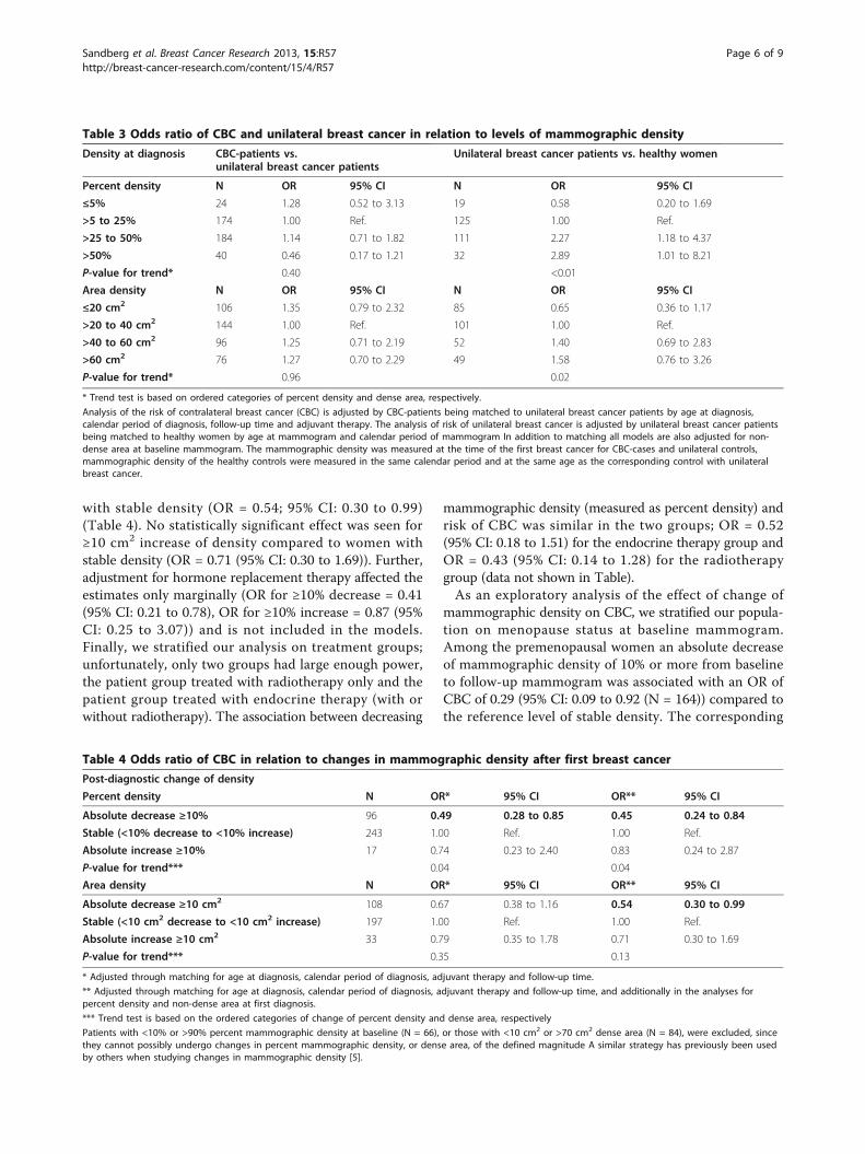
with stable density (OR = 0.54; 95% CI: 0.30 to 0.99)(Table 4). No statistically significant effect was seen for≥10 cm2 increase of density compared to women withstable density (OR = 0.71 (95% CI: 0.30 to 1.69)). Further,adjustment for hormone replacement therapy affected theestimates only marginally (OR for ≥10% decrease = 0.41(95% CI: 0.21 to 0.78), OR for ≥10% increase = 0.87 (95%CI: 0.25 to 3.07)) and is not included in the models.Finally, we stratified our analysis on treatment groups;unfortunately, only two groups had large enough power,the patient group treated with radiotherapy only and thepatient group treated with endocrine therapy (with orwithout radiotherapy). The association between decreasing
mammographic density (measured as percent density) andrisk of CBC was similar in the two groups; OR = 0.52(95% CI: 0.18 to 1.51) for the endocrine therapy group andOR = 0.43 (95% CI: 0.14 to 1.28) for the radiotherapygroup (data not shown in Table).As an exploratory analysis of the effect of change of
mammographic density on CBC, we stratified our popula-tion on menopause status at baseline mammogram.Among the premenopausal women an absolute decreaseof mammographic density of 10% or more from baselineto follow-up mammogram was associated with an OR ofCBC of 0.29 (95% CI: 0.09 to 0.92 (N = 164)) compared tothe reference level of stable density. The corresponding
Table 3 Odds ratio of CBC and unilateral breast cancer in relation to levels of mammographic density
Density at diagnosis CBC-patients vs.unilateral breast cancer patients
Unilateral breast cancer patients vs. healthy women
Percent density N OR 95% CI N OR 95% CI
≤5% 24 1.28 0.52 to 3.13 19 0.58 0.20 to 1.69
>5 to 25% 174 1.00 Ref. 125 1.00 Ref.
>25 to 50% 184 1.14 0.71 to 1.82 111 2.27 1.18 to 4.37
>50% 40 0.46 0.17 to 1.21 32 2.89 1.01 to 8.21
P-value for trend* 0.40 <0.01
Area density N OR 95% CI N OR 95% CI
≤20 cm2 106 1.35 0.79 to 2.32 85 0.65 0.36 to 1.17
>20 to 40 cm2 144 1.00 Ref. 101 1.00 Ref.
>40 to 60 cm2 96 1.25 0.71 to 2.19 52 1.40 0.69 to 2.83
>60 cm2 76 1.27 0.70 to 2.29 49 1.58 0.76 to 3.26
P-value for trend* 0.96 0.02
* Trend test is based on ordered categories of percent density and dense area, respectively.
Analysis of the risk of contralateral breast cancer (CBC) is adjusted by CBC-patients being matched to unilateral breast cancer patients by age at diagnosis,calendar period of diagnosis, follow-up time and adjuvant therapy. The analysis of risk of unilateral breast cancer is adjusted by unilateral breast cancer patientsbeing matched to healthy women by age at mammogram and calendar period of mammogram In addition to matching all models are also adjusted for non-dense area at baseline mammogram. The mammographic density was measured at the time of the first breast cancer for CBC-cases and unilateral controls,mammographic density of the healthy controls were measured in the same calendar period and at the same age as the corresponding control with unilateralbreast cancer.
Table 4 Odds ratio of CBC in relation to changes in mammographic density after first breast cancer
Post-diagnostic change of density
Percent density N OR* 95% CI OR** 95% CI
Absolute decrease ≥10% 96 0.49 0.28 to 0.85 0.45 0.24 to 0.84
Stable (<10% decrease to <10% increase) 243 1.00 Ref. 1.00 Ref.
Absolute increase ≥10% 17 0.74 0.23 to 2.40 0.83 0.24 to 2.87
P-value for trend*** 0.04 0.04
Area density N OR* 95% CI OR** 95% CI
Absolute decrease ≥10 cm2 108 0.67 0.38 to 1.16 0.54 0.30 to 0.99
Stable (<10 cm2 decrease to <10 cm2 increase) 197 1.00 Ref. 1.00 Ref.
Absolute increase ≥10 cm2 33 0.79 0.35 to 1.78 0.71 0.30 to 1.69
P-value for trend*** 0.35 0.13
* Adjusted through matching for age at diagnosis, calendar period of diagnosis, adjuvant therapy and follow-up time.
** Adjusted through matching for age at diagnosis, calendar period of diagnosis, adjuvant therapy and follow-up time, and additionally in the analyses forpercent density and non-dense area at first diagnosis.
*** Trend test is based on the ordered categories of change of percent density and dense area, respectively
Patients with <10% or >90% percent mammographic density at baseline (N = 66), or those with <10 cm2 or >70 cm2 dense area (N = 84), were excluded, sincethey cannot possibly undergo changes in percent mammographic density, or dense area, of the defined magnitude A similar strategy has previously been usedby others when studying changes in mammographic density [5].
Sandberg et al. Breast Cancer Research 2013, 15:R57http://breast-cancer-research.com/content/15/4/R57
Page 6 of 9
OR for post-menopausal women was 0.49 (95% CI: 0.16 to1.45) (N = 188). The effect of density reduction and meno-pausal status was not statistically significant (Pinteraction =0.47).Finally, we stratified the mammograms by view; CC-
view (19%) and MLO-view (81%). We found very similarestimates for the effect of decreasing mammographicdensity on the risk of CBC in the two groups, althoughthe statistical significance was lost in the CC-view group(OR for MLO-group: 0.45 (95% CI: 0.22 to 0.91), OR forthe CC-group: 0.42 (95% CI: 0.06 to 2.77)).
DiscussionTo our knowledge, this is the first study to have investi-gated the risk of CBC as an effect of mammographic den-sity at the time of diagnosis of the first cancer and of itssubsequent changes. In a population-based setting we esti-mated that an absolute decrease of mammographic densityfrom diagnosis to first follow-up mammogram of at least10% confers a significant 55% decrease in risk of CBC. Incontrast, mammographic density at diagnosis does notseem to predict risk of CBC.Strengths of the study include its ability to investigate
both baseline density and changes after first diagnosisand the use of both absolute (dense area) and relative(percent density) measures of mammographic density.The study is limited mainly by its small size, resultingfrom the large proportion of cases that had to beexcluded from the analysis because the required mam-mograms could not be traced. Exclusion of patients dueto unavailability of mammograms is a potential sourceof bias if the missingness is differential; it, however,seems unlikely that the mammograms should be missingon the basis of mammographic density. Further, una-voidably, access to the patient’s mammograms is arequirement for studies of mammographic density. Reas-suringly, the excluded patients did not differ from thoseincluded in the analysis in relation to important factorssuch as age at first diagnosis (P-value: 0.23) and calen-dar period of first diagnosis (P-value: 0.12). There were,however, differences with respect to adjuvant therapyfor the first cancer; the included patients had receivedradiotherapy and endocrine therapy to a somewhat lar-ger degree (radiotherapy; 29% vs. 23% for excludedpatients, endocrine therapy; 39% vs. 29% for excludedpatients). Since the study was matched for treatment,this difference has potential implications only for thegeneralizability of our results, as the patients receivingno adjuvant therapy and the patients receiving che-motherapy are somewhat under-represented in thisstudy. In general, the availability of the mammograms inSweden during the study period is primarily driven byarchiving policies, rather than patients not having mam-mograms taken.
In contrast to the effect of mammographic density onthe risk of unilateral breast cancer among healthywomen, mammographic density at baseline did notseem to influence the risk of CBC in breast cancerpatients. None of the categories of mammographic den-sity conferred any statistically significant change in riskfrom the reference group. The fact that some of the ORestimates were decreased is most likely due to lowpower in those specific categories (P-value for trend:0.40) (Table 3). The lack of association for mammo-graphic density at baseline is a somewhat unexpectedfinding but mimics the effect of hormonal/reproductivefactors, which increases the risk of breast cancer [28,29]but not the risk of CBC [12-14]. An alternative explana-tion for the lack of association between mammographicdensity and risk of CBC could be that there was a sys-tematic difference in mammographic density of the uni-lateral breast cancer patients selected as controls for thecurrent study, compared to unilateral breast cancerpatients in general. To investigate this concern we stu-died the effect of mammographic density on the risk ofbreast cancer in unilateral breast cancer patients com-pared to healthy women and reassuringly found theexpected strong association between mammographicdensity and the risk of breast cancer.Our study showed that women who experienced at
least 10% decrease in mammographic density after thefirst breast cancer diagnosis were at a substantially lowerrisk of developing CBC than those women whose mam-mographic density remained stable (Table 4). Two pre-vious studies have investigated the relation betweenchange of mammographic density and risk of unilateralbreast cancer and showed that decreasing density wasassociated with a decreasing risk of developing breastcancer [17,18]. However, no previous study has examinedchanges in density after a first diagnosis of breast cancerin relation to risk of CBC. Not only are CBC patients aselected subgroup of women with high susceptibility tobreast cancer, they are on average younger and have ahigher prevalence of family history of the disease.Furthermore, a large proportion of CBC patients are trea-ted with adjuvant therapy for their first breast cancer. Ina primary prevention study, women treated with tamoxi-fen or placebo showed a decreased risk of breast cancerfollowing a decrease in mammographic density [20], theeffect was stronger among the tamoxifen-treated patients,but present also among the non-treated, though not sta-tistically significantly so, indicating the presence of alsoother mechanisms, not mediated through tamoxifen. Inthe present study, we found that the decrease in the riskof CBC associated with a decline in mammographic den-sity was independent of the type of adjuvant treatmentadministered; when stratifying on adjuvant therapy wedid not see any indication of different effects in different
Sandberg et al. Breast Cancer Research 2013, 15:R57http://breast-cancer-research.com/content/15/4/R57
Page 7 of 9

treatment groups, but these analyses are associated withlow power.Kerlikowske et al. [17] suggested that the effect of
change in mammographic density on the risk of unilateralbreast cancer might be more pronounced in premenopau-sal women. We stratified our analysis on menopausalstatus at first breast cancer diagnosis and although we hadlow numbers for this analysis we still found a suggestionof a stronger association between decreased density andrisk of CBC in the premenopausal women. The majorityof the premenopausal women diagnosed with breastcancer will go through menopause relatively soon, eithernaturally, due to aging, or artificially, due to adjuvant che-motherapy [30], and the change in mammographic densityresulting from menopause is relatively large [31]. The find-ings in the pre-menopausal group, in combination withthe findings from the analysis stratified on therapy, indi-cate that a decrease in mammographic density, regardlessof the mechanism, might result in a subsequent decreasein CBC-risk.
ConclusionOur findings indicate that women who experience ≥10%absolute decrease in mammographic density from thefirst diagnosis until the first follow-up mammogram(approximately 1.6 years later) decrease their risk of CBCto about half. Furthermore, the association betweendecreasing mammographic density and risk of CBC wasindependent of therapy given for the first cancer. The10% cutoff has been previously shown as the minimumchange that could be reproducibly detected visually [20]and might, therefore, be clinically useful. In the presentstudy, 23% of the participating women experienced sucha decrease. If confirmed, change of mammographic den-sity can be used to predict the risk of CBC, and can thuscontribute to decision-making in follow-up routines andadjuvant treatment regimens.
AbbreviationsBMI: body mass index: CBC: contralateral breast cancer; CC: cranial-caudal; CI:confidence interval; ER: estrogen receptor; MLO: media-lateral-oblique; OR:odds ratio; SD: standard deviation; TNM stage: tumor-node-metastasis stage
Competing interestsThe authors declare that they have no competing interests.
Authors’ contributionsMECS participated in the design of the study and in data collection,performed the statistical analysis, and drafted the manuscript. JL carried outthe density measurements and revised the manuscript for importantintellectual content. PH, MH, IdSS and KH conceived the study, participatedin its design and revised the manuscript for important intellectual content.KC conceived, designed and coordinated the study and helped in draftingthe manuscript. All authors read and approved the final manuscript.
AcknowledgementsWe would like to acknowledge Krystyna Håkansson, Agneta Lönn andCaroline Lidén for collection of data, and the Regional Oncological Center in
Stockholm and the Stockholm Breast Cancer Group for access to theStockholm Breast Cancer Registry. This work was supported by the SwedishResearch Council [grant no: 521-2008-2728]; Swedish Cancer Society [grantno: CAN 2010/807]; Cancer Research UK [grant no: C405/A8406] A*STARGraduate Scholarship to JL; the Swedish Research Council [grant no: 523-2006-97 to KH]; and the Swedish Cancer Society [grant no: 5128-B07-01PAFto KC].
Authors’ details1Department of Medical Epidemiology and Biostatistics, Karolinska Institutet,Nobels väg 12B, Stockholm, 177 71, Sweden. 2Human Genetics, GenomeInstitute of Singapore, 60 Biopolis St, Singapore, 138672, Singapore. 3SawSwee Hock School of Public Health, National University of Singapore, 16Medical Drive Singapore, 117597, Singapore. 4Department of Surgery,National University of Singapore, 1E Kent Ridge Road, Singapore, 119228,Singapore. 5Department of Non-Communicable Disease Epidemiology,London School of Hygiene and Tropical Medicine, London, Keppel Street,London WC1E 7HT, United Kingdom.
Received: 17 January 2013 Revised: 3 April 2013Accepted: 22 July 2013 Published: 22 July 2013
References1. McCormack VA, dos Santos Silva I: Breast density and parenchymal
patterns as markers of breast cancer risk: a meta-analysis. CancerEpidemiol Biomarkers Prev 2006, 15:1159-1169.
2. Cil T, Fishell E, Hanna W, Sun P, Rawlinson E, Narod SA, McCready DR:Mammographic density and the risk of breast cancer recurrence afterbreast-conserving surgery. Cancer 2009, 115:5780-5787.
3. Chiu SY, Duffy S, Yen AM, Tabar L, Smith RA, Chen HH: Effect of baselinebreast density on breast cancer incidence, stage, mortality, andscreening parameters: 25-year follow-up of a Swedish mammographicscreening. Cancer Epidemiol Biomarkers Prev 2010, 19:1219-1228.
4. Freedman M, San Martin J, O’Gorman J, Eckert S, Lippman ME, Lo SC,Walls EL, Zeng J: Digitized mammography: a clinical trial ofpostmenopausal women randomly assigned to receive raloxifene,estrogen, or placebo. J Natl Cancer Inst 2001, 93:51-56.
5. Cuzick J, Warwick J, Pinney E, Warren RM, Duffy SW: Tamoxifen and breastdensity in women at increased risk of breast cancer. J Natl Cancer Inst2004, 96:621-628.
6. Kurian AW, McClure LA, John EM, Horn-Ross PL, Ford JM, Clarke CA: Secondprimary breast cancer occurrence according to hormone receptor status.J Natl Cancer Inst 2009, 101:1058-1065.
7. Effects of chemotherapy and hormonal therapy for early breast canceron recurrence and 15-year survival: an overview of the randomisedtrials. Lancet 2005, 365:1687-1717.
8. Chen Y, Thompson W, Semenciw R, Mao Y: Epidemiology of contralateralbreast cancer. Cancer Epidemiol Biomarkers Prev 1999, 8:855-861.
9. Robbins GF, Berg JW: Bilateral primary breast cancer: a prospectiveclinicopathological study. Cancer 1964, 17:1501-1527.
10. Chen Y, Semenciw R, Kliewer E, Shi Y, Mao Y: Incidence of second primarybreast cancer among women with a first primary in Manitoba, Canada.Breast Cancer Res Treat 2001, 67:35-40.
11. Hartman M, Czene K, Reilly M, Bergh J, Lagiou P, Trichopoulos D,Adami HO, Hall P: Genetic implications of bilateral breast cancer: apopulation based cohort study. Lancet Oncol 2005, 6:377-382.
12. Bernstein JL, Thompson WD, Risch N, Holford TR: Risk factors predictingthe incidence of second primary breast cancer among womendiagnosed with a first primary breast cancer. Am J Epidemiol 1992,136:925-936.
13. Li CI, Daling JR, Porter PL, Tang MT, Malone KE: Relationship betweenpotentially modifiable lifestyle factors and risk of second primarycontralateral breast cancer among women diagnosed with estrogenreceptor-positive invasive breast cancer. J Clin Oncol 2009, 27:5312-5318.
14. Poynter JN, Langholz B, Largent J, Mellemkjaer L, Bernstein L, Malone KE,Lynch CF, Borg A, Concannon P, Teraoka SN, Xue S, Diep AT, Torngren T,Begg CB, Capanu M, Haile RW, Bernstein JL: Reproductive factors and riskof contralateral breast cancer by BRCA1 and BRCA2 mutation status:results from the WECARE study. Cancer Causes Control 2010, 21:839-846.
15. Lacey JV Jr, Devesa SS, Brinton LA: Recent trends in breast cancerincidence and mortality. Environ Mol Mutagen 2002, 39:82-88.
Sandberg et al. Breast Cancer Research 2013, 15:R57http://breast-cancer-research.com/content/15/4/R57
Page 8 of 9

16. Hartman M, Czene K, Reilly M, Adolfsson J, Bergh J, Adami HO,Dickman PW, Hall P: Incidence and prognosis of synchronous andmetachronous bilateral breast cancer. J Clin Oncol 2007, 25:4210-4216.
17. Kerlikowske K, Ichikawa L, Miglioretti DL, Buist DS, Vacek PM, Smith-Bindman R, Yankaskas B, Carney PA, Ballard-Barbash R: Longitudinalmeasurement of clinical mammographic breast density to improveestimation of breast cancer risk. J Natl Cancer Inst 2007, 99:386-395.
18. van Gils CH, Hendriks JH, Holland R, Karssemeijer N, Otten JD, Straatman H,Verbeek AL: Changes in mammographic breast density and concomitantchanges in breast cancer risk. Eur J Cancer Prev 1999, 8:509-515.
19. Vachon CM, Pankratz VS, Scott CG, Maloney SD, Ghosh K, Brandt KR,Milanese T, Carston MJ, Sellers TA: Longitudinal trends in mammographicpercent density and breast cancer risk. Cancer Epidemiol Biomarkers Prev2007, 16:921-928.
20. Cuzick J, Warwick J, Pinney E, Duffy SW, Cawthorn S, Howell A, Forbes JF,Warren RM: Tamoxifen-induced reduction in mammographic density andbreast cancer risk reduction: a nested case-control study. J Natl CancerInst 2011, 103:744-752.
21. Rothman KJ, Lash TL, Greenland S: Modern Epidemiology. 3 edition.Philadelphia, PA: Lippincott Williams ƒ Wilkins; 2008.
22. Lundgren B, Jakobsson S: Single view mammography: a simple andefficent approach to breast cancer screening. Cancer 1976, 38:1124-1129.
23. Magnusson C, Baron JA, Correia N, Bergstrom R, Adami HO, Persson I:Breast-cancer risk following long-term oestrogen- and oestrogen-progestin-replacement therapy. Int J Cancer 1999, 81:339-344.
24. Li J, Szekely L, Eriksson L, Heddson B, Sundbom A, Czene K, Hall P,Humphreys K: High-throughput mammographic-density measurement: atool for risk prediction of breast cancer. Breast Cancer Res 2012, 14:R114.
25. Byng JW, Yaffe MJ, Jong RA, Shumak RS, Lockwood GA, Tritchler DL,Boyd NF: Analysis of mammographic density and breast cancer risk fromdigitized mammograms. Radiographics 1998, 18:1587-1598.
26. Byrne C, Schairer C, Wolfe J, Parekh N, Salane M, Brinton LA, Hoover R,Haile R: Mammographic features and breast cancer risk: effects withtime, age, and menopause status. J Natl Cancer Inst 1995, 87:1622-1629.
27. Lokate M, Peeters PH, Peelen LM, Haars G, Veldhuis WB, van Gils CH:Mammographic density and breast cancer risk: the role of the fatsurrounding the fibroglandular tissue. Breast Cancer Res 2011, 13:R103.
28. Boyd NF, Lockwood GA, Byng JW, Tritchler DL, Yaffe MJ: Mammographicdensities and breast cancer risk. Cancer Epidemiol Biomarkers Prev 1998,7:1133-1144.
29. de Waard F, Rombach JJ, Collette HJ, Slotboom B: Breast cancer riskassociated with reproductive factors and breast parenchymal patterns.J Natl Cancer Inst 1984, 72:1277-1282.
30. Del Mastro L, Venturini M, Sertoli MR, Rosso R: Amenorrhea induced byadjuvant chemotherapy in early breast cancer patients: prognostic roleand clinical implications. Breast Cancer Res Treat 1997, 43:183-190.
31. Boyd N, Martin L, Stone J, Little L, Minkin S, Yaffe M: A longitudinal studyof the effects of menopause on mammographic features. CancerEpidemiol Biomarkers Prev 2002, 11:1048-1053.
doi:10.1186/bcr3451Cite this article as: Sandberg et al.: Change of mammographic densitypredicts the risk of contralateral breast cancer - a case-control study.Breast Cancer Research 2013 15:R57.
Submit your next manuscript to BioMed Centraland take full advantage of:
• Convenient online submission
• Thorough peer review
• No space constraints or color figure charges
• Immediate publication on acceptance
• Inclusion in PubMed, CAS, Scopus and Google Scholar
• Research which is freely available for redistribution
Submit your manuscript at www.biomedcentral.com/submit
Sandberg et al. Breast Cancer Research 2013, 15:R57http://breast-cancer-research.com/content/15/4/R57
Page 9 of 9