challenges of air leak complications in a resource-limited ... · pdf filerespiratory distress...
TRANSCRIPT

February 2018
Challenges of air leak
complications in a
resource-limited country
𐇝 asa𐇝 asv
asa 𐇞
SWISS SOCIETY OF NEONATOLOGY
Berger TM, Berger S, Kamara I, Naurenge E, Ndepavali C,
NEO FOR NAMIBIA – Helping Babies Survive (BTM, BS),
Lucerne, Switzerland, Rundu State Hospital (KI, NE, NC),
Rundu, Namibia
Title figure:
The Macklin effect (asa: air surrounding artery, asv:
air surrounding vein (Source: Arch Intern Med (Chic.)
1939;64:913 – 926)
© Swiss Society of Neonatology, Thomas M Berger, Webmaster

Respiratory distress is a common, yet non-specific
symptom in sick neonates. Determination of the
underlying cause relies on the patient’s history,
clinical examination, laboratory examinations and,
last but not least, chest X-ray. Importantly, respiratory
failure is a frequent cause of death. Improving support
of neonates with respiratory failure thus has the
potential to substantially improve survival rates (1).
In low income countries, both diagnostic and thera-
peutic options for patients with respiratory distress are
frequently limited. The following two cases illustrate
some of the challenging aspects of neonatal respira-
tory care in a resource-limited setting.
INTRODUCTION
3
FIRST
CASE REPORT
4
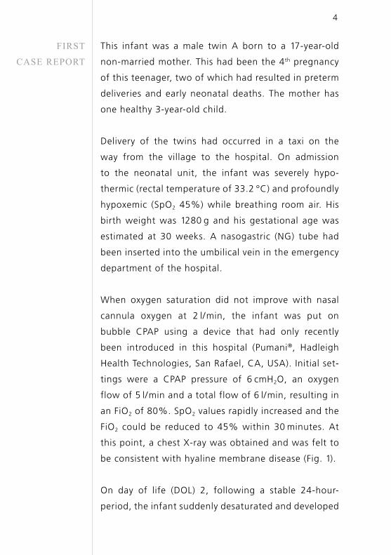
This infant was a male twin A born to a 17-year-old
non-married mother. This had been the 4th pregnancy
of this teenager, two of which had resulted in preterm
deliveries and early neonatal deaths. The mother has
one healthy 3-year-old child.
Delivery of the twins had occurred in a taxi on the
way from the village to the hospital. On admission
to the neonatal unit, the infant was severely hypo-
thermic (rectal temperature of 33.2 °C) and profoundly
hypoxemic (SpO2 45%) while breathing room air. His
birth weight was 1280 g and his gestational age was
estimated at 30 weeks. A nasogastric (NG) tube had
been inserted into the umbilical vein in the emergency
department of the hospital.
When oxygen saturation did not improve with nasal
cannula oxygen at 2 l/min, the infant was put on
bubble CPAP using a device that had only recently
been introduced in this hospital (Pumani®, Hadleigh
Health Technologies, San Rafael, CA, USA). Initial set-
tings were a CPAP pressure of 6 cmH2O, an oxygen
flow of 5 l/min and a total flow of 6 l/min, resulting in
an FiO2 of 80%. SpO2 values rapidly increased and the
FiO2 could be reduced to 45% within 30 minutes. At
this point, a chest X-ray was obtained and was felt to
be consistent with hyaline membrane disease (Fig. 1).
On day of life (DOL) 2, following a stable 24-hour-
period, the infant suddenly desaturated and deve loped

5
signs of poor perfusion. On auscultation, an unusual
rhythmic crunching sound was heard that could not
be classified. Findings on a second chest X-ray, which
became available two hours later, were consistent with
pneumopericardium (Fig. 2). SpO2 was 80% on an FiO2
of 90% and capillary refill time remained prolonged
despite the administration of a fluid bolus. At this
point, it was decided to attempt pericardiocentesis.
A 24 G venous cannula with a 2-ml syringe attached
was used. The pericardial space was entered from a
subxiphoid approach at a 30-degree-angle with the
needle directed against the left shoulder. Imme diately,
8 ml of air could be aspirated. SpO2 increased to 91%
and skin perfusion improved. The venous cannula
was left in place for intermittent aspiration (Fig. 3).
Unfortunately, the patient died during the following
night.

6
Twin A: Chest X-ray following stabilization with
bubble CPAP on DOL 1: low lung volumes, reticulo-
granular pattern and air bronchograms, consistent
with hyaline membrane disease (note intrahepatic
position of UVC and malpositioned nasogastric tube).
Fig. 1

Fig. 2
7
Twin A: Chest X-ray following sudden deterioration
on DOL 2: the heart is almost completely surrounded
by air, with the pericardium sharply outlined by air
density on either side.

8
Twin A on DOL 2: A 24 G venous catheter was used
to drain the pneumopericardium: despite initial aspi-
ration of 8 ml of air, the patient’s condition improved
only transiently (note poor skin perfusion).
Fig. 3

SECOND
CASE REPORT
9
Like his brother, twin B arrived in a critical condition
with hypothermia (rectal temperature 33.3 °C) and
significant respiratory distress and an SpO2 of 48%.
His birth weight was 1150 g. He was put on nasal can-
nula oxygen at 2 l/min and his color improved. After
stabilization of his twin brother, twin B was also put
on CPAP with a pressure of 6 cmH2O, an oxygen flow
of 4 l/min and a total flow of 6 l/min, resulting in an
FiO2 of 60%. A chest X-ray revealed findings consi-
stent with hyaline membrane disease. An NG tube had
been inserted into the umbilical vein, but was mal-
positioned in the portal vein (Fig. 4). Another NG tube
was inserted next to the first one, hoping that it would
enter the inferior vena cava. The infant responded well
to CPAP and the FiO2 could be weaned to 45%.
On DOL 2, the patient appeared stable with de creasing
signs of respiratory distress and a lower oxygen requi-
rement. However, on DOL 3, his course was compli-
cated by a right-sided pneumothorax (Fig. 5). A 24
G venous cannula was used to aspirate air (Fig. 6).
Unfortunately, there appeared to be a large air leak
and almost continuous aspiration was required. This
was done manually since no vacuum system was avai-
lable. A nurse was called to duty, sat at the bedside
and continued to intermittently aspirate air. Despite
these efforts, the patient died 6 hours later.

Fig. 4
10
Twin B: Chest X-ray following stabilization with
bubble CPAP on DOL 1: low lung volumes, reticulo-
granular pattern and air bronchograms, consistent
with hyaline membrane disease (note malposition of
the UVC in the portal vein).

Fig. 5
11
Twin B on DOL 3: chest of X-ray following gradual
deterioration on DOL 3: right-sided pneumothorax
with slight mediastinal shift to the left (note mal-
positioned UVC).

12
Twin B on DOL 3: A 24 G venous catheter was used
to drain the pneumothorax: because a continuous
suction system was lacking, intermittent manual
aspiration was performed every 5 minutes over
6 hours (until the patient’s death).
Fig. 6

13
DISCUSSIONThe two case reports illustrate multiple challenges
encountered in perinatal care in resource-limited coun-
tries. Teenage pregnancies, poor antenatal care with
late booking, delayed presentation or out-of-hospital
deliveries even in high-risk pregnancies are common
and put both mothers and babies at risk.
Until very recently, nasal cannula oxygen was the only
form of respiratory support available at Rundu State
Hospital. Flow rates between 0.5 – 4 l/min are used
and the gas is not warmed and marginally humidified
since there is only cold bubble humidification. SpO2
is measured only intermittently due to the lack of
adequate pulse oximetry equipment. Very likely, both
hypoxic and hyperoxic episodes go largely unnoticed.
The introduction of CPAP devices potentially can have a
dramatic impact on the survival rates of neonates with
respiratory distress (1, 2). In July 2017, the Pumani©
bubble CPAP device (Hadleigh Health Technologies,
San Rafael, CA, USA) was introduced at the Rundu
State Hospital. This machine is very robust, easy to use
and affordable. Initially, the two patients presented in
this report responded very nicely: SpO2 rapidly increa-
sed and FiO2 could be reduced to less than 50%.
While patients with poorly compliant lungs bene-
fit from CPAP therapy, they can still develop air leak
complications when spontaneous respiratory efforts
lead to (focal) overdistension and lung damage.

14
First described by Macklin in 1939, air can travel along
the vascular sheaths and eventually lead to pulmonary
interstitial emphysema and pneumomediastinum (3).
It has been speculated that mediastinal air dissecting
at the reflection of the parietal to visceral pericardium
near the ostia of the pulmonary veins leads to pneumo-
pericardium (4). As the two presented cases illustrate,
detection and management of air leak syndromes can
be very challenging in resource-limited countries.
Literature on pneumopericardium in neonates is
with hyaline mem brane disease is scarce. One of the
largest series was published from the University of
Minnesota Hospitals and St. Paul Children’s Hospital
in the pre-surfactant era (5). In 23 out of a total of
28 patients, pneumopericardium resulted in clinical
pericardial tampo nade. Mortality rate was 54%. The
rhythmic crunching sound heard in twin A is known
as Hamman’s sign (after Louis Hamman, Johns Hop-
kins University clinician), which can also be heard in
patients with anterior pneumomediastinum, or «bruit
de moulin» (mill-wheel murmur).
In high income countries, the described patients
would have qualified for early surfactant replacement
therapy; however, this expensive drug is currently not
avai lable at this hospital. It is noteworthy that sur-
factant preparations were included in the Essential
Drug List of WHO in 2008 (6). In its current version
(20th List, March 2017, amended August 2017), sur-

15
factant is listed in the Complementary List. This list
presents essential medicines for «priority diseases, for
which specialized diagnostic or monitoring facilities,
and/or specialist medical care, and/or specialist trai-
ning are needed.» (7).
The use of surfactant replacement therapy (SRT) in
developing countries is still limited because of a) high
cost, which may exceed the per-capita GNP (300 – 500
USD) in some countries, b) lack of skilled personnel to
administer SRT, and c) lack of support systems after
the SRT. Recent developments may have the potential
to address these constraints.
The development of SP-B and SP-C enriched syn thetic
surfactants (8) may reduce the costs of SRT. In recent
years, the INSURE (intubation-surfactant-extubation)
procedure and other forms of less invasive surfactant
administration (LISA) have become increasingly popular
in high income countries (9). The latter procedure does
not require mechanical ventilation via an endotracheal
tube, since the surfactant preparation is administered
intratracheally via a small diameter tube, while the
infant is spontaneously breathing (10 ). Alternatively,
surfactant administered through a laryngeal mask air-
way (LMA) may be a valuable option as it does not
require the skills necessary for laryngoscopy and / or
intubation (11). Robust and low-cost CPAP devices
have been developed and could be used both prior to
and after SRT (12).

CONCLUSION Improving the management of neonates with respira-
tory distress in developing countries has the potential
to greatly improve survival chances for affected neo-
nates. This will require improvements in both diagnosis
and therapy, as well as management of complications
of various causes of respiratory failure. It is highly
desirable that research in neonatology also addresses
the needs of newborn infants born in low and middle
income countries (10).
16

1. Kamath BD, Macguire ER, McClure EM, Goldenberg RL, Jobe
AH. Neonatal mortality from respiratory distress syndrome:
lessons for low-resource countries. Pediatrics
2011;127:1139 – 1146 (Abstract)
2. Kawaza K, Machen HE, Brown J, et al. Efficacy of a low-cost
bubble CPAP system in treatment of respiratory distress in a
neonatal ward in Malawi. PLoS One 2014;9:e86327 (Abstract)
3. Macklin CC. Transport of air along sheaths of pulmonic blood
vessels from alveoli to mediastinum – clinical implications. Arch
Intern Med (Chic.) 1939;64:913 – 926 (no abstract available)
4. Jokic RR, Kovacevi B, Beserminji M, Tatic M. Surgical approach
to tension pneumopericardium in newborns and infants. JPSS
2008;2:32 – 35 (Abstract)
5. Emery RW, Landes RG, Lindsay WG, Thompson T, Nicoloff DM.
Surgical treatment of pneumopericardium in the neonate.
Word J Surg 1978;2:631 – 637 (no abstract)
6. Vidyasagar D, Velaphi S, Bhat VB. Surfactant replacement
therapy in developing countries. Neonatology
2011;99:355 – 366 (Abstract)
7. WHO Model List of Essential Medicines, 20th List (March 2017)
(Website)
8. Sweet DG, Turner MA, Stranák Z, et al. A first-in-human clinical
study of a new SP-B and SP-C enriched synthetic surfactant
(CHF5633) in preterm babies with respiratory distress
syndrome. Arch Dis Child Fetal Neonatal Ed
2017;102:F497-F503 (Abstract)
9. Gortner L, Schüller SS, Herting E. Review demonstrates that
less invasive surfactant administration in preterm neonates
leads to fewer complications. Acta Paediatr 2017 Nov 24
[Epub ahead of print] (Abstract)
REFERENCES
17

10. Herting E. Less invasive surfactant administration (LISA) – ways
to deliver surfactant in spontaneously breathing infants. Early
Hum Dev 2013;89:875 – 880 (Abstract)
11. Roberts KD, Brown R, Lampland AL, et al. Laryngeal mask
airway for surfactant administration in neonates: a rando-
mized, controlled trial. J Pediatr 2017 Nov 21 (Epub ahead
of print) (Abstract)
12. Berger TM. Neonatal respiratory care: not how, but where
and when. Lancet Respir Med 2013;1:280 – 282 (no abstract
available)
18


SUPPORTED BY
CONTACT
Swiss Society of Neonatology
www.neonet.ch
con
cep
t &
des
ign
by
mes
ch.c
h