ch06: endodontic diagnostic procedures - health mantra · interest the second important asset...
TRANSCRIPT

“For I seek the truth by which no man has ever been harmed.”
–Marcus Aurelius, Meditations VI. 21, 173 AD
Before initiating treatment, one must first assemblecollective information regarding signs, symptoms, andhistory. That information is then combined withresults from the clinical examination and tests. Thisprocess is diagnosis. Stated another way, diagnosis isthe procedure of accepting a patient, recognizing thathe has a problem, determining the cause of the prob-lem, and developing a treatment plan that will solve oralleviate the problem.
The diagnostician must have a thorough knowl-edge of examination procedures—percussion, palpa-tion, probing, and pulp testing; a knowledge of patho-sis and its radiographic and clinical manifestations;an awareness of the various modalities of treatment;and, above all, a questioning mind. To be added tothese critical skills is the most basic skill of all, listen-ing to the patient.
Of all of the important diagnostic tools, the art oflistening is the most underrated. Yet careful and atten-tive listening establishes patient-dentist rapport,understanding, and trust. Such a relationship alsoenhances the patient’s reliability as a historian.1
REQUIREMENTS OF A DIAGNOSTICIAN
Diagnosis is a personal and cognitive experience; there-fore, many of the qualities of a good diagnostician are ofan interpersonal nature and are based on knowledge,experience, and diagnostic tools. Diagnosing orofacialdisease is similar to other medical diagnosis. Pulp tests,radiographs, percussion, palpation, and other tests andprocedures can facilitate the diagnosing of dental/facialdisease, just as the electrocardiograph, electroencephalo-graph, echocardiograph, computed axial tomographicand magnetic resonance imaging scan, and a host ofother radiographs can facilitate medical diagnosis.
A dentist can develop a number of assets to becomea successful diagnostician. The most important of theseare knowledge, interest, intuition, curiosity, andpatience. The successful diagnostician must also haveacute senses and the necessary equipment for diagnosis.
Knowledge
Primarily, a dentist must depend on himself, not thelaboratory. Therefore, knowledge is the most impor-tant asset the dentist must possess. This includes famil-iarity with all local orofacial causes of pain, as well asnumerous systemic, neurogenic, and psychologicalcauses. In addition, the dentist must be aware of themany physical, perceptual, emotional, and behavioralchanges brought about by chronic pain. He must knowthat constant overwhelming pain can affect the func-tion of every organ of the body. Chronic pain patientscan develop increased blood pressure, heart rate, kid-ney function, decreased bowel activity, and hormonelevels. They can have many symptoms, such as nausea,vomiting, photophobia, tinnitus, and vertigo. Theastute clinician gathers knowledge about the patientand his problem through a thorough history and anexamination. The history and examination includeevaluating the physical, emotional, behavioral, and per-ceptual aspects of the patient’s pain experience.
Under knowledge must also be listed the importantasset of knowing when and where to refer the patientfor additional consultation. This comes with experi-ence and the help of physicians, psychologists, and fel-low dentists who may be depended on to assist in diag-nosis. Often the patient is referred because examina-tion reveals a problem clearly in the province of theneurologist or otolaryngologist. Sometimes the patientis referred because the examiner has exhausted hisknowledge and needs help in diagnosis. The recogni-tion of fallibility and limitation—knowing when to yellfor help—is also a major asset to the dentist.
Chapter 6
ENDODONTIC DIAGNOSTIC PROCEDURES
John I. Ingle, Geoffrey S. Heithersay, Gary R. Hartwell, Albert C. Goerig,
F. James Marshall, Robert M. Krasny, Alfred L. Frank, and Cyril Gaum

Interest
The second important asset possessed by a good diag-nostician is interest. The dentist must have a keeninterest in the patient and his or her problem and mustevidence this interest by handling the patient withunderstanding. If this attitude is not natural to the den-tist, he will render the patient and the profession a serv-ice by referring all diagnostic problems to an interestedand competent fellow practitioner.
Intuition
In addition to interest and knowledge, the good diag-nostician is blessed with intuition or “sixth sense,” so tospeak. Good diagnosticians intuitively sense the pres-ence of something unusual. This ability, which some-times allows for “instant” diagnosis, is developedthrough broad experience with pain problems havingunusual and multiple diagnoses.
Intuition tells the dentist when the patient is hold-ing back information or is not telling the completetruth. Moreover, intuition immediately makes theexaminer subtly aware of the patient who “knows toomuch,” that is, all of the words and symptoms related toa certain condition. Intuition allows the dentist to sus-pect the unusual, but it also goes hand in hand with stillanother prime asset of a good diagnostician, curiosity.
Curiosity
The dentist must pursue or develop a natural curiosityabout the patient and his condition if perseverance is tobe maintained in arriving at a diagnosis. Dr. HarrySicher often likened dental diagnosis to the actions of agood detective, and curiosity is a detective’s greatest asset(personal communication, 1954). Medawar describeddiagnosis as the “use of the hypothetico-deductive sys-tem.”2 Again, curiosity goes with interest, and the den-tist who is bored by the painstaking methods of diagno-sis will never have the curiosity to delve a little deeper,probe a little further, or ask the unusual. All of this takestime and thus requires patience.
Patience
Often a definitive diagnosis of unusual pain may takehours, days, or even months to develop. Some patientscomplaining of unusual pain may have suffered thispain for years, so the dentist cannot expect to make aquick diagnosis in a matter of minutes. This is the rea-son, as stated earlier, why a difficult diagnosis may beunrewarding financially but very rewarding emotional-ly. Again, if the dentist is not willing to sacrifice thetime to attempt to help these individuals, he is urged torefer the patient for diagnosis rather than make an
204 Endodontics
incorrect, quick diagnosis that may result in impropertreatment, such as reaching for the forceps or removinga healthy pulp.
The dentist obviously cannot abandon other patientsto see one person repeatedly. Too frequently, the prob-lem patient is asked to return at the end of the day, whenboth dentist and patient are tired and irritable. A bettersolution is to see the patient in the morning, beforeoffice hours. The dentists who are not willing to assumethis imposition, and they are many, are urged to referthe patient to their more altruistic colleagues.
Senses
The good diagnostician must have the astuteness tograsp what his senses reveal. First, he has a voice to askquestions and ears to hear the answer; he has eyes to seeand hands to probe and palpate. In short, the dentisthas senses with which to communicate with the sickpatient. But, as Friedman pointed out, “One must learnto listen with the third ear and see with the third eye”(personal communication, 1972).
Controlling these senses, however, is the mind, andif the mind does not inquire and then reason, or hasnot accumulated the knowledge necessary to inquireand finally to analyze, then the senses are useless. Themind must list all of the possible causes of the pain andthen, more often than not, eliminate them one by oneuntil the correct diagnosis is made.
HISTORY
Anamnesis, “recollection” or “calling to memory,” is thefirst step in developing a diagnosis. The importance ofobtaining and recording this “history” goes beyondmedicolegal protection. A complete history (Table 6-1)will not determine treatment but may influence modi-fications in endodontic treatment modalities. It willseldom deny treatment. A complete medical historyshould contain, as a baseline, the vital signs; give earlywarning of unsuspected general disease; and definerisks to the health of the staff as well as identify therisks of treatment to the patient. The medical historymust be updated regularly, especially if there have beenany changes in the patient’s health status.
The procedure developed by the American Societyof Anesthesiologists is a good system for organizingand assigning risk (Table 6-2). Once the status of thepatient’s general health has been established, a dentaldiagnosis is best developed by following the time-hon-ored formula of determining the chief complaint,enlarging on this complaint with questions about thepresent dental illness, relating the history of past den-tal illness to the chief complaint, and combining this

Endodontic Diagnostic Procedures 205
Table 6-1 Medical History Form*
MEDICAL HISTORYName ________________________ Sex _______________________________________ Date of Birth __________________________Address ________________________________________________________________________________________________________Telephone _________________________________ Height _______________________ Weight _______________________________Date ________________________ Occupation ________________________________ Marital Status _________________________
MEDICAL HISTORY CIRCLE1. Are you having pain or discomfort at this time? ........................................................................................................ YES NO2. Do you feel very nervous about having dentistry treatment? ................................................................................... YES NO3. Have you ever had a bad experience in the dentistry office? ..................................................................................... YES NO4. Have you been a patient in the hospital during the past 2 years? ............................................................................. YES NO5. Have you been under the care of a medical doctor during the past 2 years? ........................................................... YES NO6. Have you taken any medicine or drugs during the past 2 years? .............................................................................. YES NO7. Are you allergic to (ie, itching, rash, swelling of hands, feet, or eyes)
or made sick by penicillin, aspirin, codeine, or any drugs or medications? ............................................................. YES NO8. Have you ever had any excessive bleeding requiring special treatment? .................................................................. YES NO9. Circle any of the following which you have had or have at present:
Heart Failure Emphysema AIDS or HIVHeart Disease or Attack Cough Hepatitis A (infectious)Angina Pectoris Tuberculosis (TB) Hepatitis B (serum)High Blood Pressure Asthma Liver DiseaseHeart Murmur Hay Fever Yellow JaundiceRheumatic Fever Sinus Trouble Blood TransfusionCongenital Heart Lesions Allergies or Hives Drug AddictionScarlet Fever Diabetes HemophiliaArtificial Heart Valve Thyroid Disease Venereal Disease (Syphilis, Gonorrhea)Heart Pacemaker X-ray or Cobalt Treatment Cold SoresHeart Surgery Chemotherapy (Cancer, Leukemia) Genital HerpesArtificial Joint Arthritis Epilepsy or SeizuresAnemia Rheumatism Fainting or Dizzy SpellsStroke Cortisone Medicine NervousnessKidney Trouble Glaucoma Psychiatric TreatmentUlcers Pain in Jaw Joints Sickle Cell Disease
Bruise Easily
10. When you walk up stairs or take a walk, do you ever have to stop because of pain in your chest, or shortness of breath, or because you are very tired? .......................................................... YES NO
11. Do your ankles swell during the day? .......................................................................................................................... YES NO12. Do you use more than two pillows to sleep? ............................................................................................................... YES NO13. Have you lost or gained more than 10 pounds in the past year? ............................................................................... YES NO14. Do you ever wake up from sleep short of breath? ....................................................................................................... YES NO15. Are you on a special diet? .............................................................................................................................................. YES NO16. Has your medical doctor ever said you have a cancer or tumor? ............................................................................... YES NO17. Do you have any disease, condition, or problem not listed? ...................................................................................... YES NO18. WOMEN: Are you pregnant now? .......................................................................................................................... YES NO
Are you practicing birth control? .......................................................................................................... YES NODo you anticipate becoming pregnant? ................................................................................................ YES NO
To the best of my knowledge, all of the preceding answers are true and correct. If I ever have any change in my health, or if my medicines change,I will inform the doctor of dentistry at the next appointment without fail.
Date Dentist Signature Signature of Patient, Parent, or Guardian
MEDICAL HISTORY/PHYSICAL EVALUATION UPDATE
Date Addition Signatures
_______________ ____________________________ ______________________________ ______________________________
_______________ ____________________________ ______________________________ ______________________________
_______________ ____________________________ ______________________________ ______________________________
This comprehensive medical history responds to contemporary advances in physical evaluation and to increasing malpractice claims.
*Reproduced with permission from McCarthy FM. A new patient administered history developed for dentistry. J Am Dent Assoc 1985;111:595.

with information about the patient’s general health(medical history) and the examination results.
Chief Complaint
The chief complaint, usually in the patient’s ownwords, is a description of the dental problem for whichthe patient seeks care. The verbal complaint may beaccompanied by the patient pointing to the generalarea of the problem.
After establishment and recording of the chief com-plaint, the examination process is continued by obtain-ing a history of the present illness.
A patient in acute distress should undergo diagnosisand examination as quickly as possible so the chiefcomplaint may be treated as expeditiously as possible.At a later time, when the patient is pain free and morerational, a complete treatment plan may be established.No treatment should be rendered unless the examineris certain of the diagnosis. Patients with severe painfrom pulpitis have difficulty in cooperating with thediagnostic procedures, but until the diagnosis has beenmade and the correct tooth identified, treatment mustnot be started (see chapter 7).
Present Dental Illness
A history of the present illness should indicate theseverity and the urgency of the problem. If the problem
206 Endodontics
is long-standing, proceed with detailed questions aboutpast episodes of pain or swelling and any previoustreatment performed to remedy the condition.
Pain is frequently the main component of thepatient’s complaint. A history of pain that persistswithout exacerbation may indicate a problem not ofdental origin. If the chief complaint is “toothache” butthe symptoms are too vague to establish a diagnosis,analgesics can be prescribed to help the patient toleratethe pain until the toothache localizes. If the patientarrives self-medicated with analgesics or sedatives, adiagnosis may be difficult to establish.3
The initial questions should help establish two basiccomponents of pain: time (chronicity) and severity (orintensity). Start by asking such questions as “How longhave you had this problem?” “How painful is it?” and“How often does it hurt?” Continue the questioningwith “When does it hurt?” “When does it go away?”“What makes it hurt?” “What makes it hurt worse?”and “What makes it hurt less or go away?”
A history of painful responses to thermal changessuggests a problem of pulpal origin and will need to befollowed up with clinical tests, using the thermal testthat would most closely duplicate the patient’s com-plaint: use ice if the complaint is pain with cold, anduse a hot stimulus if the complaint is pain with suchthings as hot drinks.4 It could also be important to
Table 6-2 American Society of Anesthesiologists (ASA) Physical Status Classification*
ASA Class Patient Description Clinical Examples Clinical Management
1 A normally healthy patient No organic, physiologic, biochemical, Routine careor psychiatric disturbance; treatment is for localized disorder
2 A patient with mild Controlled essential hypertension, Routine care but limit systemic disease pronounced obesity, psychiatric procedural stress and length
disturbance of appointment
3 A patient with severe systemic Severe diabetes mellitus, congestive heart Strict limitation of complex disease that is not failure, chronic obstructive pulmonary procedures; careful anxietyincapacitating disease control
4 A patient with an incapacitating Acute myocardial infarction; advanced Emergency or palliative care,systemic disease that is a pulmonary, cardiac, hepatic, or renal usually in a hospitalconstant threat to life insufficiency
5 A moribund patient who is not Uncontrolled massive internal bleeding, Emergency life support onlyexpected to live 24 hours with rapidly progressing cardiacor without operation insufficiency with renal failure
*Used to categorize patients following history and examination. Classification should be entered prominently in chart. Assigning risk fromtreatment and level of clinical management follows classification. American Society of Anesthesiologists. New classification of physical sta-tus. Anesthesiology 1963;24:111.
learn that a tooth has been sensitive to thermal changesbut no longer responds to such stimuli; this wouldindicate that the tooth may have a pulp that is nownecrotic.5
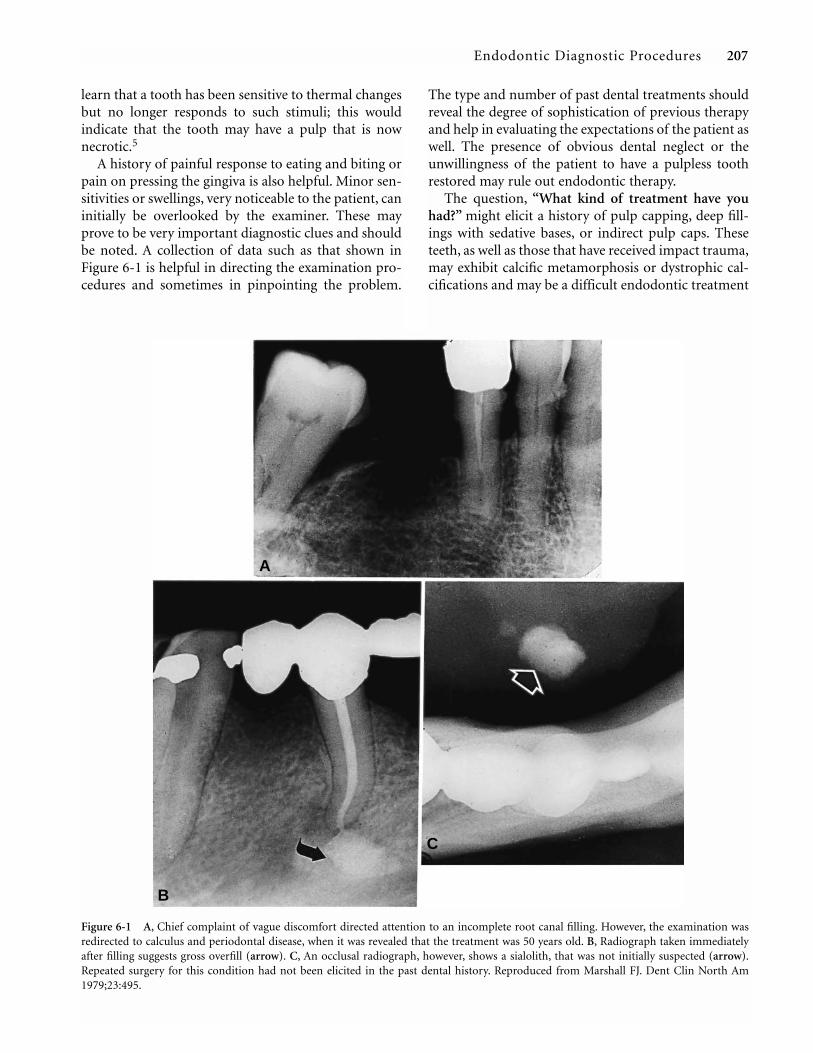
A history of painful response to eating and biting orpain on pressing the gingiva is also helpful. Minor sen-sitivities or swellings, very noticeable to the patient, caninitially be overlooked by the examiner. These mayprove to be very important diagnostic clues and shouldbe noted. A collection of data such as that shown inFigure 6-1 is helpful in directing the examination pro-cedures and sometimes in pinpointing the problem.
The type and number of past dental treatments shouldreveal the degree of sophistication of previous therapyand help in evaluating the expectations of the patient aswell. The presence of obvious dental neglect or theunwillingness of the patient to have a pulpless toothrestored may rule out endodontic therapy.
The question, “What kind of treatment have youhad?” might elicit a history of pulp capping, deep fill-ings with sedative bases, or indirect pulp caps. Theseteeth, as well as those that have received impact trauma,may exhibit calcific metamorphosis or dystrophic cal-cifications and may be a difficult endodontic treatment
Endodontic Diagnostic Procedures 207
Figure 6-1 A, Chief complaint of vague discomfort directed attention to an incomplete root canal filling. However, the examination wasredirected to calculus and periodontal disease, when it was revealed that the treatment was 50 years old. B, Radiograph taken immediatelyafter filling suggests gross overfill (arrow). C, An occlusal radiograph, however, shows a sialolith, that was not initially suspected (arrow).Repeated surgery for this condition had not been elicited in the past dental history. Reproduced from Marshall FJ. Dent Clin North Am1979;23:495.
A
B
C

problem (Figure 6-2). Although a positive and accurateanswer may not result, the question will prompt a clos-er look at radiographs for the presence or absence ofcement bases or for recurrent caries or caries remainingunder restorations (Figure 6-3).
Full-crown restorations should also raise questions tothe patient regarding “wet” or “dry” drilling. “Drydrilling” may result in an increased incidence of inflam-mation and even internal resorption (see Figure 4-44).Patients who have undergone orthodontic treatmentmay have areas of resorption or pulpal changes. In thepast decade, there has been an increased interest in adultorthodontics, and recent studies indicate that the adultpulp may be more susceptible than younger pulps tosuch iatral trauma6 (Figure 6-4).
The question, “How many times has this tooth orhave these teeth been treated?” is also pertinent. Atooth with a history of repeated restorations and mul-
208 Endodontics
tiple occurrences of caries (“stressed pulp”) should beevaluated carefully with respect to pulpal status beforeprocedures such as full crowns or bridge abutmentrestorations are initiated. Root canal therapy prior torestorations in such teeth is often indicated. The ques-tion, “How recently has this tooth or area been treat-ed?” may provide information that the problem ofthermal sensitivity is merely a reaction to a recentlyplaced restoration. If the pain is of low intensity, apatient may tolerate it in the hope that it will subside.Pain existing for several months may have become partof the patient’s lifestyle.7
A history of long-standing, severe pain should raisesuspicion that the condition may be other than pulpalin origin. Additional examinations for myofascial orneurologic pain, as well as cardiac referred pain or pos-sibly psychogenic pain, should be considered. A moredetailed discussion of this subject is found in chapter 8.
Finally, the patient must be asked about past reac-tions to dental procedures; to pain, both dental andgeneral; and to expectations for treatment. A patientwith a history of low pain threshold and strong anal-gesic dependency, as well as many previous attempts tosolve the problem, may require special treatment orreferral.
It should be noted that history taking is a process ofquestions and answers and that many questions are
Figure 6-2 A, Mandibular incisors with sclerosed canals andchronic apical periodontitis. Surgical treatment was required. B,Same case. “Dark teeth” caused the patient to seek treatment. Ifimmediate post-trauma and follow-up radiographs had been maderegularly, nonsurgical therapy could have been attempted whenchange was first noted. Reproduced from Marshall FJ. Dent ClinNorth Am 1979;23:495.
Figure 6-3 Radiolucent material (possibly a pulp cap) showsunder occlusal amalgam of a second molar. Patient had complainedof pain for 4 months, referring over the entire maxilla andmandible. Pain was not relieved completely by infiltration anesthe-sia over the second molar and persisted in the mandible. However,a posterior superior alveolar block relieved pain in both areas,demonstrating the fallibility of anesthesia in diagnosing referredpain. Reproduced with permission from Marshall FJ. Dent ClinNorth Am 1979;23:495.

repeated. These repeated questions are not redundantbut are deliberate attempts to confirm data and validatethe diagnosis.
Medical History
Patients need to share their medical problems withtheir dentists so the data can be used in planning treat-ment.8 This begins when the patient completes somestandard form (see Table 6-1 or 6-2), usually at thesame time as the receptionist records other basic data.The confidence needed to share these data builds slow-ly in some people. Thus, there is a need to review care-fully and sincerely the answers the patient has recordedon the health history form. This review may be morecomfortable for the patient after asking about the chiefcomplaint.
In reviewing the medical history, particular empha-sis must be placed on illnesses, history of bleeding, andmedications. Illness often means hospitalization topatients; consequently, they may not list weightchanges, accidents, or problems related to stress andtension. Patients who are African American should bequestioned about sickle cell anemia; there is a report ofpulp necrosis occurring in patients with this distressingcondition.9
The term bleeding is usually interpreted by thepatient to mean frank blood and seldom elicits answersrelated to bruising or healing time, chronic use ofaspirin10 (not considered a drug by many people), or ahistory of liver disease. These should all be specificallymentioned in the medical history form.
Medication means to many people only those itemsobtained by written prescription. Dentists must alsoask about “pills” and “drugs.” With the availability ofhome remedy “medical” texts, many people areself-medicating with diet pills, sleep inducers, and vita-mins, as well as “recreational drugs,” to mention only afew. Even if these self-administered medications do notinfluence treatment directly, the knowledge thatpatients have a tendency to “do their own thing” may behelpful in planning treatment.
Women should be asked if they are pregnant or ifthey have menstrual or menopausal problems. Positiveanswers to these questions must be weighed and evalu-ated along with the other responses to determine therisk of treatment against the risk of nontreatment.
When the history uncovers a serious problem, and areview of the systems involved (cardiac, respiratory,etc) does not explain the problem, the patient’s physi-cian must be consulted.
During these interviews, the dentist-patient rela-tionship tends to crystallize. A rapport is establishedthat is relevant and meaningful to all future relation-ships. This is the time when anxious, frightenedpatients may be calmed and reassured even thoughthey may not be completely at ease until the first treat-ment is completed and they learn that dental proce-dures can be uneventful and nontraumatic.
Kindness and attention to their concerns or prob-lems (chief complaint) during the history-taking willgreatly reduce most patients’ emotional trauma andstress, particularly when this phase is followed by athorough, painless examination.
CLINICAL EXAMINATION
In general, the clinical examination should follow alogical sequence from the general to the specific, fromthe more obvious to the less obvious, from the externalto the internal. The results of the examination, alongwith the information from the patient’s history, will becombined to establish the diagnosis, formulate a treat-ment plan, and determine the prognosis.
Vital Signs
The first step in examination is to record the patient’svital signs, thus establishing a baseline or a “norm” foreach patient during treatment, whether routine oremergency. Patients with test values outside the rangeof acceptable norms are at risk, as is the dentist whotreats them.8 Common sense suggests that this riskshould be shared with the patient’s physician by a tele-phone conversation at least. Information receivedshould be recorded in the chart and dated.
Endodontic Diagnostic Procedures 209
Figure 6-4 Invasive resorption (arrow) triggered by orthodonticmovement of molar tooth. (Courtesy of Robert M. Krasny.)

The vital signs may be recorded by any trainedmember of the office team. However, abnormal valuesmust be evaluated by the doctor. The person of firstcontact should also record, for later evaluation, anyadditional observations of abnormalities such asbreathlessness, color change, altered gait, or unusualbody movements observed during the initial meeting.
Blood Pressure (normal: 120/80 mm Hg for personsunder age 60; 140/90 mm /Hg for persons over age 60).Routine use of the sphygmomanometer not only estab-lishes a baseline blood pressure but occasionally bringsto light unsuspected cases of hypertension in patientswho are not regularly seeing a physician or are notmaintaining prescribed regimens of therapy. Halpernreported that only 18% of the dental clinic patientsattending Temple University Dental School “were seeingtheir physicians.”8 At times, however, elevated bloodpressure is caused only by the stress and anxiety of themoment and can be dealt with by reassurance or, if nec-essary, pretreatment sedation. Even more important,however, is the emphasis that this face-to-face proce-dure places on an examination. Both the patient and thedoctor are inclined to be more serious in their questionsand answers when the examination begins with bloodpressure records. It must be stressed that no patient,with or without a dental emergency, should be treatedwhen his diastolic blood pressure is over 100 mm.11
Pulse Rate and Respiration (normal: pulse, 60 to 100beats/minute; respiration, 16 to 18 breaths per minute).When these examinations are added to the recording ofblood pressure, the dentist increases the opportunity toknow the patient better. These examinations also showthe patient, by physical contact, how further examina-tion will proceed—deliberately, gently, and completely.Pulse and respiration rates may also be elevated owing tostress and anxiety; in fact, these signs may be even betterindicators of stress than is blood pressure. Tests withmarkedly positive findings should be repeated later inthe appointment or at a subsequent appointment.
Temperature (normal: body temperature, 98.6˚F[37˚C]). The taking and recording of body tempera-ture is a simple, significant procedure. An elevated tem-perature (fever) is one indication of a total body reac-tion to inflammatory disease. If the body temperatureis not elevated, one can assume that the body is “man-aging” its defenses well, that whatever the local signs are(pain, swelling, abscess formation, etc), systemic treat-ment, with its attendant risks, will likely not berequired. A temperature above 98.6˚ but less than 100˚Findicates localized disease.12 Localized disease can usu-ally be treated by removing the cause (eg, cleaning theroot canal) and/or incision and drainage.
210 Endodontics
Cancer Screen (soft tissue examination: lumps,bumps, white spots). Every new patient must be rou-tinely screened for cancer and other soft tissue non-odontogenic conditions as part of the examination. Andthey must be informed of the results! This examinationshould include a survey of the face, lips, neck, and intra-oral soft tissues. When such examinations are maderoutinely, without secrecy, they will usually dispel theunstated fears of the cancerphobe and add to the confi-dence and rapport of all patients with their dentists. Thesooner this examination is completed the better.
It is sometimes argued that dentists are liable if theyinform patients that they are performing an examina-tion and then miss finding disease when it is present. Infact, dentists are even more liable if they miss reportingthe disease because they have not made an examination.
Extraorally, a cancer survey includes palpation formasses and examination for asymmetry and colorchanges. Intraorally, this examination is repeated withthe additional care of directed lighting and of movingthe tongue in such a manner so that all areas can beclearly seen (Table 6-3). Detailed procedures are pre-sented elsewhere.13
Extraoral Examination
Inflammatory changes originating intraorally andobservable extraorally may indicate a serious, spread-ing problem.14 The patient must be examined forasymmetries, localized swelling, changes in color orbruises, abrasions, cuts or scars, and similar signs ofdisease, trauma, or previous treatment. Positive find-ings combined with the chief complaint and informa-tion about past injuries or previous treatments to teethor jaws will begin to clarify the extent of the patient’sproblem.
The extraoral examination includes the face, lips,and neck, which may need to be palpated if the patientreports soreness or if there are apparent areas ofinflammation. Painful and/or enlarged lymph nodesare of particular importance. They denote the spread of
Table 6-3 Oral Cancer Warning Signals*
Swelling, lump, or growth anywhere in or about the mouth
White, scaly patches inside the mouth
Any sore that does not heal
Numbness or pain anywhere in the mouth area
Repeated bleeding in the mouth without cause
*”Open Wide.” Reproduced with permission from the AmericanCancer Society, New York.

inflammation as well as possible malignant disease. Theextent and manner of jaw opening can provide infor-mation about possible myofascial pain and dysfunc-tion.15 The temporomandibular joint should be exam-ined during function for sensitivity to palpation, jointnoise, and irregular movement.16
Intraoral Examination
The intraoral examination is begun with a general eval-uation of the oral structures. The lips and cheeks areretracted while the teeth are in occlusal contact and theoral vestibules and buccal mucosa are examined forlocalized swelling and sinus tract or color changes. Withthe patient’s jaws apart, the dentist should evaluate in asimilar manner the lingual and palatal soft tissues. Also,the presence of tori should be noted. Finally, as part ofthe general inspection, carious lesions, discolorations,and other obvious abnormalities associated with theteeth, including loss of teeth and presence of supernu-merary or retained deciduous teeth, should be noted.
Often the particular tooth causing the complaint isreadily noted during this visual examination if it hasnot already been pointed out by the patient.Complaints associated with discolored or fracturedteeth, teeth with gross caries or large restorations, andteeth restored by full coverage are for the most partreadily located. True “puzzlement” begins when thecomplaint centers on teeth fully crowned and part ofextensive bridges or splints, or when only a few teethare restored, and then only with minimal restorations.
Transillumination with a fiber-optic light, directedthrough the crowns of teeth, can add further informa-tion.17 By this method, a pulpless tooth that is notnoticeably discolored may show a gross difference intranslucency when the shadow produced on a mirror iscompared to that of adjacent teeth. Transilluminationmay also locate teeth with vertical cracks or fractures.
If the involved tooth is not readily identified, it maybe necessary to thoroughly examine all of the teeth inthe half arch or opposing arch, depending on howspecifically the patient can localize the area of pain.Although the size of a carious lesion or the presence ofa crown or large restoration may point to the involvedtooth, the symptoms may be referred pain or pain froman adjoining tooth with problems. Adjacent teeth withlarge restorations or crowns can be assumed to beequally at risk. Regardless of the presence or absence offindings, the patient’s premonitions and descriptionsshould not be ignored in favor of what appears to beobvious. For the record and for the possibility of addi-tional later treatment, the condition of all teeth in theimmediate vicinity should also be recorded. This is
especially true following an accident. In fact, the gener-al state and care of the entire mouth must be notedalong with the particular tooth’s restorability andstrategic importance.
If the patient’s chief complaint includes symptomsthat occur following specific events (eg, chewing,drinking cold liquids), the specific intraoral examina-tion should include tests that duplicate or reproducethese symptoms. For example, if the chief complaint ispain with hot liquids, the clinical tests may include test-ing the suspected tooth with a hot stimulus. A positiveresponse (development of the chief complaint) will beimportant evidence in establishing the diagnosis.
In the following sections, various tests will bedescribed, detailing how to perform them and how toevaluate the results. The correct diagnosis can be estab-lished more readily the more information that is devel-oped from all sources: history, clinical examination,radiographic evaluation, and clinical tests.
Coronal Evaluation. For psychological reasonsand to expedite treatment, the most obviously affectedtooth is examined first, particularly when the patient,the history, or the general examination calls attentionto a certain tooth.
Using a mouth mirror and an explorer, and possiblya fiber-optic light source, the dentist carefully and thor-oughly examines the suspected tooth or teeth for caries,defective restorations, discoloration, enamel loss, ordefects that allow direct passage of stimuli to the pulp.Sometimes sealing off such leakage with temporarycements or periodontal dressings can be diagnostic(Figure 6-5). Vertical and horizontal fractures located bytransillumination should be further investigated by hav-
Endodontic Diagnostic Procedures 211
Figure 6-5 Zinc oxide–eugenol temporary cement packed buccal-ly and lingually to seal margins of a porcelain-fused-to-metal crownagainst external stimulus (leakage). If the patient’s complaint stops,then the margins can be permanently sealed or the crown remade.This technique is particularly helpful when several teeth arecrowned. (Courtesy of Dr. F. James Marshall.)
ing the patient bite on some firm object such as theTooth Slooth (Laguna Niguel, Calif.), or a wet cottonroll.18 Occlusal wear facets and parafunctional patternsare also sought out, as is tooth mobility.
Pulpal Evaluation
The clinical condition of the pulp can be evaluated bythermal stimuli, percussion, palpation, and vitality tests.Generally, pain of endodontic origin results from pulpinflammation that spreads from the coronal pulp api-cally to the periodontal ligament, which then spreads tothe periosteum overlying the apical bone and beyond.Pulpal and periradicular symptoms, therefore, some-times combine, making pulpal assessment difficult.
The purpose of evaluating the pulpal condition is toarrive at a diagnosis—namely, the nature of the diseaseinvolving the pulp. After determining the diagnosis,there are specific treatment options for each pulpalcondition. Irreversible pulpitis and pulp necrosisrequire removal of the pulp (pulp extirpation and rootcanal treatment, or extraction of the tooth), whereas atooth with a normal pulp or with reversible pulpitismay be treated by preserving the pulp (vital pulp ther-apy). The various methods of pulpal evaluation, then,do not dictate treatment but provide information thatcan be used with other information (history and radi-ographs) to establish a diagnosis. Pulp tests alone areusually not adequate for establishing a diagnosis butcan provide very useful information.19,20
Clinical Endodontic Tests
There are several ways to obtain information about thecondition of a tooth’s pulp and supporting structures.Probably no one test is sufficient in itself; the results ofseveral tests often have to be obtained to have enoughinformation to support a likely diagnosis or perhaps alist of differential diagnoses.
Thermal Tests. Two types of thermal tests areavailable, cold and hot stimuli. Neither is totally reliablein all cases, but both can provide very useful informa-tion in many cases of pulpal involvement.
The cold test may be used in differentiating betweenreversible and irreversible pulpitis and in identifyingteeth with necrotic pulps. It can also alleviate painbrought on by hot or warm stimuli, a finding thatpatients sometimes discover can provide them withmuch relief.
When cold is used to differentiate between reversibleand irreversible pulpitis, one must try to determine ifthe effect of stimulus application produces a lingeringeffect or if the pain subsides immediately on removal ofthe stimulus from the tooth. The “lingering” quality of
212 Endodontics
pain to a cold stimulus might be considered in cases inwhich the patient clearly feels that the pain is still pres-ent several seconds after stimulus removal. In testing, ifthe pain lingers, that is taken as evidence for irre-versible pulpitis; if pain subsides immediately afterstimulus removal, hypersensitivity or reversible pulpi-tis is the more likely diagnosis.
Cold as a test for pulp vitality (pulp necrosis versusvital pulp) is probably not entirely reliable since teethwith calcified pulp spaces may have vital pulps, but coldstimuli may not be able to excite the nerve endings owingto the insulating effect of tertiary/irritation dentin.
Cold testing can be made with an air blast, a colddrink, an ice stick, ethyl chloride or Fluori-Methane(Gebauer Chemical Co., Cleveland, Ohio) sprayed on acotton swab, or a carbon dioxide (CO2) dry “ice” stick.21
Fuss et al. found CO2 “snow” or Fluori-Methane morereliable than ethyl chloride or an ice stick.22 Rickoff et al.reported that CO2 snow applied to a tooth for as long as5 minutes did not jeopardize the health of the pulp,23
nor does it damage the surface of the enamel.24 On theother hand, CO2 does cause “pitting” of the surfacewhen applied to porcelain on porcelain-fused-to-metalrestorations for as little as 5.4 seconds.25
The CO2 dry ice stick is preferred for testing becauseit does not affect adjacent teeth, whereas the air blast andthe ice stick do, and because it gives an intense, repro-ducible response26 (Figure 6-6). This has been confirmedby Peters et al. in their studies on the effects of CO2 usedas a pulpal test.24,27–29 Small icicles can be made in theoffice by freezing water in anesthetic needle covers.
When testing with a cold stimulus, one must beginwith the most posterior tooth and advance toward theanterior teeth. Such a sequence will prevent melting icewater from dripping in a posterior direction and possiblyexcite a tooth not yet tested, giving a false response.
Hot testing can be made with a stick of heatedgutta-percha or hot water. Both have advantages, but hotwater may be preferable because it allows simulation ofthe clinical situation and also may be more effective inpenetrating porcelain-fused-to-metal crowns.30
The use of a hot stimulus in the form of hot watercan help locate a symptomatic tooth with a necrotic (ordying) pulp. The effect tends to be lingering, and themain reason for using the test is to localize which toothis symptomatic. Often other evidence (patient’s ownopinion, radiographs, history, clinical appearance) willindicate which tooth is suspected. This tooth is thenisolated with a rubber dam so the hot water will flowonly around the tooth. A positive response of pain,similar to the chief complaint, provides the informa-tion needed to identify the problem tooth.

For routine heat testing, gutta-percha, preferablybaseplate gutta-percha, is warmed, formed into a cone,applied to a warmed instrument, reheated, and appliedto the moistened tooth (so it will not adhere.) It isreheated for each tooth. If the patient is complaining ofa severe toothache, one must be ready to apply coldimmediately following a dramatic response to heat.The diagnosis is made!
Thermal testing, hot or cold, can be used for testingteeth with full coverage, to differentiate between vitaland necrotic pulps, and requires only a “yes” or “no”response: is the stimulus perceived or not?21,30
Percussion. Apical periodontitis is usually anextension of pulpal inflammation, but it may also resultfrom impact trauma, traumatic occlusion, or sinusitisaffecting maxillary teeth.31 However, since apical peri-odontitis is so frequently associated with pulpalinflammation, percussion tests are included when eval-uating pulpal conditions even though the percussionproduces a response in the periodontium rather thanthe pulp.
The procedure for testing is simple: use a mirrorhandle and very gently tap the occlusal/incisal surfacesof several teeth in the area in question. Sometimes atooth is so painful that merely touching it with a fin-gertip produces pain, so careful evaluation, prior totesting, is important.
The difficulty in evaluating percussive responses isone of quantity and quality. Does the pain signalinflammation with abscess formation, or is it just mildinflammation from an inflamed pulp? It has been stat-ed that the percussive sound offers clues: a dull notesignifies abscess formation, a sharp note merely inflam-mation.32 It is probably doubtful that such differentia-tion can be made consistently. Perhaps the most usefulinformation from percussion is to identify which toothmay be the problem tooth, whereas the final diagnosisrequires additional information.
Palpation. Sensitivity to finger pressure (palpa-tion) on the mucosa over the apex of a tooth, buccal orlingual, signals the further spread of inflammationfrom the periodontal ligament to the periosteum over-lying the bone. This examination is most effective whenit can be made bilaterally at the same time (Figure 6-7).Besides the pain response to this test, information canalso be obtained about asymmetry and fluctuation inthe areas examined. Sometimes because of excessiveswelling and associated severe pain, it is difficult todiagnose fluctuation (subperiosteal abscess).
Electric Pulp Test. Although any stimulus can ini-tiate a neural response, be it thermal change or physicalcontact with the dentin and pulp, the most frequent
Endodontic Diagnostic Procedures 213
Figure 6-6 Carbon dioxide dry ice “pencil” for thermal testingdeveloped by H. Obwegeser. A, Metal arm and plastic ice formerattached to tank of siphoned CO2 (siphoned type of CO2 should beused). B, Loaded ice former is removed and plunger inserted toextrude the CO2 ice pencil. C, Ice pencil held in gauze to preventCO2 “burns.” (Courtesy of Union Broach Co.)

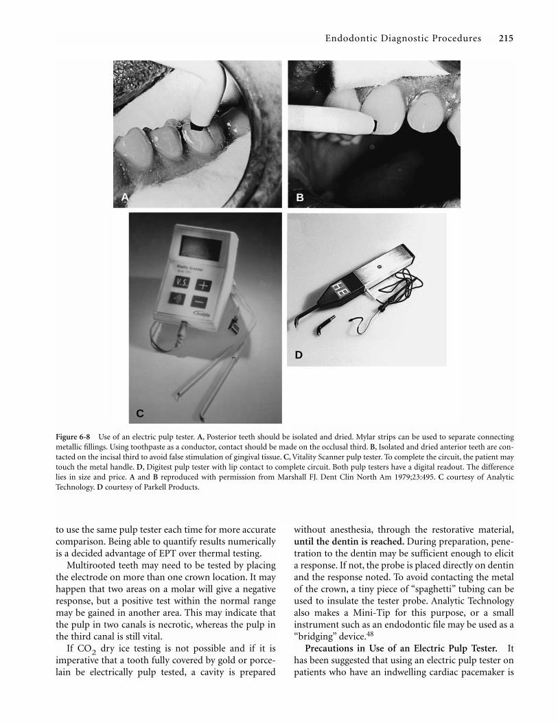
testing device has been some form of electric pulptester.33 Presently, there are a number of very efficient,battery-powered, and easily controlled devices on themarket. All have sophisticated circuitry and digital dis-play. Price is the major difference between the variousbrands, foreign or domestic. Examples are the Digitestand Gentle Pulse (Parkell Products; Farmingdale, N.Y.)Vitality Scanner and Endoanalyzer (Sybron AnalyticTechnology; Orange, Calif.), Trilite (Evident/Pulpdent,UK and USA), Pulppen (Hygenic Corp., USA), Sirotest(Siemens AG, Germany), Digipex II (Mada Equip. Co.,Japan and USA), Neotest (Amadent; Cherry Hill, N.J.),and the Dentometer (Dahlin, Denmark, UK, and USA).
In contrast to the older types of electric pulp testers,these devices produce little discomfort, even whenoperated by inexperienced examiners34–39 (Figure 6-8).It is important to follow the manufacturer’s instruc-tions to establish positive contact.
The testing procedure must be explained to thepatient. An apprehensive or confused patient or amalingering patient may give erratic responses andinvalidate the testing. It may be necessary to practicetesting on teeth other than the ones being examined tohelp the patient get used to the procedure. As withmost tests, electric pulp testing (EPT) should not beused as the only method for diagnosis.
Electric pulp testing provides limited, though oftenvery useful, information, whether or not the pulpalnerve fibers are responsive to electric stimulation.Many factors affect the level of response: enamel thick-ness, probe placement on the tooth40,41 (see Figure 6-8,A and B), dentin calcification, interfering restorativematerials, the cross-sectional area of the probe tip,36
214 Endodontics
and the patient’s level of anxiety. Comparison of EPTresults among various teeth is done primarily for thepurpose of identifying teeth with no response (ordoubtful response, ie, responses at the high end of thescale). Moreover, one needs to keep in mind that bothfalse-positive and false-negative results happen fairlyfrequently, so EPT results must be evaluated carefully.A consistently negative (or doubtful) response indi-cates a necrotic pulp. There are exceptions, of course.
A recently erupted tooth frequently gives a negativeresponse, yet never in its lifetime will the pulp be morevital. In recent studies, it has been found that the newlyerupted teeth have more large unmyelinated axonsthan do mature teeth, the speculation being that someof these large fibers may ultimately become myelinat-ed.42–45 Since it is principally the pulpal “A” fibers thatrespond to EPT, variability in the number of A fibersentering the tooth offers a possible explanation as towhy EPTs tend to be unreliable in young teeth.42,43
A young tooth traumatized by impact may notrespond to testing, yet when the pulp is opened, therush of blood illustrates the error of the test.Multirooted teeth often give bizarre pulp test readingswhen one canal may have vital pulp tissue and othercanals necrotic tissue. Practice in diagnosing and expe-rience with the EPT will help overcome some of thesedifficulties.
Electric Pulp Testing Procedures. To achieve con-sistent results with an electric pulp tester, one must fol-low a standard procedure. Dry the teeth to be tested andisolate them with cotton rolls. Cover the tip of the elec-trode with toothpaste or a similar electrical conductor.
To stimulate the pulp nerve fibers, the electric cur-rent must complete a circuit from the electrodethrough the tooth, through the patient, and back to theelectrode. When gloves were not routinely used by den-tists, the ungloved fingers of the dentists completed thecurrent by contacting both the electrode and some partof the patient’s face, usually the cheek. With glovedhands, that connection is interrupted.
To establish a complete circuit using rubber gloves,one of two methods must now be followed. A groundattachment may be clipped on the patient’s lip (seeFigure 6-8, D), or the patient may complete the circuitby placing a finger on the metal electrode handle. Thelatter method has the advantage of giving the patientmore control: simply lifting the finger off the electrodehandle when a sensation is felt will immediately inter-rupt the current and terminate the stimulation46,47 (seeFigure 6-8, C).
A record must be made of the results of each toothtested. If repeat tests are indicated, it is probably better
Figure 6-7 During palpation with the index finger, the dentistshould watch for the patient’s eyelid blink or forehead wrinkling asthe first sign of pain.
to use the same pulp tester each time for more accuratecomparison. Being able to quantify results numericallyis a decided advantage of EPT over thermal testing.
Multirooted teeth may need to be tested by placingthe electrode on more than one crown location. It mayhappen that two areas on a molar will give a negativeresponse, but a positive test within the normal rangemay be gained in another area. This may indicate thatthe pulp in two canals is necrotic, whereas the pulp inthe third canal is still vital.
If CO2 dry ice testing is not possible and if it isimperative that a tooth fully covered by gold or porce-lain be electrically pulp tested, a cavity is prepared
without anesthesia, through the restorative material,until the dentin is reached. During preparation, pene-tration to the dentin may be sufficient enough to elicita response. If not, the probe is placed directly on dentinand the response noted. To avoid contacting the metalof the crown, a tiny piece of “spaghetti” tubing can beused to insulate the tester probe. Analytic Technologyalso makes a Mini-Tip for this purpose, or a smallinstrument such as an endodontic file may be used as a“bridging” device.48
Precautions in Use of an Electric Pulp Tester. Ithas been suggested that using an electric pulp tester onpatients who have an indwelling cardiac pacemaker is
Endodontic Diagnostic Procedures 215
Figure 6-8 Use of an electric pulp tester. A, Posterior teeth should be isolated and dried. Mylar strips can be used to separate connectingmetallic fillings. Using toothpaste as a conductor, contact should be made on the occlusal third. B, Isolated and dried anterior teeth are con-tacted on the incisal third to avoid false stimulation of gingival tissue. C, Vitality Scanner pulp tester. To complete the circuit, the patient maytouch the metal handle. D, Digitest pulp tester with lip contact to complete circuit. Both pulp testers have a digital readout. The differencelies in size and price. A and B reproduced with permission from Marshall FJ. Dent Clin North Am 1979;23:495. C courtesy of AnalyticTechnology. D courtesy of Parkell Products.
C
D
BA

contraindicated. After testing the effect of electric pulptesters on dogs with artificial pacemakers, Woolley andassociates concluded that currents of the magnitude of5 to 20 milliamps are sufficient to modify normal pace-maker function.49 After testing one battery-powereddevice and three using line current, they found thatonly one caused interference with pacemaker action.They also warned against devices such as desensitizersand electrosurgical units that could produce unknowncurrent leaks, as one of the pulp testers did.
Liquid Crystal Testing. Cholesteric liquid crystalshave been used by investigators50 to show the differencein tooth temperature between teeth with vital (hotter)pulps and necrotic (cooler) pulps. The laser Dopplerflowmeter has also been shown to measure pulpalblood flow and thus the degree of vitality.51–53 Alreadyused in medicine (retina, renal cortex), this experimen-tal device might well spell the difference betweenreversible and irreversible pulpitis—the stressed pulp,if you will.
The Hughes Probeye camera, which is capable ofdetecting temperature changes as small as 0.1˚C, has alsobeen used to measure pulp vitality experimentally.54 Allthree of these methods measure blood flow in the pulp,the true measurement of pulpal status. One may emergeas the pulp tester of the future.
Occlusal Pressure Test. A frequent patient com-plaint is pain on biting or chewing. The causes for suchsymptoms include apical periodontitis, apical abscess,and incomplete tooth fractures (infractions). A clinicaltest that simulates the chief complaint is the occlusalpressure test (or biting test). Several methods exist,such as biting on an orangewood stick, a Burlew rub-ber disk, or a wet cotton roll. All have the ability tosimulate a bolus of food and allow pressure on theocclusal surfaces.
The orangewood stick, the Tooth Slooth, and Burlewdisks allow pinpoint testing of individual cusp areas,whereas the wet cotton roll has the advantage of adapt-ing to the occlusal surface, allowing for pressure over theentire occlusal table. This test is useful in identifyingteeth with symptoms of apical periodontitis, abscess, orcracks. An interesting clinical observation in patientswith tooth infractions (cracked tooth syndrome) is painoften experienced when biting force is released ratherthan during the downward chewing motion.
Anesthetic Test. Pain in the oral cavity is frequentlyreferred from one tooth to an adjacent one or even fromone quadrant to the opposing one. The anesthetic testcan help identify the quadrant from whence the focus ofpain originates. The suspected tooth should be anes-thetized, and, if the diagnosis is correct, the referred
216 Endodontics
pain should disappear, even when it is referred to theopposite arch.
Test Cavity. This test is often a last resort in testingfor pulp vitality. It is important to explain the procedureto the patient because it must be done without anesthe-sia. Make a preparation through the enamel or the exist-ing restoration until the dentin is reached. If the pulp isvital, the heat from the bur will probably generate aresponse from the patient; however, it may not necessar-ily be an accurate indication of the degree of pulpalinflammation. As with other tests, the cavity test must beused in conjunction with the history and other testingprocedures and not used as the sole determinant.
The Stressed Pulp
Abou-Rass has directed the attention of the professionto what he calls “the stressed pulp.”5 This is the pulpthat over the years has been stressed by both diseaseand treatment (caries and periodontis; trauma—impact, occlusal, and iatral; chemicals—cements,resins, and amalgams). This is the tooth that hasdecayed and been restored then re-restored, cut down,heated up—all of the insults a pulp is subjected to overa long span of time.
In addition, when the tooth is now ready to be usedas an abutment for an important partial denture, fixedor removable, still more iatral insult follows. Careful asone might be, the tooth must still be reduced somemore, heated up, cooled off, and injured by impres-sions, try-ins, and temporaries. The final insult is per-manent cementation followed by microleakage.
Is it any wonder that a number of these stressedpulps finally give up by either aching and dying or justquietly dying? But the tragedy is that the tooth is nowcovered by a beautiful new restoration that will have tobe weakened or destroyed to reach the ailing pulp.Would it not have been better, asked Abou-Rass, tohave determined the quality of life of the pulp beforetreatment and have taken action before, not after, thefinal commitment has been made?5
How does one determine which pulps are stressed?By taking a careful history and examination, of course.The dentist must consider the patient’s lengthy reporton this particular tooth, the radiographic outline of thepulp cavity, and the examination findings. The exami-nation includes transilluminating; probing; percussing;examining for cracks, crazing, and abrasion; and theresponse to pulp testing, thermal and electrical.
Any negative results should be viewed with suspicion,including past trauma, orthodontic movement, multipleor deep restorations, poor systemic health, irradiation inthe area, tooth coloration, structural cracks, defective

restorations, condensing osteitis (see Figure 6-49), pulpstones, irritation dentin, narrow canals, nearby pins,pulp capping, delayed response to heat and cold, or evena negative response to the electric pulp tester. All of thesedata must then be considered and acted on before com-mitting this tooth with this pulp to the forthcomingstressful events.
If one determines that a pulp has been severelystressed, would it not be better to intentionally removethe pulp and perform root canal therapy before pro-ceeding with treatment, rather than having to cut backthrough a fine restoration to perform the inevitable?
Clinical judgment comes with clinical experience.But when dealing with “pulps that have received repeat-ed previous injury and survived with diminishedresponses and lessened repair potential,”5 it might bewiser to act sooner rather than later.
Periodontal Evaluation
No dental examination is complete without careful eval-uation of the teeth’s periodontal support. Periodontalprobing and recording pocket depths provide informa-tion with respect to possible etiology and prognosis (seefollowing sections).
There is little question that pulpal necrosis can leadto loss of periodontal support. Whether periodontaldisease can cause pulpal degeneration is a question notclearly answered. There is agreement, however, that apotential interaction exists between the pulp and peri-odontium. For the purposes of endodontic treatmentof a single tooth, probing may be limited to the toothinvolved and at least the adjacent teeth. As part of atotal oral examination, all teeth should be included inthe probing evaluation. The number of probing loca-tions may vary depending on the tooth’s location. Fourto six areas surrounding the tooth should provide agood picture of periodontal support. Gingival and sul-cular bleeding and drainage, along with the presence ofplaque and calculus, should also be noted.
FACTORS INFLUENCING PROGNOSIS
Endodontic diagnosis should not be the sole determinantof treatment planning. Other factors contribute to deter-mine whether a suspicious tooth should be restored or apulpally involved tooth should be treated. Periodontalhealth, restorative considerations, and radiographic evi-dence of anatomic complexities associated with the toothwill have a major impact on any treatment decision.
Periodontal Disease
Periodontal stability is a basic requirement for any toothbeing considered for endodontic therapy. This stability
is determined by the amount of bony support, thehealth of that support, and the health of the overlyingsoft tissue. Examination alone cannot guarantee thefuture health of these tissues, but usually it can deter-mine existing disease. Isolated bone loss or tooth mobil-ity may or may not signify periodontal disease. It maybe owing to periradicular disease of pulpal origin, or itmay be combined periodontal-endodontal disease.55
Generalized bone loss of periodontal disease will defi-nitely affect prognosis and therefore the treatment plan.
As part of the examination, probe the sulcus of thetooth or teeth in question and record the pocket meas-urements. Also test for mobility and record the datausing a system of 0 through 3. Grade 0 means normalmobility, grade 1 slight mobility, grade 2 marked mobil-ity, and grade 3 mobility and depressable. Also record ifbleeding occurs on probing. Particular note should bemade of palatal grooves in single rooted teeth (Figure 6-9), furcas in multirooted teeth, and other anomalies(enamel projections, etc), as these may aggravate gingi-val conditions and make for unstable future periodontalhealth. These examinations will establish approximatebone levels and the crown-root ratio. The presence of 3to 5 mm pockets of a first-degree mobility indicates“moderate periodontitis” (Figure 6-10). When this isfound, the entire mouth should be screened for peri-odontal disease and treated accordingly.56
Pockets deeper than 5 mm, or mobility graded 2 or3, indicate “severe periodontitis,” and periodontaltreatment is imperative. Referral to a periodontistshould be considered. Regardless of the extent of theperiodontal disease or who renders the treatment, thepatient must be advised of the effect that periodontaldisease can have on the prognosis for endodontic ther-apy. Hiatt reported 20-year follow-up on two casesinvolving a number of teeth treated with combinedperiodontal/endodontal therapy. Only one tooth waslost, because of root resorption.57
Restorability
The restorability of a tooth requiring endodontictreatment depends on the amount of sound toothstructure remaining and the effect that the restorationwill have on the periodontal tissues—not invading the“biologic width” between restoration and the peri-odontal ligament (PDL), for example. Prior to anyendodontic treatment, and after all present coronalrestorations and caries are removed, the remainingtooth structure should be re-examined with a fiber-optic light for fractures and perforations. At this time,teeth with vertical fractures or severe perforations aregenerally untreatable.
Endodontic Diagnostic Procedures 217

It should also be noted that pulpless teeth are notstrengthened by the use of posts.58 Posts are for theretention of crown build-up. Retention of the crownand strength of the restoration depend on the designof the restoration, placing margins well onto solidtooth structure.
218 Endodontics
RADIOGRAPHIC EXAMINATION
In the sequence of examination, radiographic evalua-tions should come last. In practical terms, one usuallytakes a look at the radiographs first and then proceedswith the other evaluation. Following the examination, in
Figure 6-9 Deep palatal groove anomaly leading to irreversible pulpitis and periodontal disease and tooth loss. A, Depth and severity of thegroove. B, Combined endodontal-periodontal lesion. (Courtesy of Dr. David S. August.)
Figure 6-10 A, Five mm pocket, first-degree mobility, and bleeding indicate moderate periodontitis associated with pulpless tooth.Successful periodontal treatment may well be necessary to retain the endodontically treated tooth. A complete periodontal examination isindicated. B, A pulpless tooth is periodontally involved as well. Recession (arrows) is undoubtedly related to crown preparation invading thebiologic width of the periodontal ligament attachment. (Courtesy of Dr. F. James Marshall.)
A B
A B

hindsight, radiographs can be better interpreted whenthe results of the previous examination are available.
First, a few words about endodontic radiographs.The radiographic image is a shadow and has the elusivequalities of all shadows. First and foremost, it is atwo-dimensional representation of a three-dimension-al object (Figure 6-11). Also, like any shadow, it may betoo light or too dark, too short or too long. The centralbeam must be carefully oriented to give detail wheredetail is required (Figure 6-12). This usually requiresthe central beam to be aimed directly through the peri-apex rather than a compromise position at the crest ofthe alveolar process.
In addition to central beam positioning, two ormore exposures are frequently necessary to check outdetail from more than one horizontal angle (Figure6-13). This is especially true in the case of the normalbony foramens. The mental foramen may be directlysuperimposed over the apex of the mandibular pre-molars, for example (Figure 6-14). The nasopalatineforamen also may be superimposed on the apex ofthe maxillary central incisors. Because these forami-na are actually some distance from the apices of these
teeth, their shadows may be shifted far to the mesialor the distal merely by shifting the horizontal angleof the cone of the x-ray machine to the mesial or dis-tal during separate exposures (Figure 6-15). On theother hand, if the radiolucent area in the radiographis actually a lesion truly associated with the periapexof the involved tooth, its shadow will remain“attached” to the root end and will remain so in spiteof a mesial or distal shift in separate films. For detailsregarding the “horizontal shift,” the reader is referredto chapter 9.
Interpretation
The finest radiologist will be severely handicapped insecuring valuable information from a film that has notbeen properly placed, exposed, and processed.Conversely, the finest film is of limited value if theinterpreter is inadequately trained.
Neaverth and Goerig have emphasized the necessityof knowing the normal structures before interpretingthe abnormal (personal communication, 1995). Usingradiographs of unusual clarity, they have delineated theanatomic structures of the posterior mandible and
Endodontic Diagnostic Procedures 219
Figure 6-11 Misleading radiograph related to lack of a third dimension. A, Failing root canal therapy in the maxillary central incisorappears to be grossly overfilled. B, The extracted tooth seen in the radiograph proves that the case is not overfilled. Perforation was by hugeenlarging instruments used through restricted access cavity. (Courtesy of Dr. Eugene Meyer.) Reproduced with permission from Luebke RG,Glick DH, Ingle JI. Oral Surg 1964;18:97.

220 Endodontics
Figure 6-12 The importance of an adequate radi-ograph. A number of important details may be learnedfrom this film: (1) size and character of periradicularlesion, (2) curvature of root end, (3) relationship of rootto adjacent roots, (4) mesial or distal inclination of root,(5) approximate length of tooth, (6) relationship ofexploring instrument to root curve, (7) size of canal, and(8) divergence of coronal cavity (arrow). Periodontallesions and root fractures could also be apparent. A cen-tral beam, directed through the periradicular region,gives clarity to this important area.
Figure 6-13 Variations in horizontal angle improve radiographic interpretation. A, In this straight-on, labial-lingual pro-jection, an internal resorption defect is seen. B, In a mesially directed projection, the extension of the resorptive defect tothe mesial-lingual is apparent.

maxilla (Figure 6-16). Many times, these structures canimitate or hide lesions of endodontic or nonendodonticorigin. It is also important to identify structures such asthe mandibular canal and maxillary sinus and approachthem with caution during endodontic treatment andsurgery. Encroachment into these areas has led tonumerous lawsuits.59
An organized method of evaluating and interpretingradiographs, from a single film to a full-mouth set or apanographic plate, has been suggested by Wuehrmann.60
This technique recommends reviewing one structure ata time, such as the lamina dura. Follow this structure allthe way around the first tooth on the left and thenaround the next tooth and the next until the full-film orfull-mouth survey is scanned. The findings are recordedas normal or changed.
Brynolf has paid special attention to the continuumof the lamina dura and the periodontal ligamentspace.61 She has pointed out the normal appearance ofthe lamina dura at the apex, which, under magnifica-tion, can be seen dipping down into the apical orifice.If there is slight inflammation at the apex, the laminadura is lost as the PDL space widens62 (Figure 6-17).Kaffe and Gratt at Tel Aviv University found much thesame as Brynolf, that the best radiographic features foraccuracy in diagnosis were lamina dura continuity andPDL width and shape.63
Following Wuehrmann’s suggestion, one proceeds tothe next structure, for instance, the crowns of theteeth.60 Each crown is evaluated independently. The
Endodontic Diagnostic Procedures 221
Figure 6-14 Mental foramen is superimposed exactly at the apexof a vital mandibular premolar and may easily be mistaken for aperiradicular lesion.
Figure 6-15 Example of a methodused to determine the relationshipof radiolucency to the periapex of atooth. A, The nasopalatine foramenis superimposed over the periapexof a right central incisor. The rightlateral incisor is missing. B, Bychanging the horizontal projection,the shadow of the nasopalatineforamen may also be superimposedover the periapex of the left centralincisor, proving that the radiolucentarea is some distance lingual to theapex of both teeth.
A B

crest of the alveolar process should then be followedfrom left to right, upper to lower, and all of the struc-tures outside the alveolar process should be evaluatedas well—the sinuses, floor of the nose, foramina, and soon. In short, radiographic interpretation should bedone in an organized habitual way so that nothing isoverlooked.
Tracing the dark periodontal membrane space willreveal the number, size, and shape of the roots and theirjuxtaposition. While observing the roots, one must lookfor periradicular lesions and root defects such as anom-alies, fractures, and external resorption. The number,curvature, size, and shape of all of the canals and cham-bers should be noted along with internal resorption,pulp stones, linear calcification, and open apices.
222 Endodontics
For example, if a large pulp chamber is seen in a sin-gle adult tooth while other chambers are narrowing,one should suspect a necrotic pulp, even though a peri-radicular lesion might not be apparent. In marked con-trast, Swedish dentists have reported dramatic narrow-ing of pulp chambers in patients with serious renal dis-ease, particularly those with transplanted kidneys whoare on high doses of corticosteroids.64
Radiographic coronal evaluation includes depth ofcaries and restorations with respect to the pulp, as wellas evidence of pulp cappings or pulpotomy, densinvaginatus or dens evaginatus, and the size of thepreparations under porcelain or resin jacket crowns.
Evaluation, however, comes down to a matter of per-sonal interpretation, as demonstrated by Goldman and
Figure 6-16 Above, Anatomic landmarks in a maxillary arch. H = maxillary sinus; I = floor of the maxillary sinus; J and K = bony septumin the maxillary sinus; L = zygomatic process; M = maxillary tuberosity. Below, Anatomic landmarks in the mandibular arch. A = mandibu-lar canal; B = mental foramen; C and G = cortical bone, border of the mandible; K = lamina dura; M = root canal filling. (Courtesy of Drs.Albert C. Goerig and Elmer J. Neaverth.)

his colleagues.65,66 The radiographs of 253 cases, origi-nally examined by three faculty members at TuftsUniversity, were re-examined by them 6 to 8 monthslater. These endodontists agreed with themselves from72 to 88% of the time. Ten years later, however, thesame group repeated essentially the same experimentwith 79 other dentists and found that they not only dis-agreed with each other over half the time but, worseyet, disagreed with themselves 22% of the time.67
Much the same was found by researchers in Israel63 andGreece.68
Antrim found that holding the radiograph up to aview box produced more consistent agreement amongexaminers than either projecting the radiograph orusing a magnifying glass.69 However, Weine hasclaimed that projecting the image produces the bestinterpretative result.70
One of the reasons for these discrepancies, of course,deals with how one interprets bony lesions. Shoha et al.were able to demonstrate the variables in this interpre-tation.71 They found that lesions were always largerthan their radiographic image, especially in themandibular molar region. Lesions in the premolar areawere only slightly larger than their radiographic image.Lesions found by hindsight were often difficult todetect initially because of their vague outline. InHolland, it was found that cortical bone had to bedamaged by an osseous lesion before radiolucency
could be detected and that loss of cancellous bonealone was not enough to be visible radiographically.72
Bender has illustrated this dramatically with a radi-ograph of a molar tooth showing increased density ofthe bone at the periapex73 (Figure 6-18, A). Yet whenthe tooth was extracted, a huge granuloma wasattached, which was not at all apparent in the film(Figure 6-18, B).
Root Anatomy
The radiographic examination provides essential infor-mation relative to normal and abnormal root forma-tion. Since mandibular incisors frequently have twocanals and, at times, two roots, adding an additionalradiographic view from the mesial or distal can aid indetecting such anatomic variances.
One can expect anatomic variations in all toothlocations. Mandibular premolars and molars are noexception (Figure 6-19). By both radiographic andmechanical means (Figure 6-20), the number of canalsand foramina should be determined before canalenlargement is completed. Because of frequent varia-tions, it is a good habit to examine radiographs with amagnifying glass so as not to miss an extra canal orother variations.
Maxillary first premolars with three roots or canalsare seen more clearly if the projected horizontal direc-tion of the central beam is slightly from the mesial
Endodontic Diagnostic Procedures 223
Figure 6-17 Brynolf ’s interpretation of the normal and resorbed lamina dura. Left, Radiographic appearance of a normal lamina duraunder magnification. Note the “tit” of bone (opposite A) thrusting toward the foramen (F). Center, Loss of lamina dura at the apex alongwith apical inflammatory resorption from the necrotic canal (C). A = apical soft tissue; B = bone trabeculae; C = root canal; F = foramen;L = PDL space; M = medullary space. Right, necropsy of specimen detailed in Center. Reproduced with permission from Brynolf I61 andBrynolf I. Odontologisk Revy 1967;18 Suppl 11:27.

(Figure 6-21). The normal two roots or two canals ofthe maxillary first premolar also are easier to find if thebeam is directed from the mesial. A recent in vivo studyshowed that two canals are present in maxillary secondpremolars 59% of the time, more than had beenreported previously.74–77 The mesial angulation tech-nique is therefore beneficial in detecting the secondcanal in maxillary second premolars. In this case, thelingual root always appears to the mesial on the film(the SLOB rule—Same Lingual-Opposite Buccal).78
Slowey has demonstrated how difficult it is to detectextra roots, let alone extra canals.79 One particularly
224 Endodontics
baffling case involved a maxillary lateral incisor with anunusual second root. In the post-treatment radiograph,a bony lesion could be seen to the distal (Figure 6-22,A), but it was assumed that the lesion was related toinvagination from the cingulum. Because the lesion didnot heal, the tooth was extracted, and only then was theextra root revealed (Figure 6-22, B and C). A radi-ograph of the contralateral incisor revealed whatappeared to be a bilateral anomaly (Figure 6-22, D). AsSlowey pointed out, “whenever the outline of the rootis unclear, has an unusual contour, or strays in any wayfrom the expected radiographic appearance, one
Figure 6-18 Radiographic deception owing to the thickness of the cortical bony plates. A, Osteosclerosis distal to the second molar com-pletely masks a huge bony lesion within spongy bone. B, Same tooth extracted with an enormous space-occupying granuloma (arrows), notin the least suspected radiographically. Reproduced with permission from Bender IB.73
Figure 6-19 A, Mesial root of first molar has not only two separate canals but an additional root as well (arrow). B, Two premolars with anunusual root and canal formation. “Fast break” in canal radiodensity (arrows) indicates canal bifurcation. Both teeth have three and possi-bly four canals, but the number of apical foramens can be determined only by instrument placement (see Figure 6-21). Reproduced with per-mission from Slowey RR.79
A B

should suspect an additional root canal…”79 He quot-ed Worth, who said, “Look at the corners of the radi-ograph and the center will take care of itself.”79
The mystery of extra canals within the normal num-ber of roots is more perplexing than the extra rootproblem itself. Slowey has also given us some tips on
how to detect the undetected.79 One method is to fol-low the image of the test file in the length-of-the toothfilm, particularly in the coronal part of the root. If anextra dark line is apparent in the coronal third of theroot, running parallel to the instrument (Figure 6-23),one should suspect a second canal. This is especiallytrue in the mesiobuccal root of maxillary first molars,where a fourth canal is found 51.5 to 69% of the timein vitro75,76,80–83 but only 18.6 to 33.3% in vivo.82–84
Fourth canals frequently occur in the distal roots ofmandibular first molars as well84 (Figure 6-24).
Another diagnostic clue was pointed out bySlowey.79 It could be called “the fast break.” When view-ing a radiograph, if there is a sudden change in theradiolucency within a canal, this change in densityprobably signals the beginning of an additional canal(Figure 6-25), a frequent occurrence in maxillary firstpremolars. In mandibular canines (Figure 6-26, A),such observations should be followed up by takingadditional radiographs from a different horizontalangle (Figure 6-26, B) and possibly searching out theextra canals with the length-of-tooth instrument films(Figure 6-26, C).
Frequently, root formation results in severe curvature.When the curvature is to the mesial or distal, so fre-quently seen in the maxillary lateral incisors or occasion-ally in premolars (Figure 6-27), there is little problem
Endodontic Diagnostic Procedures 225
Figure 6-20 Mechanical means of determining separate or common foramina for two canals. The instrument is placed to full depth ineither canal (1) and the instrument in the second canal (2) is prevented from going fully to place. Single foramina can then be confirmed byraising the first instrument (3), allowing the second instrument to go fully to place (4). If turned, the instruments can be felt to grate againsteach other. Reproduced with permission from Serene TP, Krasny RM, Ziegler PE, et al. Principles of preclinical endodontics. Dubuque (IA):Kendall/Hunt Publishing Co.; 1974.
Figure 6-21 Three-rooted maxillary first premolar (arrow) andsecond premolar are readily discerned in this radiograph. Clarity ofroot structure can be improved by “aiming” a central beam directlythrough the apical region.

226 Endodontics
Figure 6-22 A, Continuing lateral lesion (mesial) and acute abscess (arrow). The tooth is finally extracted.B, Benchtop radiographic view reveals a second root and an unfilled canal (arrow), not seen in the originalfilms. C, Lingual view of second root and invagination from the cingulum. D, On the contralateral side, asimilar anomaly exists. Reproduced with permission from Slowey RR.79
A B
C D

Endodontic Diagnostic Procedures 227
Figure 6-23 A, From viewing the initial film, one would not sus-pect that there are two canals in the mesiobuccal root of the firstmolar. B, An extra dark line alongside the exploring instrument(arrow) signals the possibility of a second canal. C, Final filmshowing two separate mesiobuccal canals (arrow) apparently aris-ing from a common orifice. Reproduced with permission fromSlowey RR.79
Figure 6-24 A, Two instruments in a single canal, but a wide, extra dark line (arrow) indicates an additional canal. B, Final filling provesfour canals. Reproduced with permission from Slowey RR.79
A B
C
A B

228 Endodontics
Figure 6-25 A, Sudden change in radiographic density (arrow) signals probable canal bifurcation at that point. B, Change from right-anglehorizontal projection (A) to 20-degree projection from the distal clearly reveals two canals but a single foramen. Reproduced with permis-sion from Slowey RR.79
Figure 6-26 A,“Fast break” in radiograph of amandibular canine (arrow) indicates that asearch should be made for a second canal. B,Varying horizontal projection reveals twocanals in hourglass-shaped root. C, “Length-of-tooth” instruments in place reveal a single fora-men. Reproduced with permission fromSlowey RR.79
A B
C
A B

in detecting the curvature. However, when the curvatureis to the buccal or lingual—in the same plane as the cen-tral x-ray beam—the curvature is more difficult to detect(Figure 6-28). Careful examination may reveal increasedradiopacity at the root end as the root doubles back on
itself and is literally “x-rayed” twice. In its extreme, apeculiar “target” or “bull’s-eye” appearance will show inthe film (Figure 6-29).
In addition to “normal” variances in tooth form,such as curvatures and extra roots, anomalies that may
Endodontic Diagnostic Procedures 229
Figure 6-27 Unusual mesial and bayonet curvature seen in three maxillary premolars and easy to detect.
Figure 6-28 Double root canal curvature in a maxillary lateral incisor. A, Radiograph with instrument proves that the canal exits to distal.B, Mesiodistal view of the same tooth shows that the canal also exits to the lingual. C, Apical photograph of the same tooth demonstrates aninstrument perforating the lingual far short of the apex. (Courtesy of Drs. Howard Clausen and John R. Grady.)

affect pulp vitality may also be detected in the radi-ographs. Invagination and palatal radicular groove (seeFigure 6-9) are conditions that frequently have anom-alous tracts leading from the enamel surface directly tothe pulp (Figure 6-30). Such defects may involve theroot, either partially or all the way to the apex. Thesegrooves permit bacterial invasion of the pulp, whichleads to periradicular lesions and pulp necrosis. Otheranomalies such as odontome and microdont may alsolead to pulp necrosis (Figure 6-31).
Conditions Inside the Tooth
Pulpal changes such as inflammation and necrosis can-not be detected radiographically within the canal. Onlychanges in calcific structures can be demonstrated.However, the results of pulp tissue changes are observ-able, that is, internal resorption as a result of irreversiblepulpitis, periradicular bony changes resulting from pulpnecrosis, and lack of continued root formation inimmature teeth subjected to pulp destructive traumaticinjuries.
Pulp Stones. Chronic pulpal disease may facilitatethe formation of pulpal calcifications such as pulpstones (Figure 6-32). However, the mechanism of pulpstone formation and the various factors necessary inthe calcific process are not well known at this time, afact illustrated by the observations that pulp stones are
230 Endodontics
Figure 6-29 “Target” or “bull’s-eye” phenomena seen in roots that severely curve to the buccal or lingual, that is, in the direction of the cen-tral beam. A, Central incisor with a dilacerated root under orthodontic realignment. B, Lateral head film in which the labially dilaceratedroot (in A) is apparent (arrow). C, Resection and retrofilling of the dilacerated root seen in A and B. (A and B courtesy of Dr. Alfred T. Baum;C courtesy of Dr. Dudley H. Glick.)
B C
Figure 6-30 Dens invaginatus in the cingulum area of a maxillarylateral incisor. Anomalous tract seen in this radiograph has led topulp infection and periradicular pathosis.

found in apparently normal teeth with healthy pulps(Figure 6-33). Careful examination of radiographs mayreveal other problems beside pulp stones—root frac-tures, calcification, early pulp death (Figure 6-34), andinternal and external resorption.
Internal Resorption. Following traumatic injuriesand/or caries, internal resorption may be detected onradiographs (Figure 6-35). Differentiation between inter-nal and external resorption may be made radiographical-ly.85 First, the lesion of internal resorption usually hassharp smooth margins that can be clearly defined (seeFigure 6-35, A). However, it need not be symmetric.Another diagnostic sign is the manner in which the pulp“disappears” into the lesion, not extending through thelesion in its regular shape (Figure 6-35, B).
At times, an area of resorption may be confused withdental caries radiographically. However, dental caries isless sharply defined than is internal resorption. Neithercaries nor internal resorption should be ignored; inter-nal resorption can progress to the point of extensivetooth destruction and perforation.86
Wedenberg and Lindskog, after experimentally pro-ducing internal resorption in monkey incisors, con-cluded that there are two types of internal resorption:transient, which may repair itself, and progressive, “thelatter requiring continuous stimulation by infection.”87
Endodontic Diagnostic Procedures 231
Figure 6-31 A, Radiograph reveals 7 mm irregular radiolucency with a sclerotic border associated with an acute abscess. B, An extractedfused supernumerary microdont that had a cyst attached. Reproduced with permission from Samuels DS. Oral Surg 1992;73:131.
Figure 6-32 Huge pulp stone (arrow) that has developed in asso-ciation with chronic pulpitis. This “ball and socket” type of stonemust be fragmented to be removed.
A B

From London, Lynch and Ahlberg reported bilateralidiopathic internal resorption. The radiolucent areaswere cleanly defined, punched-out lesions. The pulpwas seen to disappear into the lesion.88
Herpes zoster was linked to resorption in othercases. The varicella-zoster virus lies dormant for yearsin a nerve ganglion, from an earlier “chickenpox”attack, and can suddenly reactivate to infect the pulp.89
Conditions Outside the Tooth
One of the most common occurrences seen radi-ographically on the outside of the root of the tooth isexternal resorption.
232 Endodontics
External Resorption. Andreasen has stated that fol-lowing traumatic injuries there are three main types ofexternal root resorption: surface, inflammatory, andreplacement resorption. Surface resorption is caused byacute injury to the periodontal ligament and root sur-face. Cell proliferation mediation removes the trauma-tized structures. If injury is not repeated, healing takesplace with new cementum and PDL. Inflammatoryresorption may occur from combined injury to the PDLand cementum complicated by bacteria from the infect-ed root canal, which, in turn, stimulate the osteoclasts.Resorption usually ceases if the root canal is thoroughlydébrided and obturated unless stimulation has provokedthe third type of resorption, replacement resorption. Inthis type, ankylosis between bone and tooth occurs with-out the intervening PDL, and the constantly remodelingbone slowly removes the tooth and replaces it with bone.This is often seen in unsuccessful replant cases.90
Frank has delineated another type of external rootresorption that he terms extra-canal invasive resorp-tion, totally different from regular external or internalresorption.91 This phenomenon has interested clini-cians and researchers over the last century and has beenvariously called odontoclastoma,92 idiopathic externalresorption,93 peripheral cervical resorption,94 cervicalexternal resorption,95 peripheral inflammatory rootresorption,96,97 cervical resorption,98 and, more recently,invasive cervical resorption.99
Frank et al. made the point that extra-canal inva-sive is the more descriptive term100 (rather than“external-internal,” which Frank originally used)because of the external origin of this resorptive defect,before it invades the dentin (Figure 6-36), but, more
Figure 6-33 Left (A) and right (B) bitewing radiographs reveal multiple pulp stones that have developed in the molar teeth of one patient.Many stones are related to advanced carious lesions and large restorations. A, One stone (arrow) has also developed in an intact tooth.
Figure 6-34 Diagnostic radiograph of a first molar reveals earlypulp death in the distal canal and calcification followed by necrosisin the mesial canals leading to a periradicular lesion. Chisel-shapedresorption of the distal root typical of long-standing chronicinflammation. (Courtesy of Dr. Craig Malin.)

Endodontic Diagnostic Procedures 233
Figure 6-35 A, Radiograph of lesion of internal resorption (arrow). Note sharp smooth margins. (Courtesy of Drs. A. H. Gartner, T. Mack,R. Somerlott, and L. Walsh.) B, Drawing of internal resorption shows how the shadow of the pulp “disappears” into the huge lesion.Reproduced with permission from a drawing modified from Lepp FH. Oral Surg 1969;27:185.
Figure 6-36 A, Radiograph of lesion of extra-canal invasive resorption (arrow). Note the ragged margins of the lesion. (Courtesy of Drs.A. H. Gartner, T. Mack, R. Somerlott, and L. Walsh.) B, Drawing of resorption shows how the shadow of pulp “passes through” the lesionunaltered. Inset depicts external lesion perforating into and resorbing dentin. Reproduced with permission from a drawing modified fromLepp LH. Oral Surg 1969;27:185.
A B
A B

important, that the destruction surrounds the rootcanal without necessarily involving the pulp (Figure6-37). This, of course, gives an entirely different radi-ographic appearance than internal resorption. Inextra-canal invasive resorption, the pulp appears topass through the lesion (Figure 6-38), whereas thepulp “disappears” in the internal resorptive lesion (seeFigure 6-35).
The histopathologic characteristics of this insidiousand often aggressive form of invasive tooth resorptionare of interest and significance to clinicians. Althoughthe invading tissue is derived from ectomesenchymalprecursor cells within the periodontal ligament, it dif-fers both in structure and behavior from the periodon-tal ligament. In early lesions, the invading tissue isfibrovascular (Figure 6-39), which accounts for thepinkish appearance that may be evident near the gingi-val margin of the affected teeth.99 Later, as resorptionextends more deeply into radicular tooth structure, the
234 Endodontics
histopathologic appearance may be described as fibro-osseous—bone-like depositions being evident—bothwithin the fibrovascular tissue and laid directly ontoresorbed dentin surfaces (Figure 6-40). Of clinical sig-nificance is the deeply infiltrating channels that ofteninterconnect with the periodontal ligament. Effectivetreatment can be achieved only if all ramifications areinactivated or removed (see chapter 12).
Jaacob examined 18 of these cases and determinedthat the pulp spaces were separated from the resorptiveareas by a “resistant dentin shell.”101 Quite possibly, thisphenomenon of not invading the pulp relates to thecharacter of the dentin surrounding the pulp. Clasticcells generally attack well-calcified structures such asbone, dentin, and cementum. The pulp, however, issurrounded by uncalcified predentin—uncalcifiedmaterial not readily amenable to cellular clastic action.In addition, there is evidence for an anti-invasive factorin tooth dentin.102
Figure 6-37 Extra-canal invasive resorption. The integrity of theroot canal is evident in the extracted molar (larger arrow). Note thefile (smaller arrow) extending into the canal. (Courtesy of Dr.Harold Gerstein.)
Figure 6-38 Frank’s classification of extra-canal invasive resorption.A, Supraosseous, the most frequent form. Inset depicts external inva-sion and dentin destruction, yet intact pulp. Note that the cross-sec-tion does not invade the pulp. B, Intraosseous: it is difficult to deter-mine the point of origin radiographically. C, Crestal, which may berelated to orthodontic movement. (Courtesy of Dr. Alfred L. Frank.)

Although most extra-canal invasive root resorptionoccurs at the immediate subgingival level —supraosseous, if you will (see Figure 6-38, A)—Franket al. described an additional intraosseous variationthat is difficult to locate radiographically since it isnot accompanied by periodontal breakdown.100
There may, however, be an open lesion just at orbeneath the gingival sulcus (Figure 6-41, A). In addi-tion, the resorption may extend into the coronal toothstructure, and a “pink tooth” may result (Figure 6-42),much the same as with coronal internal resorption.This may be more apt to happen with what Frankdescribed as a crestal variety of invasive resorption
(see Figure 6-38, C).91 If the lesion is visible radi-ographically, but not apparent visually, it may beprobed for with a curved explorer.
The intraosseous variety of extra-canal invasiveresorption (see Figure 6-38, B) is characterized radi-ographically as having an irregular moth-eaten appear-ance within the tooth—the more advanced, the moreradiolucent (see Figure 6-41, B). Again, close examina-tion will show the outline and integrity of the canalthat appears to “pass through” the lesion unaltered(Figure 6-43). The pulp usually tests vital and has beenasymptomatic. The radiographic appearance of theselesions varies. In early lesions, a small radiolucency may
Endodontic Diagnostic Procedures 235
Figure 6-39 Histologic appearance of an incisor tooth with exten-sive invasive cervical resorption. An intact layer of dentin and pre-dentin on the pulpal aspect (*) separates the pulp from the resorb-ing tissue. The resorption cavity is filled with a mass of fibrovascu-lar tissue with active mononucleated and multinucleated clasticcells lining resorption lacunae (arrows). (Original magnification×40.) (Courtesy of Dr. John McNamaara; reproduced with permis-sion from Heithersay GS.99)
Figure 6-40 Histologic appearance of an extensive invasive cervi-cal resorption with radicular extensions. Masses of ectopic calcifictissue are evident both within the fibrovascular tissue occupying theresorption cavity and on resorbed dentin surfaces. In addition,communicating channels can be seen connecting with the peri-odontal ligament (large arrows). Other channels can be seen with-in the inferior aspect of the radicular dentin (small arrows).(Original magnification × 30.) Reproduced with permission fromHeithersay GS.99

236 Endodontics
emerge—somewhat irregular in appearance—thatmay, on occasion, resemble dental caries.
Extra-canal invasive resorption may also radi-ographically resemble internal resorption (see Figure6-43, A). To differentiate, off-angle mesial and distalradiographs will “move” the extra-canal type of resorp-tion, whereas the internal resorptive defect will notmove on the film (see Figure 6-43, B).
Although the etiology of extra-canal invasive resorp-tion is unknown, several potential causative factorshave been identified and recently analyzed. Heithersaystudied 222 patients, with a total of 257 teeth, exhibit-ing varying degrees of invasive cervical resportion103
(Figure 6-44). Orthodontics alone was the most signif-icant causative factor104–106 (21.2% of patients, 24.1%of teeth), whereas trauma was the next most identifi-able potential cause accounting for 14.0% of patientsand 15.1% of teeth. Often this type of resorption wasassociated with a combination of factors, for example,trauma and bleaching. Bleaching as a sole factor wasfound in 4.5% of patients and 3.9% of teeth.
Other factors identified as potential predisposingfactors, but at a low level, were surgery, periodontics,
Figure 6-41 A, Extra-canal invasive resorption, supraosseous, of the central and lateral incisors. The integrity of the root canals remainsintact. B, Extra-canal invasive resorption, intraosseous, of the maxillary lateral incisor. Arrow marks the labial site of origin. (Courtesy of Dr.Alfred L. Frank.)
Figure 6-42 Frontal and mirror views of a “pink tooth,”supraosseous extra-canal invasive resorption of a crown (arrow)following impact trauma.

Endodontic Diagnostic Procedures 237
Figure 6-43 Extra-canal invasive resorption, supraosseous. A, Lesion (arrow) superimposed over the canal. B, Same tooth, radiographedfrom the mesial; the lesion “shifts” (arrow) within the tooth. Pulp tests are vital. (Courtesy of Dr. Alfred L. Frank.)
Figure 6-44 Heithersay’s clinical classification of invasive cervical resorption: Class l: A small invasive resorptive lesion near the cervicalarea with shallow penetration into the dentin. Class 2: A well-defined invasive resorptive lesion that has penetrated close to the coronal pulpchamber but shows little or no extension into the radicular dentin. Class 3: A deeper invasion of dentin by resorbing tissue, not only involv-ing the coronal dentin but also extending at least to the coronal third of the root. Class 4: A large invasive resorptive process that has extend-ed beyond the coronal third of the root canal. Reproduced with permission from Heithersay GS.99
1
2
3
4
A B

bruxism, delayed eruption, developmental defects, andinterproximal stripping. The potential for invasion viaa crack in extensively restored teeth was identified. Theclinical classification in Figure 6-44 is also of impor-tance when assessing teeth suitable for treatment, asdiscussed in chapter 12. Suffice it to say, Class 1 to 3lesions must be completely débrided and inactivatedand, if not pulp invasive, filled with a suitable restora-tive material such as dentin adhesive and compositeresin (Figure 6-45), glass ionomer cement, or, in somesituations, mineral trioxide aggregate. The extensiveClass 4 lesions are not suitable for current therapy, andno treatment or extraction is recommended.107
Radiographic Diagnosis: Periradicular Lesions
Pulpal inflammation and necrosis eventually produceperiradicular changes. The earliest is a widening of theperiodontal ligament space, usually at the apex.
Periradicular Lesions. Most often a widened PDLspace and inflammatory apical root resorption areassociated with pulp necrosis and infection.Occasionally, however, these signs may be associatedwith occlusal traumatism (Figure 6-46). This empha-sizes the need for additional tests beyond radiographs.A widened PDL space may also be expected with otherconditions: acute apical periodontitis, a beginningacute apical abscess, or, occasionally, acute pulpitis.
Apical External Resorption. With this condition,the apex may range in appearance from slightly blunt-ed (Figure 6-47, A) to grossly resorbed (Figure 6-47, B),caused by the pressure from orthodontic move-ment.104–106 It also may be obliquely resorbed (Figure6-48, A) or have a cupped-out appearance (Figure 6-48,
238 Endodontics
B), or it may resemble the chewed appearance of a treefelled by a beaver (Figure 6-48, C). In any event, if api-cal resorption has taken place, the apical foramen willbe in the center of the root. If the root resorption has a“moth-eaten” appearance, it is possible that the tooth,by accident, was ripped loose from its ligaments and/orwas replanted. Sometimes an unexplained lesion in theregion strongly suggests a malignancy (see Figure 5-32).
Condensing Osteitis. The exact opposite of rootresorption is condensing osteitis. Teeth with chronicpulpal inflammation sometimes exhibit increased api-cal bone calcifications, which have a radiopaqueappearance on x-ray films (Figure 6-49). Initially, theroot may have been blunted by inflammatory resorp-tion, and then this space is filled by osteoscleroticbone. Following successful root canal therapy, theregional radiopacity slowly changes back to a normalappearance108 (see Figure 5-9). Both idiopathic andsclerosing osteomyelitis result in bone hypercalcifica-tion as well.109,110
Radiographic Changes as Sequelae to PulpNecrosis. The severe periradicular osseous lesionsthat develop as sequelae of pulp necrosis are by far themost frequently observed changes.
Associated with asymptomatic apical periodontitis(AAP) is a well-circumscribed osseous lesion.Radiographically, it appears as a radiolucent area, vary-ing in size from a few millimeters to a centimeter orlarger in size. The bony perimeter of the lesion mayappear radiopaque (Figure 6-50). These lesions do notalways appear at the periapex. Occasionally, they areseen on the lateral surface of the root associated withan accessory canal (Figure 6-51).
Figure 6-45 Supraosseous extra-canal invasive resorptive lesion(seen radiographically in Figure 6-43). The defect does not invadethe pulp and may be restored with the dentin bonding agent andcomposite resin. (Courtesy of Dr. Alfred L. Frank.)
Figure 6-46 Periradicular changes associated with traumatism(arrow) are often mistaken for lesions of pulp origin. The first pre-molar is vital but highly mobile.

Endodontic Diagnostic Procedures 239
Figure 6-47 A, Blunted apices of teeth moved orthodonticallymore than 60 years ago. B, Massive root resorption associatedwith orthodontic tooth movement. Note the development ofperiodontitis as well. (B courtesy of Dr. Bradley H. Gettleman.)
Figure 6-48 A, Total pulp calcification and externalroot end resorption following injury of a mandibularsecond premolar. B and C, Two forms of apical externalresorption. B, A resorptive defect leaves a flaring apicalforamen that could not be adequately obturated byconventional methods. C, Advanced external resorptionof the apex makes conventional instrumentation haz-ardous. B and C reproduced with permission fromLuebke RG, Glick DH, Ingle JI. Oral Surg 1964;18:97.A
B C

The radiographic appearance of a asymptomaticapical abscess (AAA) is generally that of a larger, morediffuse, and irregular radiolucent lesion (Figure 6-52).This lesion may also drain into the mouth through asinus tract or extraorally onto the neck or chin (Figure6-53). An apical cyst may also develop out of a chronicabscess. In this case, the lesion appears more circum-scribed, more like a “granuloma” in appearance (Figure6-54, A). It is also pathognomonic of cysts to move theroots of teeth laterally (Figure 6-54, B). Natkin et al.
240 Endodontics
Figure 6-49 Condensing osteitis, external resorption, and pulpstones associated with chronic pulpitis of the first molar.Condensing osteitis will disappear following successful endodontictherapy.
Figure 6-50 Chronic apical periodontitis involving five mandibular anterior teeth. Pulp death from impact trauma. Well-circumscribedlesions and pear-shaped configuration periradicular to the canine are typical of chronic apical periodontitis. These may be confused with anapical cyst. A bifurcated canal (black arrow, left) is also apparent. A sudden change in radiolucency in the canal (open arrow) denotes pointof separation of canals.
Figure 6-51 A, Lateral lesion denotes pulp necrosis and drainagethrough the lateral accessory canal. B, Filling material extrudes,denoting the accessory canal. (Courtesy of Drs. Albert C. Goerigand Elmer J. Neaverth.)
A
B

Endodontic Diagnostic Procedures 241
Figure 6-52 A, Diffuse radiolucent appearance (arrow) of an asymptomatic apical abscess (AAA) (with stoma) is here contrasted with anasymptomatic apical periodontitis (AAP) associated with a lateral incisor (no stoma). B, Asymptomatic apical abscess with drainage throughbuccal stoma following chronic infection of an apical cyst. Huge diffuse radiolucency is typical of AAA. The root of the tooth is displaced later-ally by pressure from the cyst.
Figure 6-53 A, Diffuse radiographic appearance (right arrow) of an asymptomatic apical abscess may be overlooked owing to radiopacityof the heavy cortical bone. Also noted are condensing osteitis and external root resorption from chronic pulpitis (left arrow), as well as ananomalous third root of the third molar. B, Extraoral stoma (neck) of 8 years duration, draining from the chronic apical abscess seen in A.Immediately following root canal therapy, the discharge ceased, and the stoma healed to a “navel” of scar tissue.
A B
A B

242 Endodontics
Figure 6-54 Movement of teeth is pathognomonic of a cyst. A, Root separation beginning from modest cyst develop-ment. Typical circumscribed radiopaque perimeter may be confused with asymptomatic apical periodontitis. B, Pulplesslateral incisor with infected apical cyst. The canine has been particularly displaced. C, Lateral cyst (arrow) not involvedwith a pulpless tooth. D, Same case, 14 months later. Teeth remain vital. Biopsy, lateral periodontal cysts. C and D repro-duced with permission from Degering CI. Oral Surg 1971;32:498.

postulated that the larger the lesion, the more apt it isto be a cyst.111
In the event that one of these inflammatory lesionsis in contact with the floor of the maxillary sinus,chronic sinusitis may result (Figure 6-55).
Lesions of Nonendodontic Origin. A number ofpathologic changes in and near the alveolar processmay be mistaken for periradicular lesions of pulpal ori-gin. First to consider would be the nonodontogeniccysts: the so-called “globulomaxillary” cyst, the midlinepalatal cyst, and the cyst of the nasopalatine canal orforamen. Because these cysts are not associated withthe exact root end, they may be “moved” radiographi-cally around and away from the root ends if the hori-zontal direction of the central x-ray beam is variedfrom side to side (Figure 6-56). Residual cysts, as well as
globulomaxillary cysts, may be found along the lateralsurface of the root (see Figure 5-29).
To be further considered in the differential diagnosisof cysts is the vitality of the pulps of associated teeth.The pulp of a tooth with an apical cyst should test non-vital. This may not be true of the teeth near nonodon-togenic cysts, unless traumatic injuries caused pulpdeath and also triggered sutural cyst formation. Weineand Silverlade pointed out that residual cysts may mas-querade as periradicular lesions as well.112 It should benoted that there is controversy as to whether globulo-maxillary cysts exist as a clear entity.
Periodontal lesions may be mistaken for periradicu-lar lesions of endodontic origin (Figure 6-57). Theperiodontal probe and pulp tester are invaluable indetermining the origin of the lesion. An additionalmethod is to place a silver or gutta-percha point in theperiodontal pocket and take a radiograph. If the lesionis traceable to the apex of a tooth but the pulp respondsto pulp testing, the evidence for primary periodontalinvolvement is strong.
Common errors in diagnosis center around thelesions of periradicular osteofibrosis or cementoblas-toma, particularly during stage 1, when radiolucency isso apparent (Figure 6-58, A and B). By the time thelesion begins to calcify into a sclerotic lesion (Figure 6-58, C to E), little doubt should exist about the natureof the lesion. Errors will still be made, however, in spiteof all evidence to the contrary. The pulp tester willinvariably help determine the diagnosis. A group inGreat Britain has noted that osteofibrosis can be asso-ciated with hyperparathyroidism, which, in turn, maybe familial.113
In marked contrast, Marks and Dunkelberger havepointed out that Paget’s disease of bone (osteitis defor-
Endodontic Diagnostic Procedures 243
Figure 6-55 A chronic apical abscess (arrow) has perforated thefloor of the antrum, causing chronic unilateral maxillary sinusitis,which healed following endodontic therapy.
Figure 6-56 A cyst of the nasopalatine canalthat may be “moved” from side to side by vary-ing the horizontal angle of x-ray exposure. Thelesion appears to be directly over the apex ofthe central incisor (left) but has “moved” to themiddle-line position in direct labiolingual pro-jection (right). Periodontal disease complicatesthe case.

mans) often presents oral findings—atypical facial neu-ralgia and/or paralysis, hypercementosis, generalizedpulp calcification, loss of lamina dura, root blunting byresorption, and loss of normal bony trabeculation114
(Figure 6-59, A and B). Any one sign or symptom caneasily be confused with a host of localized diseases.
Importance of Endodontic Radiography
Radiography is of paramount importance to the prac-tice of endodontics. It would be virtually impossible toobtain good results from treatment without the use ofradiographs. One needs excellent diagnostic filmsbefore treatment begins, during treatment (in spite ofthe availability of electronic apex locators), and to eval-uate treatment results after completion of endodontictherapy. On the other hand, one must not becometotally dependent on the radiograph. It is mandatorythat other tests be used in conjunction with radi-ographs. Figure 6-60 is a good case in point—a serious
244 Endodontics
error in diagnosis avoided by additional films and vital-ity testing. In marked contrast, Figure 6-61 illustrates atragic misdiagnosis owing to a singular dependence onradiographs. A simple vitality test would have alertedthe dentist to an error in diagnosis.
Furthermore, it should not be assumed that a fistulais necessarily associated with the tooth to which it liesopposite. The draining sinus tract may arise some dis-tance from its orifice. Radiographing a gutta-percha orannealed silver point along the length of the tract maytotally change an initial snap diagnosis (Figure 6-62).
Initial Radiograph. Initial diagnostic radiographsmust be studied carefully, not only as an aid in diagno-sis but also as a “blueprint” for what to expect duringtreatment (Figure 6-63, A).
First, one must note the size and shape of the pulpchamber and the direction and angulation of the canalsas they leave the chamber, as well as obstructions suchas pulp stones. One realizes how valuable the initialradiograph is each time a tooth with a full crown istreated when the radiopaque metal crown eliminates apreview of coronal pulp anatomy.
Curvature of the roots and the approximate lengthof the tooth are next noted. Curvatures in the buccal orlingual directions are difficult to detect, but additionalradiographic angulations may help. Distal or mesialinclination of the roots is usually apparent on the radi-ograph; that information can be used to prevent goug-ing and perforation of the root canals.
Confirmatory Radiographs. The radiograph isnot only a valuable diagnostic tool, it is also an indis-pensable working tool. It provides pertinent informa-tion about treatment progress.
If, after the initial entry into the crown, the accessopening appears to be headed away from the canal ori-fices, a confirmatory film should be taken to evaluatealignment (see Figure 6-11). Such progress films alsoprovide assistance in searching for calcified canals.
The confirmatory length-of-tooth film is undoubted-ly the second most valuable film to be taken after the ini-tial diagnostic radiograph (Figure 6-63, B). In addition toinformation about the position of the file tip in relationto the root apex, it can also show whether the file is in thecanal as intended or has entered a root perforation.
The confirmatory trial point radiograph serves twoimportant purposes. First, it confirms the visual and“tugback” judgment of the fit of the initial filling point.Second, it provides the final opportunity to judge theadvance of instrumentation. If an error in the length ofthe tooth has been made, and the canal has beeninstrumented short or long, here is an opportunity tocorrect the error.
Figure 6-57 A periodontal lesion appears as a periradicular lesion.The involved central incisor is vital. A peculiar radiolucent, hori-zontal band lies between heavy supragingival calculus and the crestof the alveolar process. (Courtesy of Dr. Jerome Zech.)

Endodontic Diagnostic Procedures 245
Figure 6-58 Chronologic development of a suspected cementoblastoma over a 25-yearperiod. A, Initial radiograph with a lesion apparent as a radiolucent area (arrow), as well asexcess cemental formation. Note the huge pulp stone. The pulp is vital. B, Ten years later, ahuge radiolucent lesion is present as well as a gross increase in cementoma. C, Fifteen yearslater, a decrease in radiolucency and an increase in cementoma. D, Twenty years later, vir-tual disappearance of radiolucency along with a gross increase in hypercementosis. E,Twenty-five years later, total cementoblastoma. The pulp remains vital. (Courtesy of Dr.Maurice J. Friedman.)

The confirmatory post-treatment film followscompletion of the root canal filling. It should be madebefore a coronal restoration is placed because somecorrection in the canal filling may have to be made.Ideally, the final film for the record, and the duplicatefilm to be returned if a referring dentist is involved,should be taken without the rubber dam clamp(Figure 6-63, C).
Confirmatory progress films are also important dur-ing surgery. Searching for root tips, lost filling material,location of root apexes during trephination or apicoec-tomy—these are only a few of the uses made of confir-matory radiographs.
Post-Treatment Evaluation Radiograph. Historically,post-treatment evaluations have been a part of clinicalendodontic practice. This radiograph allows an oppor-tunity to evaluate changes taking place periradicularlyas a result of root canal therapy (Figure 6-63, D). A1-year interval is a realistic time frame for mostendodontic cases such as those with initial apical
246 Endodontics
lesions or those with potential post-treatment prob-lems. If any abnormal or unusual findings are noted, itmay be necessary to re-treat, either surgically or non-surgically, or if changes are uncertain, further re-evalu-ation may be indicated. Patients, however, may notconsider post-treatment evaluation to be important,particularly if they are asymptomatic. Riley surveyed159 diplomates of the American Board of Endodontics,who reported that fewer than half of their patientsreturned on recall.115
Re-treatment Radiography. Before initiating re-treatment, particularly a referred patient, the dentistmust always have a “fresh film.” Films sent with thepatient by the referring dentist may be outdated andmay also have been taken prior to a procedure such asaccess preparation. A new film may supply surprisinginformation about the tooth, perforations, brokeninstruments, failure to obturate, ineffective surgery,and a number of conditions well below the standard ofcare (Figure 6-64).
Figure 6-59 Confusion surrounding the oral radi-ologic appearance of Paget’s disease. A,Hypercementosis and pulp calcification in the rightmandibular canine (arrow). B, Same condition inthe left premolar. Note loss of trabeculation andformation of pulp stones (arrow). C, “Cottonwool”appearance (arrows) in the skull view confirmsPaget’s disease. Reproduced with permission fromMarks JM and Dunkelberger FB.114

Endodontic Diagnostic Procedures 247
Figure 6-60 Importance of multiple roentgenographic exposures. A, Lateral incisor appears to be the principal-ly involved tooth in this direct buccolingual projection. B, Projection through the central incisor shows that it isinvolved in the lesion as well. The lateral incisor is vital and the central incisor is nonvital, pulp death having devel-oped from trauma (arrow). C, Following therapy, new bone develops throughout the area, and both teeth reat-tach. (Courtesy of Dr. Pierre R. Dow.)

248 Endodontics
Figure 6-61 Chronologic development of an internal resorptive defect that has become external aswell. The patient is an adolescent female with bruxism (see chapter 4 and Figure 4–24).A, Mandibular incisors apparently involved with asymptomatic apical periodontitis. The pulps ofboth teeth are vital. B, The referring dentist mistakenly opened into vital pulp of the right centralincisor. Detecting his error, he placed a temporary filling and dismissed the patient. C, Six monthslater, internal resorption from the stimulated pulp has perforated the root, and external resorptionhas also begun. D, Total pulpectomy, root canal therapy, and amalgam repair of the resorptive lesionsave the incisor. The left central incisor remains vital. Alleviation of trauma from bruxism will allowhealing.
C D
BA

Endodontic Diagnostic Procedures 249
Figure 6-62 Although the stoma of thedraining sinus tract was over the caninepontic and the treated lateral incisor wassuspected as the origin, it was actually themore distant central incisor causing theproblem. (Courtesy of Dr. Cyril Gaum.)
Figure 6-63 Importance of initial and confirmatory radiographs. A, The initial film alerts the dentist to confusing canal anatomy and a signif-icant periradicular and lateral root lesion. B, Confirmatory length of the tooth radiograph reveals the presence of a C-shaped canal.C, Immediate postoperative confirmatory film shows a complicated root canal system obturated by vertical compaction of warm gutta-percha.Obtura II was used for back-filling. D, Confirmatory 1-year follow-up film shows the degree of healing. (Courtesy of Dr. Michael J. Scianamblo.)

DIAGNOSTIC PERPLEXITIES
Even after a careful history and examination, somedental problems still defy immediate diagnosis. Themost frequent among these have been cataloged asdiagnostic perplexities: (1) sinus tract, (2) numbness,(3) persistent discomfort, (4) cracked tooth syndrome,(5) bizarre radiographic appearance, (6) idiopathicroot resorption, and (7) treatment failures.
250 Endodontics
Sinus Tract
The stoma of a sinus tract may not always exist oppo-site the lesion. An example is the case of drainage in thearea of an extracted canine (see Figure 6-62). Initially,the adjacent pulpless lateral incisor would be suspect.However, when a tracer gutta-percha point was insert-ed through the stoma to the depth of the sinus tract, itpointed to the pulpless central incisor. Harris revealed
Figure 6-64 A, Root perforation (arrow) and associated periodontal lesion are revealed by radiography. Condensing osteitis and externalroot resorption are related to chronic pulpitis, the initial complaint. B, C, and D, Treatment errors and failures revealed by radiographs. B,Fissure bur broken in the canal; C, totally ineffective canal obliteration. An inadequate coronal access cavity is smaller than the canal and theapex to be débrided and obliterated; D, complete “butchery” and total failure in attempted multiple apicoectomies. The canals are inade-quately filled, and amputated root tips have been left in place.
A B
DC

a pulpless maxillary central incisor that was drainingextraorally through a stoma as far as the angle of the alanasi116 (Figure 6-65). A similar situation, drainingdirectly into the nostril from a periradicular lesion of acentral incisor, was reported from Israel.117
Sinus tracts related to cracked teeth are most per-plexing and often can be diagnosed only by the layingof a surgical flap. In one particular case, the fracturewas not discernible in the initial radiograph, but fol-lowing obturation of the root canal, the horizontalfracture became readily apparent (Figure 6-66).
Numbness
A symptom occasionally associated with endodonticcases is numbness in either the area containing thetooth or paresthesia of a distant part, such as the lip.When numbness occurs, one should also be concernedwith trigeminal neuropathies—tumors or cysts thatinvolve the innervation.
Numbness can also be related to large periradicularlesions encroaching on the mandibular canal (Figure 6-67, A). In this case, after root canal therapy, the numb-ness subsided, and within 14 months the lesion hadhealed and normal sensation had returned (Figure 6-67, B). Numbness associated with an acute alveolarabscess is often noted, but in such cases, the diagnosisshould be quite obvious. Paresthesia may follow grossoverfilling in the mandible, which invades the inferior
alveolar canal. Paresthesia may also follow surgery inthe region of the lower premolars.
Sixty-one cases of facial numbness were reviewed byresearchers at the Mayo Clinic,118 who found that 83%of the cases had a definite cause, including 48% inwhich the numbness was of dental origin. Thrush andSmall reported seven cases of facial numbness, only onecase of which was dental—typical mandibular numb-ness following a most profound local anesthesia andmolar extraction.119 The remaining cases of numbnesswere caused by carcinoma of the nasopharynx, tumorinvasion of the gasserian ganglion, adenocarcinoma ofthe brainstem, and cerebrovascular accident. All ofthese patients exhibited numbness of the jaws, lips,tongue, or palate. They summarized their report bysaying, “The most important task is to exclude neopla-sia.” They recommended basal views of the skull andexamination of the nasopharynx.119
Persistent Discomfort
After completion of treatment, either nonsurgical orsurgical, some patients continue to have symptoms.Others develop discomfort some time later. A patient inthe latter category was examined radiographicallymonths after root canal therapy and placement of abridge abutment. The discomfort was “midtooth,” andthe radiograph revealed a bony lesion in that area alongwith external root resorption and a poorly obturated
Endodontic Diagnostic Procedures 251
Figure 6-65 A, Pulpless central incisor with vague periradicular osseouschanges. B, Draining fistula associated with the infected pulpless incisor inA. Reproduced with permission from Harris WE.116

midcanal (Figure 6-68, A). After re-treatment throughthe crown, the discomfort subsided (Figure 6-68, B).Failing root canal therapy appears to be one of thecauses of post-treatment discomfort.
Glick reported a number of cases of persistent dis-comfort following root canal filling along with the
252 Endodontics
bizarre symptom of continued sensitivity to cold.120
After careful examination ruled out the possibility ofpulpitis in an adjacent tooth, Glick concluded that anadditional canal must be present in the treated tooth,one that was missed during treatment. Reopening thepulp chamber and searching for the extra canal was not
Figure 6-66 A, Preoperative film reveals no fracture line associated with chronic fistula. B, Cement is extruded (arrow) intothe fracture line. (Courtesy of Dr. Cyril Gaum.)
Figure 6-67 A, Extensive periradicular lesion encroaching on the mandibular canal (arrow). Numbness results. B, Successful endodontic result,14 months later. Numbness disappeared as healing advanced. Note the two distal canals and lateral canal as well. (Courtesy of Dr. Cyril Gaum.)

productive. In desperation, Glick sealed formocresol inthe chamber, which immediately and permanentlyeliminated the pain. Although not advocating this as aroutine procedure, Glick has found it effective on anumber of occasions.
In another report (Figure 6-69), cotton and Cavit(3M/Espe; St. Paul, Minn.) were sealed in a post-spacepreparation of a distal canal of a mandibular molar,resulting in continuous pain. When the post space wasnot used for the intended post and core, the space wasrefilled with gutta-percha and sealer. The cause of dis-comfort immediately became apparent when sealer wasforced out of a minute fracture line.
In a most perplexing and life-threatening situationreported by Verunac,121 a young submariner consistent-ly developed massive facial emphysema each time thesubmarine dived. The swelling would become pro-nounced after the submarine was deeply submerged for15 hours (Figure 6-70). Within 4 or 5 days after surfac-ing, the swelling would disappear. Navy physicians hadgiven him antibiotics to no avail and had recommendedhis discharge from the service. Examination by a dentistrevealed a lingual cusp fractured away from a maxillaryfirst premolar, exposing the lingual canal that was patentthrough the foramen. The air under submerged pressure
was being forced through this tiny hole into the entireside of the face and neck (see Figure 6-70).
In a recent study, the phenomenon of barodontalgia(aka aerodontologia) was also reported during simulat-ed high-altitude flights and in actual flights.Endodontically treated teeth or teeth with necrotic
Endodontic Diagnostic Procedures 253
Figure 6-68 A, Persistent discomfort after endodontic treatment and bridge placement. The lesion (arrow), midroot with some resorption,indicates a patent accessory canal. B, Re-treatment obturates the accessory lateral canal, healing occurs, and discomfort is alleviated.(Courtesy of Dr. Joel L. Dunsky.)
Figure 6-69 Cement extruded through an undisclosed hairlinefracture (arrow) reveals the source of persistent discomfort.(Courtesy of Dr. Cyril Gaum.)

pulps did not respond painfully in pressure chambersimulation. Only teeth with inflamed vital pulps react-ed to pressure change.122
Another cause of persistent discomfort reported ear-lier in this text relates to root-filled teeth wherein theapex perforates a fenestration of the buccal bone. A sim-ilar report from Paris noted that the discomfort wasrelieved when the root end was exposed surgically andthe portion of the root extending through the fenestra-tion was “trimmed back” to within the bony housing.123
Cracked Tooth Syndrome
Infraction of tooth structure (cracked tooth syn-drome) accounts for many perplexing diagnostic prob-lems. A typical situation is that of a patient who expe-rienced intermittent episodes of acute pain radiatingover the entire side of the face. The periradicular radi-ograph revealed a periradicular lesion, and vitality test-ing proved the pulp to be nonvital. Before endodontictherapy was started, a bitewing radiograph was taken,revealing a vertical fracture into the pulp (Figure 6-71).It is interesting that the crack could not be seen in theperiradicular film but did show in the bitewing. Crownfractures, if in a buccolingual direction, may be moreeasily detected by bitewing radiographs.
Bizarre Radiographic Appearances
Radiographs have many limitations, one of which isthat many conditions can produce similar appearanceson x-ray films. Just such a case is illustrated in a film ofa maxillary incisor. The tooth was exquisitely tender topercussion and was mobile. In the radiograph, a hori-zontal radiolucency appeared above the margin of the
254 Endodontics
porcelain jacket crown (Figure 6-72, A), diagnosed as afracture with pulp involvement. The crown wasremoved, and what appeared to be a fracture turnedout to be a high chamfer crown preparation. This wasverified with a second radiograph (Figure 6-72, B). Thepulp was necrotic.
Idiopathic Tooth Resorption
It is difficult at times to determine the cause (or caus-es) for cases of root resorption. One such case involvedthe roots of a mandibular molar bridge abutment,where resorption began shortly after root canal therapy(Figure 6-73, A). Within 21⁄2 years, the roots had com-pletely resorbed away (Figure 6-73, B to D). One mightspeculate that this resorption could have been relatedto the patient’s breast cancer. It has been postulatedthat a parathyroid-like hormone is secreted by tumorcells, and there is a high incidence of hypercalcemia inpatients with breast cancer.
Breast cancer could not have been an etiologic factorin a similar case reported by Poliak (personal communi-cation, 1975). His patient was a middle-aged malemotion picture producer who developed an unexplainedresorption 5 years following therapy of an endodontical-ly treated mandibular second molar. Within 2 years, theentire root structure had essentially resorbed away. FromEngland, Pankhurst et al. reported a number of teeth inthe same patient with idiopathic resorption.124
Treatment Failures
The examination of the treatment failures is importantbecause if the cause of failure can be determined andcorrected, the failure may be reversed.
Figure 6-70 During transient periods of increased pressure in adeeply submerged submarine, extracorporeal air entered facialinterstitial spaces through an empty root canal. Reproduced withpermission from Verunac JJ.121
Figure 6-71 Vertical fracture of the crown (arrow) revealed in thebitewing film was not apparent in the periradicular film. (Courtesyof Dr. Cyril Gaum.)

Endodontic Diagnostic Procedures 255
Figure 6-72 A, Radiolucent line (arrow) is suspected of being a fracture under the jacket crown. B, The jacketcrown is removed, and the “fracture” becomes a high chamfer preparation. (Courtesy of Dr. Cyril Gaum.)
Figure 6-73 Unexplained root resorption of bridge abutment in a patient with breast cancer. A, Initial film. B,One month later. C, Seven months later. D, Two and a half years later. (Courtesy of Dr. Cyril Gaum.)

Before proceeding with a detailed examination, themost obvious causes of failure should be eliminated: isthe canal incompletely cleaned and filled, or, on theother hand, is there an obvious perforation of the root,obvious overfilling, or a crown or root fracture? If all ofthe obvious factors can be eliminated, then some otherobscure cause of failure must be present. Remember,hoof beats usually means horses, not zebras!
The examiner should then begin a detailed exami-nation following a four-stage procedure. One of thesefour steps in the examination should reveal the causeof failure:
1. Complete a thorough radiographic study of theinvolved tooth with exposure from three differenthorizontal projections, the standard buccal-to-lin-gual projection, 20 degrees from the mesial, and 20degrees from the distal. The central beam must passdirectly through the apex. If this does not revealincomplete obturation of the apical one-third of thecanal, a canal obviously not filled, a perforated canal,or an extra canal or root, then proceed to
2. Examine the involved tooth for signs of occlusaltraumatism. Test the mobility of the tooth and,using the forefinger, test for movement under theforces of centric closure and both lateral excursions.Be certain to check the tooth for trauma in the non-functional, lateral position or “balance.” Look fortelltale wear facets on occluding surfaces. If the toothis not being traumatized by bruxism or an extraoralhabit, then proceed to
3. Check the vitality of adjacent teeth to be sure that theperiradicular lesion is not being maintained by anadjacent necrotic pulp. If all of these points of exam-ination check out as normal, then
4. Check the involved tooth and adjacent teeth for acoexistent periodontal lesion. This step should be leftuntil last because the area may have to be anesthetizedto complete the periodontal probing to depth.
If all of these causes are checked and eliminated byexamination, one may assume that failure is owing to anunusual factor such as a vertical root fracture or anincomplete root canal preparation and filling that doesnot show radiographically. If the unusual can be elimi-nated by careful questioning, observation, and examina-tion, the examiner should finally suspect incompletecanal treatment, and steps should be taken to eliminatethe cause.
Crump has summarized this very well, using themnemonic device of POOR PAST, an acronym inwhich each letter stands for the failure to search for thefollowing125:
256 Endodontics
P = Perforation P = Periodontal diseaseO = Obturation incomplete A = Another toothO = Overfill S = Split toothR = Root canal overlooked T = Trauma
By using the recall of POOR PAST as a check list, theclinician can go through all of the etiologic possibilitiesuntil finally arriving at the culprit. Unless the situationis hopeless, as with advanced periodontal disease or avertical fracture, re-treatment will often reverse a fail-ure into a success (see chapter 13). Occasionally, to getadditional information, occlusal films or lateral headfilms are necessary to “reach” beyond the limits of thestandard periradicular film. Teeth that “disappear”from traumatic intrusion may be shown on these filmsbut off the scope of a periradicular film.
REFERENCES
1. Krasny RM. Seven steps to better doctor-patient communica-tions. Dent Econ 1982;60:26.
2. Medawar PD. The art of the soluble. London: Metheun;3. Bolden TE, et al. Effect of prolonged use of analgesics on pul-
pal responses: a preliminary investigation. J Dent Res1975;54:198.
4. Cecic PA, Hartwell GR, Bellizzi R. Cold as a diagnostic aid incases of irreversible pulpitis. Oral Surg 1983;56:647.
5. Abou-Raas M. The stressed pulp condition: an endodonticrestorative diagnostic concept. J Prosthet Dent 1982;48:264.
6. Hamersky PA, et al. The effect of orthodontic force applicationon the pulpal tissue respiration rate in the human premo-lar. Am J Orthodont 1980;77:368.
7. Sternbach RA. Pain patients: traits and treatments. New York:Academic Press; 1974.
8. Halpern IL. Patient’s medical status—a factor in dental treat-ment. Oral Surg 1975;39:216.
9. Andrews CH, et al. Sickle cell anemia: an etiological factor inpulpal necrosis. JOE 1983;9:249.
10. Biesterfeld RC, et al. Aspirin: an update. JOE 1978;4:198.11. Abbey LM, Hargrove B. Guidelines for a dental office high
blood pressure screening program. Virg Dent J 1974;51:52.12. Summers G.W. The diagnosis and management of dental
infections. Otolaryngol Clin North Am 1976;8:717.13. American Cancer Society. Oral cancer examination procedure.
New York: The American Cancer Society.14. Tarsitano JJ. The use of antibiotics in dental practice. Dent
Clin North Am 1970;14:697.15. Cohen SR. Follow-up evaluation of 15 patients with myofas-
cial pain-dysfunction syndrome. J Am Dent Assoc1978;97:825.
16. Guralnick W, Kaban LB, Merrill RB. Temporomandibular jointaffliction. N Engl J Med 1978;299:123.
17. Hill CM. The efficacy of transillumination in vitality tests. IntEndod J 1986;19:198.
18. Cameron CE. The cracked tooth syndrome. J Am Dent Assoc1976;93:971.
19. Fulling HJ, Andreasen JO. Influence of splints and temporarycrowns upon electric and thermal pulp-testing procedures.Scand J Dent Res 1976;84:291.

45. Trowbridge HO. Review of dental pain—history and physiol-ogy. JOE 1986;12:445.
46. Cailleteau JG, Ludington JR. Using the electric pulp tester withgloves: a simplified approach. JOE 1989;15:80.
47. Anderson RW, Pantera EA. Influence of a barrier technique onelectric pulp testing. JOE 1988;14:179.
48. Pantera EA, Anderson RW. Use of dental instruments forbridging electric pulp testing. JOE 1992;18:37.
49. Woolley L, et al. A preliminary evaluation of the effect of elec-tric pulp testers on dogs with artificial pacemakers. J AmDent Assoc 1974;89:1099.
50. Howell RM, Duell RC, Mullancy TD. The determination ofpulp vitality by thermographic means using cholesteric liq-uid crystals. Oral Surg 1970;29:763.
51. Gazelius B, et al. Non-invasive recordings of blood flow inhuman dental pulp. Endodont Dent Traumatol 1986;2:219.
52. Wilder-Smith PEEB. A new method for the noninvasive meas-urement of pulpal blood flow. Int Endodont J 1988;21:307.
53. Rowe AHR, Pitt-Ford TR. The assessment of pulpal vitality. IntEndodont J 1990;23:77.
54. Pogrel MA, et al. Studies in tooth crown temperature gradientswith the use of infrared thermography. Oral Surg1989;67:583.
55. Biesterfield RC, et al. The endodontal-periodontal relation-ship. Gen Dent 1981;29:118.
56. Morgulis JR, Oliver RC. Developing a periodontal screeningexamination. J Calif Dent Assoc 1979;7:59.
57. Hiatt WH. Histologic and clinical assessment of long-termpulpal-periodontal therapy. Oral Surg 1982;54:436.
58. Trabert KC, et al. Tooth fracture—a comparison of endodon-tic and restorative treatments. JOE 1978;4:341.
59. Cohen S, Schwartz S. Endodontic complications and the law.JOE 1987;13:191.
60. Wuehrmann AH. Radiation hygiene and its practice in den-tistry are related to film viewing procedures and radi-ographic interpretation. J Am Dent Assoc 1970;80:346.
61. Brynolf I. Radiography of the periradicular region as a diag-nostic aid. I. Diagnosis of marginal changes. Dent RadiogrPhotogr 1978;51:21.
62. Brynolf I. Seminar on radiology, Loma Linda University, LomaLinda, California, November 14, 1985.
63. Kaffe I, Gratt BM. Variations in the radiographic interpreta-tion of the periapical dental region. JOE 1988;14:330.
64. Nasstrom K, et al. Narrowing the dental pulp chamber inpatients with renal disease. Oral Surg 1985;59:242.
65. Goldman M, et al. Reliability of radiographic interpretations.Oral Surg 1974;38:287.
66. Goldman M, et al. Endodontic success—who’s reading theradiograph? Oral Surg 1972;33:432.
67. Gelfand M, Sunderman EJ, Goldman M. Reliability of radio-logical interpretations. JOE 1983;9:71.
68. Lambrianidis T. Observe variations in radiographic evaluation ofendodontic therapy. Endodont Dent Traumatol 1985;1:235.
69. Antrim DD. Reading the radiograph: a comparison of viewingtechniques. JOE 1983;9:502.
70. Weine FS. Lecture on endodontics, Palm Springs Seminar,Palm Springs, California, February 5, 1990.
71. Shoha RR, Dowson J, Richards AG. Radiographic interpreta-tions of experimentally produced bony lesions. Oral Surg1974;38:294.
72. van der Stelt PF. Experimentally produced bone lesions. OralSurg 1985;59:306.
Endodontic Diagnostic Procedures 257
20. Dachi SF, et al. Standardization of a test for dental sensitivityto cold. Oral Surg 1976;29:687.
21. Ehrman EH. Pulp testers and pulp testing with particular ref-erence to the use of dry ice. Aust Dent J 1977;22:272.
22. Fuss Z, et al. Assessment of reliability of electric and thermalpulp testing agents. JOE 1986;12:301.
23. Rickoff B, et al. Effects of thermal vitality tests on human den-tal pulp. JOE 1988;14:482.
24. Peters DD, et al. Evaluation of the effects of carbon dioxide usedas a pulp test. Part III. In vivo effect on human enamel. JOE1986;12:13.
25. Krell KV, et al. The effects of CO2 ice on PFM restorations[abstract]. JOE 1985;11:
26. Erdelsky I, Dedik J. An apparatus to determine pulp vitality bycold. Pract Zub Lek 1976;24:555.
27. Augsburger RA, Peters DD. In vitro effects of ice, skin refriger-ant, and CO2 show on intrapulpal temperature. JOE1981;7:110.
28. Peters DD, et al. Evaluation of the effects of utilizing carbondioxide as a pulpal test. Part I. In vitro effect on humanenamel surface. JOE 1983;9:219.
29. Ingram TA, Peters DD. Evaluation of the effect of carbon diox-ide used as a pulpal test. Part II. In vivo effect on canineenamel and pulpal tissue. JOE 1983;9:296.
30. White JH, Cooley RL. A quantitative evaluation of thermalpulp testing. JOE 1977;3:453.
31. Bellizzi R, et al. Sinusitis secondary to pregnancy rhinitis mim-icking pain of endodontic origin. A case report. JOE1983;9:60.
32. Weisman MI. Reverberation—an aid in endodontic diagnosis.JOE 1981;7:459.
33. Matthews B, Searle BN. Some observations on pulp testers. BrDent J 1974;137:307.
34. Kidder RS, Michelich RJ, Walton RE. Clinical study of patientreaction to pulp testing instruments. Presented at the meet-ing of the American Association of Endodontists in LosAngeles, California, April 18, 1980.
35. Kitamura T, Takahashi T, Horiuchi H. Electrical characteristicsand clinical application of a new automatic pulp tester. JpnJ Conserv Dent 1979;22:202.
36. Dummer PMH, et al. A laboratory study of four electric pulptesters. Int Endodont J 1986;19:161.
37. Dummer PMH, Tanner M. The response of caries-free,unfilled teeth to electrical excitation: a comparison of twonew pulp testers. Int Endodont J 1986;19:172.
38. Cooley RL, et al. Evaluation of a digital pulp tester. Oral Surg1984;58:437.
39. Davies AL, Rawlinson A. A comparison between two electricvitality testers and ethyl chloride with special reference to anewly available device. Int Endodont J 1988;21:320.
40. Jacobson JJ. Probe placement during electric pulp testing pro-cedures. Oral Surg 1984;58:242.
41. Bender IB, et al. The optimum placement—site of the elec-trode in electric pulp testing of the 12 anterior teeth. J AmDent Assoc 1989;118:305.
42. Johnson DC, et al. Quantitative assessment of neural develop-ment in human premolars. Anat Rec 1983;205:421.
43. Fulling HJ, Andreasen JO. Influence of maturation status andtooth type of permanent teeth upon electrometric andthermal pulp testing. Scand J Dent Res 1976;84:286.
44. Fuss A, et al. Assessment of reliability of electrical and thermalpulp testing agents. JOE 1986;12:301.

73. Bender IB. Factors influencing the radiographic appearance ofbony lesions [published erratum appears in JOE1982;8:332].
74. Bellizzi R, Hartwell G. Radiographic evaluation of root canalanatomy of in vivo endodontically treated maxillary pre-molars. JOE 1985;11:37.
75. Pineda F, Kuttler Y. Mesiodistal and buccolingual roentgeno-graphic investigation of 7,275 root canals. Oral Surg1972;33:101.
76. Green D. Double canals in single roots. Oral Surg 1973;35:689.77. Vertucci FJ, et al. Root canal morphology of the human max-
illary second premolars. Oral Surg 1972;38:456.78. Goerig AC, Neaverth EJ. A simplified look at the buccal object
rule in endodontics. JOE 1987;13:570.79. Slowey RR. Radiographic aids in the detection of extra root
canals. Oral Surg 1974;37:762.80. Weine FS, Healy HJ, Gerstein H, Evanson L. Canal configura-
tion in the mesiobuccal root of the maxillary first molarand its endodontic significance. Oral Surg 1969;28:419.
81. Pineda F. Roentgenographic investigation of the mesiobuccalroot of the maxillary first molar. Oral Surg 1973;36:253.
82. Seidberg BH, et al. Frequency of two mesiobuccal root canalsin maxillary permanent first molars. J Am Dent Assoc1973; 87:852.
83. Pomeranz HH, Fishelberg G. The secondary mesiobuccalcanal of maxillary molars. J Am Dent Assoc 1974;88:119.
84. Hartwell G, Bellizzi R. Clinical investigation of in vivoendodontically treated mandibular and maxillary molars.JOE 1982;8:555.
85. Gartner AH, Sommerlott R, Walsh L. Differential diagnosis ofinternal and external resorption. Paper presented at themeeting of the American Association of Endodontists, NewOrleans, LA. April 25, 1975.
86. Wedeberg C, Zettesqvist L. Internal resorption in humanteeth—a histological scanning electron microscopic andenzyme histochemical study. JOE 1988;13:255.
87. Wedenberg C, Lindskog S. Experimental internal resorption inmonkey teeth. Endodont Dent Traumatol 1985;1:221.
88. Lynch EJ, Ahlberg KF. Bilateral idiopathic root resorption ofupper first premolars. Int Endodont J 1984;17:218.
89. Solomon CS, et al. Herpes zoster revisited: implicated in rootresorption. JOE 1986;12:210.
90. Andreasen JO. External root resorption: its implication in den-tal traumatology, paedodontics, periodontics, orthodon-tics, and endodontics. Int Endodont J 1985;18:109.
91. Frank AL. External-internal progressive resorption and itsnonsurgical correction. JOE 1981;7:473.
92. Fish EW. Benign neopolasias of tooth and bone. Proc R SocMed 1941;34:427.
93. Wade AB. Basic periodontology. Bristol (UK): Wright & Sons;1980.
94. Southan JC. Clinical and histopathological aspects of periph-eral cervical resorptions. J Periodontol 1987;38:534.
95. Makkes PC. Thoden van Velzen SK. Cervical external rootresorption. J Dent 1975;3:217.
96. Gold SI, Hasselgren G. Peripheral inflammatory root resorp-tion: a review of the literature with case reports. J ClinPeriodontol 1992;19:523.
97. Tronstad L. Root resorption: etiology and terminology and clin-ical manifestrations. Endod Dent Traumatol 1988;4:241.
98. Trope M, Chivian N. Root resorption. In: Cohen S, Burns R,editors. Pathways of the pulp. 6th ed. St. Louis: CV Mosby;1994. p. 493–503.
258 Endodontics
99. Heithersay GS. Clinical, radiologic, and histopathologic featuresof invasive cervical resorption. Quintessence Int 1999;30:27.
100. Frank AL, Simon JHS, Glick DH, Abou-Raas M. Clinical andsurgical endodontics: concepts in practice. Philadelphia: JBLippincott; 1983.
101. Jaacob HB. The resistant shell of teeth suffering from idio-pathic external resorption. Aust Dent J 1980;25:73.
102. Wedenberg C, Lindskog S. Evidence for a resorption inhibitorin dentin. Scand J Dent Res 1987;95:270.
103. Heithersay GS. Invasive cervical resorption: analysis of poten-tial predisposing factors. Quintessence Int 1999;30:83.
104. Gaudet EL Jr. Tissue changes in the monkey following roottorque with the BEGG technique. Am J Orthodont1970;58:164.
105. Remington DN, et al. Long-term evaluation of root resorptionoccurring during orthodontic treatment. Am J OrthodontDentofac Orthoped 1989;96:43.
106. Cwyk F, et al. Endodontic implications of orthodontic toothmovement [abstract]. JOE 1984;10:
107. Heithersay GS. Treatment of invasive cervical resorption: ananalysis of results using topical application of trichloraceticacid, curettage, and restoration. Quintessence Int 1999;30:96.
108. Eliasson S, et al. Periapical condensing osteitis and endodontictreatment. Oral Surg 1984;57:195.
109 Geist JR, Katz JO. The frequency and distribution of idiopath-ic osteosclerosis. Oral Surg 1990;69:388.
110. Eversole LR, et al. Focal sclerosing osteomyelitis/focal periapi-cal osteopetrosis; radiographic patterns. Oral Surg1984;58:456.
111. Natkin E, et al. The relationship of lesion size to diagnosis,incidence, and treatment of periapical cysts and granulo-mas. Oral Surg 1984;57:82.
112. Weine FS, Silverlade LB. Residual cysts masquerading as peri-apical lesions. J Am Dent Assoc 1983;106:833.
113. Warnakulasuriya S, et al. Familial hyperparathyroidism associ-ated with cementifying fibroma of the jaws in two siblings.Oral Surg 1985;59:269.
114. Marks JM, Dunkelberger FB. Paget’s disease. J Am Dent Assoc1980;101:49.
115. Riley RR. Endodontic recall procedures. Oral Surg1974;37:118.
116. Harris WE. Unusual endodontic complication: report of acase. J Am Dent Assoc 1971;83:358.
117. Heling I, Rotstein I. A persistent oronasal sinus tract ofendodontic origin. JOE 1989;15:132.
118. Goldstein N, Gibilisco J, Rushton J. Trigeminal neuropathyand neuritis. JAMA 1963;184:458.
119. Thrush DC, Small M. How benign a symptom is facial numb-ness? Lancet 1970;11:851.
120. Glick DH. Presentation before the American Association ofEndodontists, Hollywood, FL, April 1983.
121. Verunac JJ. Recurrent severe facial emphysema in a sub-mariner. J Am Dent Assoc 1973;87:1192.
122. Senia S, Cunningham KW, Marxe RE. The diagnostic dilemmaof barodontalgia. Oral Surg 1985;60:212.
123. Boucher Y, Sobel M, Sauveur G. Persistent pain related to rootcanal filling and apical fenestration. JOE 2000;26:242.
124. Pankhurst CL, et al. Multiple idiopathic external root resorp-tion. Oral Surg 1988;65:754.
125. Crump MC. Differential diagnosis in endodontic failure. DentClin North Am 1979;23:617.